Questions as text
Q1 · Which cell structure can be seen only with an electron microscope? 9700/11 Oct/Nov 2004
2 Which cell structure can be seen only with an electron microscope? A cell surface membrane B cell wall C chromosome D nucleolus
1 marks
Answer: A
This question in 9700/11 Oct/Nov 2004
Q2 · The diagram is a plan of a transverse section through a leaf, drawn using a x 5 eyepiece… 9700/11 Oct/Nov 2004
5 The diagram is a plan of a transverse section through a leaf, drawn using a x 5 eyepiece and a x 8 objective lens of a microscope. 150 mm The actual distance across the leaf section is 7.5 mm. What is the magnification of the diagram? A x 5 B x 8 C x 20 D x 40
1 marks
Answer: C
This question in 9700/11 Oct/Nov 2004
Q3 · What describes the features of an electron microscope and its use? 9700/11 Oct/Nov 2004
6 What describes the features of an electron microscope and its use? maximum resolution / nm specimen used magnification A x 2 500 250 dead B x 25 000 0.5 living C x 250 000 0.5 dead D x 500 000 250 living
1 marks
Answer: C
This question in 9700/11 Oct/Nov 2004
Q4 · What is the resolution, in nanometres, of an electron microscope and of a light… 9700/11 May/June 2006
1 What is the resolution, in nanometres, of an electron microscope and of a light microscope? electron light microscope microscope A 0.5 20 B 0.5 200 C 5.0 20 D 5.0 200
1 marks
Answer: B
This question in 9700/11 May/June 2006
Q5 · The diagram shows a cell surface membrane 9700/11 May/June 2006
6 The diagram shows a cell surface membrane. The lipid bilayer has an approximate width of 8 nm. How many times has the diagram been magnified? A 2.5 × 10² B 2.5 × 10⁴ C 2.5 × 10⁶ D 2.5 × 10⁸
1 marks
Answer: C
This question in 9700/11 May/June 2006
Q6 · The diagram shows a stage micrometer on which the small divisions are 0.1 mm 9700/11 Oct/Nov 2006
1 The diagram shows a stage micrometer on which the small divisions are 0.1 mm. It is viewed through an eyepiece containing a graticule. 0 10 20 30 40 50 60 70 80 90 100 The stage micrometer is replaced by a slide of a plant cell. 0 10 20 30 40 50 60 70 80 90 100 What is the width of a chloroplast? A 5 µm B 10 µm C 50 µm D 100 µm
1 marks
Answer: B
This question in 9700/11 Oct/Nov 2006
Q7 · The diagram shows a high-power drawing of a plant cell 9700/11 Oct/Nov 2006
5 The diagram shows a high-power drawing of a plant cell. The actual length of the cell between X and Y was 160 µm. Y X What is the magnification of the cell? A × 50 B × 100 C × 500 D × 1000
1 marks
Answer: C
This question in 9700/11 Oct/Nov 2006
Q8 · The diagram shows part of a membrane around a vacuole of a plant cell 9700/11 Oct/Nov 2007
1 The diagram shows part of a membrane around a vacuole of a plant cell. scale bar 2 nm What is the width of the membrane? A 7.5 × 10⁻³ m B 7.5 × 10⁻⁶ m C 7.5 × 10⁻⁹ m D 7.5 × 10⁻¹² m
1 marks
Answer: C
This question in 9700/11 Oct/Nov 2007
Q9 · A specimen is viewed under a microscope using green light with a wavelength of 510 nm 9700/11 Oct/Nov 2007
3 A specimen is viewed under a microscope using green light with a wavelength of 510 nm. If the same specimen is viewed under the same conditions, but using red light with a wavelength of 650 nm instead, what effect will this have on the magnification and on the resolution of the microscope? magnification resolution A decreased decreased B increased increased C remains the same increased D remains the same decreased
1 marks
Answer: D
This question in 9700/11 Oct/Nov 2007
Q10 · A student is asked to study two photographs, taken at the same magnification, of a… 9700/11 Oct/Nov 2007
5 A student is asked to study two photographs, taken at the same magnification, of a palisade mesophyll cell, one using a high quality light microscope and the other using an electron microscope. The student observed 1 the cisternae of the Golgi apparatus 2 the grana in the chloroplasts 3 the two membranes of the nuclear envelope 4 the vacuole enclosed by a tonoplast Which features can be seen because of the higher resolution of the electron microscope? A 1, 2 and 3 B 1, 2 and 4 C 1, 3 and 4 D 2, 3 and 4
1 marks
Answer: A
This question in 9700/11 Oct/Nov 2007
Q11 · The diagram shows a photomicrograph 9700/11 Oct/Nov 2007
6 The diagram shows a photomicrograph. Its magnification is ×2800. What is the diameter of the nucleolus? A 2.5 µm B 5 µm C 10 µm D 20 µm
1 marks
Answer: A
This question in 9700/11 Oct/Nov 2007
Q12 · The magnification of this electron micrograph is 5 × 10³ 9700/11 May/June 2008
3 The magnification of this electron micrograph is 5 × 10³. What is the actual size of the nucleolus? A 0.2 µm B 0.5 µm C 2 µm D 20 µm
1 marks
Answer: C
This question in 9700/11 May/June 2008
Q13 · Which eyepiece and objective lens combination enables you to see the greatest number of… 9700/11 Oct/Nov 2008
1 Which eyepiece and objective lens combination enables you to see the greatest number of cells in the field of view? eyepiece objective A ×5 ×10 B ×10 ×10 C ×5 ×40 D ×10 ×40
1 marks
Answer: A
This question in 9700/11 Oct/Nov 2008
Q14 · The diagram is a drawing made from an electron micrograph showing a cross-section of an… 9700/11 Oct/Nov 2008
4 The diagram is a drawing made from an electron micrograph showing a cross-section of an alveolus and two adjacent capillaries. alveolar air space fibre-secreting cell ×2500 What is the shortest distance travelled by an oxygen molecule diffusing from the alveolar air space into one of the red blood cells? A 1.0 µm B 3.0 µm C 10.0 µm D 30.0 µm
1 marks
Answer: B
This question in 9700/11 Oct/Nov 2008
Q15 · Which cell structure can be seen only with an electron microscope? 9700/12 Oct/Nov 2009
1 Which cell structure can be seen only with an electron microscope? A cell surface membrane B chromosome C nucleolus D vacuole
1 marks
Answer: A
This question in 9700/12 Oct/Nov 2009
Q16 · A lymphocyte has a diameter of 1 × 10⁻² millimetres (mm) 9700/12 Oct/Nov 2009
3 A lymphocyte has a diameter of 1 × 10⁻² millimetres (mm). What is the diameter in nanometres (nm)? A 1 × 101 B 1 × 10² C 1 × 10³ D 1 × 10⁴
1 marks
Answer: D
This question in 9700/12 Oct/Nov 2009
Q17 · The photomicrograph of a cell has a 2 cm scale line labelled 5 µm 9700/12 Oct/Nov 2009
5 The photomicrograph of a cell has a 2 cm scale line labelled 5 µm. 5 µm What is the magnification of the photomicrograph? A 1 × 10³ B 2 × 10³ C 4 × 10³ D 5 × 10³
1 marks
Answer: C
This question in 9700/12 Oct/Nov 2009
Q18 · Which structures are measured using these units? 9700/11 May/June 2010
1 Which structures are measured using these units? 10–3 m 10–6 m 10–9 m A chloroplast ribosome nucleus B nucleus chloroplast xylem vessel C ribosome xylem vessel chloroplast D xylem vessel nucleus ribosome
1 marks
Answer: D
This question in 9700/11 May/June 2010
Q19 · The diagram below is drawn from an electron micrograph of an animal cell 9700/11 May/June 2010
2 The diagram below is drawn from an electron micrograph of an animal cell. Which represents the same cell, seen under a light (optical) microscope at ×400 magnification?
1 marks
Answer: C
This question in 9700/11 May/June 2010
Q20 · The diagram shows a stage micrometer, with divisions 0.1 mm apart, viewed through an… 9700/11 May/June 2010
3 The diagram shows a stage micrometer, with divisions 0.1 mm apart, viewed through an eyepiece containing a graticule. 0 10 20 30 40 50 60 70 80 90 10 What is the area of the field of view of the microscope at this magnification? (π = 3.14) A π × 12.5 × 12.5 = 4.9 × 102 µm2 B π × 55 × 55 = 9.5 × 103 µm2 C π × 125 × 125 = 4.9 × 104 µm2 D π × 250 × 250 = 2.0 × 105 µm2
1 marks
Answer: C
This question in 9700/11 May/June 2010
Q21 · When making measurements in experiments, which methods have parallax errors? 9700/11 May/June 2010
6 When making measurements in experiments, which methods have parallax errors? 1 using a calibrated eyepiece graticule to measure length 2 using a measuring cylinder to measure volume 3 using a ruler to measure length of a shoot A 1 and 2 only B 2 and 3 only C 3 and 1 only D 1, 2 and 3
1 marks
Answer: B
This question in 9700/11 May/June 2010
Q22 · The diagram below is drawn from an electron micrograph of an animal cell 9700/12 May/June 2010
12 The diagram below is drawn from an electron micrograph of an animal cell. Which represents the same cell, seen under a light (optical) microscope at ×400 magnification?
1 marks
Answer: C
This question in 9700/12 May/June 2010
Q23 · The diagram shows a stage micrometer, with divisions 0.1 mm apart, viewed through an… 9700/12 May/June 2010
13 The diagram shows a stage micrometer, with divisions 0.1 mm apart, viewed through an eyepiece containing a graticule. 0 10 20 30 40 50 60 70 80 90 10 What is the area of the field of view of the microscope at this magnification? (π = 3.14) A π × 12.5 × 12.5 = 4.9 × 102 µm2 B π × 55 × 55 = 9.5 × 103 µm2 C π × 125 × 125 = 4.9 × 104 µm2 D π × 250 × 250 = 2.0 × 105 µm2
1 marks
Answer: C
This question in 9700/12 May/June 2010
Q24 · When making measurements in experiments, which methods have parallax errors? 9700/12 May/June 2010
17 When making measurements in experiments, which methods have parallax errors? 1 using a calibrated eyepiece graticule to measure length 2 using a measuring cylinder to measure volume 3 using a ruler to measure length of a shoot A 1 and 2 only B 2 and 3 only C 3 and 1 only D 1, 2 and 3
1 marks
Answer: B
This question in 9700/12 May/June 2010
Q25 · The diagram below is drawn from an electron micrograph of an animal cell 9700/13 May/June 2010
10 The diagram below is drawn from an electron micrograph of an animal cell. Which represents the same cell, seen under a light (optical) microscope at ×400 magnification?
1 marks
Answer: C
This question in 9700/13 May/June 2010
Q26 · Which structures are measured using these units? 9700/13 May/June 2010
13 Which structures are measured using these units? 10–3 m 10–6 m 10–9 m A chloroplast ribosome nucleus B nucleus chloroplast xylem vessel C ribosome xylem vessel chloroplast D xylem vessel nucleus ribosome
1 marks
Answer: D
This question in 9700/13 May/June 2010
Q27 · The diagram shows a stage micrometer, with divisions 0.1 mm apart, viewed through an… 9700/13 May/June 2010
34 The diagram shows a stage micrometer, with divisions 0.1 mm apart, viewed through an eyepiece containing a graticule. 0 10 20 30 40 50 60 70 80 90 10 What is the area of the field of view of the microscope at this magnification? (π = 3.14) A π × 12.5 × 12.5 = 4.9 × 102 µm2 B π × 55 × 55 = 9.5 × 103 µm2 C π × 125 × 125 = 4.9 × 104 µm2 D π × 250 × 250 = 2.0 × 105 µm2
1 marks
Answer: C
This question in 9700/13 May/June 2010
Q28 · When making measurements in experiments, which methods have parallax errors? 9700/13 May/June 2010
35 When making measurements in experiments, which methods have parallax errors? 1 using a calibrated eyepiece graticule to measure length 2 using a measuring cylinder to measure volume 3 using a ruler to measure length of a shoot A 1 and 2 only B 2 and 3 only C 3 and 1 only D 1, 2 and 3
1 marks
Answer: B
This question in 9700/13 May/June 2010
Q29 · Which of the cell organelles, when appropriately stained, will be clearly visible under… 9700/11 Oct/Nov 2010
1. Which of the cell organelles, when appropriately stained, will be clearly visible under the high
power (x400) of the light microscope?
lysosomes oncoplasmic mitochondria | chloroplasts
A v v x x key
B v x v x ¥ = visible
Cc x v v v X = not visible
D x x x v
1 marks
Answer: D
This question in 9700/11 Oct/Nov 2010
Q30 · The graticule and stage micrometer are used to measure cells 9700/11 Oct/Nov 2010
3 The graticule and stage micrometer are used to measure cells. Which is the correct reason why the graticule calibrated? A The graticule can be used to make measurements. B The graticule is magnified by the objective lens. C The graticule magnifies the specimen. D The graticule makes comparisons.
1 marks
Answer: A
This question in 9700/11 Oct/Nov 2010
Q31 · The diagram shows a stage micrometer, with divisions 0.1 mm apart, viewed through an… 9700/11 Oct/Nov 2010
5 The diagram shows a stage micrometer, with divisions 0.1 mm apart, viewed through an eyepiece containing a graticule. 0 10 20 30 40 50 60 70 80 90 100 The same eyepiece is now used to examine a blood smear. How many graticule divisions will cover the diameter of a white cell of 10 µm? A 1 B 4 C 10 D 20
1 marks
Answer: B
This question in 9700/11 Oct/Nov 2010
Q32 · Which steps are needed to find the actual width of a xylem vessel viewed in transverse… 9700/12 Oct/Nov 2010
1 Which steps are needed to find the actual width of a xylem vessel viewed in transverse section using a ×40 objective lens? 1 Convert from mm to µm by multiplying by 10–3. 2 Calibrate the eyepiece graticule using a stage micrometer on ×10 objective lens. 3 Measure the width of the xylem vessel using an eyepiece graticule. 4 Multiply the number of eyepiece graticule units by the calibration of the eyepiece graticule. A 1, 2, 3 and 4 B 2, 3 and 4 only C 1 and 2 only D 3 and 4 only
1 marks
Answer: D
This question in 9700/12 Oct/Nov 2010
Q33 · A specimen is viewed under a microscope using green light with a wavelength of 510 nm 9700/12 Oct/Nov 2010
2 A specimen is viewed under a microscope using green light with a wavelength of 510 nm. If the same specimen is viewed under the same conditions, but using red light with a wavelength of 650 nm instead, what effect will this have on the magnification and on the resolution of the microscope? magnification resolution A decreased decreased B increased increased C remains the same decreased D remains the same increased
1 marks
Answer: C
This question in 9700/12 Oct/Nov 2010
Q34 · A cell organelle measures 4 × 10⁻¹ mm in diameter 9700/11 May/June 2011
1 A cell organelle measures 4 × 10⁻¹ mm in diameter. What is the diameter in µm? A 4 × 101 µm B 4 × 102 µm C 4 × 103 µm D 4 × 104 µm
1 marks
Answer: B
This question in 9700/11 May/June 2011
Q35 · In the following table, which is the correct comparison between light and electron… 9700/11 May/June 2011
2 In the following table, which is the correct comparison between light and electron microscopes? light microscope electron microscope resolution magnification resolution magnification A high high low low B high low low high C low high high low D low low high high
1 marks
Answer: D
This question in 9700/11 May/June 2011
Q36 · Plant cells are stained and then viewed through a light microscope 9700/11 May/June 2011
4 Plant cells are stained and then viewed through a light microscope. Which structures would be clearly visible at a magnification of ×400? A chloroplast grana B lysosomes C nucleoli D ribosomes
1 marks
Answer: C
This question in 9700/11 May/June 2011
Q37 · Using a stage micrometer scale, one unit of an eyepiece graticule was calculated as 0.005… 9700/12 May/June 2011
1 Using a stage micrometer scale, one unit of an eyepiece graticule was calculated as 0.005 mm. The diameter of a spongy mesophyll cell was counted as 3.5 units on the eyepiece graticule. What is the estimate of the diameter of the cell? A 0.18 µm B 1.8 µm C 18.0 µm D 180 µm
1 marks
Answer: C
This question in 9700/12 May/June 2011
Q38 · A human aorta has a lumen width of 2 cm 9700/12 May/June 2011
5 A human aorta has a lumen width of 2 cm. A human red blood cell has a diameter of 7 µm. How many red blood cells could be laid end to end across the diameter of the aorta lumen? A 2.9 × 10⁻³ B 2.9 × 10⁻² C 2.9 × 10² D 2.9 × 10³
1 marks
Answer: D
This question in 9700/12 May/June 2011
Q39 · A cell organelle measures 4 × 10⁻¹ mm in diameter 9700/13 May/June 2011
9 A cell organelle measures 4 × 10⁻¹ mm in diameter. What is the diameter in µm? A 4 × 101 µm B 4 × 102 µm C 4 × 103 µm D 4 × 104 µm
1 marks
Answer: B
This question in 9700/13 May/June 2011
Q40 · Plant cells are stained and then viewed through a light microscope 9700/13 May/June 2011
10 Plant cells are stained and then viewed through a light microscope. Which structures would be clearly visible at a magnification of ×400? A chloroplast grana B lysosomes C nucleoli D ribosomes
1 marks
Answer: C
This question in 9700/13 May/June 2011
Q41 · In the following table, which is the correct comparison between light and electron… 9700/13 May/June 2011
11 In the following table, which is the correct comparison between light and electron microscopes? light microscope electron microscope resolution magnification resolution magnification A high high low low B high low low high C low high high low D low low high high
1 marks
Answer: D
This question in 9700/13 May/June 2011
Q42 · The diagram shows a stage micrometer on which the small divisions are 0.1 mm 9700/11 Oct/Nov 2011
5 The diagram shows a stage micrometer on which the small divisions are 0.1 mm. It is viewed through an eyepiece containing a graticule. 0 10 20 30 40 50 60 70 80 90 100 The stage micrometer is replaced by a slide of a plant cell. 0 10 20 30 40 50 60 70 80 90 100 What is the length of the nucleus? A 0.8 mm B 8 µm C 25 µm D 200 µm
1 marks
Answer: C
This question in 9700/11 Oct/Nov 2011
Q43 · The diagram shows a stage micrometer on which the small divisions are 0.1 mm 9700/13 Oct/Nov 2011
7 The diagram shows a stage micrometer on which the small divisions are 0.1 mm. It is viewed through an eyepiece containing a graticule. 0 10 20 30 40 50 60 70 80 90 100 The stage micrometer is replaced by a slide of a plant cell. 0 10 20 30 40 50 60 70 80 90 100 What is the length of the nucleus? A 0.8 mm B 8 µm C 25 µm D 200 µm
1 marks
Answer: C
This question in 9700/13 Oct/Nov 2011
Q44 · Which group of structures are visible in a suitably stained plant cell using a high power… 9700/11 May/June 2012
1. Which group of structures are visible in a suitably stained plant cell using a high power (x400)
light microscope?
centriole chromosomes | mitochondria starch grains
v v x v key
JY =visible
0 0O WwW >
Jv Jv x x
x Jv Jv x X =not visible
x x Jv Jv
1 marks
Answer: D
This question in 9700/11 May/June 2012
Q45 · The magnification of the photomicrograph is ×4000 9700/11 May/June 2012
4 The magnification of the photomicrograph is ×4000. What is the actual size of the nucleolus? A 1 µm B 2 µm C 5 µm D 20 µm
1 marks
Answer: B
This question in 9700/11 May/June 2012
Q46 · A microscope has a resolution of 200 nm 9700/11 May/June 2012
5 A microscope has a resolution of 200 nm. Which of the following organelles would not be resolved using this microscope? A chloroplasts B lysosomes C mitochondria D ribosomes
1 marks
Answer: D
This question in 9700/11 May/June 2012
Q47 · A student was asked to draw a plan diagram of the plant tissue shown in the… 9700/12 May/June 2012
2 A student was asked to draw a plan diagram of the plant tissue shown in the photomicrograph and to annotate two observable features. What are the correct annotations? A epidermis darkly stained layer of cells, xylem hollow vessels B epidermis formed of single layer of cells, xylem strengthened by lignin C phloem small cells, xylem empty cells to transport water D vascular bundles arranged in a regular pattern, xylem large dead cells
1 marks
Answer: A
This question in 9700/12 May/June 2012
Q48 · What restricts the resolution of the light microscope? 9700/12 May/June 2012
4 What restricts the resolution of the light microscope? A the inability to cut very thin sections B the low light intensity of microscope lamps C the low magnification produced by glass D the wavelengths of visible light
1 marks
Answer: D
This question in 9700/12 May/June 2012
Q49 · The diagram shows an electron micrograph of virus particles in a human nucleus 9700/12 May/June 2012
5 The diagram shows an electron micrograph of virus particles in a human nucleus. virus particles ×24 000 What is the diameter of the labelled virus particles? A 1.5 × 100 µm B 1.5 × 10⁻² µm C 1.5 × 100 nm D 1.5 × 10² nm
1 marks
Answer: D
This question in 9700/12 May/June 2012
Q50 · Which group of structures are visible in a suitably stained plant cell using a high power… 9700/13 May/June 2012
6 Which group of structures are visible in a suitably stained plant cell using a high power (x400)
light microscope?
centriole chromosomes | mitochondria starch grains
v v x v key
JY =visible
0 0O WwW >
Jv Jv x x
x Jv Jv x X =not visible
x x Jv Jv
1 marks
Answer: D
This question in 9700/13 May/June 2012
Q51 · A microscope has a resolution of 200 nm 9700/13 May/June 2012
7 A microscope has a resolution of 200 nm. Which of the following organelles would not be resolved using this microscope? A chloroplasts B lysosomes C mitochondria D ribosomes
1 marks
Answer: D
This question in 9700/13 May/June 2012
Q52 · The magnification of the photomicrograph is ×4000 9700/13 May/June 2012
8 The magnification of the photomicrograph is ×4000. What is the actual size of the nucleolus? A 1 µm B 2 µm C 5 µm D 20 µm
1 marks
Answer: B
This question in 9700/13 May/June 2012
Q53 · Which statement about the light microscope is correct? 9700/11 Oct/Nov 2012
5 Which statement about the light microscope is correct? A As the smallest distance to see two points as distinct separate points decreases, the resolution also decreases. B If the resolution is 220 nm, then a bacterium 0.2 µm in diameter will not be visible. C If the wavelength of light is 600 nm, then two membranes 300 nm apart will be visible as two distinct membranes. D Using visible light of a longer wavelength, such as red light, will improve the resolution.
1 marks
Answer: B
This question in 9700/11 Oct/Nov 2012
Q54 · The diagram shows the ultrastructure of a cell from a dicotyledonous leaf 9700/11 Oct/Nov 2012
6 The diagram shows the ultrastructure of a cell from a dicotyledonous leaf. 5 µm What is the magnification? A × 280 B × 2800 C × 3570 D × 7000
1 marks
Answer: B
This question in 9700/11 Oct/Nov 2012
Q55 · Which eyepiece and objective lens combination of a light microscope enables you to see… 9700/12 Oct/Nov 2012
1 Which eyepiece and objective lens combination of a light microscope enables you to see the greatest number of cells in the field of view? eyepiece lens objective lens A ×5 ×10 B ×5 ×40 C ×10 ×10 D ×10 ×40
1 marks
Answer: A
This question in 9700/12 Oct/Nov 2012
Q56 · Plant cells are fixed, stained and viewed through a light microscope 9700/12 Oct/Nov 2012
4 Plant cells are fixed, stained and viewed through a light microscope. What would be clearly visible at x400 magnification? A cristae of mitochondria B grana of chloroplasts C nucleoli D ribosomes
1 marks
Answer: C
This question in 9700/12 Oct/Nov 2012
Q57 · The photomicrograph shows some plant tissues and the magnification is ×200 9700/12 Oct/Nov 2012
6 The photomicrograph shows some plant tissues and the magnification is ×200. Z What is the diameter of the cell marked Z? A 5 µm B 10 µm C 50 µm D 100 µm
1 marks
Answer: C
This question in 9700/12 Oct/Nov 2012
Q58 · The same plant cells were viewed by a student using an electron microscope and a light… 9700/13 Oct/Nov 2012
3 The same plant cells were viewed by a student using an electron microscope and a light microscope. The electron microscope used a magnification of ×1000. The light microscope used a ×10 eyepiece lens and a ×100 objective lens. The student concluded that the image of the plant cell obtained using the electron microscope was clearer and more detailed than the image obtained using the light microscope. Which explanation supports this conclusion? A The electron microscope had a poorer resolution than the light microscope, but was better able to distinguish between two separate points. B The magnification used in the two microscopes was the same, but the electron microscope had a better resolution than the light microscope. C The student used the electron microscope at a higher magnification than the light microscope which led to an improved resolution. D The two microscopes had the same resolution, but the magnification used in the electron microscope gave an image that was ten times larger than the light microscope.
1 marks
Answer: B
This question in 9700/13 Oct/Nov 2012
Q59 · The diagram shows an organelle drawn at a magnification of ×20 000 9700/13 Oct/Nov 2012
4 The diagram shows an organelle drawn at a magnification of ×20 000. What is the maximum length of the organelle? A 3 × 10⁻¹ µm B 3 × 100 µm C 3 × 101 µm D 3 × 102 µm
1 marks
Answer: B
This question in 9700/13 Oct/Nov 2012
Q60 · Which size ranges can be viewed using a light microscope? 9700/11 May/June 2013
1 Which size ranges can be viewed using a light microscope? 1 2 3 4 1 nm 100 nm 1 µm 100 µm 1 mm A 4 only B 1 and 2 only C 2 and 3 only D 3 and 4 only
1 marks
Answer: D
This question in 9700/11 May/June 2013
Q61 · The diagram shows a mitochondrion drawn from an electronmicrograph 9700/11 May/June 2013
2 The diagram shows a mitochondrion drawn from an electronmicrograph. Y X The length of the mitochondrion from X to Y is 3000 nm. What is the magnification of the drawing of the mitochondrion? A ×100 B ×1000 C ×10 000 D ×100 000
1 marks
Answer: C
This question in 9700/11 May/June 2013
Q62 · The diagram shows a stage micrometer, with divisions 0.1 mm apart, viewed through an… 9700/11 May/June 2013
4 The diagram shows a stage micrometer, with divisions 0.1 mm apart, viewed through an eyepiece containing a graticule. 0 10 20 30 40 50 60 70 80 90 10 What is the area of the field of view of the microscope at this magnification? (π = 3.14) A π × 12.5 × 12.5 = 4.9 × 102 µm2 B π × 50 × 50 = 7.9 × 103 µm2 C π × 125 × 125 = 4.9 × 104 µm2 D π × 250 × 250 = 2.0 × 105 µm2
1 marks
Answer: C
This question in 9700/11 May/June 2013
Q63 · A light microscope is used to observe two membranes that are 200 nm apart 9700/12 May/June 2013
1 A light microscope is used to observe two membranes that are 200 nm apart. How far apart are the membranes when the objective lens is changed from low power (×40) to high power (×400)? A 2 µm B 20 µm C 200 nm D 2000 nm
1 marks
Answer: C
This question in 9700/12 May/June 2013
Q64 · The electronmicrograph is of a chloroplast 9700/12 May/June 2013
2 The electronmicrograph is of a chloroplast. The length of the chloroplast along the line shown is 80 mm. The actual length of the chloroplast is 10 µm. What is the magnification of the chloroplast? A ×8 × 102 B ×8 × 103 C ×8 × 104 D ×8 × 106
1 marks
Answer: B
This question in 9700/12 May/June 2013
Q65 · The diagram below is drawn from an electronmicrograph of an animal cell 9700/12 May/June 2013
3 The diagram below is drawn from an electronmicrograph of an animal cell. Which represents the same cell, seen under a light microscope at ×400 magnification?
1 marks
Answer: C
This question in 9700/12 May/June 2013
Q66 · An electron microscope has a higher resolution than a light microscope 9700/13 May/June 2013
1 An electron microscope has a higher resolution than a light microscope. Which is a result of the higher resolution? A the ability to produce larger images of cells B the ability to see cristae in mitochondria C the ability to see mRNA in all cells D the ability to see the nucleus in eukaryotes
1 marks
Answer: B
This question in 9700/13 May/June 2013
Q67 · The diagram shows a chloroplast drawn from an electronmicrograph 9700/13 May/June 2013
2 The diagram shows a chloroplast drawn from an electronmicrograph. X Y The length of the chloroplast from X to Y is 5000 nm. What is the magnification of the drawing of the chloroplast? A ×100 B ×1000 C ×10 000 D ×100 000
1 marks
Answer: C
This question in 9700/13 May/June 2013
Q68 · Eyepiece graticules and stage micrometers are used to measure cells 9700/13 May/June 2013
3 Eyepiece graticules and stage micrometers are used to measure cells. Which is the correct reason why an eyepiece graticule is calibrated? A An eyepiece graticule can be used to make measurements. B An eyepiece graticule is magnified by the objective lens. C An eyepiece graticule magnifies the specimen. D An eyepiece graticule makes comparisons.
1 marks
Answer: A
This question in 9700/13 May/June 2013
Q69 · Which cell structure can be seen only with an electron microscope? 9700/11 Oct/Nov 2013
1 Which cell structure can be seen only with an electron microscope? A cell wall B chromosome C nucleolus D ribosome
1 marks
Answer: D
This question in 9700/11 Oct/Nov 2013
Q70 · Which statement is not correct in its description of a light microscope or an electron… 9700/11 Oct/Nov 2013
2 Which statement is not correct in its description of a light microscope or an electron microscope? A A light microscope has a maximum resolution of 0.2 µm. B An electron microscope has a maximum resolution of 0.05 nm. C A light microscope can resolve specimens as small as 200 nm in diameter. D An electron microscope can resolve specimens as small as 0.5 nm in diameter.
1 marks
Answer: B
This question in 9700/11 Oct/Nov 2013
Q71 · Which structure is measured in the units shown using a light microscope? 9700/12 Oct/Nov 2013
1 Which structure is measured in the units shown using a light microscope? structure unit A cell surface membrane nm B cell wall nm C chloroplast µm D ribosome µm
1 marks
Answer: C
This question in 9700/12 Oct/Nov 2013
Q72 · A student studied two photographs, at the same magnification, of a palisade mesophyll… 9700/12 Oct/Nov 2013
2 A student studied two photographs, at the same magnification, of a palisade mesophyll cell, one using a light microscope and the other using an electron microscope. The student observed: 1 the cisternae of the Golgi apparatus 2 the grana in the chloroplasts 3 the two membranes of the nuclear envelope 4 the vacuole enclosed by a tonoplast. Which features can only be seen because of the higher resolution of the electron microscope? A 1, 2 and 3 only B 1, 2 and 4 only C 1, 3 and 4 only D 2, 3 and 4 only
1 marks
Answer: A
This question in 9700/12 Oct/Nov 2013
Q73 · What is not a limitation of an electron microscope? 9700/13 Oct/Nov 2013
2 What is not a limitation of an electron microscope? A Electrons do not travel far in air so the whole system must be in a vacuum. B The electron beam cannot penetrate far into biological material. C The typical specimen viewed in a vacuum must be dehydrated. D There is an increase in resolution and magnification compared with the light microscope.
1 marks
Answer: D
This question in 9700/13 Oct/Nov 2013
Q74 · What best describes an electron microscope in comparison with a light microscope? 9700/11 May/June 2014
1 What best describes an electron microscope in comparison with a light microscope? magnification resolution A higher higher B higher lower C lower higher D lower lower
1 marks
Answer: A
This question in 9700/11 May/June 2014
Q75 · At approximately which magnification is light microscopy not suitable because the… 9700/12 May/June 2014
1 At approximately which magnification is light microscopy not suitable because the resolution becomes too low? A ×100 B ×200 C ×400 D ×1500
1 marks
Answer: D
This question in 9700/12 May/June 2014
Q76 · The diagram shows a stage micrometer viewed with an eyepiece graticule scale, using a… 9700/12 May/June 2014
2 The diagram shows a stage micrometer viewed with an eyepiece graticule scale, using a magnification of ×400. 0.1mm 0 10 20 30 40 50 60 70 80 90 100 Using the same magnification, a chloroplast is measured and found to be 4 eyepiece graticule divisions long. How long is the chloroplast? A 1.0 × 101 µm B 4.0 × 102 µm C 2.5 × 10⁻¹ µm D 2.5 × 10⁻² µm
1 marks
Answer: A
This question in 9700/12 May/June 2014
Q77 · The eyepiece lens of a microscope is fitted with an eyepiece graticule 9700/13 May/June 2014
1 The eyepiece lens of a microscope is fitted with an eyepiece graticule. Which statements about the graticule are correct? 1 It allows you to measure the actual length of cells. 2 It allows you to draw cells with correct proportions. 3 It changes in size as the objective lens changes from ×10 to ×40. A 1, 2 and 3 B 1 and 3 only C 1 only D 2 only
1 marks
Answer: D
This question in 9700/13 May/June 2014
Q78 · When making measurements in experiments, which methods could have parallax errors? 9700/11 Oct/Nov 2014
1 When making measurements in experiments, which methods could have parallax errors? 1 using a calibrated eyepiece graticule to measure length 2 using a measuring cylinder to measure volume 3 using a ruler to measure length of a shoot A 1 and 2 only B 1 and 3 only C 2 and 3 only D 1, 2 and 3
1 marks
Answer: C
This question in 9700/11 Oct/Nov 2014
Q79 · Which combination of lenses for a light microscope will give the greatest magnification? 9700/12 Oct/Nov 2014
1 Which combination of lenses for a light microscope will give the greatest magnification? eyepiece lens objective lens A ×5 ×100 B ×10 ×40 C ×15 ×40 D ×15 ×100
1 marks
Answer: D
This question in 9700/12 Oct/Nov 2014
Q80 · Which steps are needed to find the actual width of a xylem vessel viewed in transverse… 9700/12 Oct/Nov 2014
2 Which steps are needed to find the actual width of a xylem vessel viewed in transverse section using a ×10 objective lens? 1 Convert from mm to µm by multiplying by 10–3. 2 Calibrate the eyepiece graticule using a stage micrometer on ×4 objective lens. 3 Measure the width of the xylem vessel using an eyepiece graticule. 4 Multiply the number of eyepiece graticule units by the calibration of the eyepiece graticule. A 1, 2, 3 and 4 B 2, 3 and 4 only C 1 and 2 only D 3 and 4 only
1 marks
Answer: D
This question in 9700/12 Oct/Nov 2014
Q81 · The eyepiece of a microscope is fitted with an eyepiece graticule and a stage micrometer… 9700/13 Oct/Nov 2014
1 The eyepiece of a microscope is fitted with an eyepiece graticule and a stage micrometer scale is placed on the microscope. Which statements about the stage micrometer scale are correct? 1 It allows you to measure the actual length of cells. 2 It allows you to calibrate the eyepiece graticule. 3 It changes in size as the objective lens changes from ×10 to ×40. A 1, 2 and 3 B 2 and 3 only C 1 only D 2 only
1 marks
Answer: B
This question in 9700/13 Oct/Nov 2014
Q82 · What are the appropriate units for measuring diameters of alveoli, diameters of white… 9700/11 May/June 2015
1 What are the appropriate units for measuring diameters of alveoli, diameters of white blood cells and the width of cell walls? diameters of diameters of width of alveoli white blood cells cell walls A mm µm µm B µm mm µm C µm µm nm D mm mm nm
1 marks
Answer: C
This question in 9700/11 May/June 2015
Q83 · The diagram shows a stage micrometer scale on which the small divisions are 0.1 mm 9700/11 May/June 2015
3 The diagram shows a stage micrometer scale on which the small divisions are 0.1 mm. It is viewed through an eyepiece containing a graticule. 0 10 20 30 40 50 60 70 80 90 100 The stage micrometer scale is replaced by a slide of a plant cell. 0 10 20 30 40 50 60 70 80 90 100 What is the width of a chloroplast? A 0.5 mm B 10 µm C 50 µm D 100 µm
1 marks
Answer: B
This question in 9700/11 May/June 2015
Q84 · The electronmicrograph shows a cell 9700/11 May/June 2015
6 The electronmicrograph shows a cell. ×5700 What is the actual diameter of the nucleus? A 0.6 µm B 6 µm C 35 µm D 350 µm
1 marks
Answer: B
This question in 9700/11 May/June 2015
Q85 · What is the diameter of a typical plant cell? 9700/12 May/June 2015
1 What is the diameter of a typical plant cell? A 4.0 × 101 µm B 1.0 × 100 µm C 4.0 × 10² nm D 1.0 × 10² nm
1 marks
Answer: A
This question in 9700/12 May/June 2015
Q86 · A specimen is viewed under a microscope using green light with a wavelength of 510 nm 9700/12 May/June 2015
2 A specimen is viewed under a microscope using green light with a wavelength of 510 nm. If the same specimen is viewed under the same conditions, but using red light with a wavelength of 650 nm instead, what effect will this have on the magnification and on the resolution of the microscope? magnification resolution A decreased remains the same B increased increased C remains the same decreased D remains the same increased
1 marks
Answer: C
This question in 9700/12 May/June 2015
Q87 · Which statements about light microscopes are correct? 9700/13 May/June 2015
2 Which statements about light microscopes are correct? 1 To calculate the magnification of a light microscope the eyepiece lens and objective lens magnifications are added together. 2 As the magnification increases the resolution decreases. 3 The resolution of a light microscope is limited by the wavelength of light. 4 The scale on a stage micrometer is resolved more clearly than an eyepiece graticule. A 1, 2, 3 and 4 B 1, 3 and 4 only C 2 and 3 only D 2 and 4 only
1 marks
Answer: C
This question in 9700/13 May/June 2015
Q88 · Different units are used when measuring biological specimens 9700/12 Oct/Nov 2015
2 Different units are used when measuring biological specimens. Which measurement in mm has not been correctly converted into both µm and nm? mm µm nm A 1.0 1.0 × 10³ 1.0 × 10⁶ B 2.5 2.5 × 10³ 2.5 × 10⁶ C 5.0 5.0 × 10⁴ 5.0 × 10⁷ D 25.0 2.5 × 10⁴ 2.5 × 10⁷
1 marks
Answer: C
This question in 9700/12 Oct/Nov 2015
Q89 · Different units are used when measuring biological specimens 9700/13 Oct/Nov 2015
2 Different units are used when measuring biological specimens. In which rows are the same measurements correctly expressed in each of the units shown in the column headings? mm µm nm 1 1.0 1.0 × 10³ 1.0 × 10⁶ 2 2.5 2.5 × 10³ 2.5 × 10⁶ 3 5.0 5.0 × 10⁴ 5.0 × 10⁷ 4 25.0 2.5 × 10⁴ 2.5 × 10⁷ A 1, 2, 3 and 4 B 1, 2 and 4 only C 1 and 2 only D 3 and 4 only
1 marks
Answer: B
This question in 9700/13 Oct/Nov 2015
Q90 · A student has drawn a cell structure as seen using a light microscope 9700/12 Feb/March 2016
1 A student has drawn a cell structure as seen using a light microscope. The magnification of the drawing is ×600. The length of the structure on the drawing is 6 mm. What is the actual length of the cell structure? A 1 × 10⁻¹ µm B 1 × 100 µm C 1 × 101 µm D 1 × 102 µm
1 marks
Answer: C
This question in 9700/12 Feb/March 2016
Q91 · Which units are the most appropriate to record the diameter of a lymphocyte and a red… 9700/12 Feb/March 2016
4 Which units are the most appropriate to record the diameter of a lymphocyte and a red blood cell? lymphocyte red blood cell A mm mm B mm µm C µm mm D µm µm
1 marks
Answer: D
This question in 9700/12 Feb/March 2016
Q92 · Which of the cell organelles will be clearly visible under the high power (x400) of the… 9700/11 May/June 2016
1. Which of the cell organelles will be clearly visible under the high power (x400) of the light
microscope?
lysosomes oncoplasmic mitochondria | chloroplasts
A Jv v x x key
B v x v x ¥ = clearly visible
Cc x v v v X = not clearly visible
D x x x v
1 marks
Answer: D
This question in 9700/11 May/June 2016
Q93 · A student was presented with a photomicrograph of a cell organelle 9700/12 May/June 2016
1 A student was presented with a photomicrograph of a cell organelle. The magnification of the photomicrograph is known. Which calculation of the actual length of the organelle in µm is correct? A actual size in cm × 100 divided by the magnification B actual size in mm × 100 divided by the magnification C image size in cm × 1000 divided by the magnification D image size in mm × 1000 divided by the magnification
1 marks
Answer: D
This question in 9700/12 May/June 2016
Q94 · An eyepiece graticule can be calibrated using a stage micrometer 9700/13 May/June 2016
1 An eyepiece graticule can be calibrated using a stage micrometer. What is the correct reason why an eyepiece graticule is calibrated? A An eyepiece graticule can be used to make measurements. B An eyepiece graticule is magnified by the objective lens. C An eyepiece graticule magnifies the specimen. D An eyepiece graticule makes comparisons.
1 marks
This question in 9700/13 May/June 2016
Q95 · The diagram shows a transverse section through a blood capillary 9700/11 Oct/Nov 2016
1 The diagram shows a transverse section through a blood capillary. wall of endothelium lumen 7 µm What is the magnification of the drawing? A × 200 B × 245 C × 500 D × 5000
1 marks
Answer: D
This question in 9700/11 Oct/Nov 2016
Q96 · A culture of human cells had its cell surface membranes removed, releasing the cell… 9700/11 Oct/Nov 2016
2 A culture of human cells had its cell surface membranes removed, releasing the cell contents. This material became contaminated by bacteria. The material was then centrifuged, separating out the various cell structures according to size and mass. Which cell structure would be separated out along with the bacteria? A endoplasmic reticulum B mitochondria C nuclei D ribosomes
1 marks
Answer: B
This question in 9700/11 Oct/Nov 2016
Q97 · Which calculation is used to find the actual length of an organelle from an image? 9700/13 Oct/Nov 2016
3 Which calculation is used to find the actual length of an organelle from an image? A image size ÷ magnification B image size × magnification C image size × resolution D magnification ÷ image size
1 marks
Answer: A
This question in 9700/13 Oct/Nov 2016
Q98 · A light microscope is used to observe two structures that are 200 nm apart 9700/12 Feb/March 2017
2 A light microscope is used to observe two structures that are 200 nm apart. How far apart are the structures when the magnification is changed from ×40 to ×400? A 2 µm B 20 µm C 200 nm D 2000 nm
1 marks
Answer: C
This question in 9700/12 Feb/March 2017
Q99 · The diagram shows a stage micrometer scale viewed through an eyepiece containing a… 9700/12 Feb/March 2017
3 The diagram shows a stage micrometer scale viewed through an eyepiece containing a graticule. The small divisions of the stage micrometer scale are 0.1 mm. 0 10 20 30 40 50 60 70 80 90 100 The stage micrometer scale is replaced by a slide of a plant cell. 0 10 20 30 40 50 60 70 80 90 100 What is the length of the nucleus in the plant cell? A 0.8 mm B 8 µm C 25 µm D 200 µm
1 marks
Answer: C
This question in 9700/12 Feb/March 2017
Q100 · Which definition of the magnification of a drawing of a leaf is correct? 9700/11 May/June 2017
1 Which definition of the magnification of a drawing of a leaf is correct? A the actual size of an object multiplied by the magnification of the microscope B the difference in size between an actual object and a drawing of the object C the increase in size of an object when observed using a microscope D the size of the drawing of a specimen in comparison to the actual size
1 marks
Answer: D
This question in 9700/11 May/June 2017
Q101 · Plant cells are fixed, stained and viewed using a student microscope 9700/11 May/June 2017
3 Plant cells are fixed, stained and viewed using a student microscope. The light source was natural light. What would be clearly visible at ×400 magnification? A cristae of mitochondria B grana of chloroplasts C nucleoli D ribosomes
1 marks
Answer: C
This question in 9700/11 May/June 2017
Q102 · Which lengths are equivalent to 1 µm? 9700/11 May/June 2017
4 Which lengths are equivalent to 1 µm? 1 1000 mm 2 0.001 nm 3 0.001 mm 4 1 000 000 nm 5 0.01 mm 6 1000 nm A 1 and 4 B 2 and 5 C 3 and 4 D 3 and 6
1 marks
Answer: D
This question in 9700/11 May/June 2017
Q103 · The recently discovered Pandoravirus measures 1000 nm in diameter 9700/12 May/June 2017
3 The recently discovered Pandoravirus measures 1000 nm in diameter. The Mimivirus has a diameter of 400 nm. What can be detected using a light microscope with a maximum resolution of 0.25 µm? A both the Mimivirus and the Pandoravirus B neither the Mimivirus nor the Pandoravirus C the Mimivirus, but not the Pandoravirus D the Pandoravirus, but not the Mimivirus
1 marks
Answer: A
This question in 9700/12 May/June 2017
Q104 · The diagram shows an eyepiece graticule and part of a stage micrometer scale as seen… 9700/13 May/June 2017
2 The diagram shows an eyepiece graticule and part of a stage micrometer scale as seen using ×100 magnification. eyepiece graticule stage micrometer scale 0.1 mm How is the value, in µm, of one eyepiece graticule unit calculated? A divide 100 by 0.1 and multiply by 1000 B divide 100 by 0.1 and multiply by 1000 divided by 100 C multiply 0.1 by 1000 and divide by 100 D multiply 0.1 by 1000 and divide by 100 then divide again by 100
1 marks
Answer: C
This question in 9700/13 May/June 2017
Q105 · Which equation for calculating the actual size of a specimen, A, or image size, I, or… 9700/12 Oct/Nov 2017
1 Which equation for calculating the actual size of a specimen, A, or image size, I, or magnification, M, is correct? A A = M ÷ I B A = I × M C I = M ÷A D M = I ÷ A
1 marks
Answer: D
This question in 9700/12 Oct/Nov 2017
Q106 · Which statement about the light microscope is correct? 9700/13 Oct/Nov 2017
1 Which statement about the light microscope is correct? A As the smallest distance to see two points as distinct separate points decreases, the resolution also decreases. B If the resolution is 220 nm, then a bacterium 0.2 µm in diameter will not be visible. C If the wavelength of light is 600 nm, then two membranes 300 nm apart will be visible as two distinct membranes. D Using visible light of a longer wavelength, such as red light, will improve the resolution.
1 marks
Answer: B
This question in 9700/13 Oct/Nov 2017
Q107 · The diagram shows a stage micrometer scale viewed with an eyepiece graticule, using a… 9700/13 Oct/Nov 2017
2 The diagram shows a stage micrometer scale viewed with an eyepiece graticule, using a magnification of ×200. 0.1 mm 0 10 20 30 40 50 60 70 80 90 100 Using the same magnification, a chloroplast is measured as 4 eyepiece graticule divisions long. How long is the chloroplast? A 1.0 × 101 µm B 4.0 × 102 µm C 2.5 × 10⁻¹ µm D 2.5 × 10⁻² µm
1 marks
Answer: A
This question in 9700/13 Oct/Nov 2017
Q108 · The eyepiece lens of a microscope can be fitted with an eyepiece graticule 9700/12 Feb/March 2018
1 The eyepiece lens of a microscope can be fitted with an eyepiece graticule. Which of these statements about eyepiece graticules are correct? 1 They measure the actual length of cells in micrometres. 2 They help biologists to draw cells with correct proportions. 3 They change in size when the objective lens is changed from ×10 to ×40. A 1, 2 and 3 B 1 and 3 only C 1 only D 2 only
1 marks
Answer: D
This question in 9700/12 Feb/March 2018
Q109 · A student was asked to use the scale bar shown to calculate the magnification of a cell… 9700/12 Feb/March 2018
2 A student was asked to use the scale bar shown to calculate the magnification of a cell on a photomicrograph. 2 μm Which method could the student use to calculate the magnification of the cell? A divide the diameter of the cell by the length of the scale bar, both measured in the same units of length B measure the diameter of the cell in millimetres, multiply by 2000 and divide by the length of the scale bar measured in millimetres C measure the length of the scale bar in millimetres, convert to micrometres and divide by 2 D measure the length of the scale bar in millimetres, convert to micrometres and multiply by 2
1 marks
Answer: C
This question in 9700/12 Feb/March 2018
Q110 · Which eyepiece and objective lens combination of a light microscope allows the greatest… 9700/12 Feb/March 2018
3 Which eyepiece and objective lens combination of a light microscope allows the greatest number of cells in a field of view to be seen? eyepiece lens objective lens A ×5 ×10 B ×5 ×40 C ×10 ×10 D ×10 ×40
1 marks
Answer: A
This question in 9700/12 Feb/March 2018
Q111 · Which statements about resolution and magnification are correct? 9700/11 May/June 2018
1 Which statements about resolution and magnification are correct? resolution magnification A the ability to distinguish between two the number of times larger an image is separate objects that are very close compared with the real size of the object together B the clarity of the image formed the power of the microscope to focus by the microscope on very small objects C the number of times larger an image is the ability to distinguish between two compared with the real size of the object separate objects that are very close together D the power of the microscope to focus the clarity of the image formed on very small objects by the microscope
1 marks
Answer: A
This question in 9700/11 May/June 2018
Q112 · An eyepiece graticule has a scale with 100 divisions 9700/11 May/June 2018
2 An eyepiece graticule has a scale with 100 divisions. A stage micrometer has a scale with 50 divisions, each of which is 0.040 mm apart. Using a ×40 objective lens, the whole length of this stage micrometer scale lines up with 15 divisions of the eyepiece graticule. What is the actual length of the 100 division scale of the eyepiece graticule? A 1.3 mm B 13 mm C 75 µm D 750 µm
1 marks
Answer: B
This question in 9700/11 May/June 2018
Q113 · A prokaryotic cell which is 1 µm in diameter, is magnified 50 000 times in an electron… 9700/11 May/June 2018
3 A prokaryotic cell which is 1 µm in diameter, is magnified 50 000 times in an electron micrograph. What is the diameter of the cell in the electron micrograph? A 5 × 10⁻¹ mm B 5 × 100 mm C 5 × 101 mm D 5 × 102 mm
1 marks
Answer: C
This question in 9700/11 May/June 2018
Q114 · Which of these statements about light microscopy are correct? 9700/12 May/June 2018
2 Which of these statements about light microscopy are correct? 1 The greater the resolution of a light microscope, the greater the detail that can be seen. 2 The greater the magnification of a light microscope, the greater the detail that can be seen. 3 Increasing the magnification of a light microscope up to its limit of resolution allows more detail to be seen. 4 The shorter the wavelength of light used in a light microscope, the greater the detail that can be seen. A 1, 2, 3 and 4 B 1, 3 and 4 only C 1 and 2 only D 4 only
1 marks
Answer: B
This question in 9700/12 May/June 2018
Q115 · Which steps are needed to find the actual width of a xylem vessel viewed in transverse… 9700/13 May/June 2018
1 Which steps are needed to find the actual width of a xylem vessel viewed in transverse section using a ×10 objective lens? 1 Convert from mm to µm by multiplying by 10–3. 2 Calibrate the eyepiece graticule using a stage micrometer on ×4 objective lens. 3 Measure the width of the xylem vessel using an eyepiece graticule. 4 Multiply the number of eyepiece graticule units by the calibration of the eyepiece graticule. A 1, 2, 3 and 4 B 1 and 2 only C 2, 3 and 4 only D 3 and 4 only
1 marks
Answer: D
This question in 9700/13 May/June 2018
Q116 · A student made notes describing photomicrographs of four cells 9700/11 Oct/Nov 2018
1 A student made notes describing photomicrographs of four cells. cell 1 Grey cytoplasm at edge of cell contains many black lines and spots. Large white area in centre of cell. cell 2 Grey cytoplasm contains many black lines and spots which fill the entire cell. cell 3 Pale blue cytoplasm surrounds a single dark blue spot. cell 4 Many green structures are enclosed within a rectangular shape with visible boundaries. Which table identifies the type of cell and the type of microscope used to take each photograph? A B animal cell plant cell animal cell plant cell electron electron 1 2 1 2 microscope microscope light light 3 4 4 3 microscope microscope C D animal cell plant cell animal cell plant cell electron electron 2 1 2 1 microscope microscope light light 3 4 4 3 microscope microscope
1 marks
Answer: C
This question in 9700/11 Oct/Nov 2018
Q117 · The diagram shows a slide of a transverse section of a stem 9700/11 Oct/Nov 2018
2 The diagram shows a slide of a transverse section of a stem. This diagram is the same size as the actual slide. A student observed this slide using a light microscope at a magnification of ×40. The student made a plan drawing of the stem, which was 20 cm in diameter. The student labelled the plan ‘Transverse section of a stem ×40’. Which statement explains why this label is not correct? A The actual size of the stem should have been checked using an eyepiece graticule. B The actual size of the stem was smaller under low power. C The image size in the drawing was larger than ×40. D The image size in the drawing was smaller than ×40.
1 marks
Answer: C
This question in 9700/11 Oct/Nov 2018
Q118 · A specimen of plant tissue is first observed under a microscope using red light with a… 9700/12 Oct/Nov 2018
2 A specimen of plant tissue is first observed under a microscope using red light with a wavelength of 650 nm. The same specimen is then observed under the same conditions, but using green light with a wavelength of 510 nm. What happens to the magnification and resolution when using green light compared to red light? magnification resolution A decreases decreases B increases increases C remains the same decreases D remains the same increases
1 marks
Answer: D
This question in 9700/12 Oct/Nov 2018
Q119 · A prokaryotic cell which is 0.25 µm in diameter, is magnified 50 000 times on an electron… 9700/13 Oct/Nov 2018
2 A prokaryotic cell which is 0.25 µm in diameter, is magnified 50 000 times on an electron micrograph. How big will its diameter be in the electron micrograph? A 1.25 × 10⁻¹ mm B 1.25 × 100 mm C 1.25 × 101 mm D 1.25 × 102 mm
1 marks
Answer: C
This question in 9700/13 Oct/Nov 2018
Q120 · When making measurements in experiments, which methods could have parallax errors? 9700/13 Oct/Nov 2018
3 When making measurements in experiments, which methods could have parallax errors? 1 using a calibrated eyepiece graticule to measure length 2 using a measuring cylinder to measure volume 3 using a ruler to measure length of a shoot A 1, 2 and 3 B 1 and 2 only C 1 and 3 only D 2 and 3 only
1 marks
Answer: D
This question in 9700/13 Oct/Nov 2018
Q121 · The diagram below was drawn from an electron micrograph of an animal cell 9700/12 Feb/March 2019
1 The diagram below was drawn from an electron micrograph of an animal cell. Which diagram would represent the same cell seen under a simple light microscope, using daylight as the only light source? A B C D
1 marks
Answer: A
This question in 9700/12 Feb/March 2019
Q122 · The diameter of a red blood cell in a diagram was measured as 2.5 cm 9700/12 Feb/March 2019
2 The diameter of a red blood cell in a diagram was measured as 2.5 cm. The actual diameter of the red blood cell was 7 µm. Which calculation would give the correct magnification for the red blood cell in the diagram? 7 2500 25 000 25 000 A B C D 25 000 7 7 7000
1 marks
Answer: C
This question in 9700/12 Feb/March 2019
Q123 · The diagram shows a fin whale drawn to scale 9700/12 Feb/March 2019
3 The diagram shows a fin whale drawn to scale. 3 m A student made three statements about the diagram. 1 The magnification is ×0.006. 2 The ratio of actual size to diagram size is 1667 : 1. 3 The fin whale has an actual length of 24 m. Which statements are correct? A 1, 2 and 3 B 1 and 2 only C 1 and 3 only D 3 only
1 marks
Answer: C
This question in 9700/12 Feb/March 2019
Q124 · A student was told that the actual length of a cell structure is 5 µm 9700/11 May/June 2019
1 A student was told that the actual length of a cell structure is 5 µm. The student was asked to state an equation that can be used to calculate the magnification of an electron micrograph of this cell structure. The student used some of the letters q to u in the equation. q = the length of the cell structure image on the micrograph in centimetres r = the length of the cell structure image on the micrograph in millimetres s = 1000 t = 5 1 u = 5 Which is the correct equation to calculate the magnification? q r A s × u B q × s × t C s × u D r × s × t
1 marks
Answer: D
This question in 9700/11 May/June 2019
Q125 · The actual length of a cell structure is 8 µm 9700/12 May/June 2019
1 The actual length of a cell structure is 8 µm. Which steps are used to calculate the magnification of an electron micrograph of this cell structure? step 1 measure the length of the cell structure image on the micrograph in centimetres step 2 measure the length of the cell structure image on the micrograph in millimetres step 3 divide the image length by 1000 step 4 multiply the image length by 1000 step 5 divide by 8 step 6 multiply by 8 A steps 1, 3 and 6 B steps 1, 4 and 6 C steps 2, 3 and 5 D steps 2, 4 and 5
1 marks
Answer: D
This question in 9700/12 May/June 2019
Q126 · What is the typical resolution of a microscope using daylight as a light source with a… 9700/12 May/June 2019
2 What is the typical resolution of a microscope using daylight as a light source with a ×10 eyepiece lens and a ×40 objective lens? A 0.20 nm B 200 nm C 100 µm D 400 µm
1 marks
Answer: B
This question in 9700/12 May/June 2019
Q127 · Which combination of lenses for a light microscope will give the greatest magnification? 9700/13 May/June 2019
1 Which combination of lenses for a light microscope will give the greatest magnification? eyepiece lens objective lens A ×5 ×100 B ×10 ×40 C ×15 ×40 D ×15 ×100
1 marks
Answer: D
This question in 9700/13 May/June 2019
Q128 · The photomicrograph shows some mesophyll tissue from a dicotyledonous leaf 9700/13 May/June 2019
2 The photomicrograph shows some mesophyll tissue from a dicotyledonous leaf. 5 µm What is the magnification of the photomicrograph? A × 280 B × 2800 C × 3570 D × 7000
1 marks
Answer: B
This question in 9700/13 May/June 2019
Q129 · The drawing has been made from a section showing part of an alveolus and a red blood cell… 9700/11 Oct/Nov 2019
3 The drawing has been made from a section showing part of an alveolus and a red blood cell in a capillary. The magnification of the drawing is ×5000. alveolus red blood cell What is the minimum distance that oxygen must diffuse from air in an alveolus into the red blood cell? A 0.1 nm B 1.0 nm C 0.1 µm D 1.0 µm
1 marks
Answer: D
This question in 9700/11 Oct/Nov 2019
Q130 · A student calibrated the scale on an eyepiece graticule in the eyepiece lens of a light… 9700/12 Oct/Nov 2019
1 A student calibrated the scale on an eyepiece graticule in the eyepiece lens of a light microscope. The student was given a stage micrometer scale to use. The divisions on the stage micrometer scale were 0.1 mm apart. Which data must the student collect in order to calibrate the eyepiece graticule? 1 magnification of the eyepiece lens of the microscope 2 number of divisions of the stage micrometer scale seen in one field of view of the microscope 3 number of divisions of the eyepiece graticule scale equivalent to each division of the stage micrometer scale A 1 and 3 B 2 and 3 C 2 only D 3 only
1 marks
Answer: D
This question in 9700/12 Oct/Nov 2019
Q131 · Plant cells are stained and then seen with a simple light microscope using daylight as… 9700/13 Oct/Nov 2019
1 Plant cells are stained and then seen with a simple light microscope using daylight as the only light source. Which cell structures are clearly visible at a magnification of ×400? A chloroplast grana B lysosomes C nucleoli D ribosomes
1 marks
Answer: C
This question in 9700/13 Oct/Nov 2019
Q132 · How many nanometres are there in one millimetre? 9700/13 Oct/Nov 2019
2 How many nanometres are there in one millimetre? A 1000 B 10 000 C 100 000 D 1 000 000
1 marks
Answer: D
This question in 9700/13 Oct/Nov 2019
Q133 · The diagram shows an eyepiece graticule and cell viewed through a microscope 9700/12 Feb/March 2020
1 The diagram shows an eyepiece graticule and cell viewed through a microscope. When the eyepiece graticule was calibrated at this magnification, the whole length of the graticule shown covered 12 divisions of a stage micrometer scale. There were 100 divisions in 10 mm of the stage micrometer. 0 10 20 30 40 50 60 70 80 90 100 What is the actual length of the cell? A 2.5 µm B 3.6 µm C 360 µm D 3 mm
1 marks
Answer: C
This question in 9700/12 Feb/March 2020
Q134 · A student examined a slide of human blood with a light microscope and made a careful… 9700/11 May/June 2020
3 A student examined a slide of human blood with a light microscope and made a careful drawing of the different cell types. The student used an eyepiece graticule so that the relative sizes of the different cell types were drawn accurately. In the drawing: ● red blood cells were 7 mm in diameter ● lymphocytes were 6 mm in diameter ● neutrophils were 14 mm in diameter. What is the linear magnification of the drawing? A ×10 B ×40 C ×100 D ×1000
1 marks
Answer: D
This question in 9700/11 May/June 2020
Q135 · The diagram shows a graduated slide, with divisions of 0.1 mm viewed using an eyepiece… 9700/11 May/June 2020
4 The diagram shows a graduated slide, with divisions of 0.1 mm viewed using an eyepiece graticule. 0 10 20 30 40 50 60 70 80 90 100 Pollen grains were grown in a sugar solution and viewed using the eyepiece graticule. Diagram 1 shows the pollen grains at the start. Diagram 2 shows the pollen grains after four hours. diagram 1 diagram 2 pollen tube 0 10 20 30 40 50 60 70 80 90 100 0 10 20 30 40 50 60 70 80 90 100 at start after 4 hours What is the growth rate of the pollen tubes? A 5 µmh–1 B 10 µmh–1 C 5 mmh–1 D 10 mmh–1
1 marks
Answer: A
This question in 9700/11 May/June 2020
Q136 · What is the definition of the resolution of a light microscope? 9700/12 May/June 2020
1 What is the definition of the resolution of a light microscope? A the degree of sharpness produced by the microscope B the greatest distance between two objects visible in the same field of view C the minimum distance that allows two objects to be viewed as separate D the size of the smallest object visible using the microscope
1 marks
Answer: C
This question in 9700/12 May/June 2020
Q137 · The electron micrograph shows a type of virus at a magnification of ×30 000 9700/12 May/June 2020
2 The electron micrograph shows a type of virus at a magnification of ×30 000. What is the length of the virus? A 2.2 × 10³ nm B 2.2 × 10² nm C 2.2 × 101 nm D 2.2 × 100 nm
1 marks
Answer: A
This question in 9700/12 May/June 2020
Q138 · A student was given a photomicrograph of a cell and told the magnification of the image 9700/13 May/June 2020
1 A student was given a photomicrograph of a cell and told the magnification of the image. The student was asked to calculate the actual size of the cell. Which row in the table explains how to do this? measure the convert to µm by rearrange the image in multiplying by formula to M A cm 1.0 × 10⁴ I B cm 1.0 × 10⁶ I × M I C mm 1.0 × 10³ M D mm 1.0 × 10⁴ I × M
1 marks
Answer: C
This question in 9700/13 May/June 2020
Q139 · The diagram shows a stage micrometer scale viewed through an eyepiece containing a… 9700/11 Oct/Nov 2020
2 The diagram shows a stage micrometer scale viewed through an eyepiece containing a graticule. The small divisions of the stage micrometer scale are 0.1 mm. 0 10 20 30 40 50 60 70 80 90 100 The stage micrometer scale is replaced by a slide of a plant cell. 0 10 20 30 40 50 60 70 80 90 100 What is the actual length of the nucleus in the plant cell? A 8 m B 25 m C 200 m D 0.8 mm
1 marks
Answer: B
This question in 9700/11 Oct/Nov 2020
Q140 · What are the appropriate units for measuring diameters of alveoli, diameters of white… 9700/13 Oct/Nov 2020
1 What are the appropriate units for measuring diameters of alveoli, diameters of white blood cells and the width of cell walls? diameters of diameters of width of alveoli white blood cells cell walls A mm mm nm B mm m m C m mm m D m m nm
1 marks
Answer: D
This question in 9700/13 Oct/Nov 2020
Q141 · The actual diameter of a prokaryotic cell is 0.5 m 9700/13 Oct/Nov 2020
2 The actual diameter of a prokaryotic cell is 0.5 m. An electron micrograph of the cell has a magnification of 50 000. What is the diameter of the cell in the image? A 2.5 10–1 mm B 2.5 100 mm C 2.5 101 mm D 2.5 102 mm
1 marks
Answer: C
This question in 9700/13 Oct/Nov 2020
Q142 · The electron micrograph shows some cell structures 9700/13 Oct/Nov 2020
3 The electron micrograph shows some cell structures. Some cell structures are listed. 1 nucleolus 2 chloroplast 3 microtubules 4 ribosomes 5 mitochondria 6 plasmodesmata Which cell structures can be seen in the electron micrograph? A 1, 2, 3 and 4 B 1, 4, 5 and 6 C 2, 3, 5 and 6 D 2, 4, 5 and 6
1 marks
Answer: B
This question in 9700/13 Oct/Nov 2020
Q143 · The photomicrograph shows onion cells 9700/12 Feb/March 2021
1 The photomicrograph shows onion cells. Y The actual length of the onion cell labelled Y is 350 m. What is the magnification of the photomicrograph? A 2.5 B 25 C 40 D 400
1 marks
Answer: D
This question in 9700/12 Feb/March 2021
Q144 · The diagram shows a mitochondrion drawn from an electron micrograph 9700/11 May/June 2021
1 The diagram shows a mitochondrion drawn from an electron micrograph. Y X The actual length of the mitochondrion, using the line X–Y, is 3000 nm. What is the magnification of the drawing of the mitochondrion? A 100 B 1000 C 10 000 D 100 000
1 marks
Answer: C
This question in 9700/11 May/June 2021
Q145 · A specimen of plant tissue is observed twice with a microscope, firstly using red light… 9700/11 May/June 2021
2 A specimen of plant tissue is observed twice with a microscope, firstly using red light with a wavelength of 650 nm and then using green light with a wavelength of 510 nm. What happens to the magnification and resolution when using green light compared to red light? magnification resolution A decreases decreases B increases increases C remains the same decreases D remains the same increases
1 marks
Answer: D
This question in 9700/11 May/June 2021
Q146 · The mean width of mitochondria in an electron micrograph is 6 mm 9700/12 May/June 2021
2 The mean width of mitochondria in an electron micrograph is 6 mm. The magnification of the electron micrograph is 9600. What is the actual mean width of the mitochondria? A 6 10–3 mm B 6 10–4 mm C 6 10–2 m D 6 103 m
1 marks
Answer: B
This question in 9700/12 May/June 2021
Q147 · A prokaryotic cell is 5.0 m in length 9700/12 May/June 2021
5 A prokaryotic cell is 5.0 m in length. A virus particle is 300 nm in length. How many times larger is the prokaryotic cell compared to the virus particle? A 2 B 17 C 60 D 167
1 marks
Answer: B
This question in 9700/12 May/June 2021
Q148 · The diagram shows an eyepiece graticule and part of a stage micrometer scale as seen… 9700/13 May/June 2021
2 The diagram shows an eyepiece graticule and part of a stage micrometer scale as seen using 100 magnification. eyepiece graticule stage micrometer scale 0.1 mm Which is the correct method for calculating the value of one eyepiece graticule unit in micrometres (m)? A divide 100 by 0.1 then multiply by 1000 B divide 100 by 0.1 then multiply by 1000 divided by 100 C multiply 0.1 by 1000 then divide by 100 D multiply 0.1 by 1000 then divide by 100 then divide again by 100
1 marks
Answer: C
This question in 9700/13 May/June 2021
Q149 · A prokaryotic cell, 1 m in diameter, is magnified 50 000 times on an electron micrograph 9700/13 May/June 2021
3 A prokaryotic cell, 1 m in diameter, is magnified 50 000 times on an electron micrograph. What is the diameter as shown in the electron micrograph? A 0.5 mm B 5 mm C 50 mm D 500 mm
1 marks
Answer: C
This question in 9700/13 May/June 2021
Q150 · A student observes a cell using a light microscope 9700/11 Oct/Nov 2021
1 A student observes a cell using a light microscope. The student then draws the cell. Which items will the student need to calculate the magnification of the drawing? 1 eyepiece graticule 2 ruler 3 hand lens 4 stage micrometer scale A 1, 2, 3 and 4 B 1, 2 and 4 only C 1 and 3 only D 2 and 4 only
1 marks
Answer: B
This question in 9700/11 Oct/Nov 2021
Q151 · Which equations correctly show the relationship between magnification, image size and… 9700/11 Oct/Nov 2021
2 Which equations correctly show the relationship between magnification, image size and actual size in microscopy? magnification = image size 1 actual size magnification = actual size 2 image size actual size = image size 3 magnification actual size = magnification 4 image size A 1 and 3 B 1 and 4 C 2 and 3 D 2 and 4
1 marks
Answer: A
This question in 9700/11 Oct/Nov 2021
Q152 · A scale bar on an electron micrograph is 2 cm long and represents an actual length of 1 m 9700/12 Oct/Nov 2021
1 A scale bar on an electron micrograph is 2 cm long and represents an actual length of 1 m. What is the magnification of the electron micrograph? A 200 B 2000 C 20 000 D 200 000
1 marks
Answer: C
This question in 9700/12 Oct/Nov 2021
Q153 · The eyepiece of a microscope is fitted with an eyepiece graticule and a stage micrometer… 9700/12 Oct/Nov 2021
2 The eyepiece of a microscope is fitted with an eyepiece graticule and a stage micrometer scale is placed on the microscope. Which statements about the stage micrometer scale are correct? 1 The scale can be used to measure the actual length of cells directly. 2 The scale allows you to calibrate the eyepiece graticule. 3 Less of the scale is visible as the objective lens changes from 10 to 40. A 1, 2 and 3 B 2 and 3 only C 1 only D 2 only
1 marks
Answer: B
This question in 9700/12 Oct/Nov 2021
Q154 · A student was asked to use the scale bar shown to calculate the magnification of a cell… 9700/13 Oct/Nov 2021
1 A student was asked to use the scale bar shown to calculate the magnification of a cell on a photomicrograph. 2 μm Which method could the student use to calculate the magnification of the cell? A divide the diameter of the cell by the length of the scale bar, with both measured in the same units of length B measure the diameter of the cell in millimetres, multiply by 2000 and divide by the length of the scale bar measured in millimetres C measure the length of the scale bar in millimetres, convert to micrometres and divide by 2 D measure the length of the scale bar in millimetres, convert to micrometres and multiply by 2
1 marks
Answer: C
This question in 9700/13 Oct/Nov 2021
Q155 · Which statements about light microscopy are always correct? 9700/13 Oct/Nov 2021
2 Which statements about light microscopy are always correct? 1 The greater the resolution of a light microscope, the greater the detail that can be seen. 2 The greater the magnification of a light microscope, the greater the detail that can be seen. 3 Increasing the magnification of a light microscope up to its limit of resolution allows more detail to be seen. 4 The shorter the wavelength of light used in a light microscope, the greater the detail that can be seen. A 1, 2, 3 and 4 B 1, 3 and 4 only C 1 and 2 only D 4 only
1 marks
Answer: B
This question in 9700/13 Oct/Nov 2021
Q156 · Which cell organelles are clearly visible when viewed with a light microscope at high… 9700/12 Feb/March 2022
1. Which cell organelles are clearly visible when viewed with a light microscope at high power
(x400)?
ribosomes endoplasmic centrioles chloroplasts
reticulum
A v Jv x x key
B J x Jv x ¥ = clearly visible
Cc x Jv v v X =not clearly visible
D x x x Jv
1 marks
Answer: D
This question in 9700/12 Feb/March 2022
Q157 · The photomicrograph is of a plant cell 9700/12 Feb/March 2022
3 The photomicrograph is of a plant cell. The cell is 25 µm in width from X to Y. X Y What is the magnification of the photomicrograph? A 2.0 × 101 B 2.0 × 10² C 2.0 × 10³ D 2.0 × 10⁴
1 marks
Answer: C
This question in 9700/12 Feb/March 2022
Q158 · Some students were asked to look at the photomicrograph of a cross-section of unfamiliar… 9700/12 Feb/March 2022
25 Some students were asked to look at the photomicrograph of a cross-section of unfamiliar material and describe what they could see. F The students described the cross-section of F as: 1 circular 2 a hollow tube 3 spherical. Which descriptions of the cross-section of F correctly state what the students could actually see? A 1, 2 and 3 B 1 and 2 only C 1 only D 3 only
1 marks
Answer: C
This question in 9700/12 Feb/March 2022
Q159 · A student used a stage micrometer scale to calibrate an eyepiece graticule 9700/11 May/June 2022
1 A student used a stage micrometer scale to calibrate an eyepiece graticule. The diagram shows the view of both the stage micrometer scale and the eyepiece graticule seen by the student. The divisions on the stage micrometer scale are 0.1 mm apart. stage micrometer scale eyepiece graticule 0 10 20 30 40 50 60 70 80 90 10 The student removed the stage micrometer scale and viewed a slide with blood cells on it. The same lenses were used so that the magnification remained unchanged. The student measured the diameter of one of the white blood cells on the slide using the eyepiece graticule and recorded that it was 8 eyepiece units. What is the correct diameter of this white blood cell in micrometers? A 0.2 B 0.8 C 20 D 800
1 marks
Answer: C
This question in 9700/11 May/June 2022
Q160 · The photomicrograph shows a bronchiole and alveoli 9700/12 May/June 2022
1 The photomicrograph shows a bronchiole and alveoli. The magnification of the image is 360. What is the maximum diameter of the bronchiole lumen? A 14 m B 80 m C 140 m D 170 m
1 marks
Answer: C
This question in 9700/12 May/June 2022
Q161 · A specimen is observed twice with a microscope, firstly using green light with a… 9700/12 May/June 2022
2 A specimen is observed twice with a microscope, firstly using green light with a wavelength of 510 nm and then using red light with a wavelength of 650 nm. What happens to the magnification and resolution when using red light compared to green light? magnification resolution A decreases remains the same B increases increases C remains the same decreases D remains the same increases
1 marks
Answer: C
This question in 9700/12 May/June 2022
Q162 · The diagram shows a section through epithelium found in part of the respiratory system 9700/13 May/June 2022
1 The diagram shows a section through epithelium found in part of the respiratory system. 80 μm What is the magnification of the diagram? A 35 B 350 C 3500 D 35 000
1 marks
Answer: B
This question in 9700/13 May/June 2022
Q163 · The photomicrograph shows part of a plant cell 9700/11 Oct/Nov 2022
1 The photomicrograph shows part of a plant cell. 5 μm What is the magnification of the cell? A ×250 B ×400 C ×1000 D ×4000
1 marks
Answer: D
This question in 9700/11 Oct/Nov 2022
Q164 · The diagram shows a stage micrometer, with divisions 0.10 mm apart, viewed through an… 9700/12 Oct/Nov 2022
1 The diagram shows a stage micrometer, with divisions 0.10 mm apart, viewed through an eyepiece containing a graticule. 0 10 20 30 40 50 60 70 80 90 10 The area of the field of view of the microscope can be calculated using this formula. area = πr2 A student calculated the area of the field of view of the microscope using the information provided and a value for π of 3.142. Which answer has been rounded correctly to an appropriate number of significant figures? A 0.04909 mm2 B 5 × 10⁻² mm2 C 4.909 × 104 µm2 D 4.91 × 104 µm2
1 marks
Answer: D
This question in 9700/12 Oct/Nov 2022
Q165 · The electron micrograph shows a chloroplast from a tobacco leaf 9700/13 Oct/Nov 2022
1 The electron micrograph shows a chloroplast from a tobacco leaf. X X Y Y If the actual length of this chloroplast measured along X–Y is 10 µm, what is the magnification of the image? A ×6.3 B ×63 C ×630 D ×6300
1 marks
Answer: D
This question in 9700/13 Oct/Nov 2022
Q166 · The photomicrograph shows cells from a human blood smear 9700/12 Feb/March 2023
1 The photomicrograph shows cells from a human blood smear. neutrophil P Q Which calculation shows a correct method to calculate the actual diameter of the neutrophil shown in the photomicrograph in micrometres (m)? length of line PQ in mm 1000 A magnification of photomicrograph length of line PQ in mm 10 000 B magnification of photomicrograph magnification of photomicrograph C length of line PQ in mm 1000 magnification of photomicrograph D length of line PQ in mm 10 000
1 marks
Answer: A
This question in 9700/12 Feb/March 2023
Q167 · A student calibrated an eyepiece graticule using a stage micrometer 9700/12 Feb/March 2023
2 A student calibrated an eyepiece graticule using a stage micrometer. Each division of the stage micrometer was 0.01 mm. With a 10 magnification objective lens, 10 eyepiece graticule units matched 10 divisions on the stage micrometer. The same microscope was used with a 40, instead of a 10, magnification objective lens to measure the diameter of an alveolus. The diameter of the alveolus was found to be 96 eyepiece graticule units. The eyepiece lens was not changed. What is the best estimate for the diameter of the alveolus? A 0.960 mm B 3.84 mm C 240 m D 384 m
1 marks
Answer: C
This question in 9700/12 Feb/March 2023
Q168 · The electron micrograph shows onion root cells prepared using a freeze-fracture technique 9700/11 May/June 2023
1 The electron micrograph shows onion root cells prepared using a freeze-fracture technique. The cells were quickly frozen and then physically broken apart. Freeze fracture breaks apart cells along weak areas, such as membranes and the surfaces of organelles. structure X ×20 000 Which statement best explains the appearance of the electron micrograph? A The cells were broken apart at the endoplasmic reticulum; structure X is a ribosome. B The cells were broken apart at the nuclear envelope; structure X is a nuclear pore. C The cells were broken apart at the nuclear envelope; structure X is a ribosome. D The cells were broken apart at the tonoplast; structure X is a plasmodesma.
1 marks
Answer: B
This question in 9700/11 May/June 2023
Q169 · A graticule and a micrometer scale can be used to measure the size of biological… 9700/12 May/June 2023
1 A graticule and a micrometer scale can be used to measure the size of biological structures that are viewed with a microscope. 1 2 3 Which row shows the correct locations for the placement of a graticule and a micrometer scale on the microscope shown? micrometer graticule scale A 1 2 B 1 3 C 2 3 D 3 1
1 marks
Answer: B
This question in 9700/12 May/June 2023
Q170 · Which feature is visible with a light microscope using a natural light source? 9700/13 May/June 2023
1 Which feature is visible with a light microscope using a natural light source? A DNA molecule of diameter 2 nm B Paramecium cell of diameter 200 µm C phospholipid bilayer of width 8 nm D ribosome of diameter 20 nm
1 marks
Answer: B
This question in 9700/13 May/June 2023
Q171 · Which combination of lenses for a light microscope will give the greatest magnification? 9700/12 Oct/Nov 2023
1 Which combination of lenses for a light microscope will give the greatest magnification? eyepiece lens objective lens A 5 100 B 10 40 C 15 40 D 15 100
1 marks
Answer: D
This question in 9700/12 Oct/Nov 2023
Q172 · The diagram shows an electron micrograph of virus particles in a human nucleus 9700/12 Oct/Nov 2023
2 The diagram shows an electron micrograph of virus particles in a human nucleus. virus particle ×24 000 What is the diameter of the labelled virus particle? A 1.5 100 m B 1.5 10–2 m C 1.5 100 nm D 1.5 102 nm
1 marks
Answer: D
This question in 9700/12 Oct/Nov 2023
Q173 · Which statement about light microscopy is correct? 9700/13 Oct/Nov 2023
1 Which statement about light microscopy is correct? A As the resolution increases, the magnification increases. B As the magnification increases, the image always becomes clearer. C The resolution will decrease as coloured light of increasing wavelengths is used. D Magnification and resolution are terms that relate to the same factor.
1 marks
Answer: C
This question in 9700/13 Oct/Nov 2023
Q174 · The diagram shows a stage micrometer scale viewed through an eyepiece containing a… 9700/13 Oct/Nov 2023
2 The diagram shows a stage micrometer scale viewed through an eyepiece containing a graticule. The small divisions of the stage micrometer scale are 0.1 mm. 0 10 20 30 40 50 60 70 80 90 100 The stage micrometer scale is replaced by a slide of a plant cell. 0 10 20 30 40 50 60 70 80 90 100 What is the maximum actual length of the nucleus in the plant cell? A 8 µm B 25 µm C 200 µm D 0.8 mm
1 marks
Answer: B
This question in 9700/13 Oct/Nov 2023
Q175 · A student used a light microscope to observe a blood smear on a microscope slide 9700/12 Feb/March 2024
1 A student used a light microscope to observe a blood smear on a microscope slide. An eyepiece graticule was used to measure the diameter of a white blood cell on the slide. The student recorded that the white blood cell was 5 eyepiece graticule units in diameter. Which additional information does the student need to determine the diameter of the white blood cell in micrometres? A calibration of the eyepiece graticule using a stage micrometer only B calibration of the eyepiece graticule using a stage micrometer and the magnification of the eyepiece lens C the magnification of the eyepiece lens only D the magnification of the eyepiece lens and the magnification of the objective lens
1 marks
Answer: A
This question in 9700/12 Feb/March 2024
Q176 · Which statement explains why it is necessary to use an electron microscope to see the… 9700/12 Feb/March 2024
2 Which statement explains why it is necessary to use an electron microscope to see the cristae of a mitochondrion? A The magnification of the electron microscope is greater than that of the light microscope. B The membranes of the cristae are separated by a distance greater than 200 nm. C The maximum resolution of a microscope using visible light is too low. D The wavelength of an electron beam is longer than the wavelength of visible light.
1 marks
Answer: C
This question in 9700/12 Feb/March 2024
Q177 · The diagram shows a stage micrometer scale viewed with an eyepiece graticule, using a… 9700/11 May/June 2024
1 The diagram shows a stage micrometer scale viewed with an eyepiece graticule, using a magnification of 200. 0.1 mm 0 10 20 30 40 50 60 70 80 90 100 Using the same magnification, a chloroplast is measured as 4 eyepiece graticule divisions long. How long is the chloroplast? A 1.0 101 m B 4.0 102 m C 2.5 10–1 m D 2.5 10–2 m
1 marks
Answer: A
This question in 9700/11 May/June 2024
Q178 · A prokaryotic cell which is 1 m in diameter is magnified 50 000 times in an electron… 9700/12 May/June 2024
1 A prokaryotic cell which is 1 m in diameter is magnified 50 000 times in an electron micrograph. What is the diameter of the cell in the electron micrograph? A 5 10–1 mm B 5 100 mm C 5 101 mm D 5 102 mm
1 marks
Answer: C
This question in 9700/12 May/June 2024
Q179 · An eyepiece graticule can be calibrated using a stage micrometer 9700/13 May/June 2024
1 An eyepiece graticule can be calibrated using a stage micrometer. What is the correct reason why an eyepiece graticule is calibrated? A An eyepiece graticule can be used to make measurements. B An eyepiece graticule is magnified by the objective lens. C An eyepiece graticule magnifies the specimen. D An eyepiece graticule makes comparisons.
1 marks
Answer: A
This question in 9700/13 May/June 2024
Q180 · The diagram shows a transverse section through a blood capillary 9700/11 Oct/Nov 2024
1 The diagram shows a transverse section through a blood capillary. wall of endothelium lumen 7 µm What is the magnification of the drawing? A 200 B 245 C 500 D 5000
1 marks
Answer: D
This question in 9700/11 Oct/Nov 2024
Q181 · Density gradient centrifugation is used to separate cell structures by their relative… 9700/11 Oct/Nov 2024
3 Density gradient centrifugation is used to separate cell structures by their relative density. Larger cell structures have greater density and sink further down the centrifuge tube. What is the correct order of the cell structures, starting from the top of the centrifuge tube? A chloroplasts nuclei mitochondria ribosomes B nuclei chloroplasts mitochondria ribosomes C ribosomes chloroplasts nuclei mitochondria D ribosomes mitochondria chloroplasts nuclei
1 marks
Answer: D
This question in 9700/11 Oct/Nov 2024
Q182 · A light microscope is used to observe two structures that are 200 nm apart on the slide 9700/12 Oct/Nov 2024
1 A light microscope is used to observe two structures that are 200 nm apart on the slide. What is the actual distance between the two structures when the magnification is changed from 40 to 400? A 2 m B 20 m C 200 nm D 2000 nm
1 marks
Answer: C
This question in 9700/12 Oct/Nov 2024
Q183 · A cell is shown in the micrograph 9700/12 Oct/Nov 2024
2 A cell is shown in the micrograph. Which statement explains how it is possible to identify the type of microscope used to produce the micrograph? A The nucleus is visible, so an electron microscope was used. B The endoplasmic reticulum is not visible, so a light microscope was used. C Chloroplasts are visible, so a light microscope was used. D Ribosomes are visible, so an electron microscope was used.
1 marks
Answer: D
This question in 9700/12 Oct/Nov 2024
Q184 · Which steps are needed to find the actual width of a xylem vessel viewed in transverse… 9700/13 Oct/Nov 2024
1 Which steps are needed to find the actual width of a xylem vessel viewed in transverse section using a 10 objective lens? 1 Convert from mm to m by multiplying by 10–3. 2 Calibrate the eyepiece graticule using a stage micrometer on a 4 objective lens. 3 Measure the width of the xylem vessel using an eyepiece graticule. 4 Multiply the number of eyepiece graticule units by the calibration of the eyepiece graticule. A 1, 2, 3 and 4 B 1 and 2 only C 2, 3 and 4 only D 3 and 4 only
1 marks
Answer: D
This question in 9700/13 Oct/Nov 2024
Q185 · A student drew a plan diagram of some plant tissue 9700/13 Oct/Nov 2024
26 A student drew a plan diagram of some plant tissue. The diagram is shown. It contains a mistake in the way that it is drawn. R The maximum width of R is 15 mm on the diagram and its actual width is 250 m. The student was asked to draw a scale bar on their diagram. Which row is correct? length of scale bar part of the location of in mm to correctly plant drawn drawing mistake represent 100 m A root phloem 6 B root xylem 170 C stem phloem 170 D stem xylem 6
1 marks
Answer: D
This question in 9700/13 Oct/Nov 2024
Q186 · In an electron micrograph, the length of a mitochondrion is measured as 17.1 cm 9700/12 Feb/March 2025
1 In an electron micrograph, the length of a mitochondrion is measured as 17.1 cm. The magnification of the electron micrograph is 38 000. What is the actual length of the mitochondrion? A 0.22 m B 0.45 m C 2.22 m D 4.50 m
1 marks
Answer: D
This question in 9700/12 Feb/March 2025
Q187 · Which row shows possible uses of an eyepiece graticule? 9700/12 May/June 2025
1
Which row shows possible uses of an eyepiece graticule?
calibrating a stage
micrometer viewed with
a x10 objective lens
comparing the diameter
of two cells viewed with
a x40 objective lens
v v
v x
x v
x x
key
/ = possible
X = not possible
1 marks
Answer: B
This question in 9700/12 May/June 2025
Q188 · Which statement about a light microscope is correct? 9700/12 May/June 2025
2 Which statement about a light microscope is correct? A As the distance to see two points as separate points decreases, the resolution also decreases. B A bacterium 0.2 m in diameter will not be visible if the resolution is 220 nm. C Two membranes that are less than 300 nm apart will be visible as two separate membranes if the wavelength of light is 600 nm. D The resolution will improve with visible light of a longer wavelength, such as red light.
1 marks
Answer: B
This question in 9700/12 May/June 2025
Q189 · The electron micrograph shows a cell 9700/12 May/June 2025
3 The electron micrograph shows a cell. ×108 000 What is the actual maximum length of the cell? A 0.509 m B 0.676 m C 1.48 m D 6.76 m
1 marks
Answer: B
This question in 9700/12 May/June 2025
Q190 · The diagram shows a view of an eyepiece graticule being calibrated using a stage… 9700/13 May/June 2025
1 The diagram shows a view of an eyepiece graticule being calibrated using a stage micrometer. 0 10 20 30 40 50 60 70 80 90 10 Each division on the stage micrometer scale is 0.1 mm. Which row shows a correct calculation to calibrate each eyepiece graticule unit and shows the appropriate units? calculation units 0.1 A 1000 mm 27 0.1 1000 B m 52 0.2 C 1000 m 54 0.2 1 000 000 D nm 79
1 marks
Answer: C
This question in 9700/13 May/June 2025
Q191 · The diagram was drawn from an electron micrograph of an animal cell 9700/13 May/June 2025
2 The diagram was drawn from an electron micrograph of an animal cell. Which diagram would represent the same cell viewed with a simple light microscope, using daylight as the only light source? A B C D
1 marks
Answer: A
This question in 9700/13 May/June 2025
Q192 · Norovirus has a diameter of 30 nm 9700/13 May/June 2025
3 Norovirus has a diameter of 30 nm.
Mimivirus has a diameter of 400 nm.
Which viruses can be detected using a light microscope with a maximum resolution of 0.25 um?
Mimivirus
Jv key
/ = can be detected
X = cannot be detected
x \ x
1 marks
Answer: C
This question in 9700/13 May/June 2025
Q193 · A culture of human cells had its cell surface membranes removed, releasing the cell… 9700/13 May/June 2025
6 A culture of human cells had its cell surface membranes removed, releasing the cell contents. This material became contaminated by bacteria. The material was then centrifuged, separating out the various cell structures according to size and mass. Which cell structure would be separated out along with the bacteria? A endoplasmic reticulum B mitochondria C nuclei D ribosomes
1 marks
Answer: B
This question in 9700/13 May/June 2025
Q194 · Plant cells are stained and then viewed with a simple light microscope, using daylight as… 9700/14 May/June 2025
1 Plant cells are stained and then viewed with a simple light microscope, using daylight as the only light source. Which cell structures are clearly visible at a magnification of 400? A chloroplast grana B lysosomes C nucleoli D ribosomes
1 marks
Answer: C
This question in 9700/14 May/June 2025
Q195 · The diagram shows an image of a cell with a scale bar 9700/14 May/June 2025
2 The diagram shows an image of a cell with a scale bar. The scale bar (Z) represents an actual size of 15 m. Distance Y represents the diameter of the cell image. Y Z Which calculation to find the actual diameter of the cell is correct? A 15 (Y + Z) Z B Y 15 Y C 15 Z Y D 15
1 marks
Answer: C
This question in 9700/14 May/June 2025
Q196 · The diagram shows a stage micrometer, with divisions of 0.1 mm, viewed using an eyepiece… 9700/14 May/June 2025
3 The diagram shows a stage micrometer, with divisions of 0.1 mm, viewed using an eyepiece graticule. 0 10 20 30 40 50 60 70 80 90 100 Pollen grains were grown in a sugar solution and viewed using the eyepiece graticule. Diagram 1 shows the pollen grains at the start. Diagram 2 shows the pollen grains after 4 hours. diagram 1 diagram 2 pollen tube 0 10 20 30 40 50 60 70 80 90 100 0 10 20 30 40 50 60 70 80 90 100 at start after 4 hours What is the growth rate of the pollen tubes? A 5 m h–1 B 10 m h–1 C 5 mm h–1 D 10 mm h–1
1 marks
Answer: A
This question in 9700/14 May/June 2025
Q197 · In a photomicrograph of magnification 5000, a chloroplast measures 25 mm in diameter 9700/11 Oct/Nov 2025
1 In a photomicrograph of magnification 5000, a chloroplast measures 25 mm in diameter. What is the actual diameter of the chloroplast? A 0.2 m B 0.5 m C 2 m D 5 m
1 marks
Answer: D
This question in 9700/11 Oct/Nov 2025
Q198 · The diagram shows a stage micrometer, with divisions 0.1 mm apart, viewed through an… 9700/13 Oct/Nov 2025
1 The diagram shows a stage micrometer, with divisions 0.1 mm apart, viewed through an eyepiece containing a graticule. 0 10 20 30 40 50 60 70 80 90 100 The same eyepiece is now used to examine a blood smear. How many graticule divisions will cover the diameter of a lymphocyte of 10 m? A 1 B 4 C 10 D 20
1 marks
Answer: B
This question in 9700/13 Oct/Nov 2025
Q199 · The image shown is produced using a microscope 9700/13 Oct/Nov 2025
4 The image shown is produced using a microscope. How many statements about this image are correct? ● It is an electron micrograph. ● It shows part of a eukaryotic cell. ● It shows at least one mitochondrion. ● It shows a specimen viewed at more than 400 magnification. A 1 B 2 C 3 D 4
1 marks
Answer: C
This question in 9700/13 Oct/Nov 2025

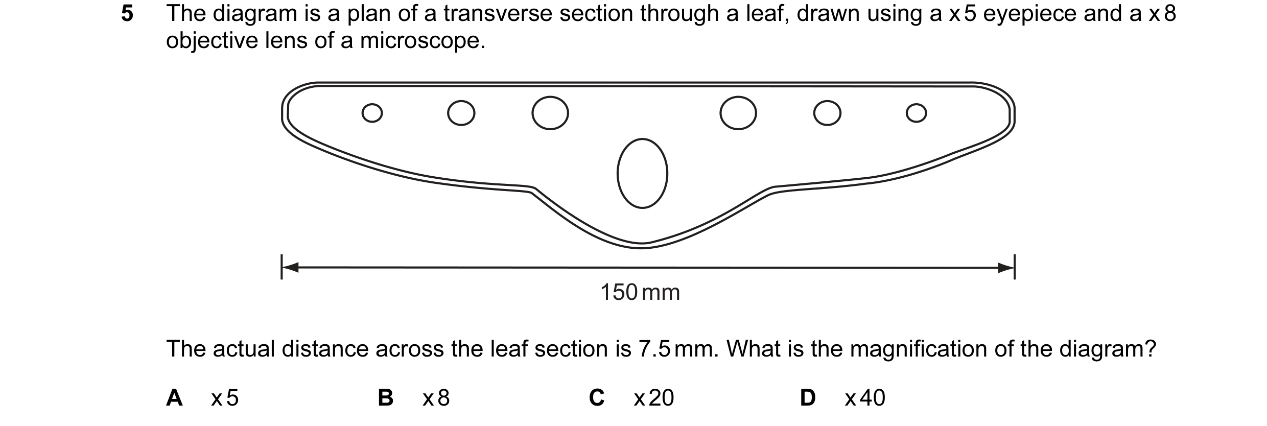
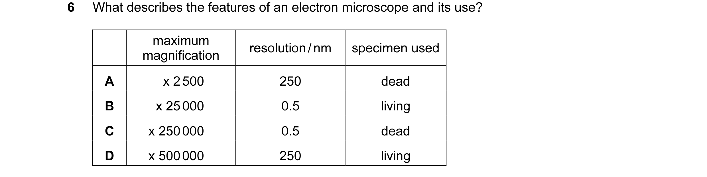
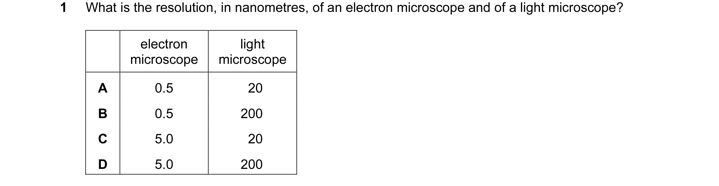 1 / 72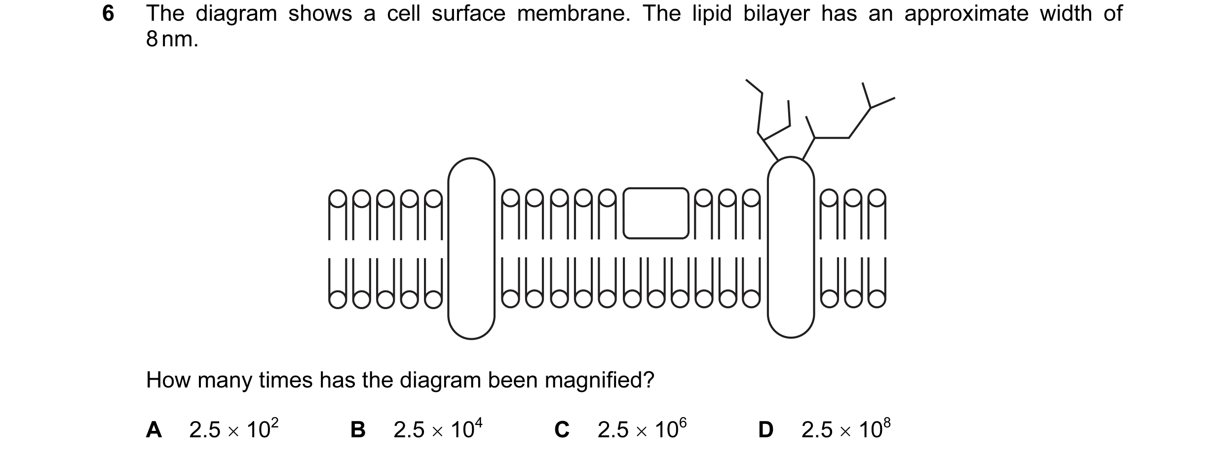
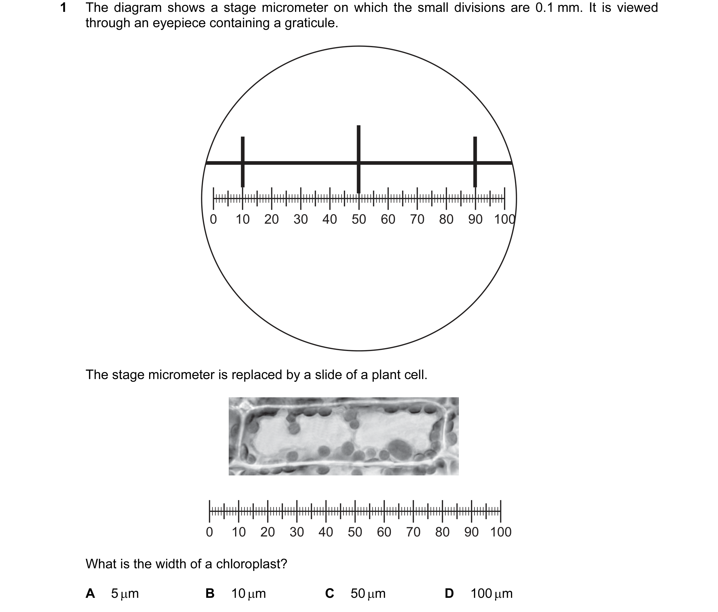 2 / 72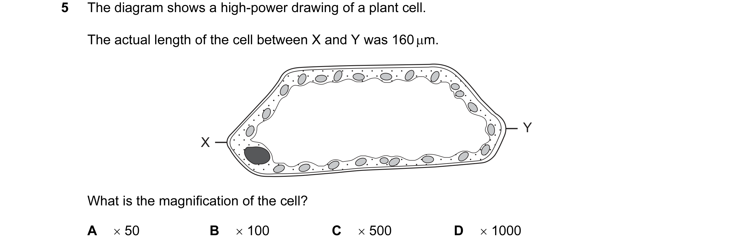
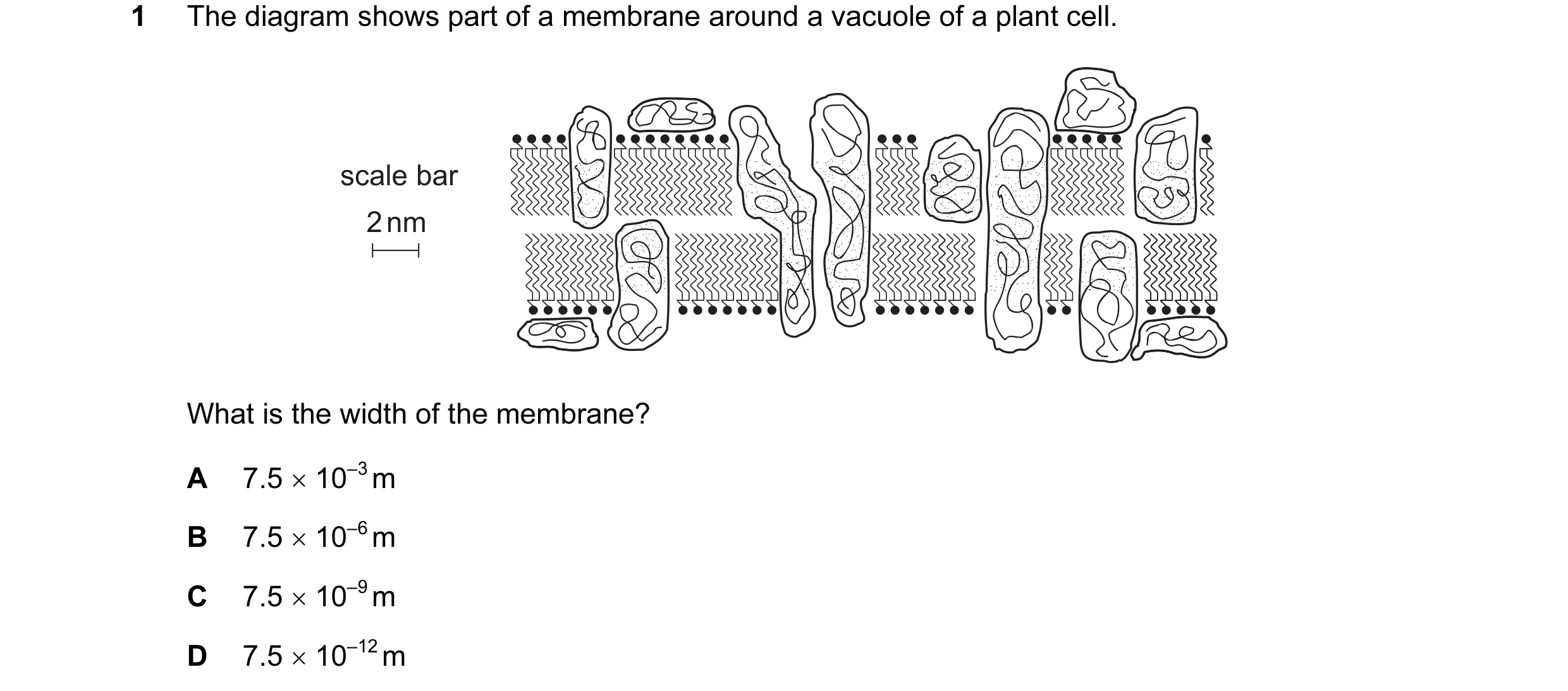
 3 / 72
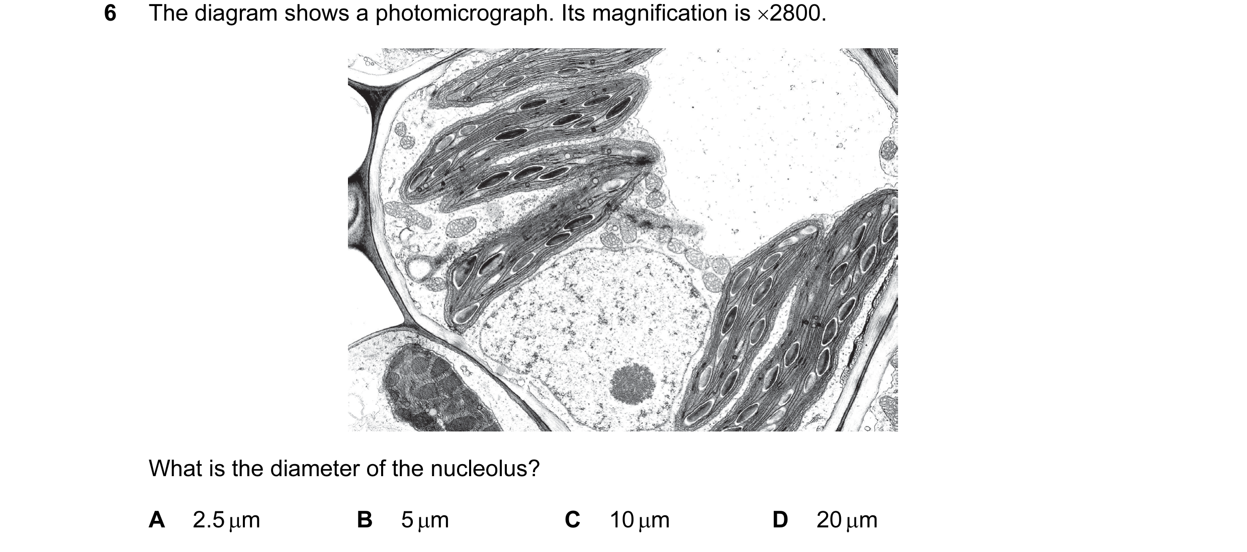 4 / 72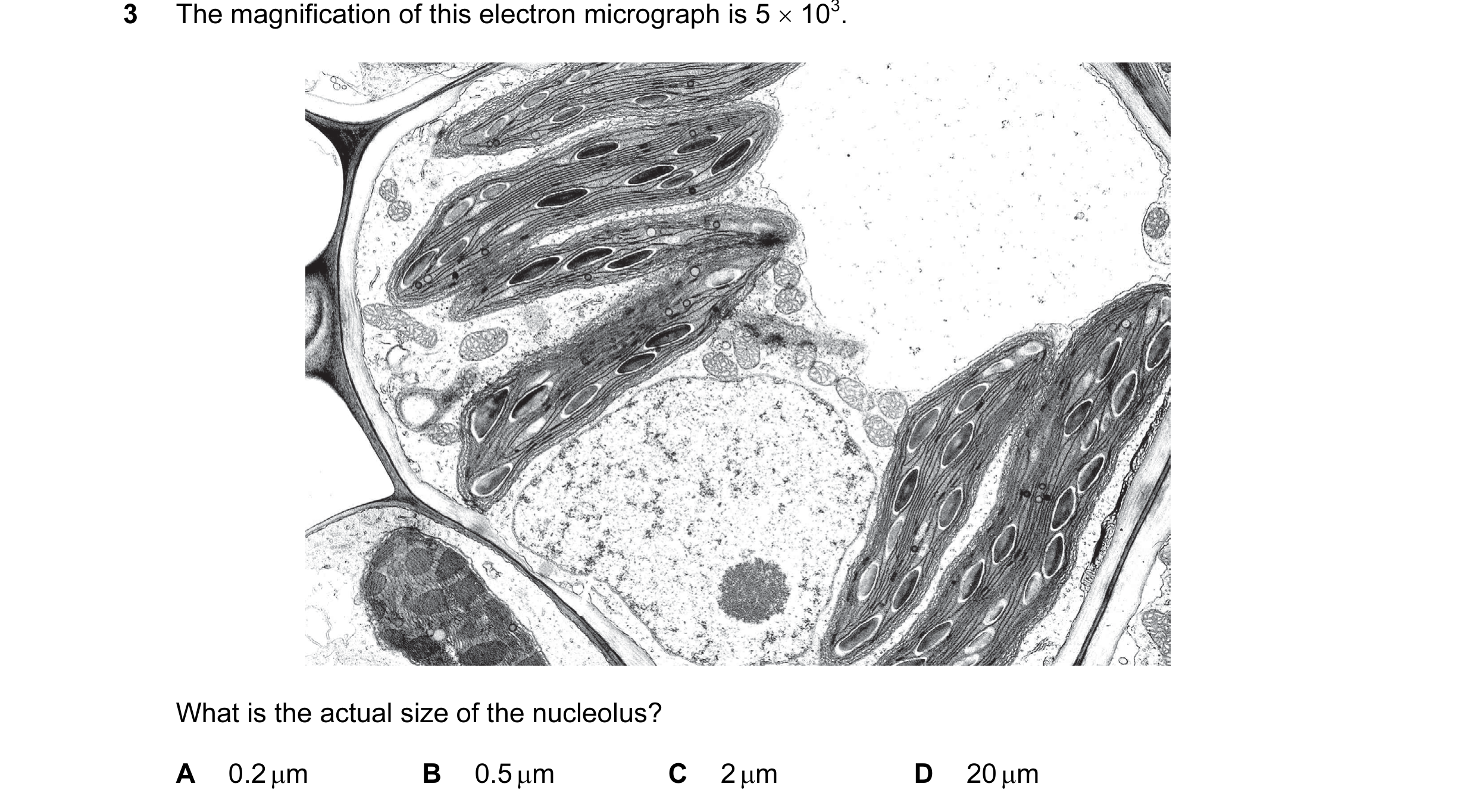
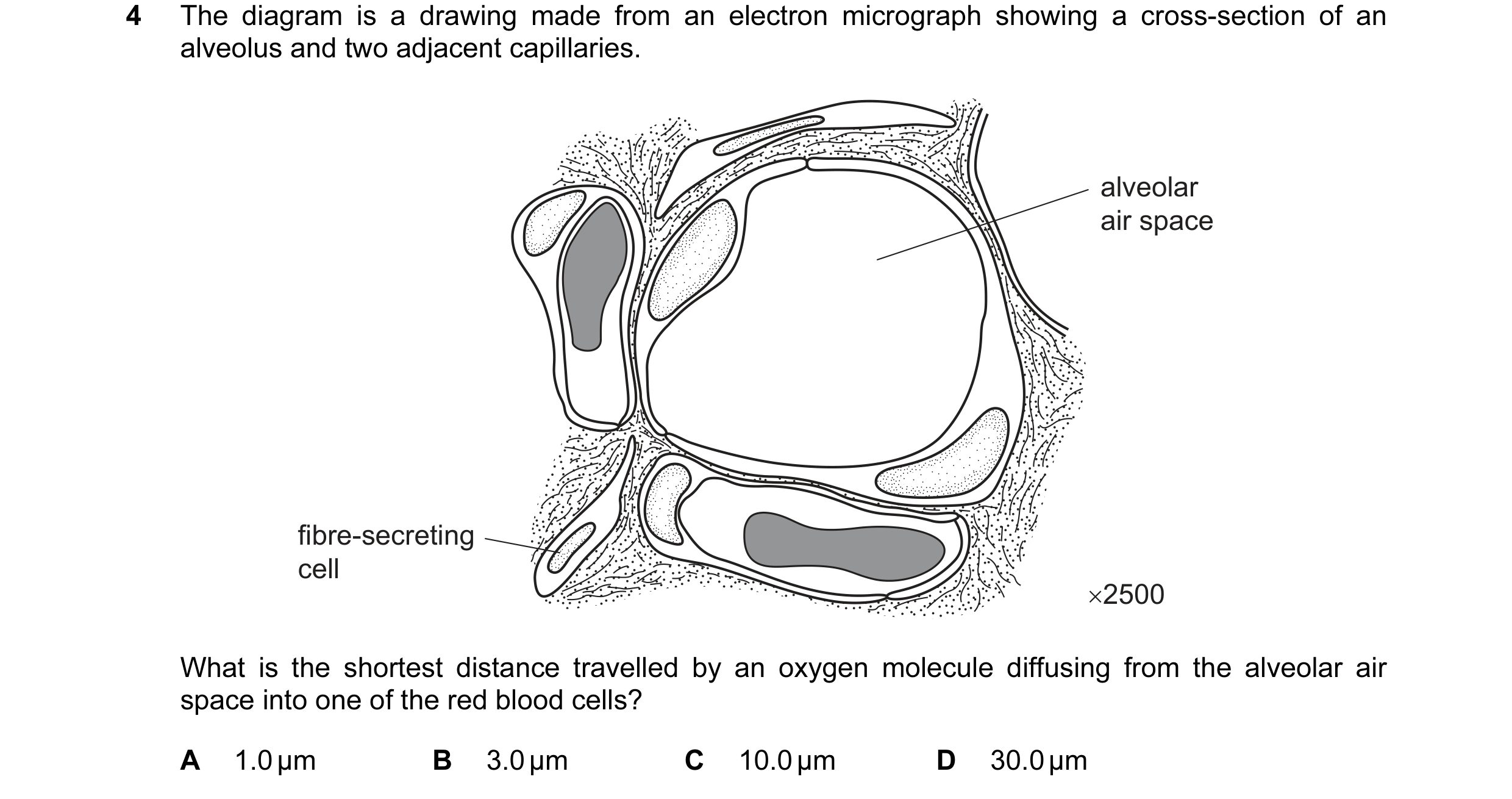 5 / 72

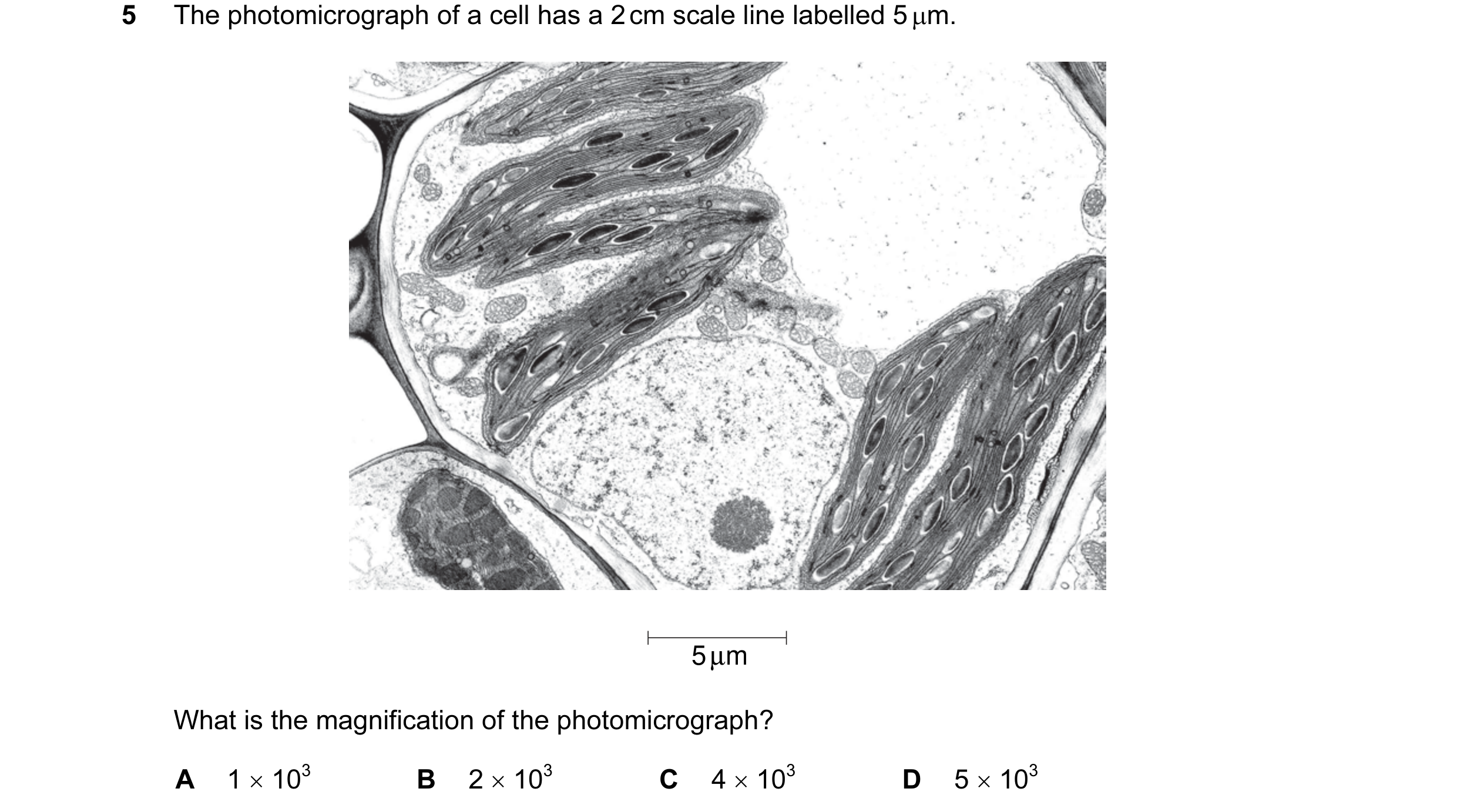
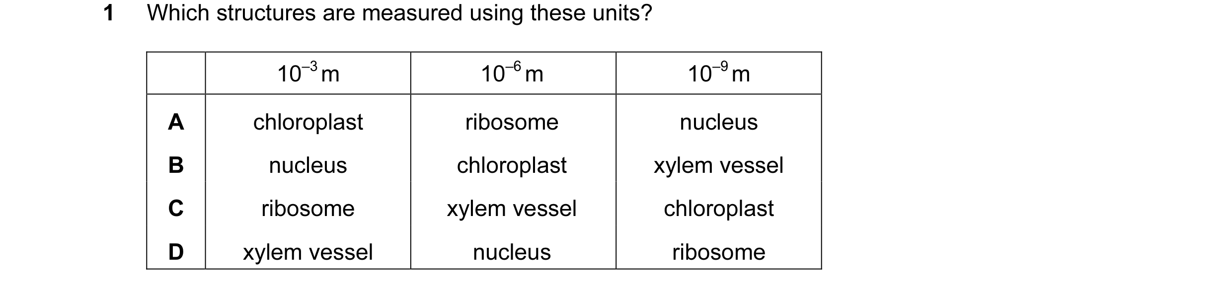 6 / 72
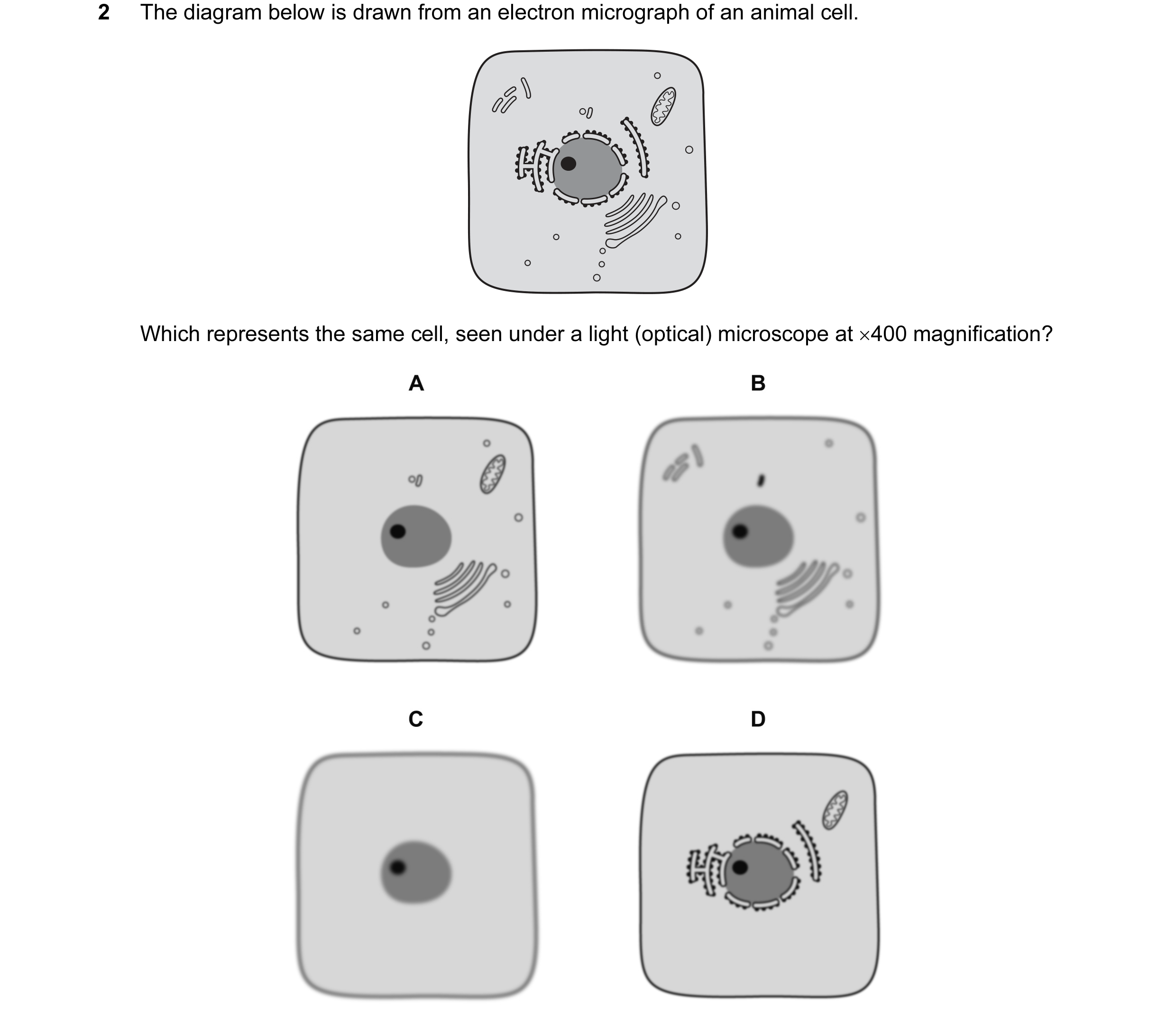 7 / 72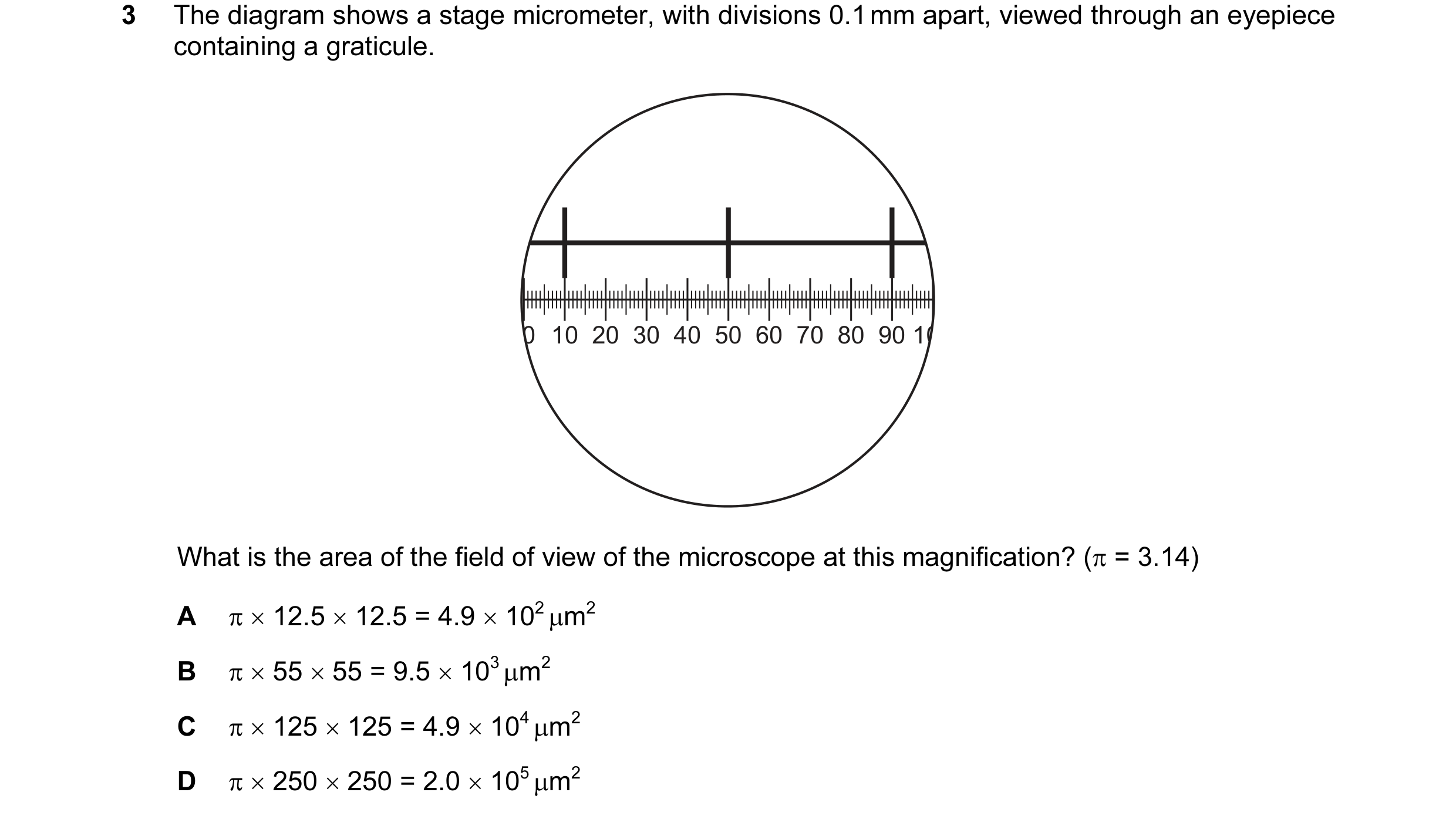
 8 / 72
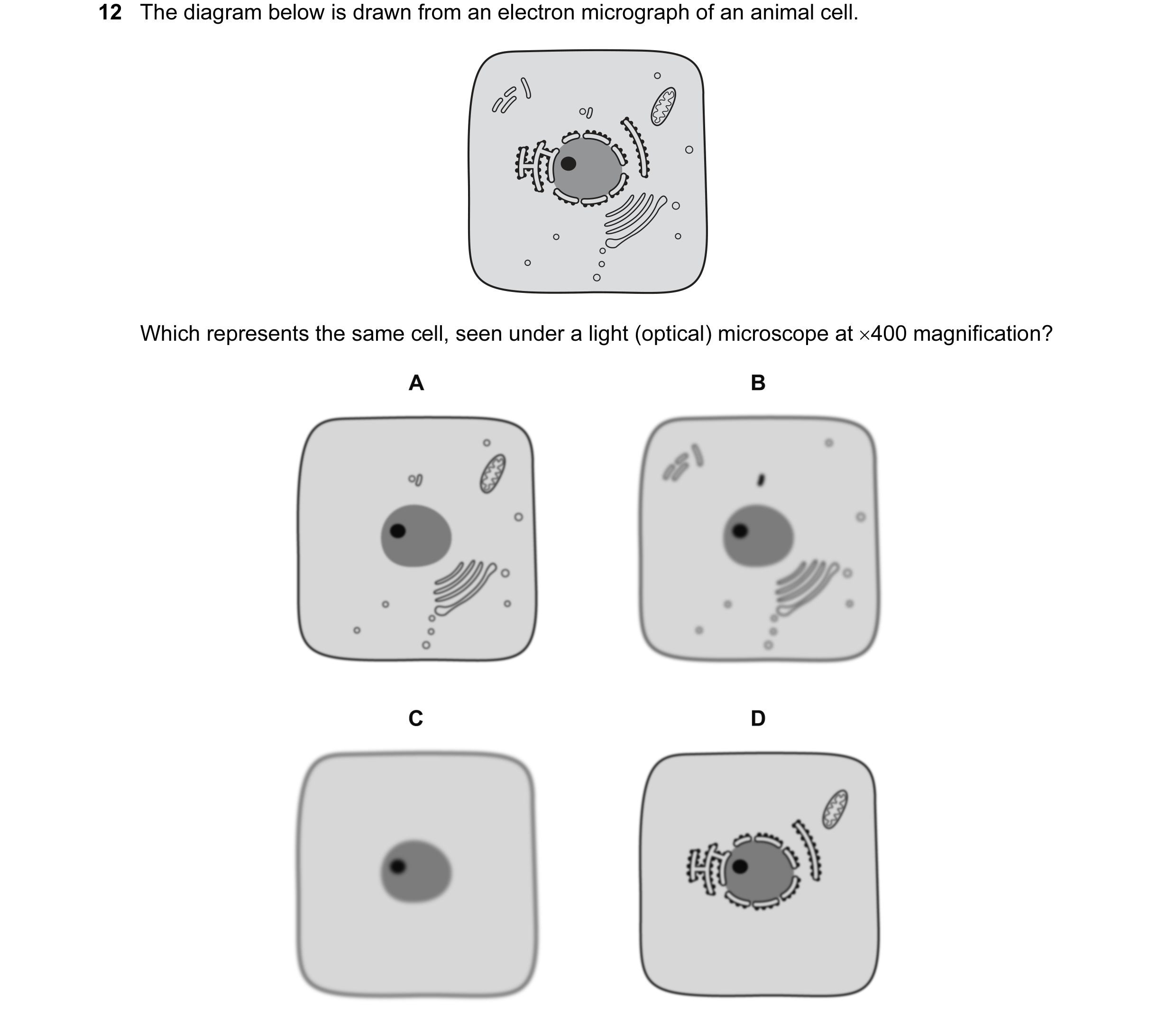 9 / 72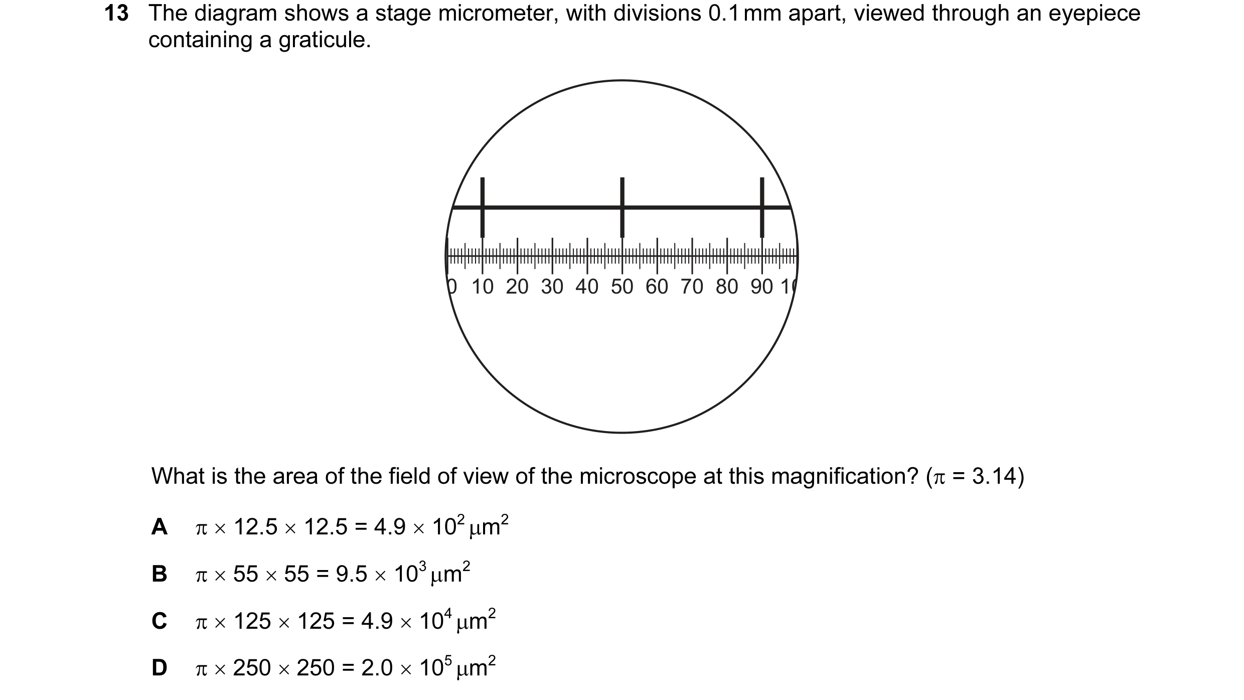
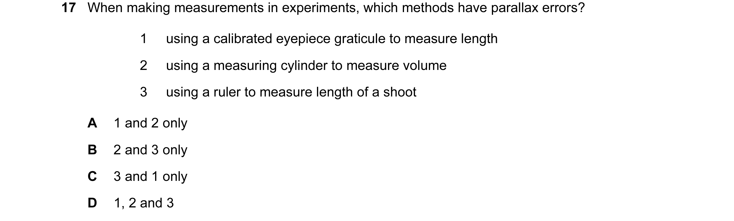 10 / 72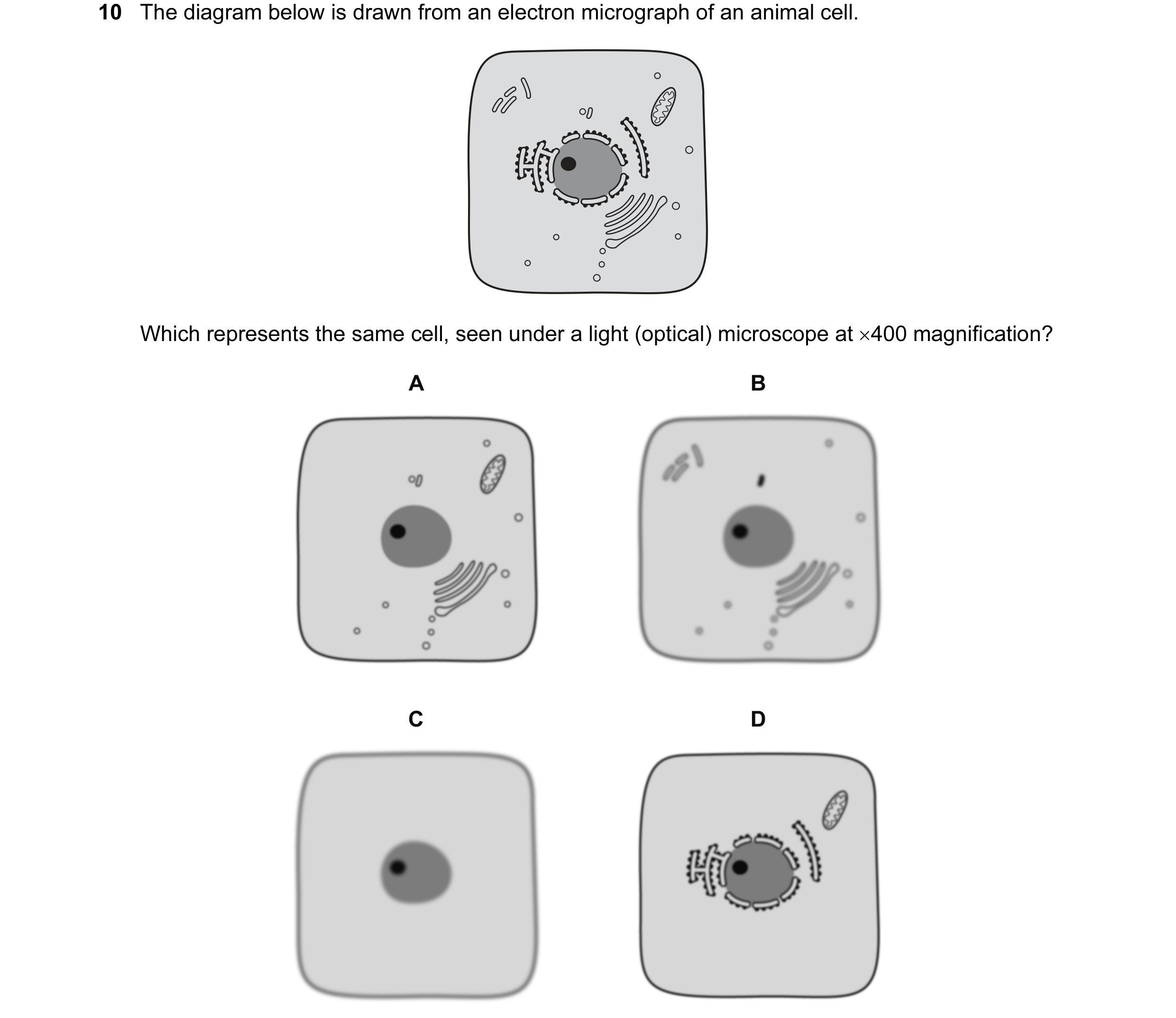
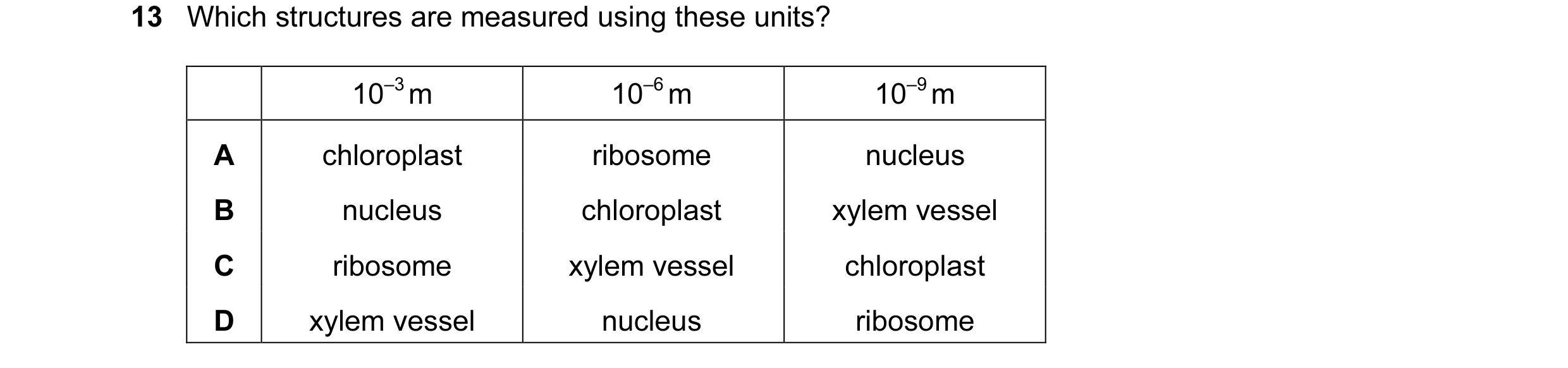 11 / 72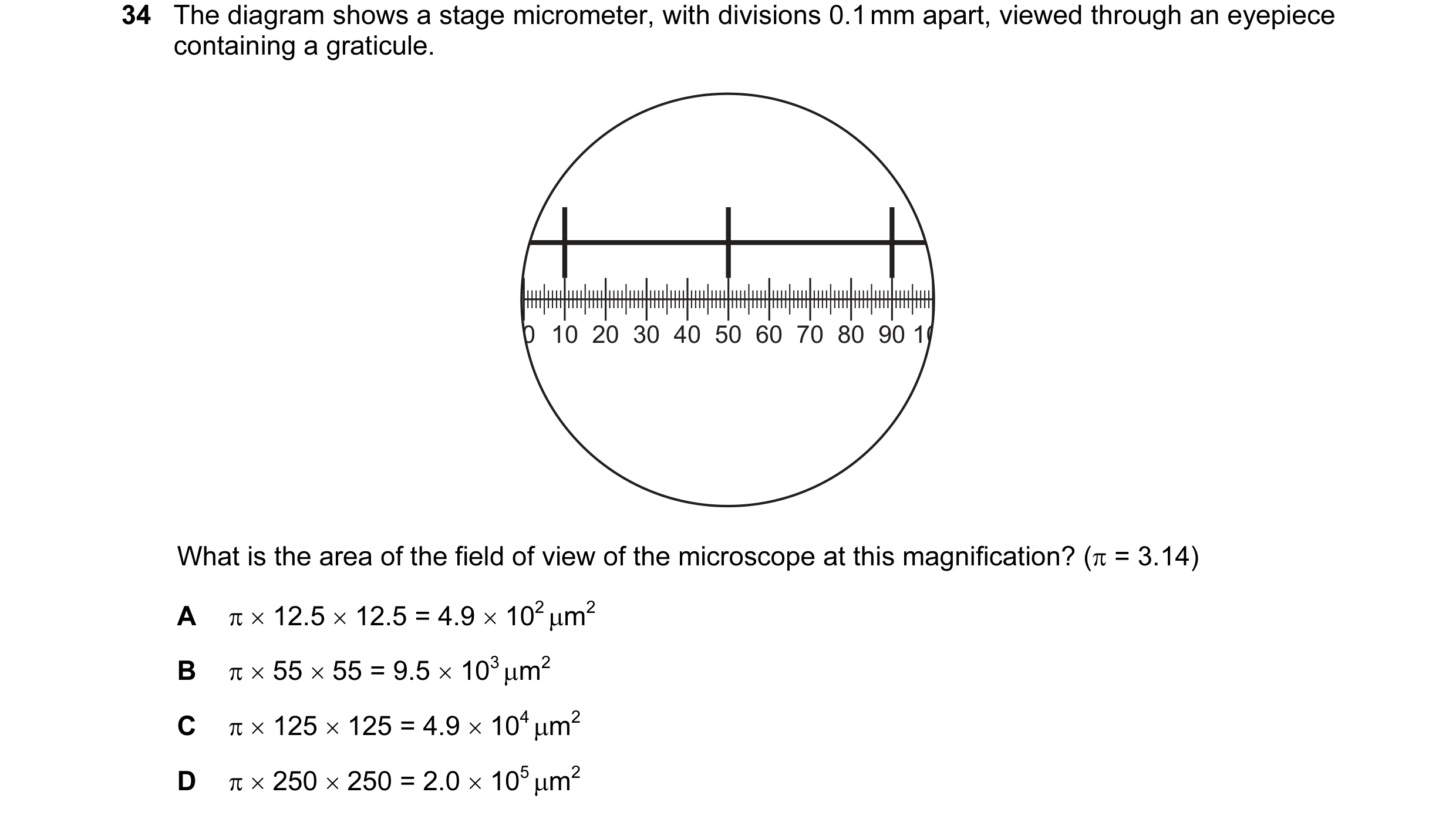
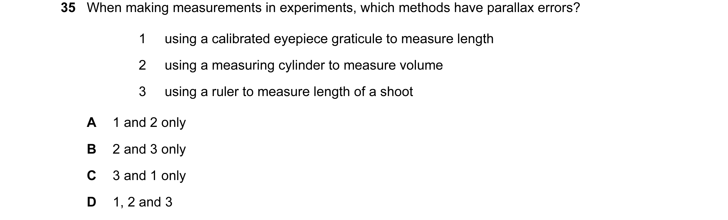
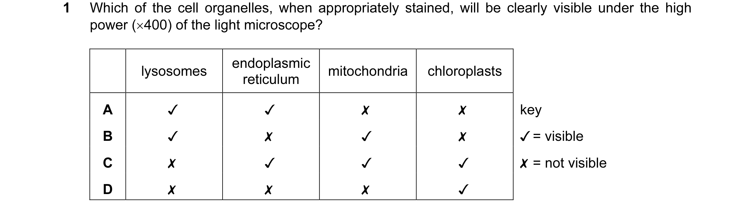 12 / 72
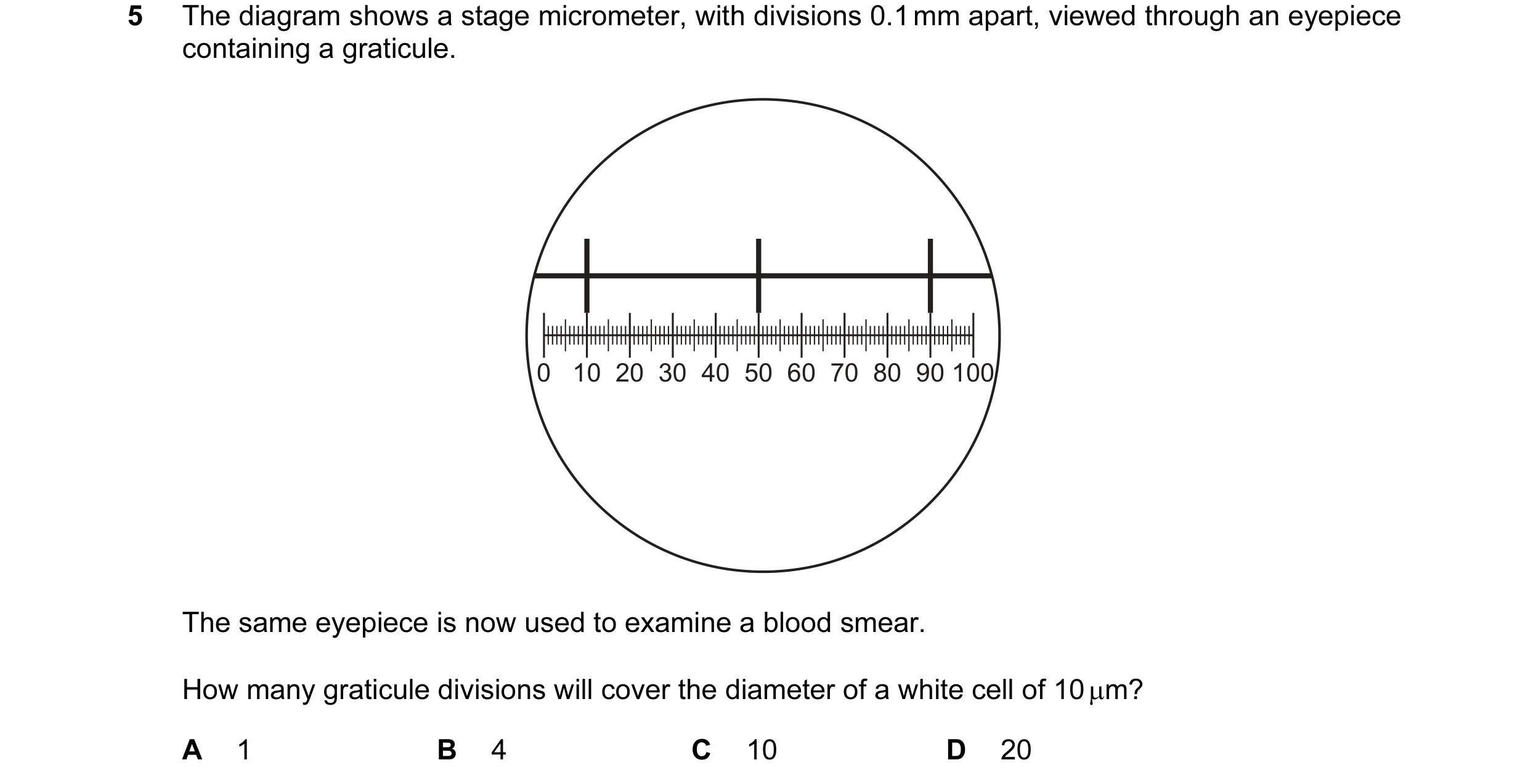
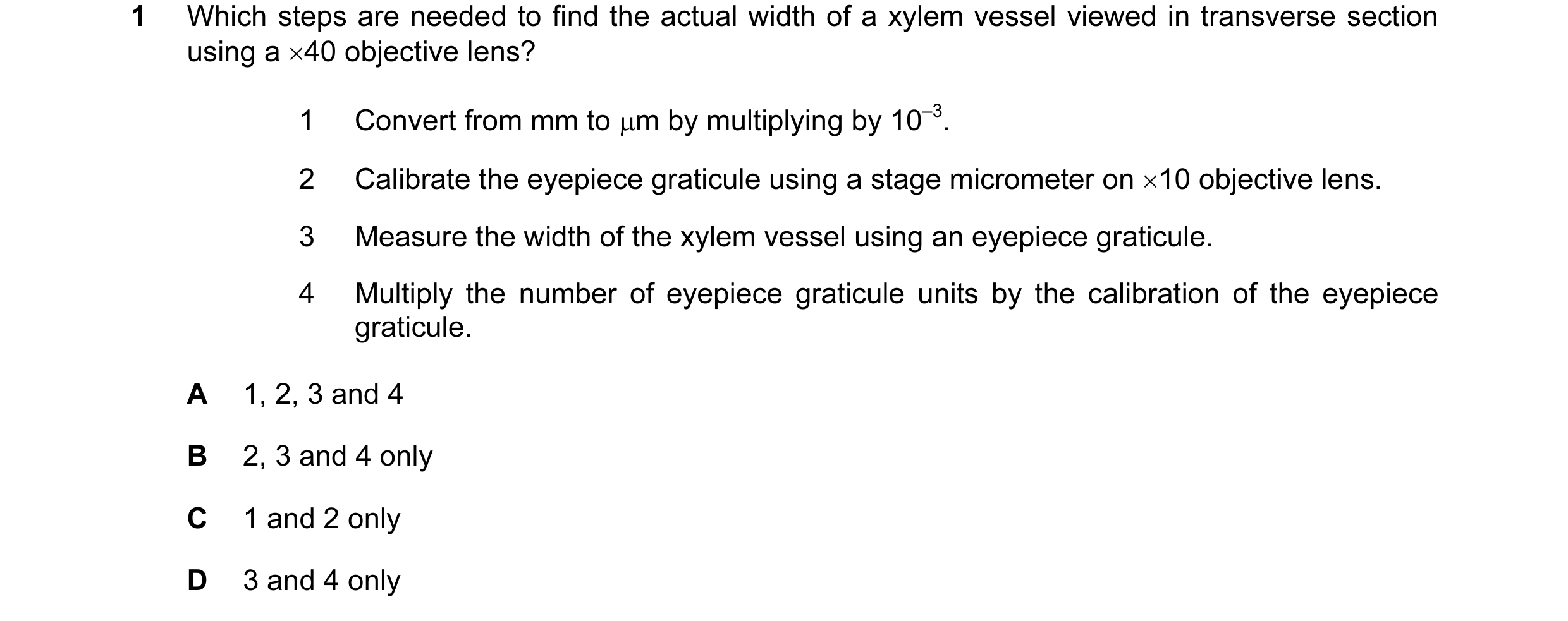 13 / 72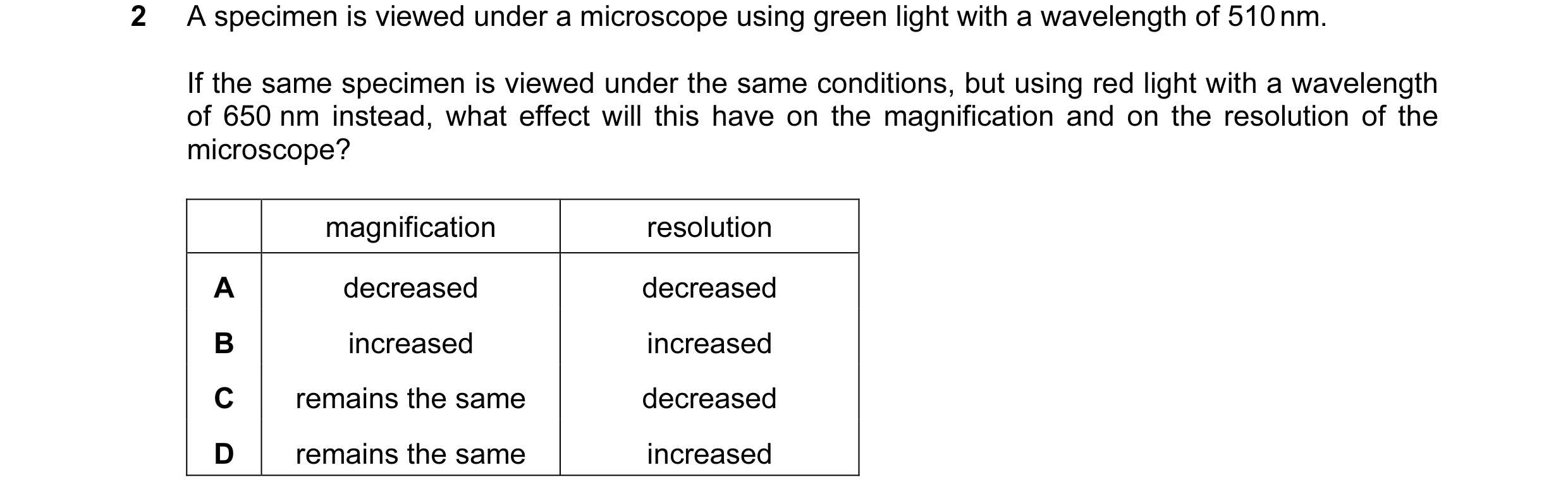

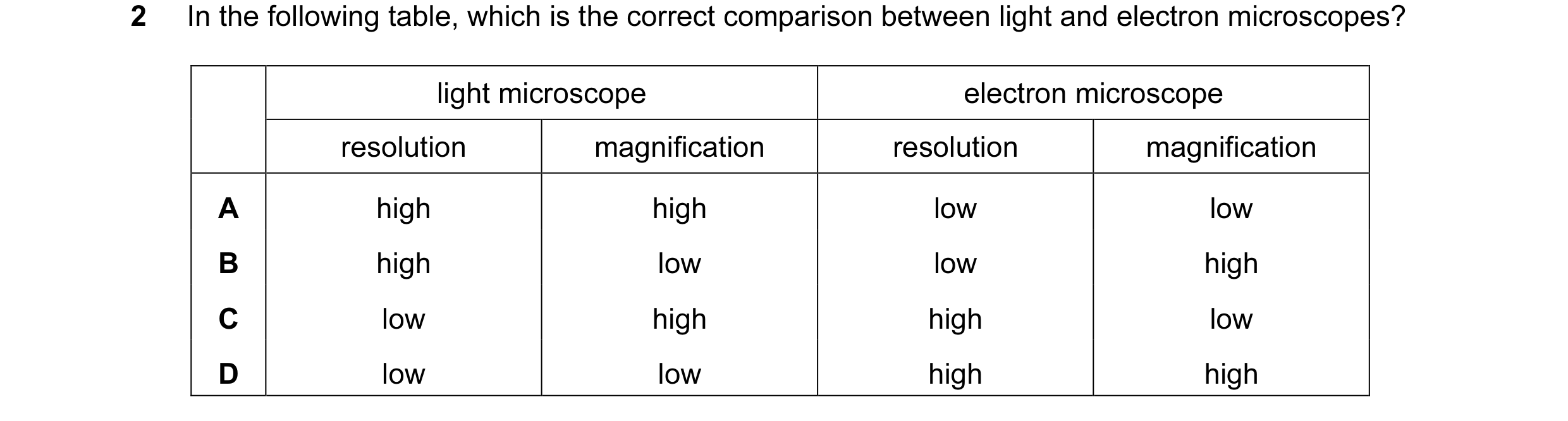
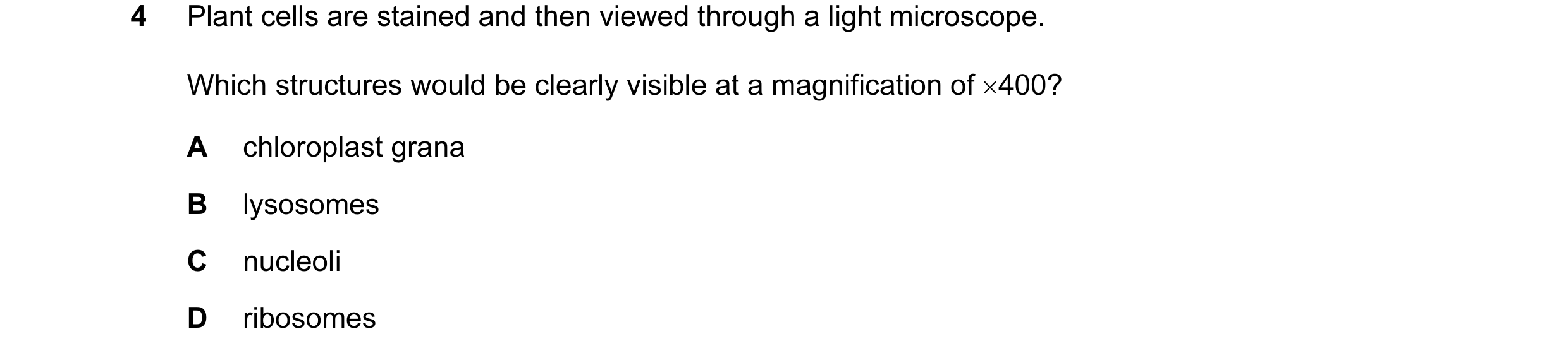

 14 / 72
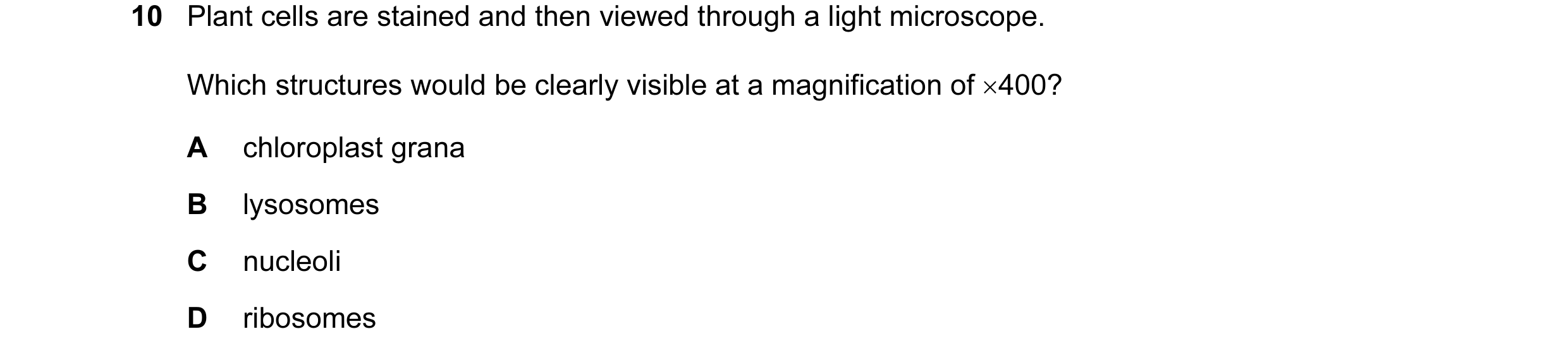
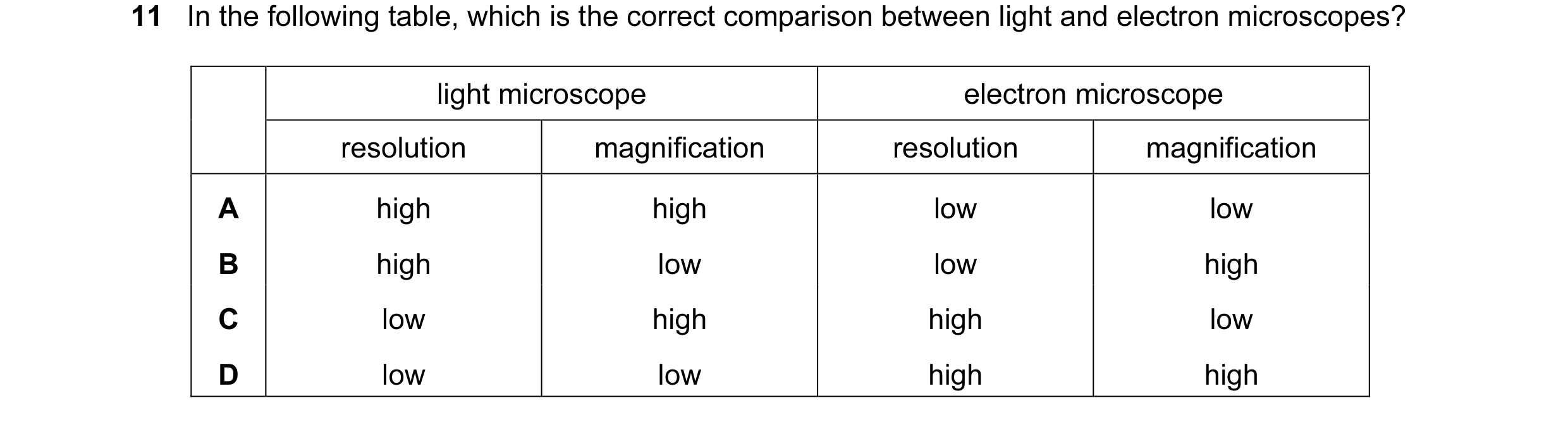 15 / 72
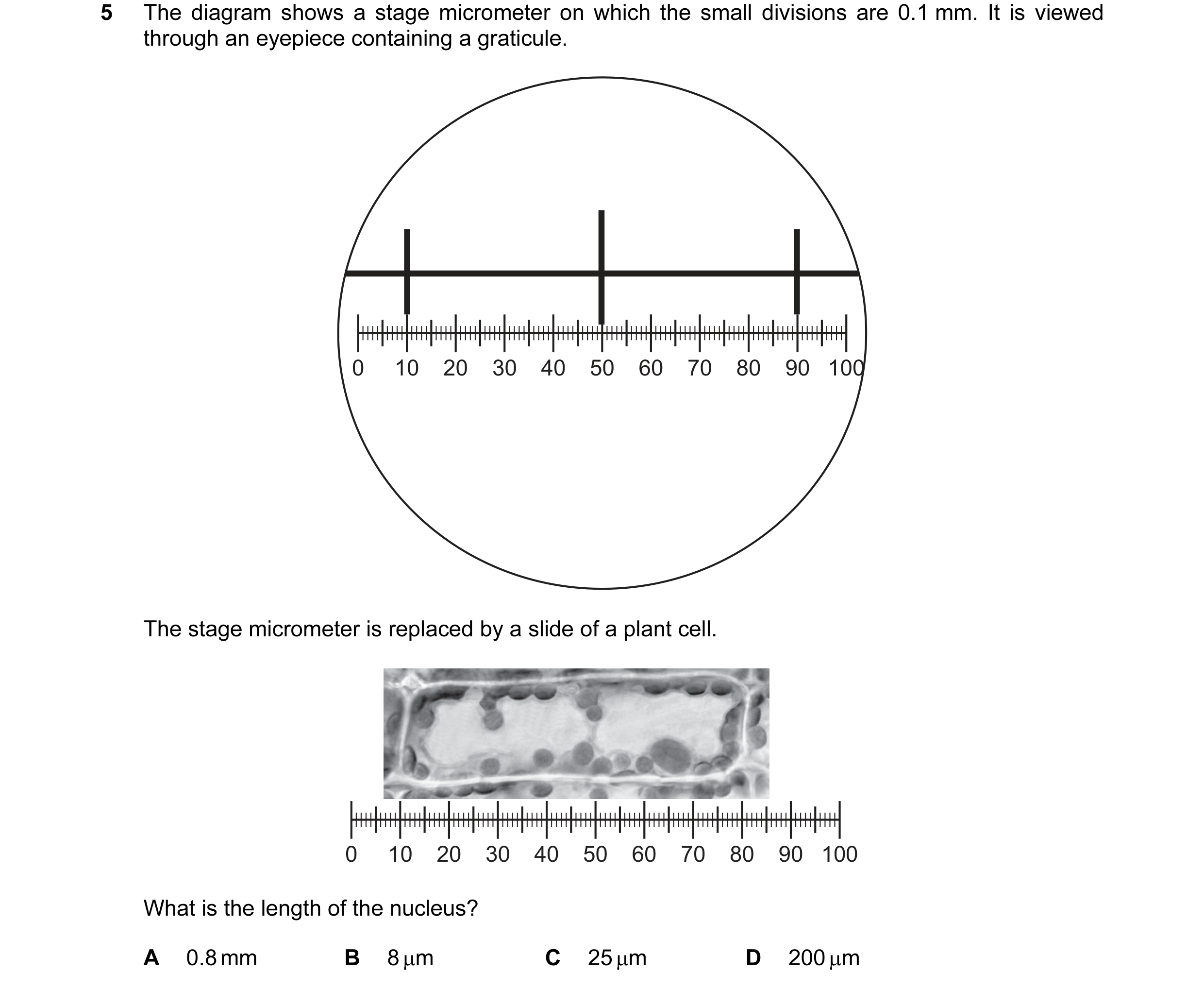 16 / 72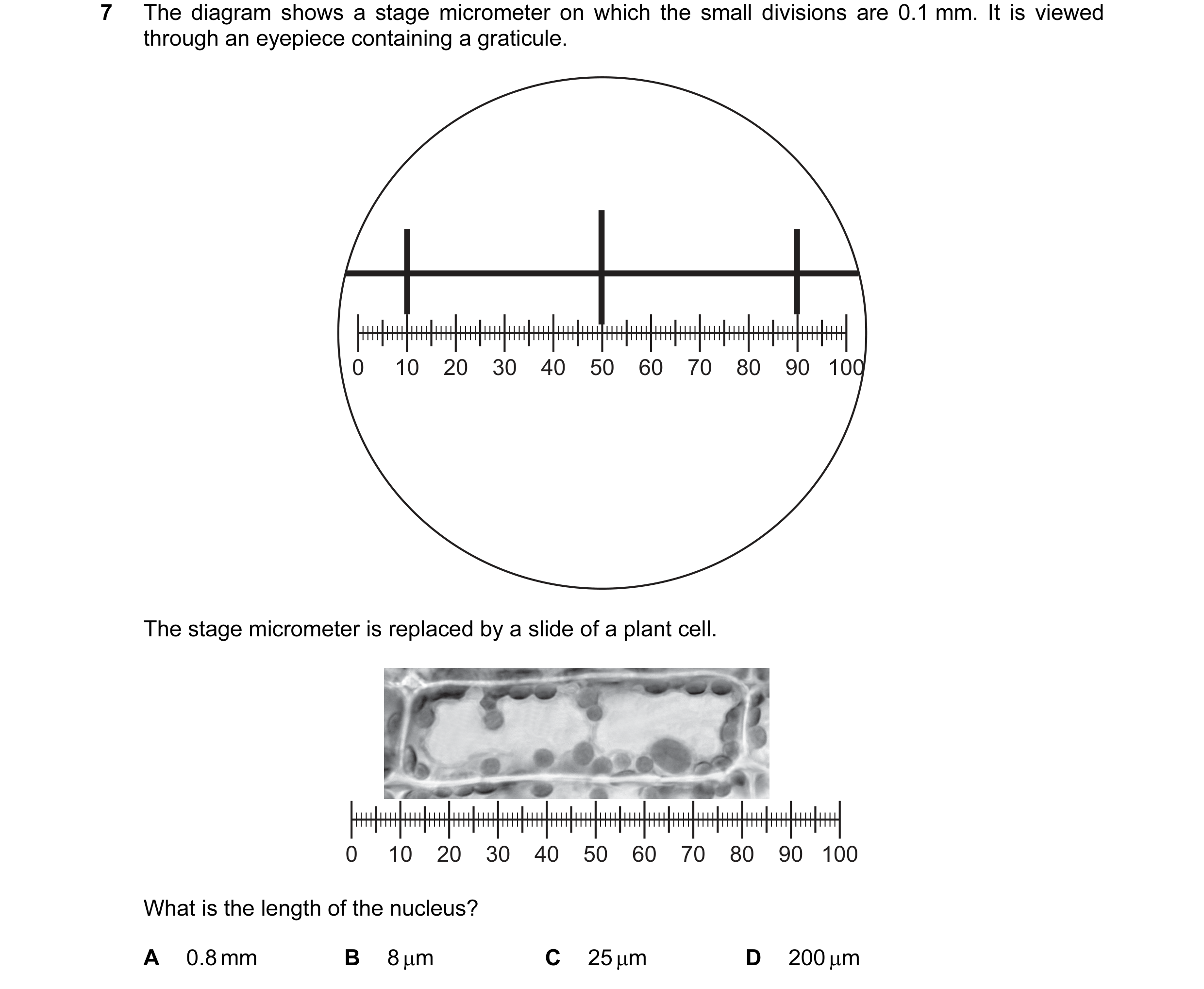
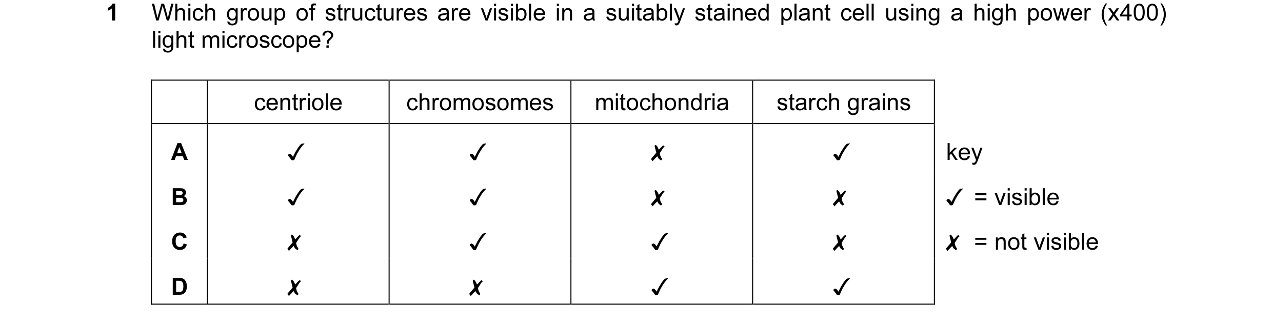 17 / 72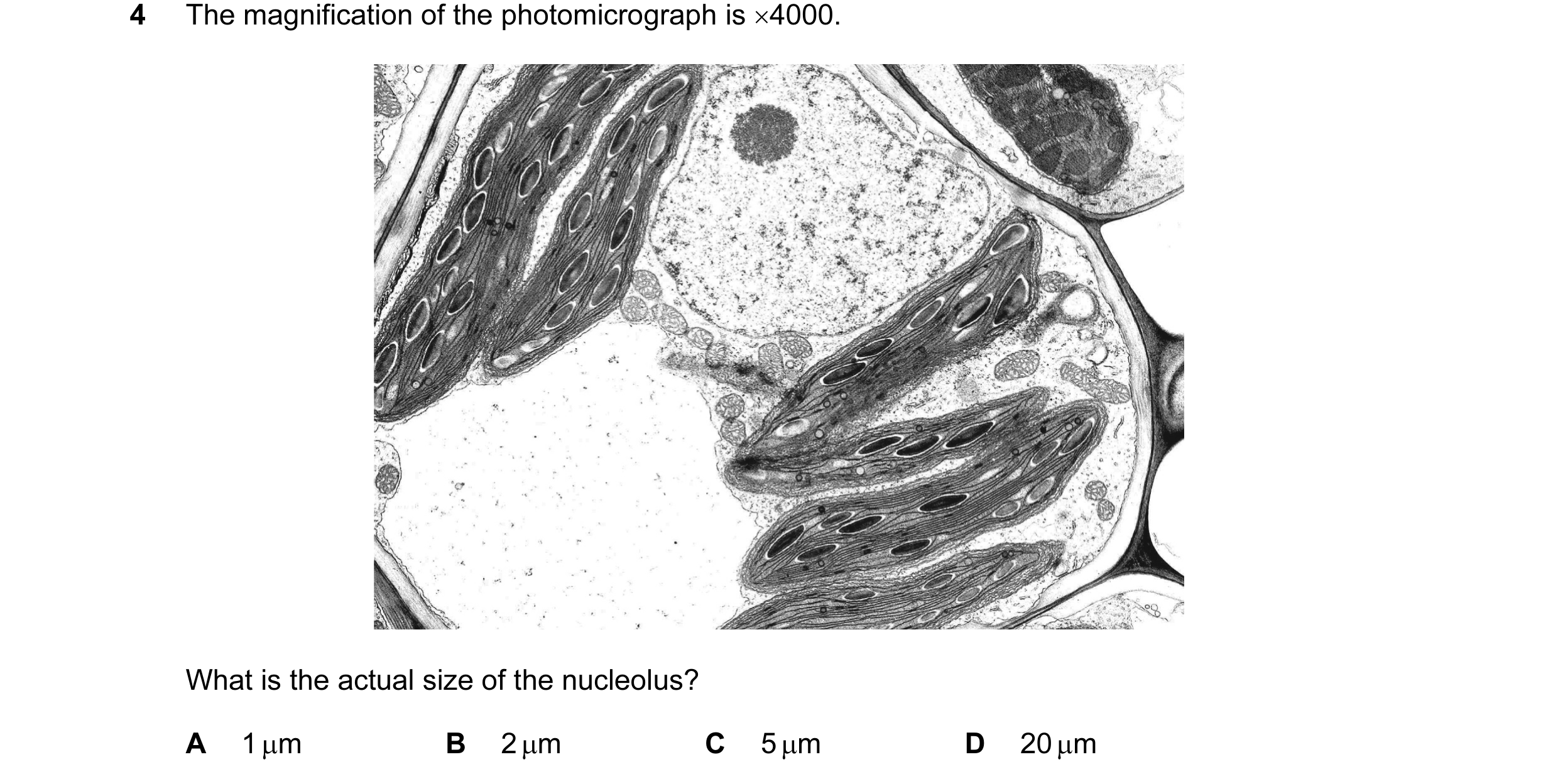

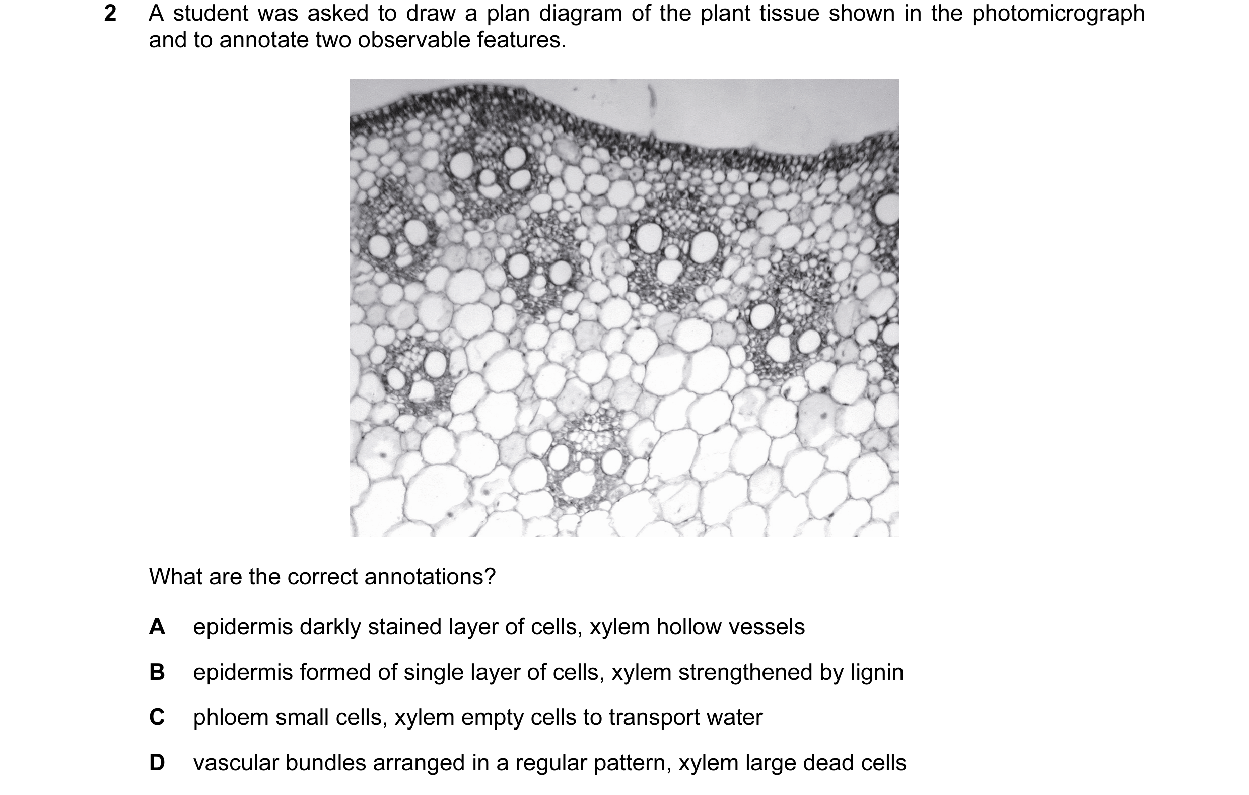 18 / 72
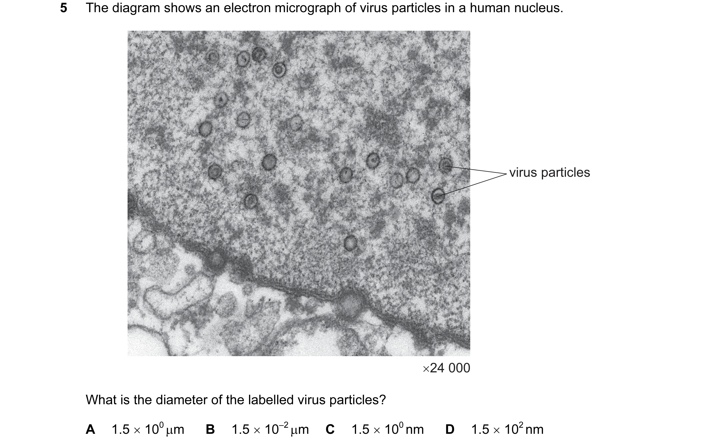
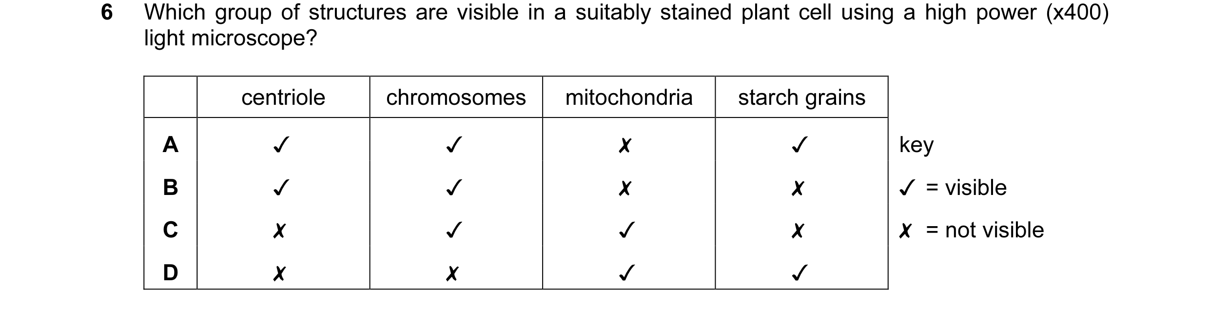
 19 / 72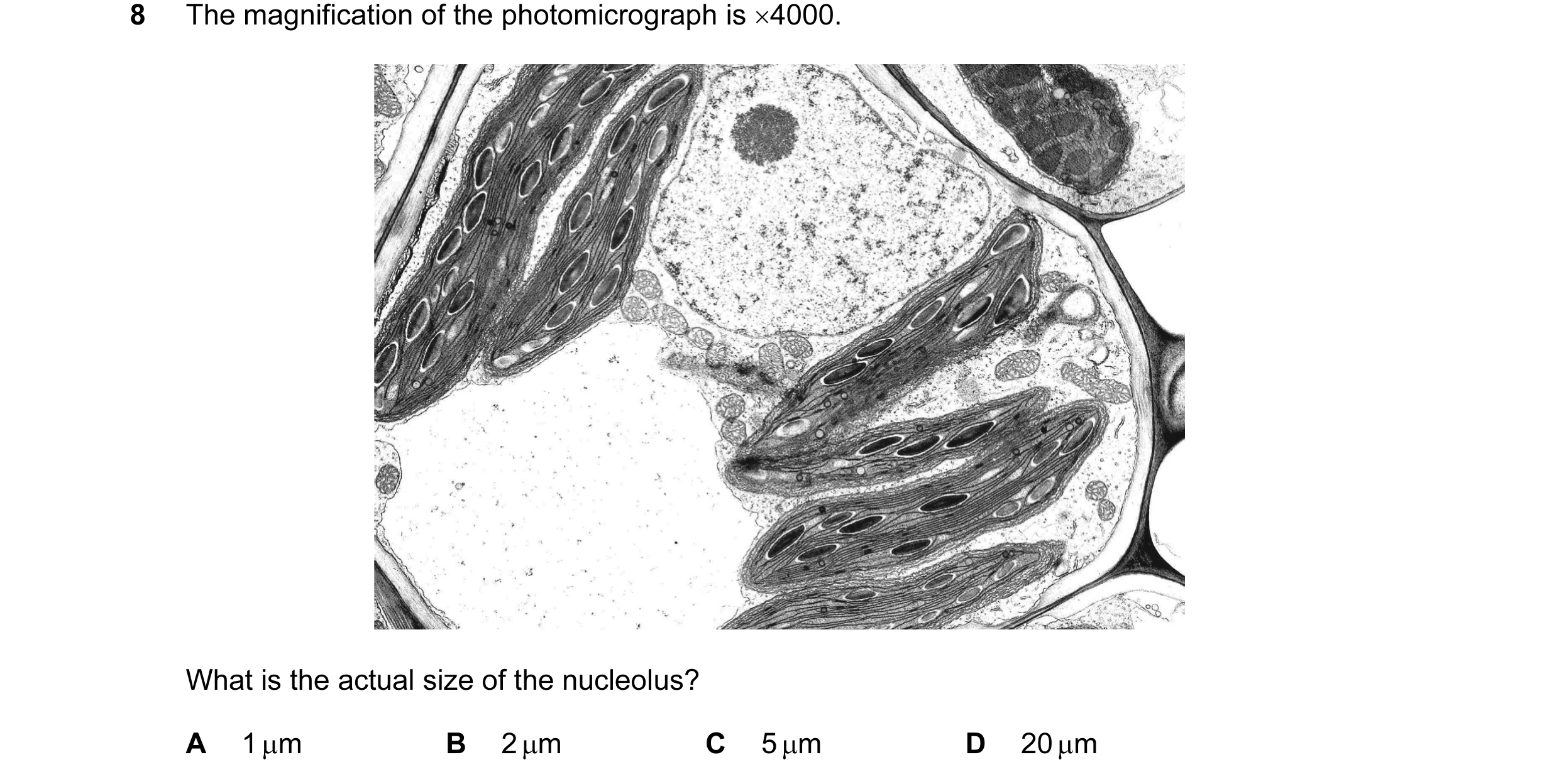
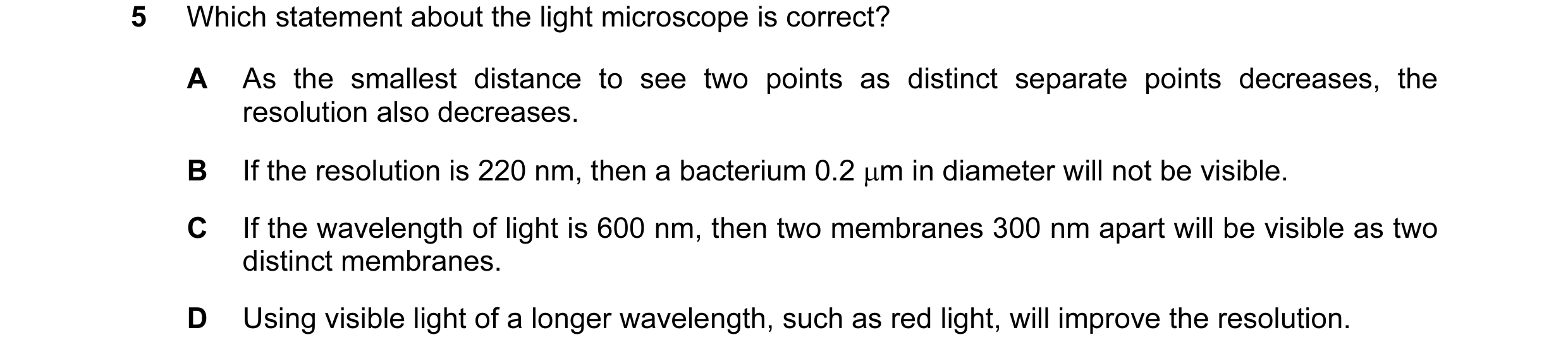
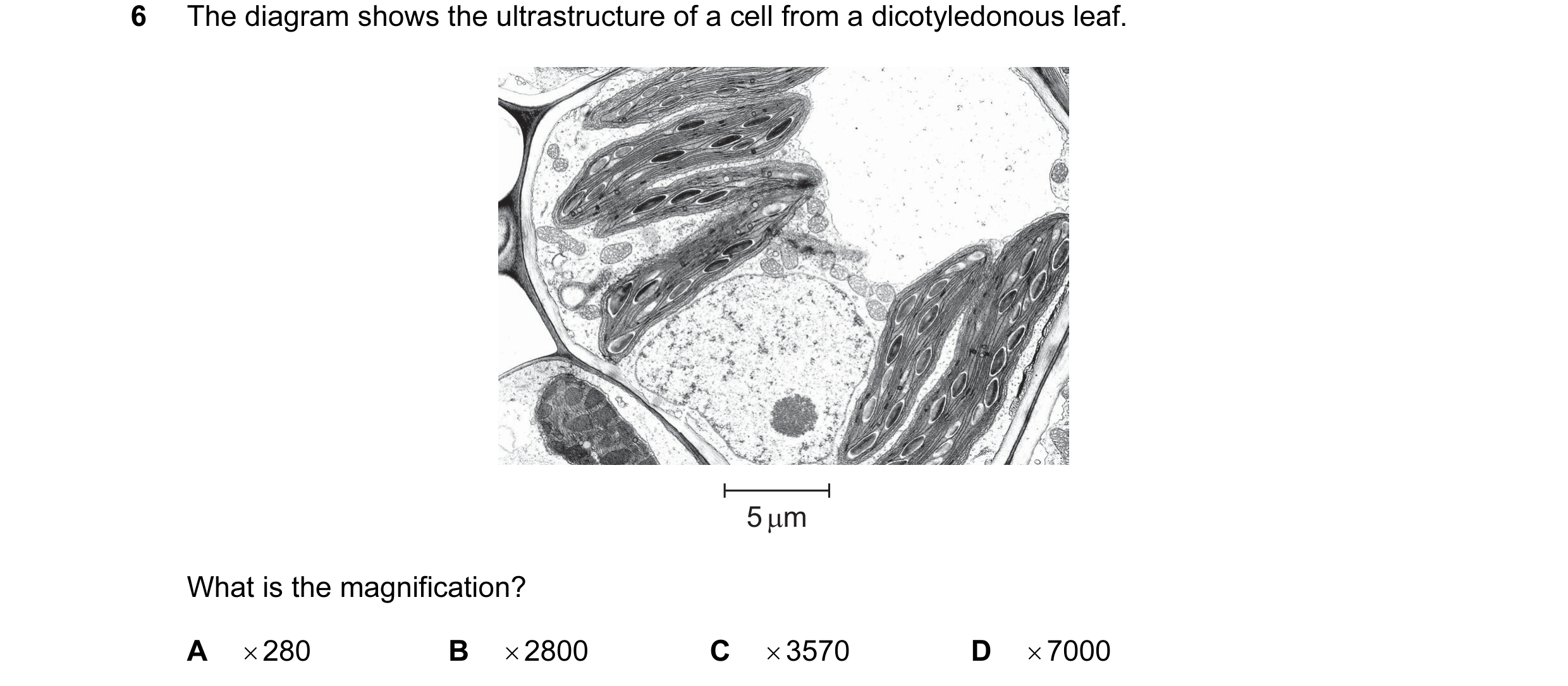 20 / 72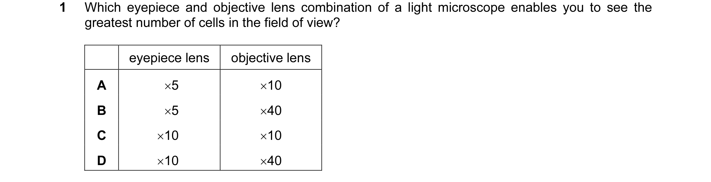
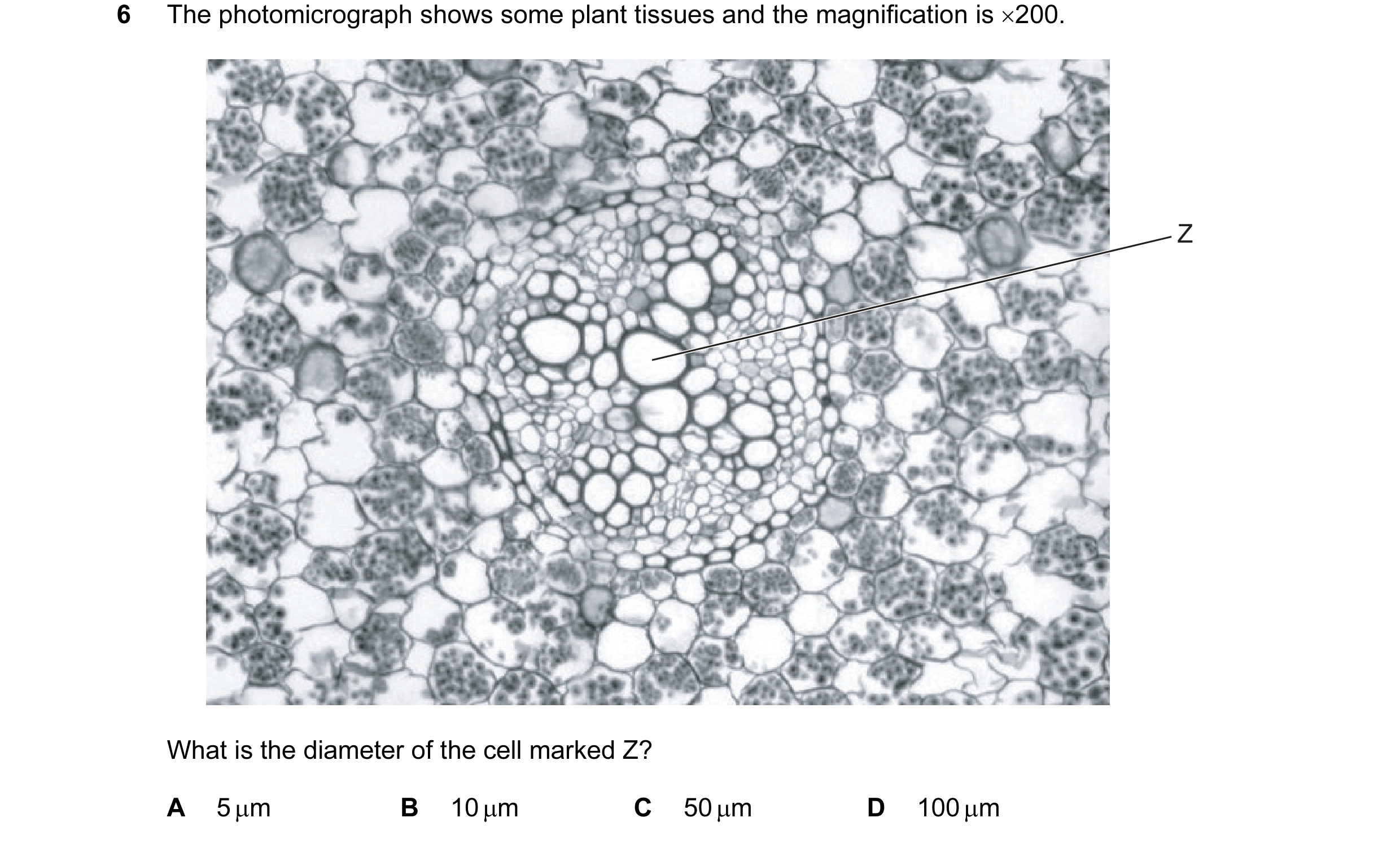 21 / 72
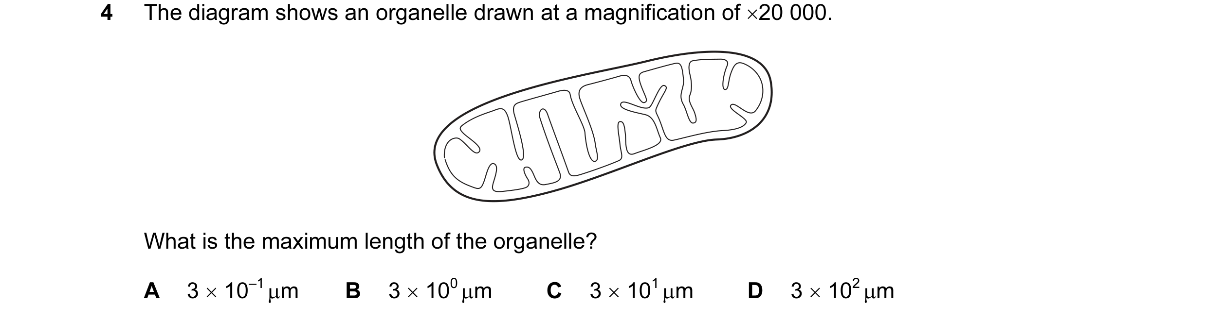
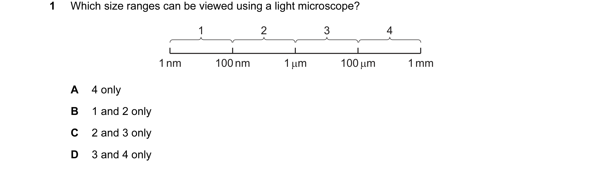
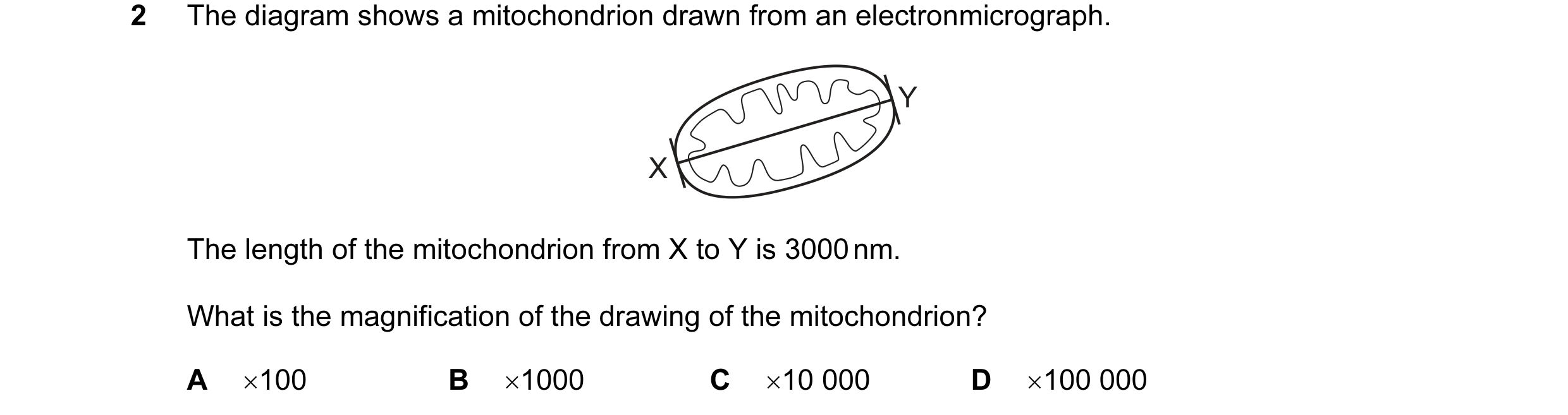 22 / 72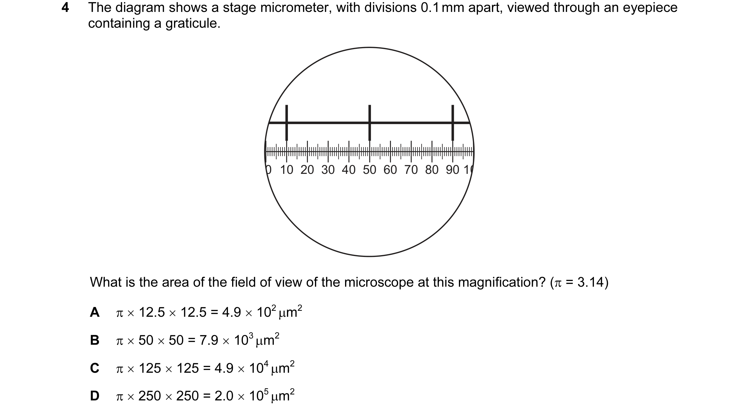
 23 / 72
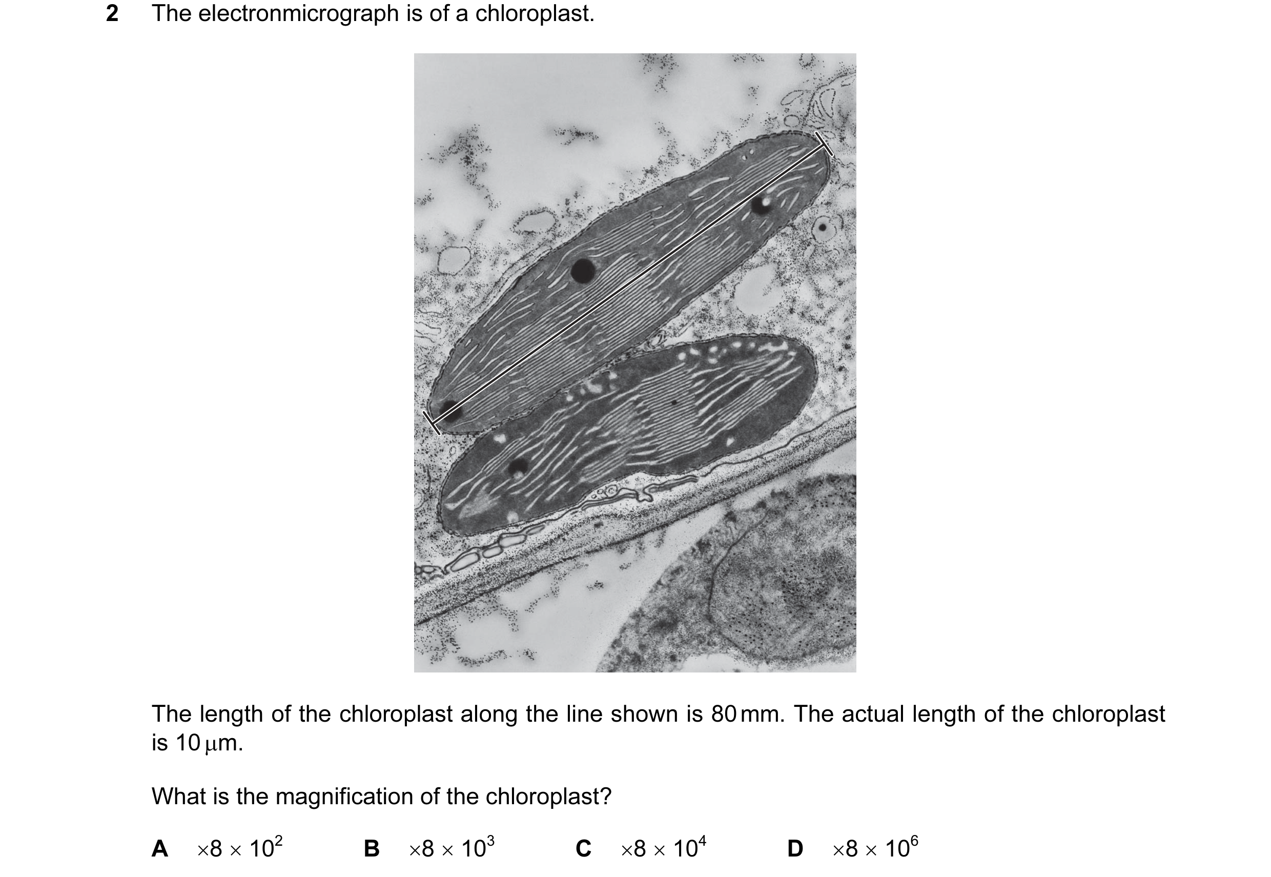 24 / 72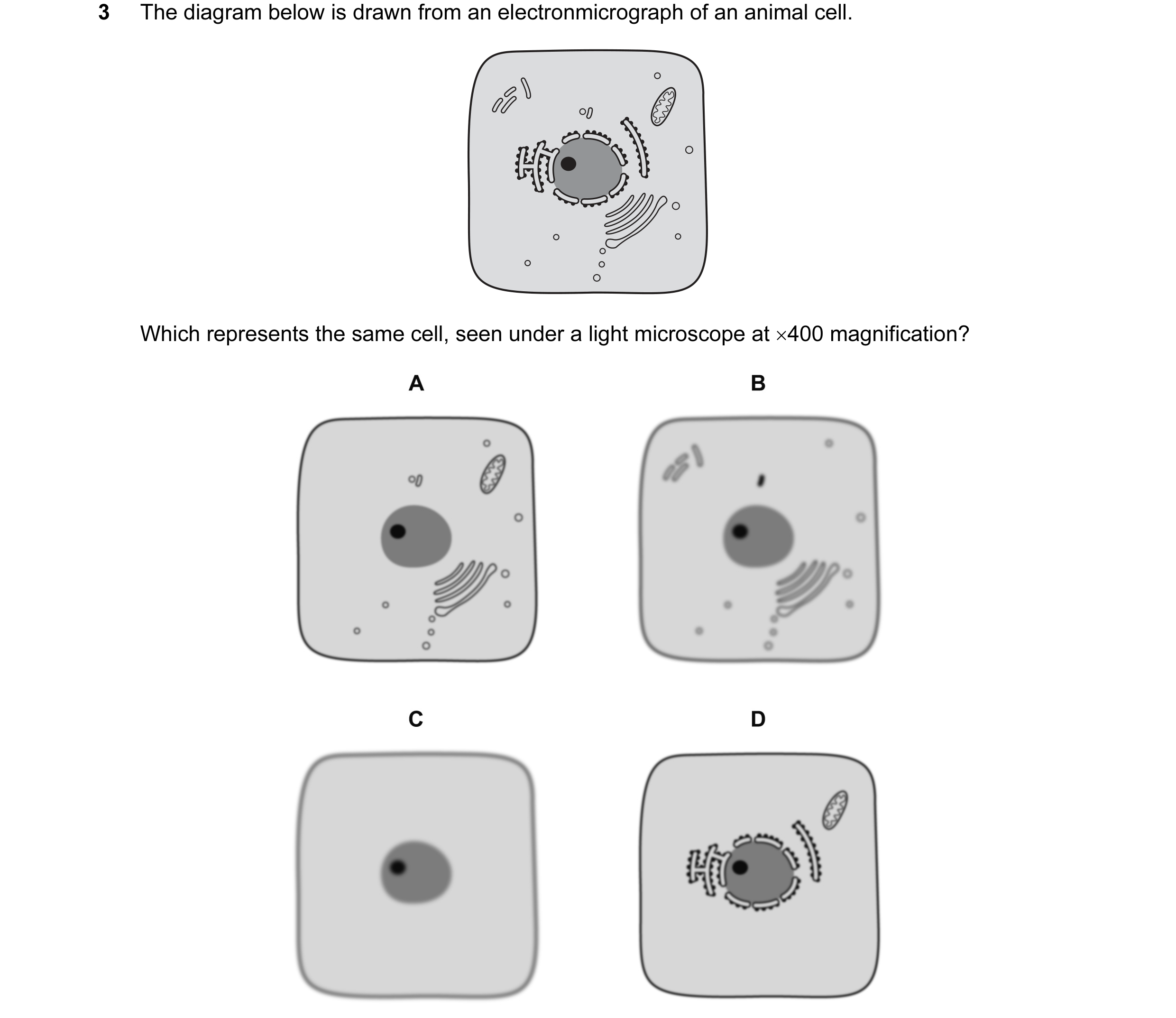
 25 / 72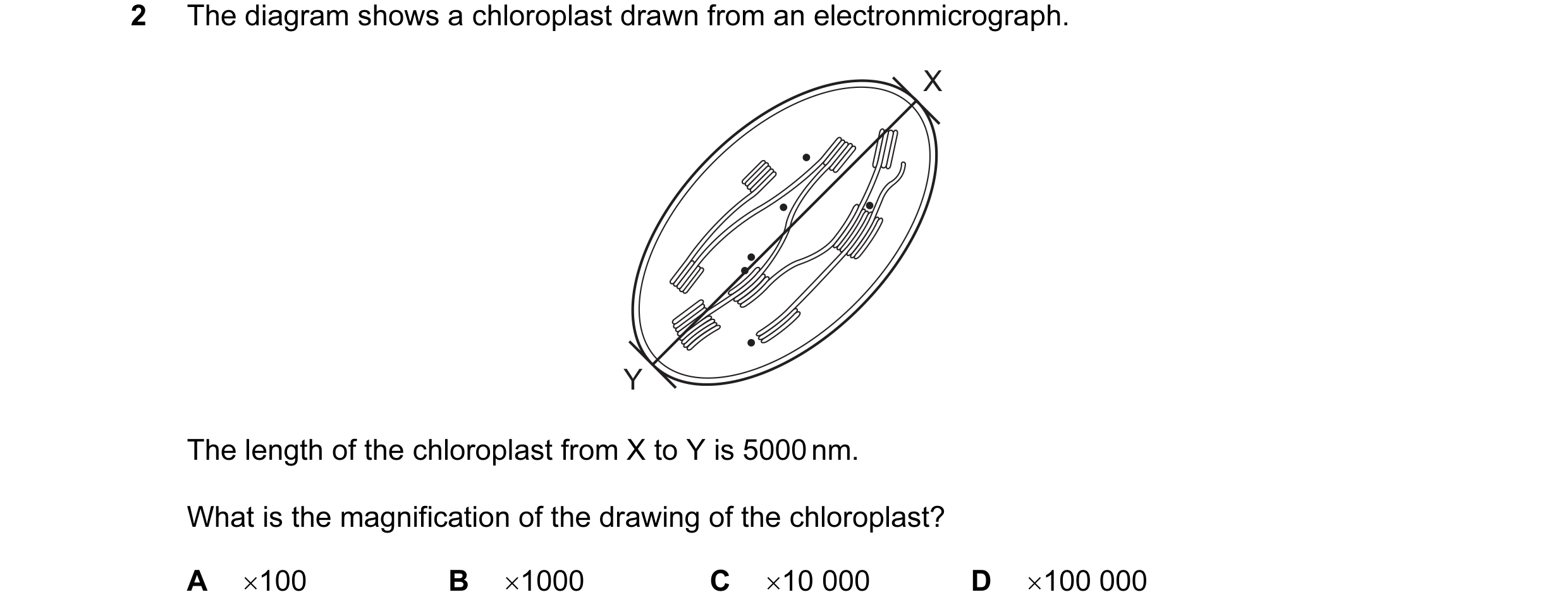



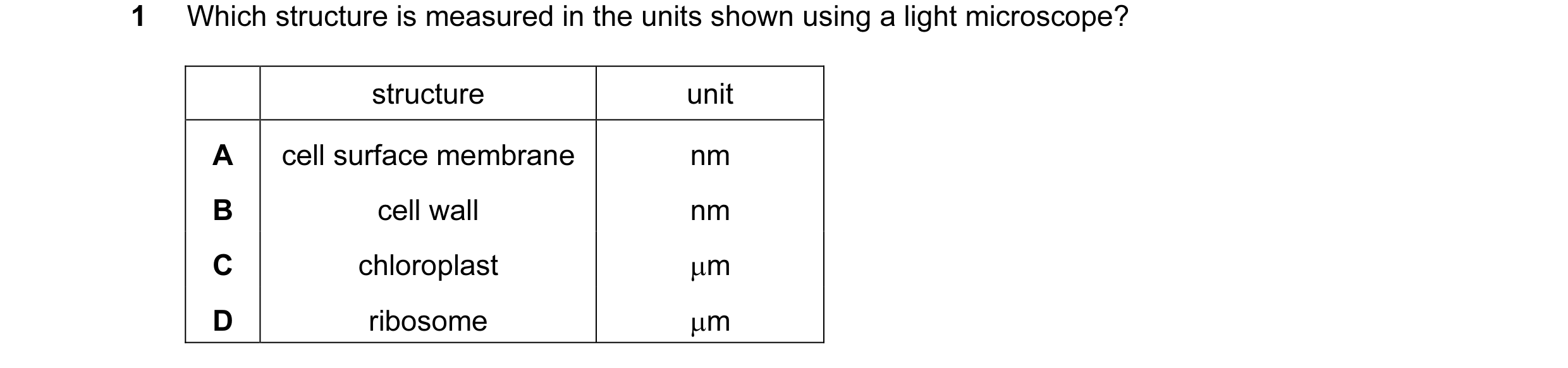 26 / 72

 27 / 72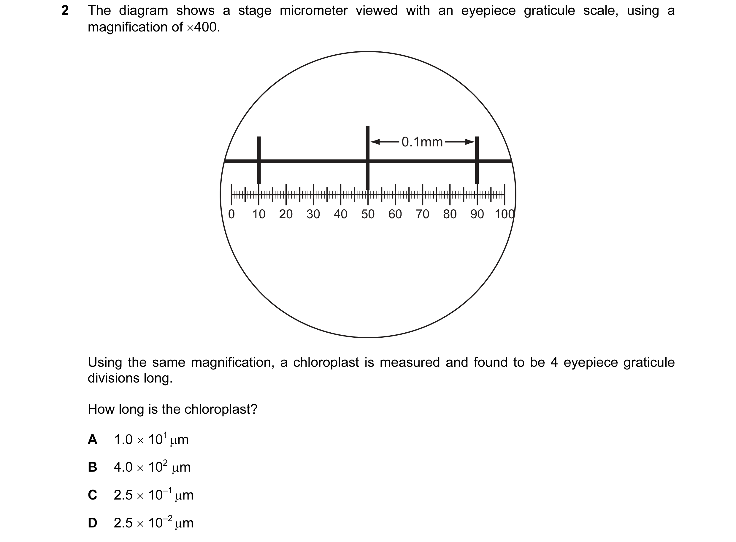
 28 / 72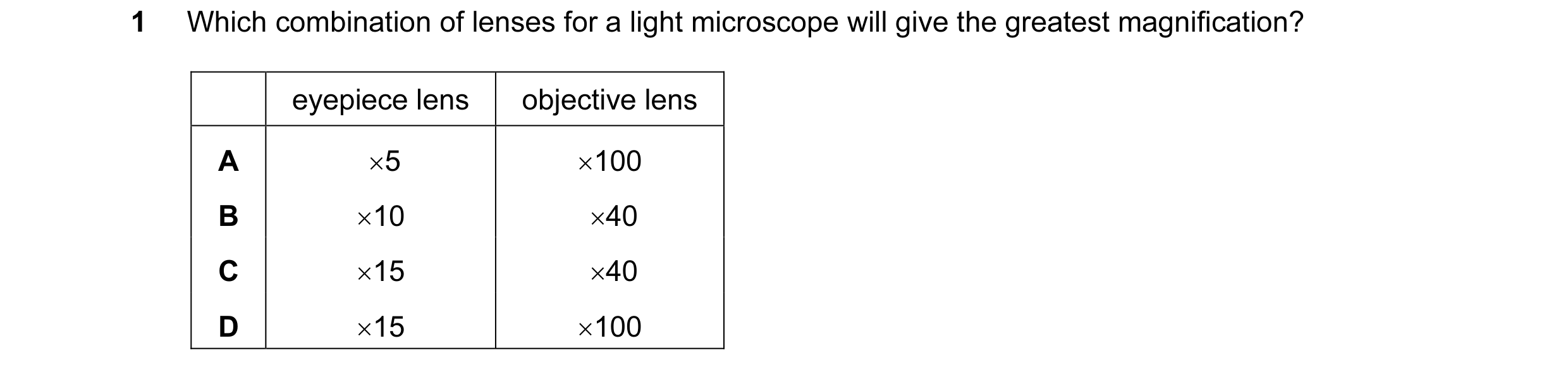
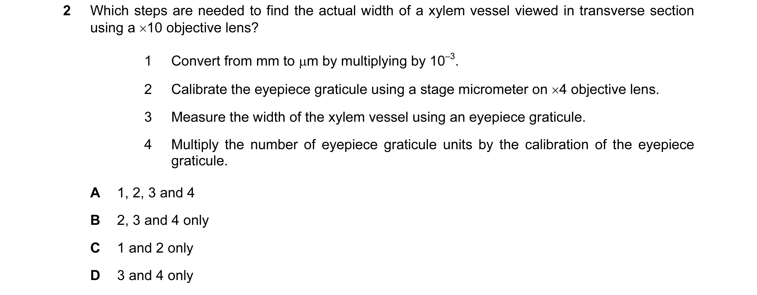
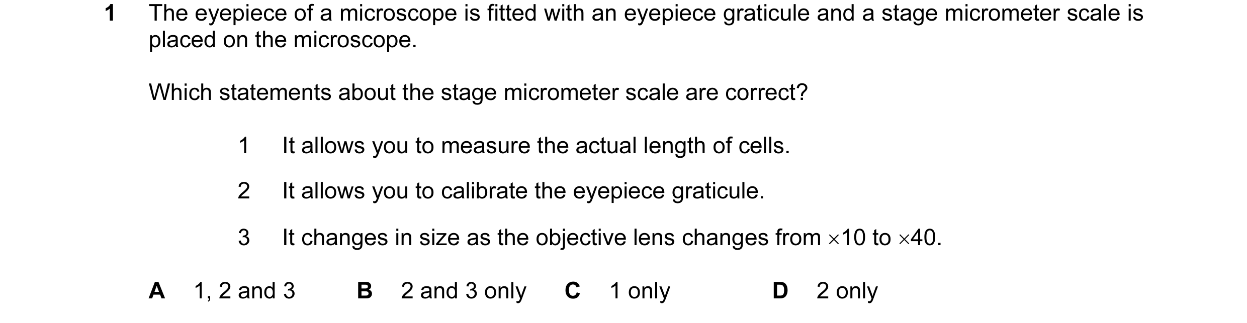
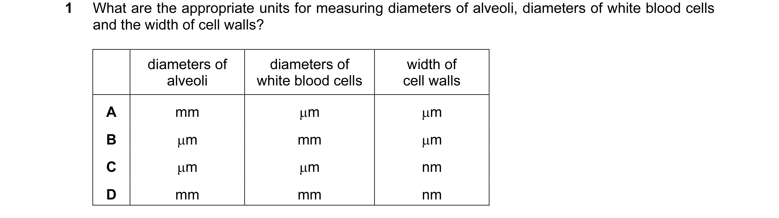 29 / 72
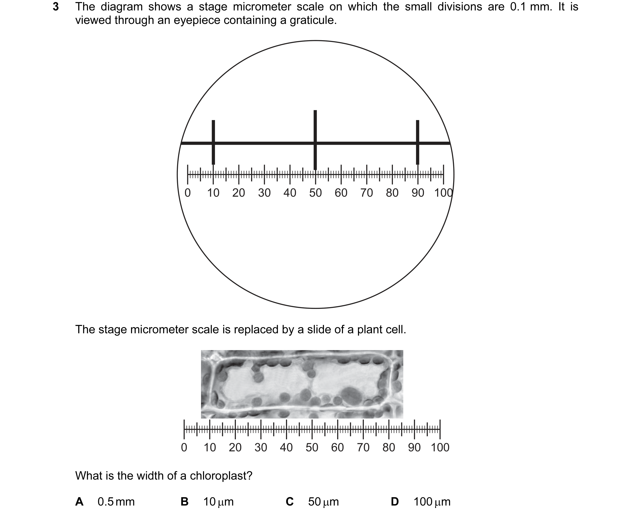 30 / 72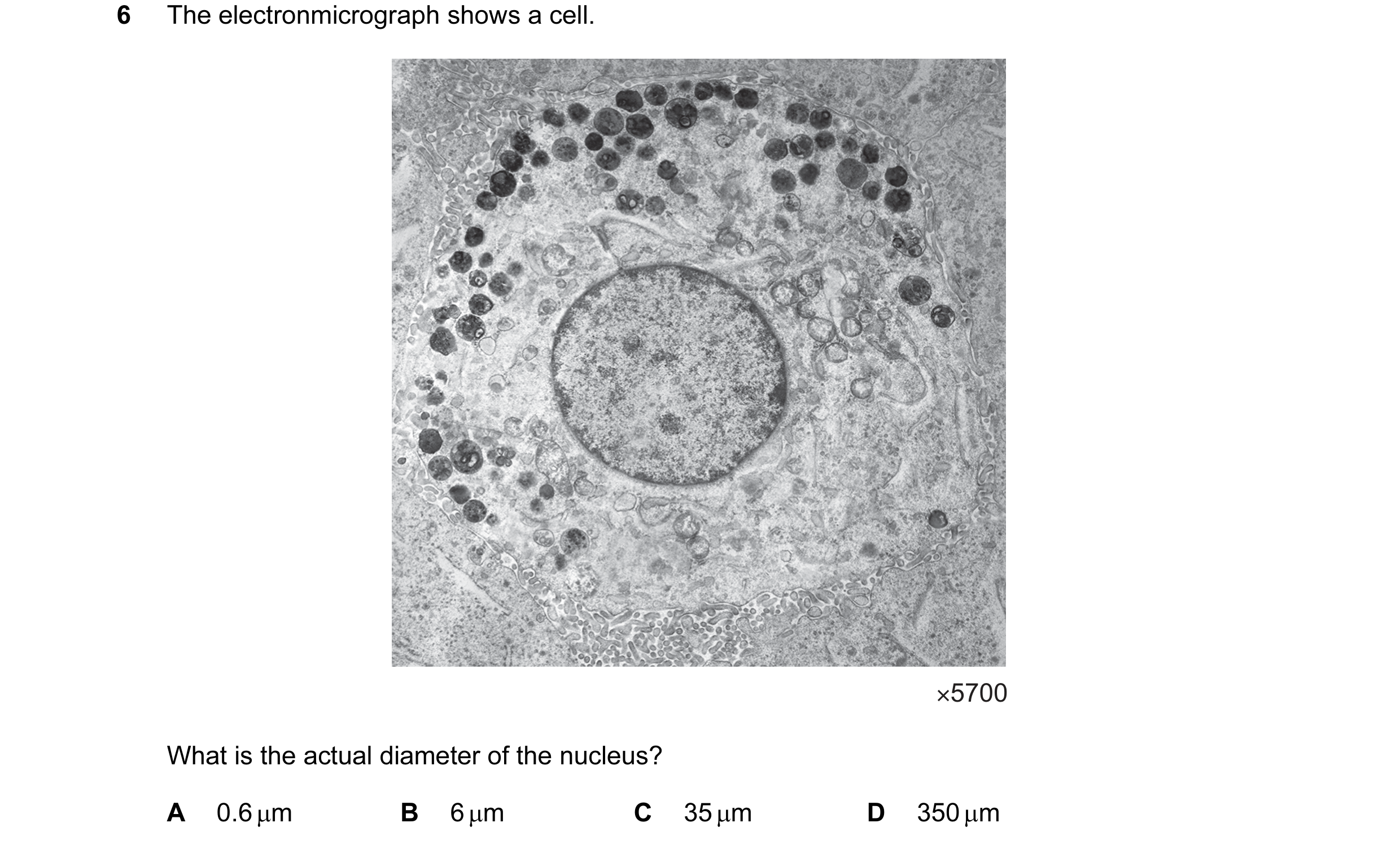

 31 / 72
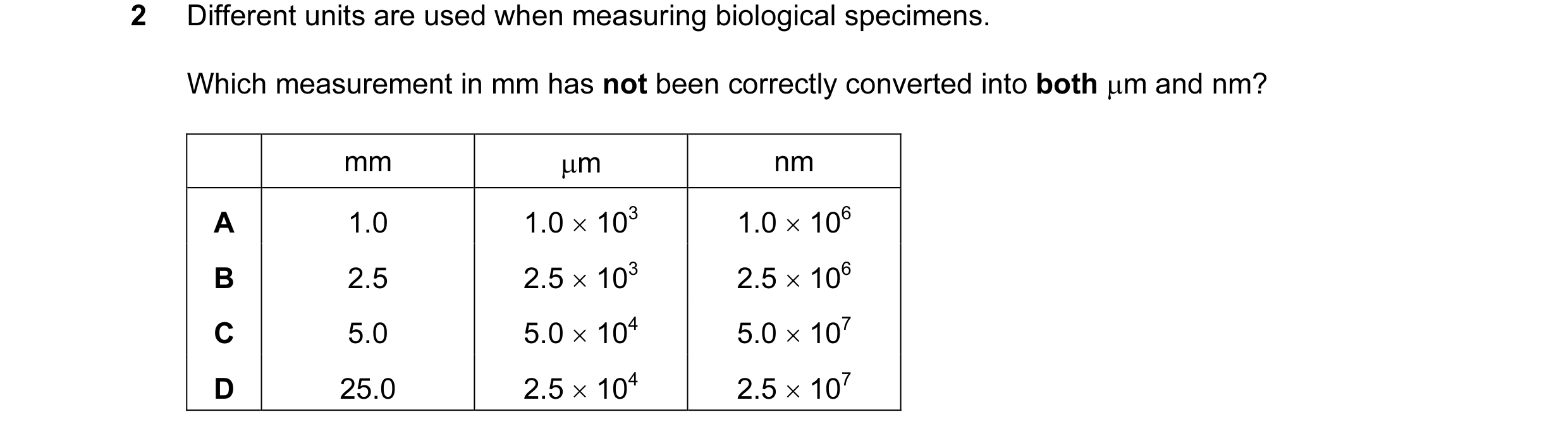
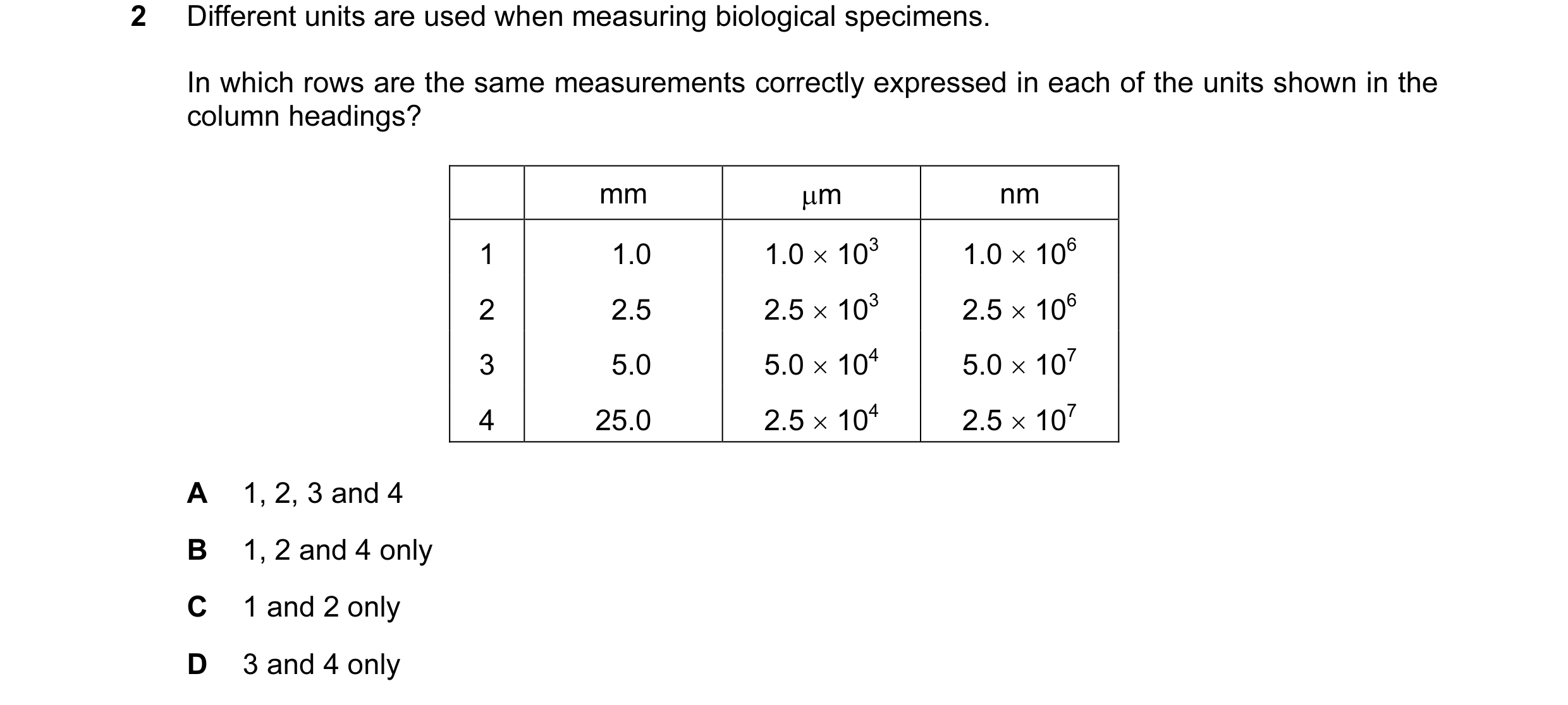
 32 / 72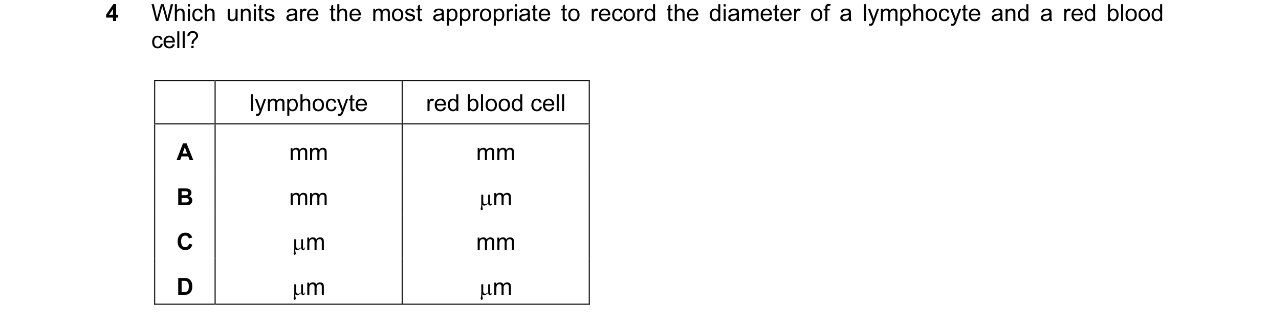
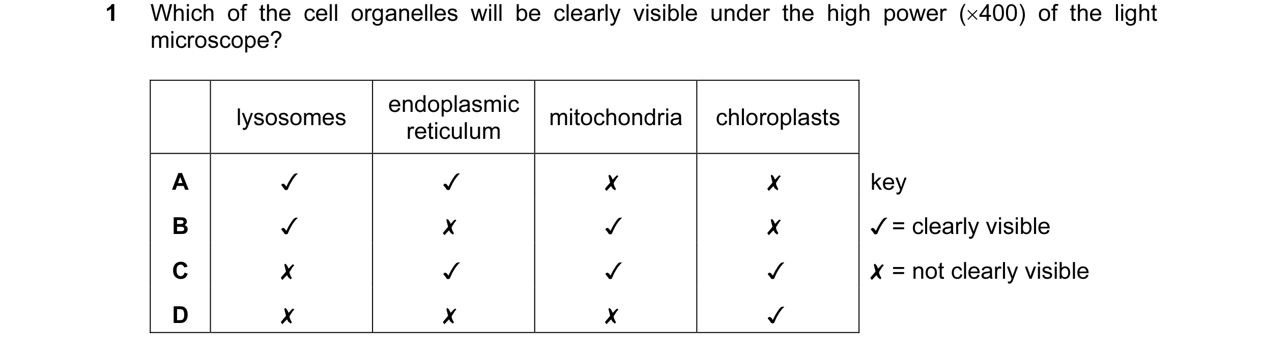
 33 / 72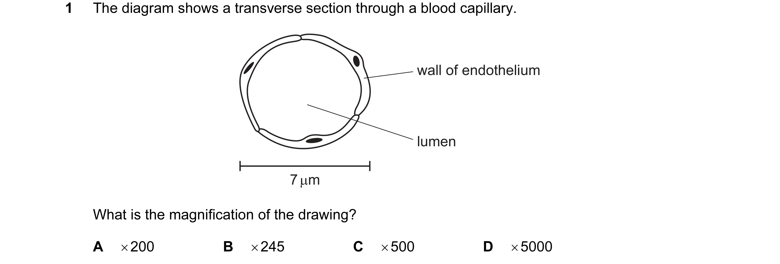

 34 / 72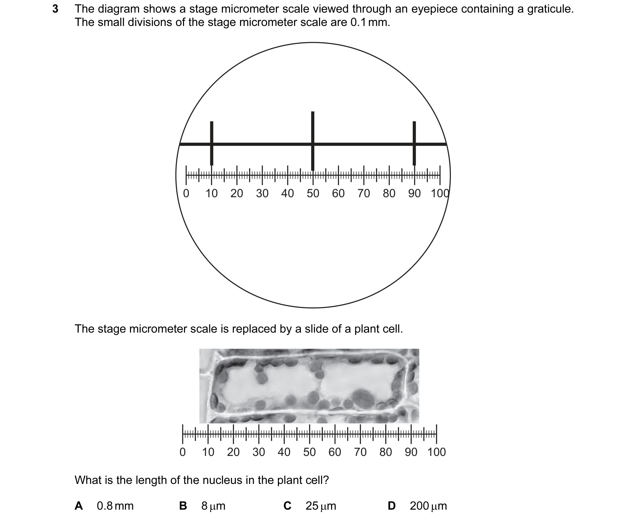

 35 / 72
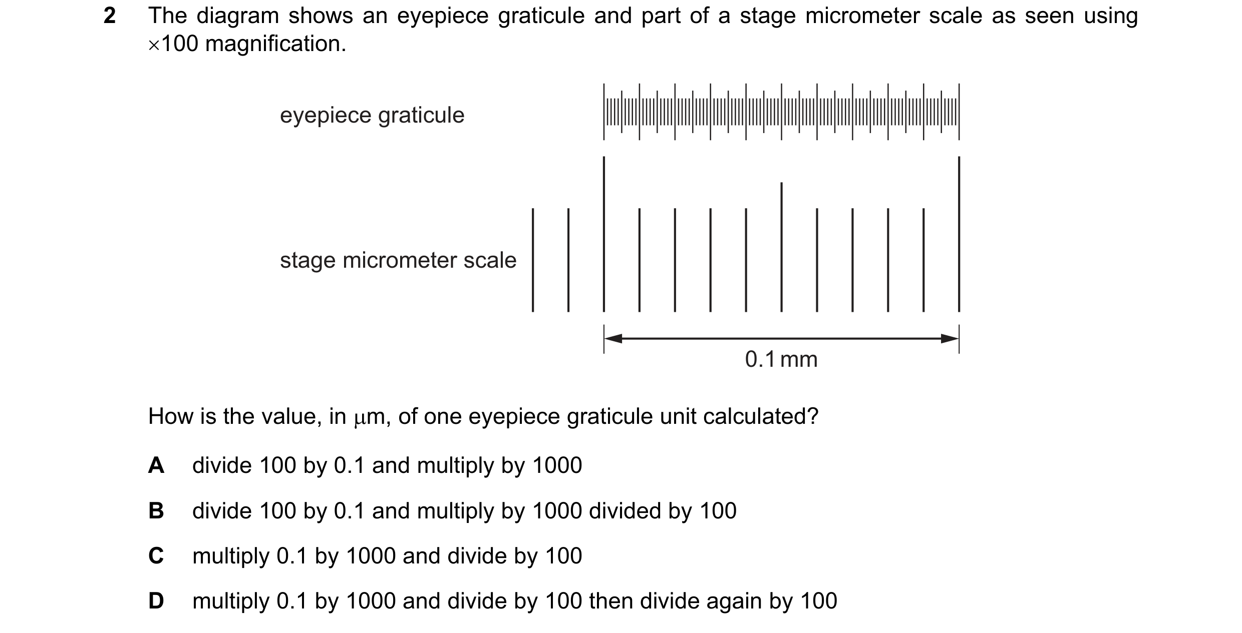
 36 / 72
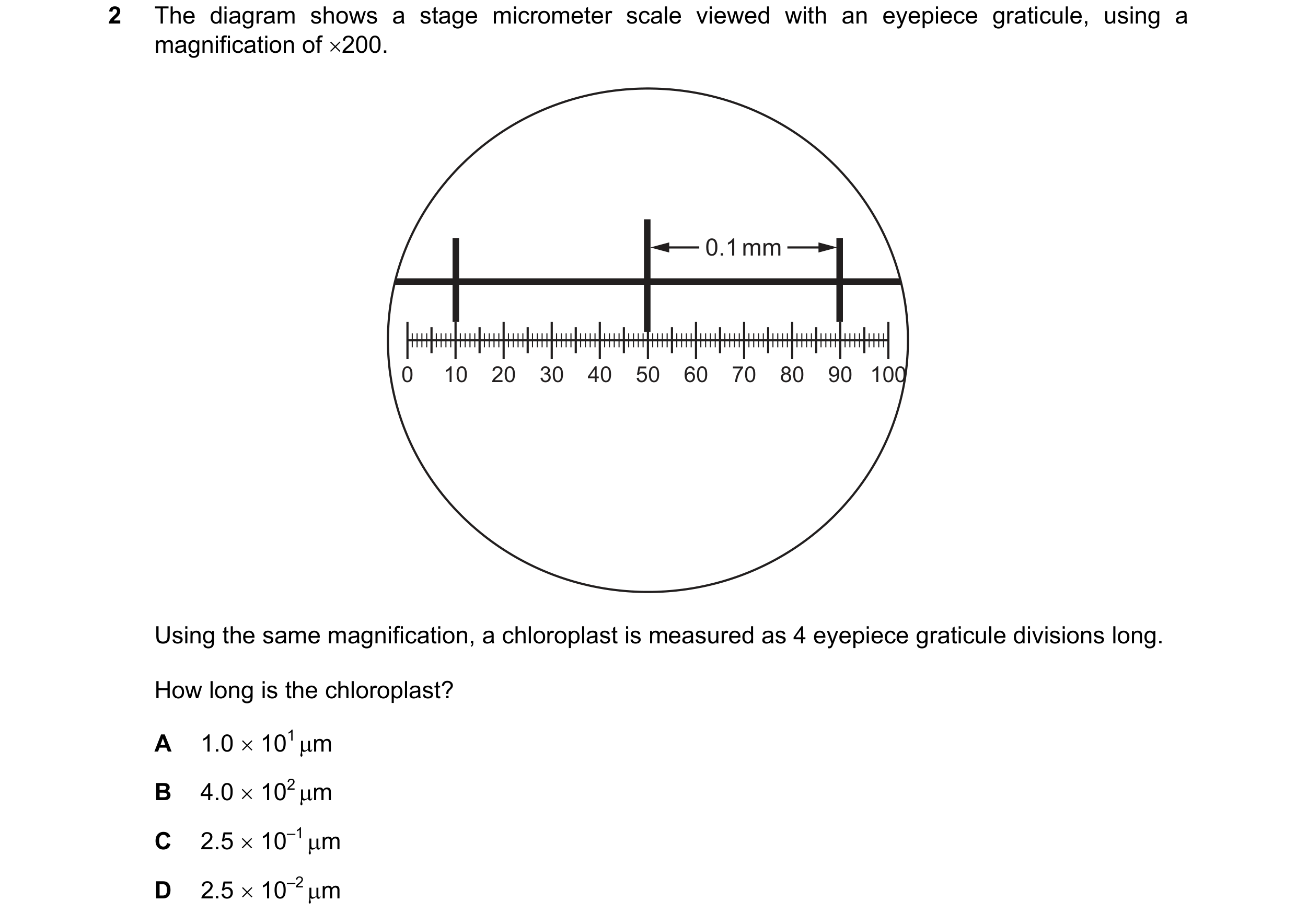
 37 / 72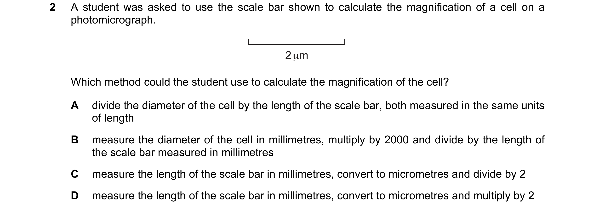
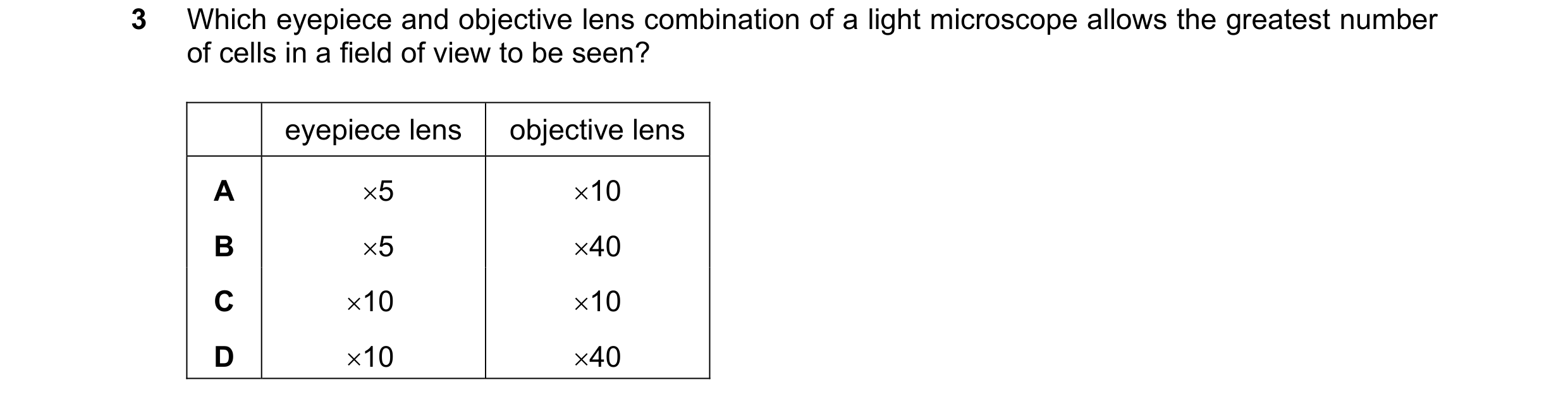
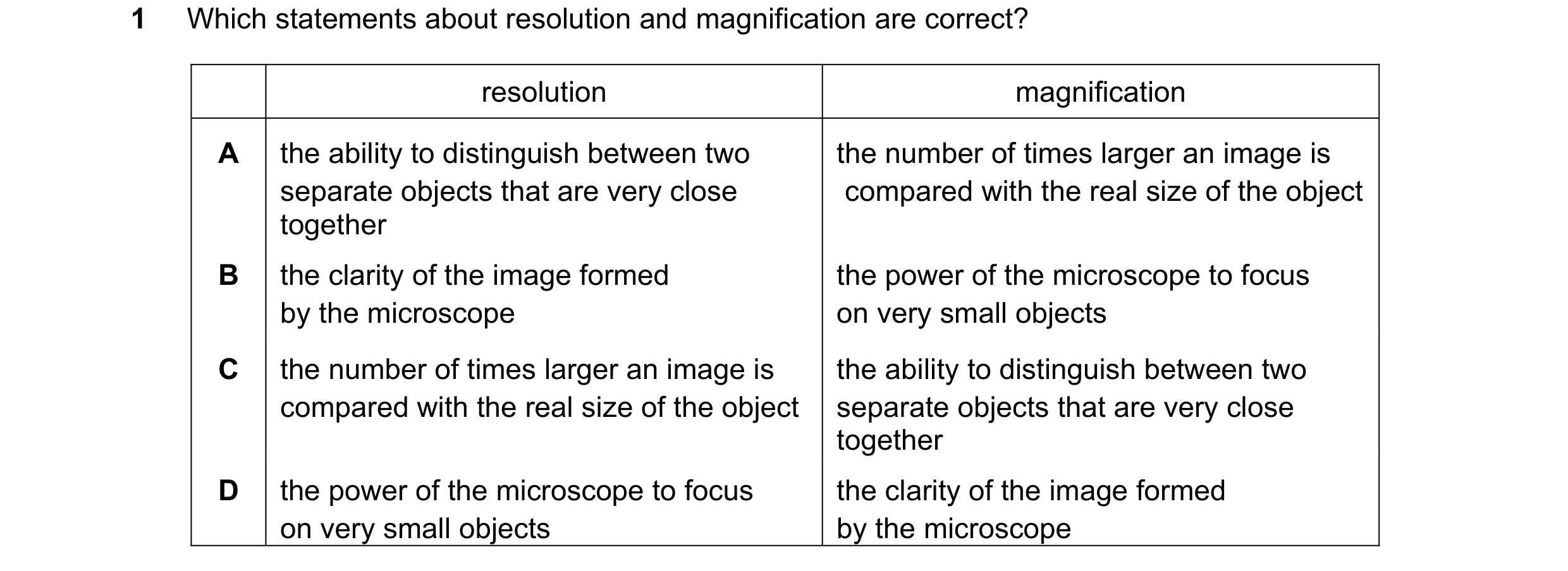
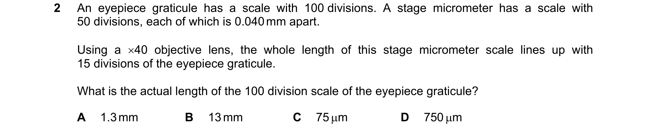 38 / 72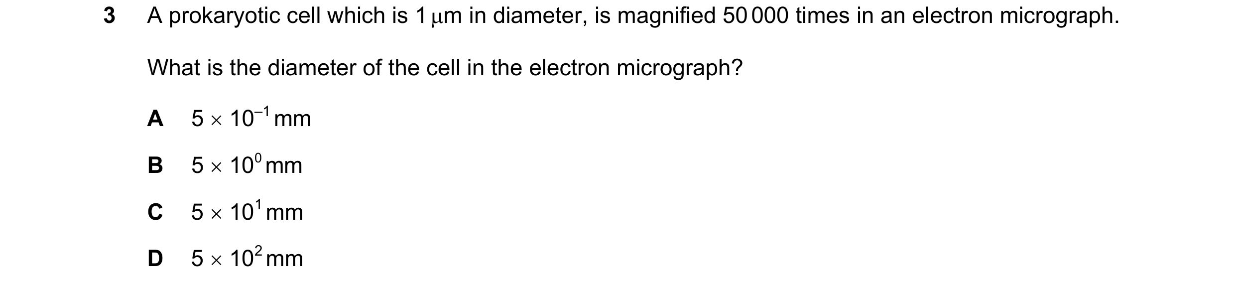
 39 / 72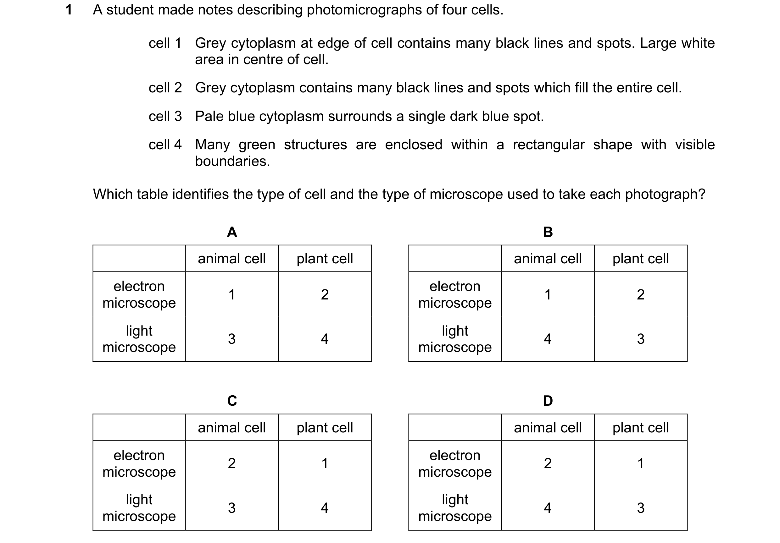
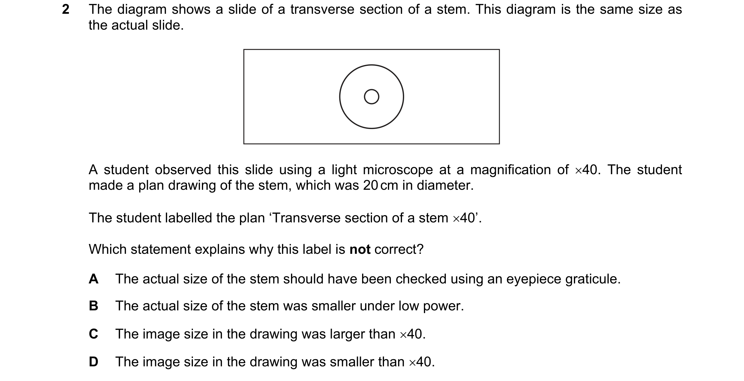 40 / 72
 41 / 72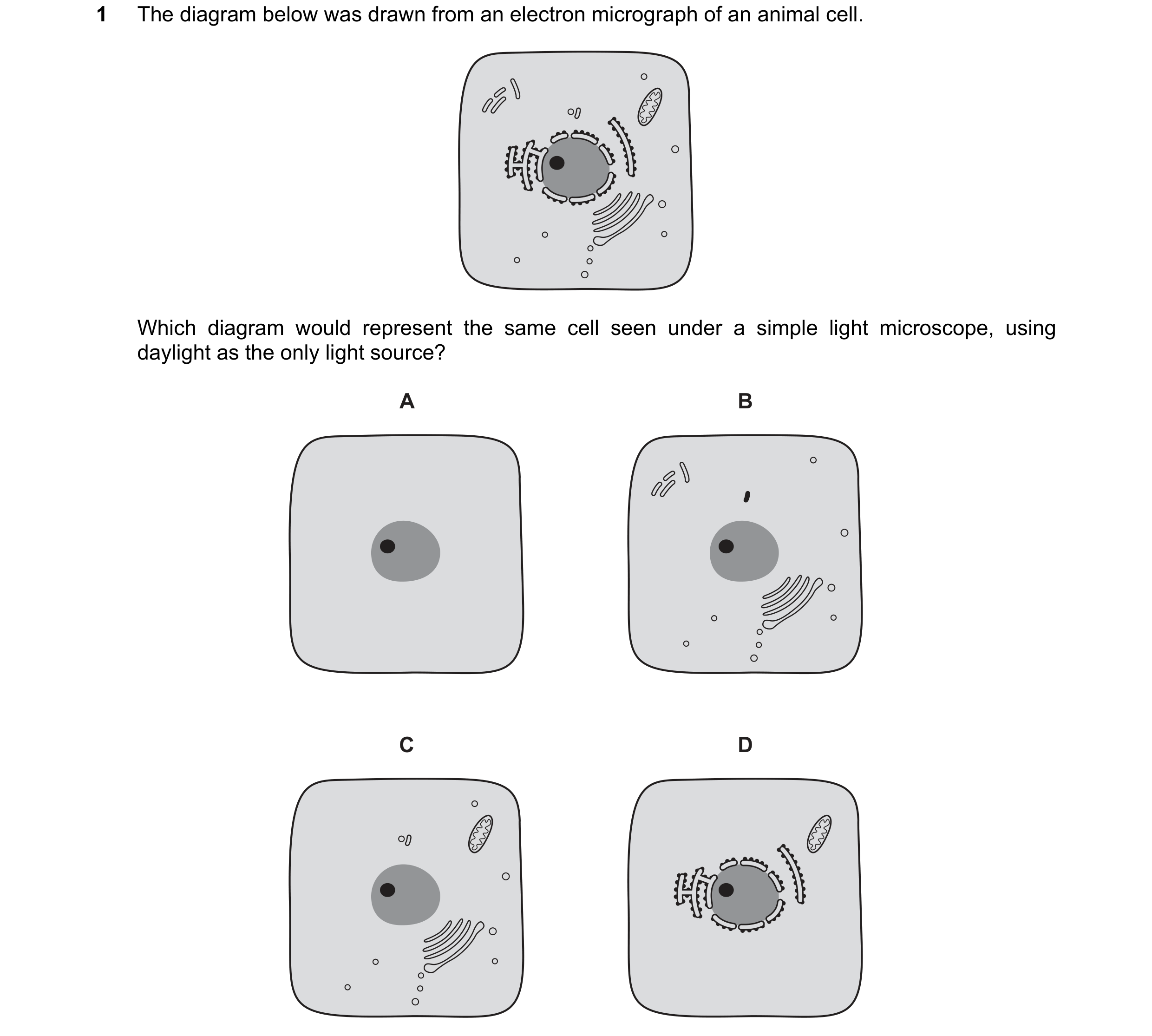
 42 / 72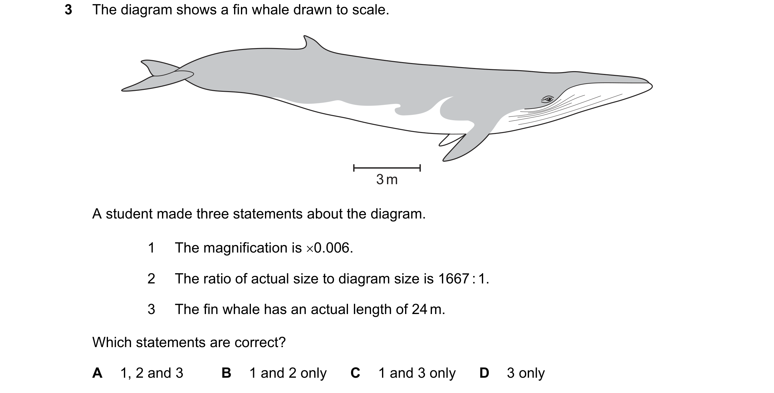
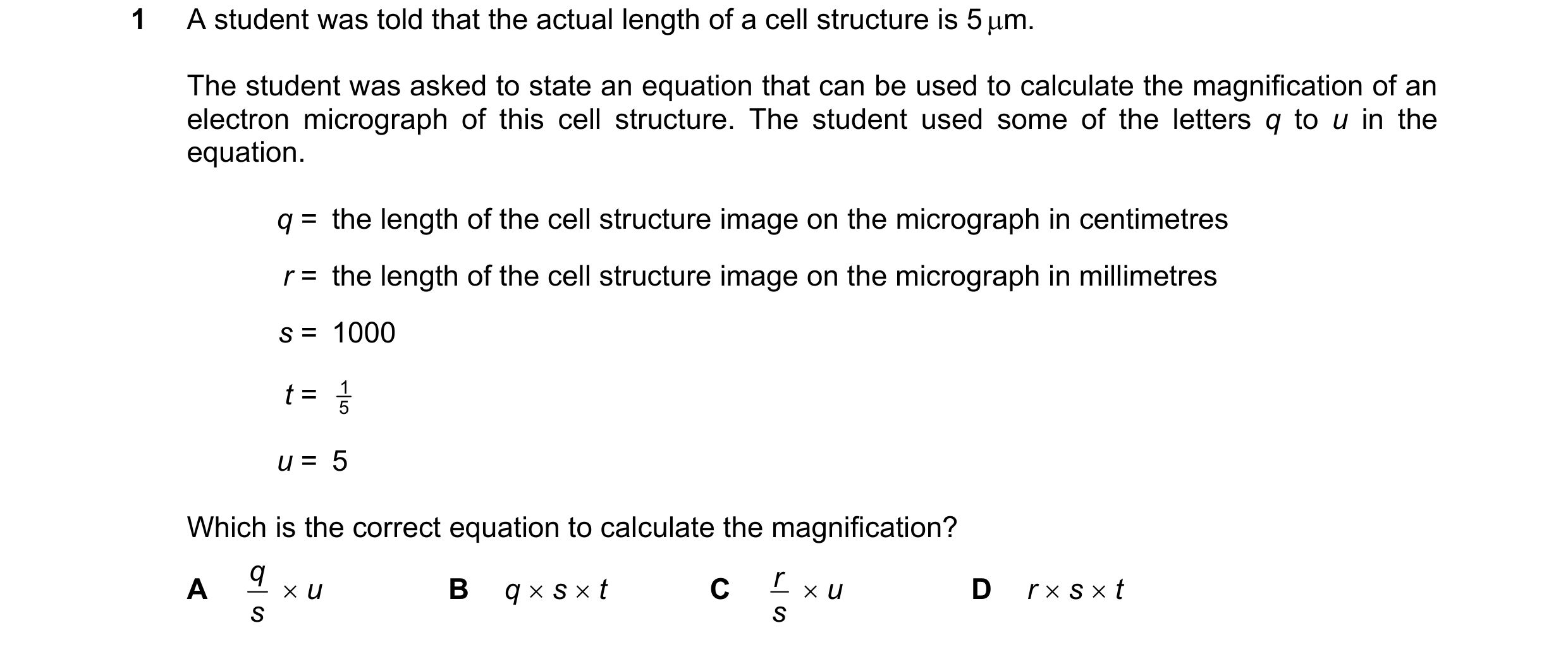 43 / 72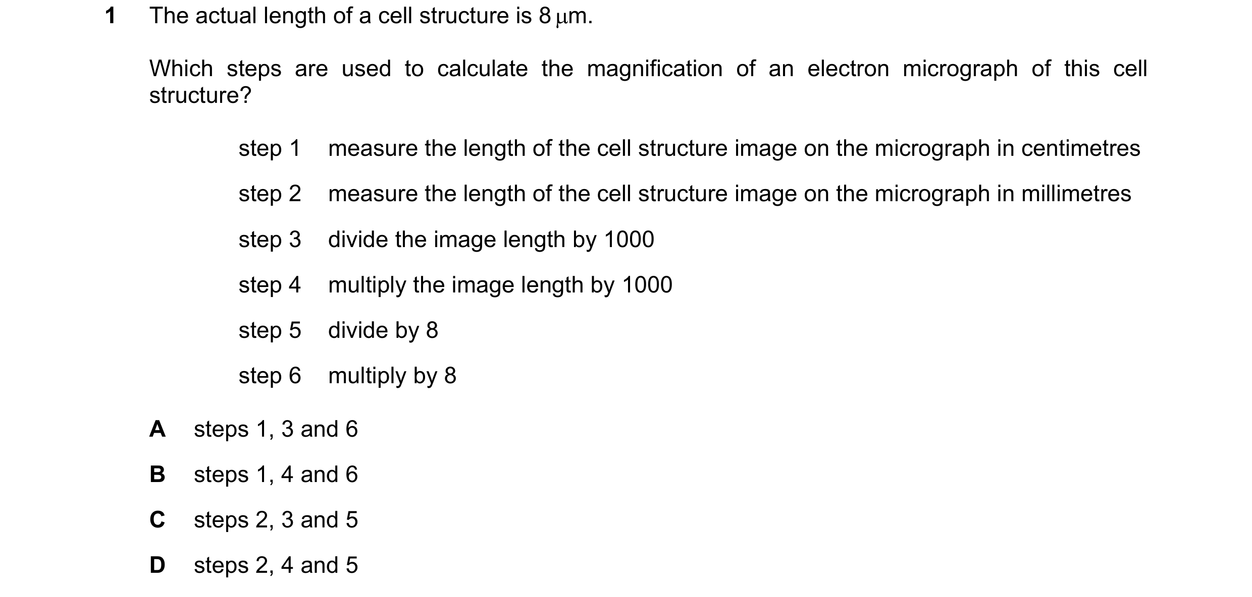

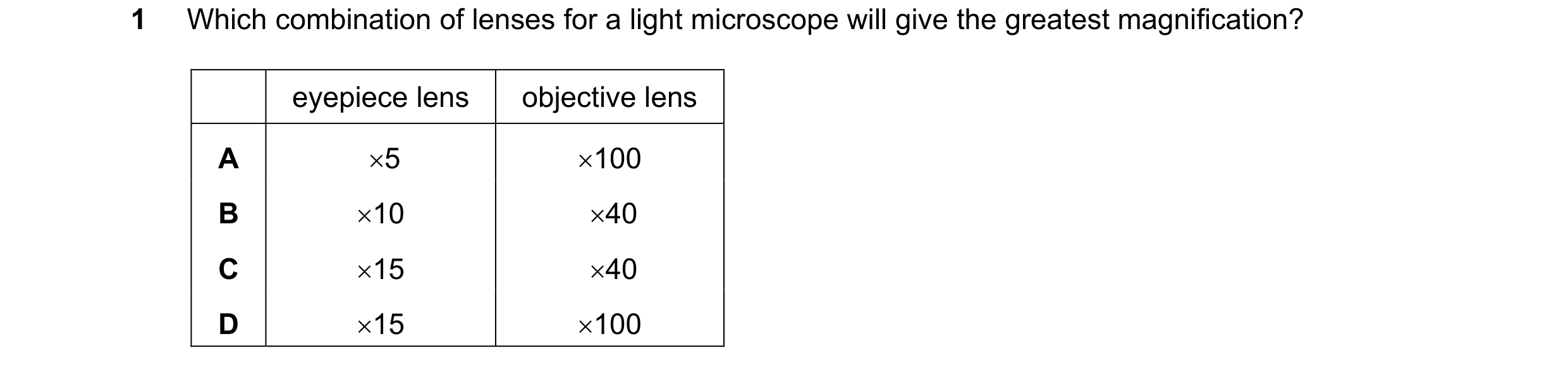
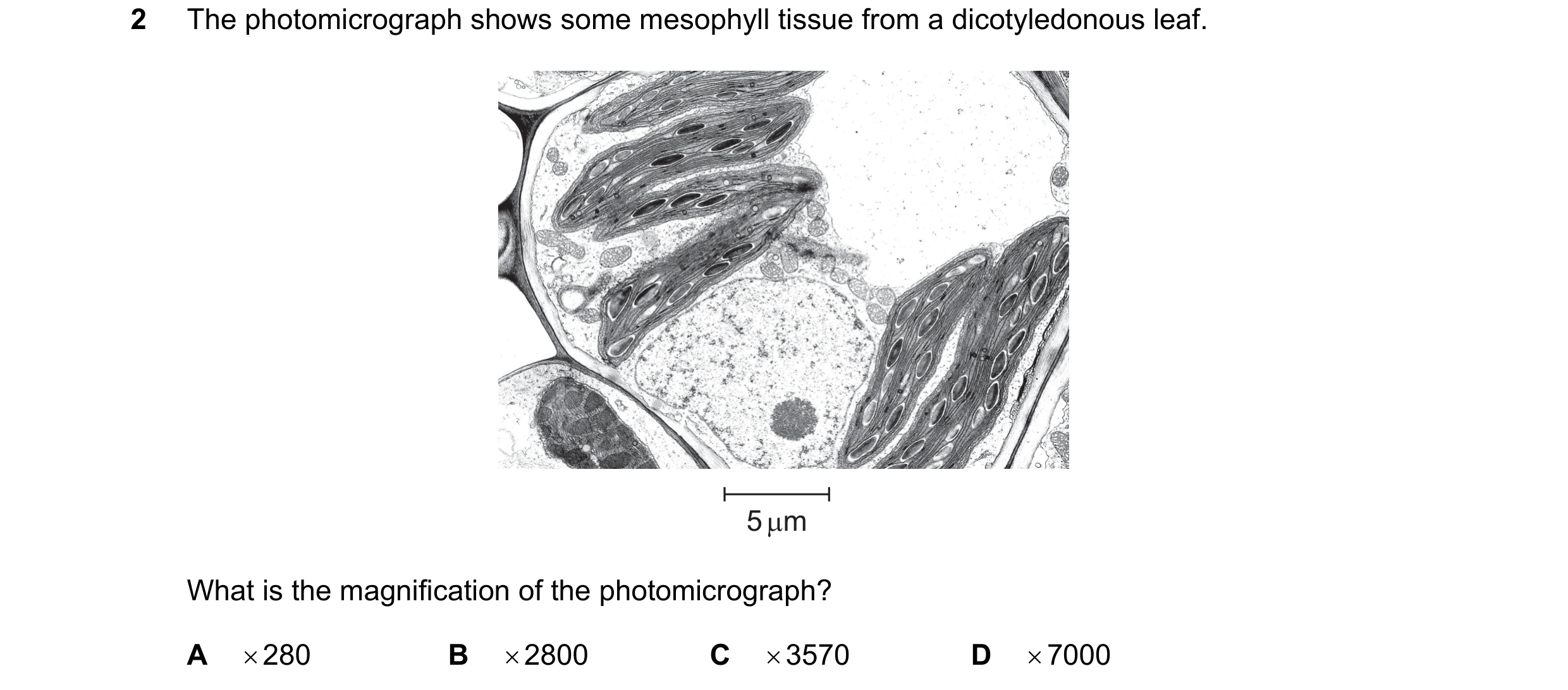 44 / 72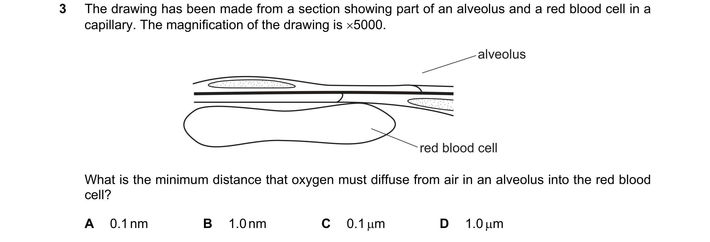
 45 / 72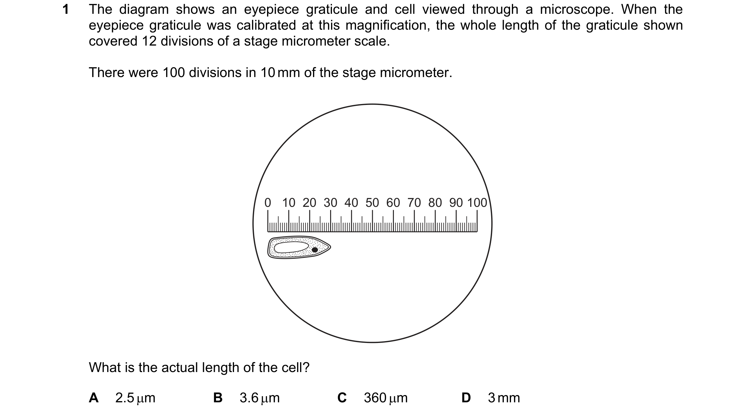
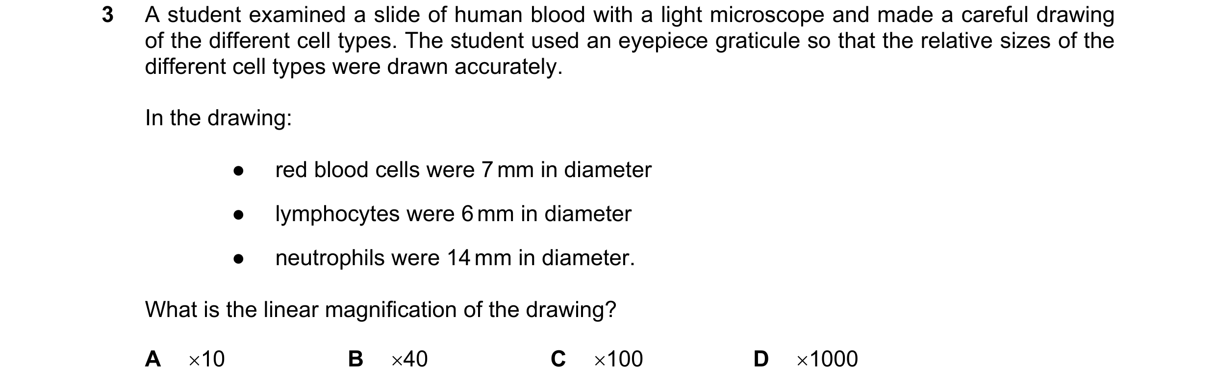 46 / 72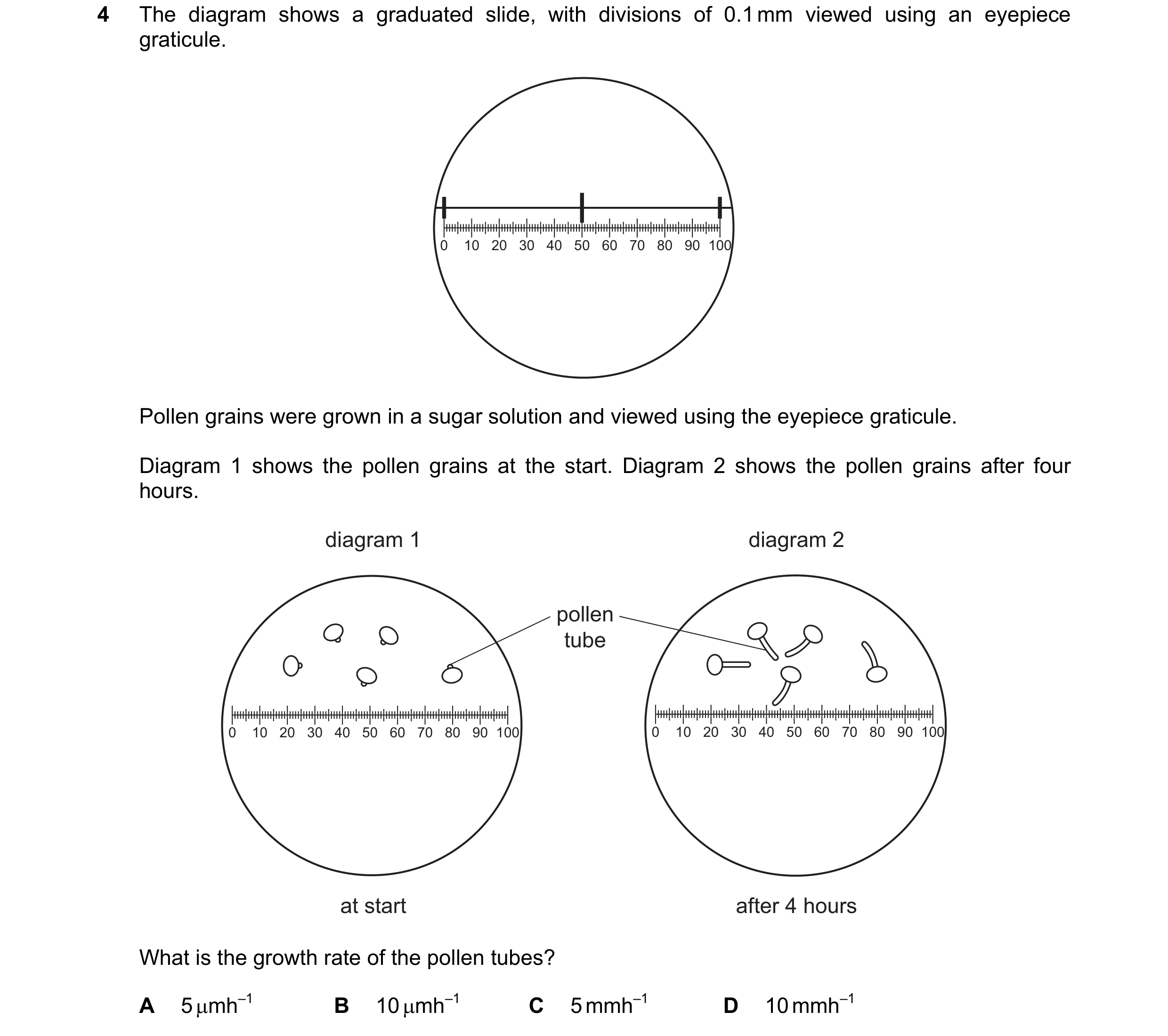
 47 / 72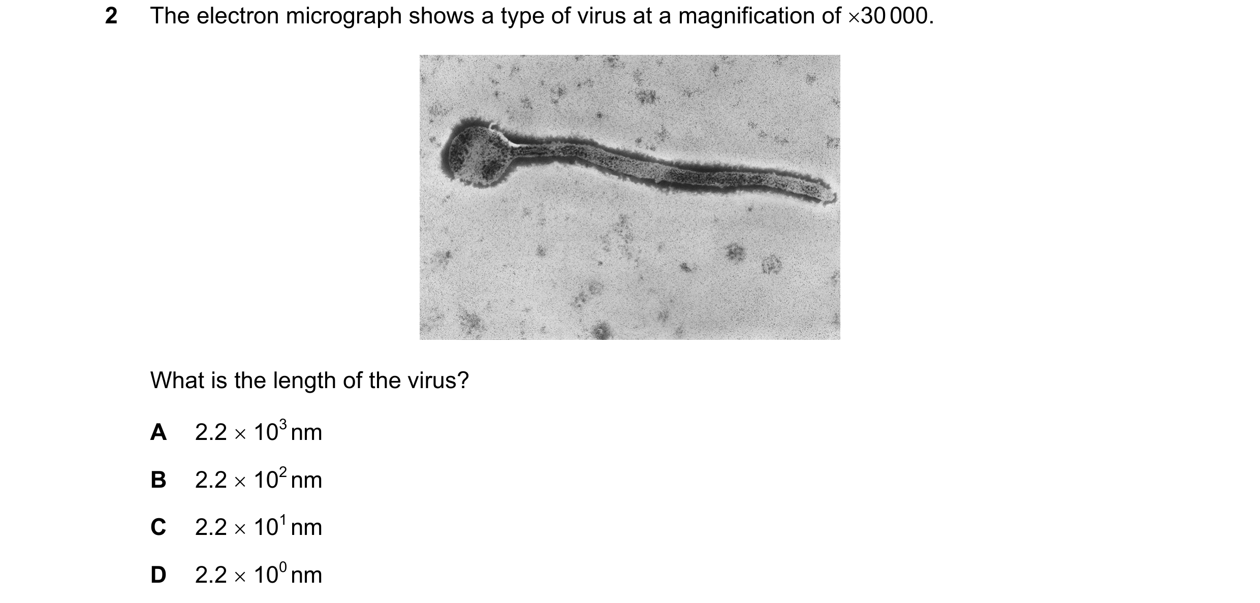
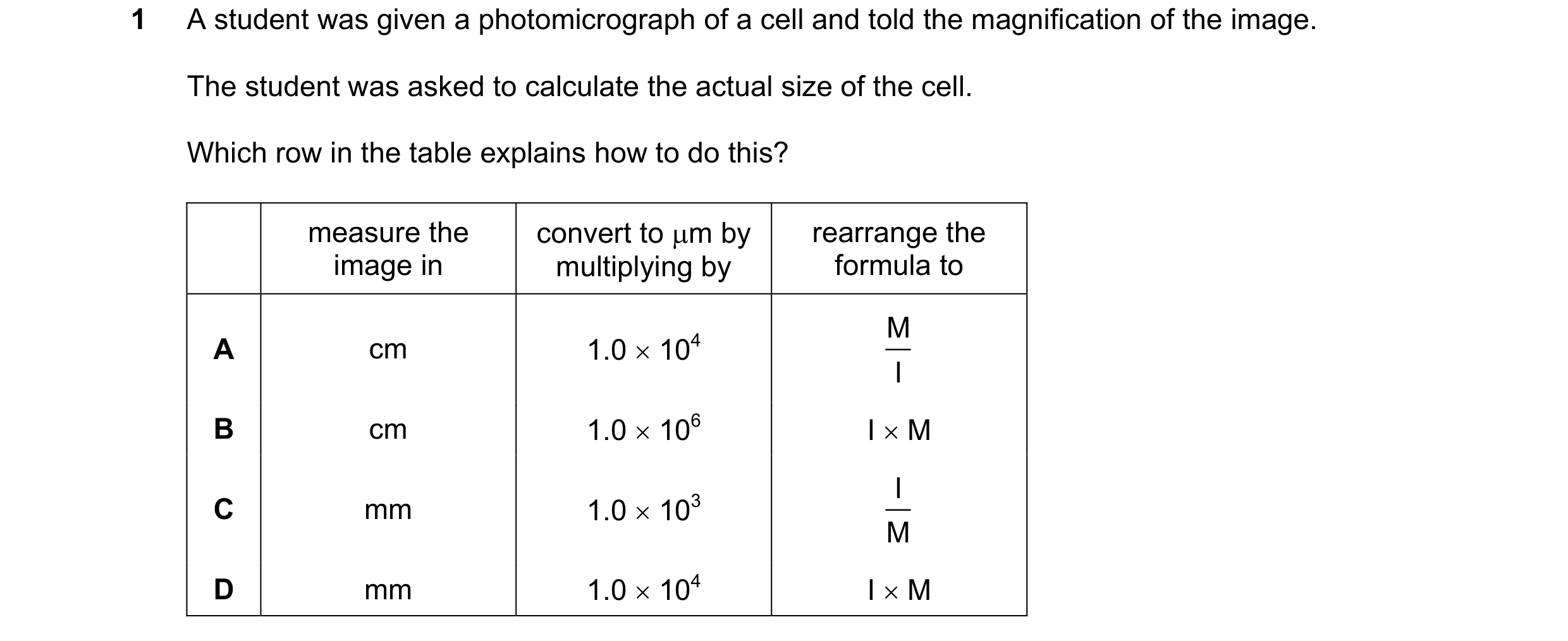 48 / 72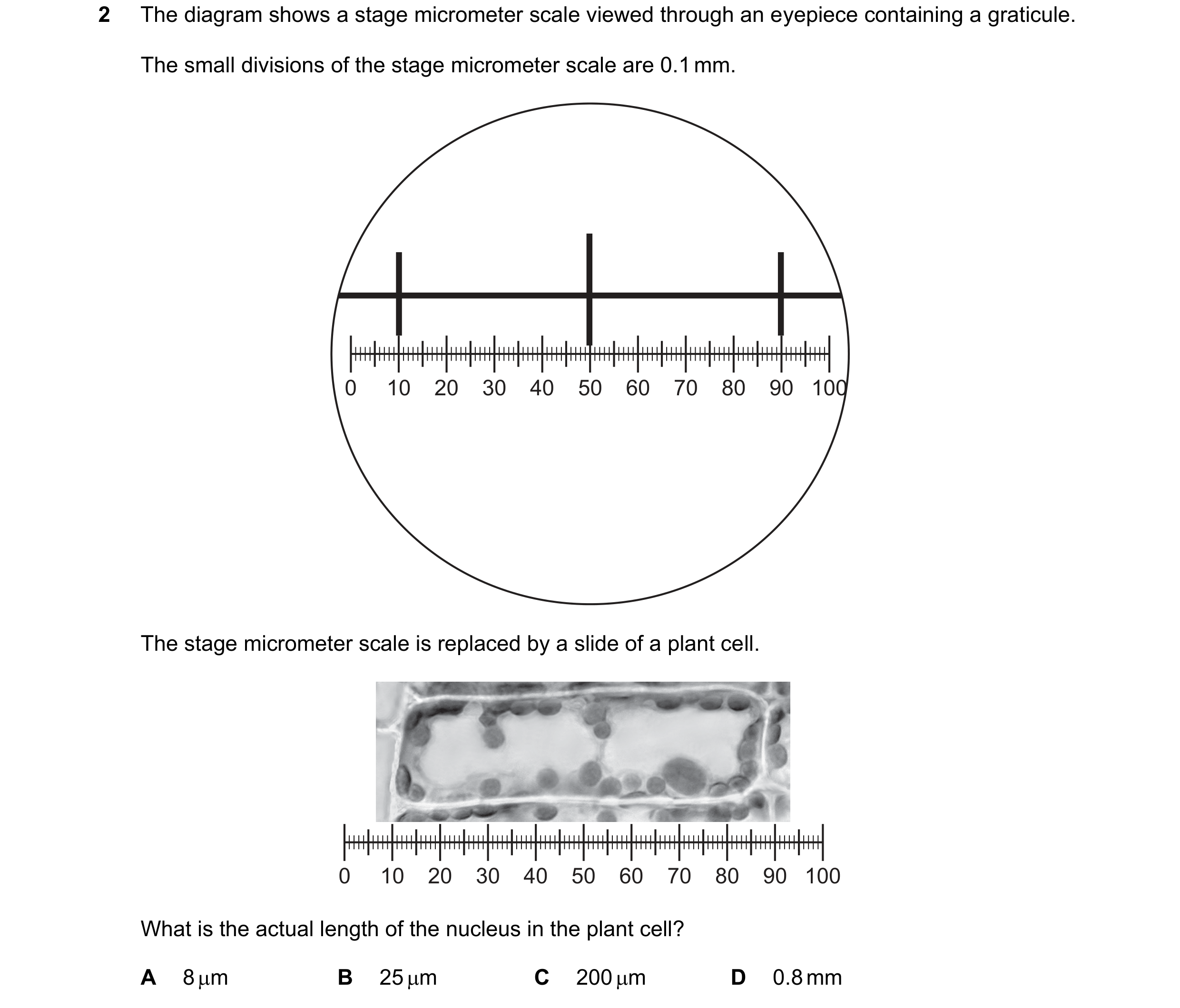
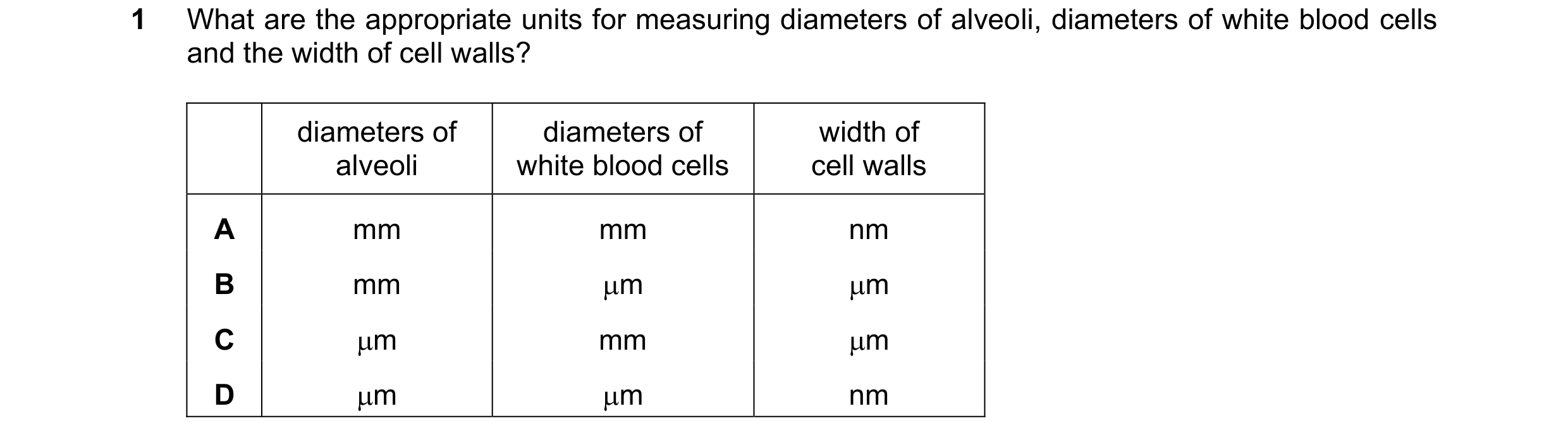 49 / 72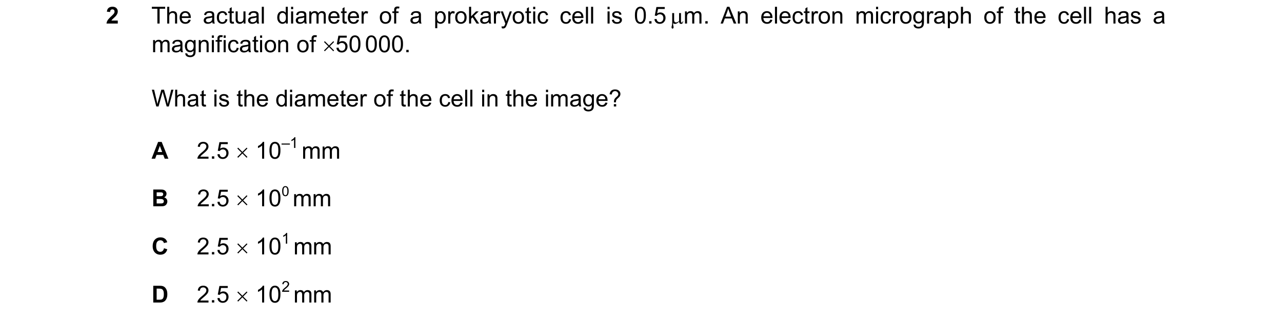
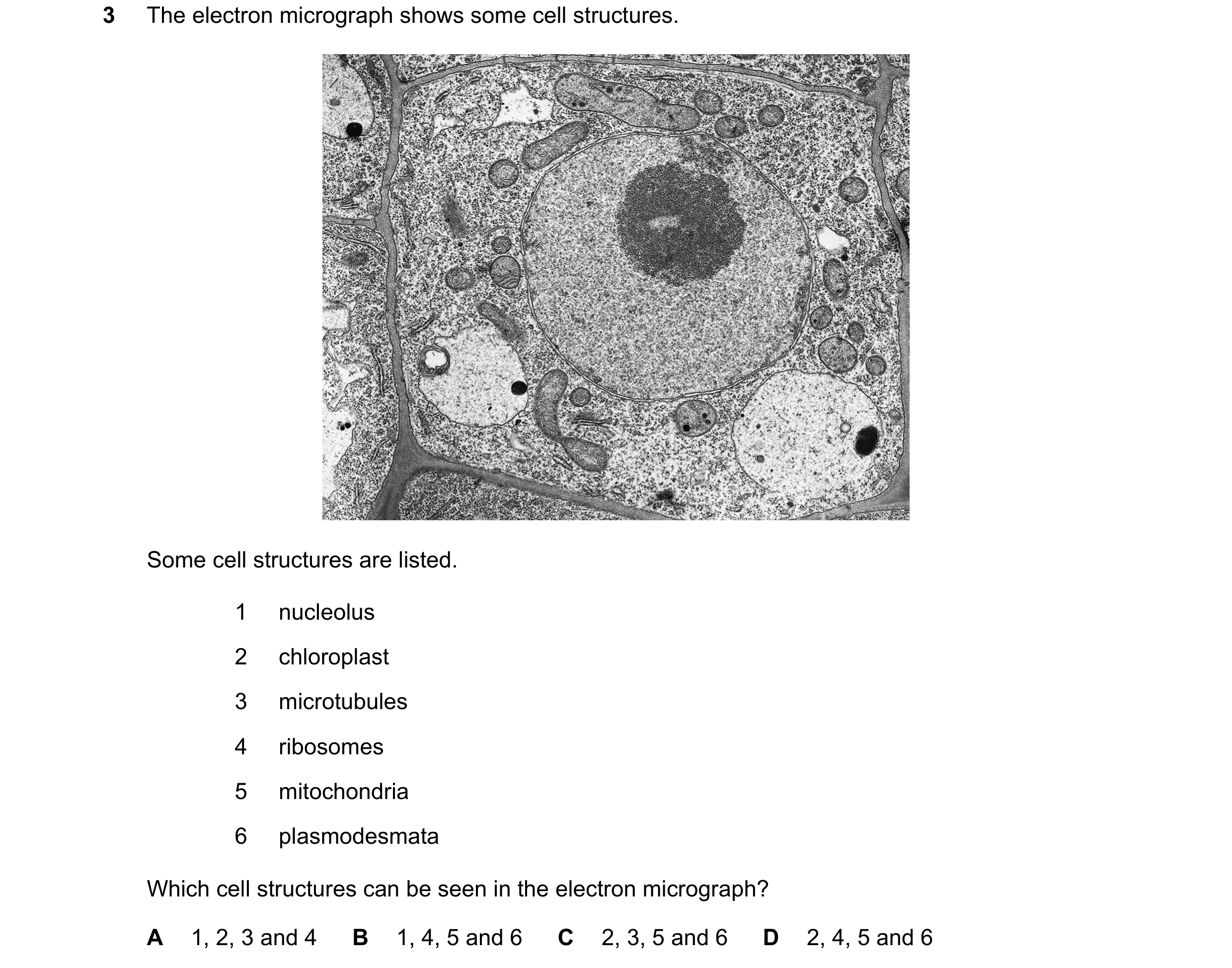 50 / 72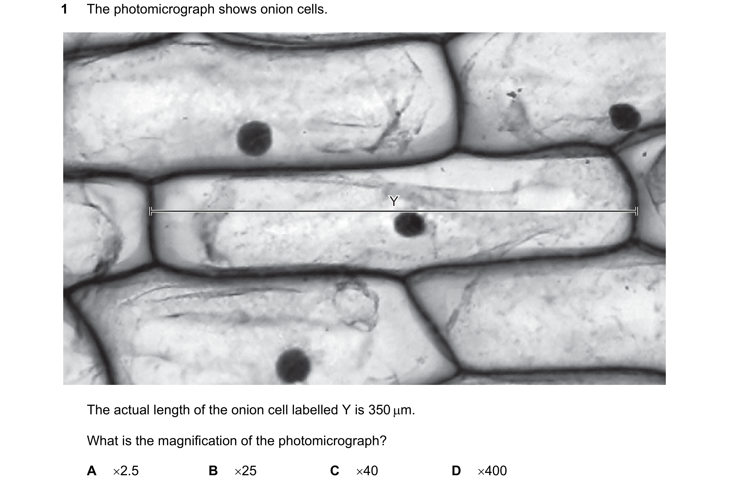
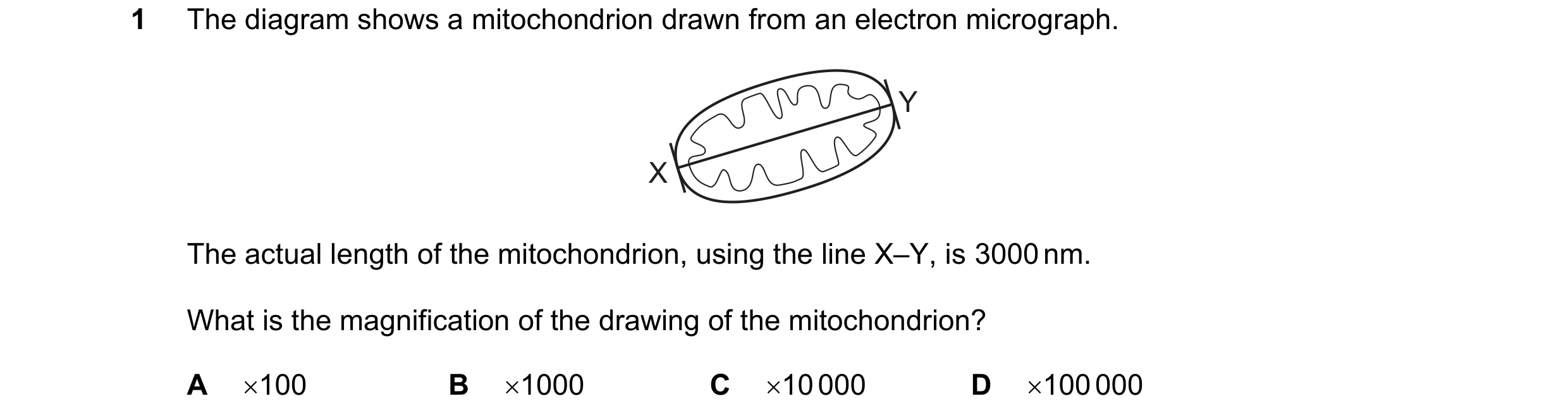
 51 / 72

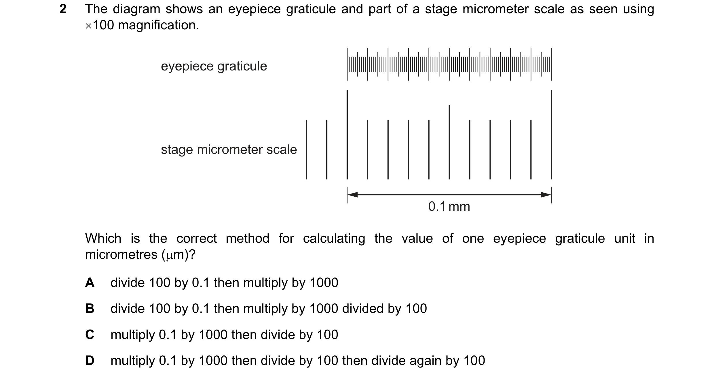
 52 / 72
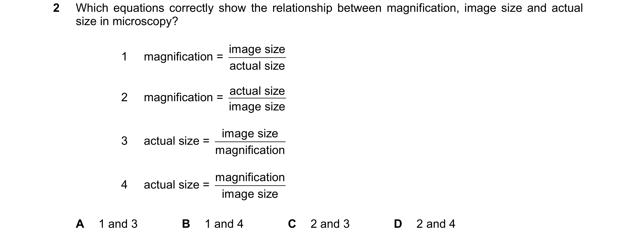

 53 / 72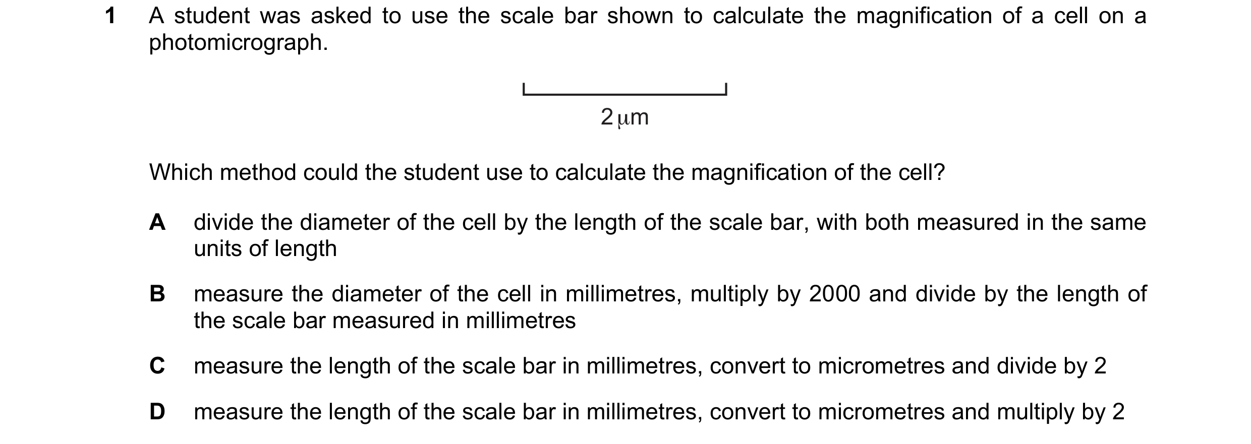
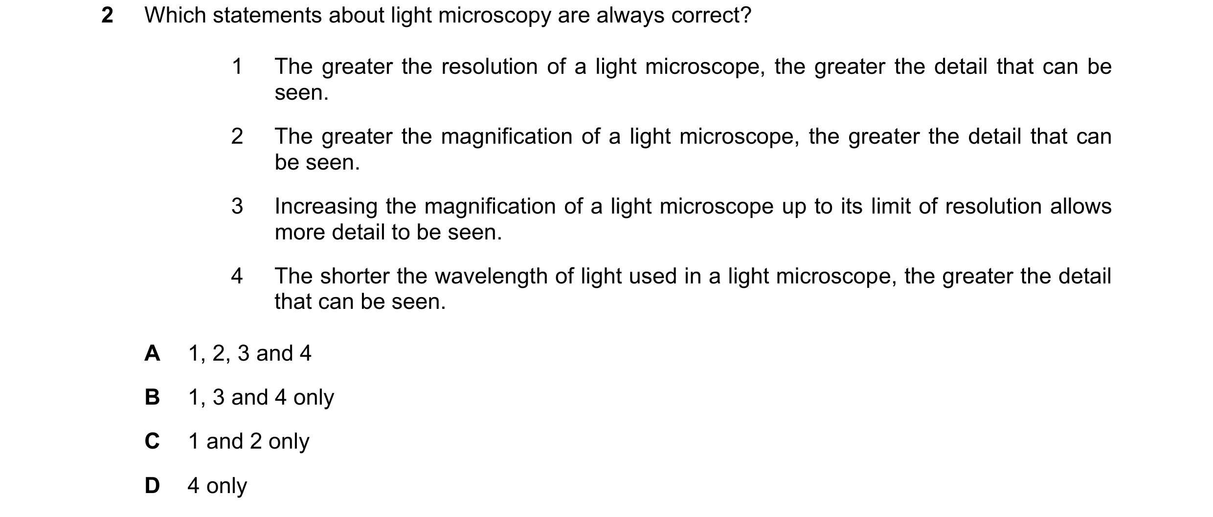
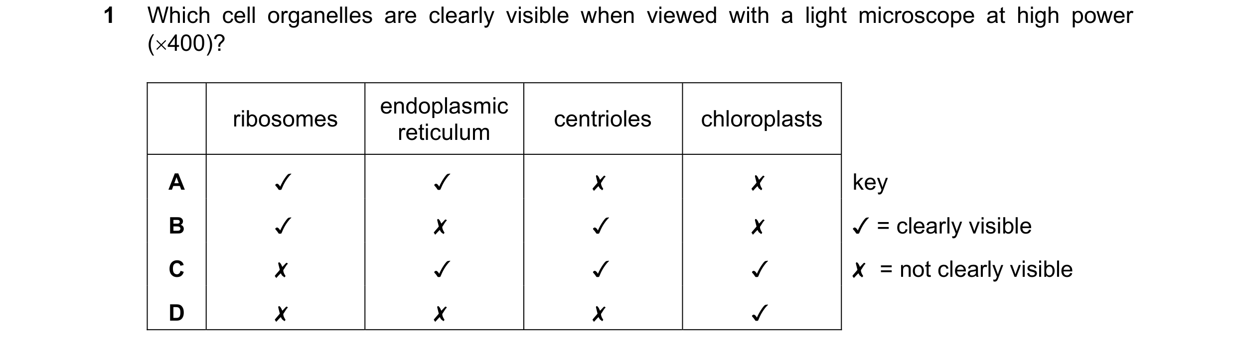 54 / 72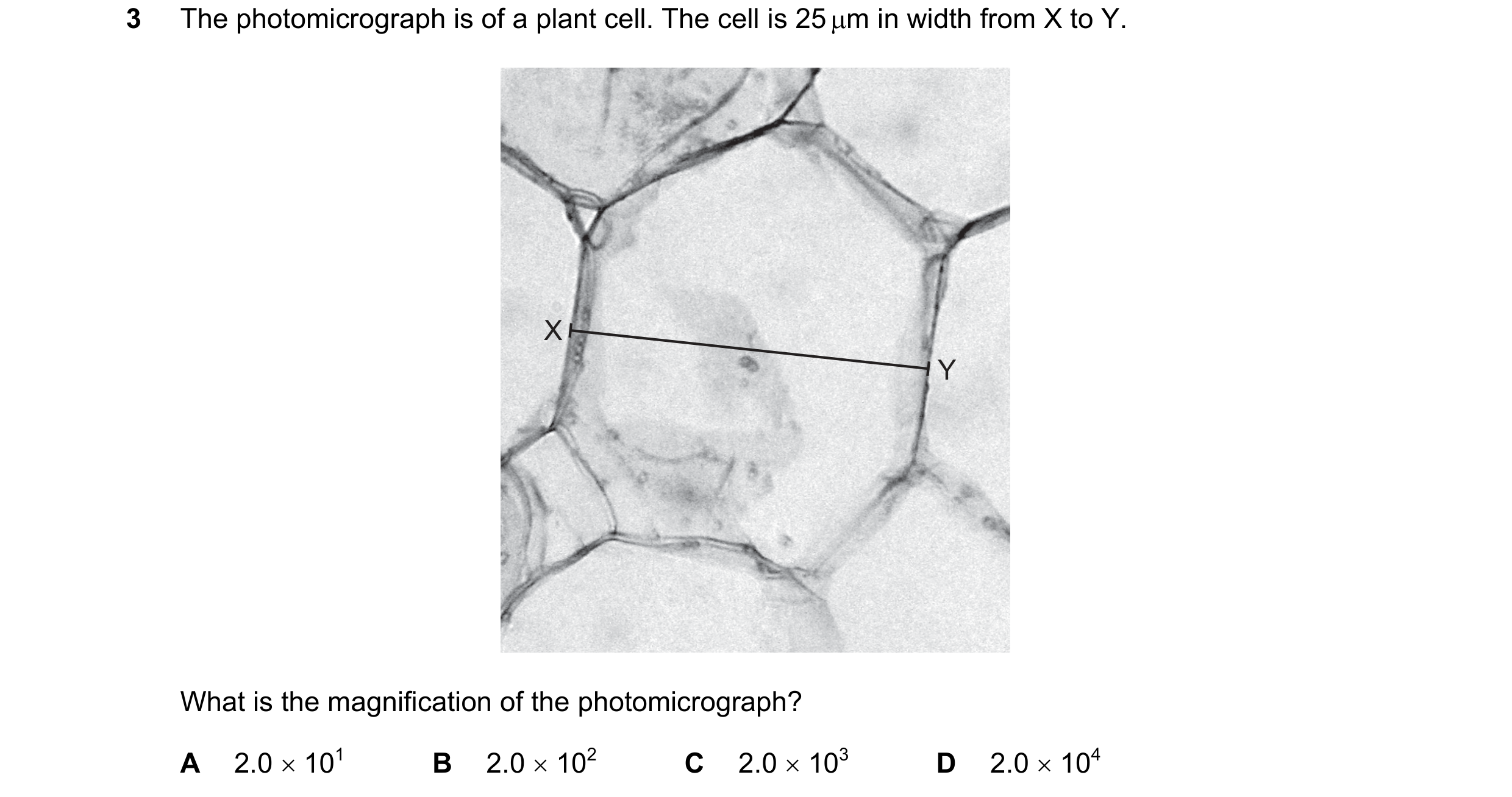
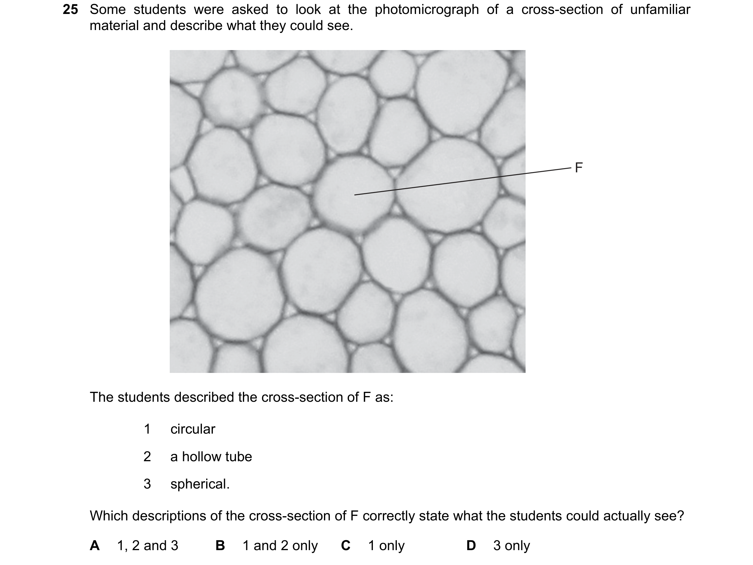 55 / 72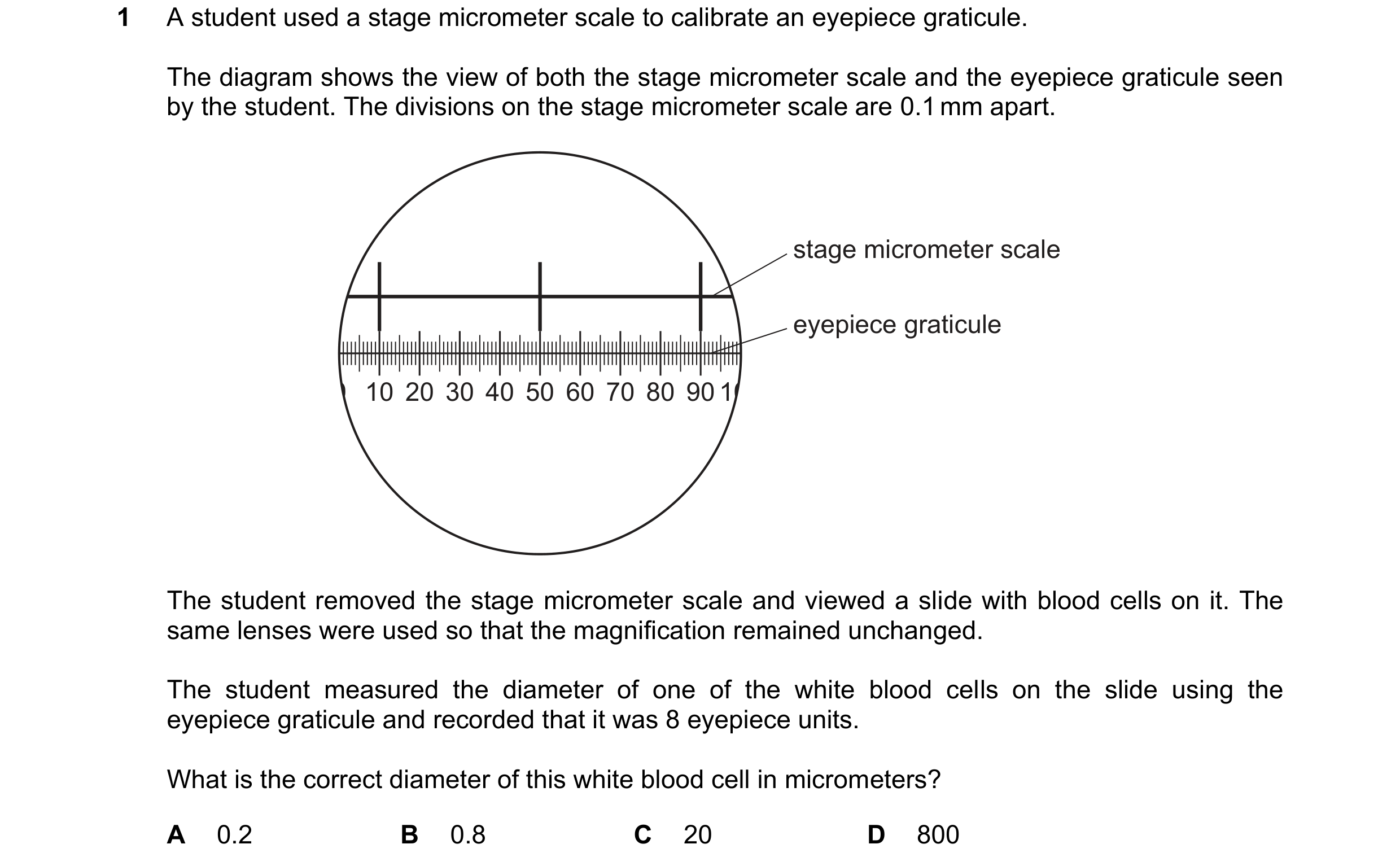
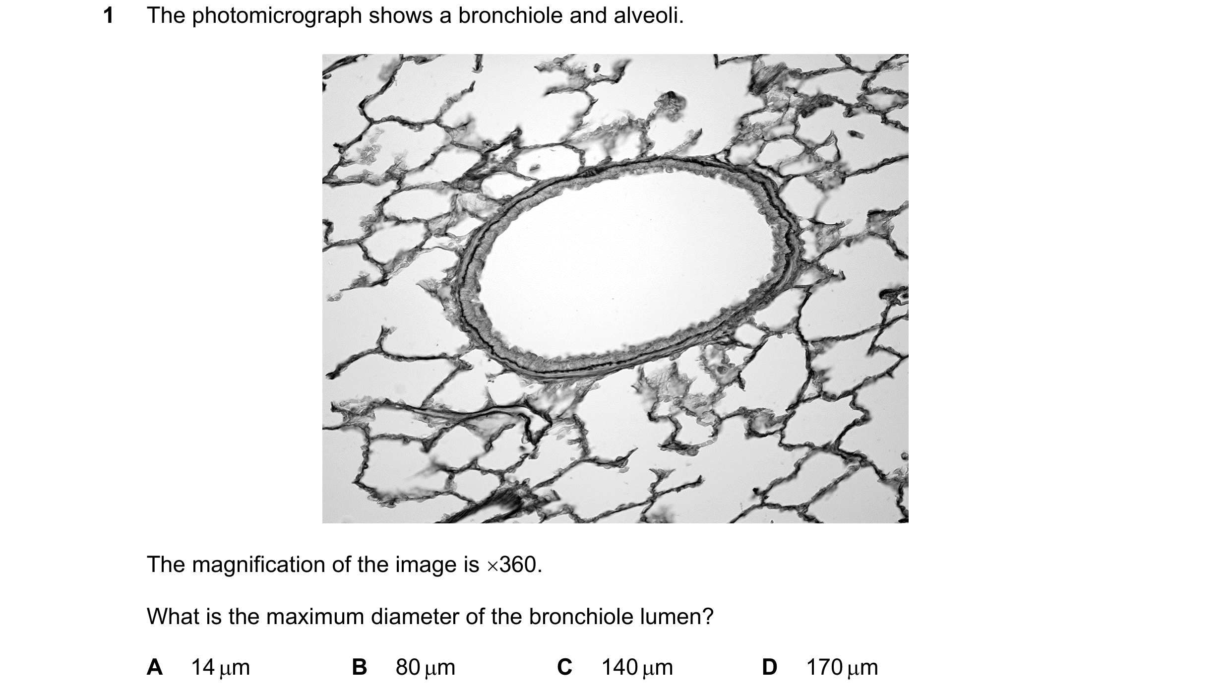 56 / 72
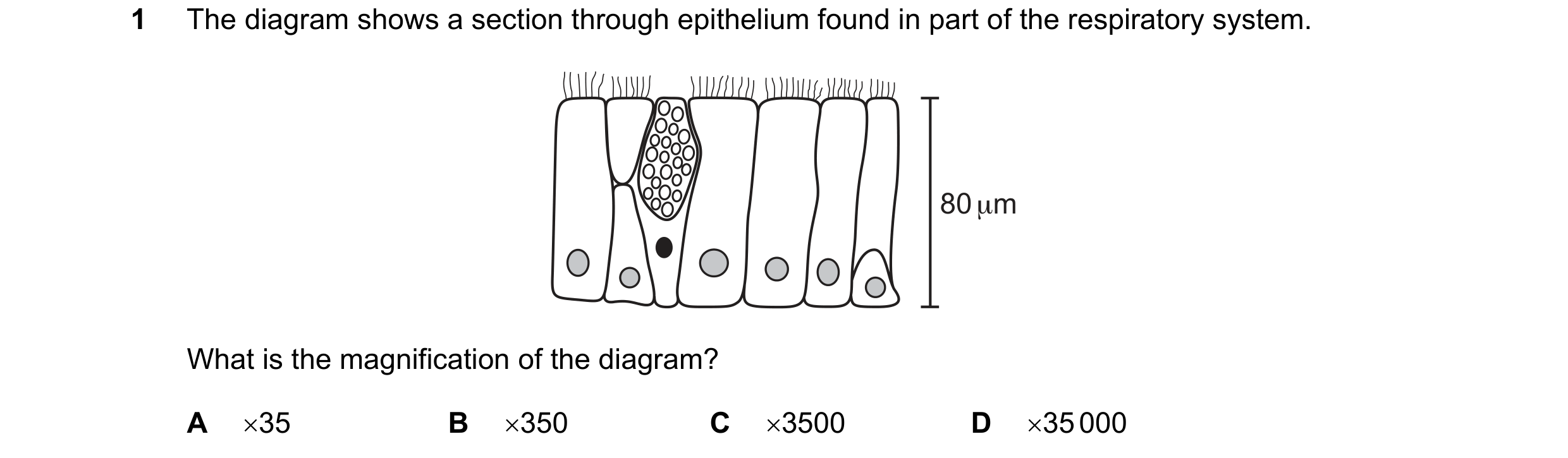
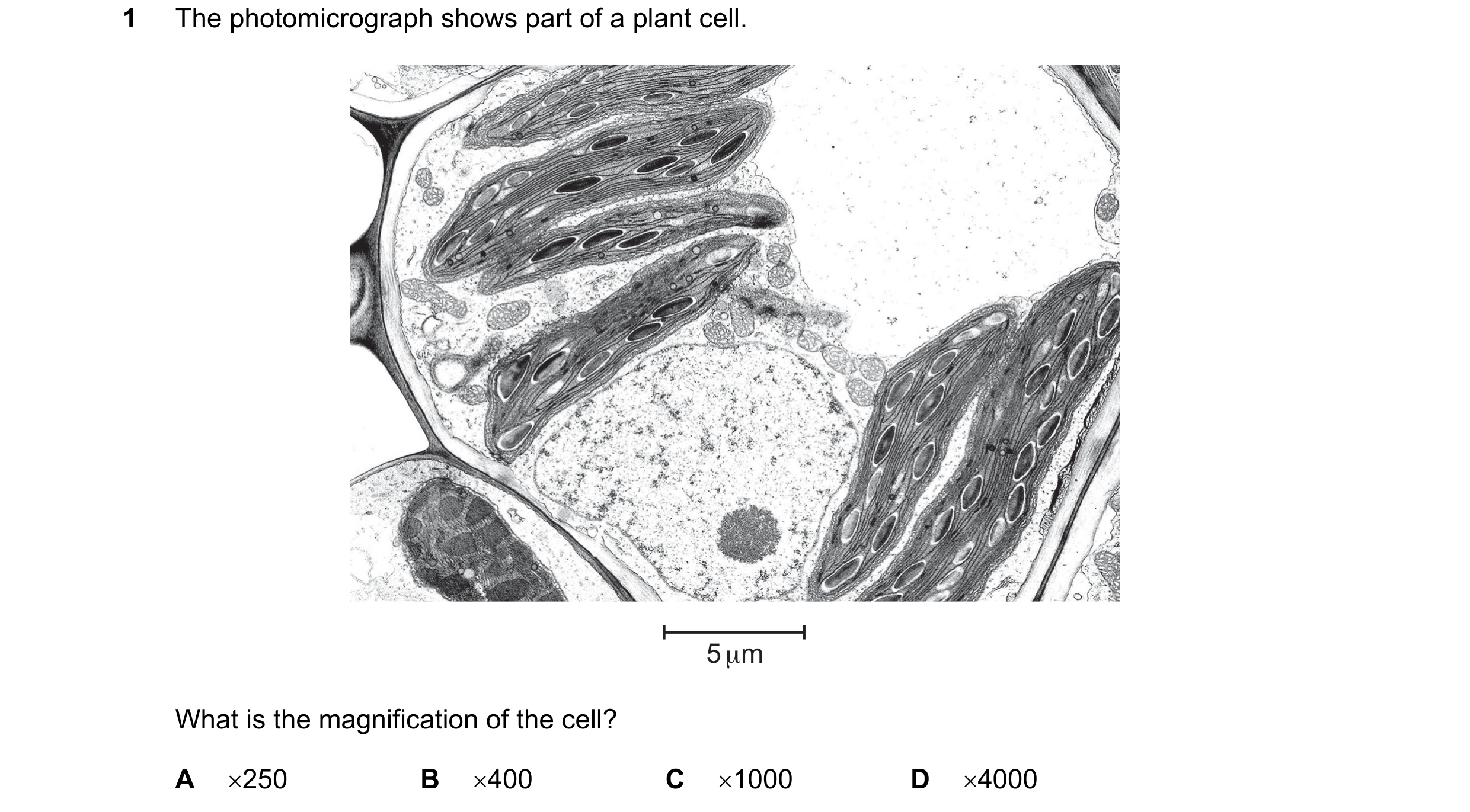 57 / 72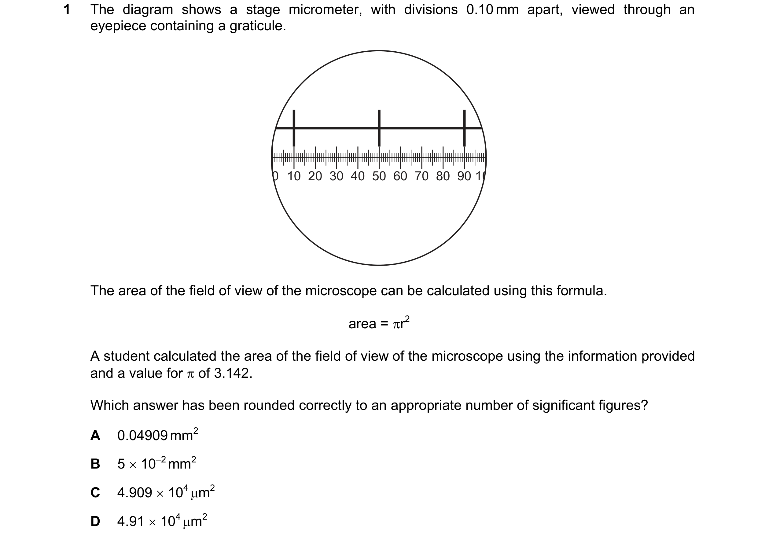
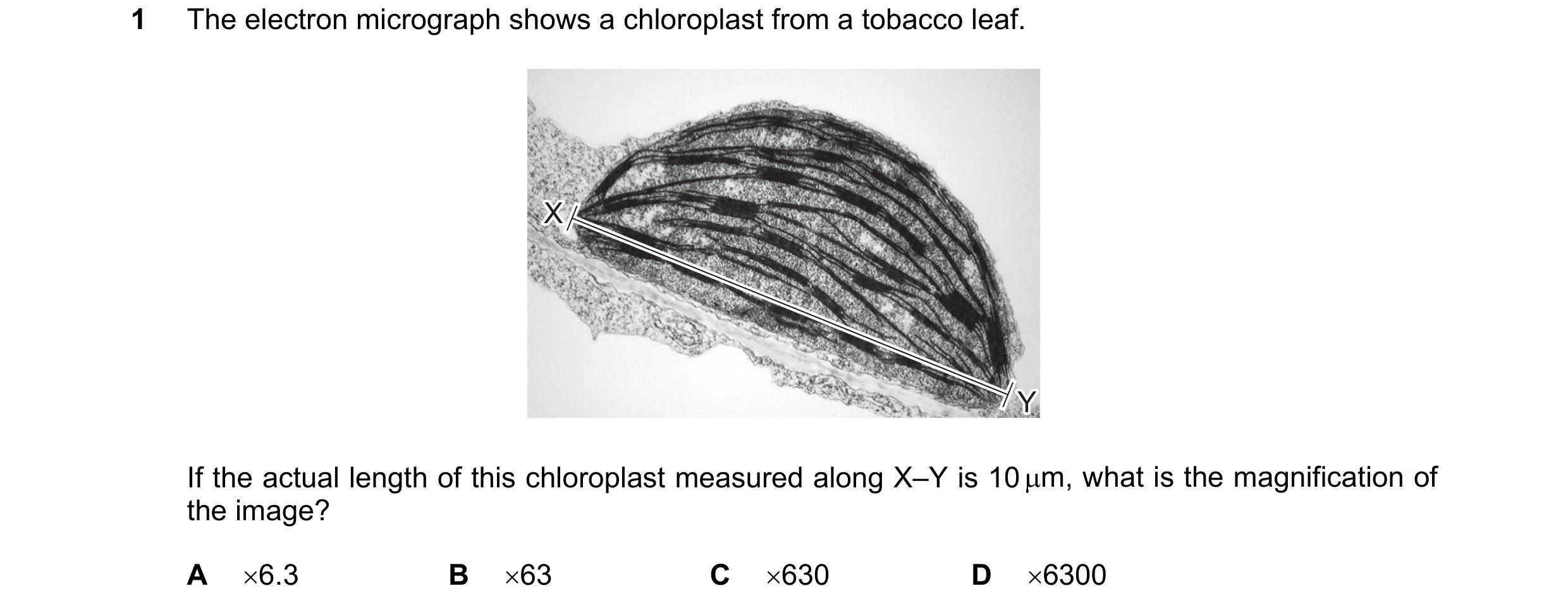 58 / 72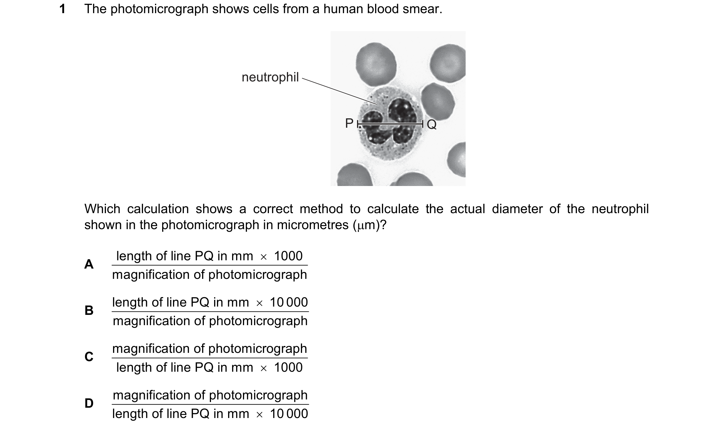
 59 / 72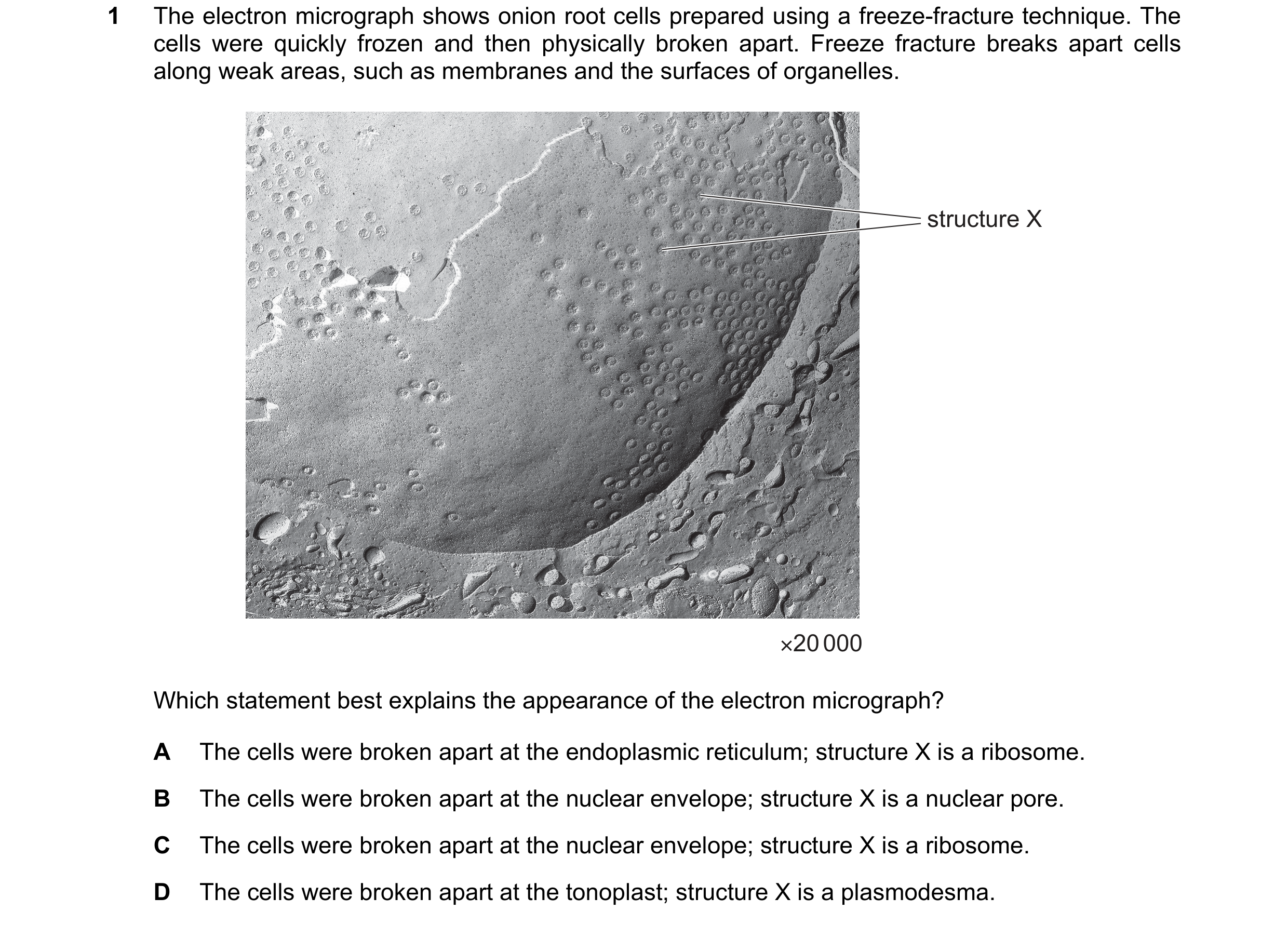
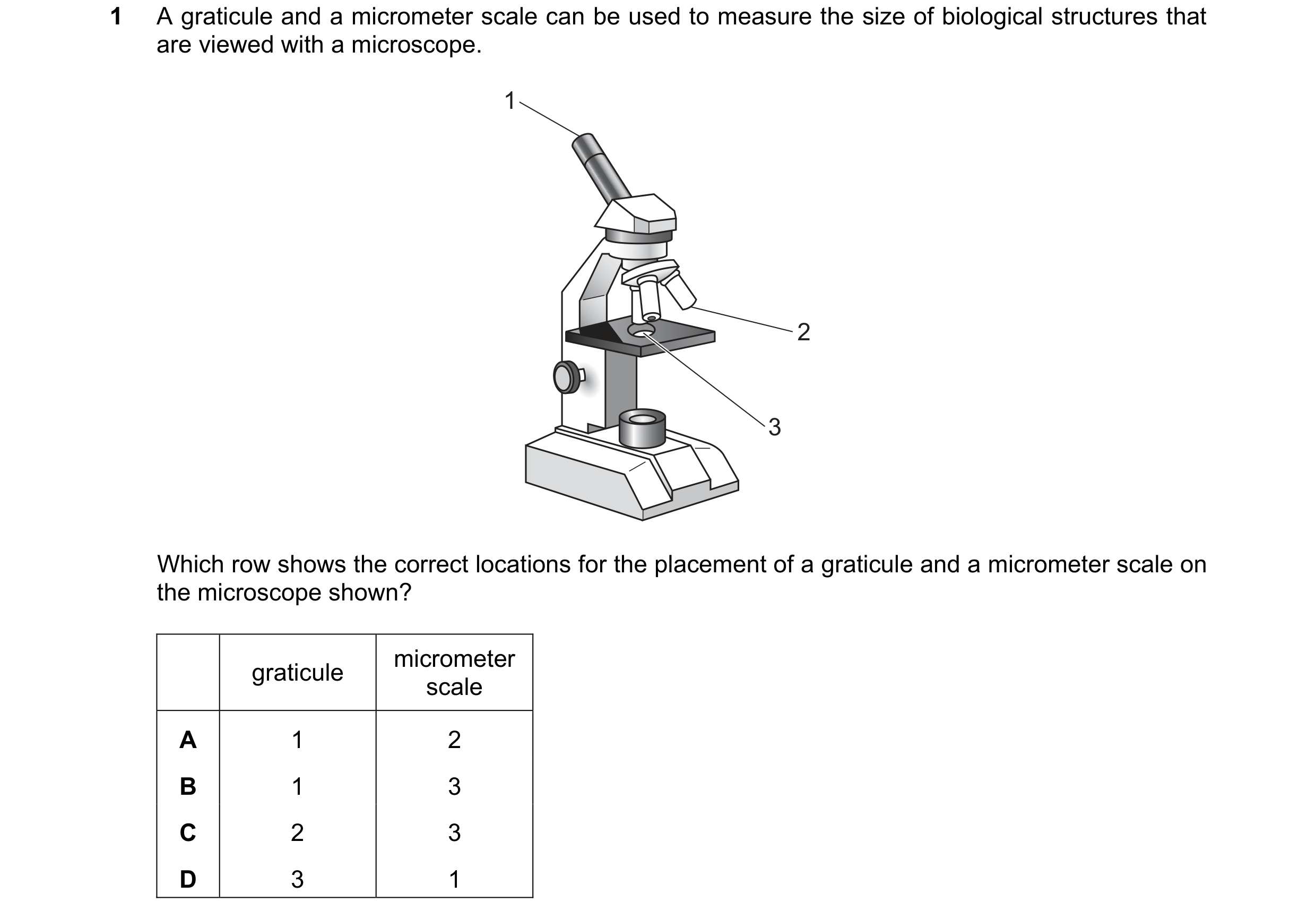 60 / 72
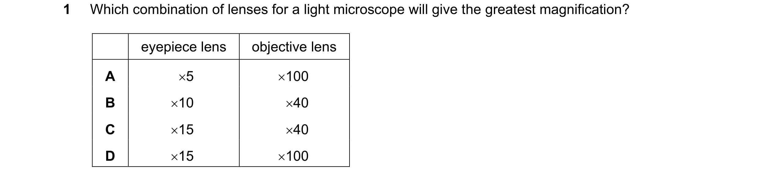
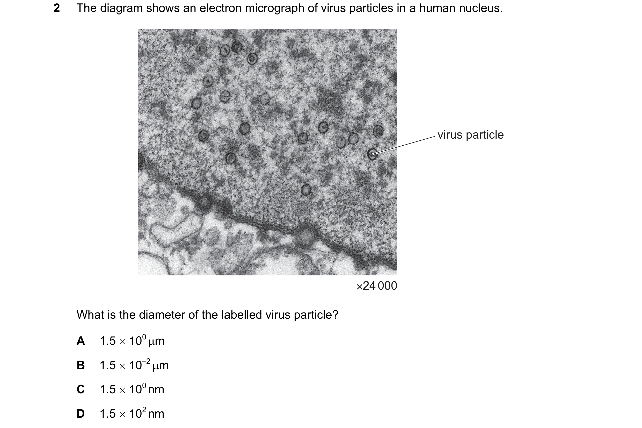
 61 / 72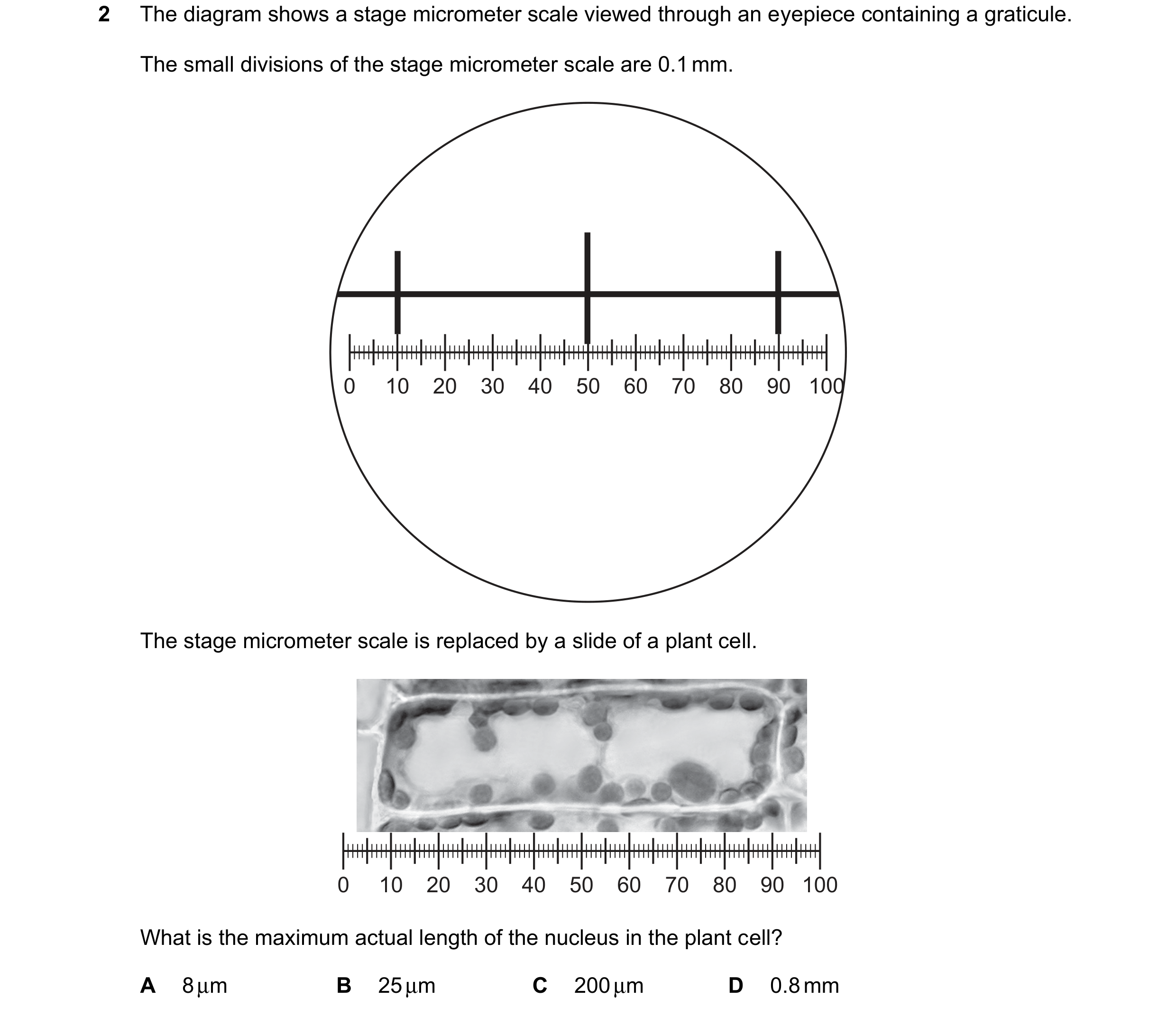
 62 / 72
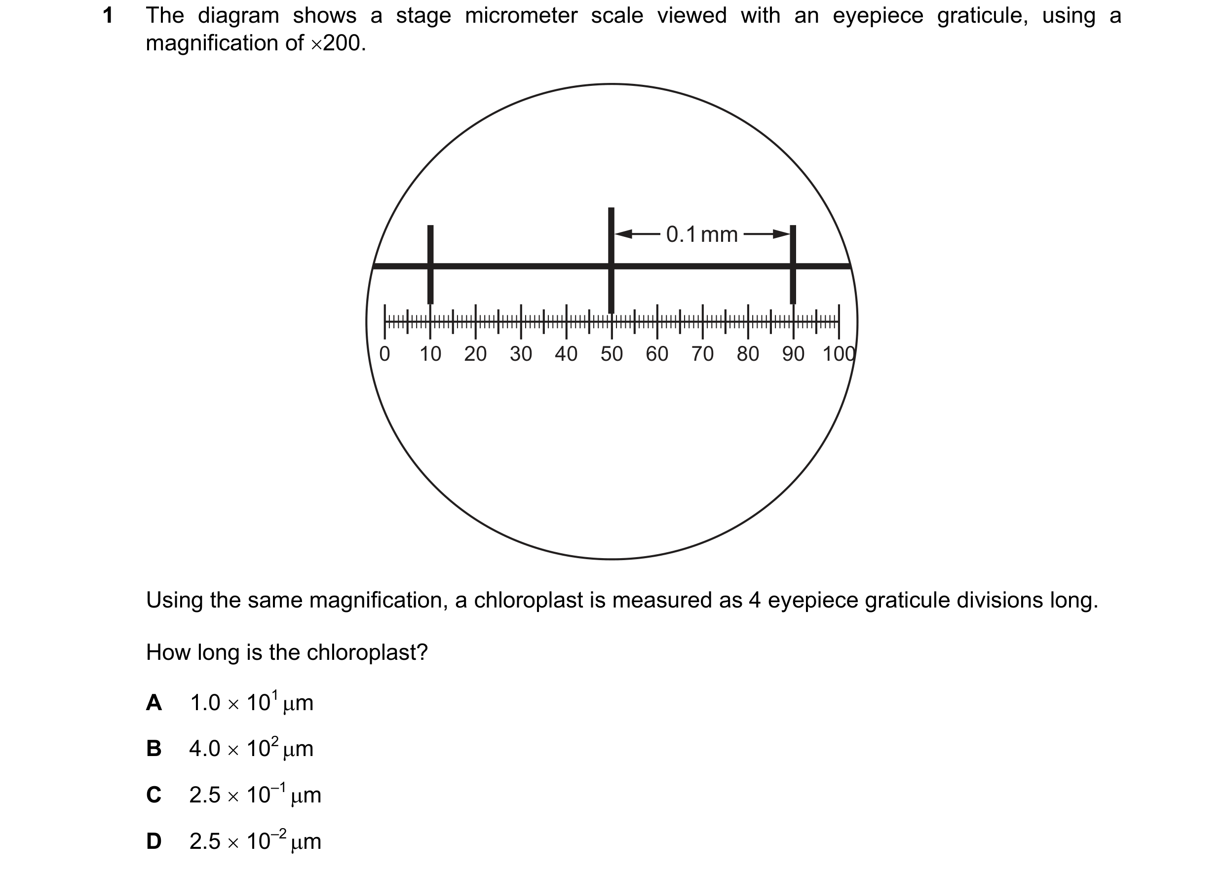
 63 / 72
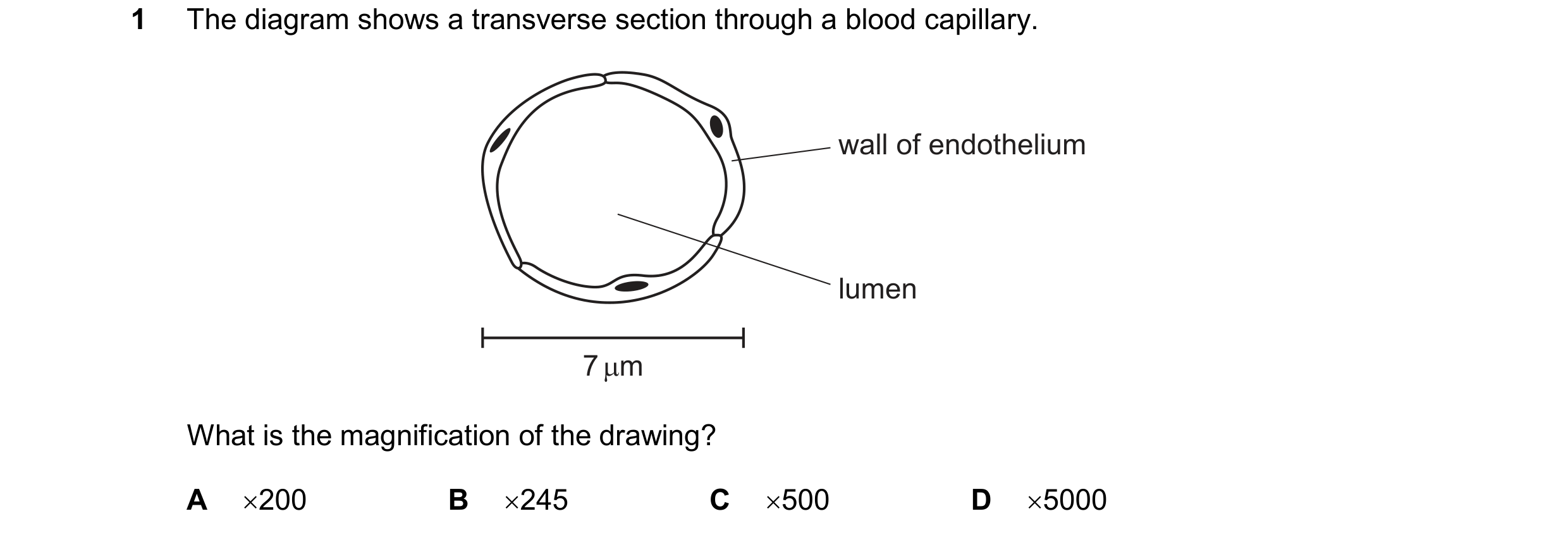
 64 / 72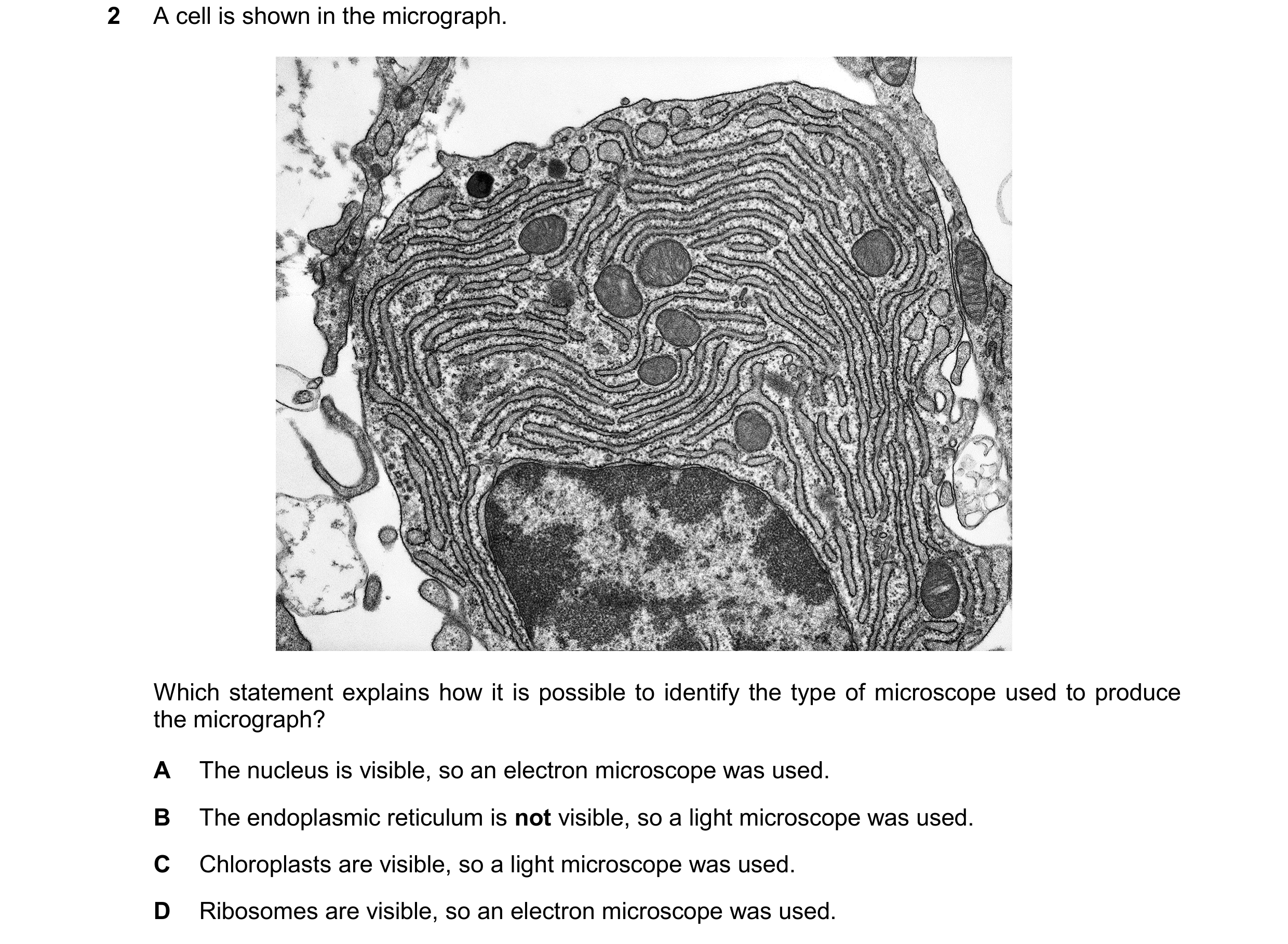
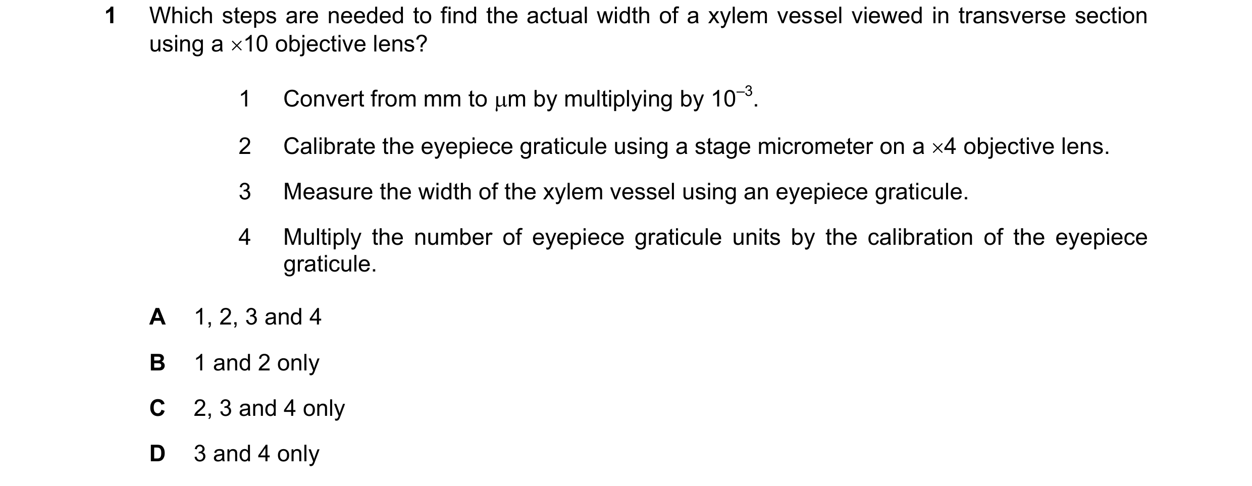 65 / 72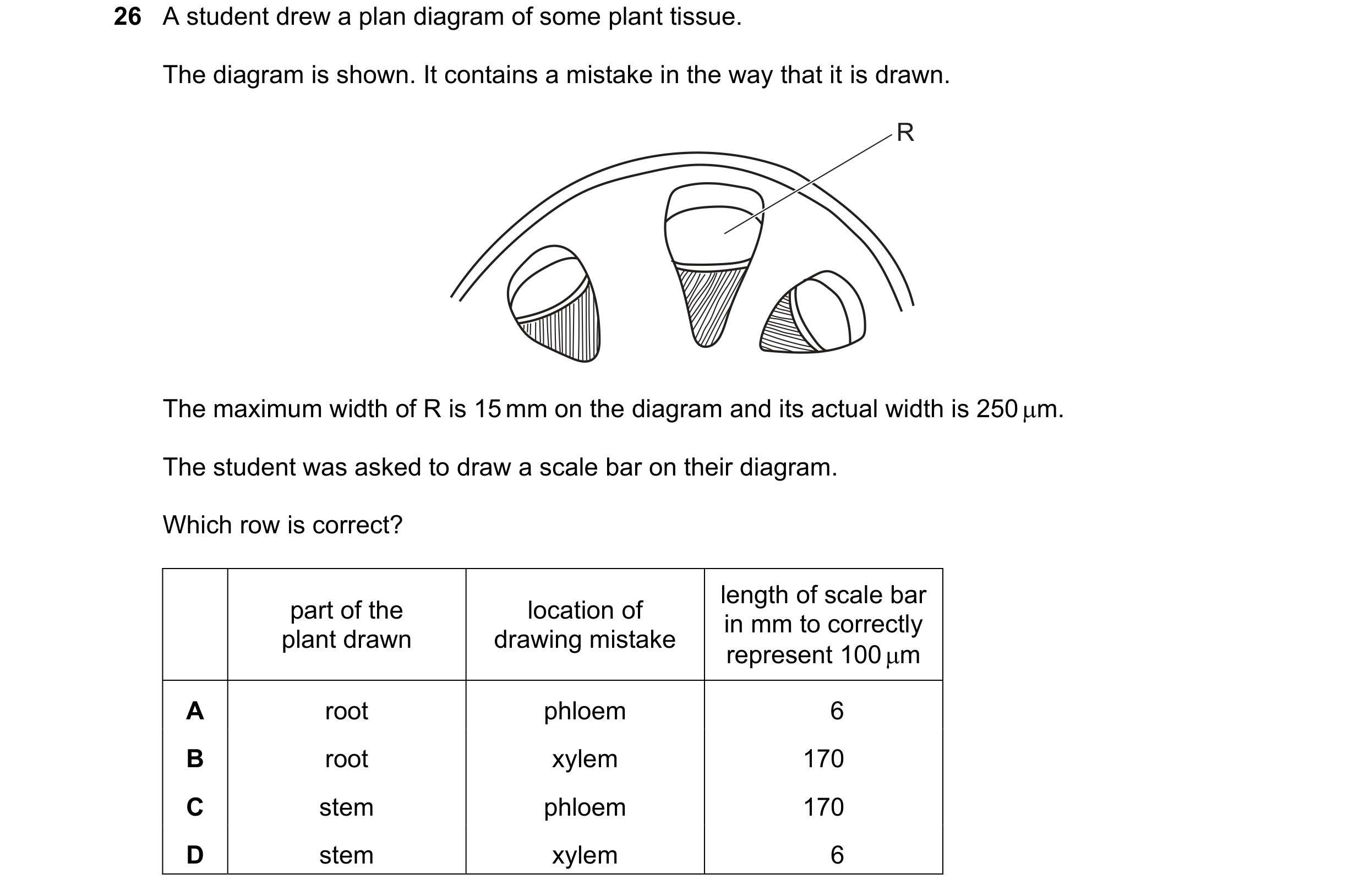

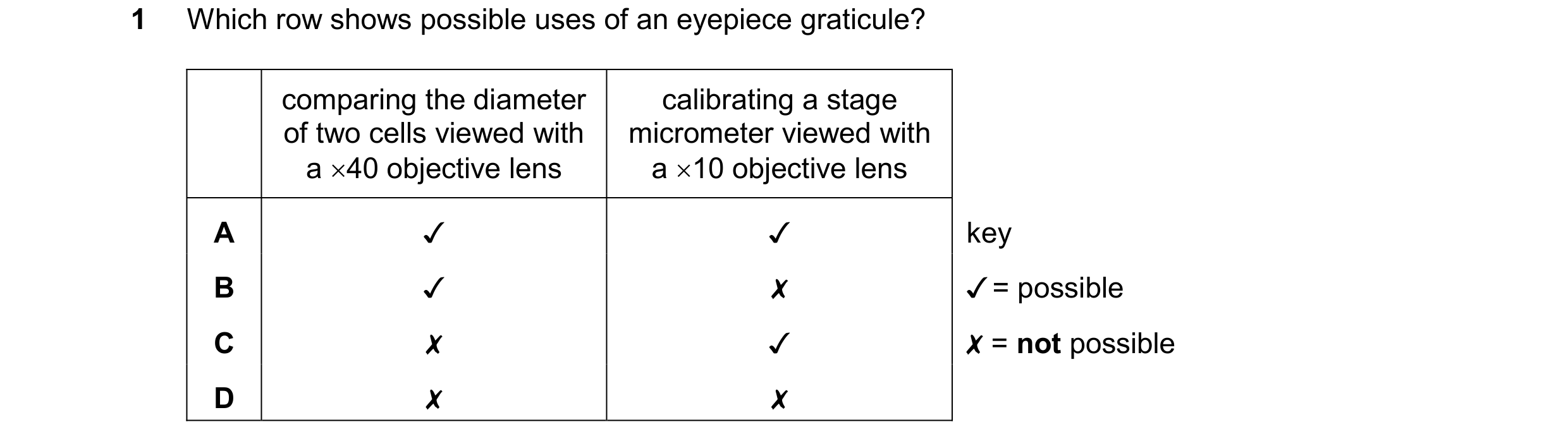
 66 / 72
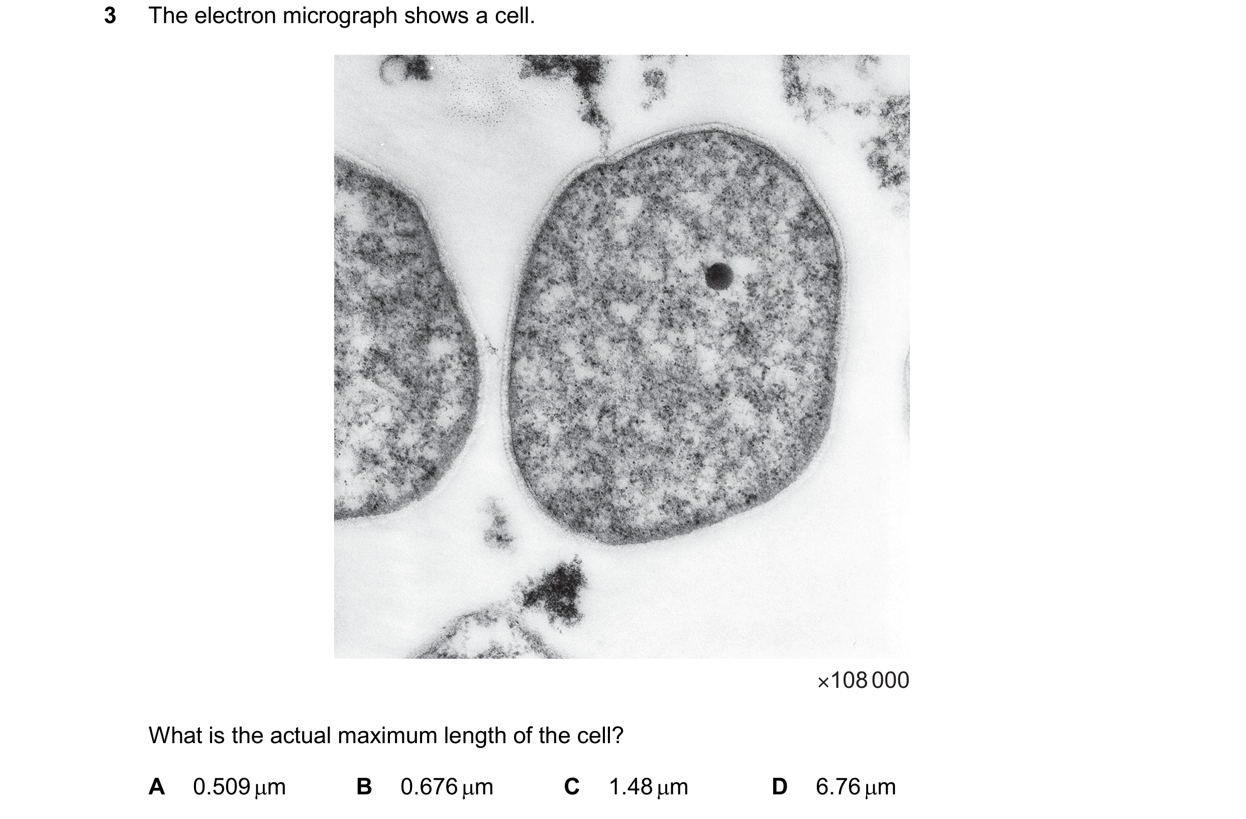 67 / 72
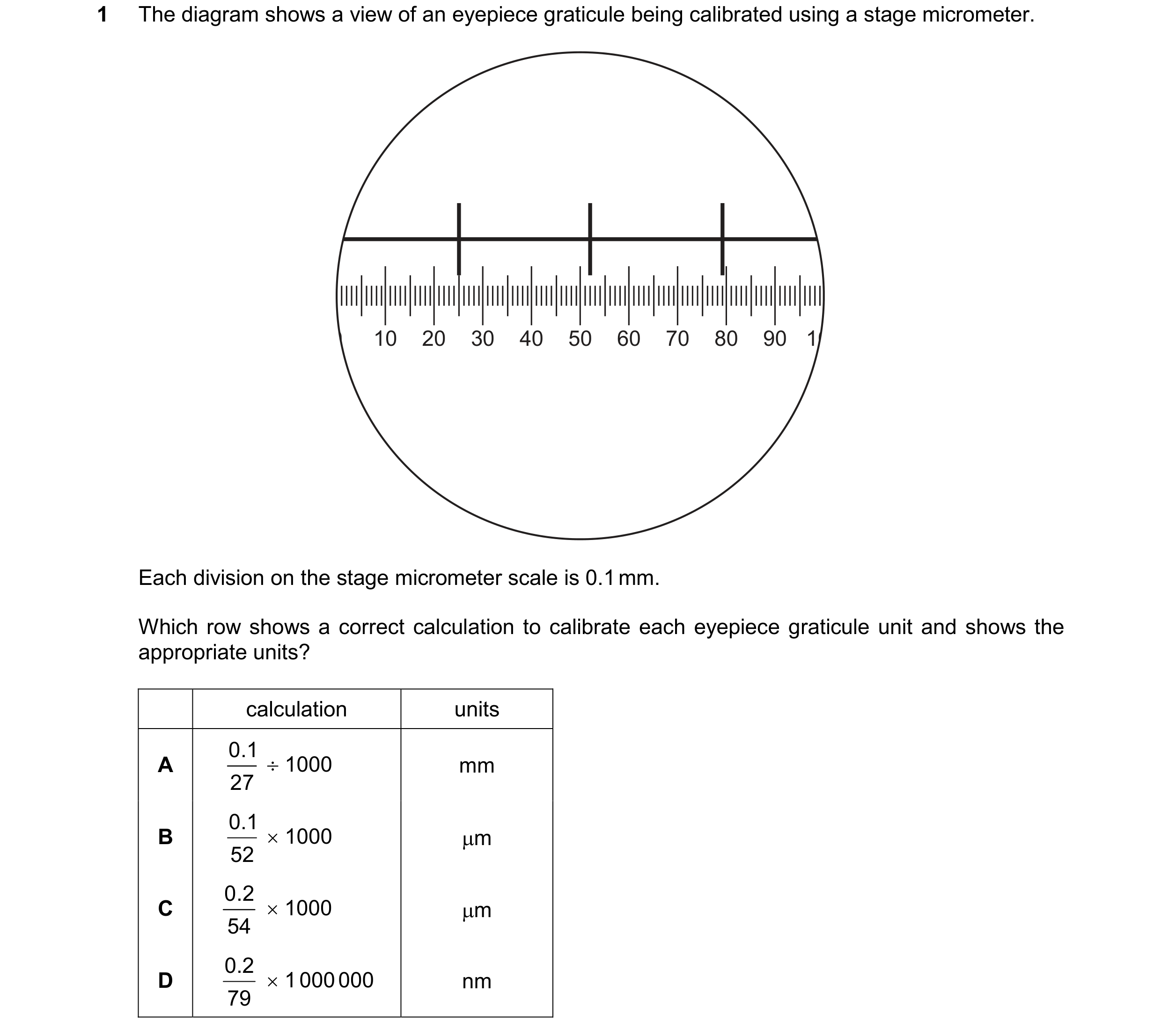 68 / 72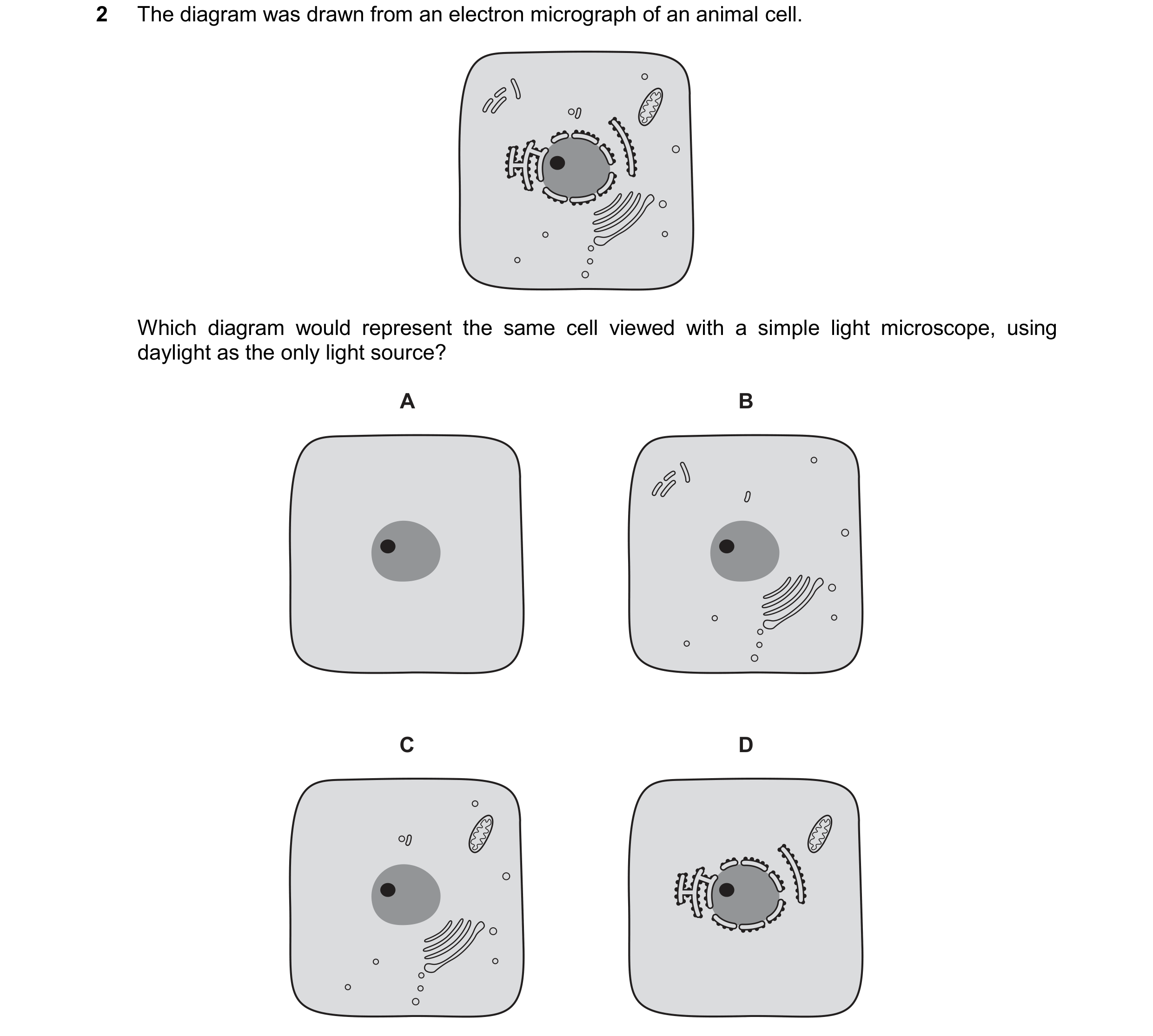
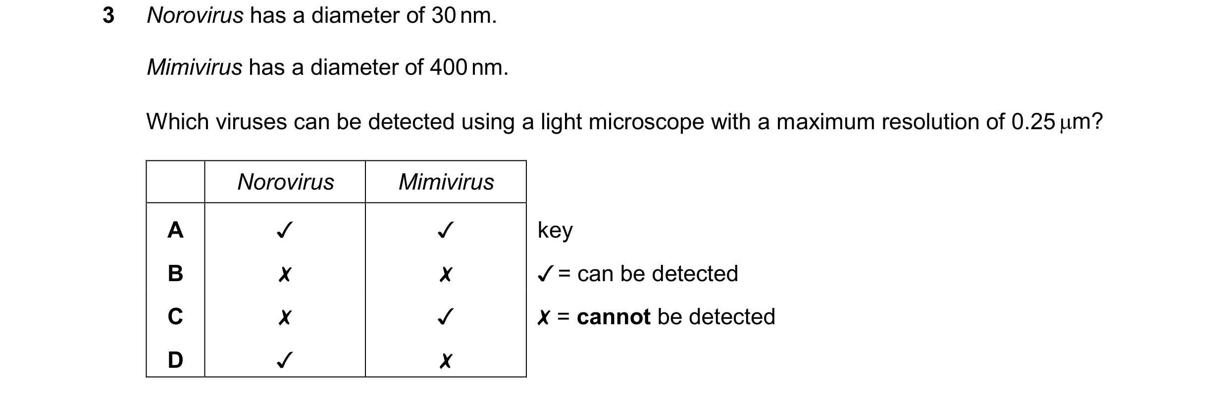 69 / 72
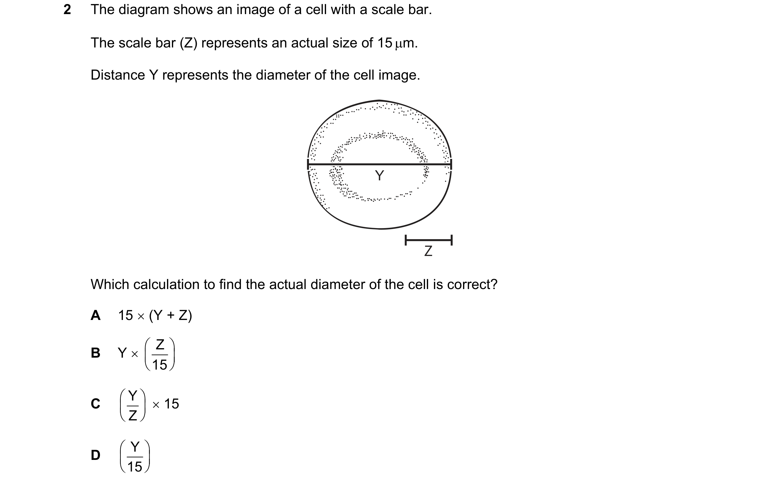 70 / 72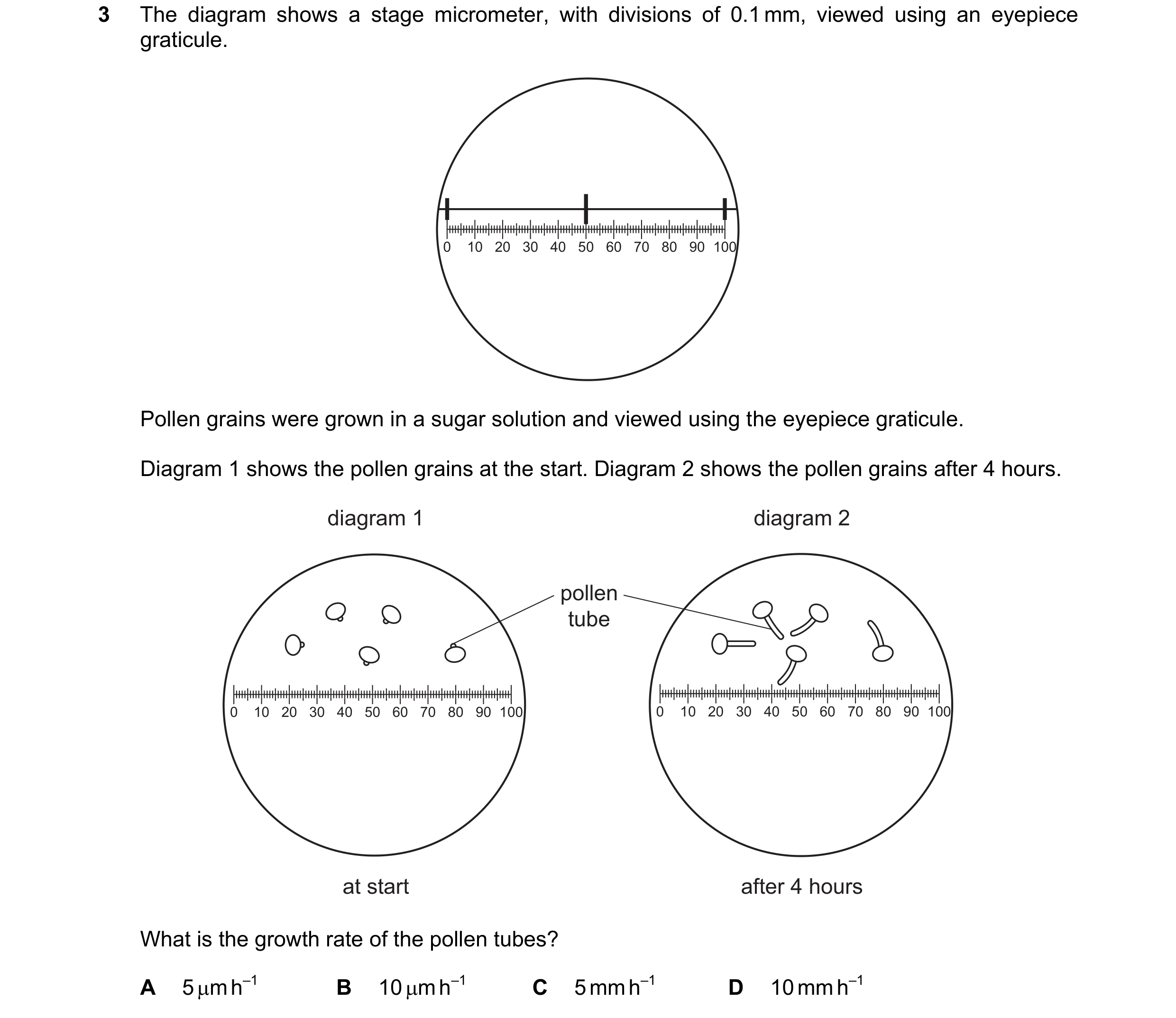
 71 / 72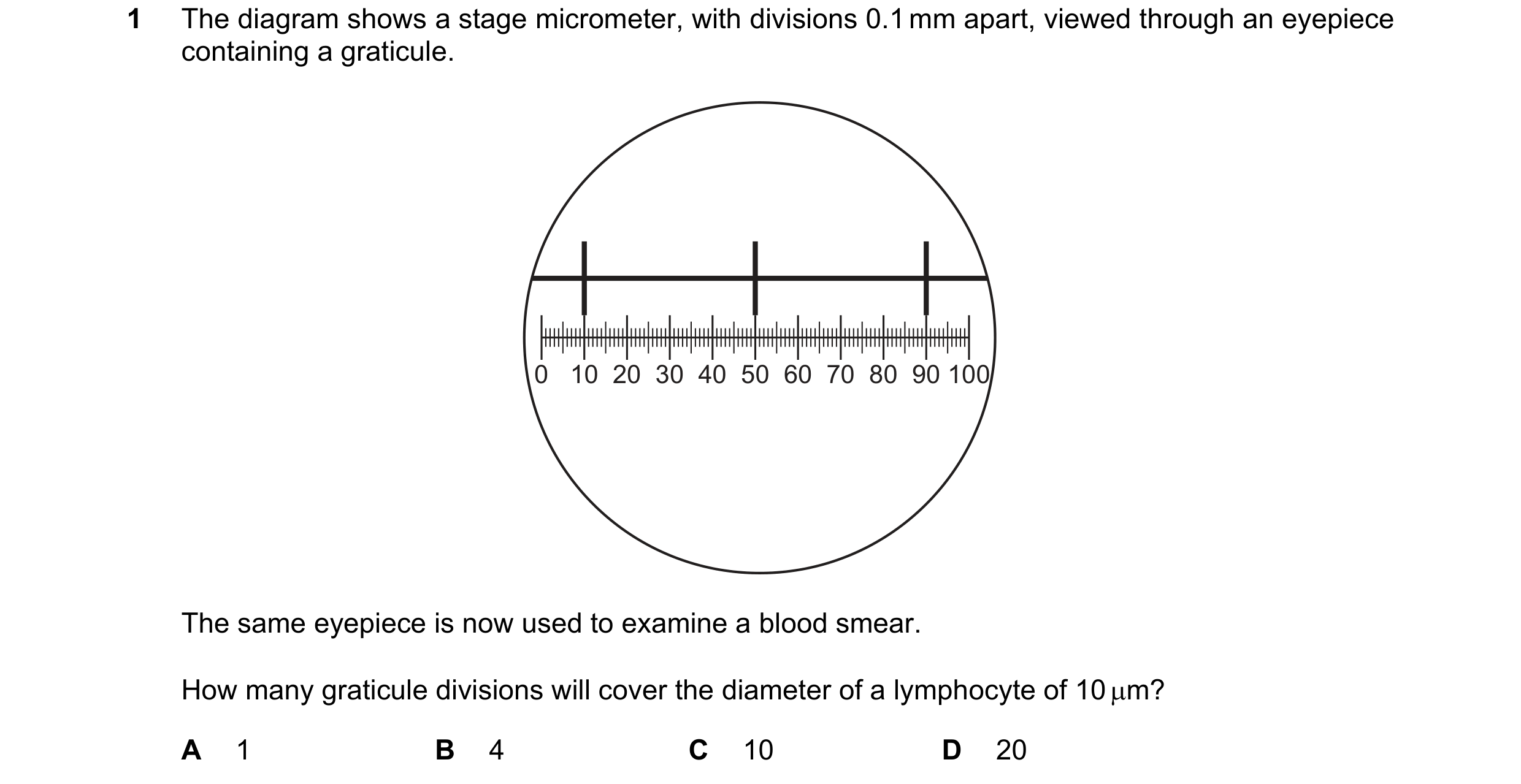
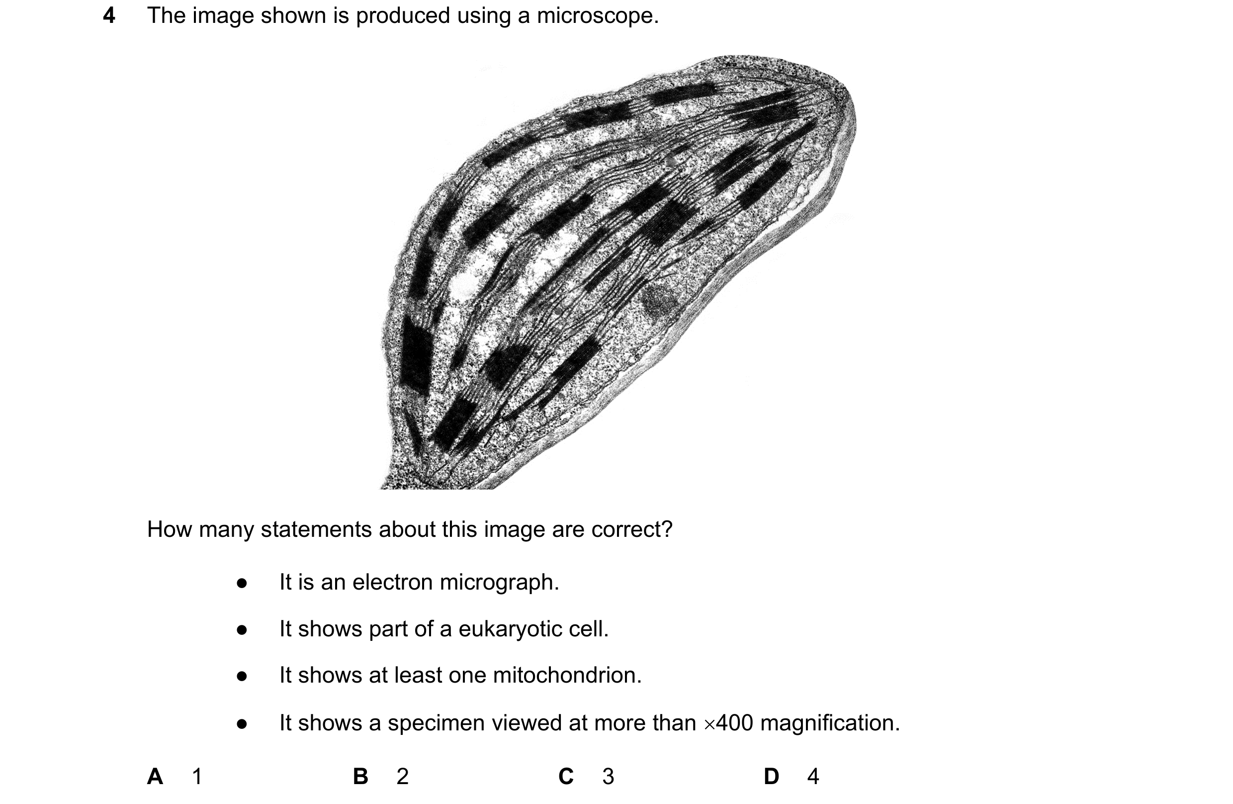 72 / 72