Questions as text
Q1 · Use the following words or phrases to complete the flow chart about the structures the… 0653/43 Oct/Nov 2017
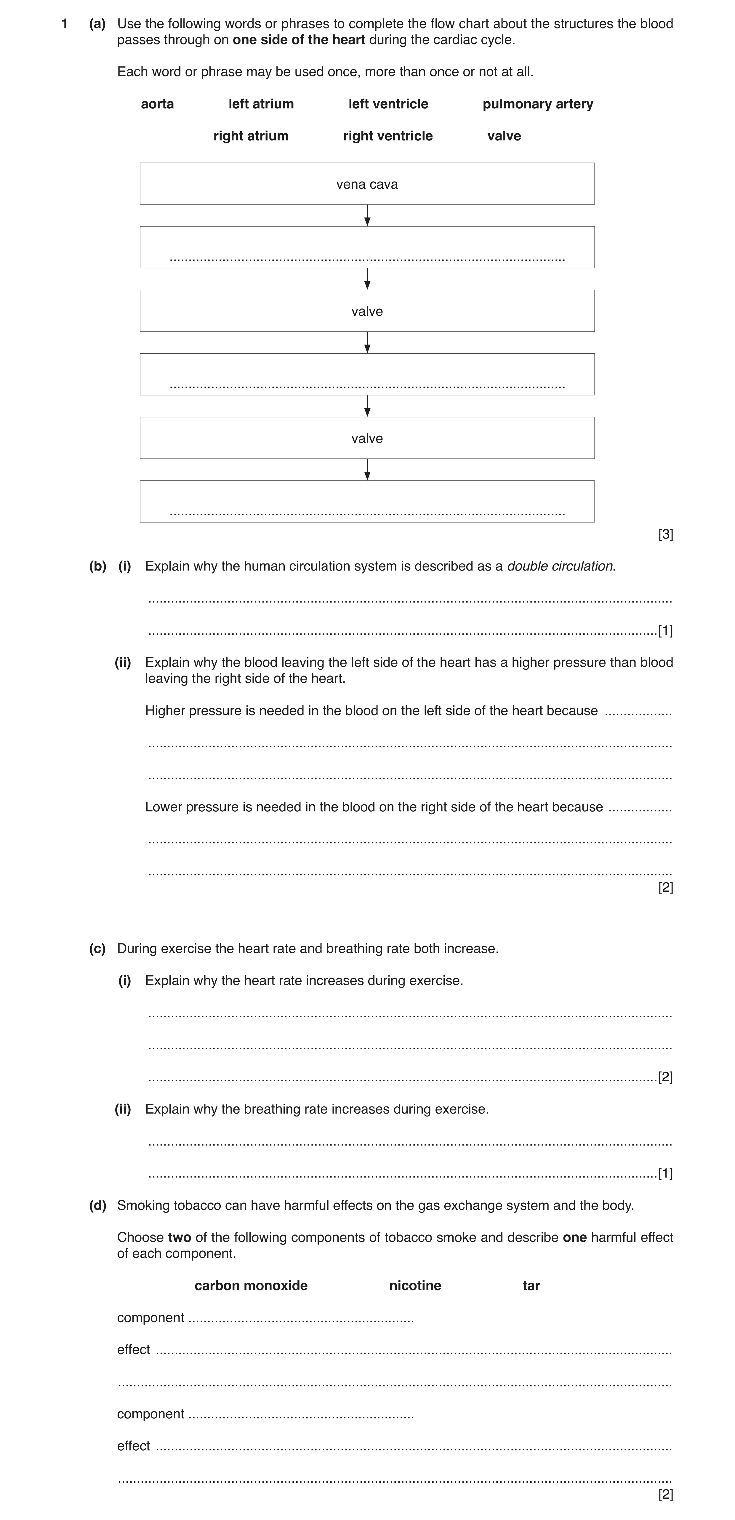
1 (a) Use the following words or phrases to complete the flow chart about the structures the blood passes through on one side of the heart during the cardiac cycle. Each word or phrase may be used once, more than once or not at all. aorta left atrium left ventricle pulmonary artery right atrium right ventricle valve vena cava … valve … valve … [3] (b) (i) Explain why the human circulation system is described as a double circulation. … … [1] (ii) Explain why the blood leaving the left side of the heart has a higher pressure than blood leaving the right side of the heart. Higher pressure is needed in the blood on the left side of the heart because … … … Lower pressure is needed in the blood on the right side of the heart because … … … [2] (c) During exercise the heart rate and breathing rate both increase. (i) Explain why the heart rate increases during exercise. … … … [2] (ii) Explain why the breathing rate increases during exercise. … … [1] (d) Smoking tobacco can have harmful effects on the gas exchange system and the body. Choose two of the following components of tobacco smoke and describe one harmful effect of each component. carbon monoxide nicotine tar component … effect … … component … effect … … [2]
11 marks
Mark scheme: 1(a) right atrium ; right ventricle ; pulmonary artery ; 3 1(b)(i) blood passes through heart twice, in each cycle / blood has two circulation paths (to the lungs and to the body) ; 1 1(b)(ii) (higher pressure on left side) needed for blood going all round the body / blood travels further ; (lower pressure on right side) needed for blood going to the lungs / shorter distance / so the blood capillaries are not damaged ; 2 1(c)(i) to take oxygen / glucose to the cells / muscles more quickly / to take more oxygen / more glucose to cells / muscles remove carbon dioxide from cells / muscles more quickly / remove more carbon dioxide from cells / muscles ; correct reference to respiration ; max 2 1(c)(ii) to take more oxygen (into blood) / remove carbon dioxide (from blood) more quickly ; 1 1(d) any two from tar increases mucus / tar builds up in lungs / paralyses / destroys cilia / causes cancer ; or nicotine causes addiction / increases blood pressure / leads to heart disease ; or carbon monoxide reduces the concentration of oxygen carried by the blood / makes carboxyhaemoglobin ; 2
This question in 0653/43 Oct/Nov 2017
Q2 · Four substances found in food, and elements they may contain 0653/41 May/June 2018
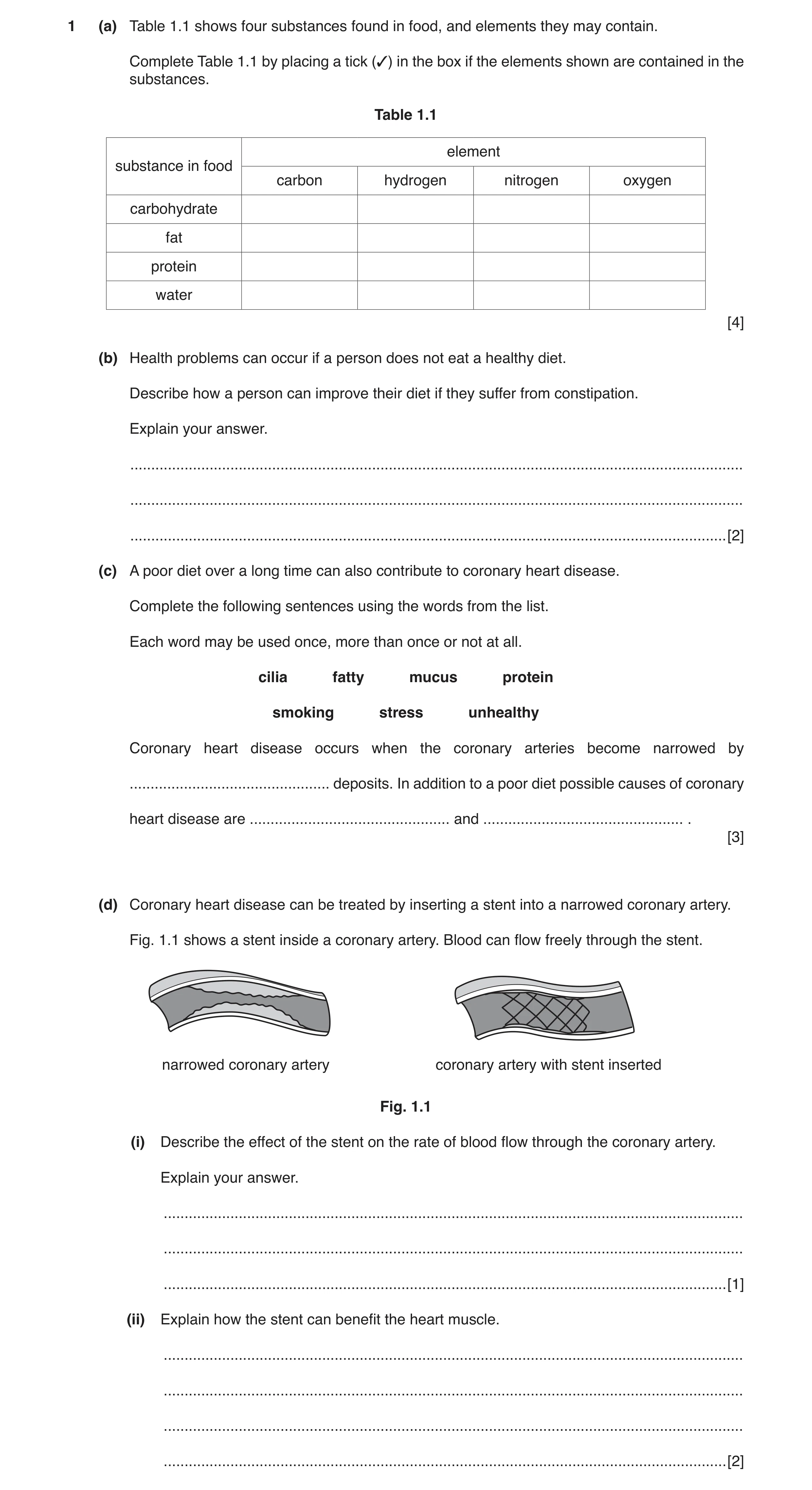
1 (a) Table 1.1 shows four substances found in food, and elements they may contain. Complete Table 1.1 by placing a tick (3) in the box if the elements shown are contained in the substances. Table 1.1 element substance in food carbon hydrogen nitrogen oxygen carbohydrate fat protein water [4] (b) Health problems can occur if a person does not eat a healthy diet. Describe how a person can improve their diet if they suffer from constipation. Explain your answer. … … … [2] (c) A poor diet over a long time can also contribute to coronary heart disease. Complete the following sentences using the words from the list. Each word may be used once, more than once or not at all. cilia fatty mucus protein smoking stress unhealthy Coronary heart disease occurs when the coronary arteries become narrowed by … deposits. In addition to a poor diet possible causes of coronary heart disease are … and … . [3] (d) Coronary heart disease can be treated by inserting a stent into a narrowed coronary artery. Fig. 1.1 shows a stent inside a coronary artery. Blood can flow freely through the stent. narrowed coronary artery coronary artery with stent inserted Fig. 1.1 (i) Describe the effect of the stent on the rate of blood flow through the coronary artery. Explain your answer. … … … [1] (ii) Explain how the stent can benefit the heart muscle. … … … … [2]
12 marks
Mark scheme: 1(a) substance in food element carbon hydrogen nitrogen oxygen carbohydrate 9 9 9 fat 9 9 9 protein 9 9 9 9 water 9 9 one mark for each correct row ;;;; 4 1(b) include more fibre / roughage / fruit / vegetables ; (fibre) helps to move food through digestive system ; 2 1(c) fatty ; (first space) smoking ; stress ; 3 1(d)(i) (increases blood flow) (because the artery is) wider / the idea of more space (for blood to pass through) ; 1 1(d)(ii) increases supply of glucose / oxygen (reaching the heart muscle) ; for respiration (in heart muscle cells) / to release energy (in muscle cells) / for contraction / action (of heart muscle) ; 2
This question in 0653/41 May/June 2018
Q3 · A diagram of the internal structure of the heart 0653/42 Oct/Nov 2018
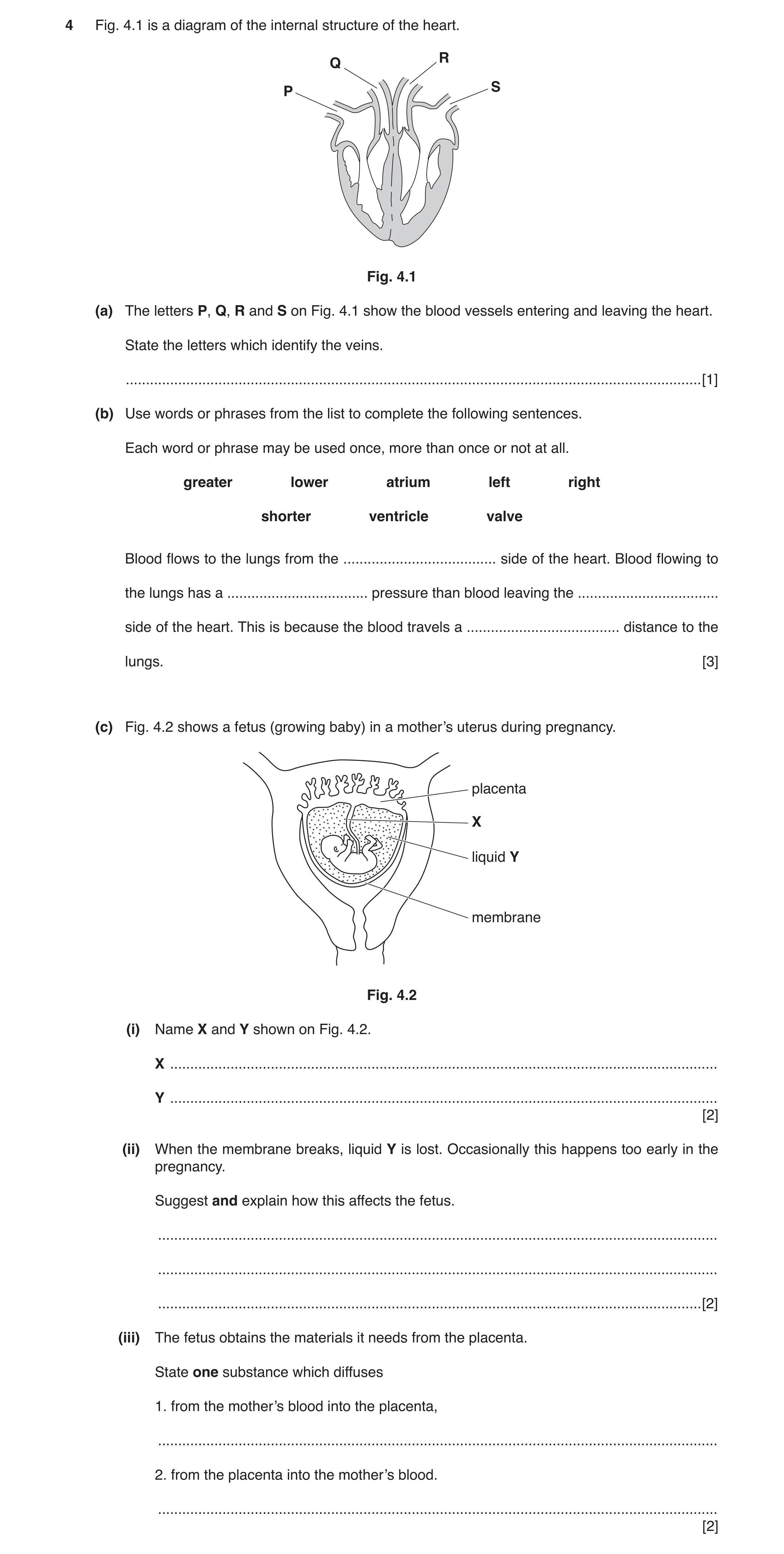
4 Fig. 4.1 is a diagram of the internal structure of the heart. R Q S P Fig. 4.1 (a) The letters P, Q, R and S on Fig. 4.1 show the blood vessels entering and leaving the heart. State the letters which identify the veins. … [1] (b) Use words or phrases from the list to complete the following sentences. Each word or phrase may be used once, more than once or not at all. greater lower atrium left right shorter ventricle valve Blood flows to the lungs from the … side of the heart. Blood flowing to the lungs has a … pressure than blood leaving the … side of the heart. This is because the blood travels a … distance to the lungs. [3] (c) Fig. 4.2 shows a fetus (growing baby) in a mother’s uterus during pregnancy. placenta X liquid Y membrane Fig. 4.2 (i) Name X and Y shown on Fig. 4.2. X … Y … [2] (ii) When the membrane breaks, liquid Y is lost. Occasionally this happens too early in the pregnancy. Suggest and explain how this affects the fetus. … … … [2] (iii) The fetus obtains the materials it needs from the placenta. State one substance which diffuses 1. from the mother’s blood into the placenta, … 2. from the placenta into the mother’s blood. … [2]
10 marks
Mark scheme: 4(a) P and S ; 1 Question Answer Marks 4(b) right ; lower ; left ; shorter ; 3 4(c)(i) X umbilical cord ; Y amniotic fluid ; 2 4(c)(ii) reference to the loss of protection (of fetus) ; higher risk of physical damage / infection / premature birth ; 2 4(c)(iii) oxygen / glucose / any named dissolved nutrient ; carbon dioxide ; 2
This question in 0653/42 Oct/Nov 2018
Q4 · The external structures of the heart 0653/43 Oct/Nov 2018
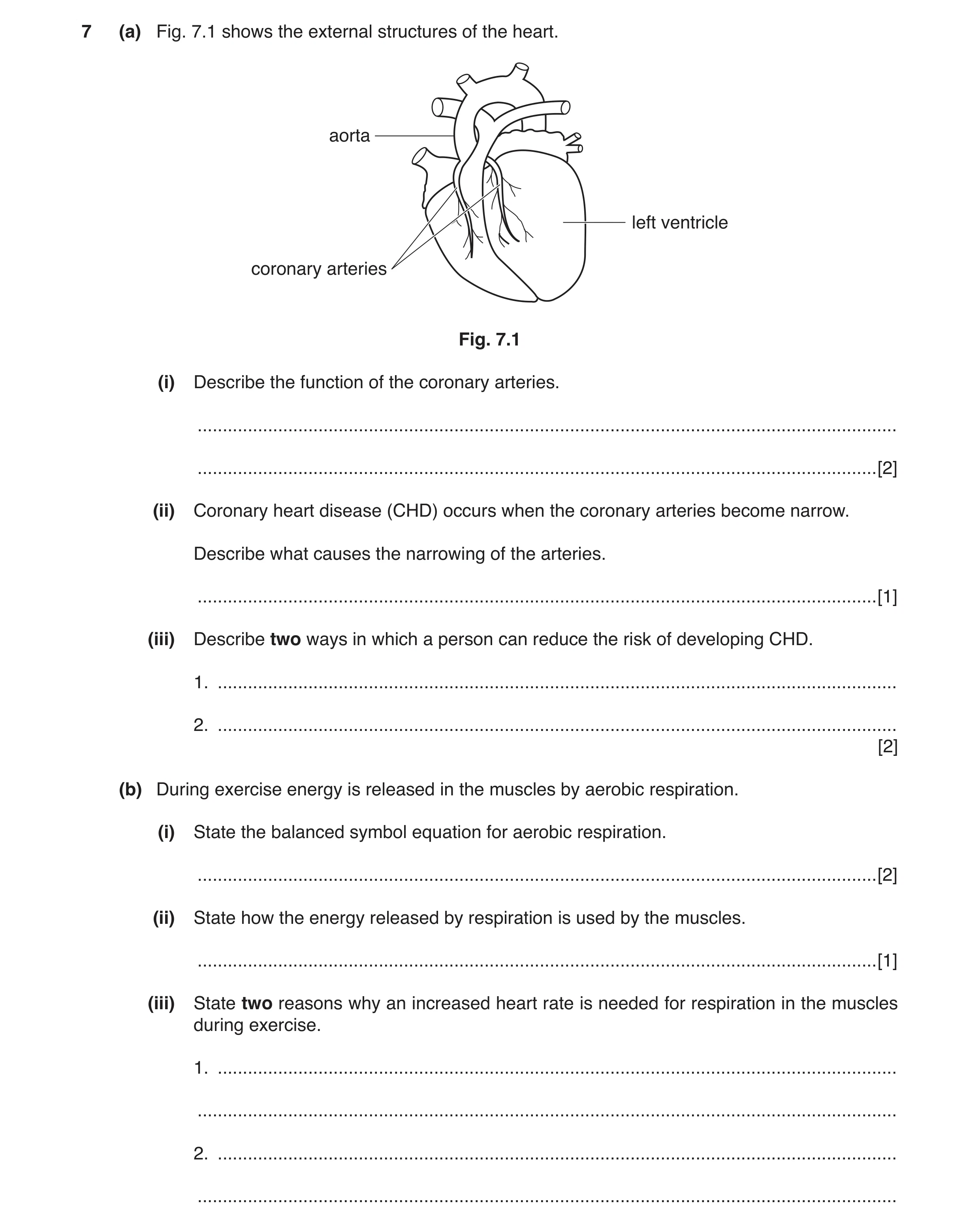
7 (a) Fig. 7.1 shows the external structures of the heart. aorta left ventricle coronary arteries Fig. 7.1 (i) Describe the function of the coronary arteries. … … [2] (ii) Coronary heart disease (CHD) occurs when the coronary arteries become narrow. Describe what causes the narrowing of the arteries. … [1] (iii) Describe two ways in which a person can reduce the risk of developing CHD. 1. … 2. … [2] (b) During exercise energy is released in the muscles by aerobic respiration. (i) State the balanced symbol equation for aerobic respiration. … [2] (ii) State how the energy released by respiration is used by the muscles. … [1] (iii) State two reasons why an increased heart rate is needed for respiration in the muscles during exercise. 1. … … 2. … …
10 marks
Mark scheme: 7(a)(i) to supply the heart muscle / tissue ; with oxygen / glucose / oxygenated blood ; for respiration ; 2 7(a)(ii) fatty deposits ; 1 7(a)(iii) any two of take plenty of exercise ; eat a diet with little (saturated) fat ; try to reduce stress ; stop / do not smoke ; Max 2 2 7(b)(i) C6H12O6 + 6O2 → 6CO2 + 6H2O formulae ; balanced ; 2 Question Answer Marks 7(b)(ii) contraction ; 1 7(b)(iii) to transport more glucose / oxygen (to muscles) ; to transport more carbon dioxide / distribute heat ; 2
This question in 0653/43 Oct/Nov 2018
Q5 · A diagram of the internal structure of the heart and the blood vessels entering and… 0653/42 Feb/March 2019
4 Fig. 4.1 shows a diagram of the internal structure of the heart and the blood vessels entering and leaving the heart. pulmonary artery aorta pulmonary vein vena cava valve Z Fig. 4.1 (a) Describe the function of valve Z in the heart shown in Fig. 4.1. … … … [2] (b) The concentrations of oxygen gas and of carbon dioxide gas in the blood in each side of the heart are measured. The results are shown on Fig. 4.2. concentration Key of gas in oxygen heart carbon dioxide right side left side side of heart Fig. 4.2 (i) Explain why the concentration of carbon dioxide in the blood in the right side of the heart is higher than in the left side. … … … [2] (ii) Describe what causes the oxygen concentration in the blood to be higher in the left side of the heart compared with the right side. … … [1] (c) (i) State what is meant by coronary heart disease. … … … [2] (ii) Describe one lifestyle choice a person can make to reduce the chance of developing coronary heart disease. … … [1] [Total: 8]
8 marks
Mark scheme: 4(a) prevents backflow of blood ; from ventricle to atrium ; 2 4(b)(i) produced by respiration / in body cells ; the idea that blood has come from the body (to heart) ; 2 4(b)(ii) oxygen collected at the lungs / the idea that oxygenated blood comes from the lungs (to the left hand side) ; 1 4(c)(i) blockage / narrowing of artery ; reference to coronary (arteries) ; 2 4(c)(ii) avoid smoking ; reduce stress ; reduce fat in diet ; reference to exercise ; max 1
This question in 0653/42 Feb/March 2019
Q6 · A balanced diet for a person contains all nutrients in the correct amounts for their needs 0653/41 May/June 2019
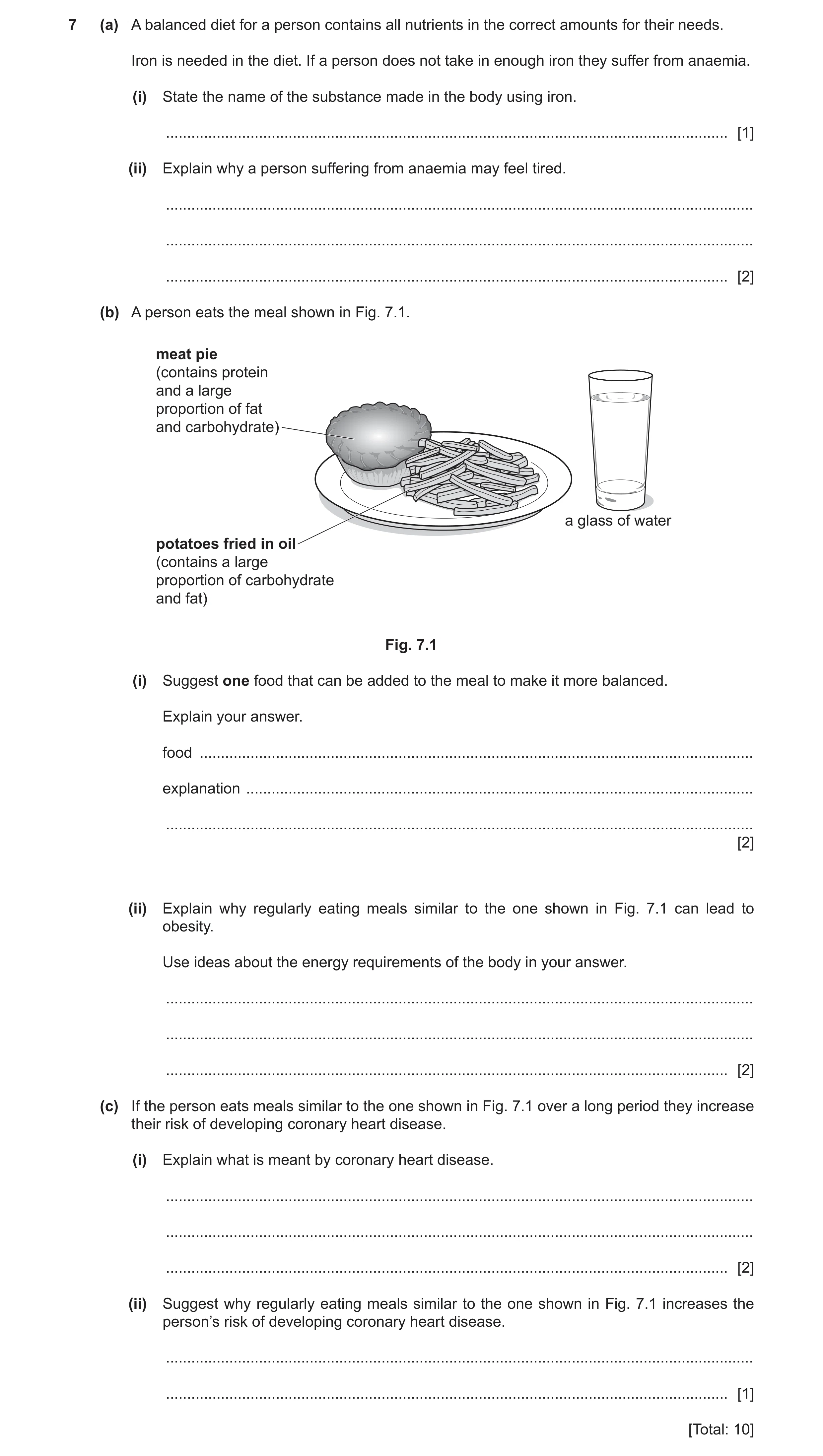
7 (a) A balanced diet for a person contains all nutrients in the correct amounts for their needs. Iron is needed in the diet. If a person does not take in enough iron they suffer from anaemia. (i) State the name of the substance made in the body using iron. … [1] (ii) Explain why a person suffering from anaemia may feel tired. … … … [2] (b) A person eats the meal shown in Fig. 7.1. meat pie (contains protein and a large proportion of fat and carbohydrate) a glass of water potatoes fried in oil (contains a large proportion of carbohydrate and fat) Fig. 7.1 (i) Suggest one food that can be added to the meal to make it more balanced. Explain your answer. food … explanation … … [2] (ii) Explain why regularly eating meals similar to the one shown in Fig. 7.1 can lead to obesity. Use ideas about the energy requirements of the body in your answer. … … … [2] (c) If the person eats meals similar to the one shown in Fig. 7.1 over a long period they increase their risk of developing coronary heart disease. (i) Explain what is meant by coronary heart disease. … … … [2] (ii) Suggest why regularly eating meals similar to the one shown in Fig. 7.1 increases the person’s risk of developing coronary heart disease. … … [1] [Total: 10]
10 marks
Mark scheme: 7(a)(i) haemoglobin ; 1 7(a)(ii) any two of not enough red blood cells / haemoglobin ; not enough oxygen taken to body / cells / muscles ; less (energy released by) respiration ; max 2 7(b)(i) fruit / vegetable / named example ; to add vitamins / minerals / fibre ; 2 7(b)(ii) energy taken in by body exceeds energy needed by body ; excess (energy) is stored as fat ; 2 7(c)(i) coronary arteries ; become blocked / narrowed ; 2 7(c)(ii) too much fat (contained in the food) ; 1
This question in 0653/41 May/June 2019
Q7 · If the light shining on a shoot tip is more intense on one side, the shoot shows a growth… 0653/41 May/June 2020
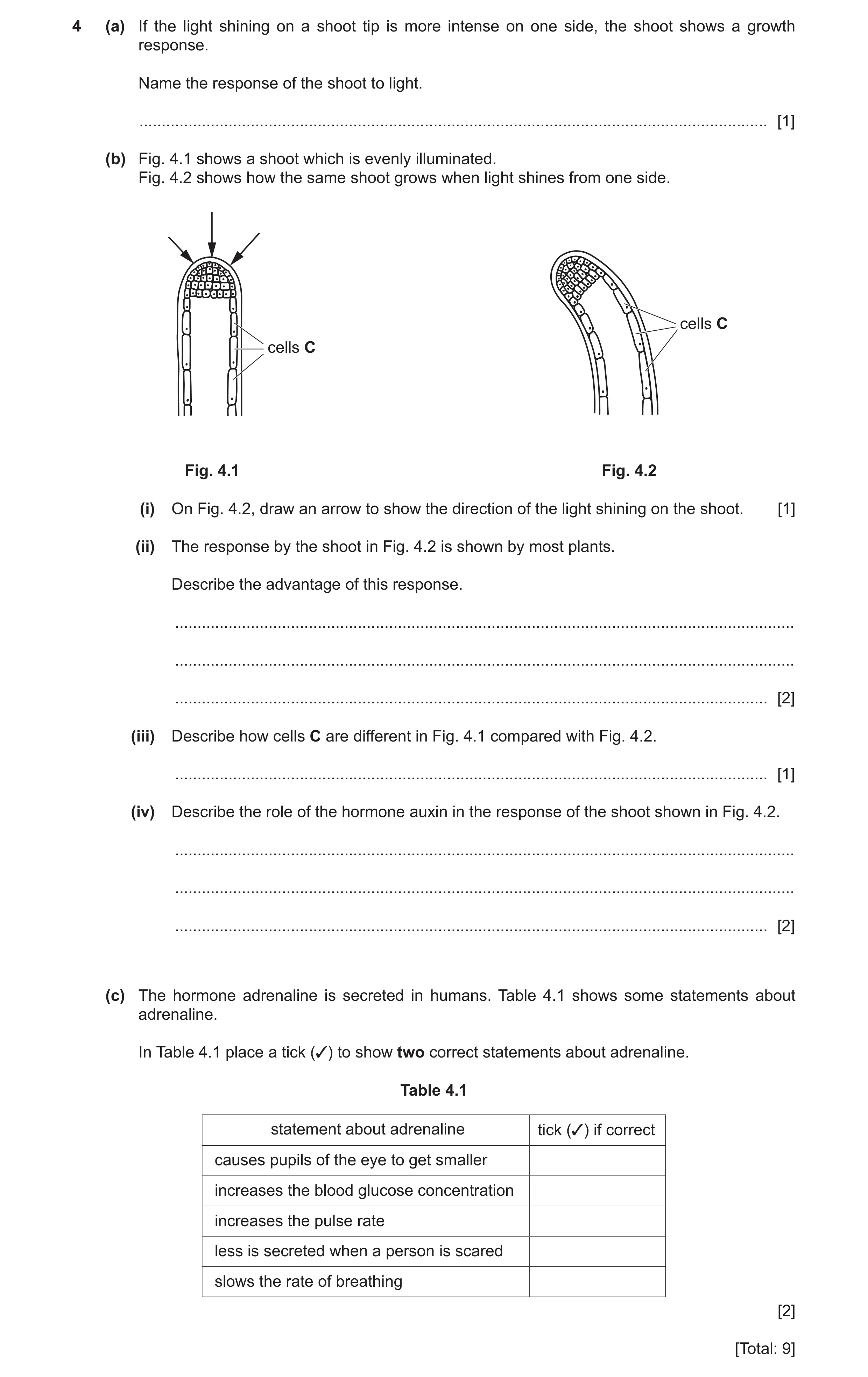
4 (a) If the light shining on a shoot tip is more intense on one side, the shoot shows a growth response. Name the response of the shoot to light. … [1] (b) Fig. 4.1 shows a shoot which is evenly illuminated. Fig. 4.2 shows how the same shoot grows when light shines from one side. cells C cells C Fig. 4.1 Fig. 4.2 (i) On Fig. 4.2, draw an arrow to show the direction of the light shining on the shoot. [1] (ii) The response by the shoot in Fig. 4.2 is shown by most plants. Describe the advantage of this response. … … … [2] (iii) Describe how cells C are different in Fig. 4.1 compared with Fig. 4.2. … [1] (iv) Describe the role of the hormone auxin in the response of the shoot shown in Fig. 4.2. … … … [2] (c) The hormone adrenaline is secreted in humans. Table 4.1 shows some statements about adrenaline. In Table 4.1 place a tick (3) to show two correct statements about adrenaline. Table 4.1 statement about adrenaline tick (3) if correct causes pupils of the eye to get smaller increases the blood glucose concentration increases the pulse rate less is secreted when a person is scared slows the rate of breathing [2] [Total: 9]
9 marks
Mark scheme: 4(a) phototropism ; 1 4(b)(i) arrow drawn coming from the left towards the top of the shoot ; 1 4(b)(ii) increases light intensity on shoot ; for photosynthesis ; 2 4(b)(iii) cells C are, longer / elongated, in Fig. 4.2 ; 1 4(b)(iv) more auxin present, in cells on the dark side / in cells C, in Fig. 4.2 ; auxin stimulates cell elongation ; 2 4(c) ticks placed in boxes next to: increases the blood glucose concentration ; increases the pulse rate ; 2
This question in 0653/41 May/June 2020
Q8 · A diagram of the internal structure of the heart and the blood vessels to and from the… 0653/42 May/June 2020
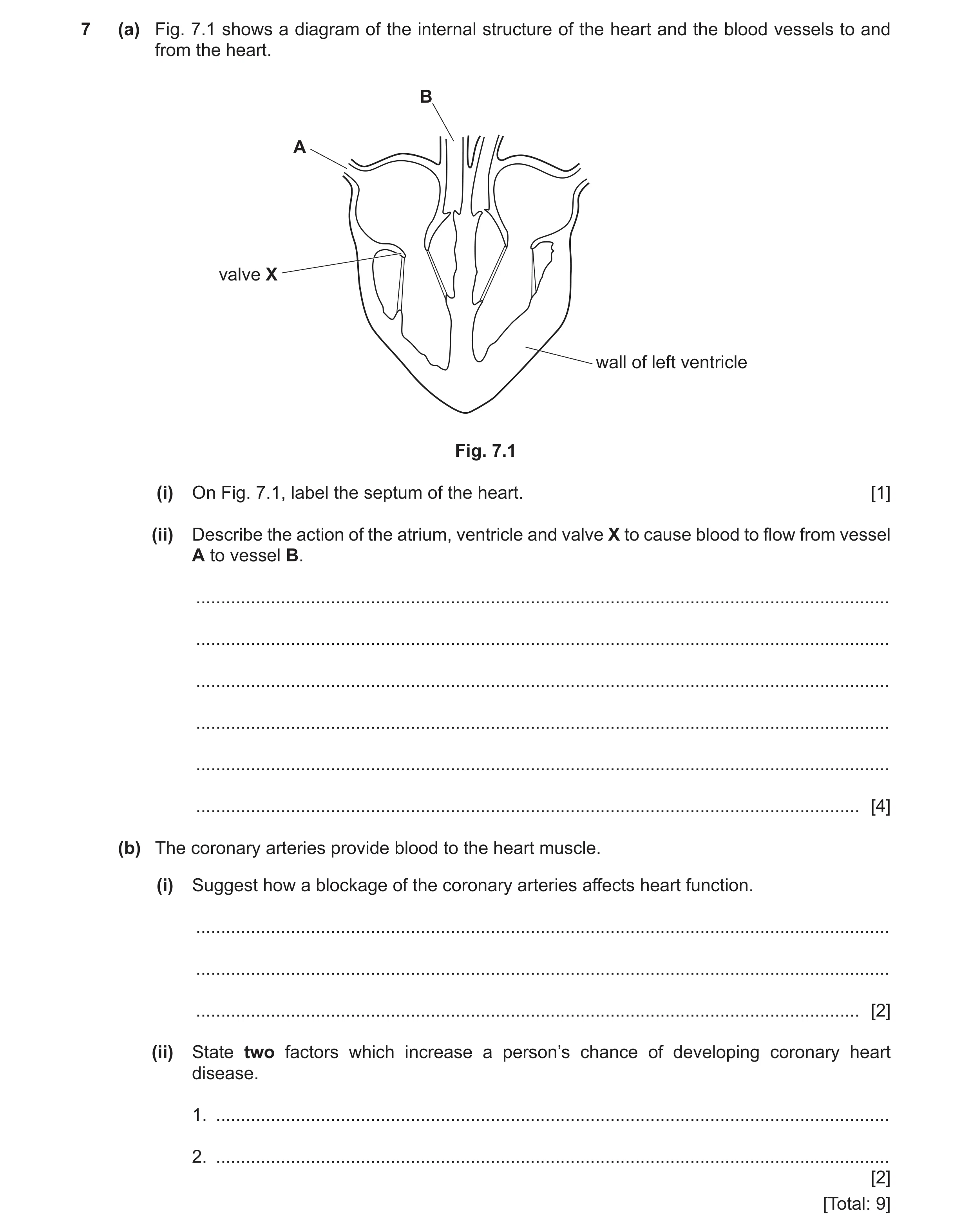
7 (a) Fig. 7.1 shows a diagram of the internal structure of the heart and the blood vessels to and from the heart. B A valve X wall of left ventricle Fig. 7.1 (i) On Fig. 7.1, label the septum of the heart. [1] (ii) Describe the action of the atrium, ventricle and valve X to cause blood to flow from vessel A to vessel B. … … … … … … [4] (b) The coronary arteries provide blood to the heart muscle. (i) Suggest how a blockage of the coronary arteries affects heart function. … … … [2] (ii) State two factors which increase a person’s chance of developing coronary heart disease. 1. … 2. … [2] [Total: 9]
9 marks
Mark scheme: 7(a)(i) septum labelled ; 1 7(a)(ii) any four from: (deoxygenated) blood enters (right) atrium from, vein A / vena cava) ; (right) atrium contracts and valve X opens ; blood flows into (right) ventricle ; (right) ventricle contracts and valve X closes ; blood flows into, B / pulmonary artery ; valve X prevents backflow of blood ; 4 7(b)(i) less, oxygen / glucose supplied to heart muscle ; and any one from: reduces respiration in heart muscle ; reduces, ability / efficiency of contraction of heart muscle ; 2 Question Answer Marks 7(b)(ii) any two from: poor diet ; stress ; smoking ; genetic predisposition ; (increasing risk with) age ; sex / males more likely to get CHD ; AVP, e.g. lack of exercise / overweight ; 2
This question in 0653/42 May/June 2020
Q9 · An external view of the heart 0653/41 Oct/Nov 2020
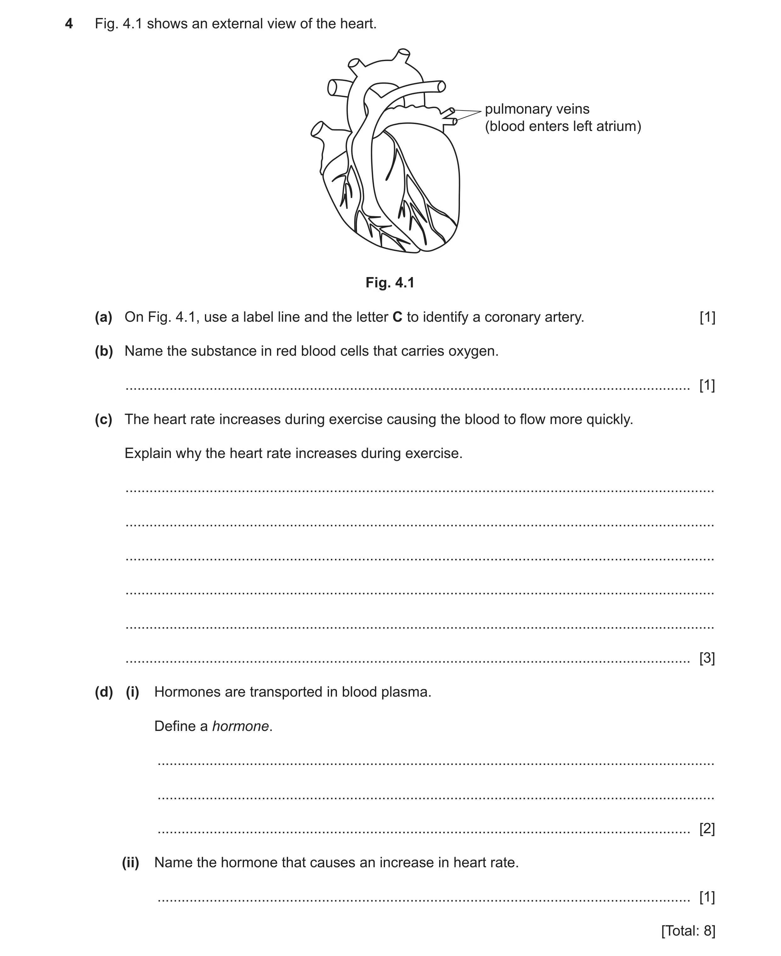
4 Fig. 4.1 shows an external view of the heart. pulmonary veins (blood enters left atrium) Fig. 4.1 (a) On Fig. 4.1, use a label line and the letter C to identify a coronary artery. [1] (b) Name the substance in red blood cells that carries oxygen. … [1] (c) The heart rate increases during exercise causing the blood to flow more quickly. Explain why the heart rate increases during exercise. … … … … … … [3] (d) (i) Hormones are transported in blood plasma. Define a hormone. … … … [2] (ii) Name the hormone that causes an increase in heart rate. … [1] [Total: 8]
8 marks
Mark scheme: 4(a) coronary artery correctly labelled ; 1 4(b) haemoglobin ; 1 4(c) any three from: so more oxygen / glucose, taken to the muscles ; so more energy can be released ; correct reference to respiration ; so more carbon dioxide can be removed (from the muscles) ; 3 4(d)(i) a chemical substance produced by a gland, carried by blood ; which alters the activity of one or more specific target organs ; 2 4(d)(ii) adrenaline ; 1
This question in 0653/41 Oct/Nov 2020
Q10 · A diagram of a cross-section through an artery 0653/42 Oct/Nov 2020
7 (a) Fig. 7.1 is a diagram of a cross-section through an artery. wall lumen Fig. 7.1 Explain two ways the structure of the artery is adapted for its function. 1 … … 2 … … [2] (b) Describe how the structure of capillaries allows efficient exchange of materials. … … [1] (c) Fig. 7.2 shows a diagram of the internal structure of the heart. aorta valve 1 A left atrium right atrium valve 2 left ventricle Fig. 7.2 (i) Blood from the body enters the right atrium through blood vessel A. State the name of blood vessel A. … [1] (ii) Blood is forced through the aorta by contraction of the muscle in the wall of the left ventricle. Describe the action of valves 1 and 2 in Fig. 7.2 during this contraction. valve 1 … valve 2 … [1] (d) Describe the function of valves in veins. … … [1] [Total: 6]
6 marks
Mark scheme: 7(a) any two paired responses: strong / thick wall, prevents bursting / withstands high pressure ; elastic fibres in wall, prevent bursting / stretch to accommodate high pressure of blood / enable wall to recoil and maintain pressure ; narrow lumen, maintains high pressure ; 7(b) (capillaries have) thin walls ; 1 7(c)(i) vena cava ; 1 7(c)(ii) valve 1 opens AND valve 2 closes ; 1 7(d) prevent backflow of blood / ensure that blood only travels in one direction ; 1
This question in 0653/42 Oct/Nov 2020
Q11 · A heart with coronary heart disease 0653/42 Feb/March 2021
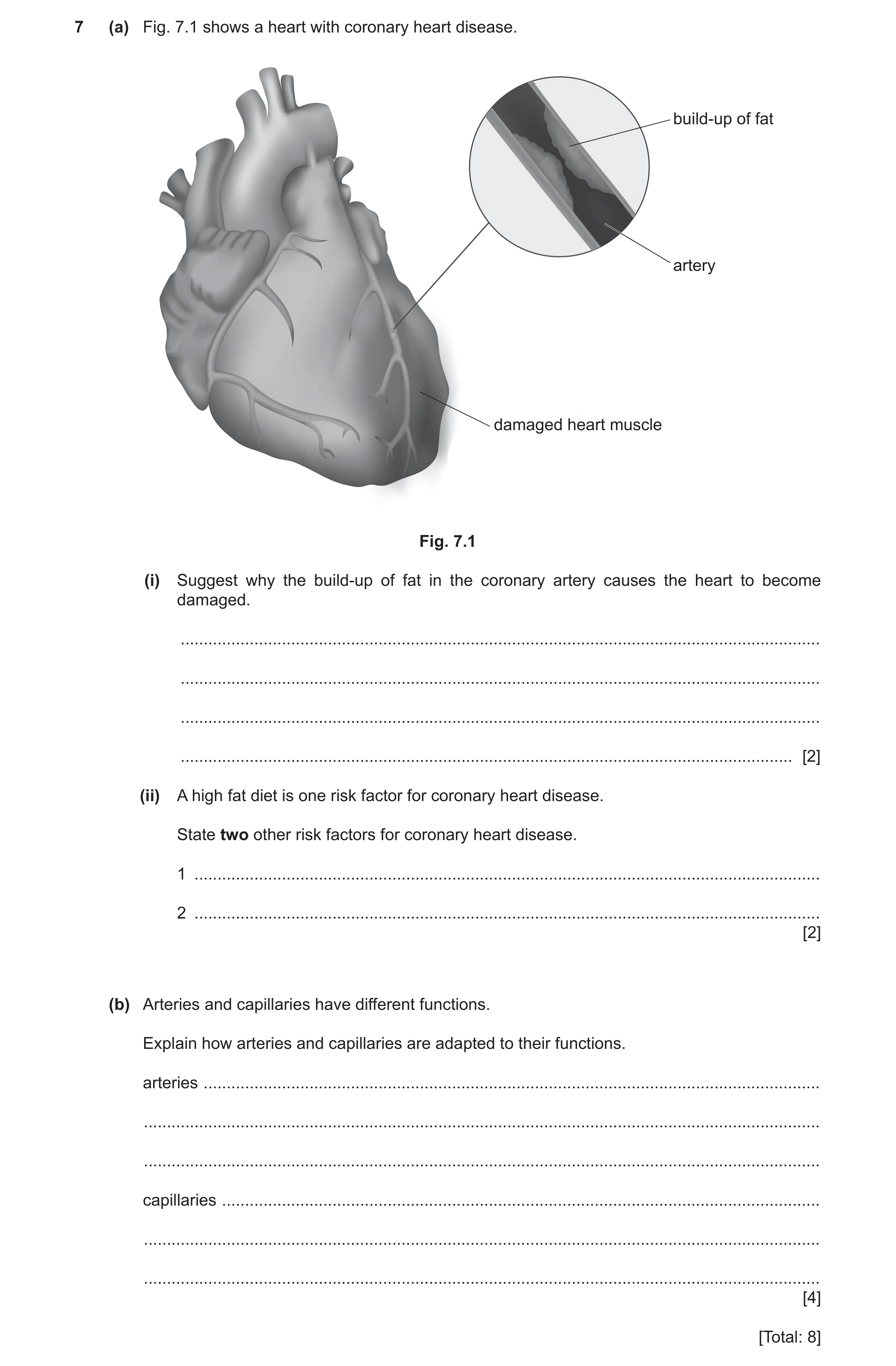
7 (a) Fig. 7.1 shows a heart with coronary heart disease. build-up of fat artery damaged heart muscle Fig. 7.1 (i) Suggest why the build-up of fat in the coronary artery causes the heart to become damaged. … … … … [2] (ii) A high fat diet is one risk factor for coronary heart disease. State two other risk factors for coronary heart disease. 1 … 2 … [2] (b) Arteries and capillaries have different functions. Explain how arteries and capillaries are adapted to their functions. arteries … … … capillaries … … … [4] [Total: 8]
8 marks
Mark scheme: 7(a)(i) less, blood / oxygen transported to heart (muscle) ; (heart) muscle unable to respire effectively ; 2 7(a)(ii) any two from: gender / sex ; stress ; smoking ; age ; genetic predisposition ; AVP, e.g. obesity / lack of exercise ; 2 7(b) any one adaptation and explanation pairing for arteries from: thick muscular wall ; to withstand high pressure ; OR elastic (muscle fibres in walls) ; to allow, contraction / relaxation / maintain blood flow rate ; OR narrow lumen ; to maintain, blood pressure / flow rate ; adaptation and explanation for capillaries: thin walls / walls only one cell thick ; to allow for exchange of materials / to allow substances to diffuse in and out ; 4
This question in 0653/42 Feb/March 2021
Q12 · A diagram of a section through a human heart 0653/43 May/June 2021
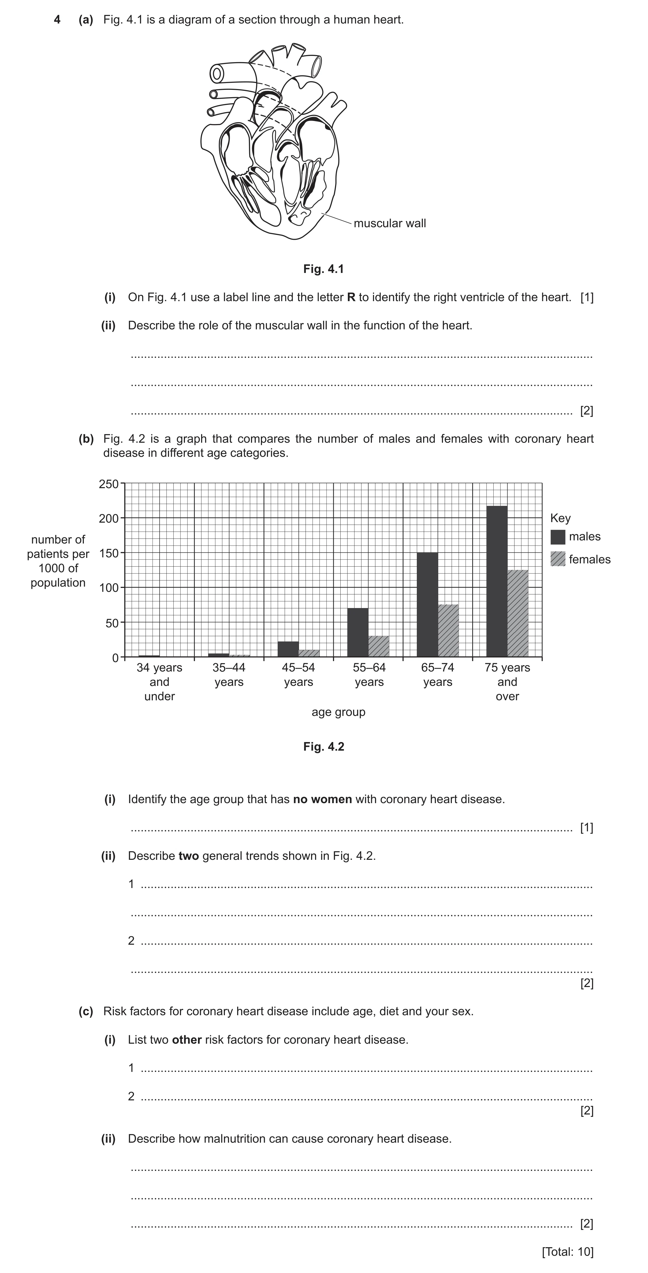
4 (a) Fig. 4.1 is a diagram of a section through a human heart. muscular wall Fig. 4.1 (i) On Fig. 4.1 use a label line and the letter R to identify the right ventricle of the heart. [1] (ii) Describe the role of the muscular wall in the function of the heart. … … … [2] (b) Fig. 4.2 is a graph that compares the number of males and females with coronary heart disease in different age categories. 250 200 Key number of males patients per 150 females 1000 of population 100 50 0 34 years 35–44 45–54 55–64 65–74 75 years and years years years years and under over age group Fig. 4.2 (i) Identify the age group that has no women with coronary heart disease. … [1] (ii) Describe two general trends shown in Fig. 4.2. 1 … … 2 … … [2] (c) Risk factors for coronary heart disease include age, diet and your sex. (i) List two other risk factors for coronary heart disease. 1 … 2 … [2] (ii) Describe how malnutrition can cause coronary heart disease. … … … [2] [Total: 10]
10 marks
Mark scheme: 4(a)(i) right ventricle labelled ; 1 4(a)(ii) contracts ; to push / pump, blood out of heart ; 2 4(b)(i) 34 years and under ; 1 4(b)(ii) coronary heart disease increases with age ; more men than women have coronary heart disease (in each age group) ; 2 4(c)(i) any two from: genetic predisposition ; stress ; smoking ; 2 4(c)(ii) high fat diet ; cause blockage of coronary artery ; 2
This question in 0653/43 May/June 2021
Q13 · A healthy human heart and a damaged human heart 0653/42 May/June 2022
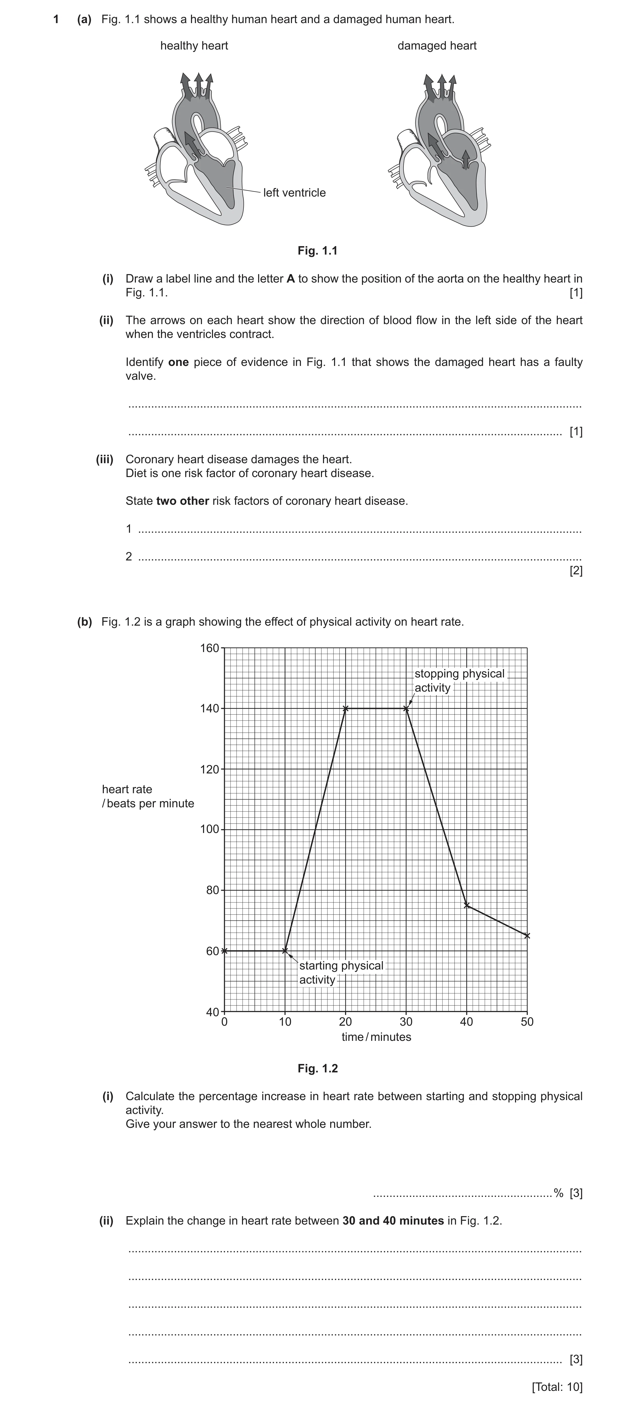
1 (a) Fig. 1.1 shows a healthy human heart and a damaged human heart. healthy heart damaged heart left ventricle Fig. 1.1 (i) Draw a label line and the letter A to show the position of the aorta on the healthy heart in Fig. 1.1. [1] (ii) The arrows on each heart show the direction of blood flow in the left side of the heart when the ventricles contract. Identify one piece of evidence in Fig. 1.1 that shows the damaged heart has a faulty valve. … … [1] (iii) Coronary heart disease damages the heart. Diet is one risk factor of coronary heart disease. State two other risk factors of coronary heart disease. 1 … 2 … [2] (b) Fig. 1.2 is a graph showing the effect of physical activity on heart rate. 160 stopping physical activity 140 120 heart rate / beats per minute 100 80 60 starting physical activity 40 0 10 20 30 40 50 time / minutes Fig. 1.2 (i) Calculate the percentage increase in heart rate between starting and stopping physical activity. Give your answer to the nearest whole number. … % [3] (ii) Explain the change in heart rate between 30 and 40 minutes in Fig. 1.2. … … … … … [3] [Total: 10]
10 marks
Mark scheme: 1(a)(i) label to aorta on healthy heart ; 1 1(a)(ii) blood flowing, backwards / into (left) atrium ; 1 1(a)(iii) any two from: stress ; smoking ; genetic predisposition ; age ; gender ; AVP ; 2 1(b)(i) 80 60 100 ; 133.33333 ; 133 ; 3 1(b)(ii) any three from: heart rate decreases ; less energy required / less muscular contraction ; less oxygen needs to be transported to muscles ; (due to) decreased respiration (rate) ; AVP ; 3
This question in 0653/42 May/June 2022
Q14 · A diagram of the human heart 0653/42 Oct/Nov 2022
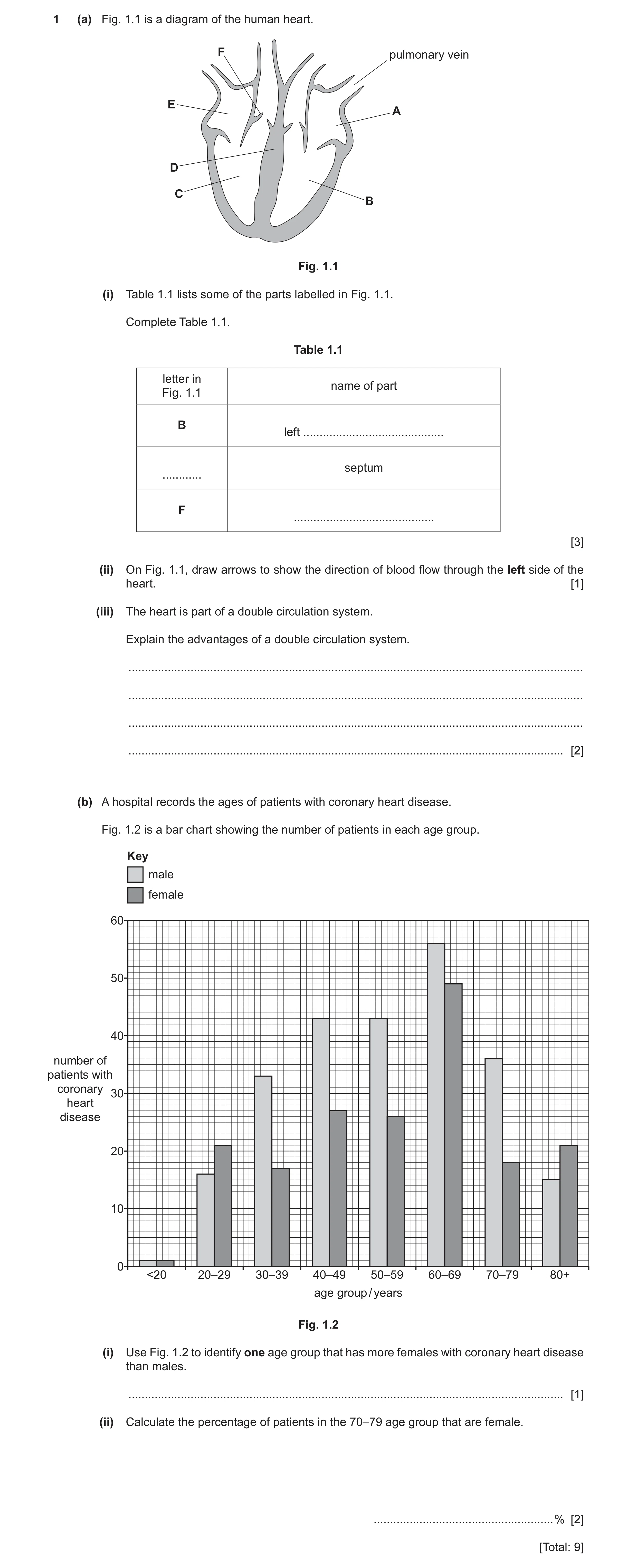
1 (a) Fig. 1.1 is a diagram of the human heart. F pulmonary vein E A D C B Fig. 1.1 (i) Table 1.1 lists some of the parts labelled in Fig. 1.1. Complete Table 1.1. Table 1.1 letter in name of part Fig. 1.1 B left … septum … F … [3] (ii) On Fig. 1.1, draw arrows to show the direction of blood flow through the left side of the heart. [1] (iii) The heart is part of a double circulation system. Explain the advantages of a double circulation system. … … … … [2] (b) A hospital records the ages of patients with coronary heart disease. Fig. 1.2 is a bar chart showing the number of patients in each age group. Key male female 60 50 40 number of patients with coronary 30 heart disease 20 10 0 <20 20–29 30–39 40–49 50–59 60–69 70–79 80+ age group / years Fig. 1.2 (i) Use Fig. 1.2 to identify one age group that has more females with coronary heart disease than males. … [1] (ii) Calculate the percentage of patients in the 70–79 age group that are female. … % [2] [Total: 9]
9 marks
Mark scheme: Question Answer Marks 1(a)(i) 3 letter in name of part Fig. 1.1 (B) (left) ventricle ; D ; (septum) (F) valve ; 1(a)(ii) arrow(s) showing correct direction ; 1 1(a)(iii) separates oxygenated and deoxygenated blood ; 2 allows for different levels of blood pressure / AW ; 1(b)(i) 20–29 / 80+ ; 1 1(b)(ii) 18 100 ÷ 54 / 18 + 36 / 54 ; 2 33(%) ;
This question in 0653/42 Oct/Nov 2022
Q15 · A diagram of a human heart 0653/43 Oct/Nov 2022
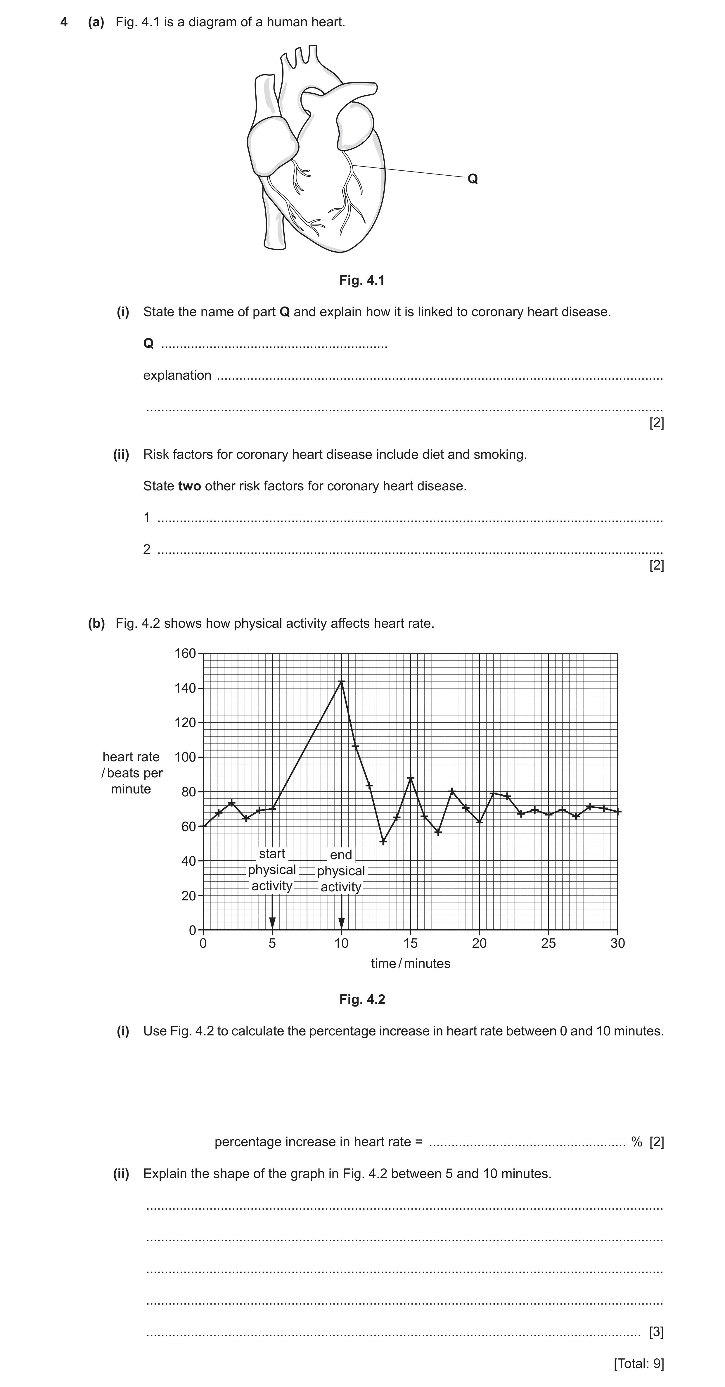
4 (a) Fig. 4.1 is a diagram of a human heart. Q Fig. 4.1 (i) State the name of part Q and explain how it is linked to coronary heart disease. Q … explanation … … [2] (ii) Risk factors for coronary heart disease include diet and smoking. State two other risk factors for coronary heart disease. 1 … 2 … [2] (b) Fig. 4.2 shows how physical activity affects heart rate. 160 140 120 heart rate 100 / beats per minute 80 60 start end 40 physical physical activity activity 20 0 0 5 10 15 20 25 30 time / minutes Fig. 4.2 (i) Use Fig. 4.2 to calculate the percentage increase in heart rate between 0 and 10 minutes. percentage increase in heart rate = … % [2] (ii) Explain the shape of the graph in Fig. 4.2 between 5 and 10 minutes. … … … … … [3] [Total: 9]
9 marks
Mark scheme: 4(a)(i) Q coronary artery ; 2 explanation (CHD occurs) when it becomes blocked ; 4(a)(ii) any two from: 2 stress ; genetic predisposition ; age ; gender / sex ; 4(b)(i) correct numbers from graph / calculation of percentage, e.g. 144 and 60 or (144 – 60) ÷ 60 100 ; 2 140(%) ; 4(b)(ii) any three from: 3 increase blood flow to muscle ; muscles require (more), energy / oxygen ; increased muscle contraction ; reference to respiration ; need to remove more carbon dioxide from muscle ;
This question in 0653/43 Oct/Nov 2022
Q16 · A diagram of a heart 0653/42 Feb/March 2023
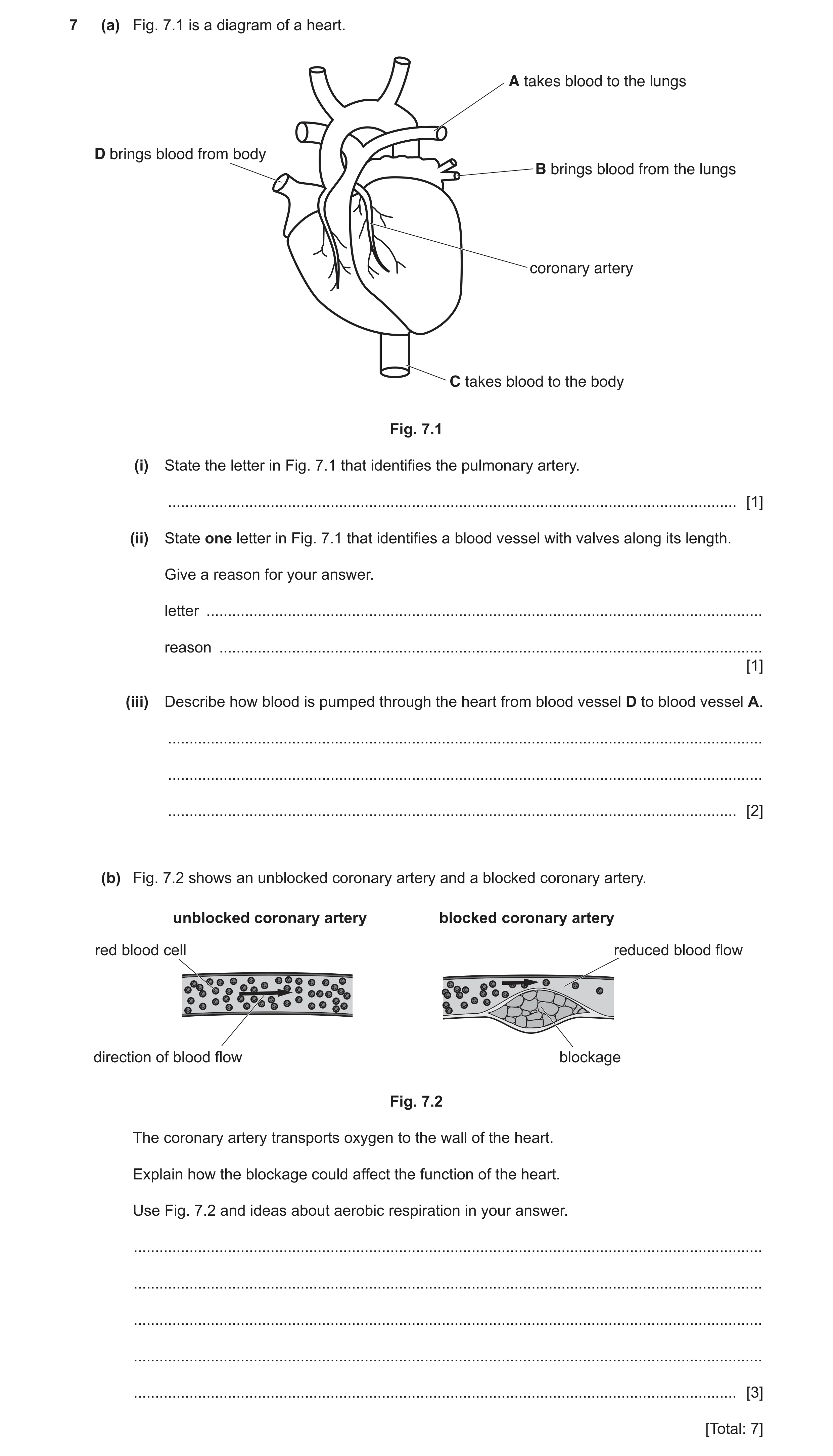
7 (a) Fig. 7.1 is a diagram of a heart. A takes blood to the lungs D brings blood from body B brings blood from the lungs coronary artery C takes blood to the body Fig. 7.1 (i) State the letter in Fig. 7.1 that identifies the pulmonary artery. … [1] (ii) State one letter in Fig. 7.1 that identifies a blood vessel with valves along its length. Give a reason for your answer. letter … reason … [1] (iii) Describe how blood is pumped through the heart from blood vessel D to blood vessel A. … … … [2] (b) Fig. 7.2 shows an unblocked coronary artery and a blocked coronary artery. unblocked coronary artery blocked coronary artery red blood cell reduced blood flow direction of blood flow blockage Fig. 7.2 The coronary artery transports oxygen to the wall of the heart. Explain how the blockage could affect the function of the heart. Use Fig. 7.2 and ideas about aerobic respiration in your answer. … … … … … [3] [Total: 7]
7 marks
Mark scheme: 7(a)(i) A ; 1 7(a)(ii) B / D because it is a vein ; 1 7(a)(iii) idea of blood flows from atrium to ventricle ; 2 idea of flow of blood caused by (muscular) contraction (of chambers) ; 7(b) idea that less oxygen is transported (to heart muscle / wall) ; 3 therefore reduced (aerobic) respiration ; so less energy available (for muscle contraction) ;
This question in 0653/42 Feb/March 2023
Q17 · The circulatory system of frogs and the circulatory system of humans 0653/41 May/June 2023
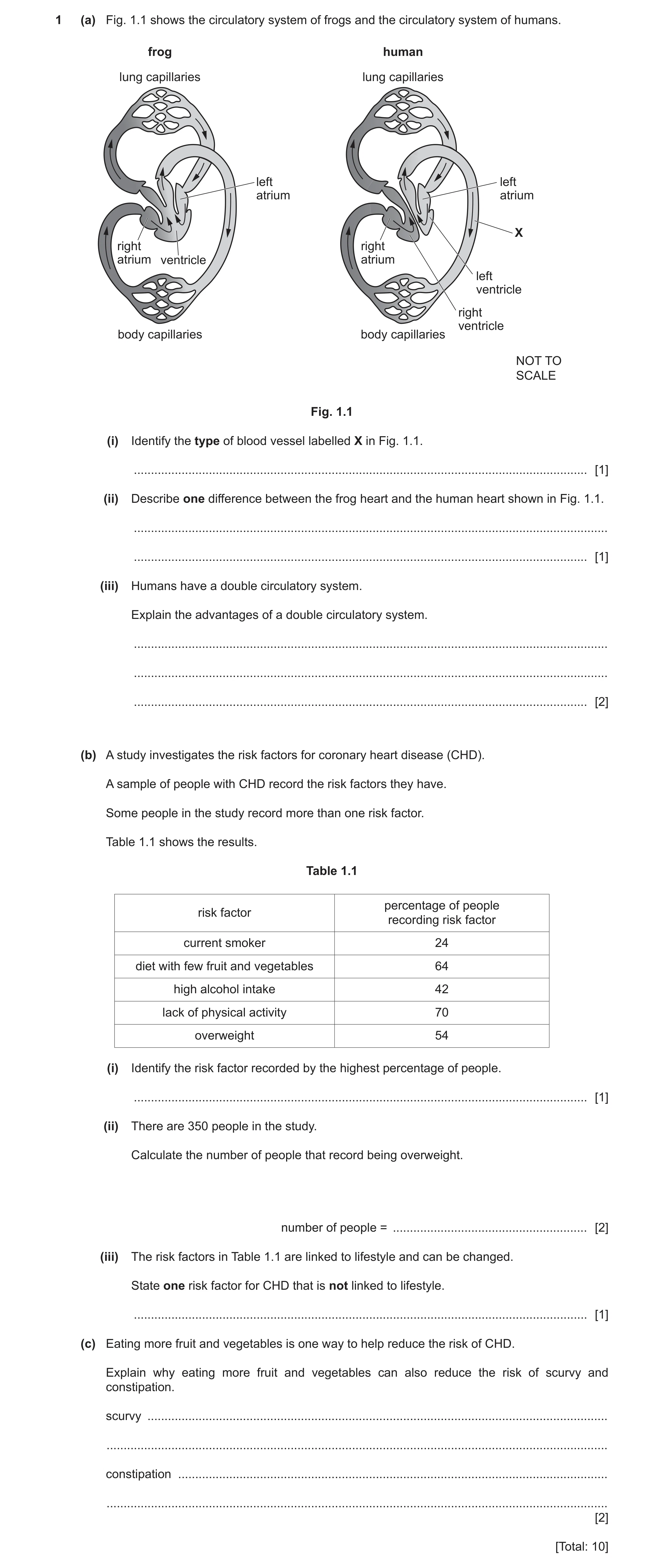
1 (a) Fig. 1.1 shows the circulatory system of frogs and the circulatory system of humans. frog human lung capillaries lung capillaries left left atrium atrium X right right atrium ventricle atrium left ventricle right ventricle body capillaries body capillaries NOT TO SCALE Fig. 1.1 (i) Identify the type of blood vessel labelled X in Fig. 1.1. … [1] (ii) Describe one difference between the frog heart and the human heart shown in Fig. 1.1. … … [1] (iii) Humans have a double circulatory system. Explain the advantages of a double circulatory system. … … … [2] (b) A study investigates the risk factors for coronary heart disease (CHD). A sample of people with CHD record the risk factors they have. Some people in the study record more than one risk factor. Table 1.1 shows the results. Table 1.1 percentage of people risk factor recording risk factor current smoker 24 diet with few fruit and vegetables 64 high alcohol intake 42 lack of physical activity 70 overweight 54 (i) Identify the risk factor recorded by the highest percentage of people. … [1] (ii) There are 350 people in the study. Calculate the number of people that record being overweight. number of people = … [2] (iii) The risk factors in Table 1.1 are linked to lifestyle and can be changed. State one risk factor for CHD that is not linked to lifestyle. … [1] (c) Eating more fruit and vegetables is one way to help reduce the risk of CHD. Explain why eating more fruit and vegetables can also reduce the risk of scurvy and constipation. scurvy … … constipation … … [2] [Total: 10]
10 marks
Mark scheme: 1(a)(i) artery ; 1 1(a)(ii) any one from: frog has only 3 chambers / frog has one ventricle ; humans have four chambers / humans have two ventricles ; no septum in frog / AW ; 1 1(a)(iii) oxygenated and deoxygenated blood kept separate ; high blood pressure around body / low blood pressure to lungs ; 2 1(b)(i) (lack of) physical activity ; 1 1(b)(ii) (54 350) ÷ 100 ; 189 ; 2 1(b)(iii) age / genetics / inheritance ; 1 1(c) scurvy – (fruit and vegetables) are a source of Vit C / Vit C prevents scurvy ; constipation – (vegetables) are a source of fibre / fibre needed for bulk ; 2
This question in 0653/41 May/June 2023
Q18 · The structure of the human heart 0653/43 May/June 2023
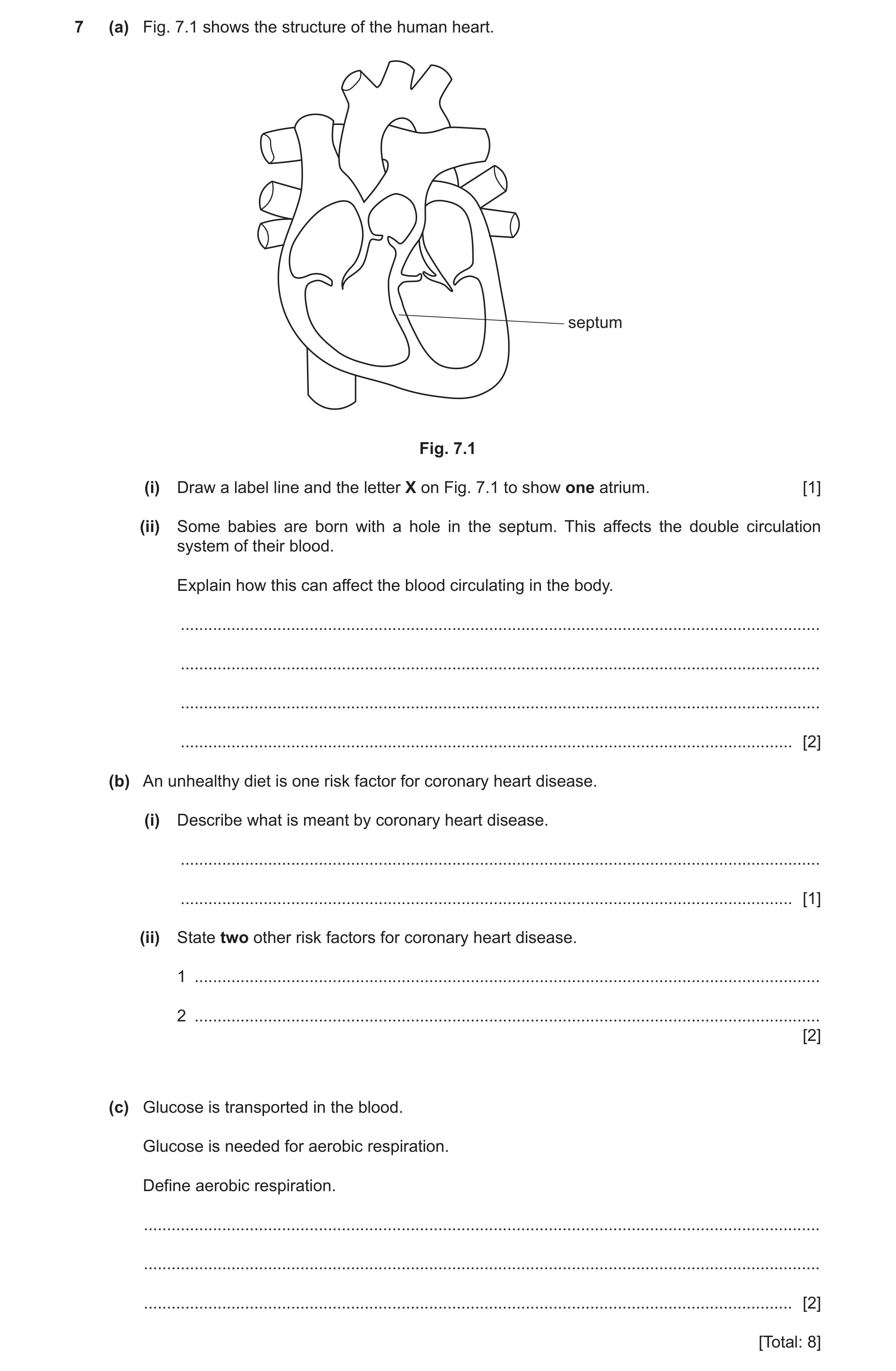
7 (a) Fig. 7.1 shows the structure of the human heart. septum Fig. 7.1 (i) Draw a label line and the letter X on Fig. 7.1 to show one atrium. [1] (ii) Some babies are born with a hole in the septum. This affects the double circulation system of their blood. Explain how this can affect the blood circulating in the body. … … … … [2] (b) An unhealthy diet is one risk factor for coronary heart disease. (i) Describe what is meant by coronary heart disease. … … [1] (ii) State two other risk factors for coronary heart disease. 1 … 2 … [2] (c) Glucose is transported in the blood. Glucose is needed for aerobic respiration. Define aerobic respiration. … … … [2] [Total: 8]
8 marks
Mark scheme: 7(a)(i) either atrium labelled ; 1 7(a)(ii) any two from: mixing of blood from lungs and body ; less oxygen transported (to body) ; decrease in blood pressure (to body) ; AVP, e.g. reduction in (aerobic) respiration in cells ; max 2 7(b)(i) blockage of coronary artery ; 1 7(b)(ii) any two from: stress ; smoking ; genetic predisposition ; age ; gender / sex ; AVP, e.g. lack of exercise ; 2 Question Answer Marks 7(c) (respiration is a) reaction with oxygen ; breakdown of nutrient (molecules) to release energy ; 2
This question in 0653/43 May/June 2023
Q19 · The structure of the human heart 0653/41 May/June 2024
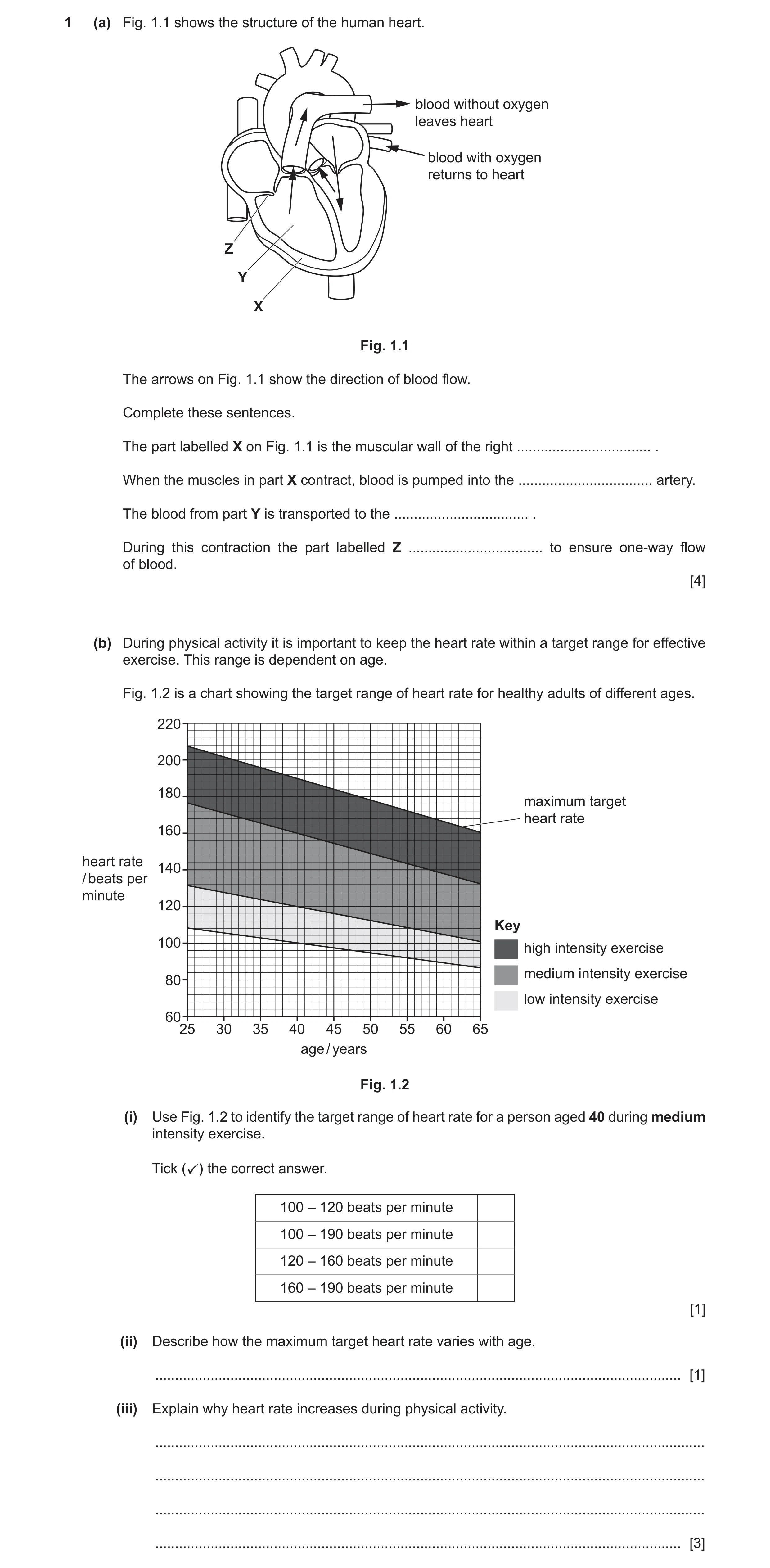
1 (a) Fig. 1.1 shows the structure of the human heart. blood without oxygen leaves heart blood with oxygen returns to heart Z Y X Fig. 1.1 The arrows on Fig. 1.1 show the direction of blood flow. Complete these sentences. The part labelled X on Fig. 1.1 is the muscular wall of the right … . When the muscles in part X contract, blood is pumped into the … artery. The blood from part Y is transported to the … . During this contraction the part labelled Z … to ensure one‑way flow of blood. [4] (b) During physical activity it is important to keep the heart rate within a target range for effective exercise. This range is dependent on age. Fig. 1.2 is a chart showing the target range of heart rate for healthy adults of different ages. 220 200 180 maximum target heart rate 160 heart rate 140 / beats per minute 120 Key 100 high intensity exercise medium intensity exercise 80 low intensity exercise 60 25 30 35 40 45 50 55 60 65 age / years Fig. 1.2 (i) Use Fig. 1.2 to identify the target range of heart rate for a person aged 40 during medium intensity exercise. Tick (ü) the correct answer. 100 – 120 beats per minute 100 – 190 beats per minute 120 – 160 beats per minute 160 – 190 beats per minute [1] (ii) Describe how the maximum target heart rate varies with age. … [1] (iii) Explain why heart rate increases during physical activity. … … … … [3]
9 marks
Mark scheme: 1(a) ventricle ; pulmonary ; lungs ; closes / shuts ; 1(b)(i) 120 – 160 beats per minute (ticked) ; 1 1(b)(ii) decreases (with age) ; 1 1(b)(iii) any three from: muscles require more energy (for contraction) ; more oxygen (and glucose) transported (to muscle) ; reference to increases (rate of) respiration ; remove more carbon dioxide (from lungs) / increased carbon dioxide in the, blood / muscle / more carbon dioxide released (from respiration) ; 3
This question in 0653/41 May/June 2024
Q20 · Drawings of three different blood vessels, W, X and Y 0653/42 Oct/Nov 2024
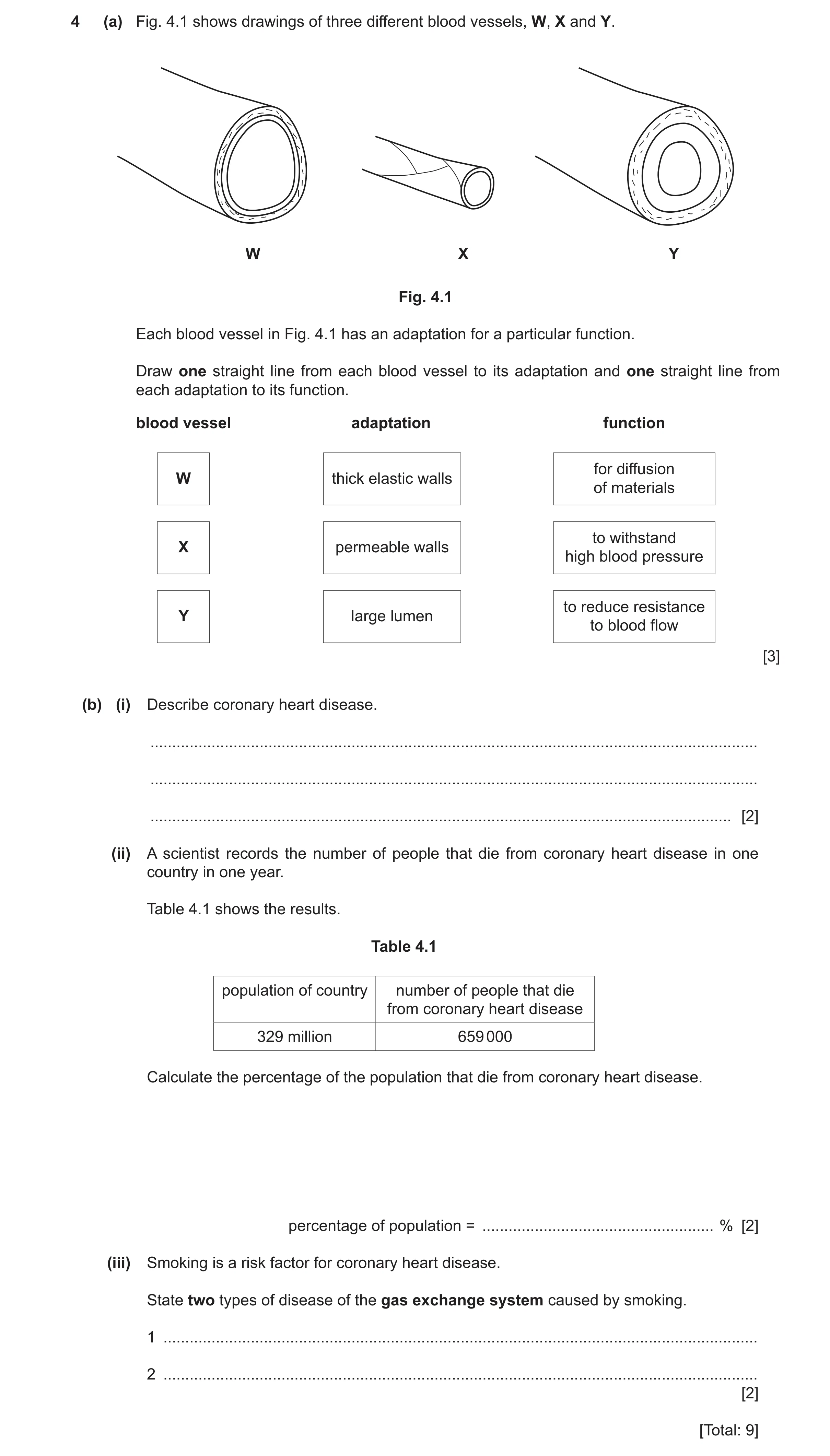
4 (a) Fig. 4.1 shows drawings of three different blood vessels, W, X and Y. W X Y Fig. 4.1 Each blood vessel in Fig. 4.1 has an adaptation for a particular function. Draw one straight line from each blood vessel to its adaptation and one straight line from each adaptation to its function. blood vessel adaptation function for diffusion W thick elastic walls of materials to withstand X permeable walls high blood pressure to reduce resistance Y large lumen to blood flow [3] (b) (i) Describe coronary heart disease. … … … [2] (ii) A scientist records the number of people that die from coronary heart disease in one country in one year. Table 4.1 shows the results. Table 4.1 population of country number of people that die from coronary heart disease 329 million 659 000 Calculate the percentage of the population that die from coronary heart disease. percentage of population = … % [2] (iii) Smoking is a risk factor for coronary heart disease. State two types of disease of the gas exchange system caused by smoking. 1 … 2 … [2] [Total: 9]
9 marks
Mark scheme: 4(a) 3 two lines correct ; four lines correct ; six lines correct ; 4(b)(i) idea of blockage of, a blood vessel / blood flow ; 2 coronary artery ; 4(b)(ii) 329 000 000 / 0.659 million / 0.002 / ratio of N:P 100 ; 2 0.200 (%) ; 4(b)(iii) any two from: 2 chronic obstructive pulmonary disease / COPD ; lung cancer ; AVP ;
This question in 0653/42 Oct/Nov 2024
Q21 · A diagram of the structure of the human heart 0653/42 Feb/March 2025
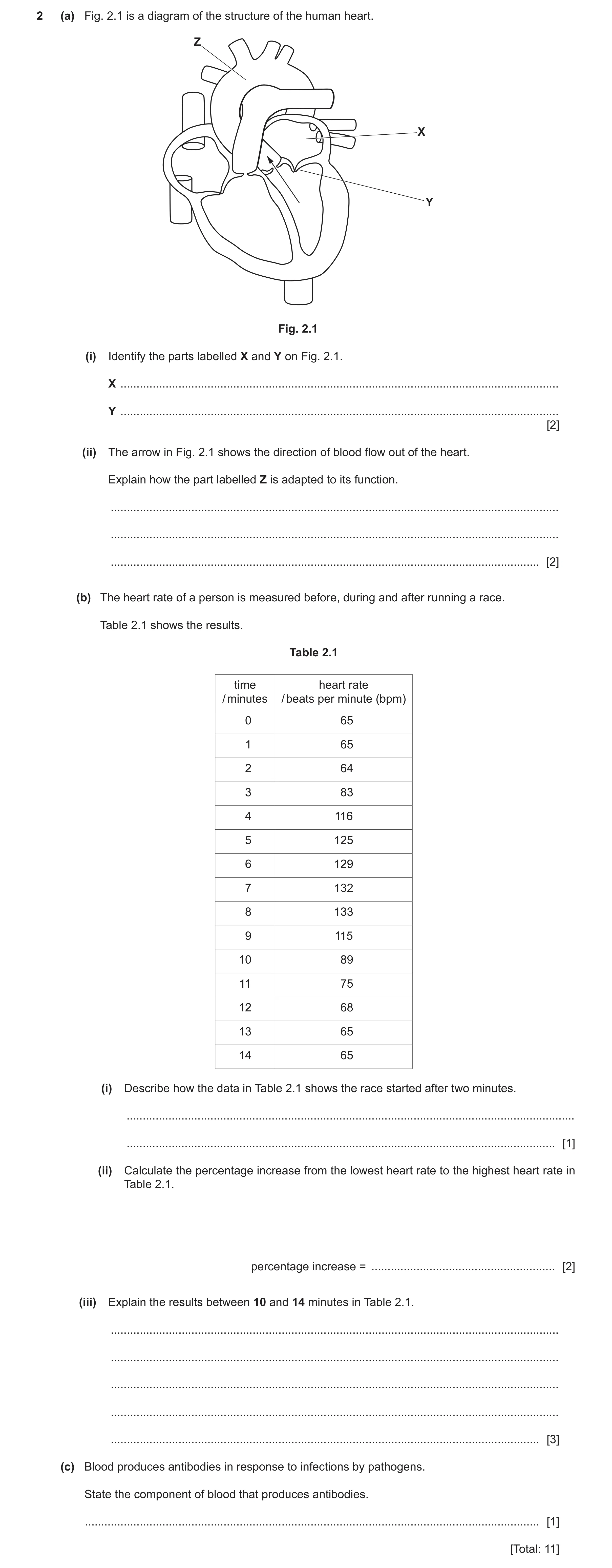
2 (a) Fig. 2.1 is a diagram of the structure of the human heart. Z X Y Fig. 2.1 (i) Identify the parts labelled X and Y on Fig. 2.1. X … Y … [2] (ii) The arrow in Fig. 2.1 shows the direction of blood flow out of the heart. Explain how the part labelled Z is adapted to its function. … … … [2] (b) The heart rate of a person is measured before, during and after running a race. Table 2.1 shows the results. Table 2.1 time heart rate / minutes / beats per minute (bpm) 0 65 1 65 2 64 3 83 4 116 5 125 6 129 7 132 8 133 9 115 10 89 11 75 12 68 13 65 14 65 (i) Describe how the data in Table 2.1 shows the race started after two minutes. … … [1] (ii) Calculate the percentage increase from the lowest heart rate to the highest heart rate in Table 2.1. percentage increase = … [2] (iii) Explain the results between 10 and 14 minutes in Table 2.1. … … … … … [3] (c) Blood produces antibodies in response to infections by pathogens. State the component of blood that produces antibodies. … [1] [Total: 11]
11 marks
Mark scheme: 2(a)(i) (left) atrium ; 2 valve ; 2(a)(ii) thick (muscular) wall / elastic wall ; 2 the idea of allowing or withstanding the transport of blood at high pressure ; OR narrow lumen ; to maintain high blood pressure ; 2(b)(i) heart rate increases (from 3 min) ; 1 2(b)(ii) 133 – 64 / 69 ; 2 133 − 64 100 / 107.8(125) / 108 ; 64 2(b)(iii) any three from: 3 the person has stopped running / owtte ; muscle contraction decreases ; less energy / oxygen required ; reduction in respiration rate ; 2(c) white blood cells ; 1
This question in 0653/42 Feb/March 2025
Q22 · A diagram of a human heart 0653/41 Oct/Nov 2025
3 (a) Fig. 3.1 is a diagram of a human heart. Fig. 3.1 (i) Draw a label line and the letter C to identify a coronary artery in Fig. 3.1. [1] (ii) Describe what happens to the coronary artery when a person has coronary heart disease. … … [1] (iii) Diet is one risk factor for coronary heart disease. State two other risk factors for coronary heart disease. 1 … 2 … [2] (b) Arteries transport blood away from the heart. Explain how the structure of an artery is adapted to its function. … … … … … … [3] [Total: 7]
7 marks
Mark scheme: 3(a)(i) either coronary artery labelled ; 1 3(a)(ii) blocked / AW ; 1 3(a)(iii) any two from: 2 lack of exercise / AW ; stress ; smoking ; genetic predisposition ; age ; sex ; high blood pressure ; AVP;; 3(b) Any two from 3 (wall) is muscular (wall) is elastic; narrow lumen ; and then to withstand high (blood) pressure / maintain high (blood) pressure ;
This question in 0653/41 Oct/Nov 2025
 6 / 33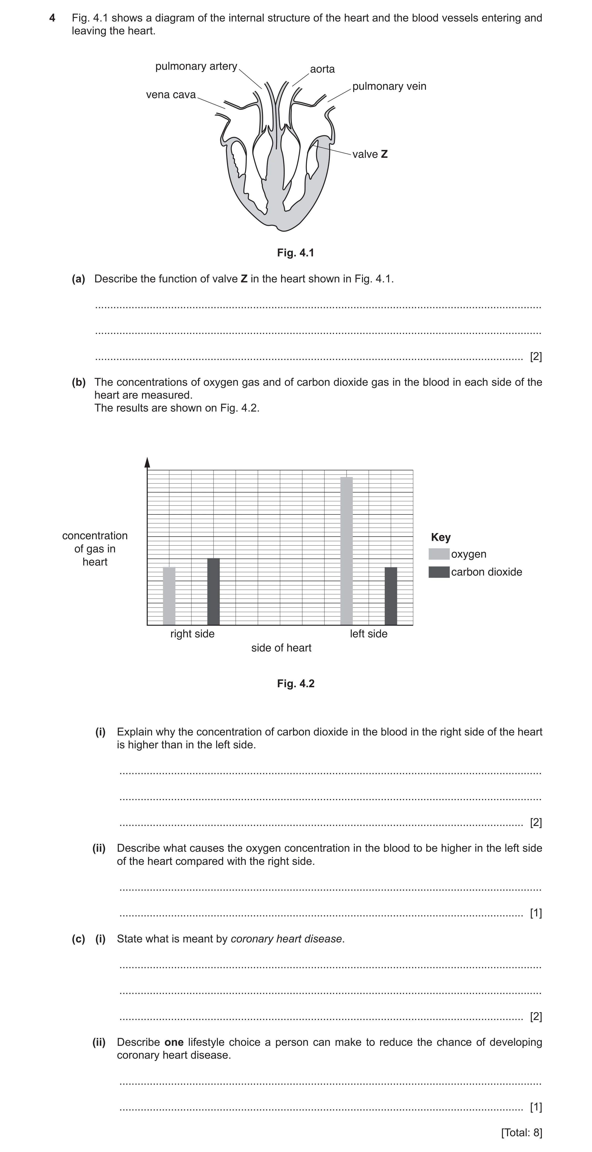
 11 / 33
 12 / 33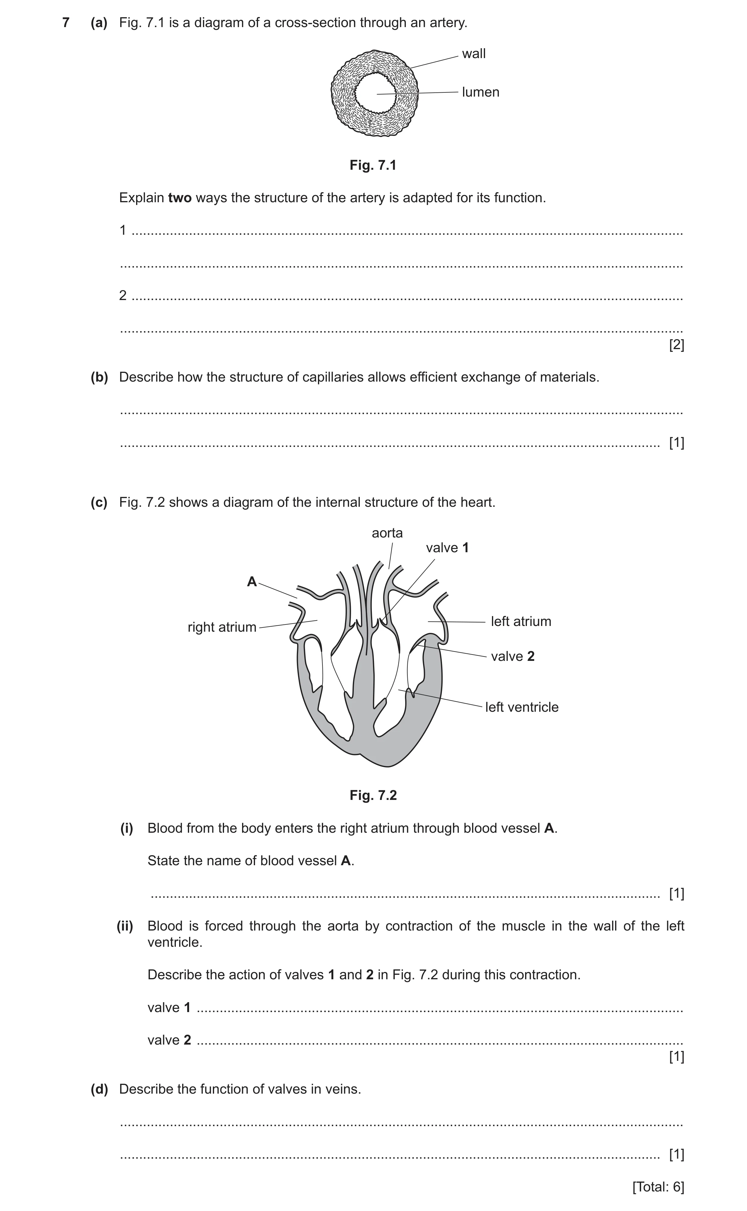
![Question 22: (a) Fig. 3.1 is a diagram of a human heart. Fig. 3.1 (i) Draw a label line and the letter C to identify a coronary artery in Fig. 3.1. [1] …](https://img.pastlit.com/crops/6fc12538-e60d-48ee-a0ed-7796e6999497/q3.webp) 33 / 33
33 / 33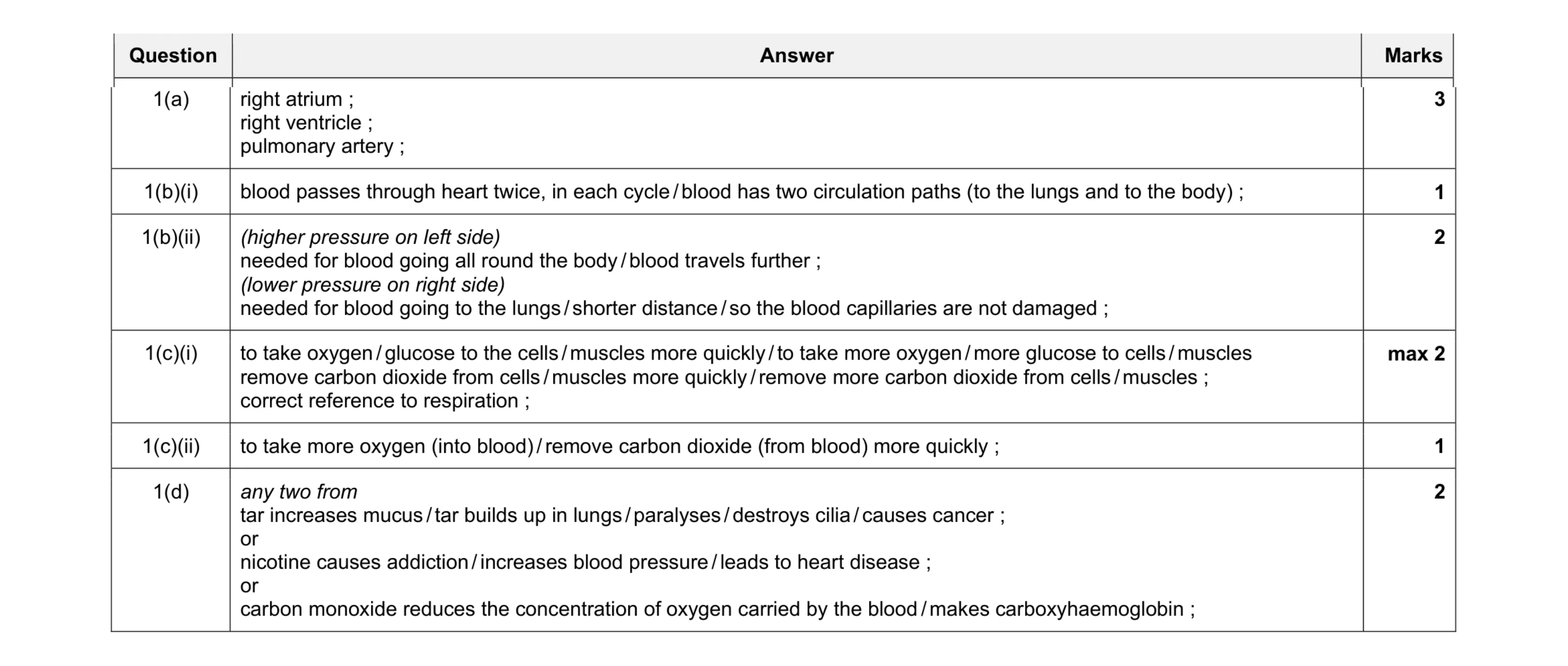 11
11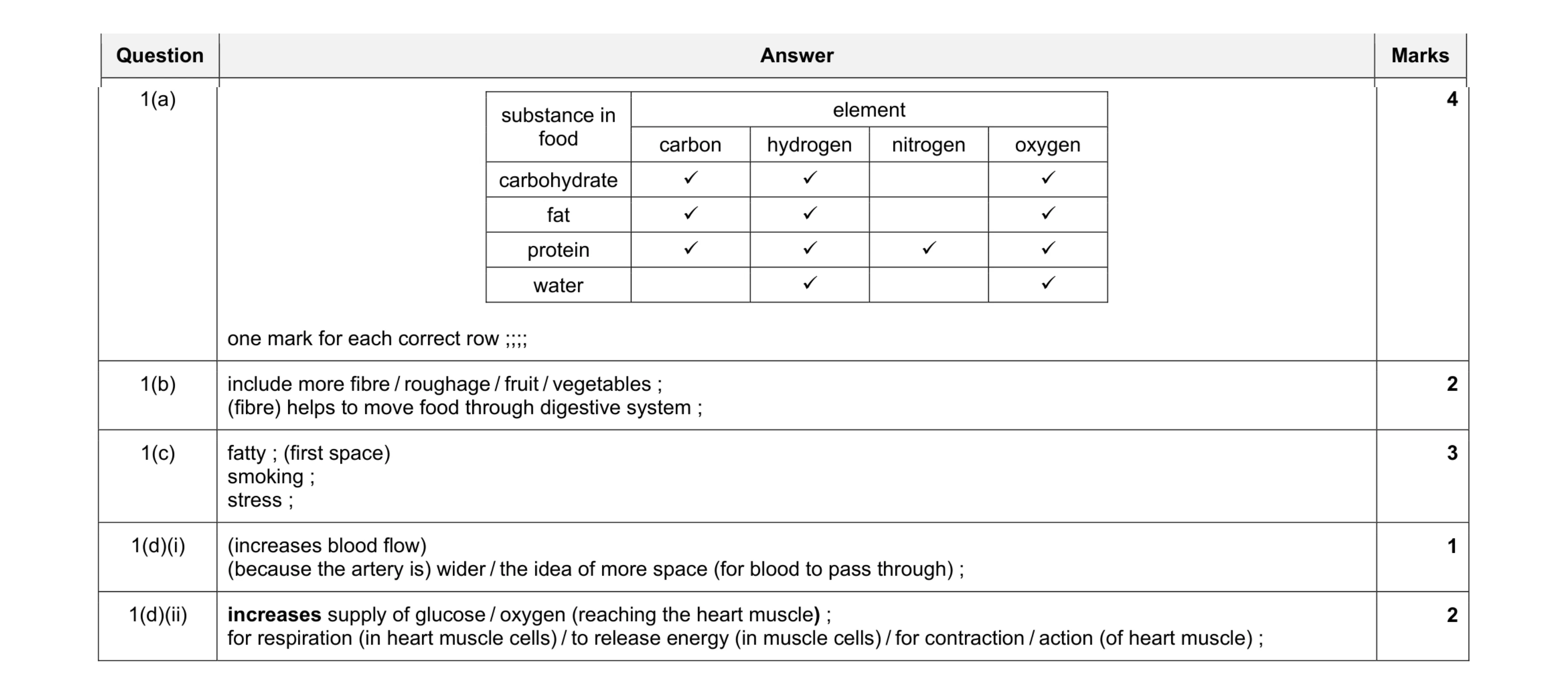 12
12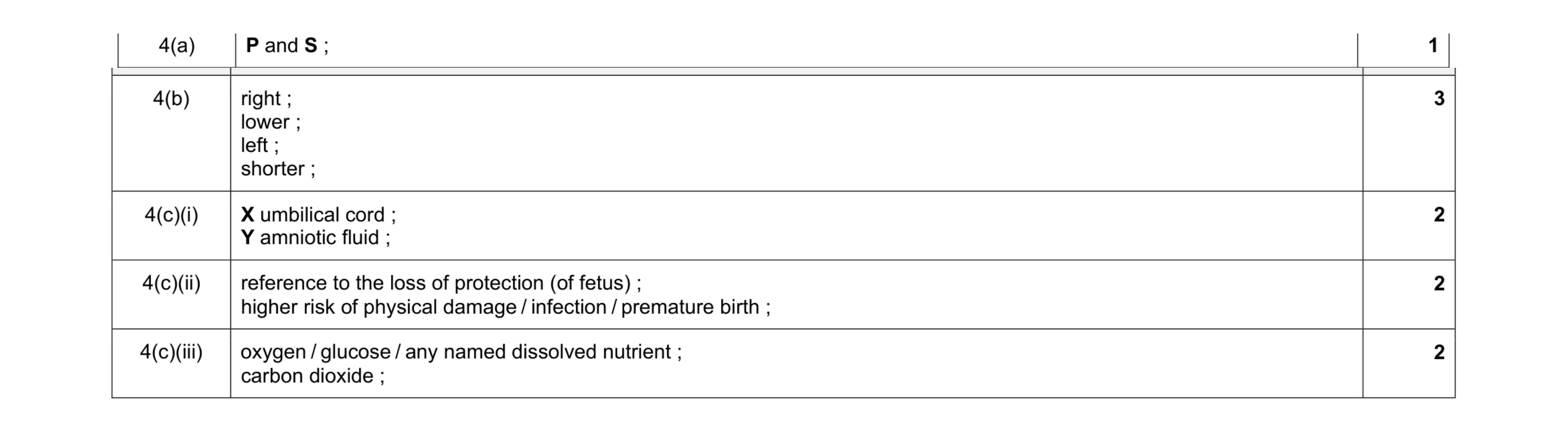 10
10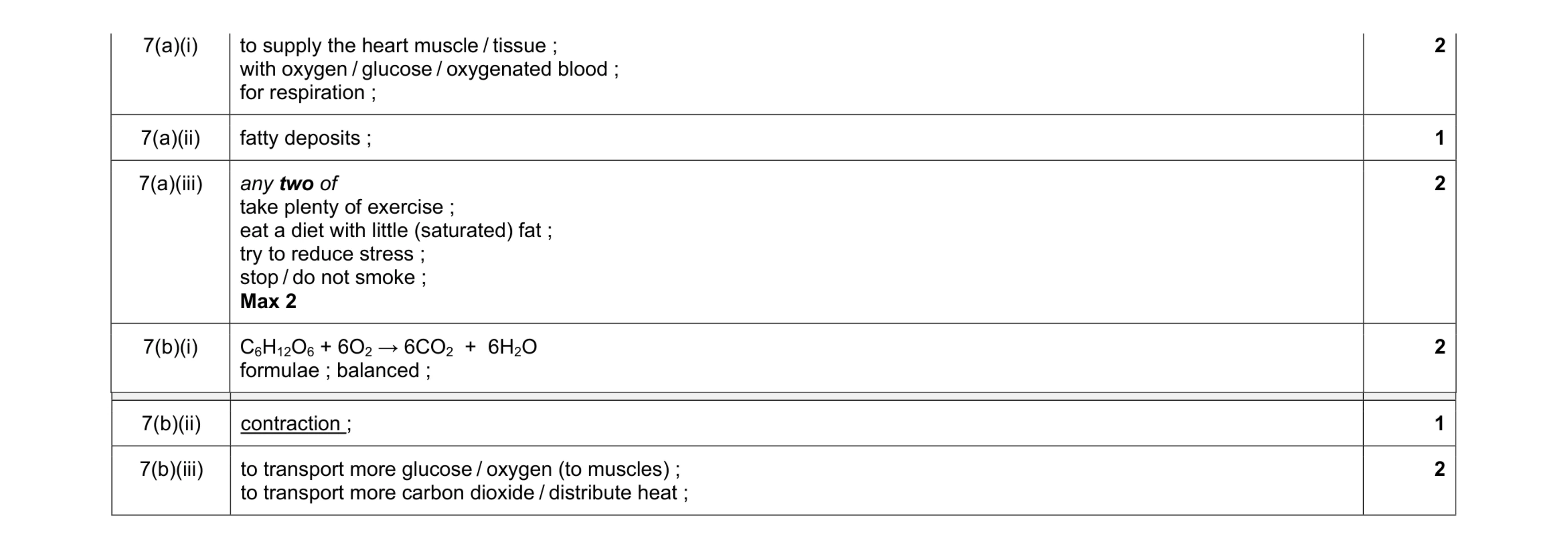 10
10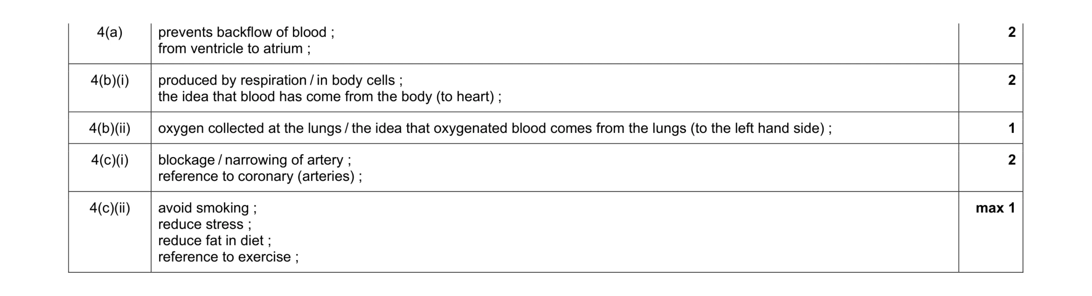 8
8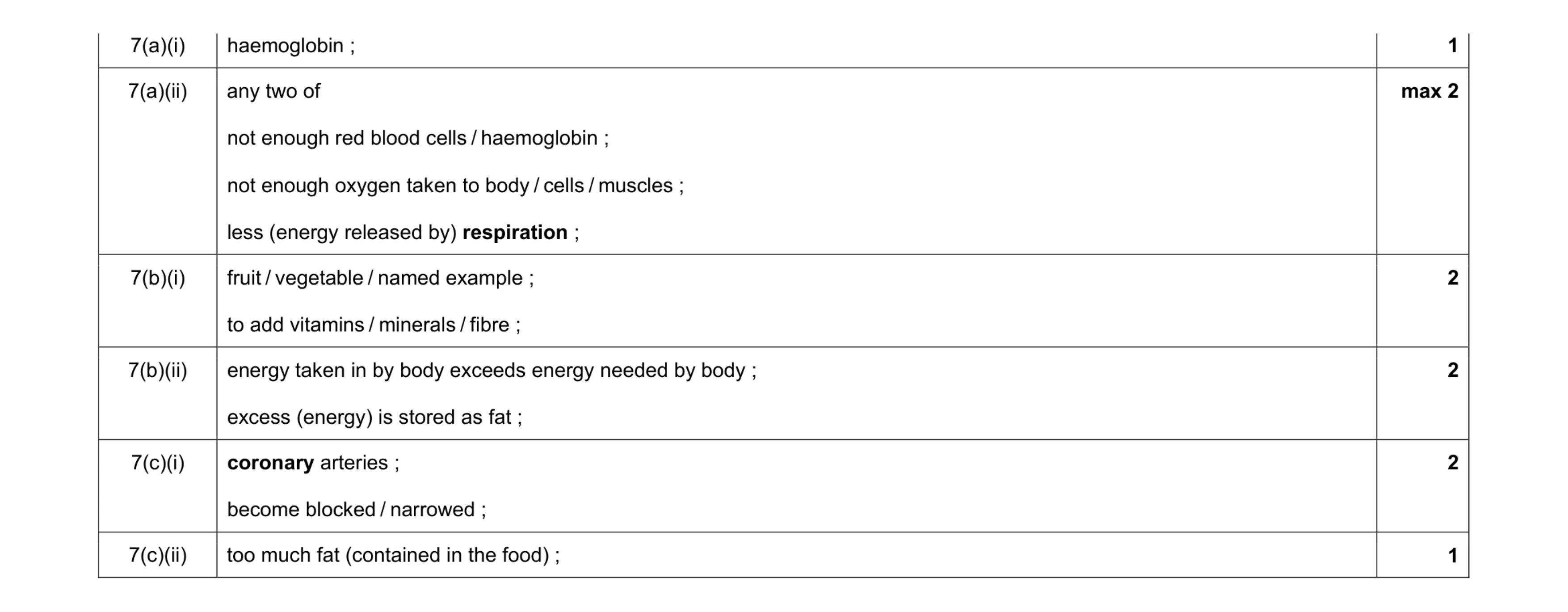 10
10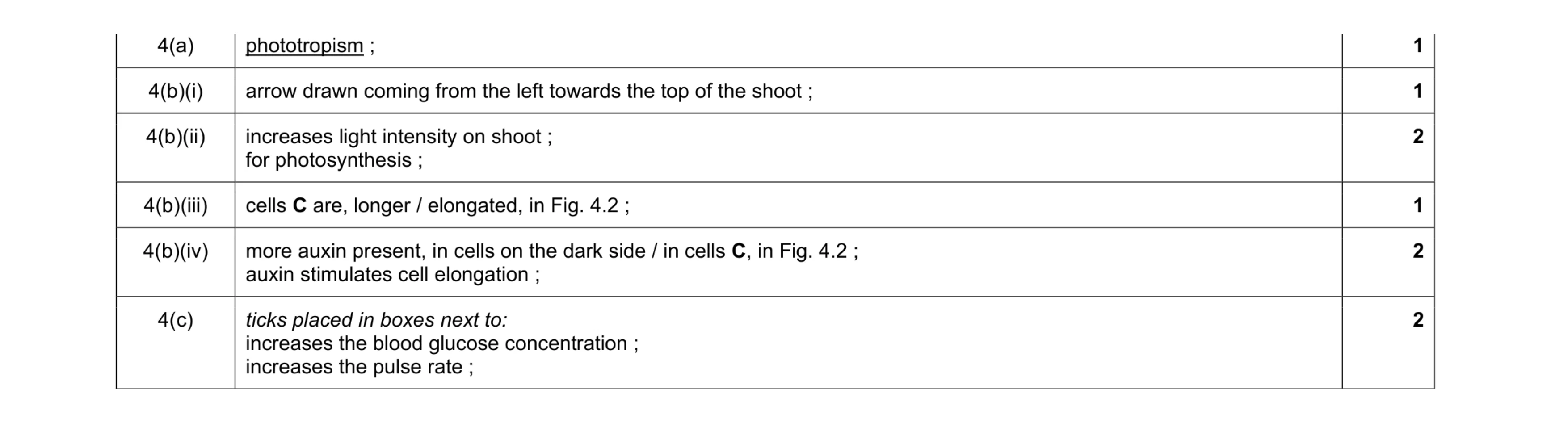 9
9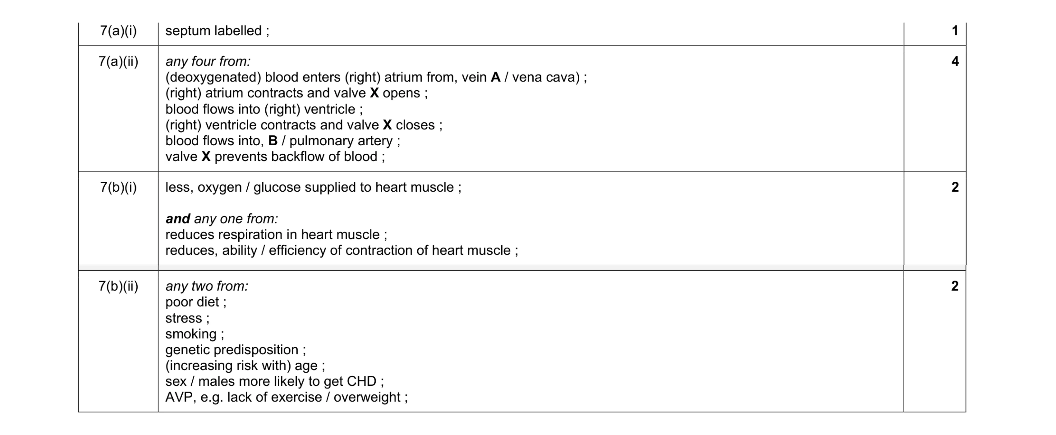 9
9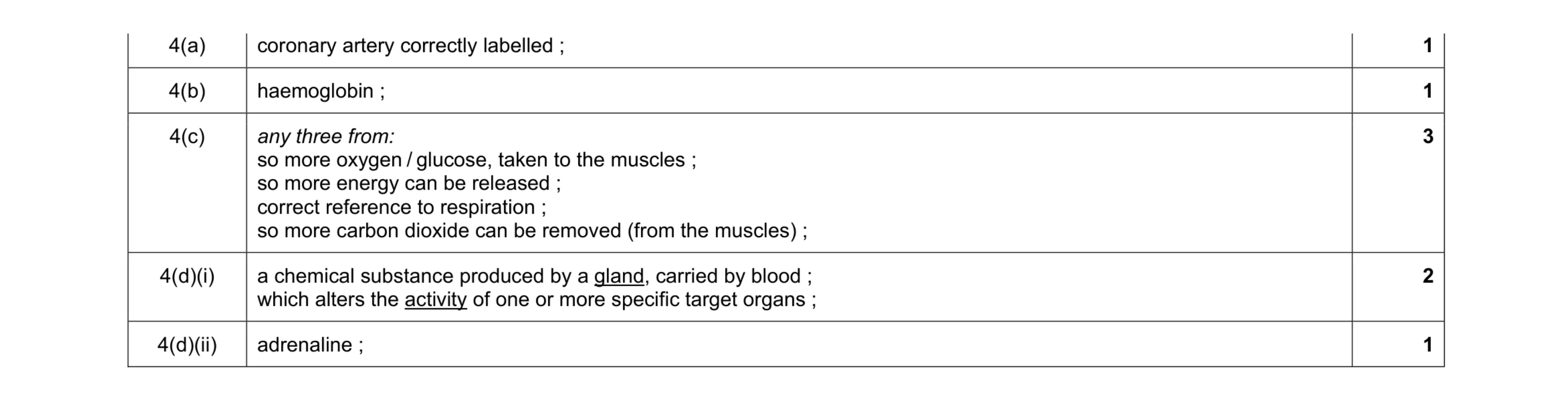 8
8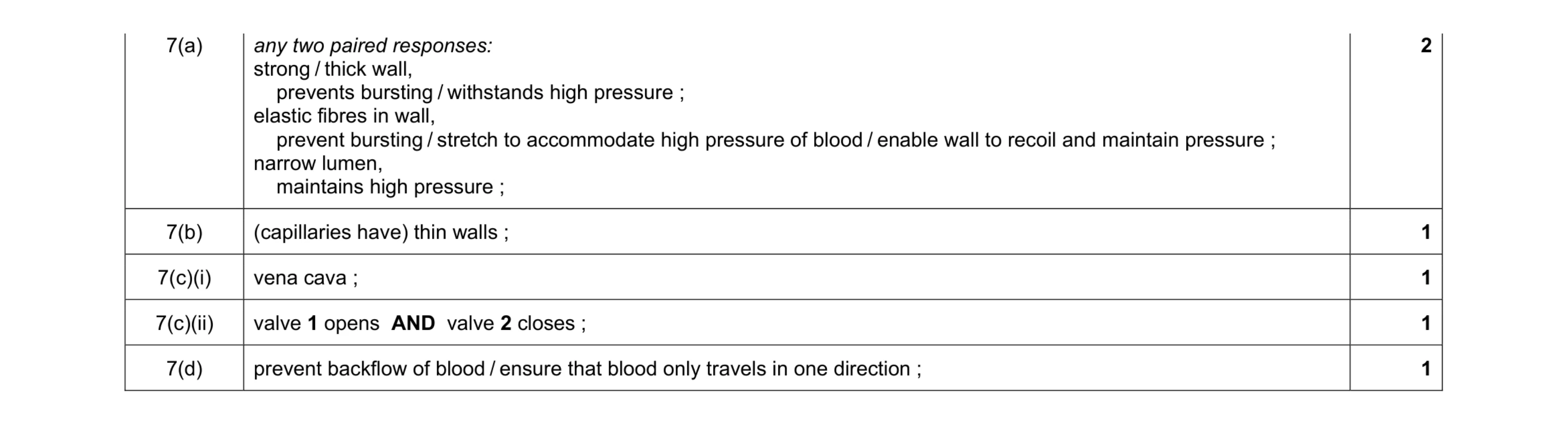 6
6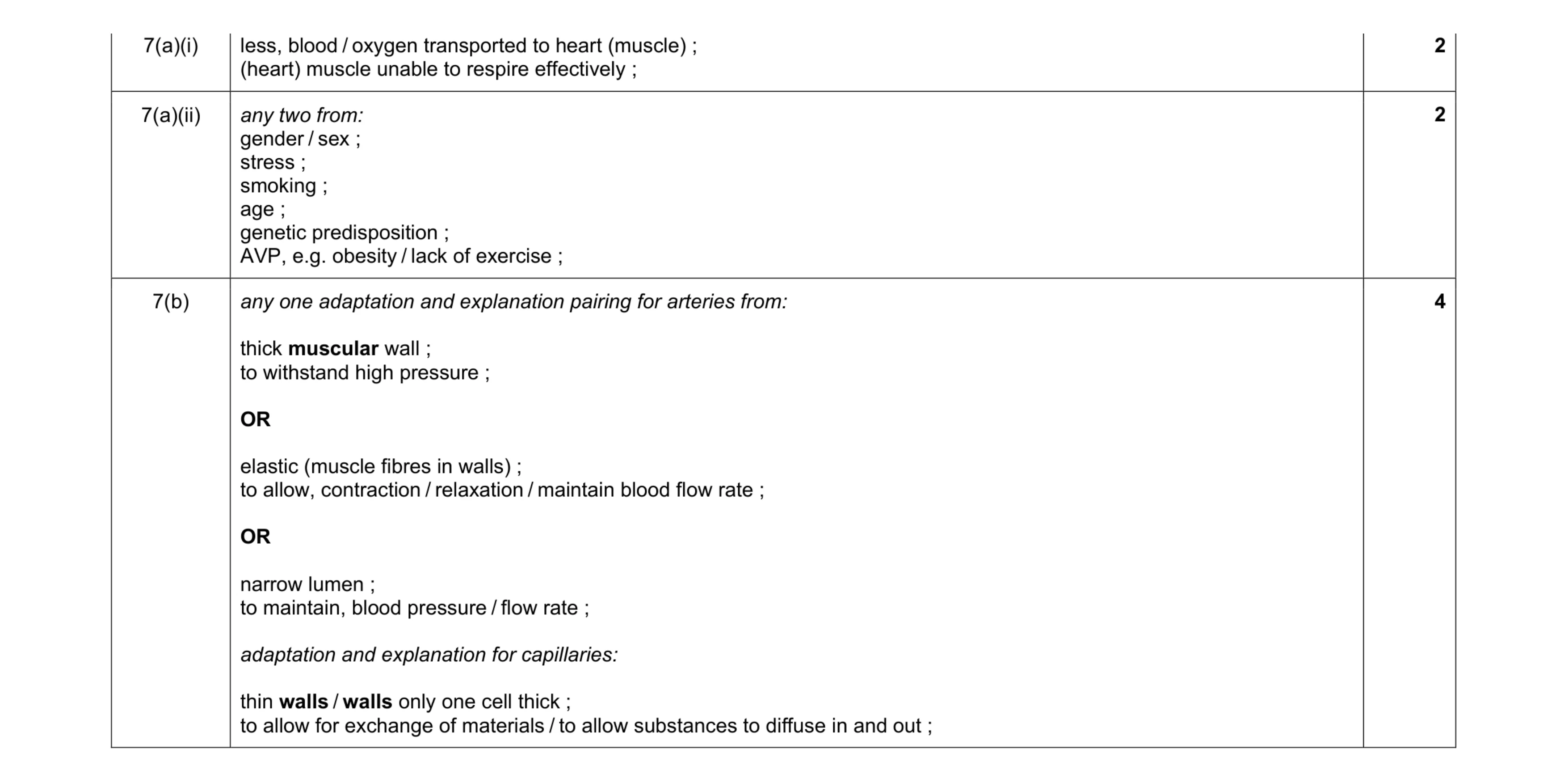 8
8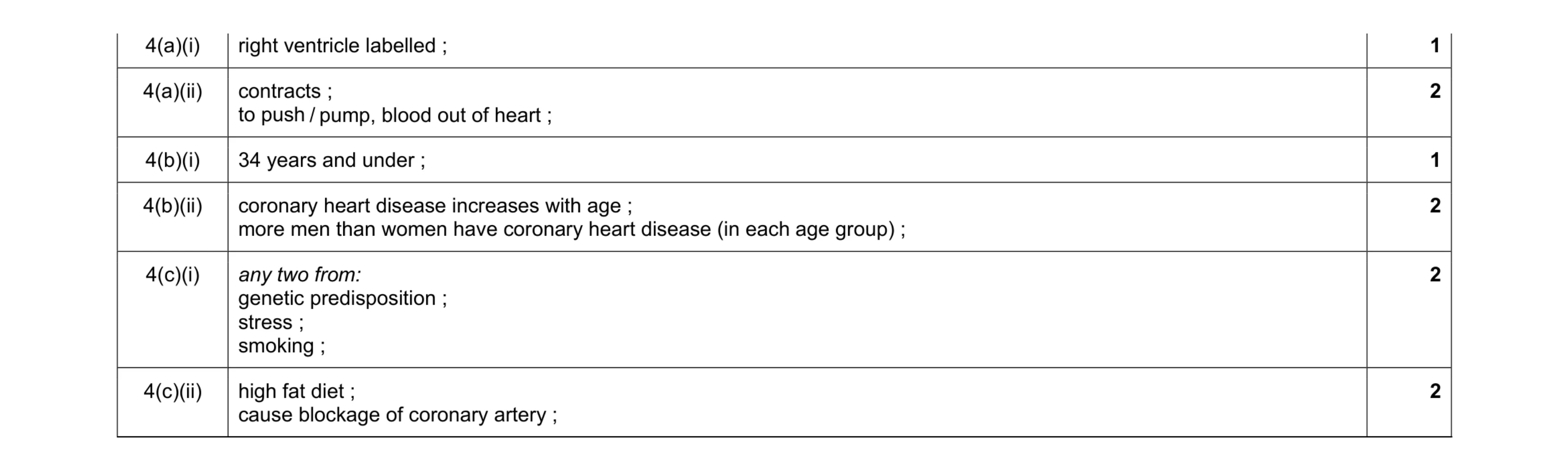 10
10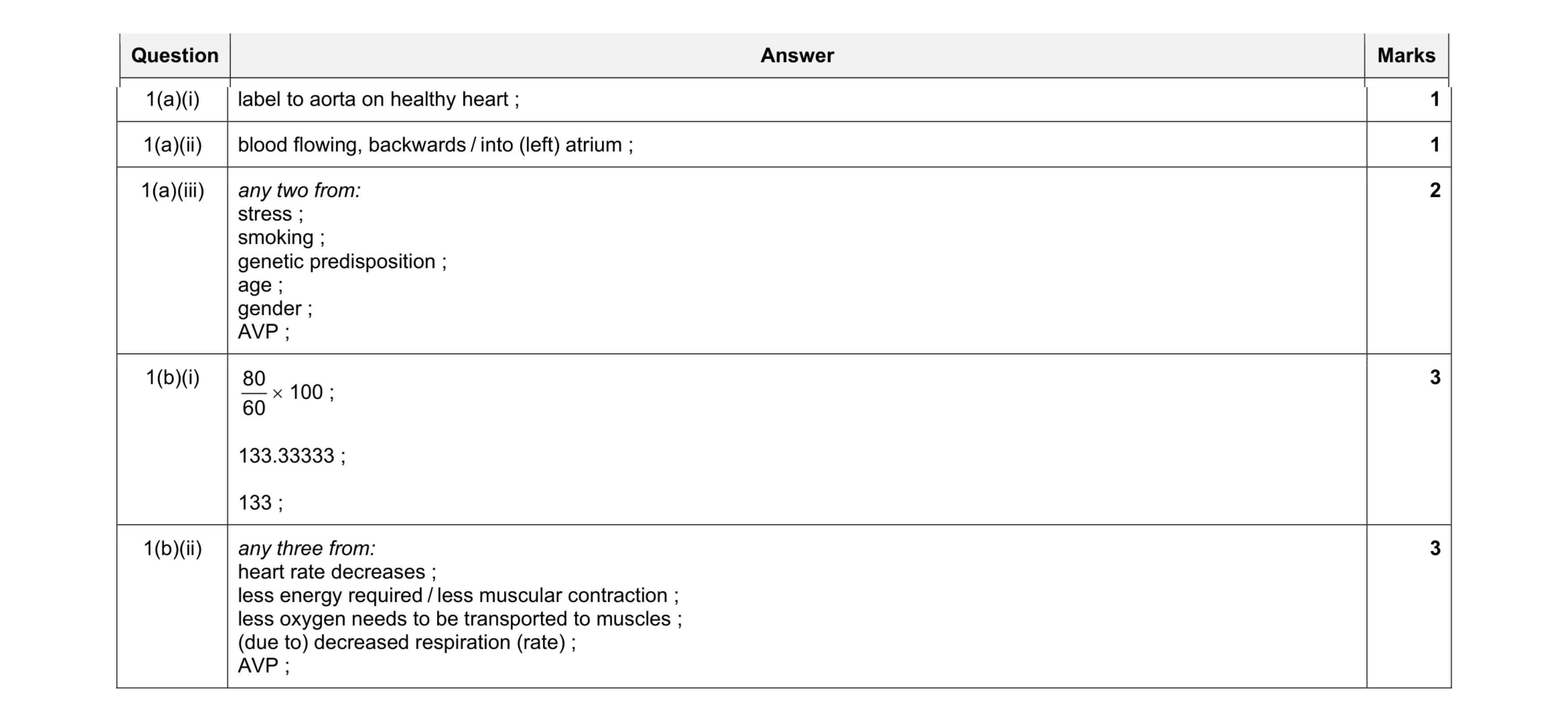 10
10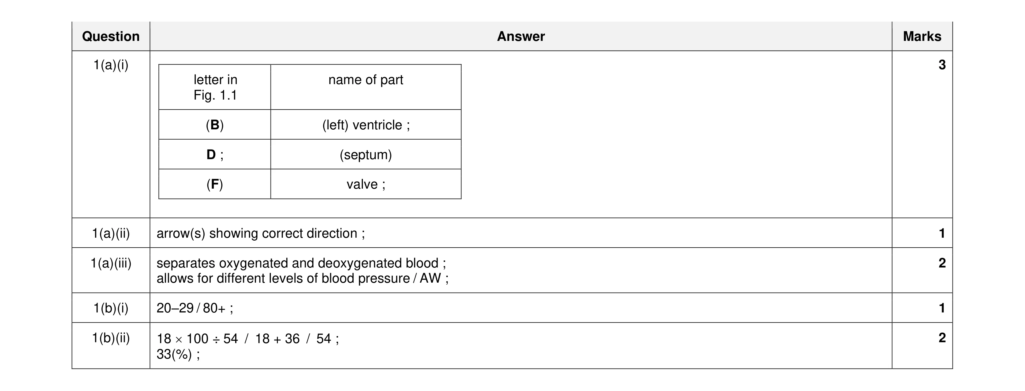 9
9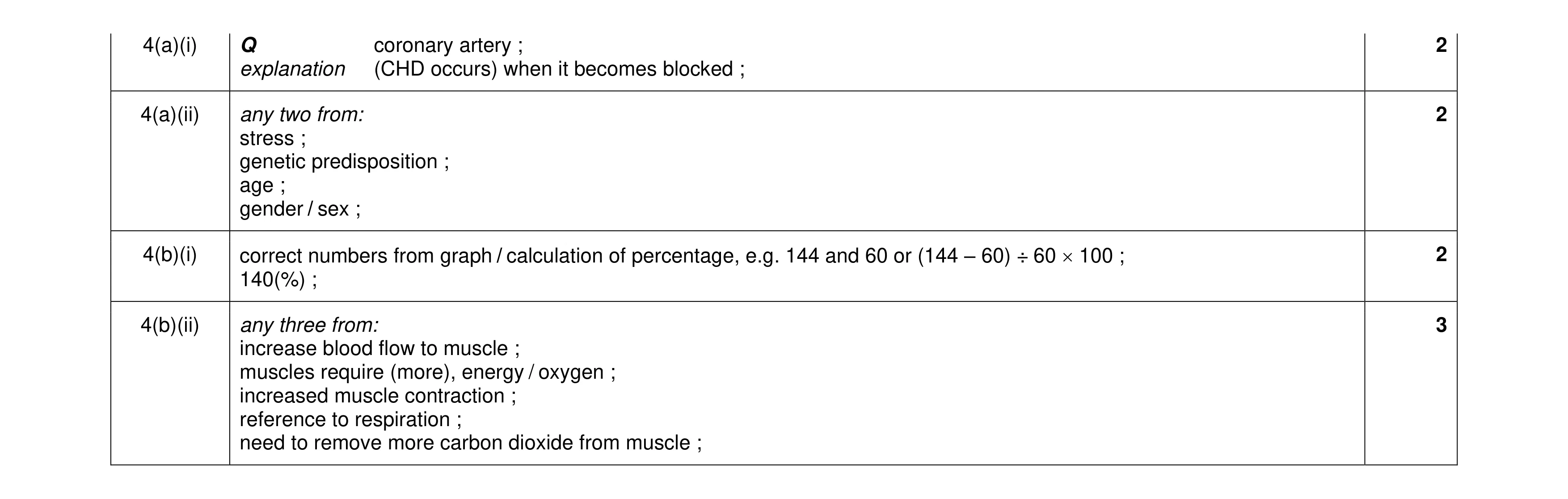 9
9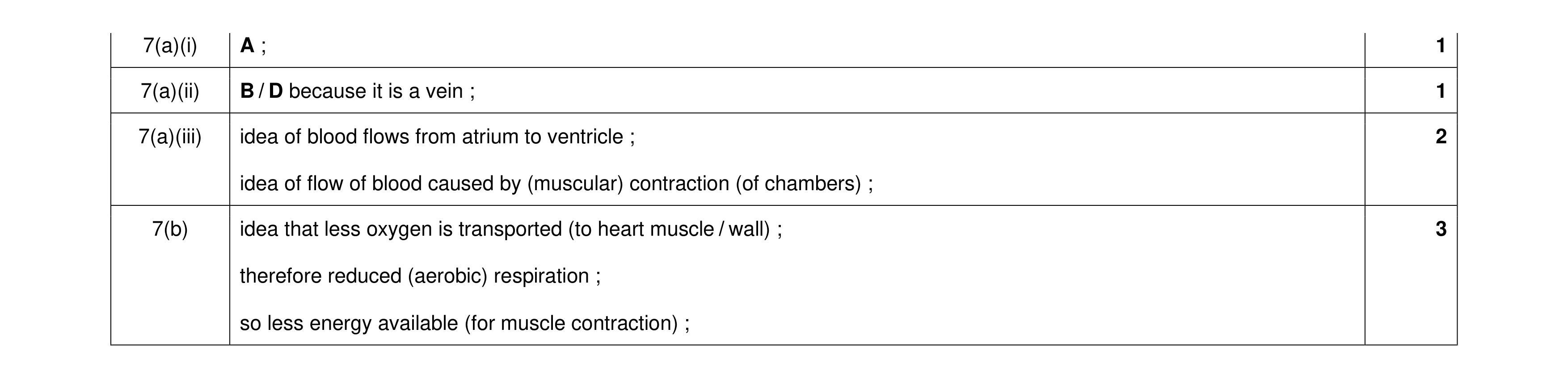 7
7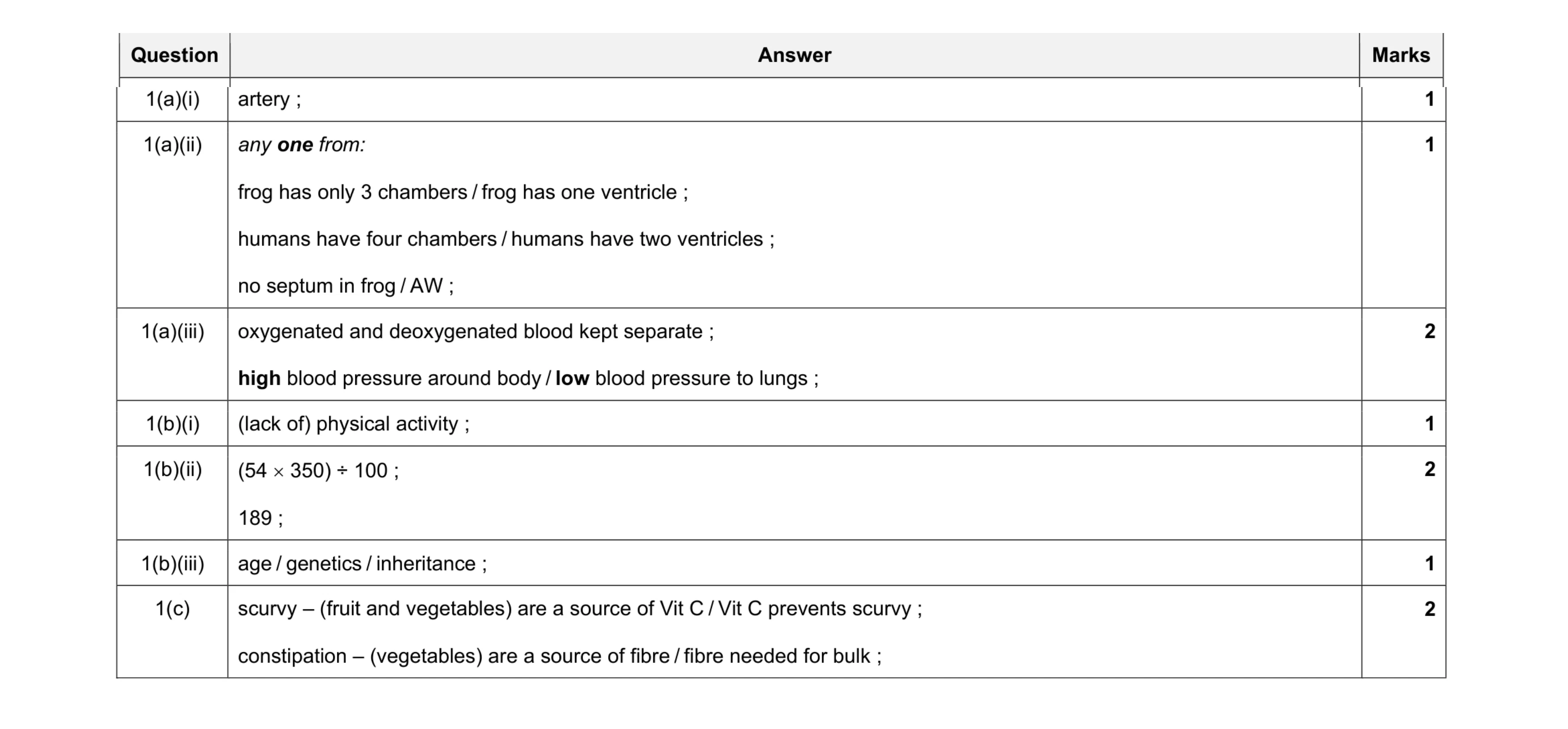 10
10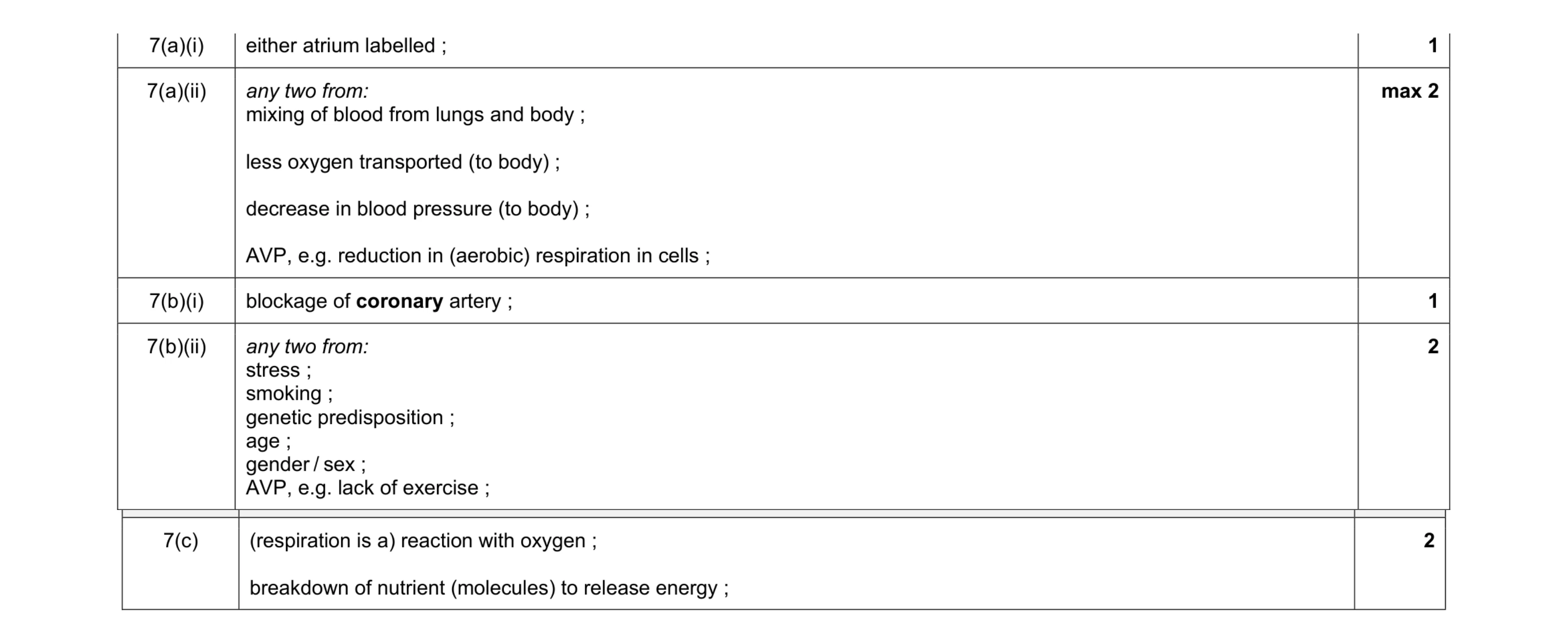 8
8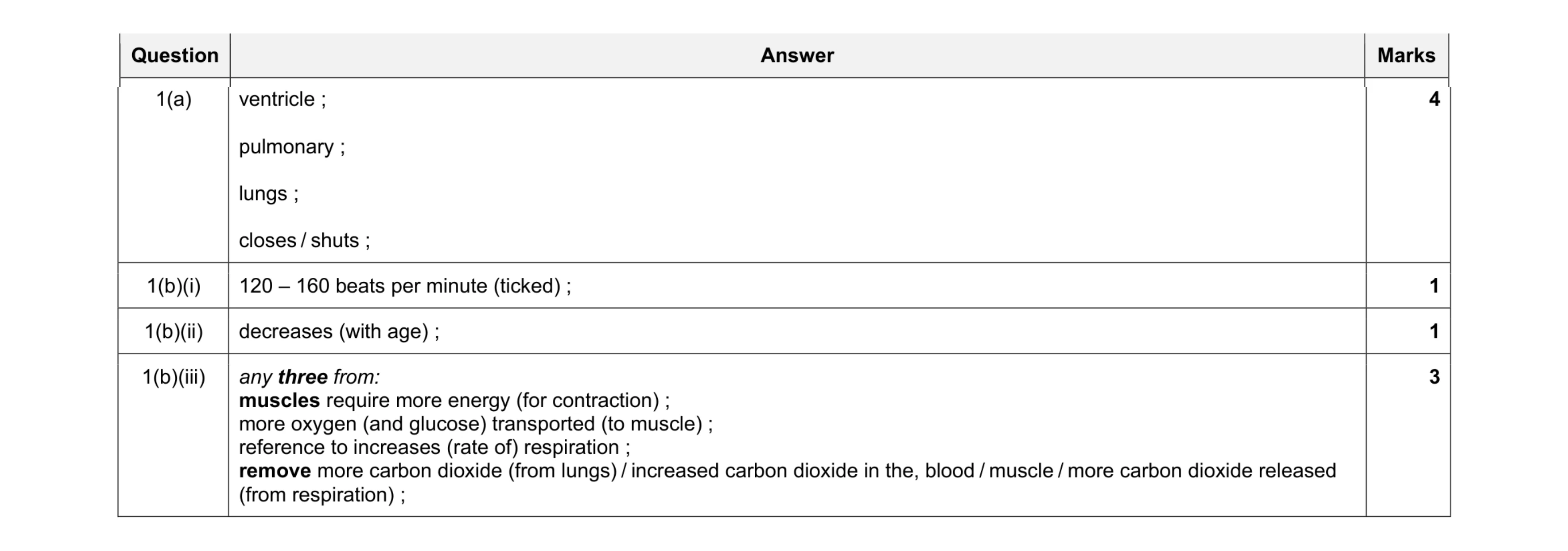 9
9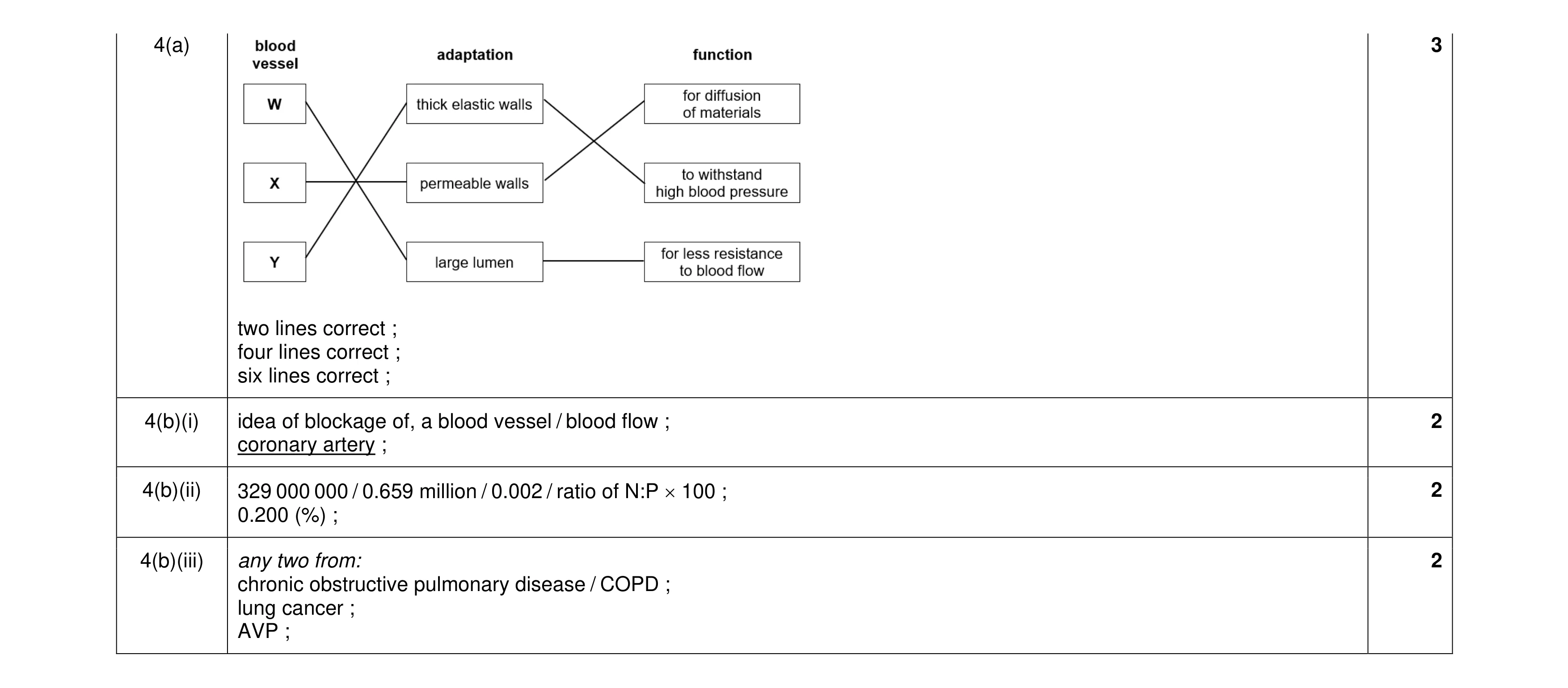 9
9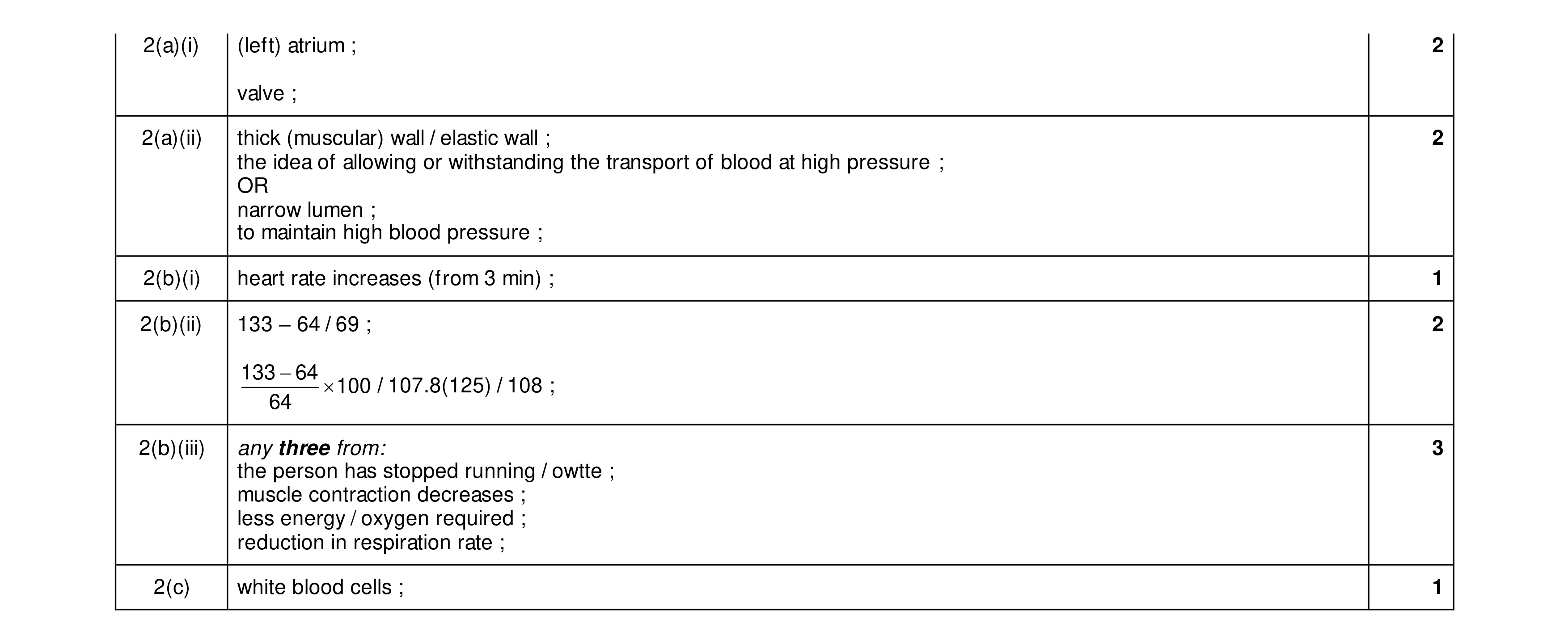 11
11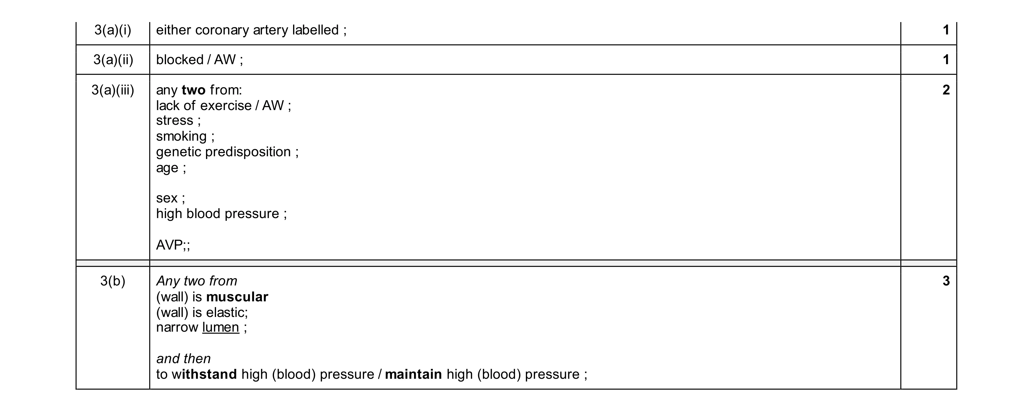 7
7