Questions as text
Q1 · A diagram of the internal structure of the heart 0653/32 May/June 2017
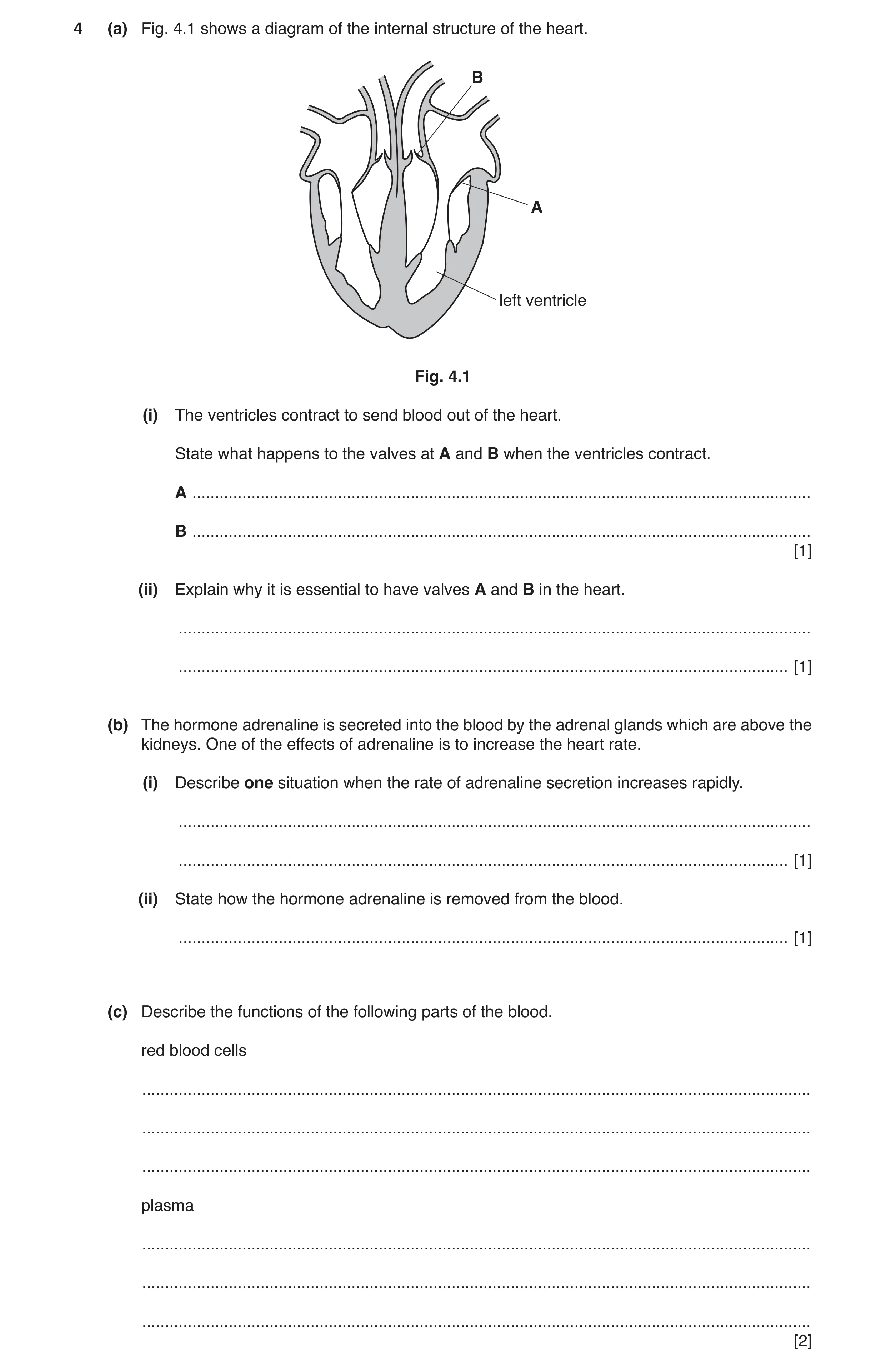
4 (a) Fig. 4.1 shows a diagram of the internal structure of the heart. B A left ventricle Fig. 4.1 (i) The ventricles contract to send blood out of the heart. State what happens to the valves at A and B when the ventricles contract. A … B … [1] (ii) Explain why it is essential to have valves A and B in the heart. … … [1] (b) The hormone adrenaline is secreted into the blood by the adrenal glands which are above the kidneys. One of the effects of adrenaline is to increase the heart rate. (i) Describe one situation when the rate of adrenaline secretion increases rapidly. … … [1] (ii) State how the hormone adrenaline is removed from the blood. … [1] (c) Describe the functions of the following parts of the blood. red blood cells … … … plasma … … … [2]
6 marks
Mark scheme: 4(a)(i) A closes and B opens ; 1 4(a)(ii) to prevent backflow of blood ; 1 4(b)(i) any suitable flight or fight situation described ; 1 4(b)(ii) destroyed by the liver ; 1 4(c) transport of oxygen / haemoglobin ; transport of blood cells / ions / soluble nutrients / named soluble nutrient / hormones / carbon dioxide ; 2
This question in 0653/32 May/June 2017
Q2 · A diagram of the internal structure of the heart 0653/33 May/June 2017
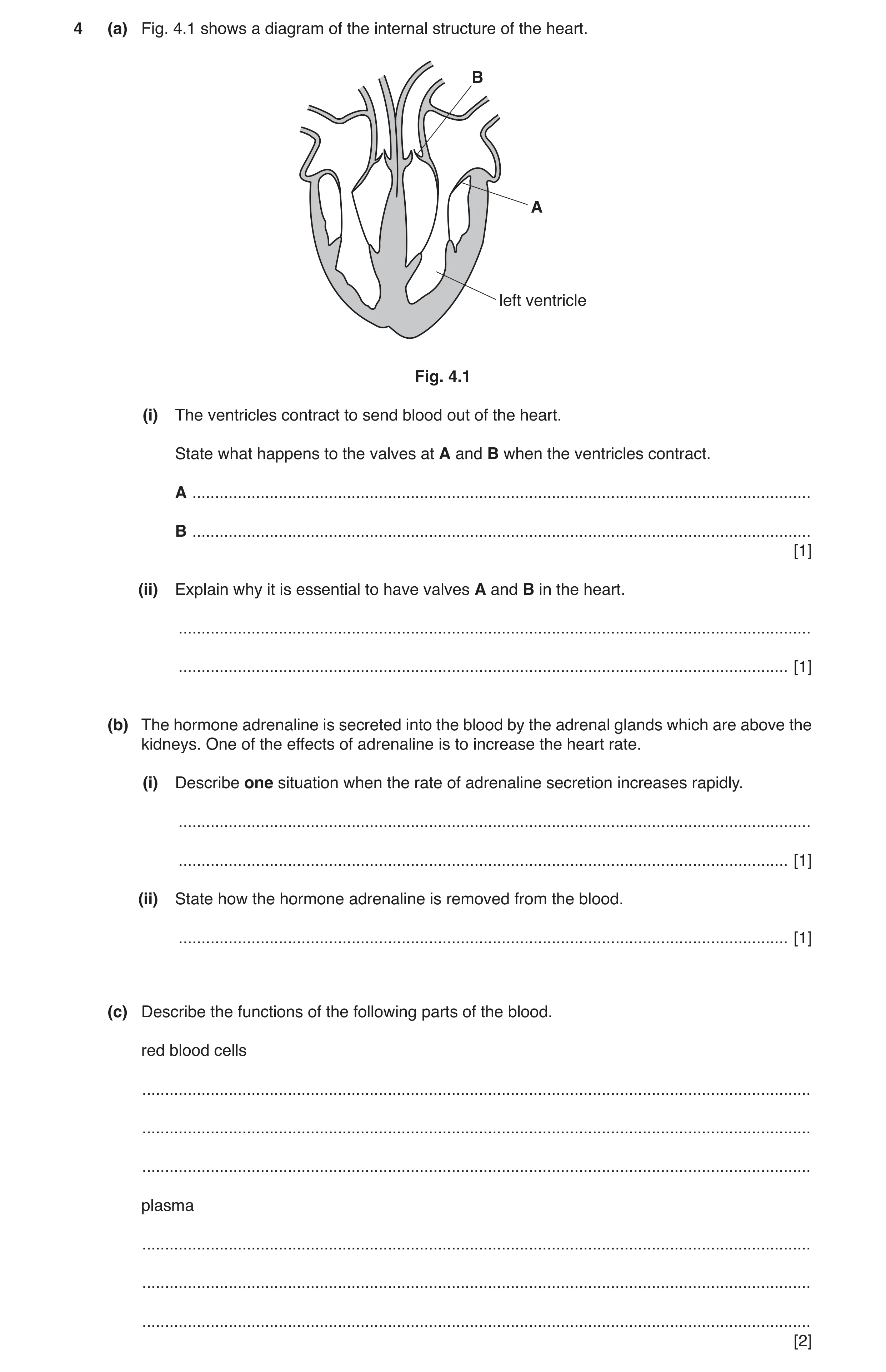
4 (a) Fig. 4.1 shows a diagram of the internal structure of the heart. B A left ventricle Fig. 4.1 (i) The ventricles contract to send blood out of the heart. State what happens to the valves at A and B when the ventricles contract. A … B … [1] (ii) Explain why it is essential to have valves A and B in the heart. … … [1] (b) The hormone adrenaline is secreted into the blood by the adrenal glands which are above the kidneys. One of the effects of adrenaline is to increase the heart rate. (i) Describe one situation when the rate of adrenaline secretion increases rapidly. … … [1] (ii) State how the hormone adrenaline is removed from the blood. … [1] (c) Describe the functions of the following parts of the blood. red blood cells … … … plasma … … … [2]
6 marks
Mark scheme: 4(a)(i) A closes and B opens ; 1 4(a)(ii) to prevent backflow of blood ; 1 4(b)(i) any suitable flight or fight situation described ; 1 4(b)(ii) destroyed by the liver ; 1 4(c) transport of oxygen / haemoglobin ; transport of blood cells / ions / soluble nutrients / named soluble nutrient / hormones / carbon dioxide ; 2
This question in 0653/33 May/June 2017
Q3 · A diagram of the internal structure of the heart 0653/32 Feb/March 2019
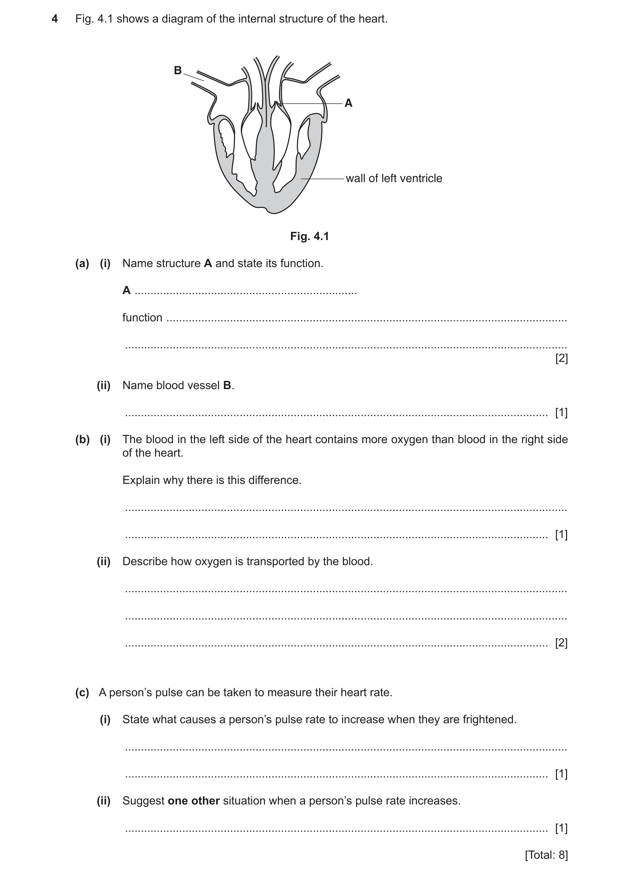
4 Fig. 4.1 shows a diagram of the internal structure of the heart. B A wall of left ventricle Fig. 4.1 (a) (i) Name structure A and state its function. A … function … … [2] (ii) Name blood vessel B. … [1] (b) (i) The blood in the left side of the heart contains more oxygen than blood in the right side of the heart. Explain why there is this difference. … … [1] (ii) Describe how oxygen is transported by the blood. … … … [2] (c) A person’s pulse can be taken to measure their heart rate. (i) State what causes a person’s pulse rate to increase when they are frightened. … … [1] (ii) Suggest one other situation when a person’s pulse rate increases. … [1] [Total: 8]
8 marks
Mark scheme: 4(a)(i) (one-way) valve ; prevents back flow of blood / ensures blood flows in one direction ; 2 4(a)(ii) vena cava ; 1 4(b)(i) oxygen is picked up at the lungs ; 1 4(b)(ii) in red blood cells ; by haemoglobin ; 2 4(c)(i) adrenaline is released ; 1 4(c)(ii) when exercising ; 1
This question in 0653/32 Feb/March 2019
Q4 · A diagram of a human heart 0653/32 Feb/March 2020
1 (a) Fig. 1.1 is a diagram of a human heart. Y X Fig. 1.1 (i) Name artery X shown in Fig. 1.1. … [1] (ii) Vein Y brings blood to the heart from the body. Name vein Y shown in Fig. 1.1. … [1] (iii) Name the structures that make sure there is one-way flow of blood through the heart. … [1] (iv) Fig. 1.2 is a diagram of the cross-section of three different blood vessels, P, Q and R. P Q R Fig. 1.2 Identify which blood vessel, P, Q or R, represents a vein. Explain your answer. blood vessel P, Q or R … explanation … … [2] (b) Fig. 1.3 shows the pulse rate of two students before, during and after exercise. recovery time 140 120 100 pulse rate 80 student A(beats per minute) 60 student B recovery 40 time 20 0 before exercise exercise after exercise time Fig. 1.3 Describe one similarity and two differences in the pulse rates of students A and B shown in Fig. 1.3. similarity … … difference 1 … … difference 2 … … [3] [Total: 8]
8 marks
Mark scheme: 1(a)(i) coronary ; 1 1(a)(ii) vena cava ; 1 1(a)(iii) valves ; 1 1(a)(iv) Q ; and any one from: has a large lumen ; has a thin (muscle) wall ; 2 1(b) (similarity) any one from: pulse rates increase during exercise ; pulse rates decrease after exercise ; pulse rates return, to normal / to rate before exercise ; (differences) any two from: Student A has higher pulse rate (before or during or after exercise) / ora ; Student A has higher increase in pulse rate (during exercise) / ora ; Student A has longer recovery time / ora ; 3
This question in 0653/32 Feb/March 2020
Q5 · The structure of a human heart 0653/31 May/June 2020
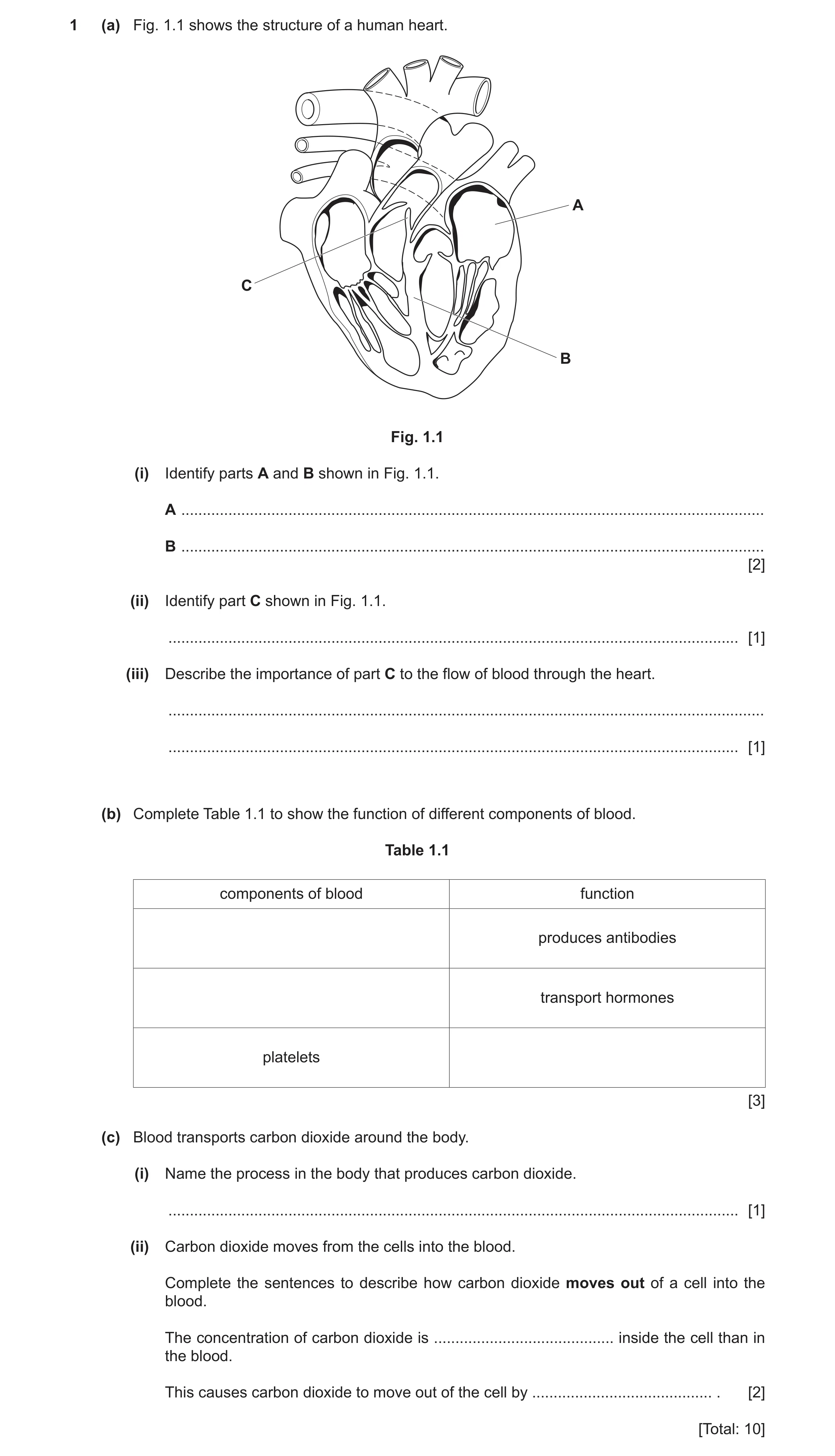
1 (a) Fig. 1.1 shows the structure of a human heart. A C B Fig. 1.1 (i) Identify parts A and B shown in Fig. 1.1. A … B … [2] (ii) Identify part C shown in Fig. 1.1. … [1] (iii) Describe the importance of part C to the flow of blood through the heart. … … [1] (b) Complete Table 1.1 to show the function of different components of blood. Table 1.1 components of blood function produces antibodies transport hormones platelets [3] (c) Blood transports carbon dioxide around the body. (i) Name the process in the body that produces carbon dioxide. … [1] (ii) Carbon dioxide moves from the cells into the blood. Complete the sentences to describe how carbon dioxide moves out of a cell into the blood. The concentration of carbon dioxide is … inside the cell than in the blood. This causes carbon dioxide to move out of the cell by … . [2] [Total: 10]
10 marks
Mark scheme: 1(a)(i) A (left) atrium ; B septum ; 2 1(a)(ii) valve ; 1 1(a)(iii) makes sure the blood flows in one direction / prevents backflow ; 1 1(b) white blood cells (produces antibodies) ; plasma (transports hormones) ; (platelets) blood clotting ; 3 1(c)(i) respiration ; 1 1(c)(ii) higher / greater ; diffusion ; 2
This question in 0653/31 May/June 2020
Q6 · A diagram of muscle cells 0653/33 Oct/Nov 2020
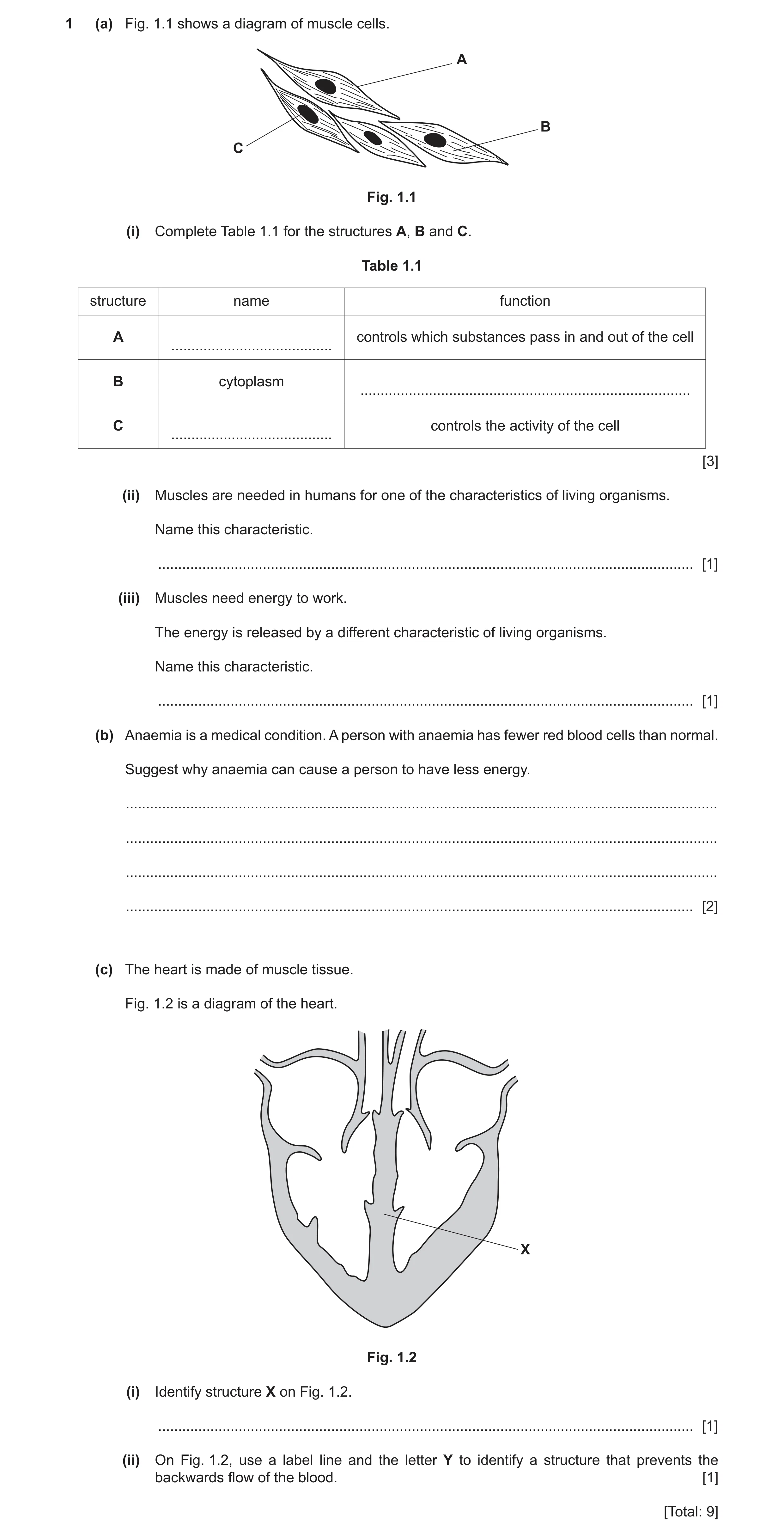
1 (a) Fig. 1.1 shows a diagram of muscle cells. A B C Fig. 1.1 (i) Complete Table 1.1 for the structures A, B and C. Table 1.1 structure name function A controls which substances pass in and out of the cell … B cytoplasm … C controls the activity of the cell … [3] (ii) Muscles are needed in humans for one of the characteristics of living organisms. Name this characteristic. … [1] (iii) Muscles need energy to work. The energy is released by a different characteristic of living organisms. Name this characteristic. … [1] (b) Anaemia is a medical condition. A person with anaemia has fewer red blood cells than normal. Suggest why anaemia can cause a person to have less energy. … … … … [2] (c) The heart is made of muscle tissue. Fig. 1.2 is a diagram of the heart. X Fig. 1.2 (i) Identify structure X on Fig. 1.2. … [1] (ii) On Fig. 1.2, use a label line and the letter Y to identify a structure that prevents the backwards flow of the blood. [1] [Total: 9]
9 marks
Mark scheme: 1(a)(i) (cell) membrane ; site of different (metabolic) reactions ; nucleus ; 3 1(a)(ii) movement ; 1 1(a)(iii) respiration ; 1 1(b) any two from: red blood cells transport oxygen ; less oxygen, transported / available ; for (aerobic) respiration / less (aerobic) respiration OR oxygen is needed for respiration ; 2 1(c)(i) septum ; 1 1(c)(ii) label line to any valve ; 1
This question in 0653/33 Oct/Nov 2020
Q7 · A diagram of the human heart 0653/32 May/June 2021
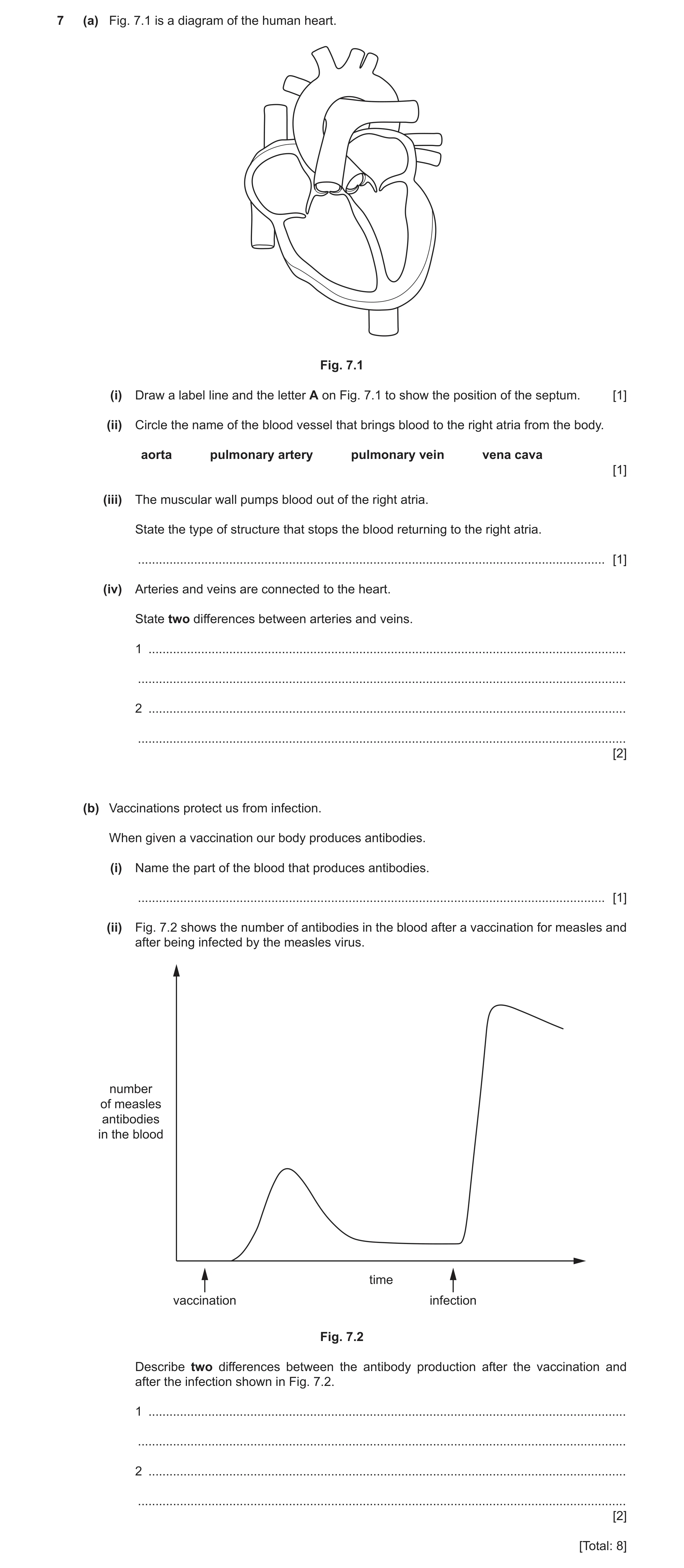
7 (a) Fig. 7.1 is a diagram of the human heart. Fig. 7.1 (i) Draw a label line and the letter A on Fig. 7.1 to show the position of the septum. [1] (ii) Circle the name of the blood vessel that brings blood to the right atria from the body. aorta pulmonary artery pulmonary vein vena cava [1] (iii) The muscular wall pumps blood out of the right atria. State the type of structure that stops the blood returning to the right atria. … [1] (iv) Arteries and veins are connected to the heart. State two differences between arteries and veins. 1 … … 2 … … [2] (b) Vaccinations protect us from infection. When given a vaccination our body produces antibodies. (i) Name the part of the blood that produces antibodies. … [1] (ii) Fig. 7.2 shows the number of antibodies in the blood after a vaccination for measles and after being infected by the measles virus. number of measles antibodies in the blood time vaccination infection Fig. 7.2 Describe two differences between the antibody production after the vaccination and after the infection shown in Fig. 7.2. 1 … … 2 … … [2] [Total: 8]
8 marks
Mark scheme: 7(a)(i) line touching septum ; 1 7(a)(ii) vena cava ; 1 7(a)(iii) valve ; 1 7(a)(iv) any two from: arteries have thicker (muscular) walls ORA ; arteries have smaller, cavity ORA ; arteries take blood away from heart / veins bring blood to heart ; veins have valves (arteries do not) ; 2 7(b)(i) white blood cells ; 1 7(b)(ii) any two from: fewer antibodies made after vaccination (than after infection) ORA ; antibodies reach maximum quicker after infection ORA ; antibody numbers remain higher for longer after infection ; 2
This question in 0653/32 May/June 2021
Q8 · A diagram of the circulatory system in humans 0653/31 Oct/Nov 2021
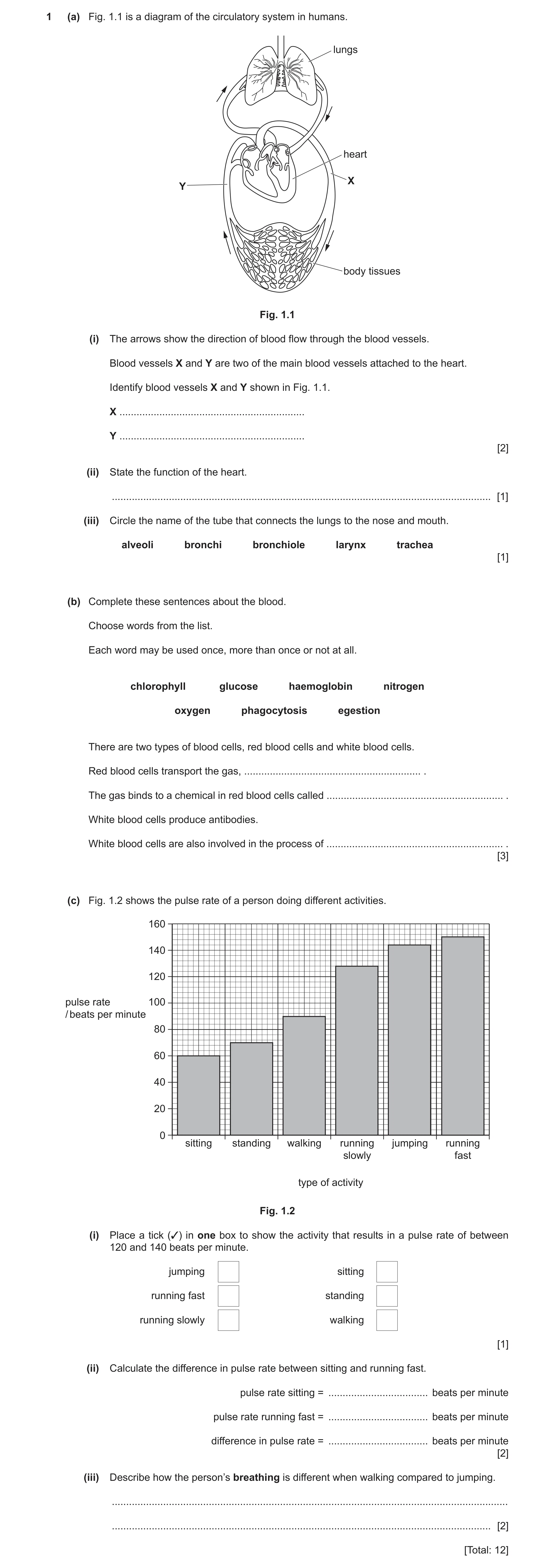
1 (a) Fig. 1.1 is a diagram of the circulatory system in humans. lungs heart X Y body tissues Fig. 1.1 (i) The arrows show the direction of blood flow through the blood vessels. Blood vessels X and Y are two of the main blood vessels attached to the heart. Identify blood vessels X and Y shown in Fig. 1.1. X … Y … [2] (ii) State the function of the heart. … [1] (iii) Circle the name of the tube that connects the lungs to the nose and mouth. alveoli bronchi bronchiole larynx trachea [1] (b) Complete these sentences about the blood. Choose words from the list. Each word may be used once, more than once or not at all. chlorophyll glucose haemoglobin nitrogen oxygen phagocytosis egestion There are two types of blood cells, red blood cells and white blood cells. Red blood cells transport the gas, … . The gas binds to a chemical in red blood cells called … . White blood cells produce antibodies. White blood cells are also involved in the process of … . [3] (c) Fig. 1.2 shows the pulse rate of a person doing different activities. 160 140 120 pulse rate 100 / beats per minute 80 60 40 20 0 sitting standing walking running jumping running slowly fast type of activity Fig. 1.2 (i) Place a tick (3) in one box to show the activity that results in a pulse rate of between 120 and 140 beats per minute. jumping sitting running fast standing running slowly walking [1] (ii) Calculate the difference in pulse rate between sitting and running fast. pulse rate sitting = … beats per minute pulse rate running fast = … beats per minute difference in pulse rate = … beats per minute [2] (iii) Describe how the person’s breathing is different when walking compared to jumping. … … [2] [Total: 12]
12 marks
Mark scheme: 1(a)(i) aorta ; vena cava ; 2 1(a)(ii) pump blood ; 1 1(a)(iii) trachea ; 1 1(b) oxygen ; haemoglobin ; phagocytosis ; 3 1(c)(i) running slowly box ticked ; 1 1(c)(ii) reading values from graph 60 and 150 ; correct subtraction / 90 ; 2 1(c)(iii) lower breathing rate / AW ; lesser depth of breathing / AW ; 2
This question in 0653/31 Oct/Nov 2021
Q9 · A biology teacher measures the pulse rate of five students A–E before and after exercise 0653/32 Oct/Nov 2021
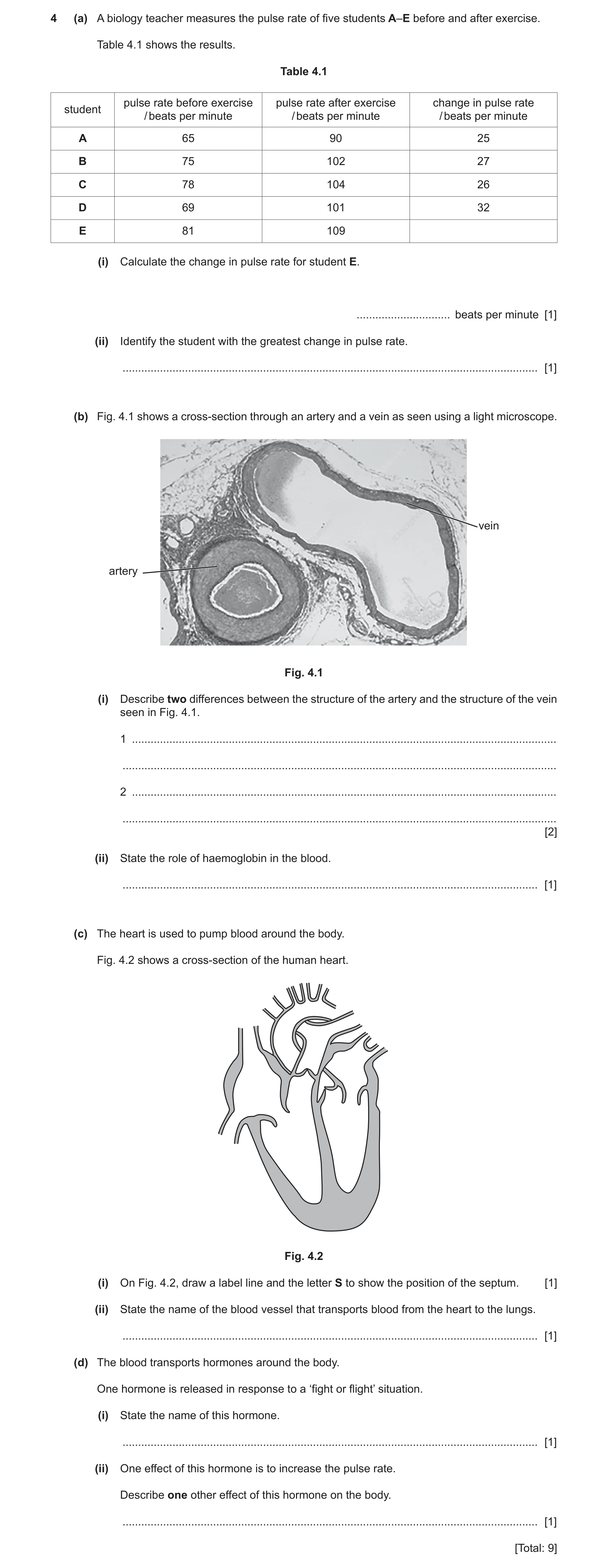
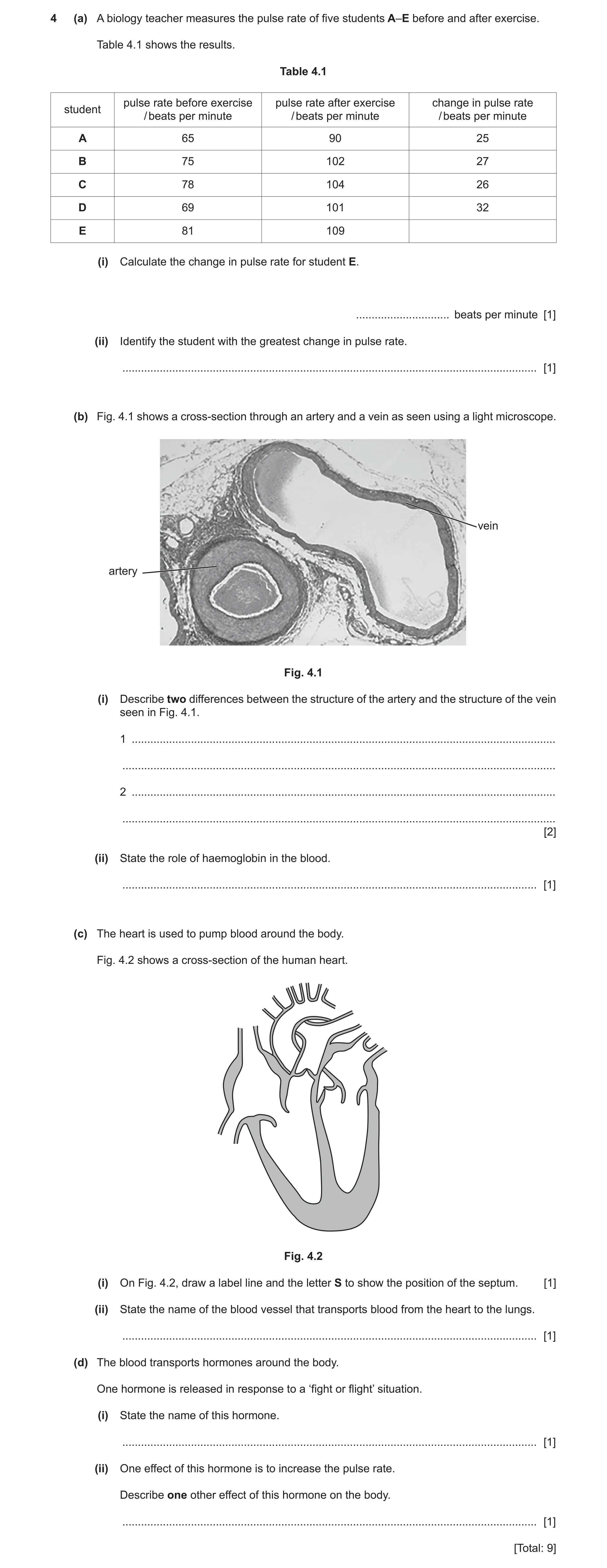
4 (a) A biology teacher measures the pulse rate of five students A–E before and after exercise. Table 4.1 shows the results. Table 4.1 pulse rate before exercise pulse rate after exercise change in pulse rate student / beats per minute / beats per minute / beats per minute A 65 90 25 B 75 102 27 C 78 104 26 D 69 101 32 E 81 109 (i) Calculate the change in pulse rate for student E. … beats per minute [1] (ii) Identify the student with the greatest change in pulse rate. … [1] (b) Fig. 4.1 shows a cross-section through an artery and a vein as seen using a light microscope. vein artery Fig. 4.1 (i) Describe two differences between the structure of the artery and the structure of the vein seen in Fig. 4.1. 1 … … 2 … … [2] (ii) State the role of haemoglobin in the blood. … [1] (c) The heart is used to pump blood around the body. Fig. 4.2 shows a cross-section of the human heart. Fig. 4.2 (i) On Fig. 4.2, draw a label line and the letter S to show the position of the septum. [1] (ii) State the name of the blood vessel that transports blood from the heart to the lungs. … [1] (d) The blood transports hormones around the body. One hormone is released in response to a ‘fight or flight’ situation. (i) State the name of this hormone. … [1] (ii) One effect of this hormone is to increase the pulse rate. Describe one other effect of this hormone on the body. … [1] [Total: 9]
9 marks
Mark scheme: 4(a)(i) 28 ; 1 4(a)(ii) D ; 1 4(b)(i) artery has a smaller internal area / ORA ; artery has a thicker (muscular) wall / ORA ; 2 4b(ii) transport oxygen ; 1 4(c)(i) label line in septum ; 1 4(c)(ii) pulmonary artery ; 1 4(d)(i) adrenaline ; 1 4(d)(ii) increase breathing rate / widen pupils ; 1
This question in 0653/32 Oct/Nov 2021
Q10 · A biology teacher measures the pulse rate of five students A–E before and after exercise 0653/33 Oct/Nov 2021
4 (a) A biology teacher measures the pulse rate of five students A–E before and after exercise. Table 4.1 shows the results. Table 4.1 pulse rate before exercise pulse rate after exercise change in pulse rate student / beats per minute / beats per minute / beats per minute A 65 90 25 B 75 102 27 C 78 104 26 D 69 101 32 E 81 109 (i) Calculate the change in pulse rate for student E. … beats per minute [1] (ii) Identify the student with the greatest change in pulse rate. … [1] (b) Fig. 4.1 shows a cross-section through an artery and a vein as seen using a light microscope. vein artery Fig. 4.1 (i) Describe two differences between the structure of the artery and the structure of the vein seen in Fig. 4.1. 1 … … 2 … … [2] (ii) State the role of haemoglobin in the blood. … [1] (c) The heart is used to pump blood around the body. Fig. 4.2 shows a cross-section of the human heart. Fig. 4.2 (i) On Fig. 4.2, draw a label line and the letter S to show the position of the septum. [1] (ii) State the name of the blood vessel that transports blood from the heart to the lungs. … [1] (d) The blood transports hormones around the body. One hormone is released in response to a ‘fight or flight’ situation. (i) State the name of this hormone. … [1] (ii) One effect of this hormone is to increase the pulse rate. Describe one other effect of this hormone on the body. … [1] [Total: 9]
9 marks
Mark scheme: 4(a)(i) 28 ; 1 4(a)(ii) D ; 1 4(b)(i) artery has a smaller internal area / ORA ; artery has a thicker (muscular) wall / ORA ; 2 4b(ii) transport oxygen ; 1 4(c)(i) label line in septum ; 1 4(c)(ii) pulmonary artery ; 1 4(d)(i) adrenaline ; 1 4(d)(ii) increase breathing rate / widen pupils ; 1
This question in 0653/33 Oct/Nov 2021
Q11 · The boxes on the left show some of the components of blood 0653/32 Feb/March 2022
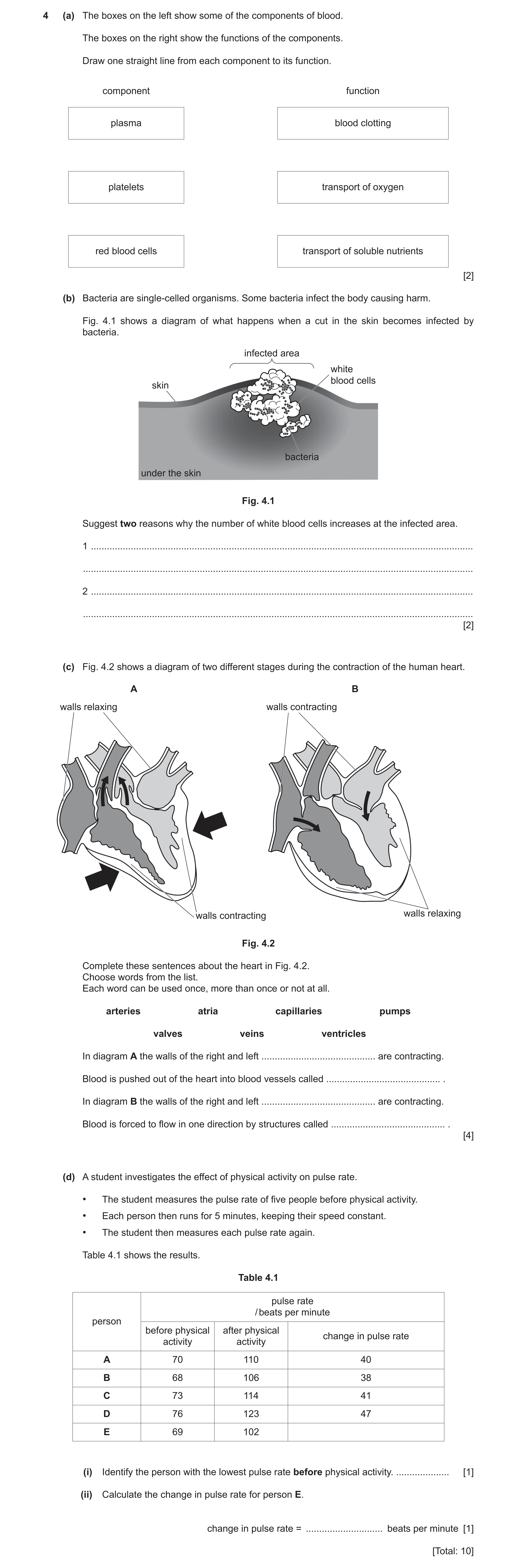
4 (a) The boxes on the left show some of the components of blood. The boxes on the right show the functions of the components. Draw one straight line from each component to its function. component function plasma blood clotting platelets transport of oxygen red blood cells transport of soluble nutrients [2] (b) Bacteria are single-celled organisms. Some bacteria infect the body causing harm. Fig. 4.1 shows a diagram of what happens when a cut in the skin becomes infected by bacteria. infected area white blood cells skin bacteria under the skin Fig. 4.1 Suggest two reasons why the number of white blood cells increases at the infected area. 1 … … 2 … … [2] (c) Fig. 4.2 shows a diagram of two different stages during the contraction of the human heart. A B walls relaxing walls contracting walls contracting walls relaxing Fig. 4.2 Complete these sentences about the heart in Fig. 4.2. Choose words from the list. Each word can be used once, more than once or not at all. arteries atria capillaries pumps valves veins ventricles In diagram A the walls of the right and left … are contracting. Blood is pushed out of the heart into blood vessels called … . In diagram B the walls of the right and left … are contracting. Blood is forced to flow in one direction by structures called … . [4] (d) A student investigates the effect of physical activity on pulse rate. • The student measures the pulse rate of five people before physical activity. • Each person then runs for 5 minutes, keeping their speed constant. • The student then measures each pulse rate again. Table 4.1 shows the results. Table 4.1 pulse rate / beats per minute person before physical after physical change in pulse rate activity activity A 70 110 40 B 68 106 38 C 73 114 41 D 76 123 47 E 69 102 (i) Identify the person with the lowest pulse rate before physical activity. … [1] (ii) Calculate the change in pulse rate for person E. change in pulse rate = … beats per minute [1] [Total: 10]
10 marks
Mark scheme: 4(a) plasma blood clotting platelets transport of oxygen red blood cells transport of soluble nutrients ;; 4(b) phagocytosis ; to produce antibodies ; 2 4(c) ventricles ; arteries ; atria ; valves ; 4 4(d)(i) B ; 1 4(d)(ii) 33 (bpm) ; 1
This question in 0653/32 Feb/March 2022
Q12 · A diagram of the human heart 0653/33 May/June 2022
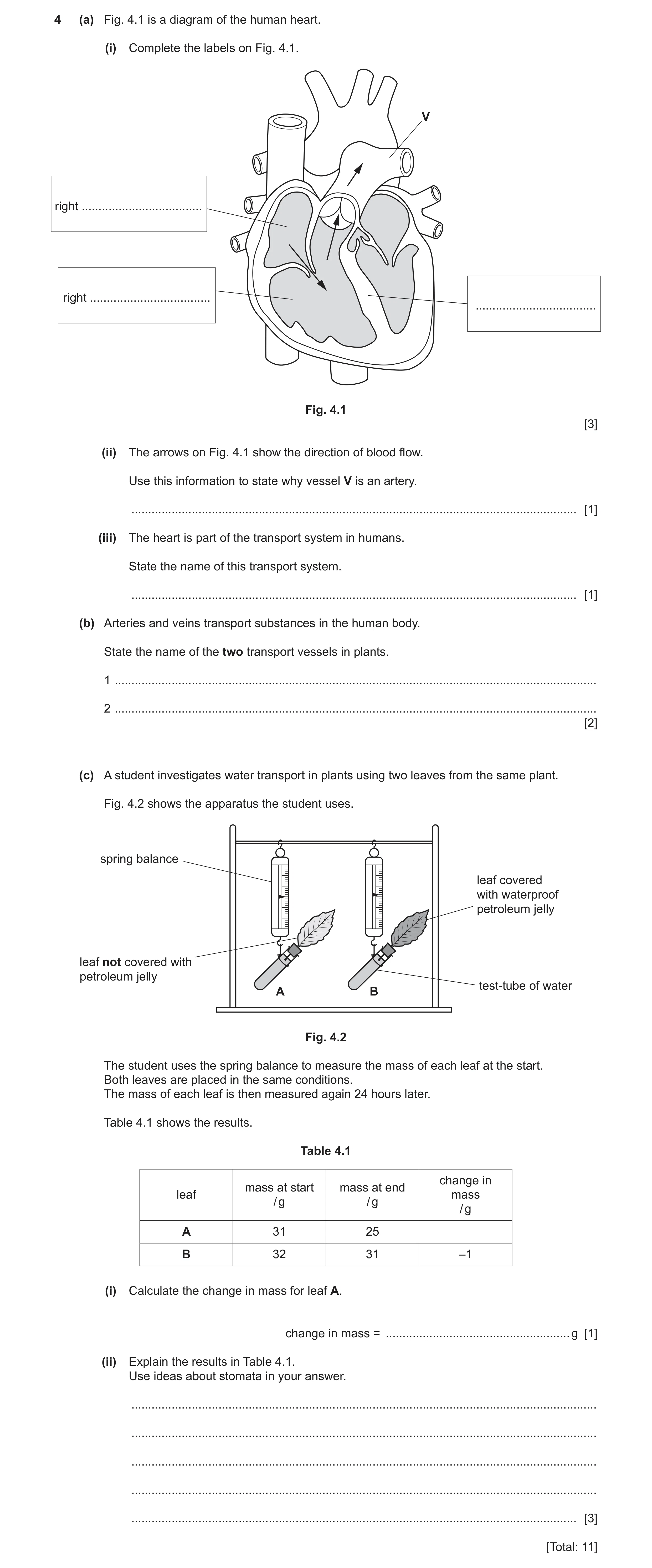
4 (a) Fig. 4.1 is a diagram of the human heart. (i) Complete the labels on Fig. 4.1. V right … right … … Fig. 4.1 [3] (ii) The arrows on Fig. 4.1 show the direction of blood flow. Use this information to state why vessel V is an artery. … [1] (iii) The heart is part of the transport system in humans. State the name of this transport system. … [1] (b) Arteries and veins transport substances in the human body. State the name of the two transport vessels in plants. 1 … 2 … [2] (c) A student investigates water transport in plants using two leaves from the same plant. Fig. 4.2 shows the apparatus the student uses. spring balance leaf covered with waterproof petroleum jelly leaf not covered with petroleum jelly test-tube of water A B Fig. 4.2 The student uses the spring balance to measure the mass of each leaf at the start. Both leaves are placed in the same conditions. The mass of each leaf is then measured again 24 hours later. Table 4.1 shows the results. Table 4.1 change in mass at start mass at end leaf mass / g / g / g A 31 25 B 32 31 –1 (i) Calculate the change in mass for leaf A. change in mass = … g [1] (ii) Explain the results in Table 4.1. Use ideas about stomata in your answer. … … … … … [3] [Total: 11]
11 marks
Mark scheme: 4(a)(i) atrium ; ventricle ; septum ; 3 4(a)(ii) idea it is taking blood away from the heart ; 1 4(a)(iii) circulatory / circulation ; 1 4(b) phloem ; xylem ; 2 4(c)(i) –6 (g) ; 1 4(c)(ii) any three from: water is lost from stomata ; by transpiration ; in B, stomata are blocked / water cannot get through stomata ; (B) less water loss so less mass loss ; Max 3 marks 3
This question in 0653/33 May/June 2022
Q13 · A student draws a labelled diagram of a human heart 0653/31 Oct/Nov 2022
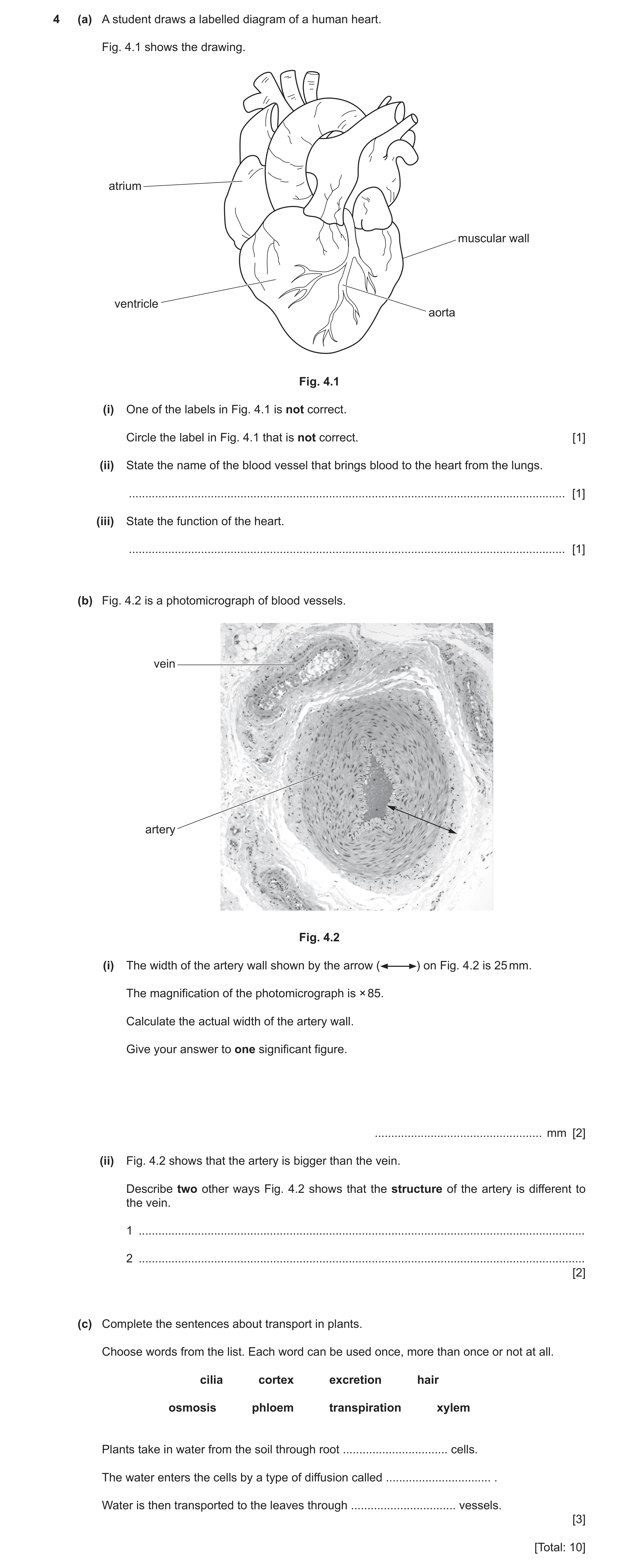
4 (a) A student draws a labelled diagram of a human heart. Fig. 4.1 shows the drawing. atrium muscular wall ventricle aorta Fig. 4.1 (i) One of the labels in Fig. 4.1 is not correct. Circle the label in Fig. 4.1 that is not correct. [1] (ii) State the name of the blood vessel that brings blood to the heart from the lungs. … [1] (iii) State the function of the heart. … [1] (b) Fig. 4.2 is a photomicrograph of blood vessels. vein artery Fig. 4.2 (i) The width of the artery wall shown by the arrow ( ) on Fig. 4.2 is 25 mm. The magnification of the photomicrograph is × 85. Calculate the actual width of the artery wall. Give your answer to one significant figure. … mm [2] (ii) Fig. 4.2 shows that the artery is bigger than the vein. Describe two other ways Fig. 4.2 shows that the structure of the artery is different to the vein. 1 … 2 … [2] (c) Complete the sentences about transport in plants. Choose words from the list. Each word can be used once, more than once or not at all. cilia cortex excretion hair osmosis phloem transpiration xylem Plants take in water from the soil through root … cells. The water enters the cells by a type of diffusion called … . Water is then transported to the leaves through … vessels. [3] [Total: 10]
10 marks
Mark scheme: 4(a)(i) aorta ; 1 4(a)(ii) pulmonary vein ; 1 4(a)(iii) to pump blood ; 1 4(b)(i) 25 ÷ 85 / 0.294117647 ; 2 answer to one sig. fig. / 0.3 ; 4(b)(ii) (artery has / ORA) 2 thicker wall ; smaller hole in centre ; 4(c) hair ; 3 osmosis ; xylem ;
This question in 0653/31 Oct/Nov 2022
Q14 · A diagram of a human heart 0653/32 May/June 2023
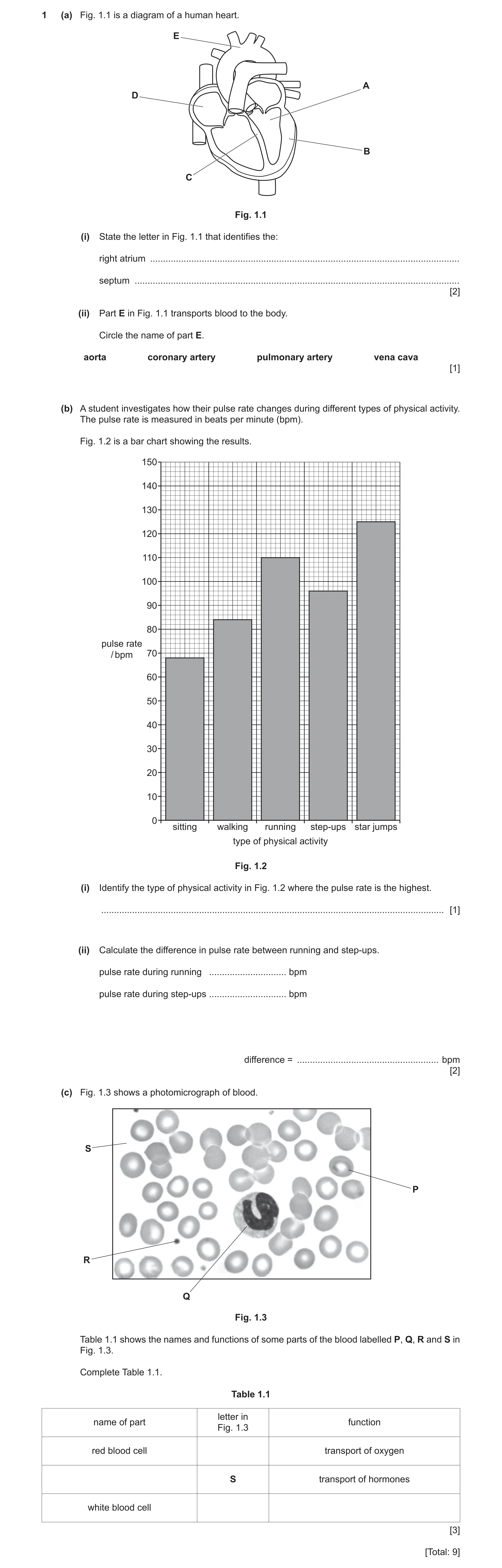
1 (a) Fig. 1.1 is a diagram of a human heart. E A D B C Fig. 1.1 (i) State the letter in Fig. 1.1 that identifies the: right atrium … septum … [2] (ii) Part E in Fig. 1.1 transports blood to the body. Circle the name of part E. aorta coronary artery pulmonary artery vena cava [1] (b) A student investigates how their pulse rate changes during different types of physical activity. The pulse rate is measured in beats per minute (bpm). Fig. 1.2 is a bar chart showing the results. 150 140 130 120 110 100 90 80 pulse rate / bpm 70 60 50 40 30 20 10 0 sitting walking running step-ups star jumps type of physical activity Fig. 1.2 (i) Identify the type of physical activity in Fig. 1.2 where the pulse rate is the highest. … [1] (ii) Calculate the difference in pulse rate between running and step‑ups. pulse rate during running … bpm pulse rate during step‑ups … bpm difference = … bpm [2] (c) Fig. 1.3 shows a photomicrograph of blood. S P R Q Fig. 1.3 Table 1.1 shows the names and functions of some parts of the blood labelled P, Q, R and S in Fig. 1.3. Complete Table 1.1. Table 1.1 letter in name of part function Fig. 1.3 red blood cell transport of oxygen S transport of hormones white blood cell [3] [Total: 9]
9 marks
Mark scheme: 1(a)(i) D ; C ; 2 1(a)(ii) aorta (circled) ; 1 1(b)(i) star jumps ; 1 1(b)(ii) (running) 110 (bpm) and (step-ups) 96 (bpm) ; (difference =) 14 (bpm) ; 2 1(c) name of part letter in Fig. 1.3 function red blood cell P transport of oxygen plasma S transport of hormones white blood cells Q phagocytosis / produce antibodies ;;; one mark for each correct row 3
This question in 0653/32 May/June 2023
Q15 · A diagram of the human heart 0653/33 May/June 2024
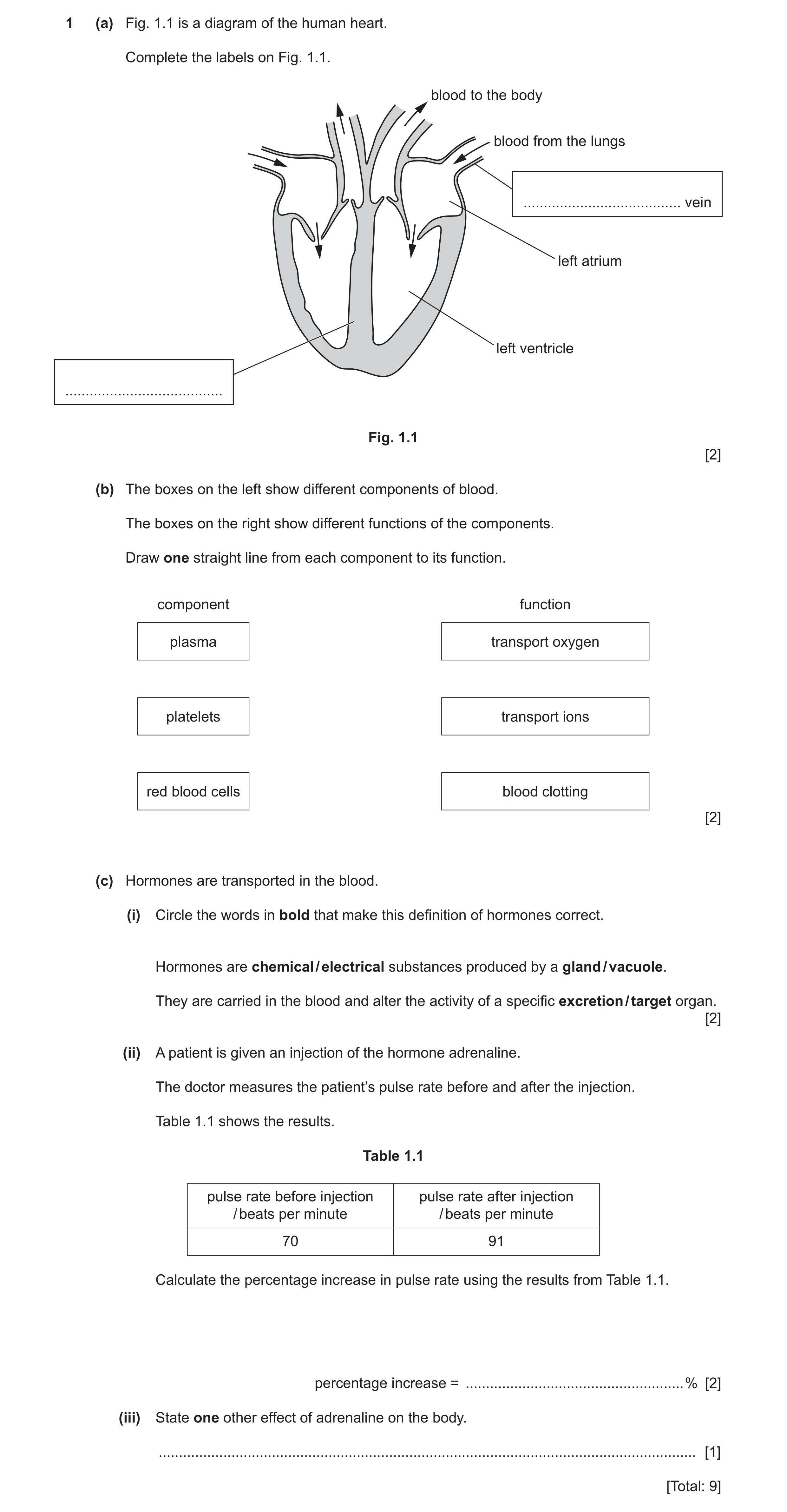
1 (a) Fig. 1.1 is a diagram of the human heart. Complete the labels on Fig. 1.1. blood to the body blood from the lungs … vein left atrium left ventricle … Fig. 1.1 [2] (b) The boxes on the left show different components of blood. The boxes on the right show different functions of the components. Draw one straight line from each component to its function. component function plasma transport oxygen platelets transport ions red blood cells blood clotting [2] (c) Hormones are transported in the blood. (i) Circle the words in bold that make this definition of hormones correct. Hormones are chemical / electrical substances produced by a gland / vacuole. They are carried in the blood and alter the activity of a specific excretion / target organ. [2] (ii) A patient is given an injection of the hormone adrenaline. The doctor measures the patient’s pulse rate before and after the injection. Table 1.1 shows the results. Table 1.1 pulse rate before injection pulse rate after injection / beats per minute / beats per minute 70 91 Calculate the percentage increase in pulse rate using the results from Table 1.1. percentage increase = … % [2] (iii) State one other effect of adrenaline on the body. … [1] [Total: 9]
9 marks
Mark scheme: 1(a) pulmonary (vein) ; septum ; 2 1(b) plasma transport oxygen platelets transport ions red blood cells blood clotting ;; all three correct = 2 marks one or two correct = 1 mark 2 1(c)(i) chemical gland target ;; all three correct = 2 marks two correct = 1 mark 2 1(c)(ii) (91–70 =) 21 ; 21 100 70 30 (%) ; 2 1(c)(iii) increases breathing rate / widens pupils ; 1
This question in 0653/33 May/June 2024
Q16 · A diagram of the circulatory system in humans where the arrows show the direction of… 0653/32 Oct/Nov 2024
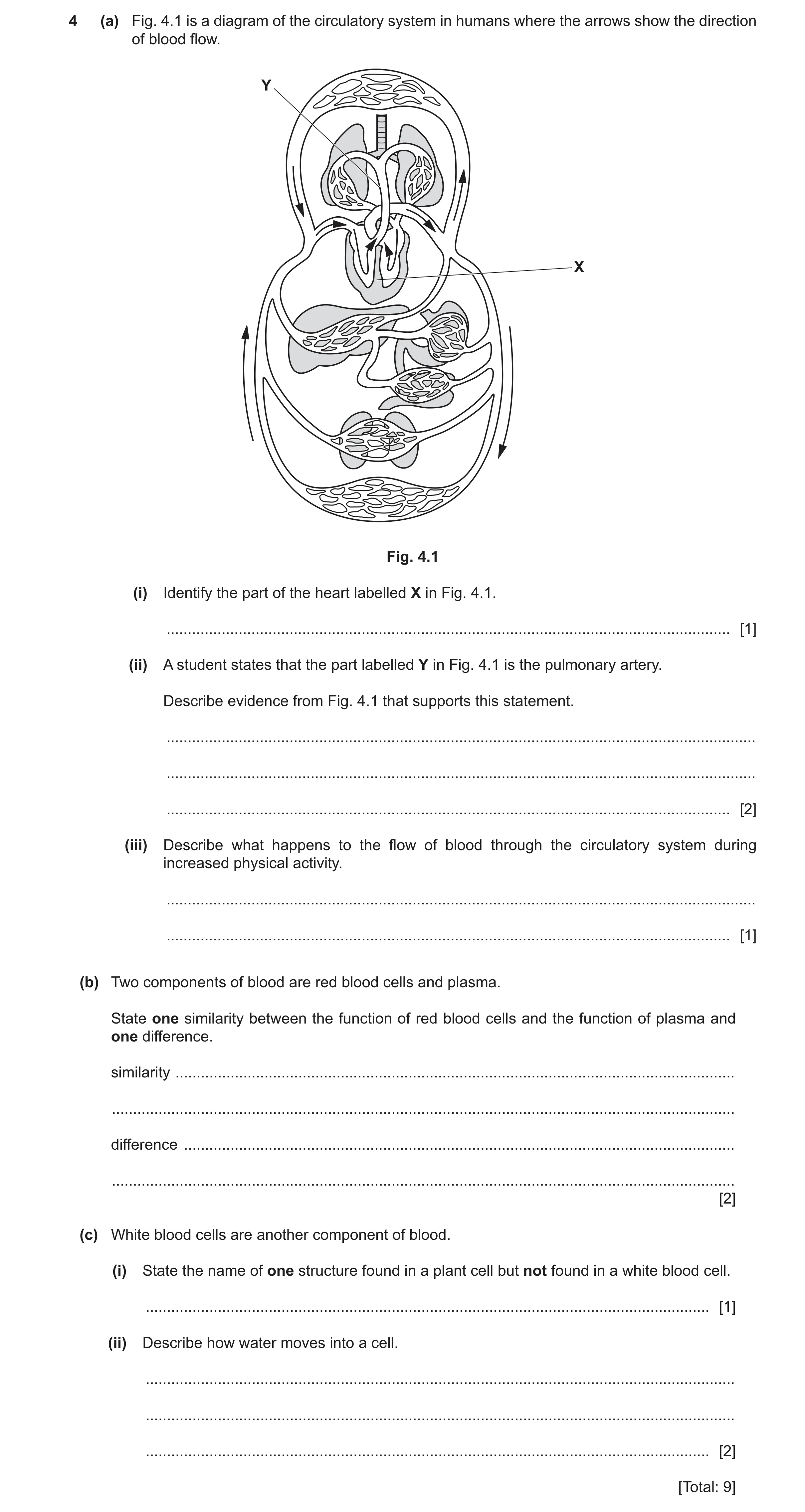
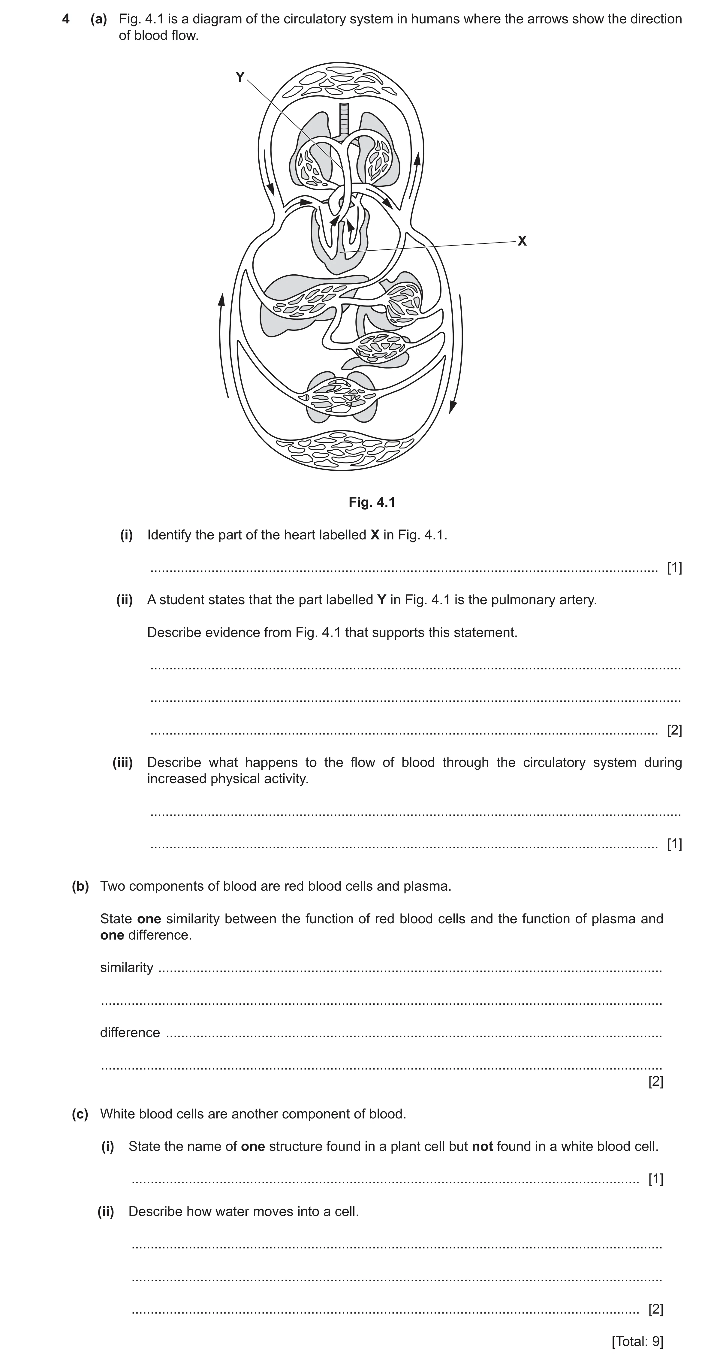
4 (a) Fig. 4.1 is a diagram of the circulatory system in humans where the arrows show the direction of blood flow. Y X Fig. 4.1 (i) Identify the part of the heart labelled X in Fig. 4.1. … [1] (ii) A student states that the part labelled Y in Fig. 4.1 is the pulmonary artery. Describe evidence from Fig. 4.1 that supports this statement. … … … [2] (iii) Describe what happens to the flow of blood through the circulatory system during increased physical activity. … … [1] (b) Two components of blood are red blood cells and plasma. State one similarity between the function of red blood cells and the function of plasma and one difference. similarity … … difference … … [2] (c) White blood cells are another component of blood. (i) State the name of one structure found in a plant cell but not found in a white blood cell. … [1] (ii) Describe how water moves into a cell. … … … [2] [Total: 9]
9 marks
Mark scheme: 4(a)(i) septum ; 1 4(a)(ii) (arrows show) blood flows, away from heart ; 2 towards the lungs ; 4(a)(iii) rate (of blood flow) increases ; 1 4(b) similarity both transport / carry substances ; 2 red blood cells transport oxygen OR plasma transports, hormones / nutrients / carbon dioxide / ions / amino acids ; 4(c)(i) vacuole / cell wall / chloroplast ; 1 4(c)(ii) by osmosis ; 2 through partially permeable membrane ;
This question in 0653/32 Oct/Nov 2024
Q17 · A diagram of the circulatory system in humans where the arrows show the direction of… 0653/33 Oct/Nov 2024
4 (a) Fig. 4.1 is a diagram of the circulatory system in humans where the arrows show the direction of blood flow. Y X Fig. 4.1 (i) Identify the part of the heart labelled X in Fig. 4.1. … [1] (ii) A student states that the part labelled Y in Fig. 4.1 is the pulmonary artery. Describe evidence from Fig. 4.1 that supports this statement. … … … [2] (iii) Describe what happens to the flow of blood through the circulatory system during increased physical activity. … … [1] (b) Two components of blood are red blood cells and plasma. State one similarity between the function of red blood cells and the function of plasma and one difference. similarity … … difference … … [2] (c) White blood cells are another component of blood. (i) State the name of one structure found in a plant cell but not found in a white blood cell. … [1] (ii) Describe how water moves into a cell. … … … [2] [Total: 9]
9 marks
Mark scheme: 4(a)(i) septum ; 1 4(a)(ii) (arrows show) blood flows, away from heart ; 2 towards the lungs ; 4(a)(iii) rate (of blood flow) increases ; 1 4(b) similarity both transport / carry substances ; 2 red blood cells transport oxygen OR plasma transports, hormones / nutrients / carbon dioxide / ions / amino acids ; 4(c)(i) vacuole / cell wall / chloroplast ; 1 4(c)(ii) by osmosis ; 2 through partially permeable membrane ;
This question in 0653/33 Oct/Nov 2024
Q18 · The structure of a human heart 0653/31 May/June 2025
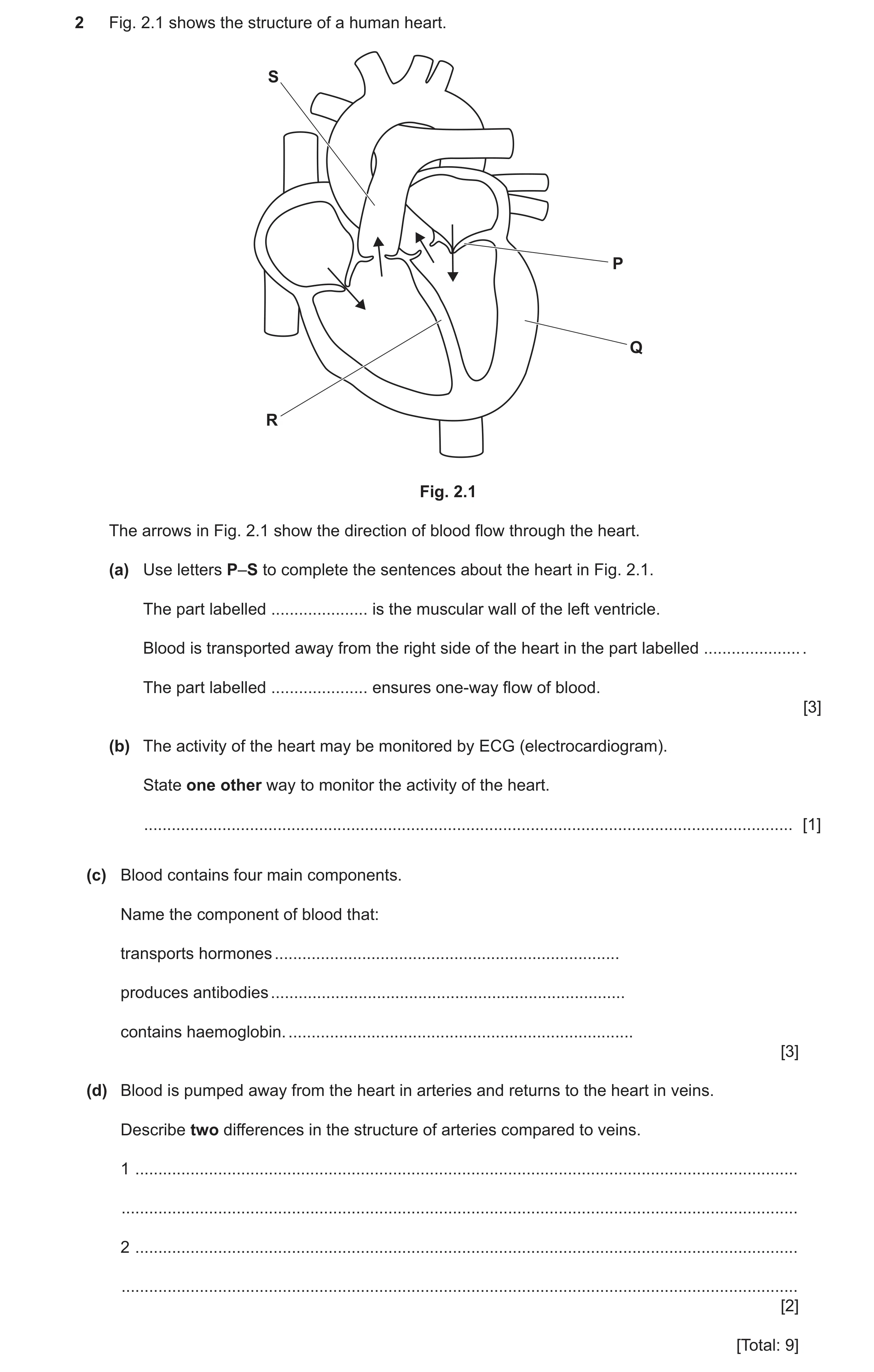
2 Fig. 2.1 shows the structure of a human heart. S P Q R Fig. 2.1 The arrows in Fig. 2.1 show the direction of blood flow through the heart. (a) Use letters P–S to complete the sentences about the heart in Fig. 2.1. The part labelled … is the muscular wall of the left ventricle. Blood is transported away from the right side of the heart in the part labelled … . The part labelled … ensures one‑way flow of blood. [3] (b) The activity of the heart may be monitored by ECG (electrocardiogram). State one other way to monitor the activity of the heart. … [1] (c) Blood contains four main components. Name the component of blood that: transports hormones … produces antibodies … contains haemoglobin. … [3] (d) Blood is pumped away from the heart in arteries and returns to the heart in veins. Describe two differences in the structure of arteries compared to veins. 1 … … 2 … … [2] [Total: 9]
9 marks
Mark scheme: 2(a) Q ; 3 S ; P ; 2(b) pulse (rate) / listening to sound of valves (closing) / stethoscope ; 1 2(c) plasma ; 3 white blood cells ; red blood cells ; 2(d) any two from: 2 (arteries have) thicker walls ; smaller lumen ; no valves ;
This question in 0653/31 May/June 2025
 4 / 32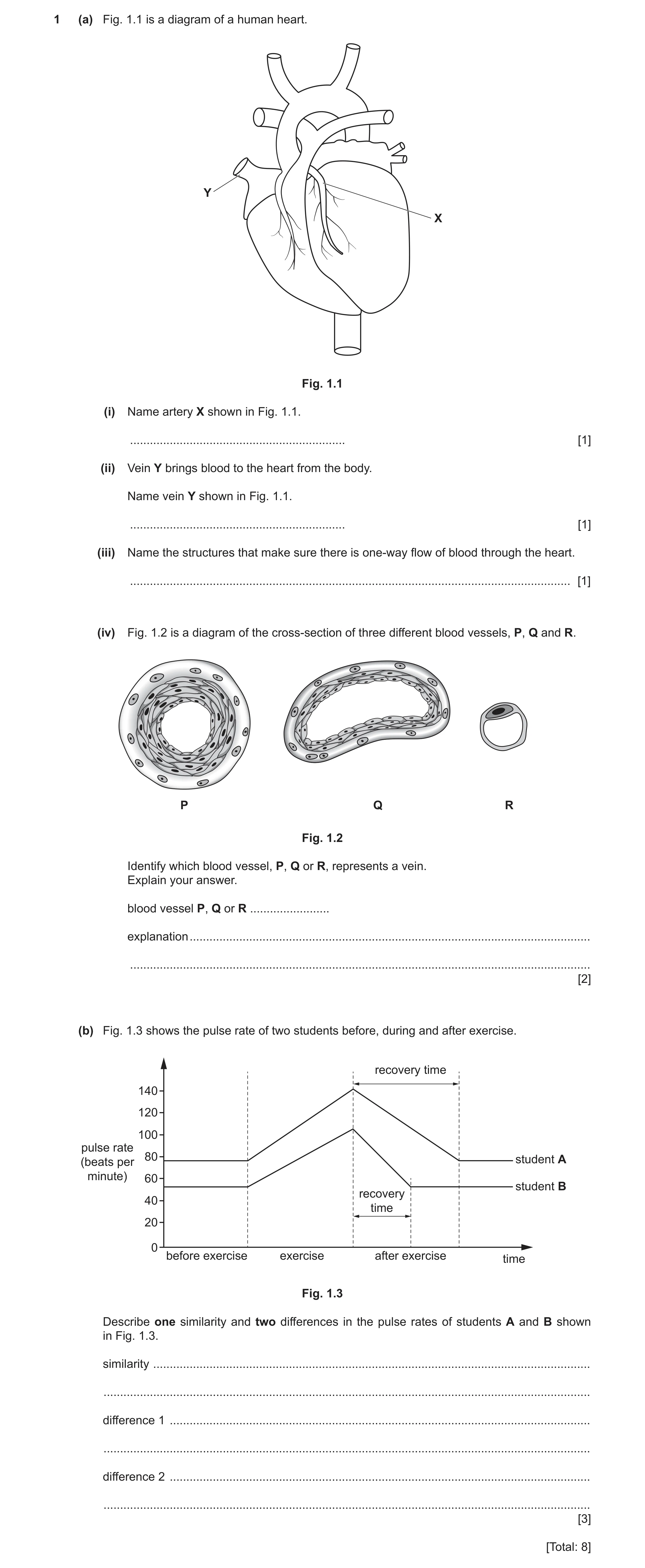
 6
6 6
6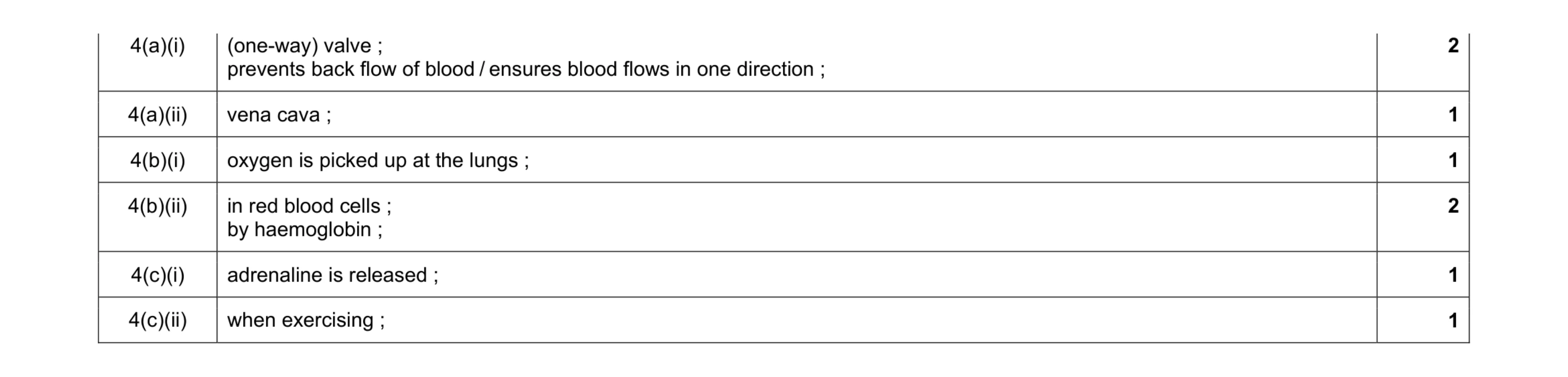 8
8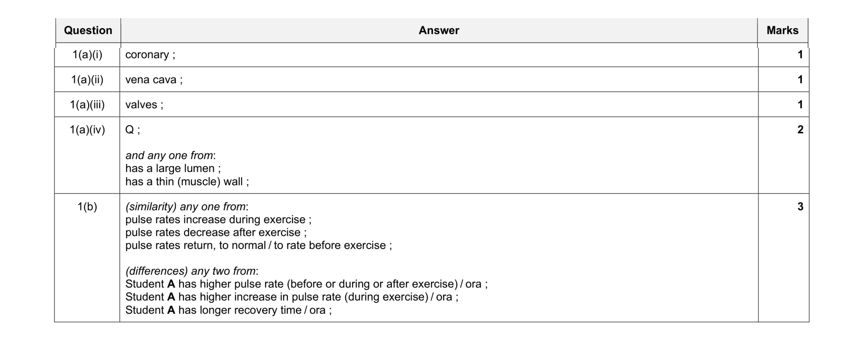 8
8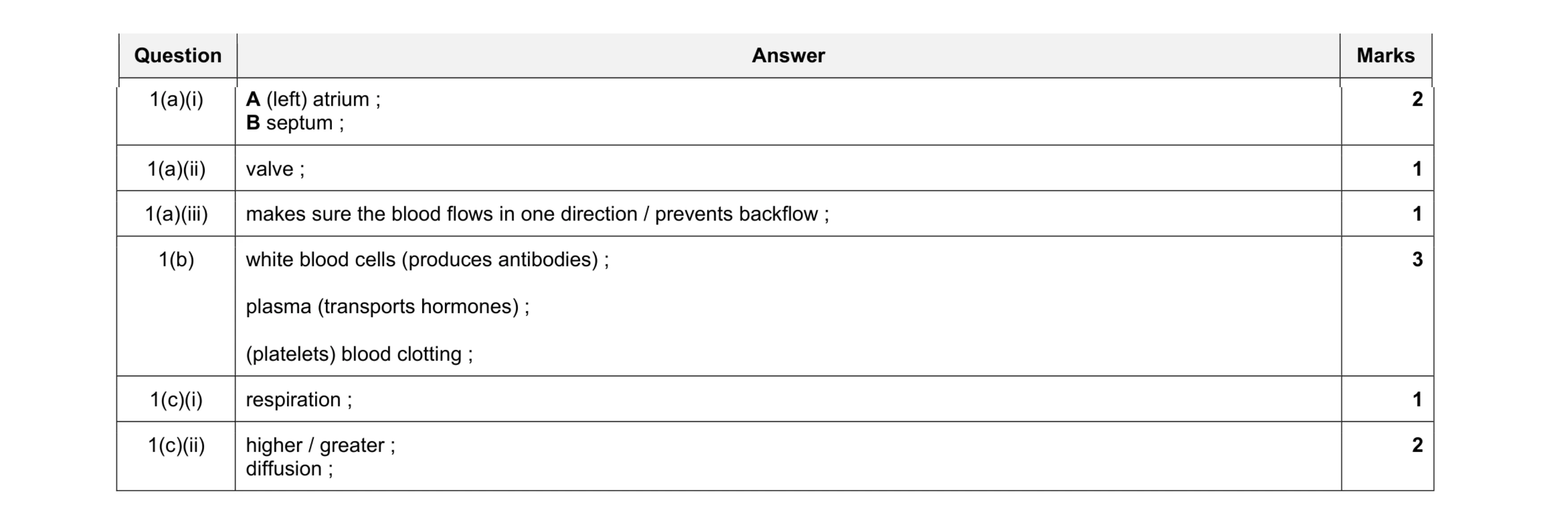 10
10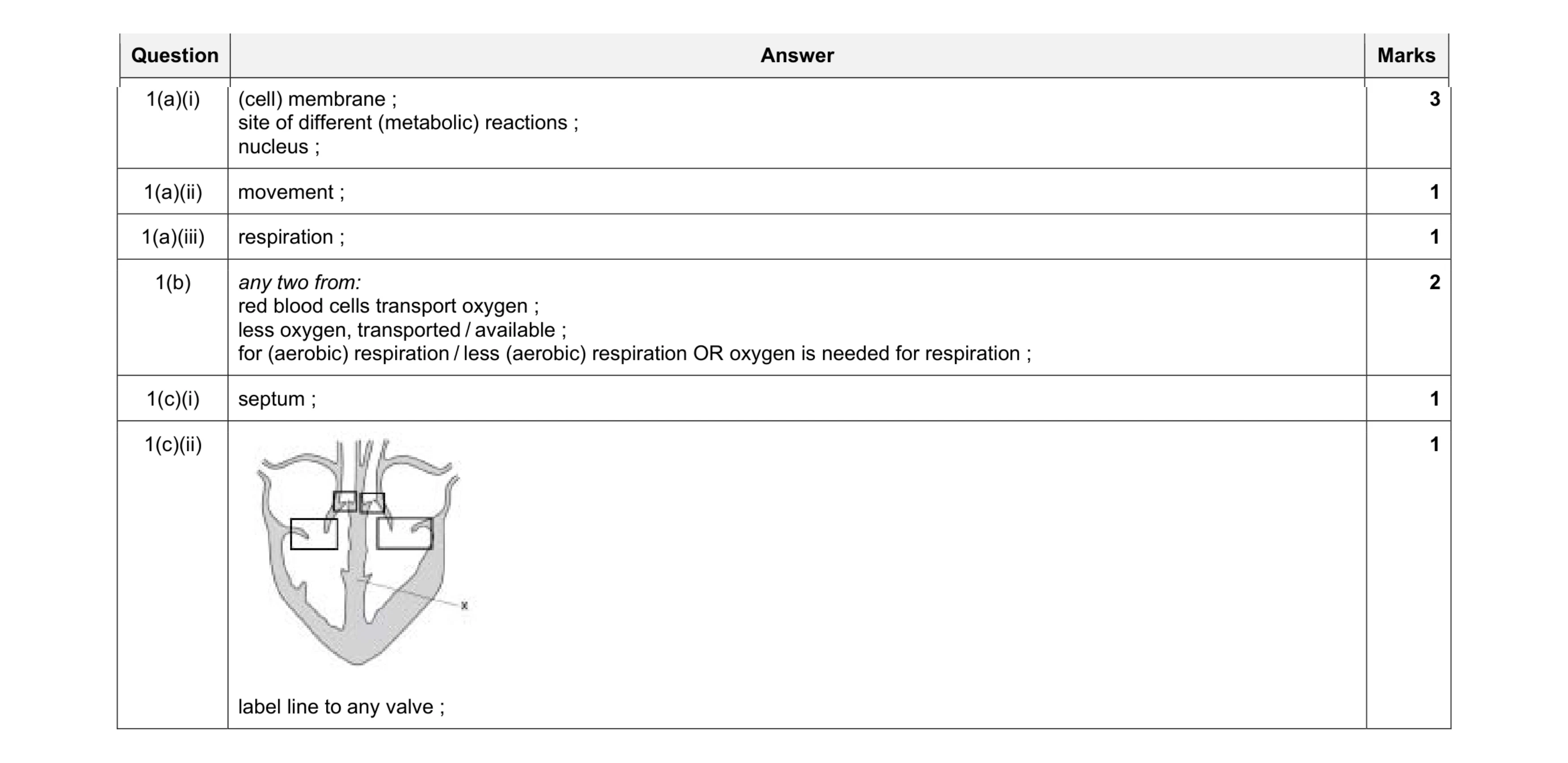 9
9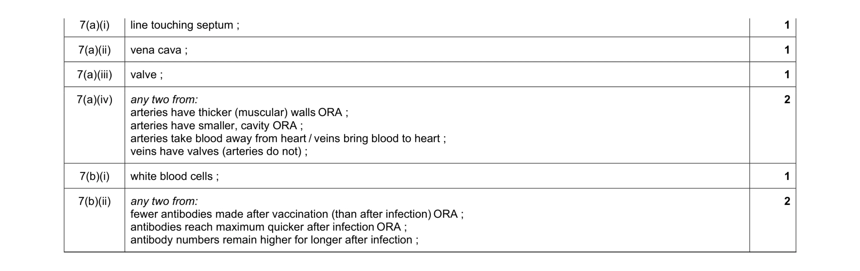 8
8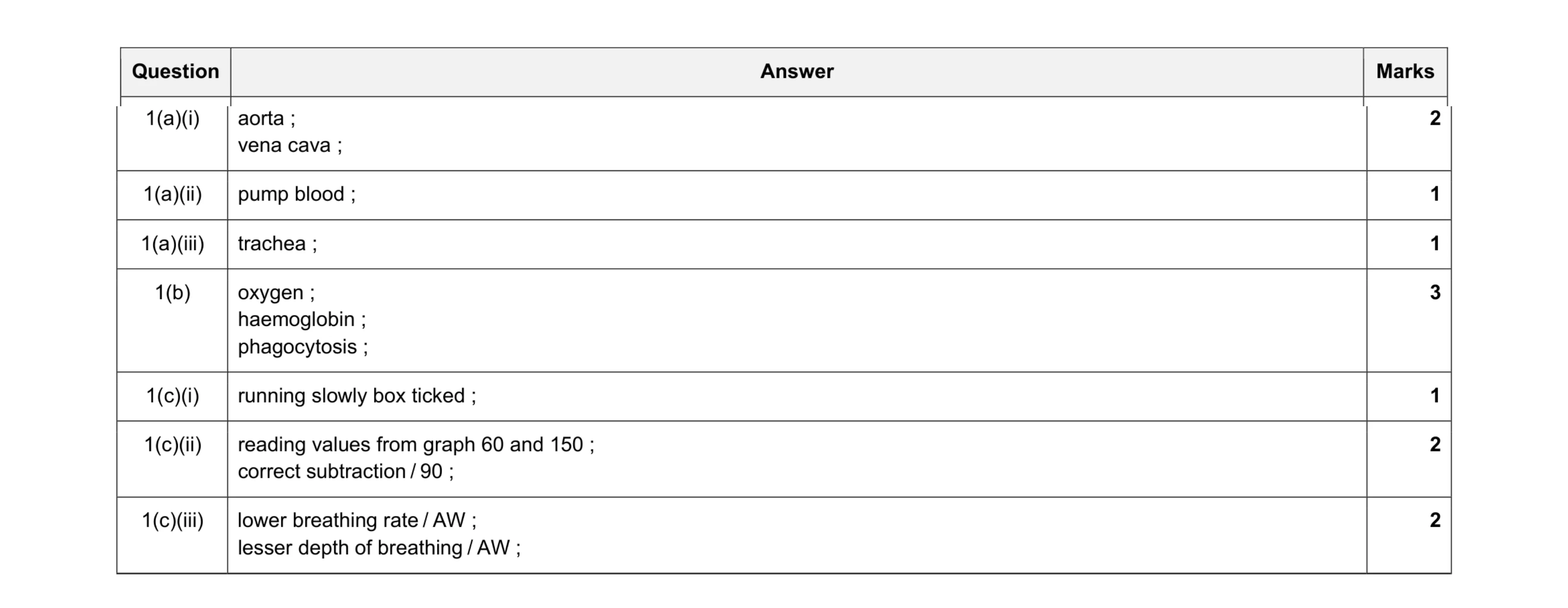 12
12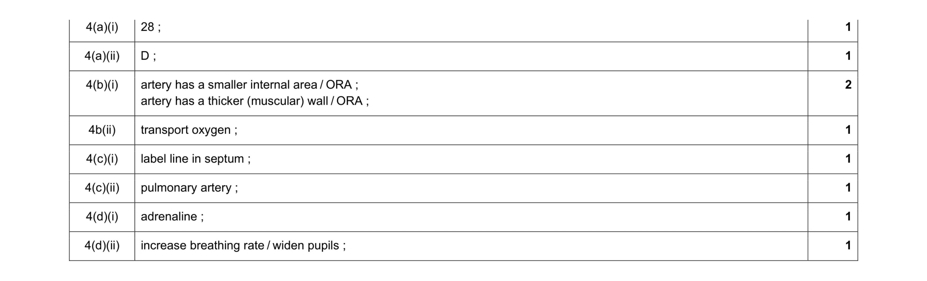 9
9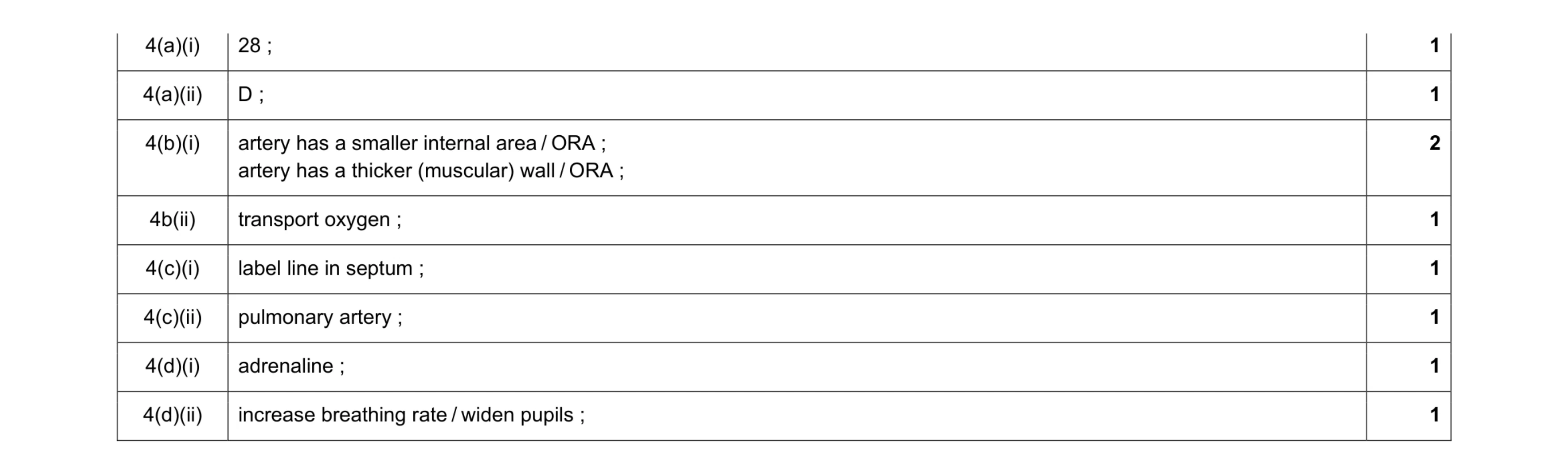 9
9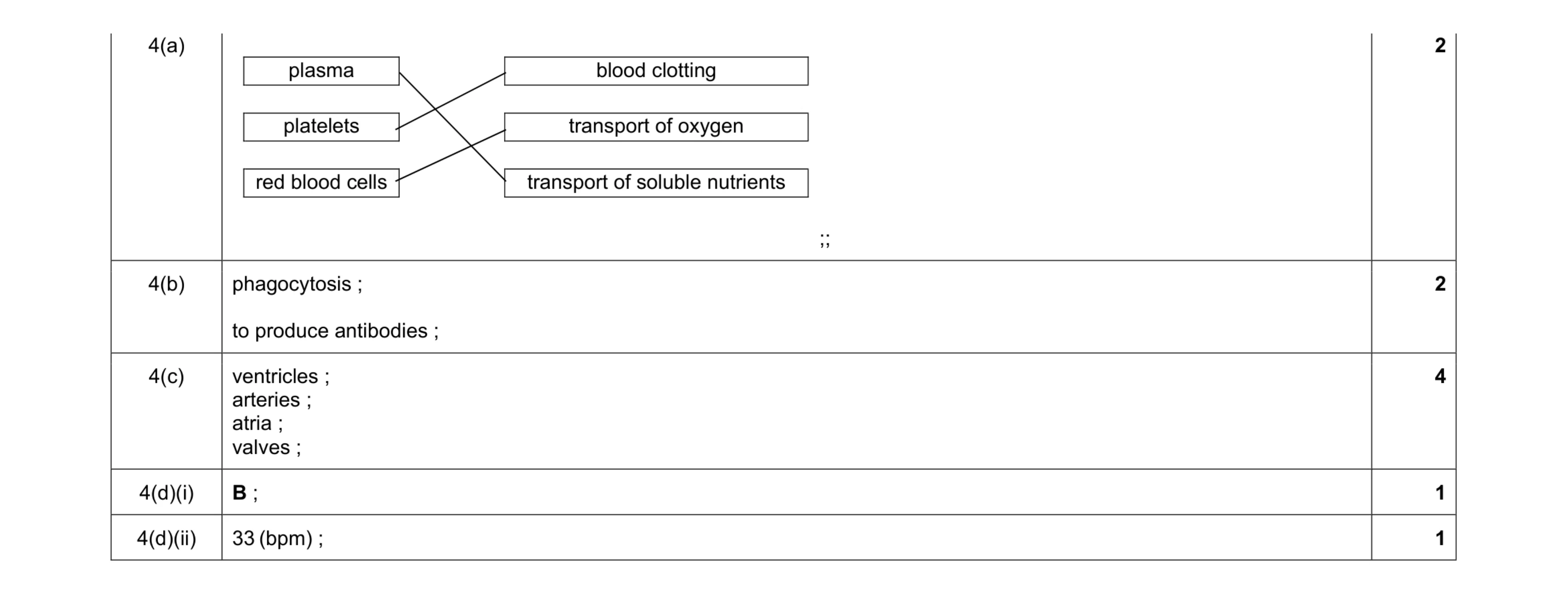 10
10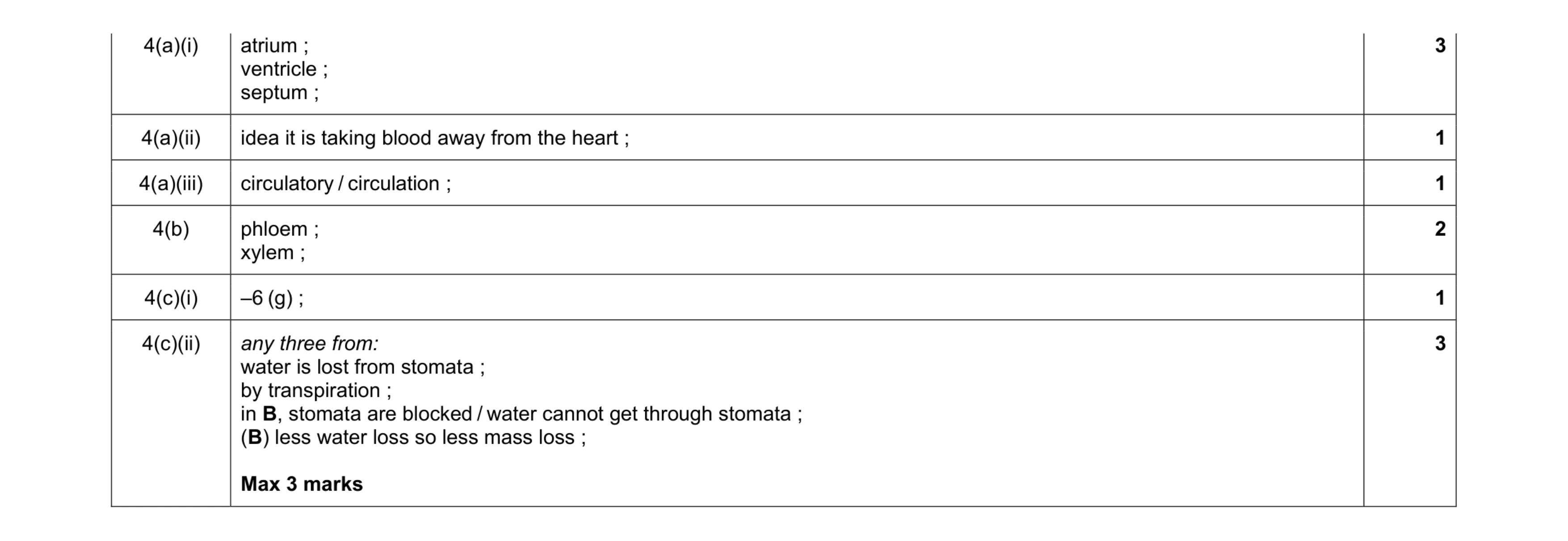 11
11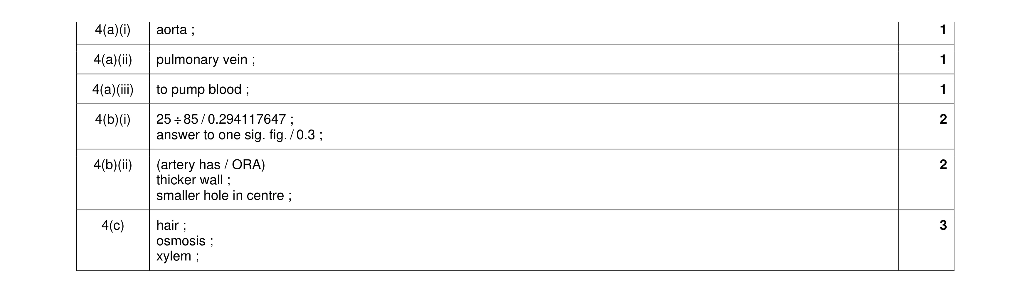 10
10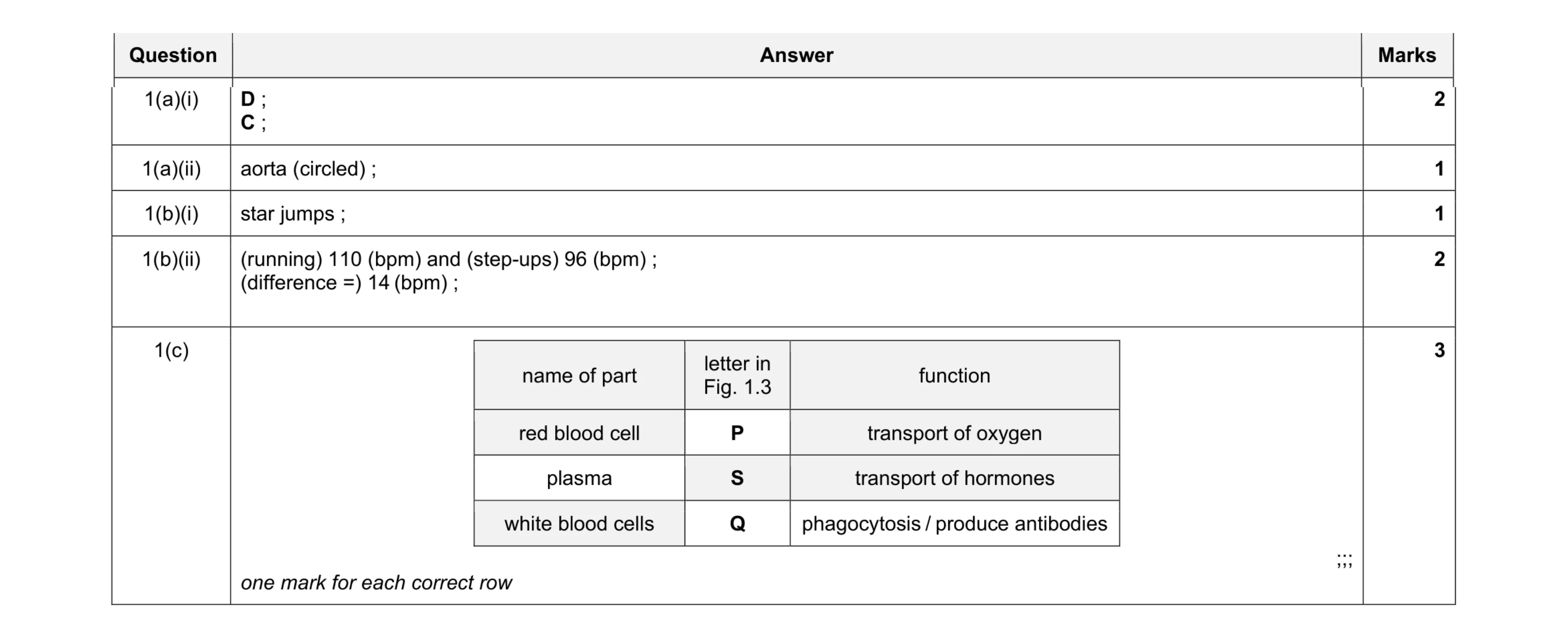 9
9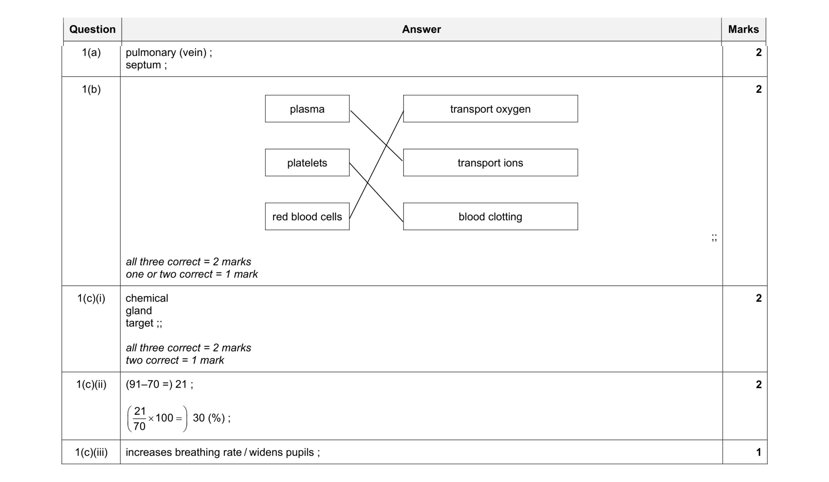 9
9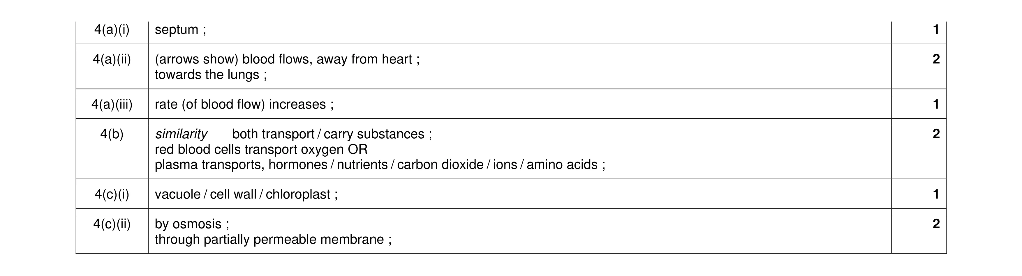 9
9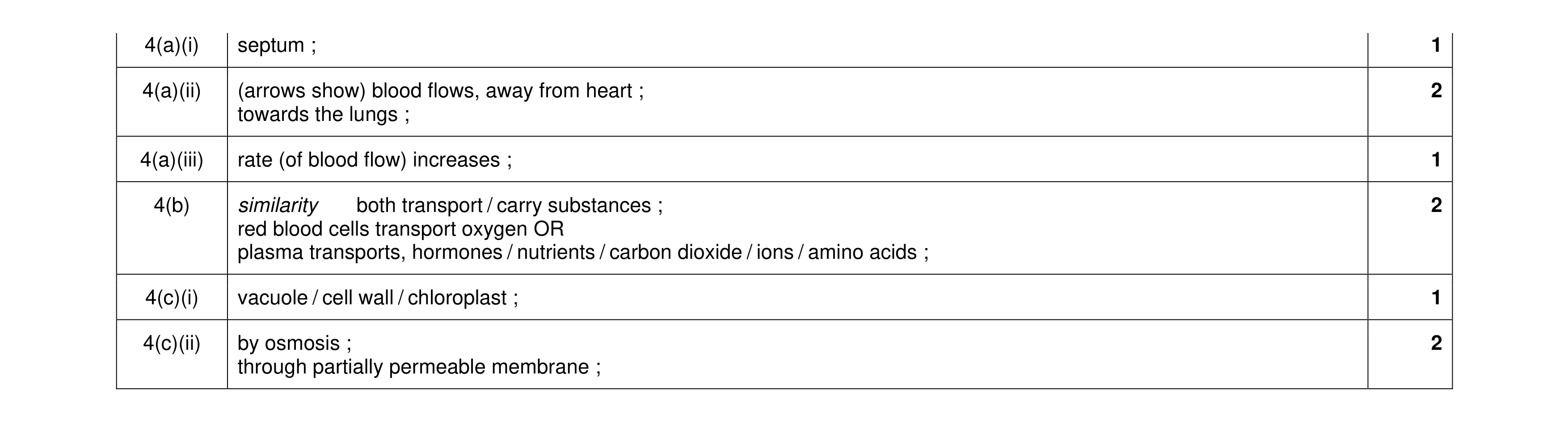 9
9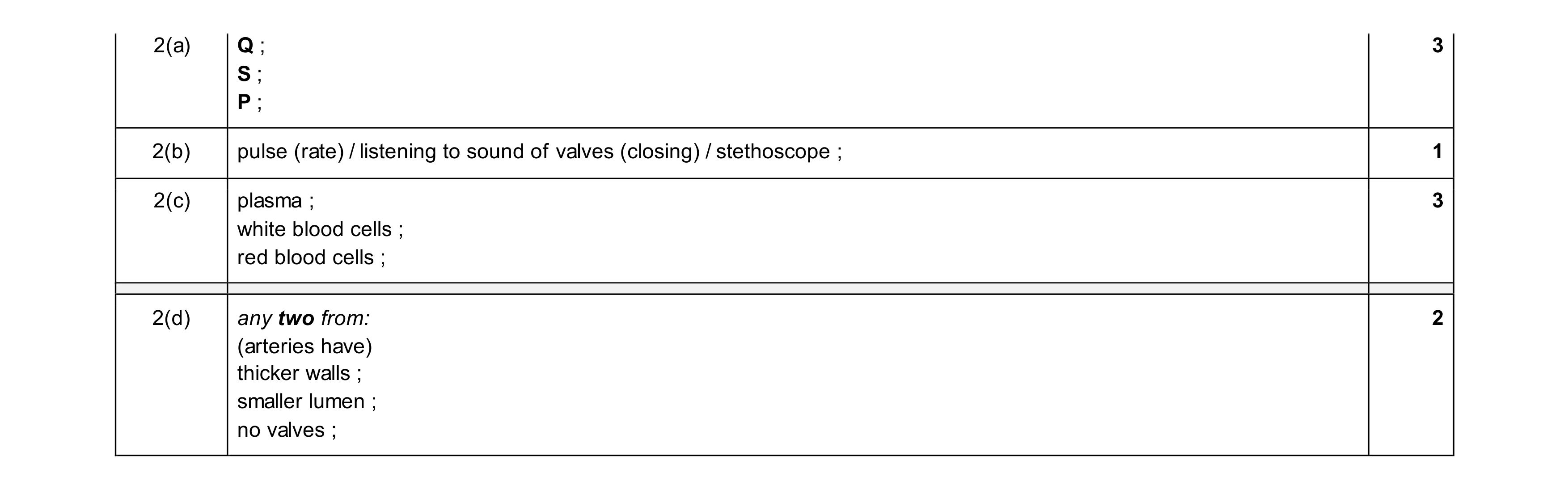 9
9