Questions as text
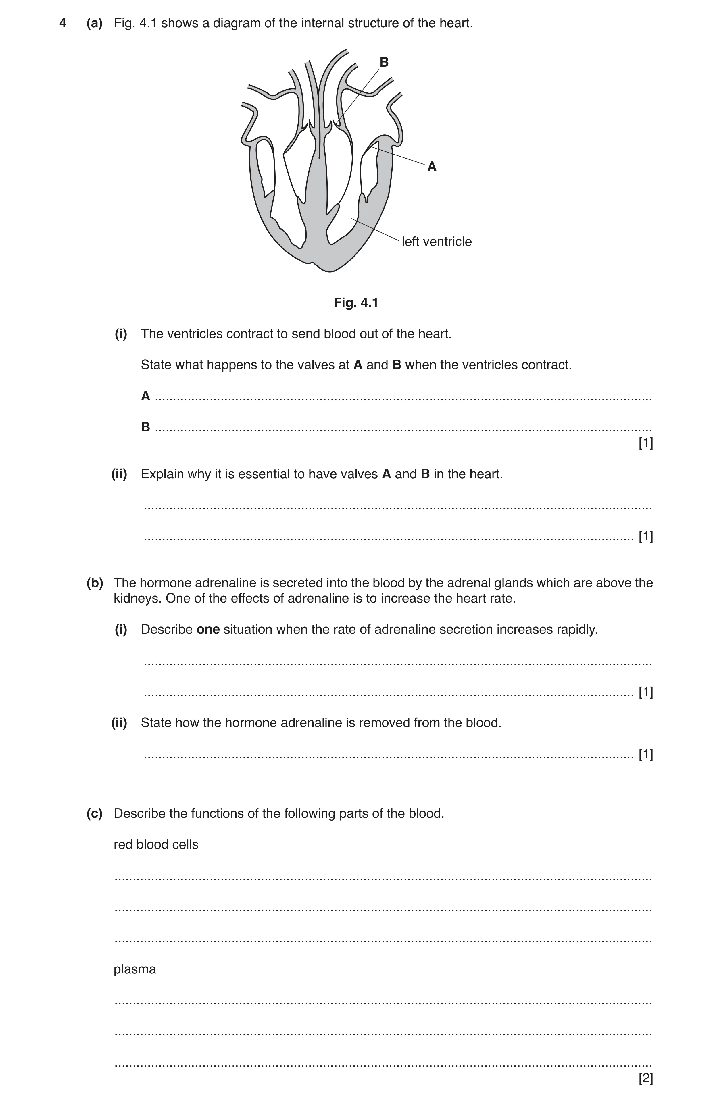
Q1 · A diagram of the internal structure of the heart 0653/32 May/June 2017
4 (a) Fig. 4.1 shows a diagram of the internal structure of the heart. B A left ventricle Fig. 4.1 (i) The ventricles contract to send blood out of the heart. State what happens to the valves at A and B when the ventricles contract. A … B … [1] (ii) Explain why it is essential to have valves A and B in the heart. … … [1] (b) The hormone adrenaline is secreted into the blood by the adrenal glands which are above the kidneys. One of the effects of adrenaline is to increase the heart rate. (i) Describe one situation when the rate of adrenaline secretion increases rapidly. … … [1] (ii) State how the hormone adrenaline is removed from the blood. … [1] (c) Describe the functions of the following parts of the blood. red blood cells … … … plasma … … … [2]
6 marks
Mark scheme: 4(a)(i) A closes and B opens ; 1 4(a)(ii) to prevent backflow of blood ; 1 4(b)(i) any suitable flight or fight situation described ; 1 4(b)(ii) destroyed by the liver ; 1 4(c) transport of oxygen / haemoglobin ; transport of blood cells / ions / soluble nutrients / named soluble nutrient / hormones / carbon dioxide ; 2
This question in 0653/32 May/June 2017
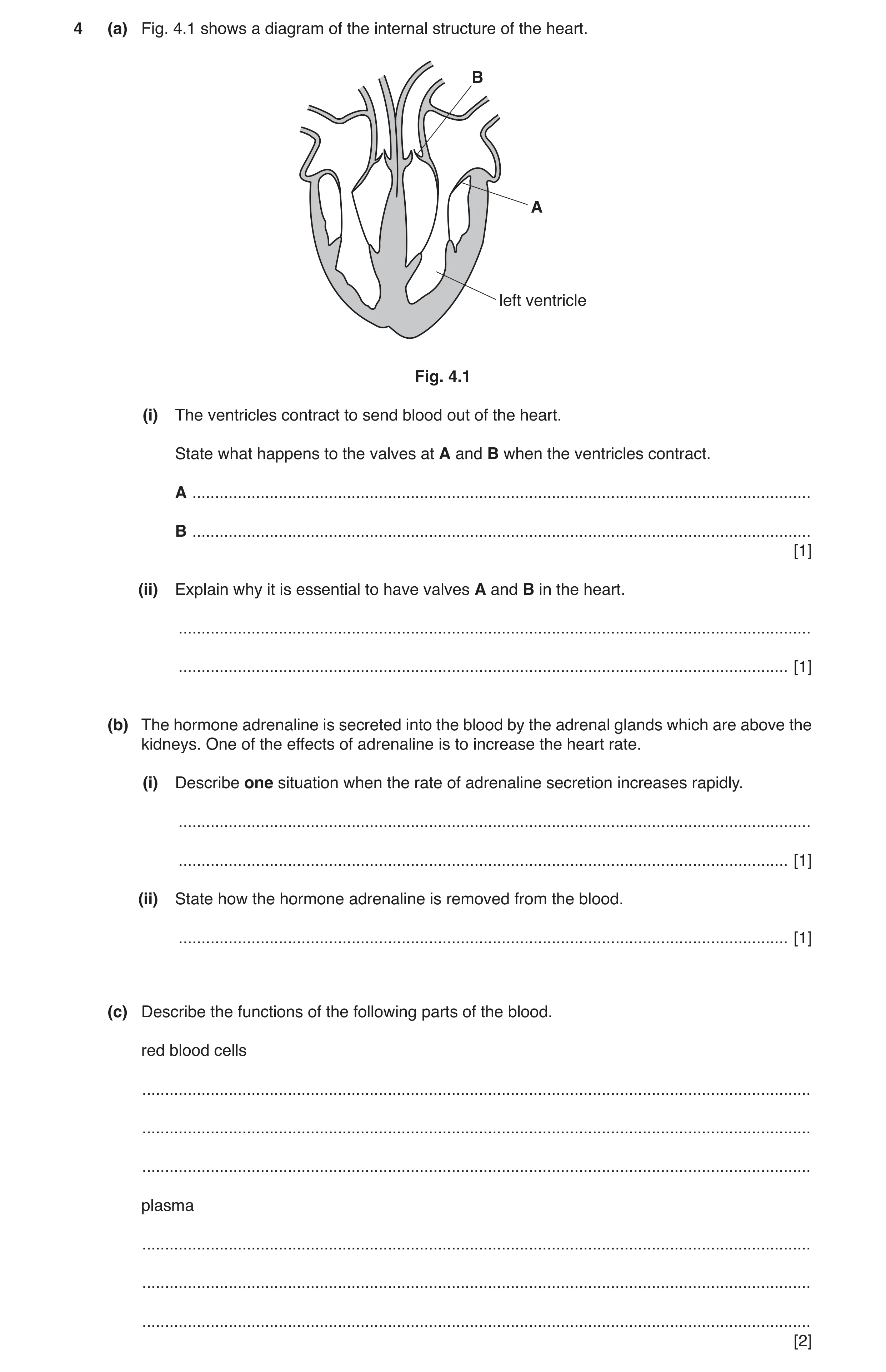
Q2 · A diagram of the internal structure of the heart 0653/33 May/June 2017
4 (a) Fig. 4.1 shows a diagram of the internal structure of the heart. B A left ventricle Fig. 4.1 (i) The ventricles contract to send blood out of the heart. State what happens to the valves at A and B when the ventricles contract. A … B … [1] (ii) Explain why it is essential to have valves A and B in the heart. … … [1] (b) The hormone adrenaline is secreted into the blood by the adrenal glands which are above the kidneys. One of the effects of adrenaline is to increase the heart rate. (i) Describe one situation when the rate of adrenaline secretion increases rapidly. … … [1] (ii) State how the hormone adrenaline is removed from the blood. … [1] (c) Describe the functions of the following parts of the blood. red blood cells … … … plasma … … … [2]
6 marks
Mark scheme: 4(a)(i) A closes and B opens ; 1 4(a)(ii) to prevent backflow of blood ; 1 4(b)(i) any suitable flight or fight situation described ; 1 4(b)(ii) destroyed by the liver ; 1 4(c) transport of oxygen / haemoglobin ; transport of blood cells / ions / soluble nutrients / named soluble nutrient / hormones / carbon dioxide ; 2
This question in 0653/33 May/June 2017
Q3 · Use the following words or phrases to complete the flow chart about a possible pathway… 0653/33 Oct/Nov 2017
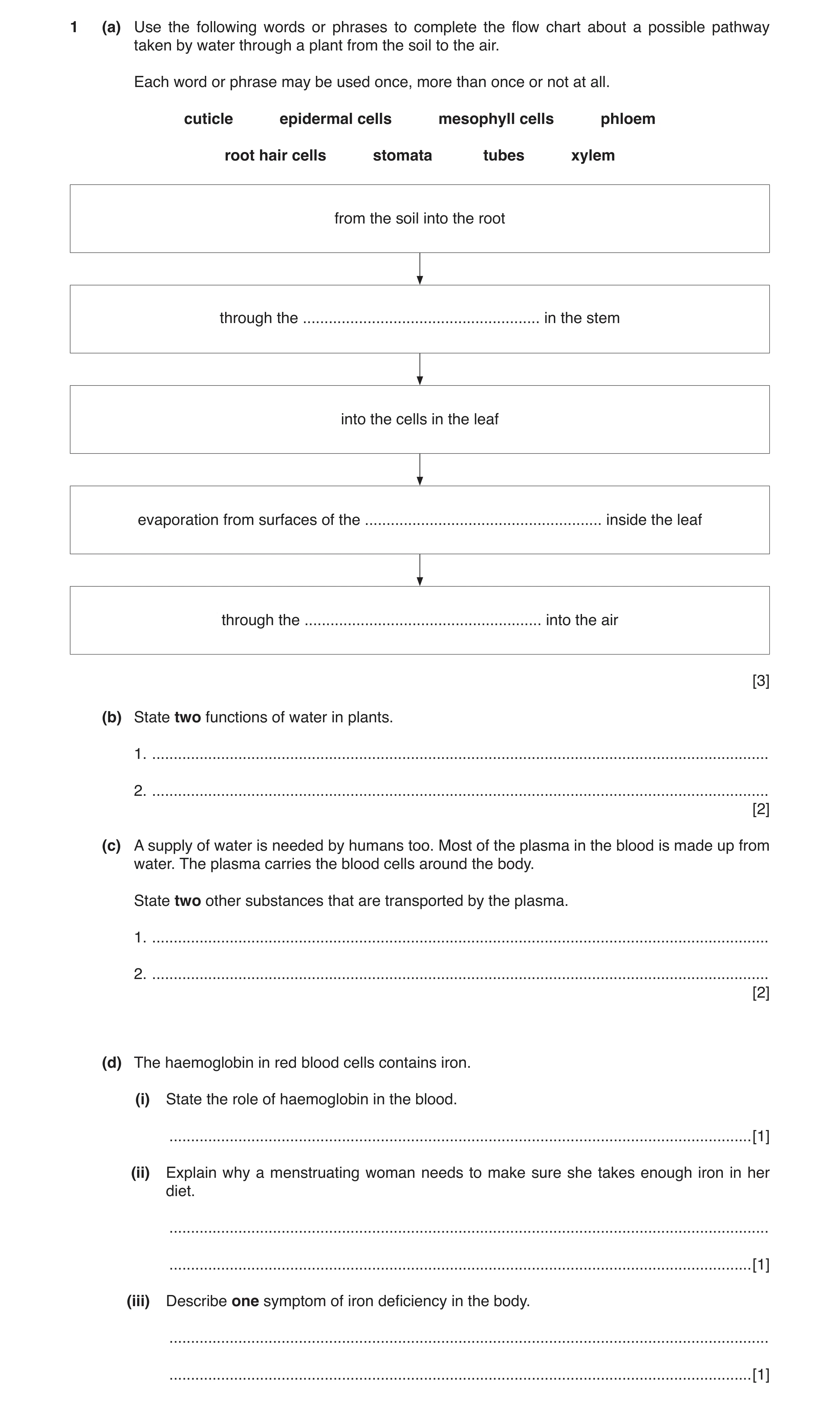
1 (a) Use the following words or phrases to complete the flow chart about a possible pathway taken by water through a plant from the soil to the air. Each word or phrase may be used once, more than once or not at all. cuticle epidermal cells mesophyll cells phloem root hair cells stomata tubes xylem from the soil into the root through the … in the stem into the cells in the leaf evaporation from surfaces of the … inside the leaf through the … into the air [3] (b) State two functions of water in plants. 1. … 2. … [2] (c) A supply of water is needed by humans too. Most of the plasma in the blood is made up from water. The plasma carries the blood cells around the body. State two other substances that are transported by the plasma. 1. … 2. … [2] (d) The haemoglobin in red blood cells contains iron. (i) State the role of haemoglobin in the blood. … [1] (ii) Explain why a menstruating woman needs to make sure she takes enough iron in her diet. … … [1] (iii) Describe one symptom of iron deficiency in the body. … … [1]
10 marks
Mark scheme: 1(a) xylem ; mesophyll cells ; stomata ; 3 1(b) any two from photosynthesis ; transport of ions ; support / turgor ; AVP ; max 2 1(c) ions / named ion ; soluble nutrients / named soluble nutrient ; hormones / named hormone ; carbon dioxide ; AVP ; max 2 1(d)(i) transport of oxygen ; 1 1(d)(ii) to replace iron lost during menstruation / period ; 1 1(d)(iii) feeling tired / dizzy ; 1
This question in 0653/33 Oct/Nov 2017
Q4 · The boxes in Fig 0653/32 Oct/Nov 2018
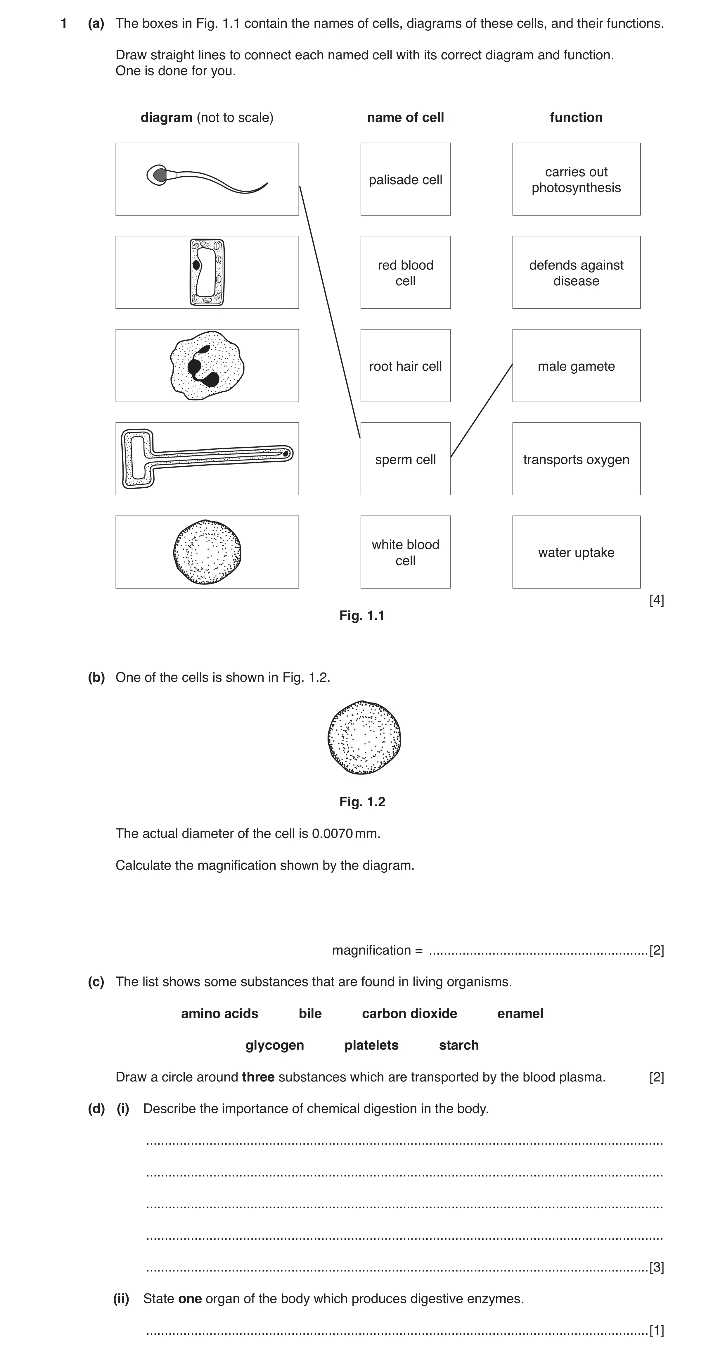
1 (a) The boxes in Fig. 1.1 contain the names of cells, diagrams of these cells, and their functions. Draw straight lines to connect each named cell with its correct diagram and function. One is done for you. diagram (not to scale) name of cell function carries out palisade cell photosynthesis red blood defends against cell disease root hair cell male gamete sperm cell transports oxygen white blood water uptake cell [4] Fig. 1.1 (b) One of the cells is shown in Fig. 1.2. Fig. 1.2 The actual diameter of the cell is 0.0070 mm. Calculate the magnification shown by the diagram. magnification = … [2] (c) The list shows some substances that are found in living organisms. amino acids bile carbon dioxide enamel glycogen platelets starch Draw a circle around three substances which are transported by the blood plasma. [2] (d) (i) Describe the importance of chemical digestion in the body. … … … … … [3] (ii) State one organ of the body which produces digestive enzymes. … [1]
12 marks
Mark scheme: 1(a) palisade cell carries out photosynthesis red blood cell defends against disease root hair cell male gamete sperm cell transports oxygen white blood cell water uptake 1 mark for each correct diagram / name / function ;;;; 4 1(b) diameter of red blood cell = 21 mm ; magnification = 21 / 0.007 = 3000 (times) ; 2 1(c) amino acid ; carbon dioxide ; platelets ; All 3 correct = 2 2 or 1 correct = 1 2 Question Answer Marks 1(d)(i) breaks down ; large / insoluble molecules ; into small / soluble molecules ; that can be absorbed ; Max 3 3 1(d)(ii) salivary glands / stomach / pancreas / ileum / small intestine ; 1
This question in 0653/32 Oct/Nov 2018
Q5 · A diagram of part of the alimentary canal and its associated organs 0653/33 Oct/Nov 2018
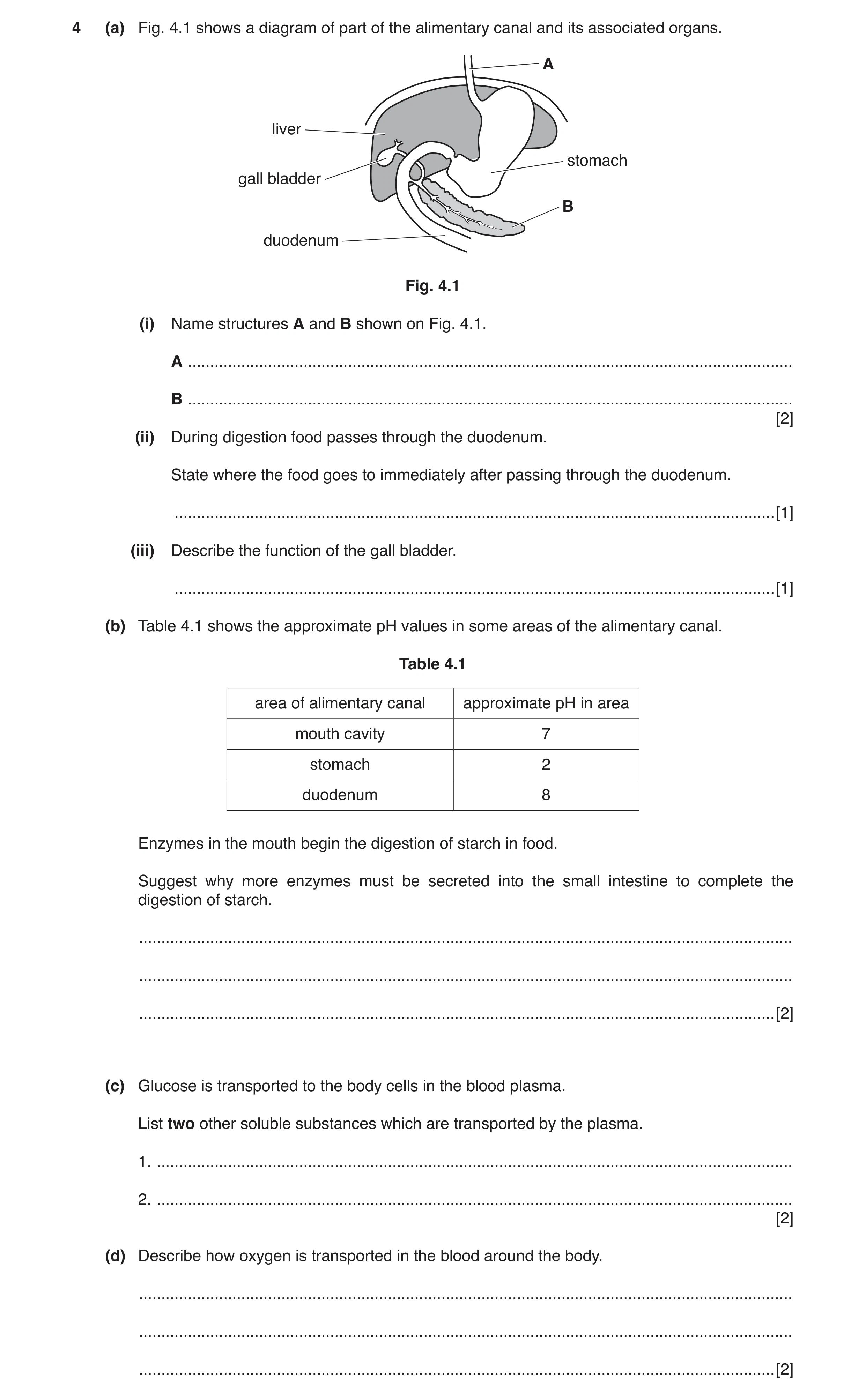
4 (a) Fig. 4.1 shows a diagram of part of the alimentary canal and its associated organs. A liver stomach gall bladder B duodenum Fig. 4.1 (i) Name structures A and B shown on Fig. 4.1. A … B … [2] (ii) During digestion food passes through the duodenum. State where the food goes to immediately after passing through the duodenum. … [1] (iii) Describe the function of the gall bladder. … [1] (b) Table 4.1 shows the approximate pH values in some areas of the alimentary canal. Table 4.1 area of alimentary canal approximate pH in area mouth cavity 7 stomach 2 duodenum 8 Enzymes in the mouth begin the digestion of starch in food. Suggest why more enzymes must be secreted into the small intestine to complete the digestion of starch. … … … [2] (c) Glucose is transported to the body cells in the blood plasma. List two other soluble substances which are transported by the plasma. 1. … 2. … [2] (d) Describe how oxygen is transported in the blood around the body. … … … [2]
10 marks
Mark scheme: 4(a)(i) A oesophagus ; B pancreas ; 2 4(a)(ii) ileum ; 1 4(a)(iii) stores bile ; 1 4(b) amylase from mouth cavity doesn’t work ; in the acidic environment of the stomach ; 2 Question Answer Marks 4(c) any two of ions / named ion, e.g. chloride ; carbon dioxide ; hormones ; any other soluble nutrient, e.g. amino acids ; Max 2 2 4(d) in red (blood) cells ; by haemoglobin ; 2
This question in 0653/33 Oct/Nov 2018
Q6 · Cell membranes are partially permeable 0653/31 May/June 2019
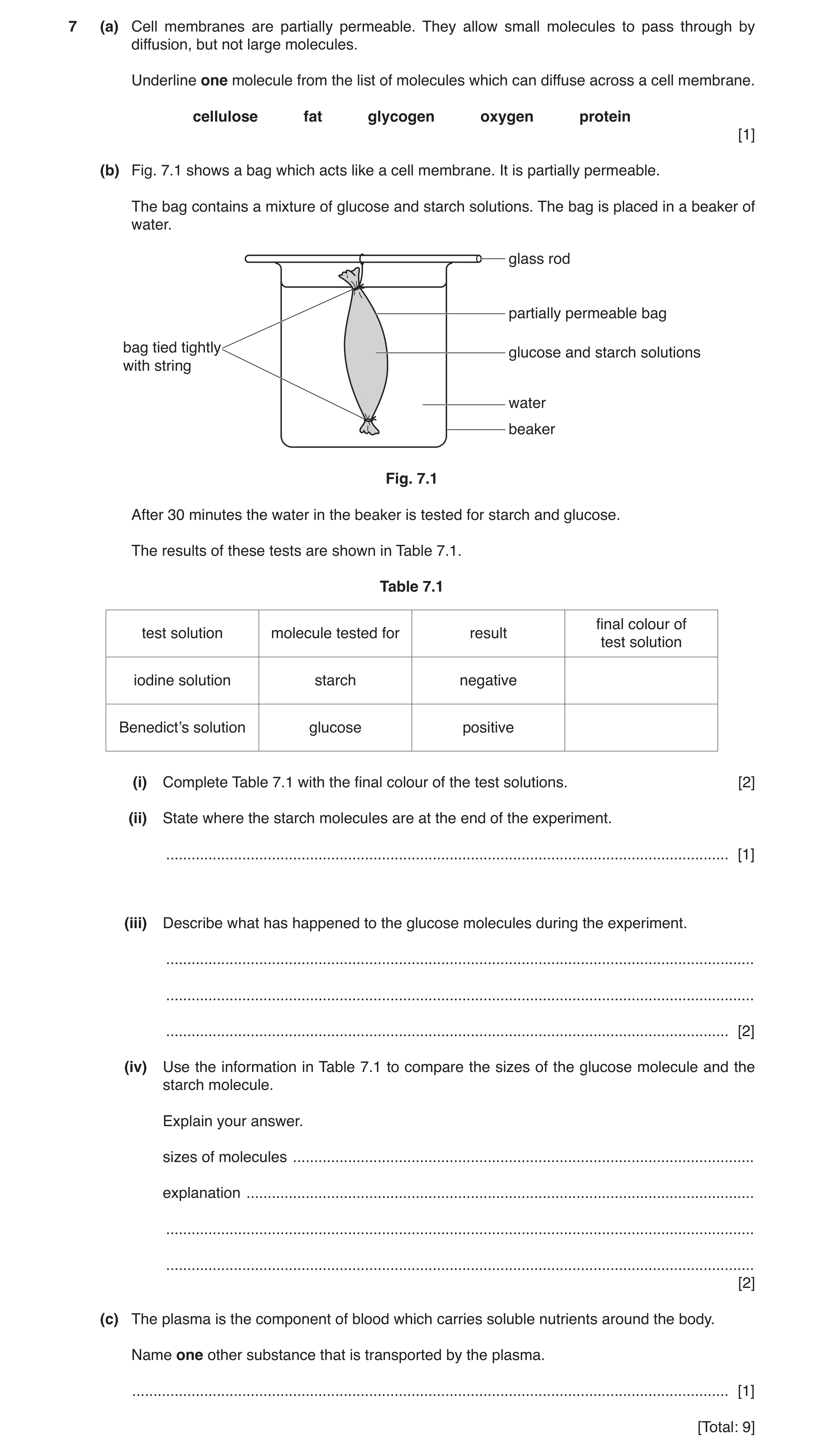
7 (a) Cell membranes are partially permeable. They allow small molecules to pass through by diffusion, but not large molecules. Underline one molecule from the list of molecules which can diffuse across a cell membrane. cellulose fat glycogen oxygen protein [1] (b) Fig. 7.1 shows a bag which acts like a cell membrane. It is partially permeable. The bag contains a mixture of glucose and starch solutions. The bag is placed in a beaker of water. glass rod partially permeable bag bag tied tightly glucose and starch solutions with string water beaker Fig. 7.1 After 30 minutes the water in the beaker is tested for starch and glucose. The results of these tests are shown in Table 7.1. Table 7.1 final colour of test solution molecule tested for result test solution iodine solution starch negative Benedict’s solution glucose positive (i) Complete Table 7.1 with the final colour of the test solutions. [2] (ii) State where the starch molecules are at the end of the experiment. … [1] (iii) Describe what has happened to the glucose molecules during the experiment. … … … [2] (iv) Use the information in Table 7.1 to compare the sizes of the glucose molecule and the starch molecule. Explain your answer. sizes of molecules … explanation … … … [2] (c) The plasma is the component of blood which carries soluble nutrients around the body. Name one other substance that is transported by the plasma. … [1] [Total: 9]
9 marks
Mark scheme: 7(a) oxygen ; 1 7(b)(i) brown ; (brick) red ; 2 7(b)(ii) inside the bag ; 1 7(b)(iii) some / they have moved out of the bag / into the water ; by diffusion / through the membrane ; 2 7(b)(iv) glucose molecules are small(er) / starch molecules are bigg(er) ; glucose molecules can pass through (the bag) / starch molecules cannot pass through (the bag) ; 2 7(c) carbon dioxide / hormones / ions ; 1
This question in 0653/31 May/June 2019
Q7 · The human gas exchange system 0653/33 May/June 2019
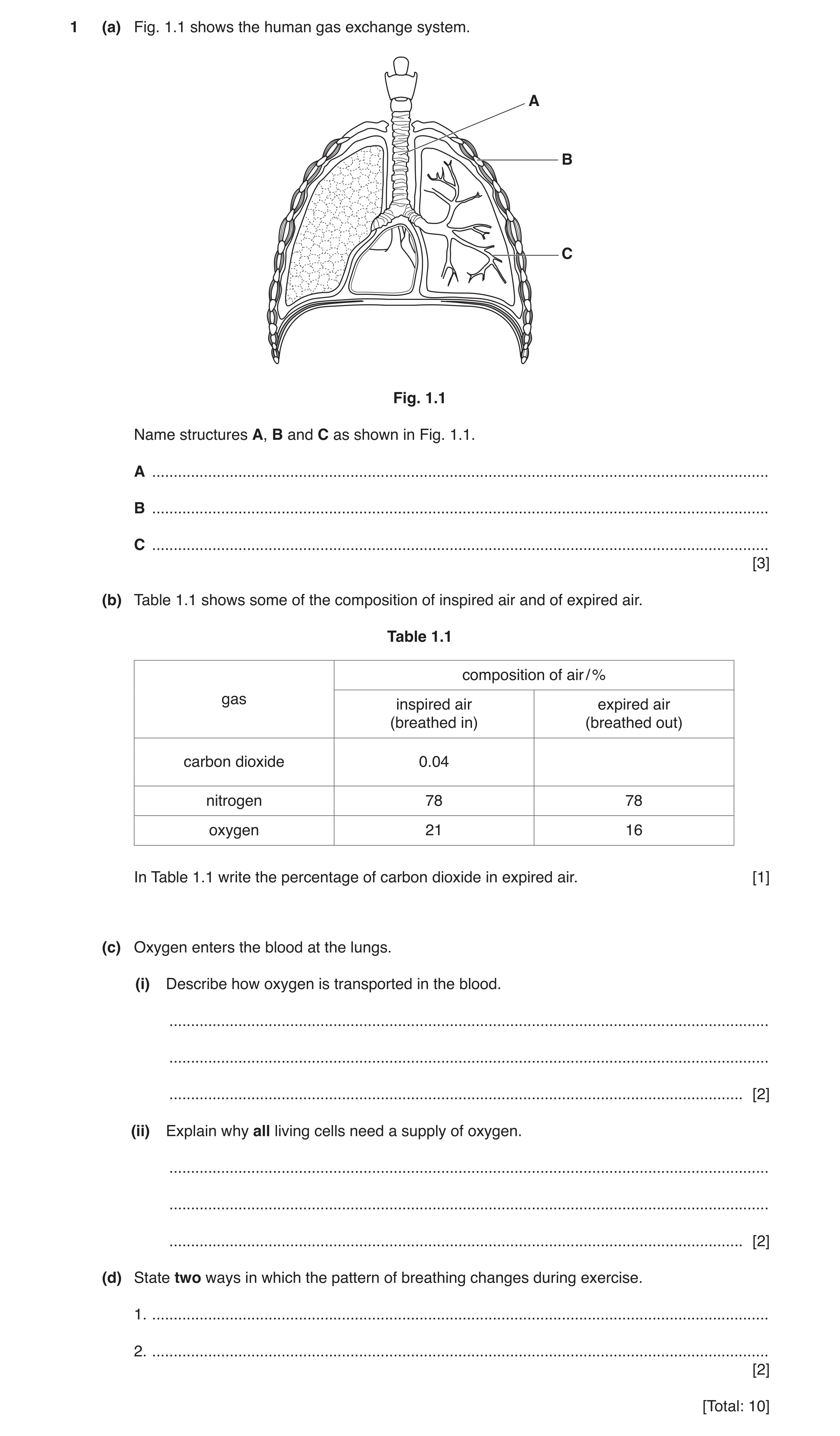
1 (a) Fig. 1.1 shows the human gas exchange system. A B C Fig. 1.1 Name structures A, B and C as shown in Fig. 1.1. A … B … C … [3] (b) Table 1.1 shows some of the composition of inspired air and of expired air. Table 1.1 composition of air / % gas inspired air expired air (breathed in) (breathed out) carbon dioxide 0.04 nitrogen 78 78 oxygen 21 16 In Table 1.1 write the percentage of carbon dioxide in expired air. [1] (c) Oxygen enters the blood at the lungs. (i) Describe how oxygen is transported in the blood. … … … [2] (ii) Explain why all living cells need a supply of oxygen. … … … [2] (d) State two ways in which the pattern of breathing changes during exercise. 1. … 2. … [2] [Total: 10]
10 marks
Mark scheme: 1(a) A trachea ; B rib ; C bronchiole ; 3 1(b) 4 (%) ; 1 1(c)(i) carried by haemoglobin ; in red blood cells ; 2 1(c)(ii) needed for respiration ; reacts with glucose / releases energy ; 2 1(d) breathing rate increases ; depth of breathing increases ; 2
This question in 0653/33 May/June 2019
Q8 · Diagrams of cells as they are seen under a light microscope 0653/31 Oct/Nov 2019
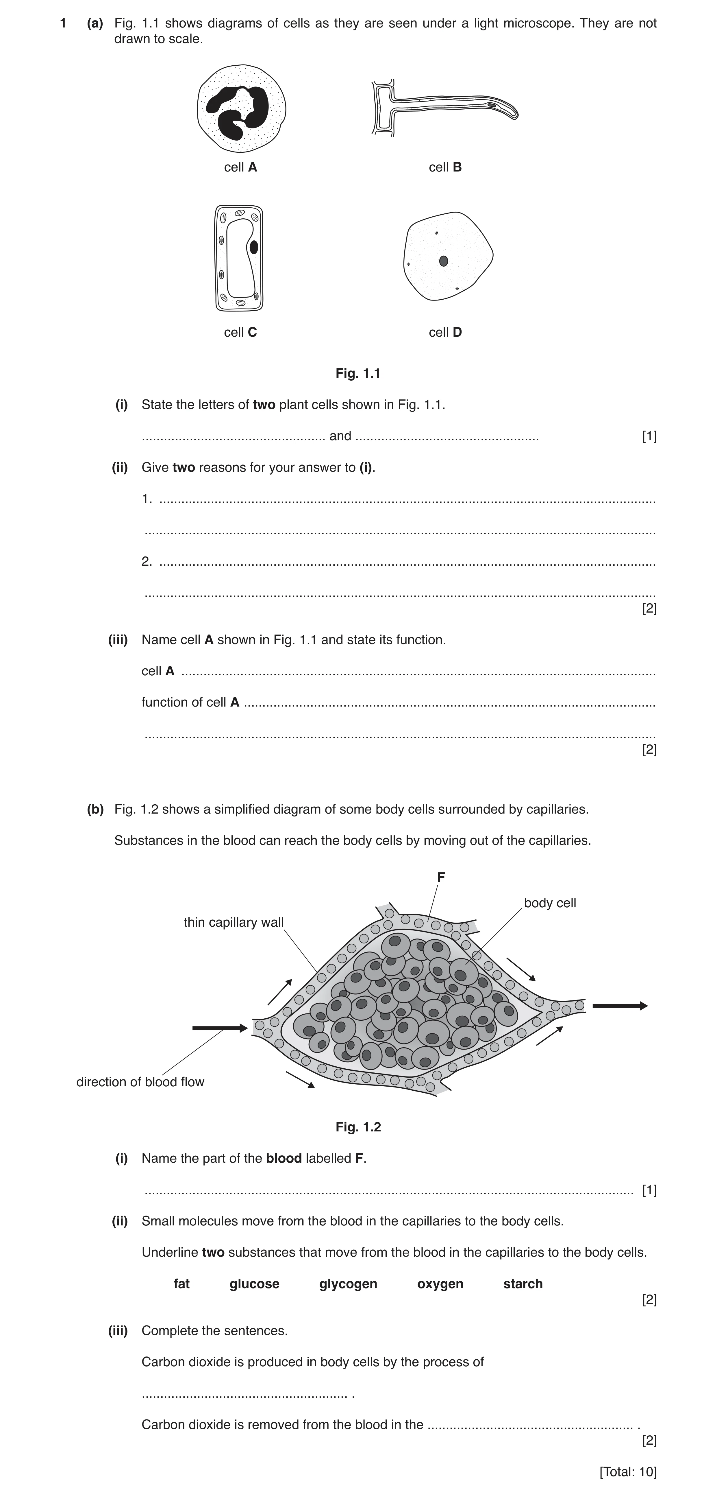
1 (a) Fig. 1.1 shows diagrams of cells as they are seen under a light microscope. They are not drawn to scale. cell A cell B cell C cell D Fig. 1.1 (i) State the letters of two plant cells shown in Fig. 1.1. … and … [1] (ii) Give two reasons for your answer to (i). 1. … … 2. … … [2] (iii) Name cell A shown in Fig. 1.1 and state its function. cell A … function of cell A … … [2] (b) Fig. 1.2 shows a simplified diagram of some body cells surrounded by capillaries. Substances in the blood can reach the body cells by moving out of the capillaries. F body cell thin capillary wall direction of blood flow Fig. 1.2 (i) Name the part of the blood labelled F. … [1] (ii) Small molecules move from the blood in the capillaries to the body cells. Underline two substances that move from the blood in the capillaries to the body cells. fat glucose glycogen oxygen starch [2] (iii) Complete the sentences. Carbon dioxide is produced in body cells by the process of … . Carbon dioxide is removed from the blood in the … . [2] [Total: 10]
10 marks
Mark scheme: 1(a)(i) B and C ; 1 1(a)(ii) any two from: vacuoles ; cell walls ; chloroplasts ; 2 1(a)(iii) white blood cell ; phagocytosis / description of phagocytosis ; 2 1(b)(i) plasma ; 1 1(b)(ii) glucose underlined ; oxygen underlined ; 2 1(b)(iii) respiration ; lungs / alveoli ; 2
This question in 0653/31 Oct/Nov 2019
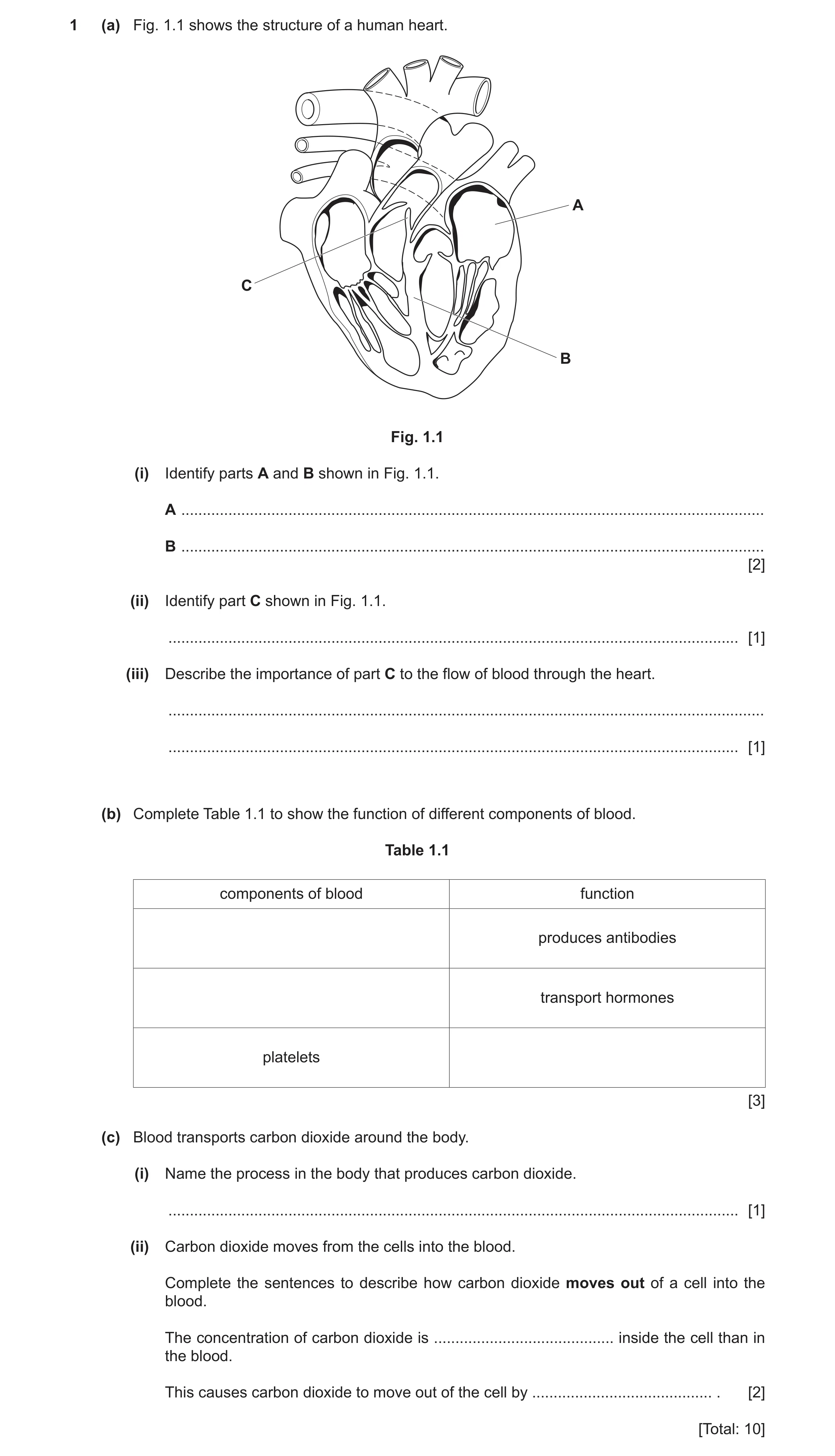
Q9 · The structure of a human heart 0653/31 May/June 2020
1 (a) Fig. 1.1 shows the structure of a human heart. A C B Fig. 1.1 (i) Identify parts A and B shown in Fig. 1.1. A … B … [2] (ii) Identify part C shown in Fig. 1.1. … [1] (iii) Describe the importance of part C to the flow of blood through the heart. … … [1] (b) Complete Table 1.1 to show the function of different components of blood. Table 1.1 components of blood function produces antibodies transport hormones platelets [3] (c) Blood transports carbon dioxide around the body. (i) Name the process in the body that produces carbon dioxide. … [1] (ii) Carbon dioxide moves from the cells into the blood. Complete the sentences to describe how carbon dioxide moves out of a cell into the blood. The concentration of carbon dioxide is … inside the cell than in the blood. This causes carbon dioxide to move out of the cell by … . [2] [Total: 10]
10 marks
Mark scheme: 1(a)(i) A (left) atrium ; B septum ; 2 1(a)(ii) valve ; 1 1(a)(iii) makes sure the blood flows in one direction / prevents backflow ; 1 1(b) white blood cells (produces antibodies) ; plasma (transports hormones) ; (platelets) blood clotting ; 3 1(c)(i) respiration ; 1 1(c)(ii) higher / greater ; diffusion ; 2
This question in 0653/31 May/June 2020
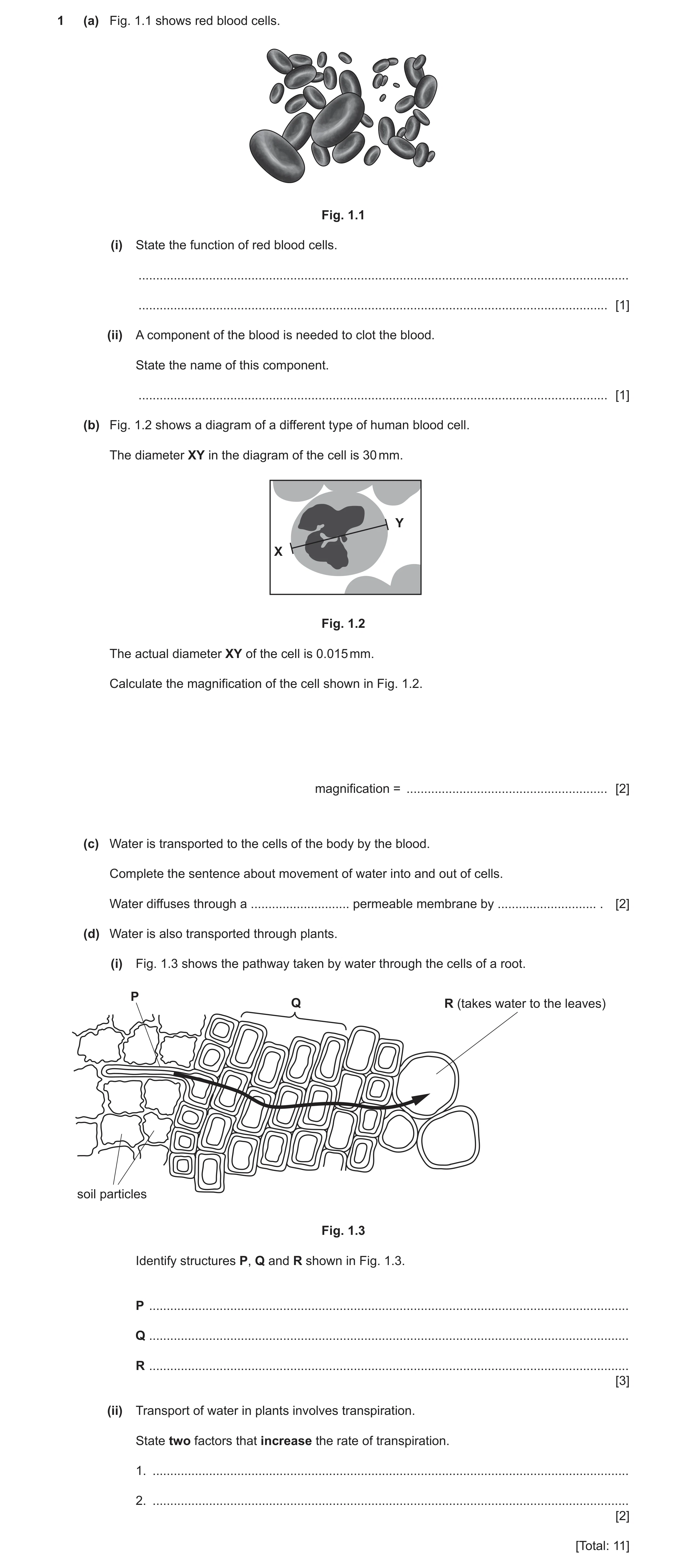
Question 10 0653/32 May/June 2020
1 (a) Fig. 1.1 shows red blood cells. Fig. 1.1 (i) State the function of red blood cells. … … [1] (ii) A component of the blood is needed to clot the blood. State the name of this component. … [1] (b) Fig. 1.2 shows a diagram of a different type of human blood cell. The diameter XY in the diagram of the cell is 30 mm. Y X Fig. 1.2 The actual diameter XY of the cell is 0.015 mm. Calculate the magnification of the cell shown in Fig. 1.2. magnification = … [2] (c) Water is transported to the cells of the body by the blood. Complete the sentence about movement of water into and out of cells. Water diffuses through a … permeable membrane by … . [2] (d) Water is also transported through plants. (i) Fig. 1.3 shows the pathway taken by water through the cells of a root. P Q R (takes water to the leaves) soil particles Fig. 1.3 Identify structures P, Q and R shown in Fig. 1.3. P … Q … R … [3] (ii) Transport of water in plants involves transpiration. State two factors that increase the rate of transpiration. 1. … 2. … [2] [Total: 11]
11 marks
Mark scheme: 1(a)(i) transport oxygen ; 1 1(a)(ii) platelets ; 1 1(b) = 30 / 0.015 ; = 2000 ; 2 1(c) partially ; osmosis ; 2 1(d)(i) P root hair (cell) ; Q root cortex (cell) ; R xylem ; 3 1(d)(ii) increased temperature ; decreased humidity ; 2
This question in 0653/32 May/June 2020
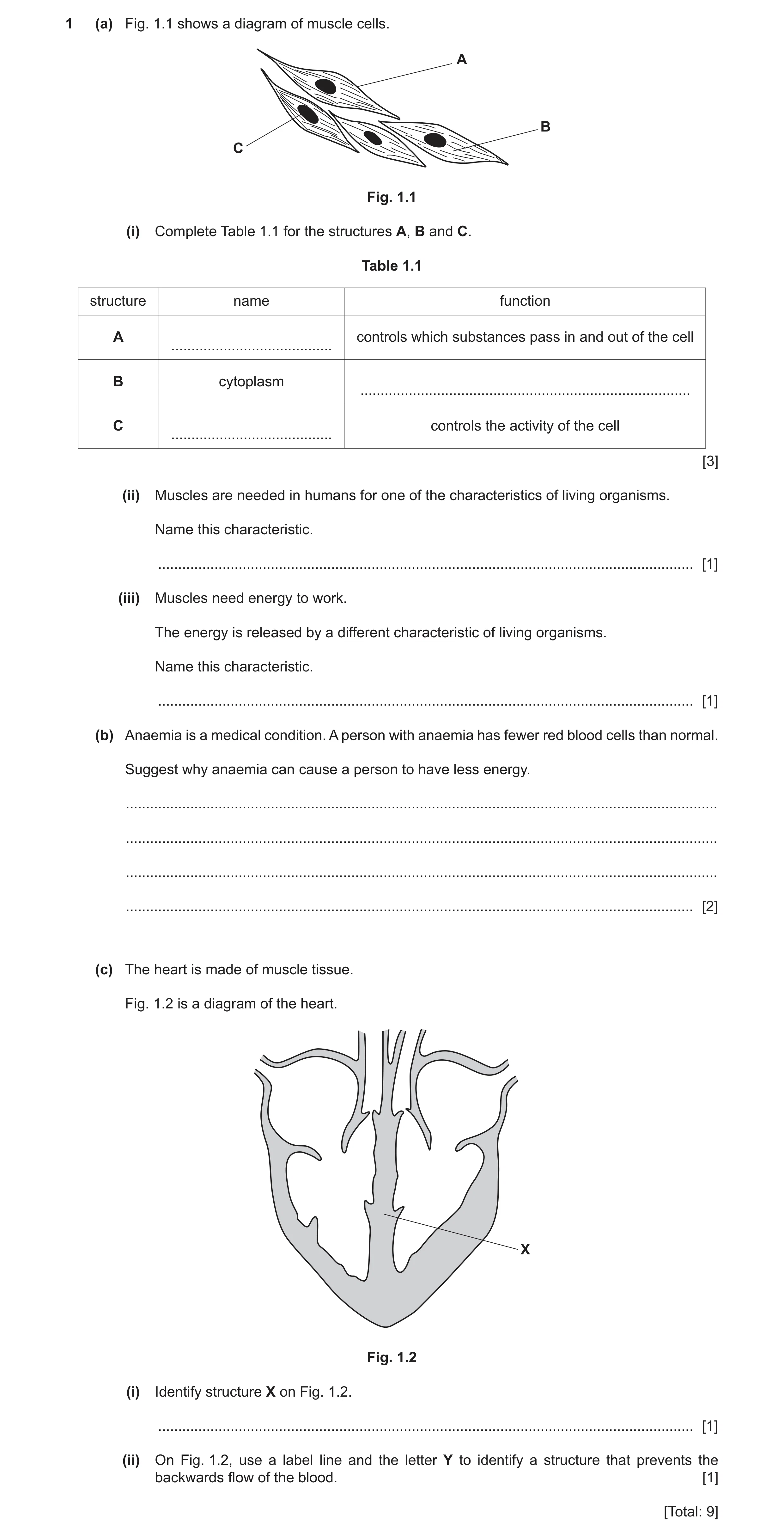
Q11 · A diagram of muscle cells 0653/33 Oct/Nov 2020
1 (a) Fig. 1.1 shows a diagram of muscle cells. A B C Fig. 1.1 (i) Complete Table 1.1 for the structures A, B and C. Table 1.1 structure name function A controls which substances pass in and out of the cell … B cytoplasm … C controls the activity of the cell … [3] (ii) Muscles are needed in humans for one of the characteristics of living organisms. Name this characteristic. … [1] (iii) Muscles need energy to work. The energy is released by a different characteristic of living organisms. Name this characteristic. … [1] (b) Anaemia is a medical condition. A person with anaemia has fewer red blood cells than normal. Suggest why anaemia can cause a person to have less energy. … … … … [2] (c) The heart is made of muscle tissue. Fig. 1.2 is a diagram of the heart. X Fig. 1.2 (i) Identify structure X on Fig. 1.2. … [1] (ii) On Fig. 1.2, use a label line and the letter Y to identify a structure that prevents the backwards flow of the blood. [1] [Total: 9]
9 marks
Mark scheme: 1(a)(i) (cell) membrane ; site of different (metabolic) reactions ; nucleus ; 3 1(a)(ii) movement ; 1 1(a)(iii) respiration ; 1 1(b) any two from: red blood cells transport oxygen ; less oxygen, transported / available ; for (aerobic) respiration / less (aerobic) respiration OR oxygen is needed for respiration ; 2 1(c)(i) septum ; 1 1(c)(ii) label line to any valve ; 1
This question in 0653/33 Oct/Nov 2020
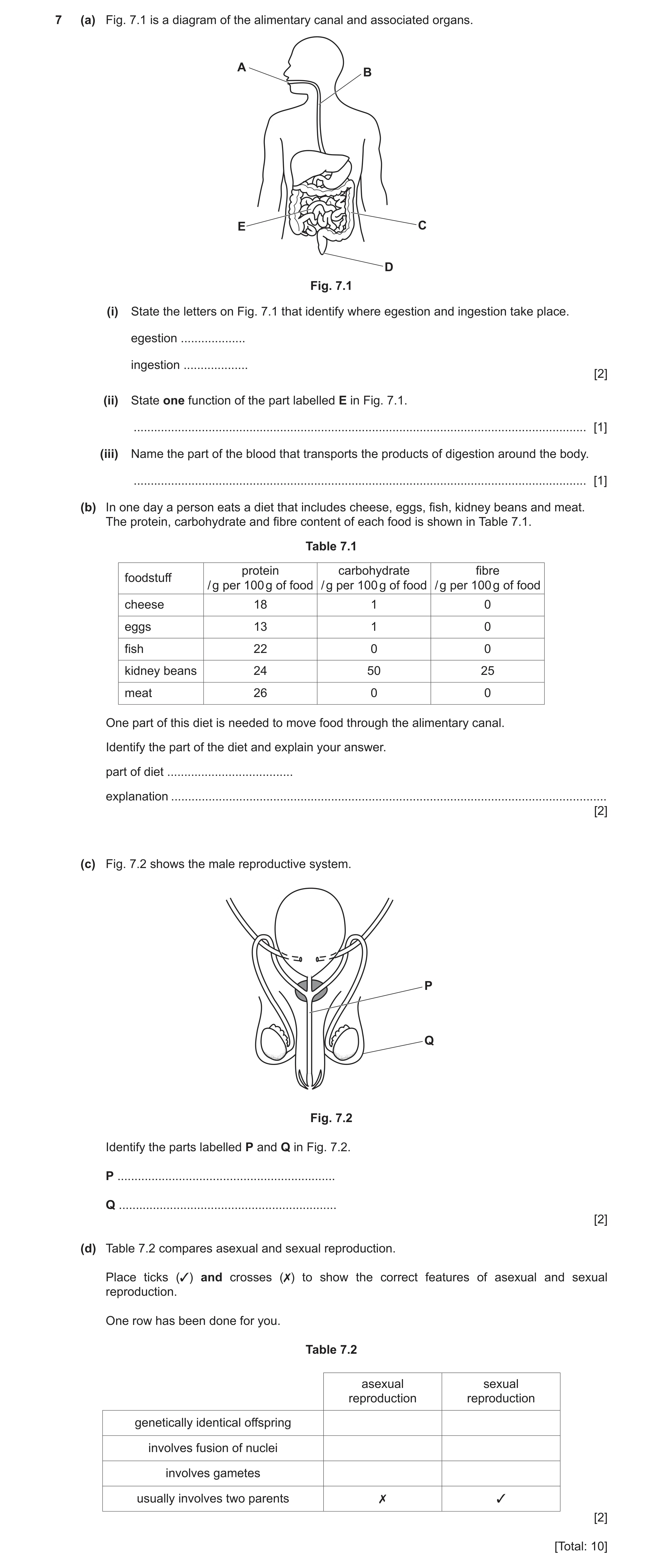
Q12 · A diagram of the alimentary canal and associated organs 0653/31 May/June 2021
7 (a) Fig. 7.1 is a diagram of the alimentary canal and associated organs. A B E C D Fig. 7.1 (i) State the letters on Fig. 7.1 that identify where egestion and ingestion take place. egestion … ingestion … [2] (ii) State one function of the part labelled E in Fig. 7.1. … [1] (iii) Name the part of the blood that transports the products of digestion around the body. … [1] (b) In one day a person eats a diet that includes cheese, eggs, fish, kidney beans and meat. The protein, carbohydrate and fibre content of each food is shown in Table 7.1. Table 7.1 protein carbohydrate fibre foodstuff / g per 100 g of food / g per 100 g of food / g per 100 g of food cheese 18 1 0 eggs 13 1 0 fish 22 0 0 kidney beans 24 50 25 meat 26 0 0 One part of this diet is needed to move food through the alimentary canal. Identify the part of the diet and explain your answer. part of diet … explanation … [2] (c) Fig. 7.2 shows the male reproductive system. P Q Fig. 7.2 Identify the parts labelled P and Q in Fig. 7.2. P … Q … [2] (d) Table 7.2 compares asexual and sexual reproduction. Place ticks (✓) and crosses (✗) to show the correct features of asexual and sexual reproduction. One row has been done for you. Table 7.2 asexual sexual reproduction reproduction genetically identical offspring involves fusion of nuclei involves gametes usually involves two parents ✗ ✓ [2] [Total: 10]
10 marks
Mark scheme: 7(a)(i) egestion: D ; ingestion: A ; 2 7(a)(ii) digestion / absorption ; 1 7(a)(iii) plasma ; 1 7(b) part of diet: kidney beans ; explanation: contain fibre ; 2 7(c) P urethra ; Q scrotum ; 2 7(d) asexual reproduction sexual reproduction genetically identical offspring x involves fusion of nuclei x involves gametes x usually involves two parents (x) () ;; 2 marks for all 6 correct 1 mark for 4 correct 2
This question in 0653/31 May/June 2021
Q13 · The boxes on the left show different components of blood 0653/33 May/June 2021
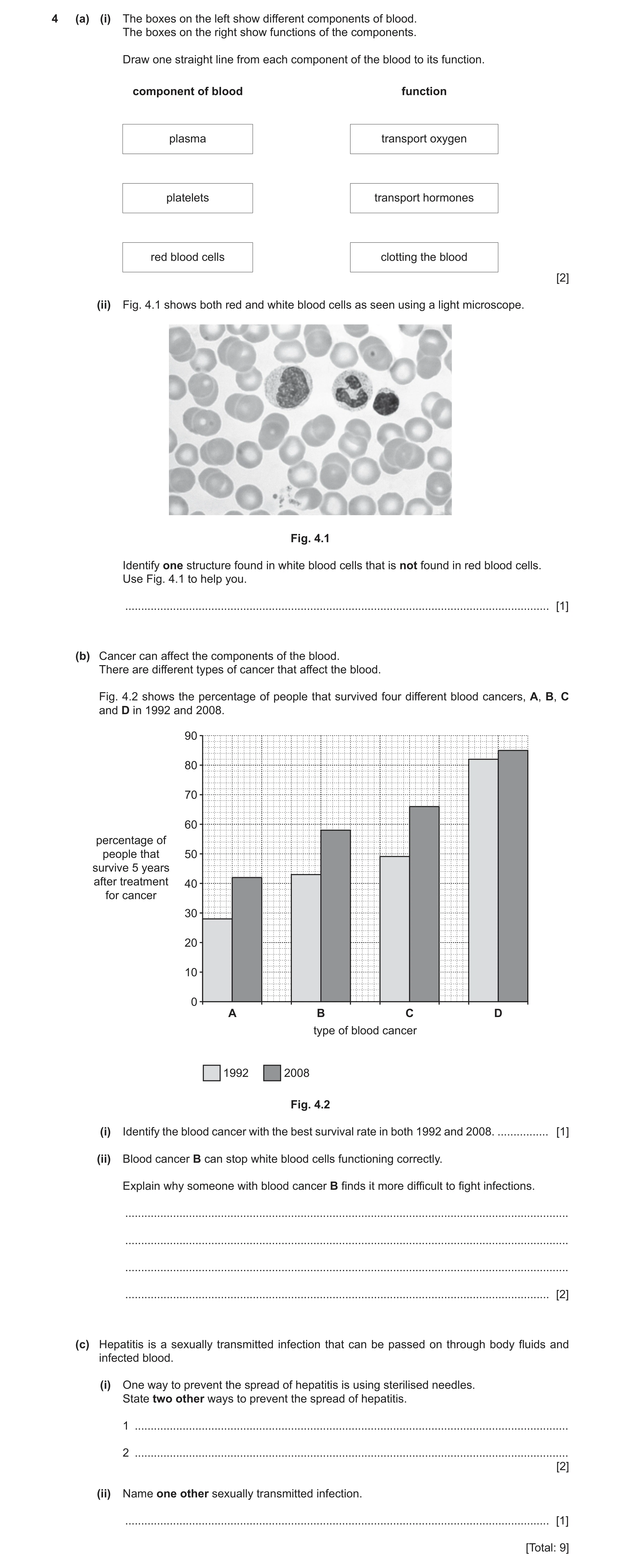
4 (a) (i) The boxes on the left show different components of blood. The boxes on the right show functions of the components. Draw one straight line from each component of the blood to its function. component of blood function plasma transport oxygen platelets transport hormones red blood cells clotting the blood [2] (ii) Fig. 4.1 shows both red and white blood cells as seen using a light microscope. Fig. 4.1 Identify one structure found in white blood cells that is not found in red blood cells. Use Fig. 4.1 to help you. … [1] (b) Cancer can affect the components of the blood. There are different types of cancer that affect the blood. Fig. 4.2 shows the percentage of people that survived four different blood cancers, A, B, C and D in 1992 and 2008. 90 80 70 60 percentage of people that 50 survive 5 years after treatment 40 for cancer 30 20 10 0 A B C D type of blood cancer 1992 2008 Fig. 4.2 (i) Identify the blood cancer with the best survival rate in both 1992 and 2008. … [1] (ii) Blood cancer B can stop white blood cells functioning correctly. Explain why someone with blood cancer B finds it more difficult to fight infections. … … … … [2] (c) Hepatitis is a sexually transmitted infection that can be passed on through body fluids and infected blood. (i) One way to prevent the spread of hepatitis is using sterilised needles. State two other ways to prevent the spread of hepatitis. 1 … 2 … [2] (ii) Name one other sexually transmitted infection. … [1] [Total: 9]
9 marks
Mark scheme: 4(a)(i) plasma transport oxygen platelets transport hormones red blood cells clotting the blood ;; all correct = 2 marks one or two correct = 1 mark 2 4(a)(ii) nucleus ; 1 4(b)(i) D ; 1 4(b)(ii) idea of not being able to make antibodies ; idea of not being able to engulf microbes / not able to carry out phagocytosis ; 2 4(c)(i) any two from: abstinence from, sex / sexual intercourse ; use of condom / femidom ; blood screening ; not sharing (syringe) needles ; gloves used by health professionals ; better education / raise awareness ; 2 4(c)(ii) HIV / AIDS ; 1
This question in 0653/33 May/June 2021
Q14 · The boxes on the left show some of the components of blood 0653/32 Feb/March 2022
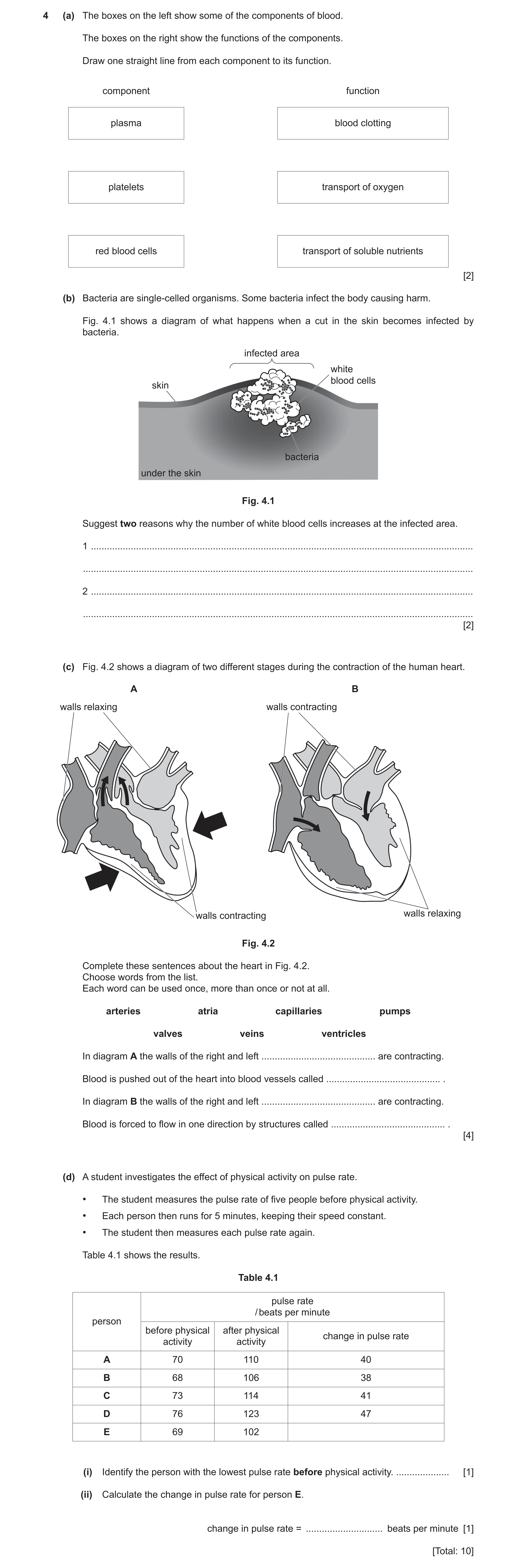
4 (a) The boxes on the left show some of the components of blood. The boxes on the right show the functions of the components. Draw one straight line from each component to its function. component function plasma blood clotting platelets transport of oxygen red blood cells transport of soluble nutrients [2] (b) Bacteria are single-celled organisms. Some bacteria infect the body causing harm. Fig. 4.1 shows a diagram of what happens when a cut in the skin becomes infected by bacteria. infected area white blood cells skin bacteria under the skin Fig. 4.1 Suggest two reasons why the number of white blood cells increases at the infected area. 1 … … 2 … … [2] (c) Fig. 4.2 shows a diagram of two different stages during the contraction of the human heart. A B walls relaxing walls contracting walls contracting walls relaxing Fig. 4.2 Complete these sentences about the heart in Fig. 4.2. Choose words from the list. Each word can be used once, more than once or not at all. arteries atria capillaries pumps valves veins ventricles In diagram A the walls of the right and left … are contracting. Blood is pushed out of the heart into blood vessels called … . In diagram B the walls of the right and left … are contracting. Blood is forced to flow in one direction by structures called … . [4] (d) A student investigates the effect of physical activity on pulse rate. • The student measures the pulse rate of five people before physical activity. • Each person then runs for 5 minutes, keeping their speed constant. • The student then measures each pulse rate again. Table 4.1 shows the results. Table 4.1 pulse rate / beats per minute person before physical after physical change in pulse rate activity activity A 70 110 40 B 68 106 38 C 73 114 41 D 76 123 47 E 69 102 (i) Identify the person with the lowest pulse rate before physical activity. … [1] (ii) Calculate the change in pulse rate for person E. change in pulse rate = … beats per minute [1] [Total: 10]
10 marks
Mark scheme: 4(a) plasma blood clotting platelets transport of oxygen red blood cells transport of soluble nutrients ;; 4(b) phagocytosis ; to produce antibodies ; 2 4(c) ventricles ; arteries ; atria ; valves ; 4 4(d)(i) B ; 1 4(d)(ii) 33 (bpm) ; 1
This question in 0653/32 Feb/March 2022
Q15 · An image of a white blood cell 0653/31 May/June 2022
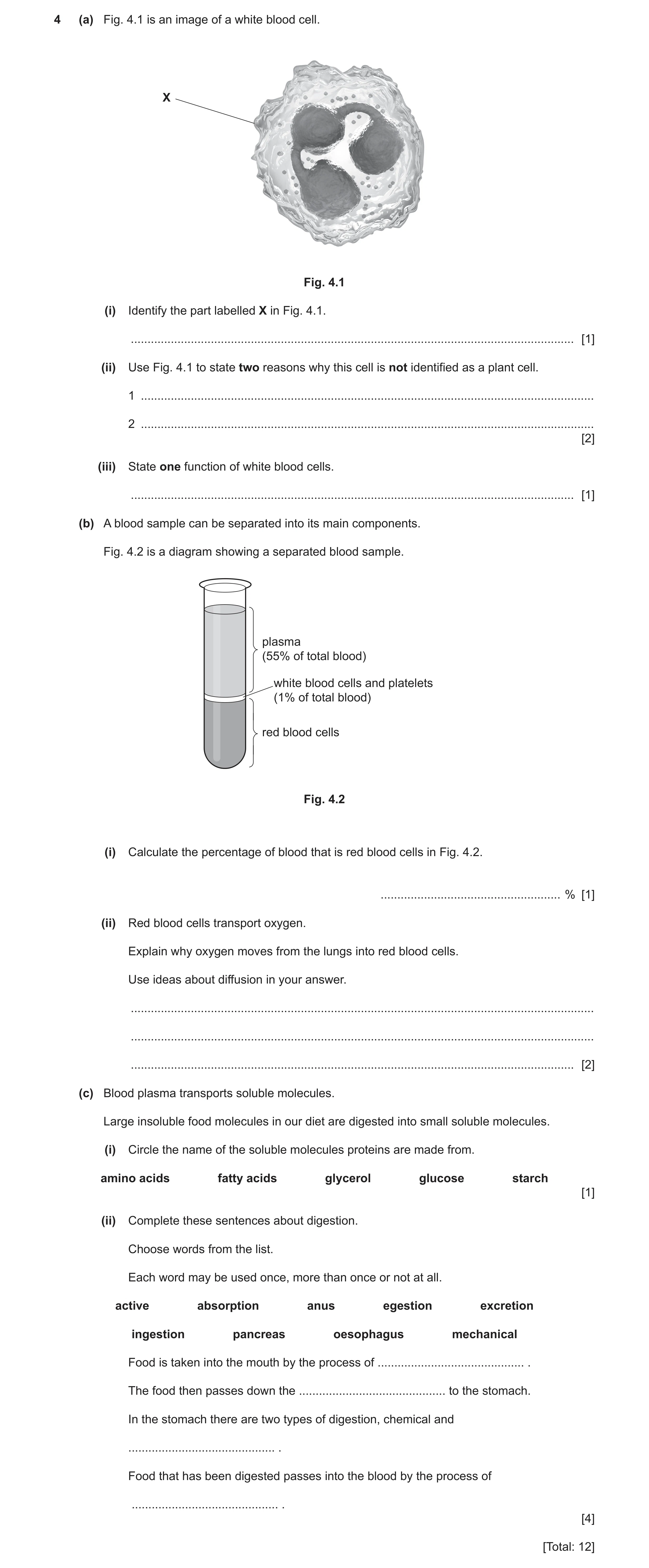
4 (a) Fig. 4.1 is an image of a white blood cell. X Fig. 4.1 (i) Identify the part labelled X in Fig. 4.1. … [1] (ii) Use Fig. 4.1 to state two reasons why this cell is not identified as a plant cell. 1 … 2 … [2] (iii) State one function of white blood cells. … [1] (b) A blood sample can be separated into its main components. Fig. 4.2 is a diagram showing a separated blood sample. plasma (55% of total blood) white blood cells and platelets (1% of total blood) red blood cells Fig. 4.2 (i) Calculate the percentage of blood that is red blood cells in Fig. 4.2. … % [1] (ii) Red blood cells transport oxygen. Explain why oxygen moves from the lungs into red blood cells. Use ideas about diffusion in your answer. … … … [2] (c) Blood plasma transports soluble molecules. Large insoluble food molecules in our diet are digested into small soluble molecules. (i) Circle the name of the soluble molecules proteins are made from. amino acids fatty acids glycerol glucose starch [1] (ii) Complete these sentences about digestion. Choose words from the list. Each word may be used once, more than once or not at all. active absorption anus egestion excretion ingestion pancreas oesophagus mechanical Food is taken into the mouth by the process of … . The food then passes down the … to the stomach. In the stomach there are two types of digestion, chemical and … . Food that has been digested passes into the blood by the process of … . [4] [Total: 12]
12 marks
Mark scheme: 4(a)(i) cell membrane ; 1 4(a)(ii) any two from: no cell wall ; no (permanent) vacuole ; no chloroplast ; 2 4(a)(iii) produce antibodies / phagocytosis ; 1 4(b)(i) 44(%) ; 1 4(b)(ii) higher concentration oxygen (in lungs) / ora ; oxygen moves down concentration gradient / oxygen moves (from a higher concentration) to a lower concentration ; 2 4(c)(i) amino acids (circled) ; 1 Question Answer Marks 4(c)(ii) ingestion ; oesophagus ; mechanical ; absorption ; 4
This question in 0653/31 May/June 2022
Q16 · A diagram of the human gas exchange system 0653/32 May/June 2022
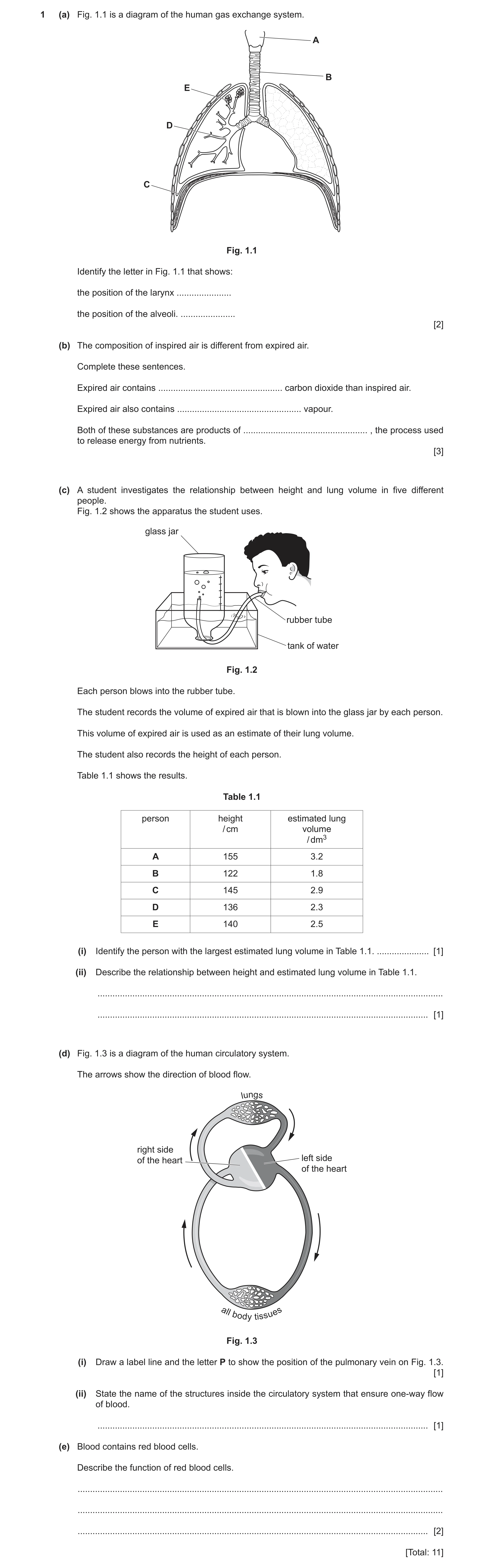
1 (a) Fig. 1.1 is a diagram of the human gas exchange system. A B E D C Fig. 1.1 Identify the letter in Fig. 1.1 that shows: the position of the larynx … the position of the alveoli. … [2] (b) The composition of inspired air is different from expired air. Complete these sentences. Expired air contains … carbon dioxide than inspired air. Expired air also contains … vapour. Both of these substances are products of … , the process used to release energy from nutrients. [3] (c) A student investigates the relationship between height and lung volume in five different people. Fig. 1.2 shows the apparatus the student uses. glass jar rubber tube tank of water Fig. 1.2 Each person blows into the rubber tube. The student records the volume of expired air that is blown into the glass jar by each person. This volume of expired air is used as an estimate of their lung volume. The student also records the height of each person. Table 1.1 shows the results. Table 1.1 person height estimated lung / cm volume / dm3 A 155 3.2 B 122 1.8 C 145 2.9 D 136 2.3 E 140 2.5 (i) Identify the person with the largest estimated lung volume in Table 1.1. … [1] (ii) Describe the relationship between height and estimated lung volume in Table 1.1. … … [1] (d) Fig. 1.3 is a diagram of the human circulatory system. The arrows show the direction of blood flow. lungs right side of the heart left side of the heart all body tissues Fig. 1.3 (i) Draw a label line and the letter P to show the position of the pulmonary vein on Fig. 1.3. [1] (ii) State the name of the structures inside the circulatory system that ensure one-way flow of blood. … [1] (e) Blood contains red blood cells. Describe the function of red blood cells. … … … [2] [Total: 11]
11 marks
Mark scheme: 1(a) A ; E ; 2 1(b) more ; water ; respiration ; 3 1(c)(i) (person) A ; 1 1(c)(ii) taller the person / as the height increases, the greater the (estimated) lung volume ; 1 1(d)(i) pulmonary vein labelled ; 1 1(d)(ii) valves ; 1 1(e) transport oxygen ; ref. haemoglobin ; 2
This question in 0653/32 May/June 2022
Q17 · A diagram of a human heart 0653/32 May/June 2023
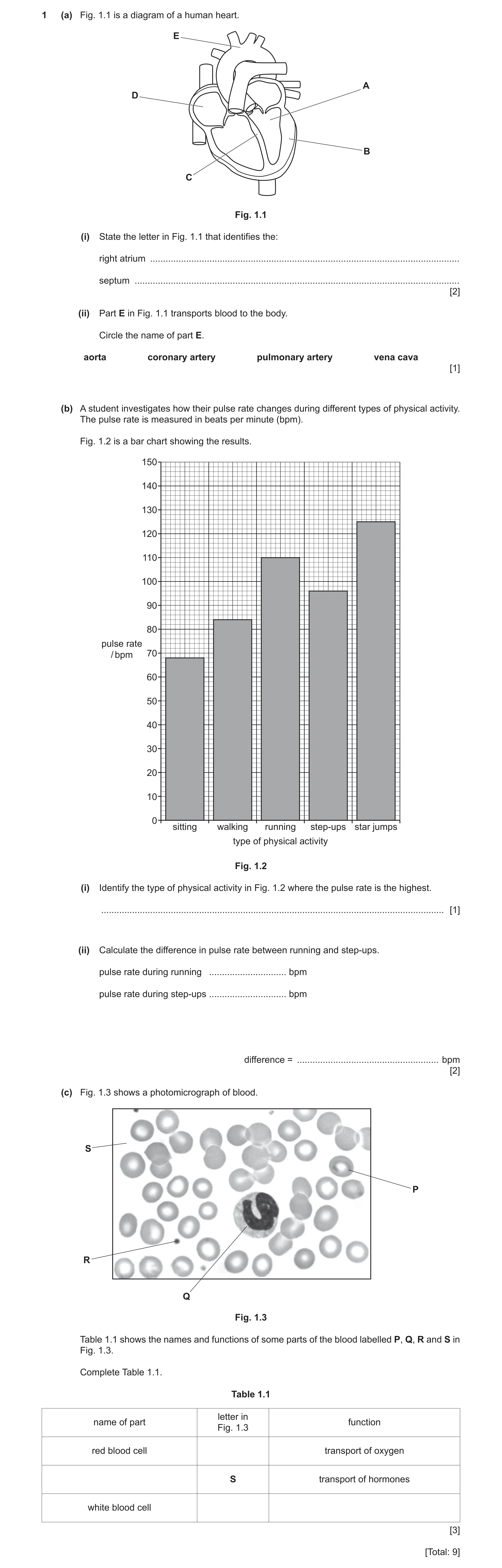
1 (a) Fig. 1.1 is a diagram of a human heart. E A D B C Fig. 1.1 (i) State the letter in Fig. 1.1 that identifies the: right atrium … septum … [2] (ii) Part E in Fig. 1.1 transports blood to the body. Circle the name of part E. aorta coronary artery pulmonary artery vena cava [1] (b) A student investigates how their pulse rate changes during different types of physical activity. The pulse rate is measured in beats per minute (bpm). Fig. 1.2 is a bar chart showing the results. 150 140 130 120 110 100 90 80 pulse rate / bpm 70 60 50 40 30 20 10 0 sitting walking running step-ups star jumps type of physical activity Fig. 1.2 (i) Identify the type of physical activity in Fig. 1.2 where the pulse rate is the highest. … [1] (ii) Calculate the difference in pulse rate between running and step‑ups. pulse rate during running … bpm pulse rate during step‑ups … bpm difference = … bpm [2] (c) Fig. 1.3 shows a photomicrograph of blood. S P R Q Fig. 1.3 Table 1.1 shows the names and functions of some parts of the blood labelled P, Q, R and S in Fig. 1.3. Complete Table 1.1. Table 1.1 letter in name of part function Fig. 1.3 red blood cell transport of oxygen S transport of hormones white blood cell [3] [Total: 9]
9 marks
Mark scheme: 1(a)(i) D ; C ; 2 1(a)(ii) aorta (circled) ; 1 1(b)(i) star jumps ; 1 1(b)(ii) (running) 110 (bpm) and (step-ups) 96 (bpm) ; (difference =) 14 (bpm) ; 2 1(c) name of part letter in Fig. 1.3 function red blood cell P transport of oxygen plasma S transport of hormones white blood cells Q phagocytosis / produce antibodies ;;; one mark for each correct row 3
This question in 0653/32 May/June 2023
Q18 · A diagram of the alimentary canal and associated organs 0653/33 May/June 2023
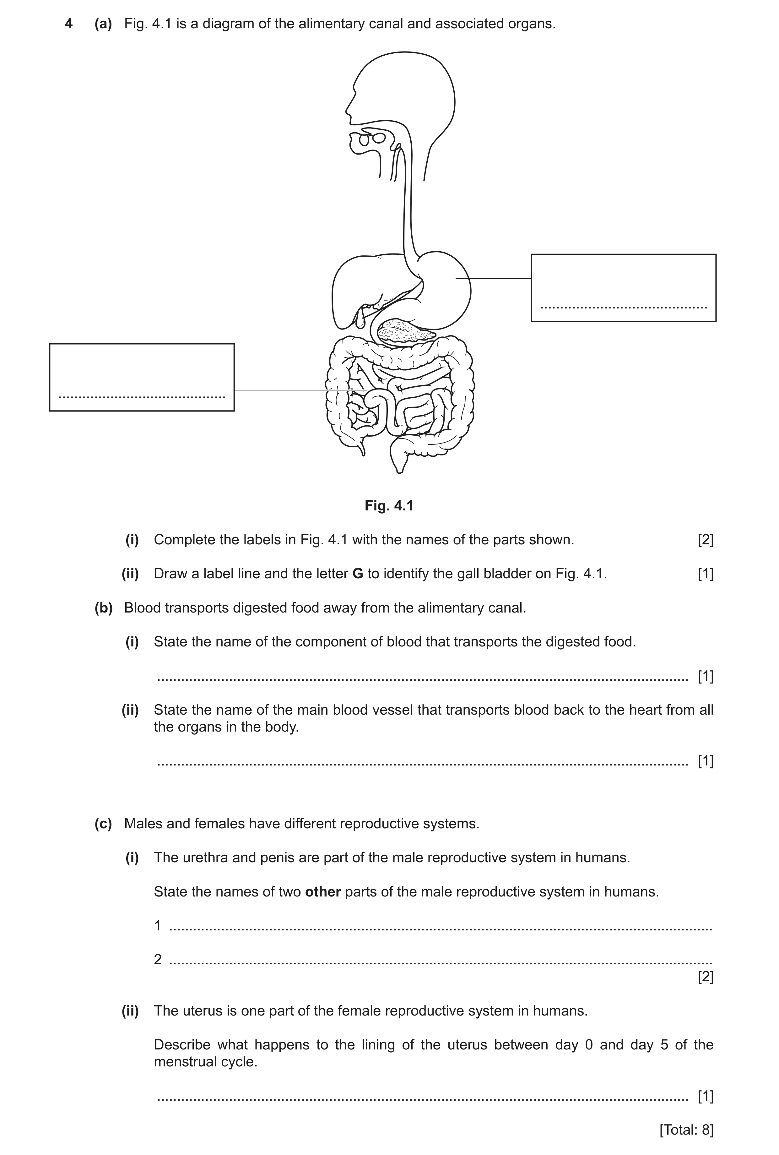
4 (a) Fig. 4.1 is a diagram of the alimentary canal and associated organs. … … Fig. 4.1 (i) Complete the labels in Fig. 4.1 with the names of the parts shown. [2] (ii) Draw a label line and the letter G to identify the gall bladder on Fig. 4.1. [1] (b) Blood transports digested food away from the alimentary canal. (i) State the name of the component of blood that transports the digested food. … [1] (ii) State the name of the main blood vessel that transports blood back to the heart from all the organs in the body. … [1] (c) Males and females have different reproductive systems. (i) The urethra and penis are part of the male reproductive system in humans. State the names of two other parts of the male reproductive system in humans. 1 … 2 … [2] (ii) The uterus is one part of the female reproductive system in humans. Describe what happens to the lining of the uterus between day 0 and day 5 of the menstrual cycle. … [1] [Total: 8]
8 marks
Mark scheme: 4(a)(i) small intestine ; stomach ; 2 4(a)(ii) gall bladder labelled ; 1 4(b)(i) plasma ; 1 4(b)(ii) vena cava ; 1 4(c)(i) any two from: testes ; scrotum ; sperm duct ; prostate gland ; 2 4(c)(ii) thickness decreases / is shed ; 1
This question in 0653/33 May/June 2023
Q19 · Blood and blood vessels are part of the circulatory system in humans 0653/32 Feb/March 2024
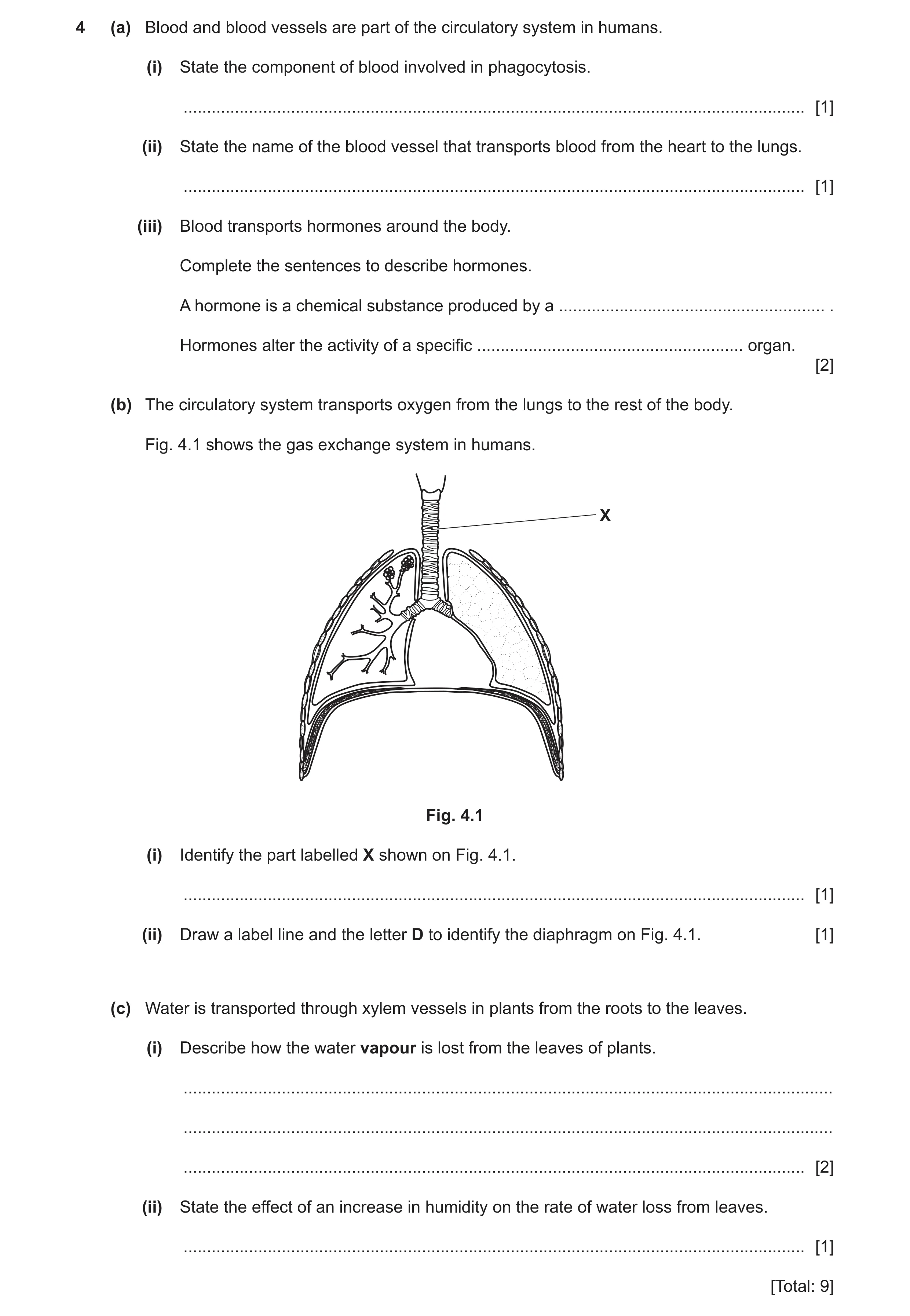
4 (a) Blood and blood vessels are part of the circulatory system in humans. (i) State the component of blood involved in phagocytosis. … [1] (ii) State the name of the blood vessel that transports blood from the heart to the lungs. … [1] (iii) Blood transports hormones around the body. Complete the sentences to describe hormones. A hormone is a chemical substance produced by a … . Hormones alter the activity of a specific … organ. [2] (b) The circulatory system transports oxygen from the lungs to the rest of the body. Fig. 4.1 shows the gas exchange system in humans. X Fig. 4.1 (i) Identify the part labelled X shown on Fig. 4.1. … [1] (ii) Draw a label line and the letter D to identify the diaphragm on Fig. 4.1. [1] (c) Water is transported through xylem vessels in plants from the roots to the leaves. (i) Describe how the water vapour is lost from the leaves of plants. … … … [2] (ii) State the effect of an increase in humidity on the rate of water loss from leaves. … [1] [Total: 9]
9 marks
Mark scheme: 4(a)(i) white blood cells ; 1 4(a)(ii) pulmonary artery ; 1 4(a)(iii) gland ; 2 target ; 4(b)(i) trachea ; 1 4(b)(ii) diaphragm labelled ; 1 4(c)(i) diffusion ; 2 through stomata ; 4(c)(ii) decreases ; 1
This question in 0653/32 Feb/March 2024
Question 20 0653/32 May/June 2024
4 (a) Fig. 4.1a shows a white blood cell. Fig. 4.1b shows a type of plant cell. NOT TO SCALE Fig. 4.1a Fig. 4.1b (i) State the type of plant cell shown in Fig. 4.1b. … [1] (ii) Identify one visible structure in the plant cell in Fig. 4.1b that is: also visible in the white blood cell … not present in the white blood cell. … [2] (b) Water moves into the plant cell from the soil. (i) Describe the process of water movement into the plant cell. … … … [2] (ii) State the name of the vessels that transport water up the stem to the leaves. … [1] (c) White blood cells are part of the circulatory system. (i) State two functions of white blood cells. 1 … 2 … [2] (ii) Fig. 4.2 shows a photomicrograph of blood vessels from the circulatory system. lumen (inside space) of vein artery wall Fig. 4.2 Complete these sentences about Fig. 4.2. The artery has a much … wall compared to the vein. This is because the artery transports blood away from the … at high pressure. [2] [Total: 10]
10 marks
Mark scheme: 4(a)(i) root hair (cell) ; 1 4(a)(ii) (also visible in the white blood cell) nucleus / cytoplasm ; (not present in the white blood cell) vacuole / (cell) wall ; 2 4(b)(i) any two from: diffuses ; by osmosis ; through, cell membrane / partially permeable membrane ; 2 Question Answer Marks 4(b)(ii) xylem ; 1 4(c)(i) phagocytosis ; produce antibodies ; 2 4(c)(ii) thicker / more elastic ; heart ; 2
This question in 0653/32 May/June 2024
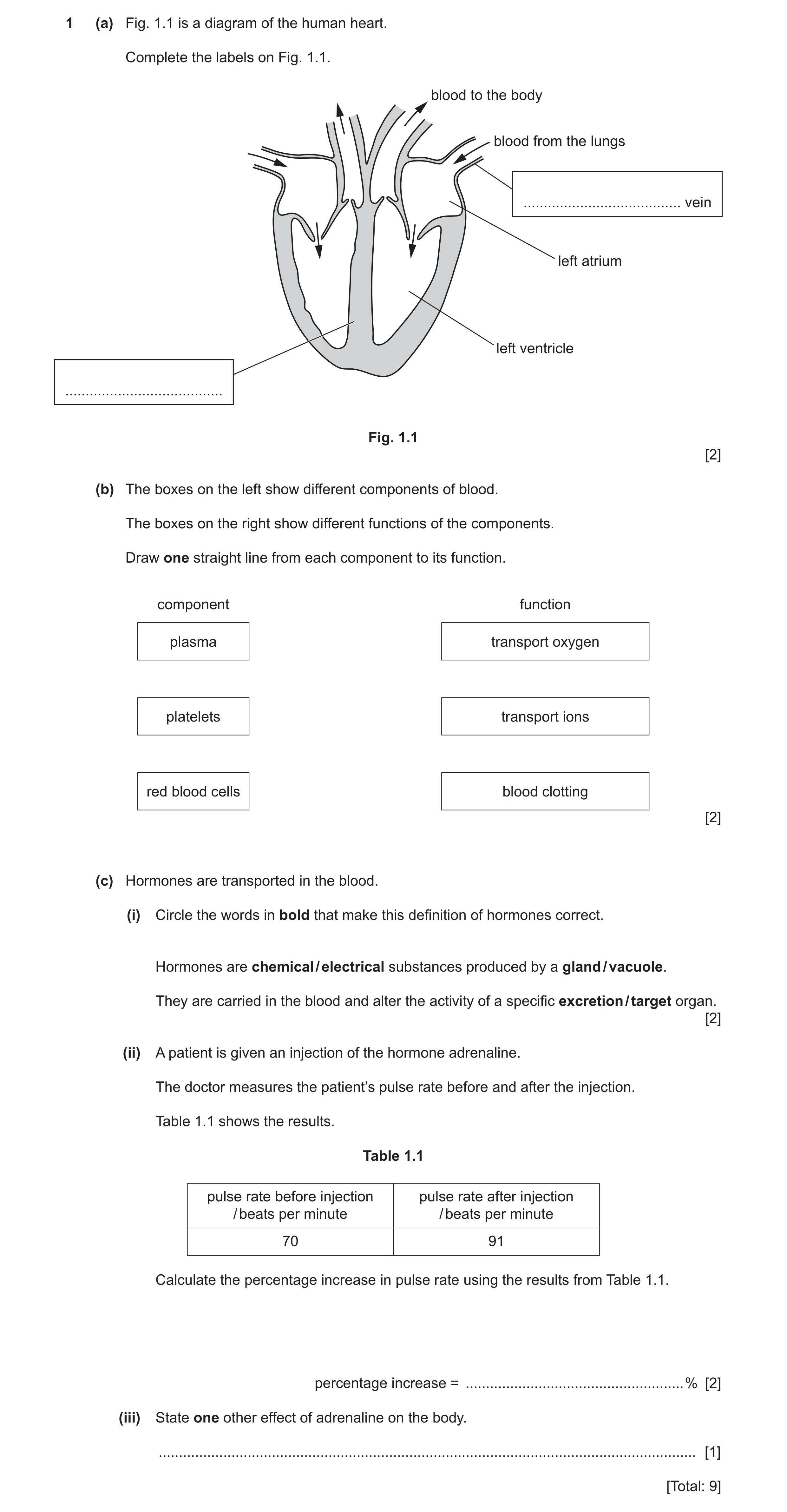
Q21 · A diagram of the human heart 0653/33 May/June 2024
1 (a) Fig. 1.1 is a diagram of the human heart. Complete the labels on Fig. 1.1. blood to the body blood from the lungs … vein left atrium left ventricle … Fig. 1.1 [2] (b) The boxes on the left show different components of blood. The boxes on the right show different functions of the components. Draw one straight line from each component to its function. component function plasma transport oxygen platelets transport ions red blood cells blood clotting [2] (c) Hormones are transported in the blood. (i) Circle the words in bold that make this definition of hormones correct. Hormones are chemical / electrical substances produced by a gland / vacuole. They are carried in the blood and alter the activity of a specific excretion / target organ. [2] (ii) A patient is given an injection of the hormone adrenaline. The doctor measures the patient’s pulse rate before and after the injection. Table 1.1 shows the results. Table 1.1 pulse rate before injection pulse rate after injection / beats per minute / beats per minute 70 91 Calculate the percentage increase in pulse rate using the results from Table 1.1. percentage increase = … % [2] (iii) State one other effect of adrenaline on the body. … [1] [Total: 9]
9 marks
Mark scheme: 1(a) pulmonary (vein) ; septum ; 2 1(b) plasma transport oxygen platelets transport ions red blood cells blood clotting ;; all three correct = 2 marks one or two correct = 1 mark 2 1(c)(i) chemical gland target ;; all three correct = 2 marks two correct = 1 mark 2 1(c)(ii) (91–70 =) 21 ; 21 100 70 30 (%) ; 2 1(c)(iii) increases breathing rate / widens pupils ; 1
This question in 0653/33 May/June 2024
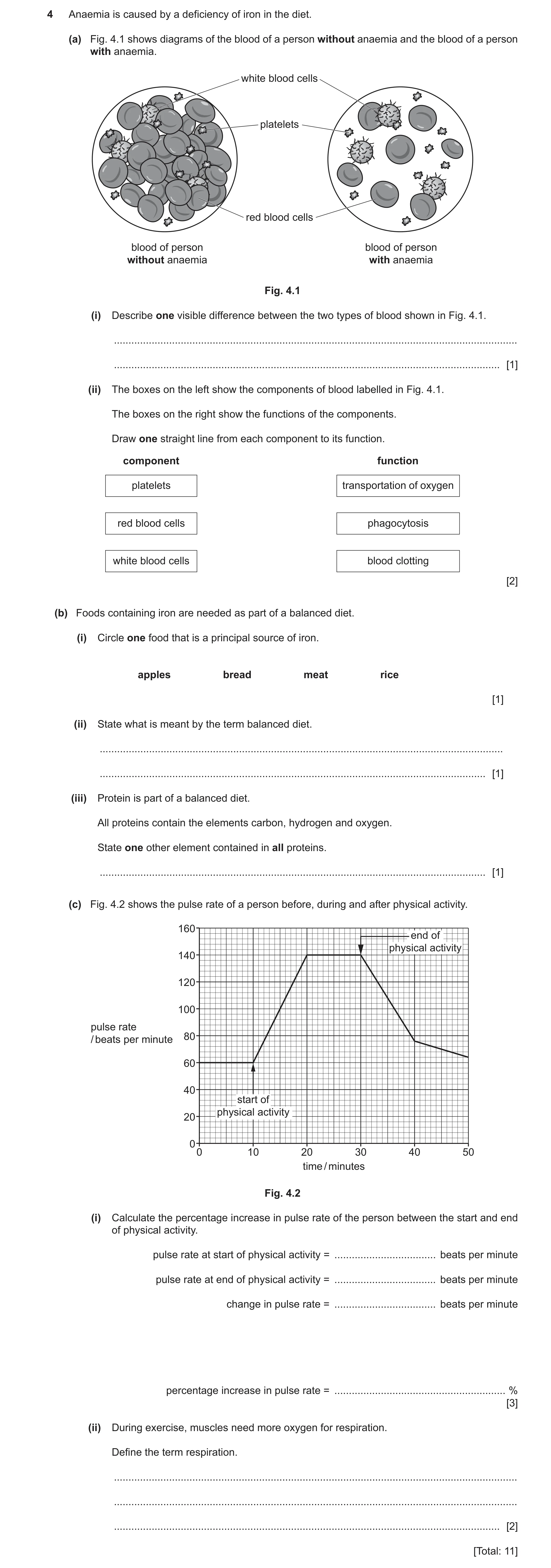
Q22 · Anaemia is caused by a deficiency of iron in the diet 0653/31 Oct/Nov 2024
4 Anaemia is caused by a deficiency of iron in the diet. (a) Fig. 4.1 shows diagrams of the blood of a person without anaemia and the blood of a person with anaemia. white blood cells platelets red blood cells blood of person blood of person without anaemia with anaemia Fig. 4.1 (i) Describe one visible difference between the two types of blood shown in Fig. 4.1. … … [1] (ii) The boxes on the left show the components of blood labelled in Fig. 4.1. The boxes on the right show the functions of the components. Draw one straight line from each component to its function. component function platelets transportation of oxygen red blood cells phagocytosis white blood cells blood clotting [2] (b) Foods containing iron are needed as part of a balanced diet. (i) Circle one food that is a principal source of iron. apples bread meat rice [1] (ii) State what is meant by the term balanced diet. … … [1] (iii) Protein is part of a balanced diet. All proteins contain the elements carbon, hydrogen and oxygen. State one other element contained in all proteins. … [1] (c) Fig. 4.2 shows the pulse rate of a person before, during and after physical activity. 160 end of physical activity 140 120 100 pulse rate 80 / beats per minute 60 40 start of physical activity 20 0 0 10 20 30 40 50 time / minutes Fig. 4.2 (i) Calculate the percentage increase in pulse rate of the person between the start and end of physical activity. pulse rate at start of physical activity = … beats per minute pulse rate at end of physical activity = … beats per minute change in pulse rate = … beats per minute percentage increase in pulse rate = … % [3] (ii) During exercise, muscles need more oxygen for respiration. Define the term respiration. … … … [2] [Total: 11]
11 marks
Mark scheme: 4(a)(i) blood from person without anaemia has more red blood cells ORA ; 1 4(a)(ii) 2 platelets transportation of oxygen red blood cells phagocytosis white blood cells blood clotting one correct ; all correct ; 4(b)(i) meat ; 1 4(b)(ii) idea of diet that contains suitable amounts of each type of nutrient ; 1 4(b)(iii) nitrogen ; 1 4(c)(i) 60 AND 140 ; 3 80 ; 133.3(%) ; 4(c)(ii) chemical reaction (in cells) that breaks down nutrient molecules ; 2 to release energy ;
This question in 0653/31 Oct/Nov 2024
Q23 · Blood contains different components 0653/32 Feb/March 2025
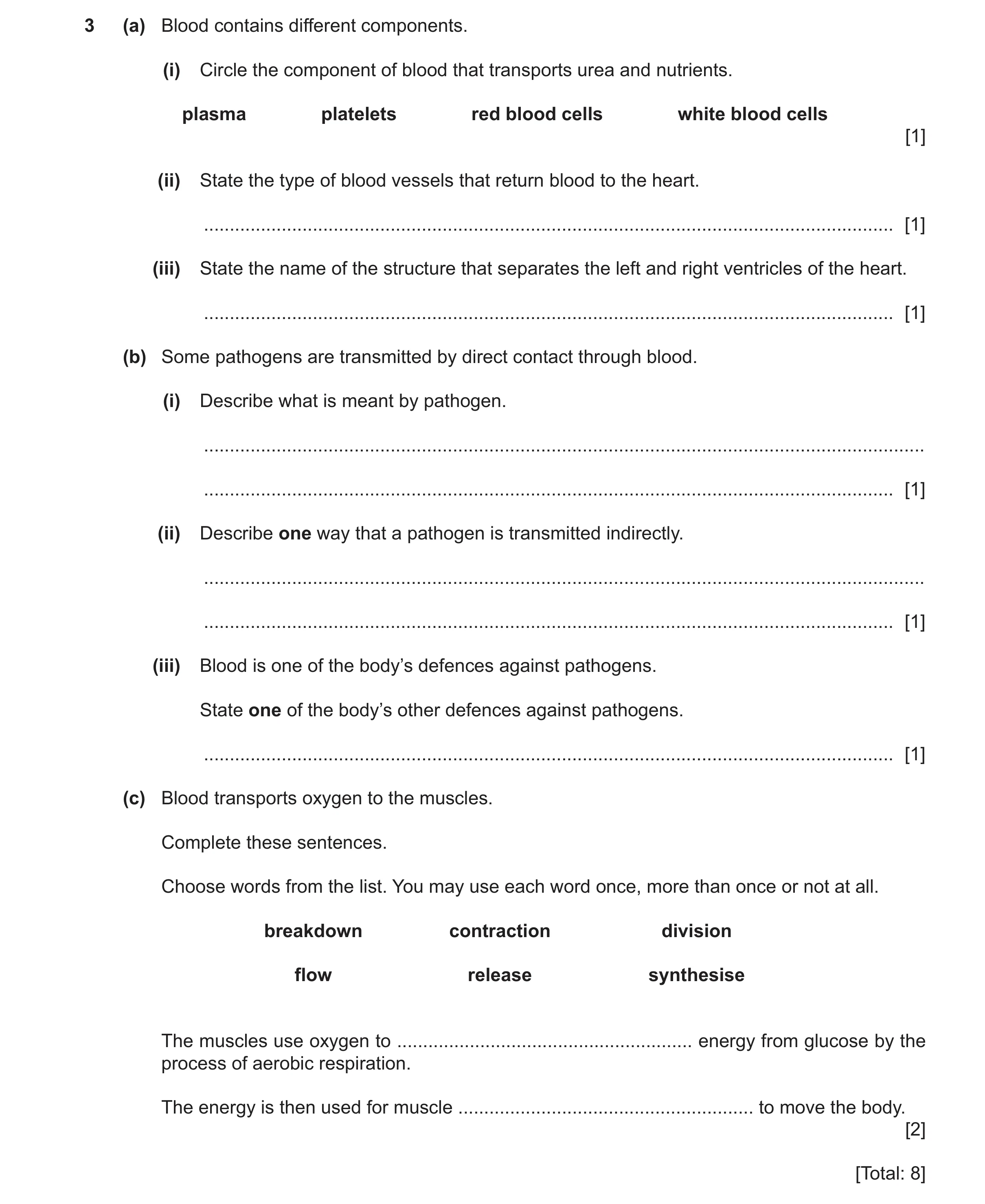
3 (a) Blood contains different components. (i) Circle the component of blood that transports urea and nutrients. plasma platelets red blood cells white blood cells [1] (ii) State the type of blood vessels that return blood to the heart. … [1] (iii) State the name of the structure that separates the left and right ventricles of the heart. … [1] (b) Some pathogens are transmitted by direct contact through blood. (i) Describe what is meant by pathogen. … … [1] (ii) Describe one way that a pathogen is transmitted indirectly. … … [1] (iii) Blood is one of the body’s defences against pathogens. State one of the body’s other defences against pathogens. … [1] (c) Blood transports oxygen to the muscles. Complete these sentences. Choose words from the list. You may use each word once, more than once or not at all. breakdown contraction division flow release synthesise The muscles use oxygen to … energy from glucose by the process of aerobic respiration. The energy is then used for muscle … to move the body. [2] [Total: 8]
8 marks
Mark scheme: 3(a)(i) plasma ; 1 more than one answer circled = 0 marks 3(a)(ii) vein ; 1 3(a)(iii) septum ; 1 3(b)(i) disease-causing organism ; 1 3(b)(ii) (contaminated) surfaces / food / animals / air ; 1 3(b)(iii) skin / nose hairs / mucus / stomach acid ; 1 3(c) release ; 2 contraction ;
This question in 0653/32 Feb/March 2025
Q24 · The structure of a human heart 0653/31 May/June 2025
2 Fig. 2.1 shows the structure of a human heart. S P Q R Fig. 2.1 The arrows in Fig. 2.1 show the direction of blood flow through the heart. (a) Use letters P–S to complete the sentences about the heart in Fig. 2.1. The part labelled … is the muscular wall of the left ventricle. Blood is transported away from the right side of the heart in the part labelled … . The part labelled … ensures one‑way flow of blood. [3] (b) The activity of the heart may be monitored by ECG (electrocardiogram). State one other way to monitor the activity of the heart. … [1] (c) Blood contains four main components. Name the component of blood that: transports hormones … produces antibodies … contains haemoglobin. … [3] (d) Blood is pumped away from the heart in arteries and returns to the heart in veins. Describe two differences in the structure of arteries compared to veins. 1 … … 2 … … [2] [Total: 9]
9 marks
Mark scheme: 2(a) Q ; 3 S ; P ; 2(b) pulse (rate) / listening to sound of valves (closing) / stethoscope ; 1 2(c) plasma ; 3 white blood cells ; red blood cells ; 2(d) any two from: 2 (arteries have) thicker walls ; smaller lumen ; no valves ;
This question in 0653/31 May/June 2025
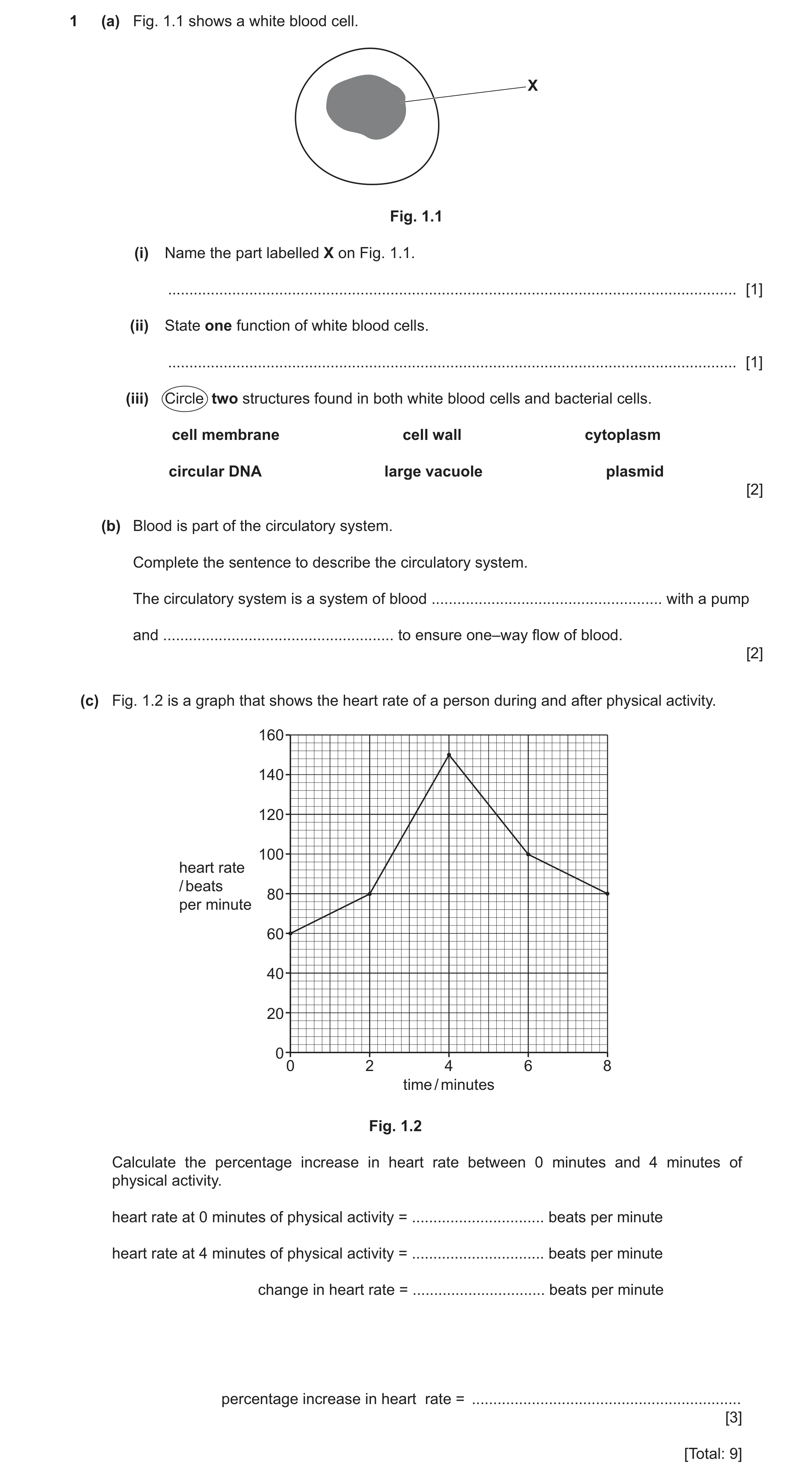
Q25 · A white blood cell 0653/32 May/June 2025
1 (a) Fig. 1.1 shows a white blood cell. X Fig. 1.1 (i) Name the part labelled X on Fig. 1.1. … [1] (ii) State one function of white blood cells. … [1] (iii) Circle two structures found in both white blood cells and bacterial cells. cell membrane cell wall cytoplasm circular DNA large vacuole plasmid [2] (b) Blood is part of the circulatory system. Complete the sentence to describe the circulatory system. The circulatory system is a system of blood … with a pump and … to ensure one–way flow of blood. [2] (c) Fig. 1.2 is a graph that shows the heart rate of a person during and after physical activity. 160 140 120 100 heart rate / beats 80 per minute 60 40 20 00 2 4 6 8 time / minutes Fig. 1.2 Calculate the percentage increase in heart rate between 0 minutes and 4 minutes of physical activity. heart rate at 0 minutes of physical activity = … beats per minute heart rate at 4 minutes of physical activity = … beats per minute change in heart rate = … beats per minute percentage increase in heart rate = … [3] [Total: 9]
9 marks
Mark scheme: Question Answer Marks 1(a)(i) nucleus ; 1 1(a)(ii) produce antibodies / phagocytosis ; 1 1(a)(iii) cell membrane ; 2 cytoplasm ; 1(b) vessels ; 2 valves ; 1(c) 60 and 150 ; 3 (150 – 60) = 90 ; (90 100) = 150(%) ; 60
This question in 0653/32 May/June 2025
 32 / 42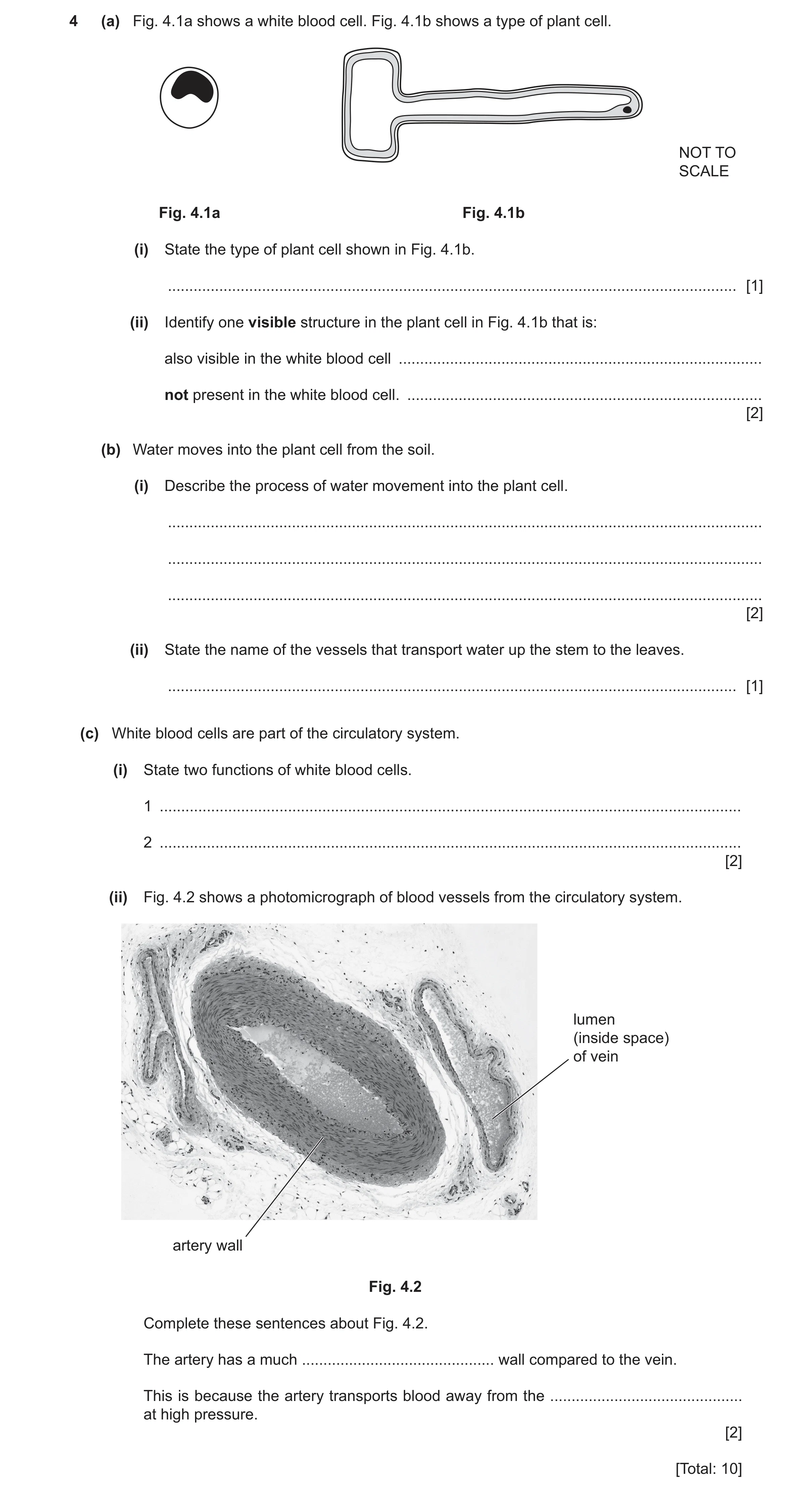
 39 / 42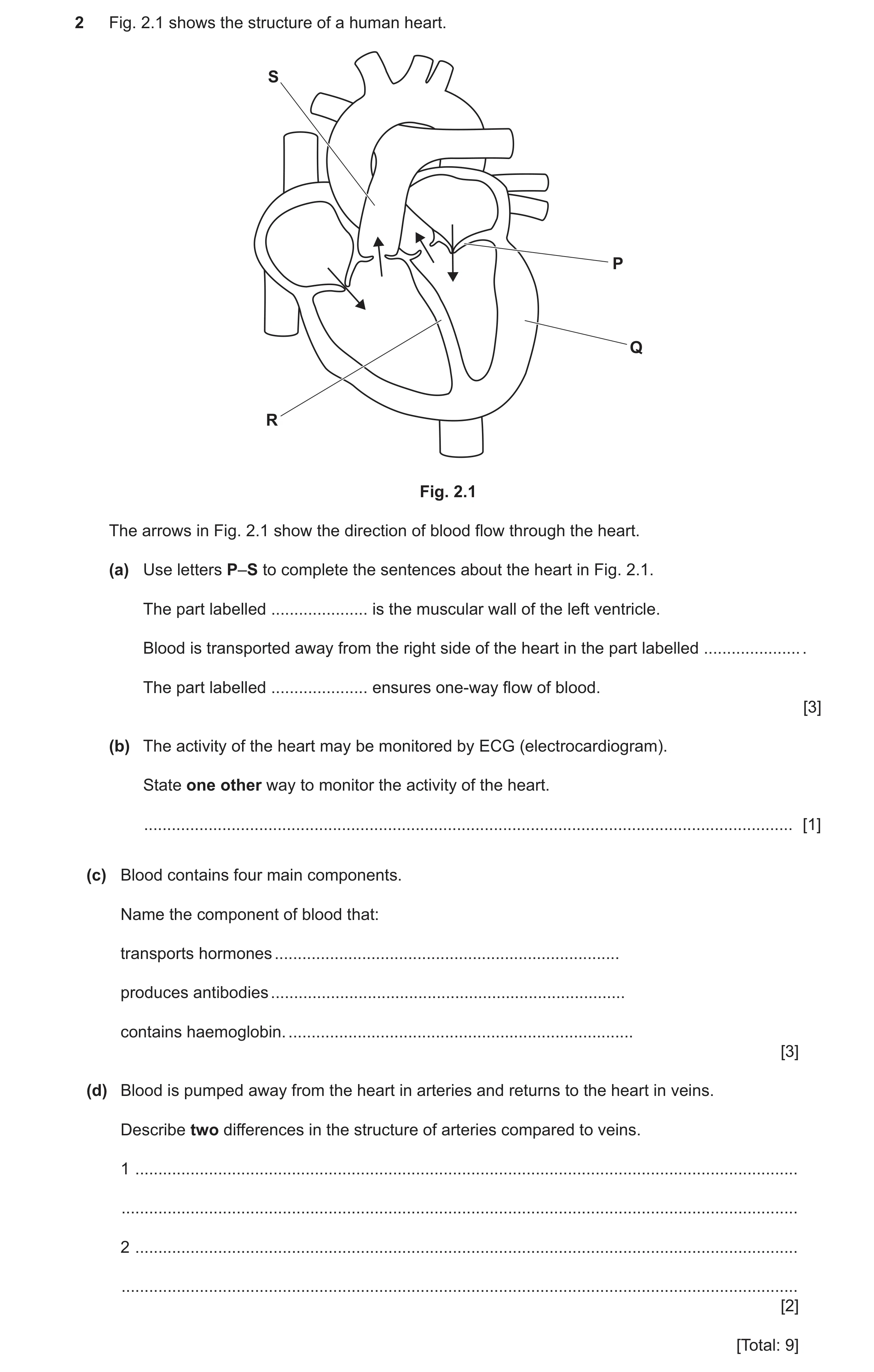
 6
6 6
6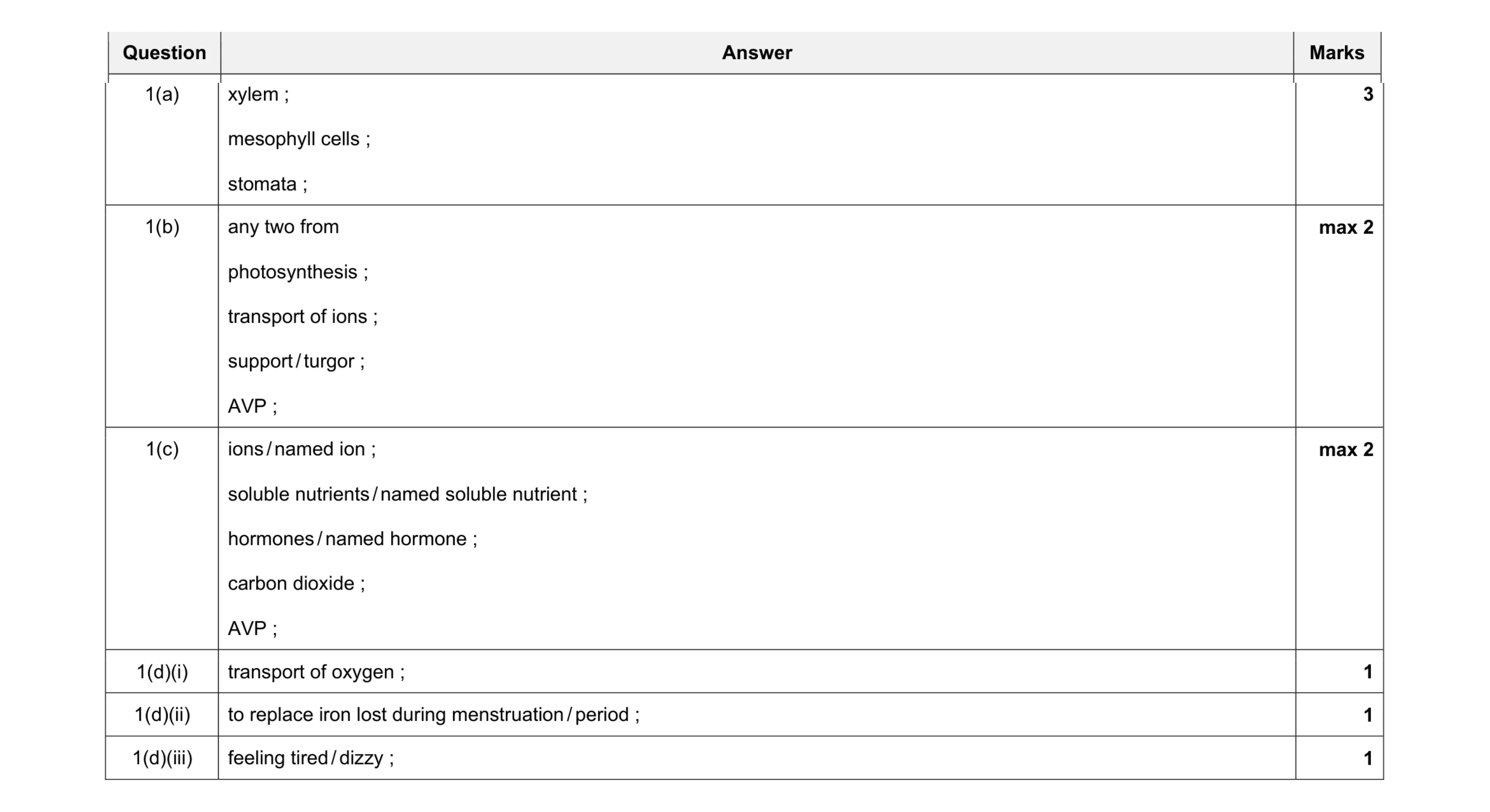 10
10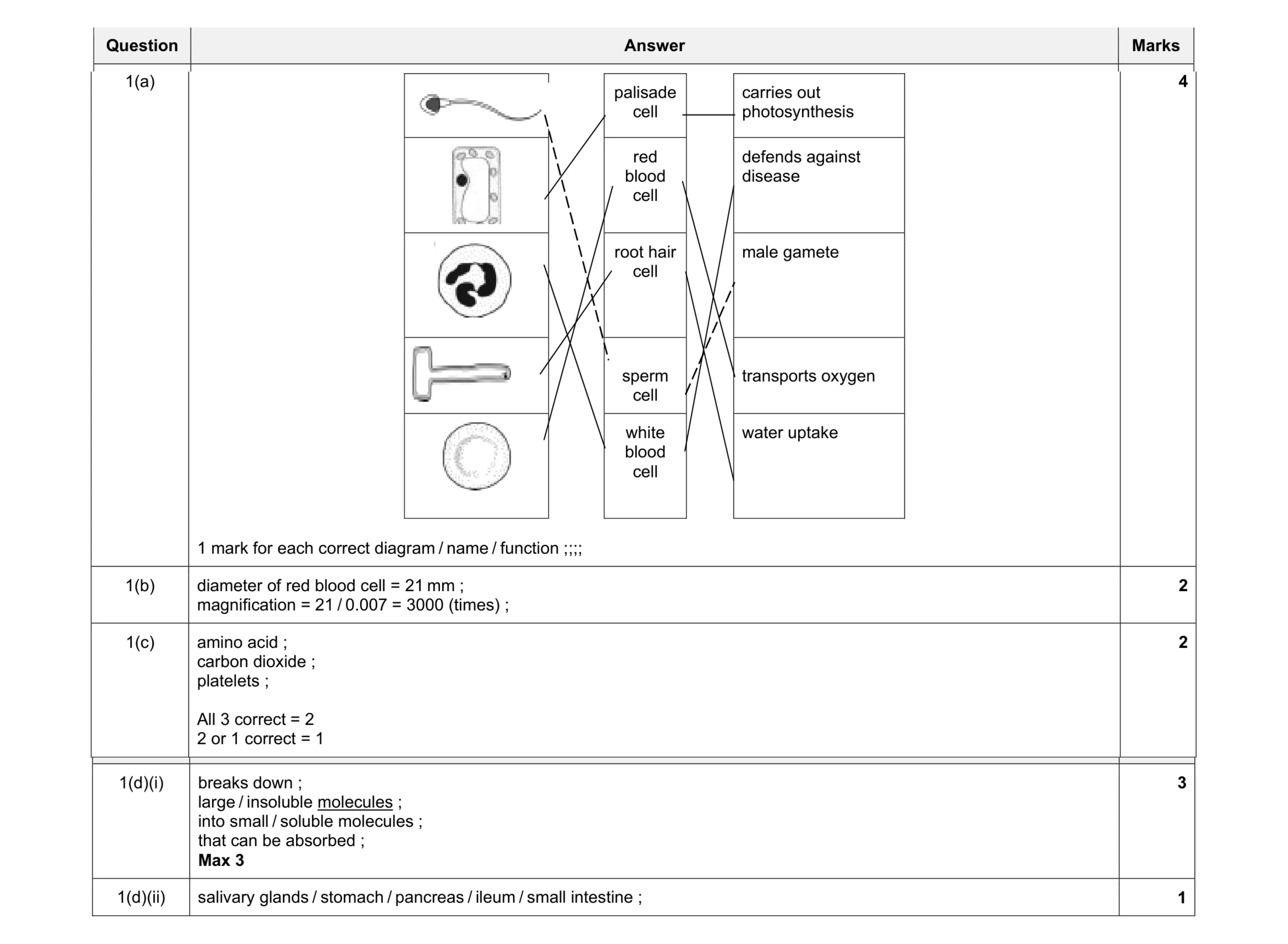 12
12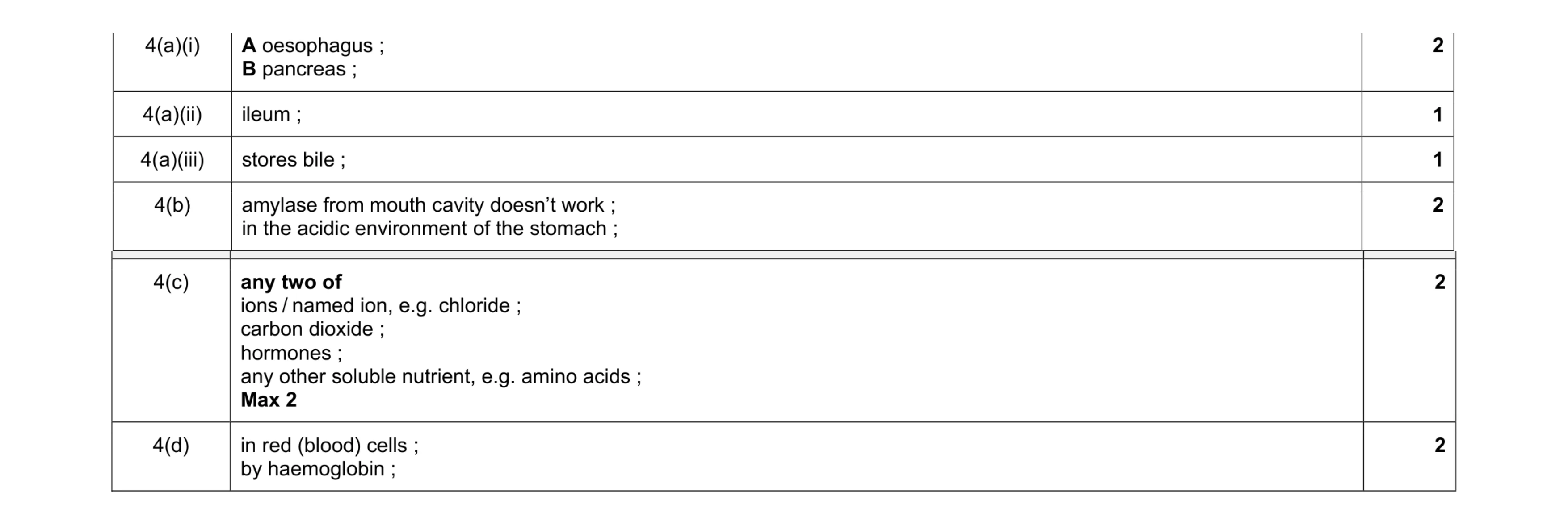 10
10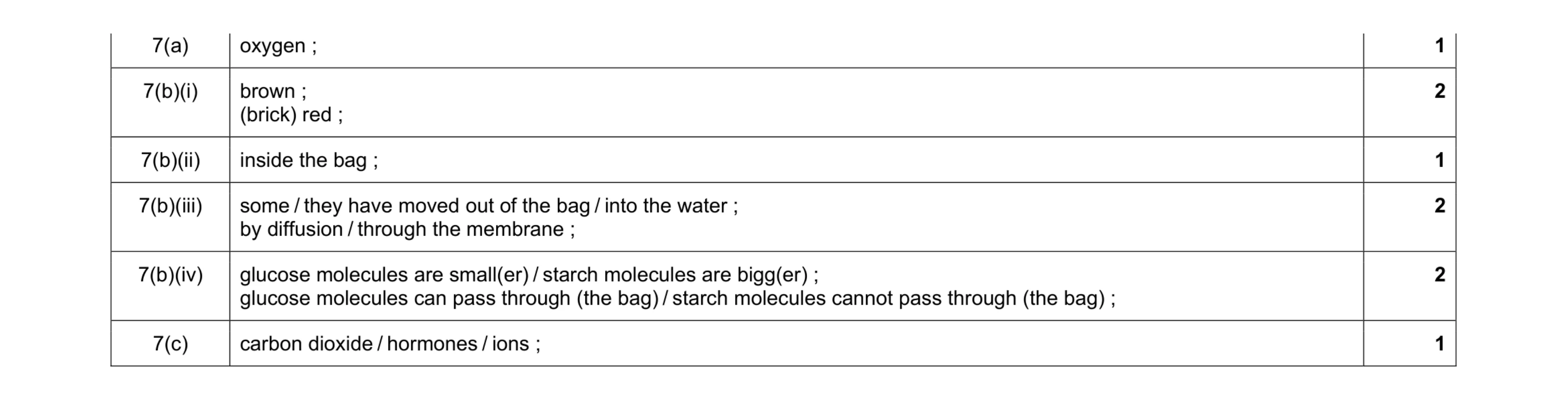 9
9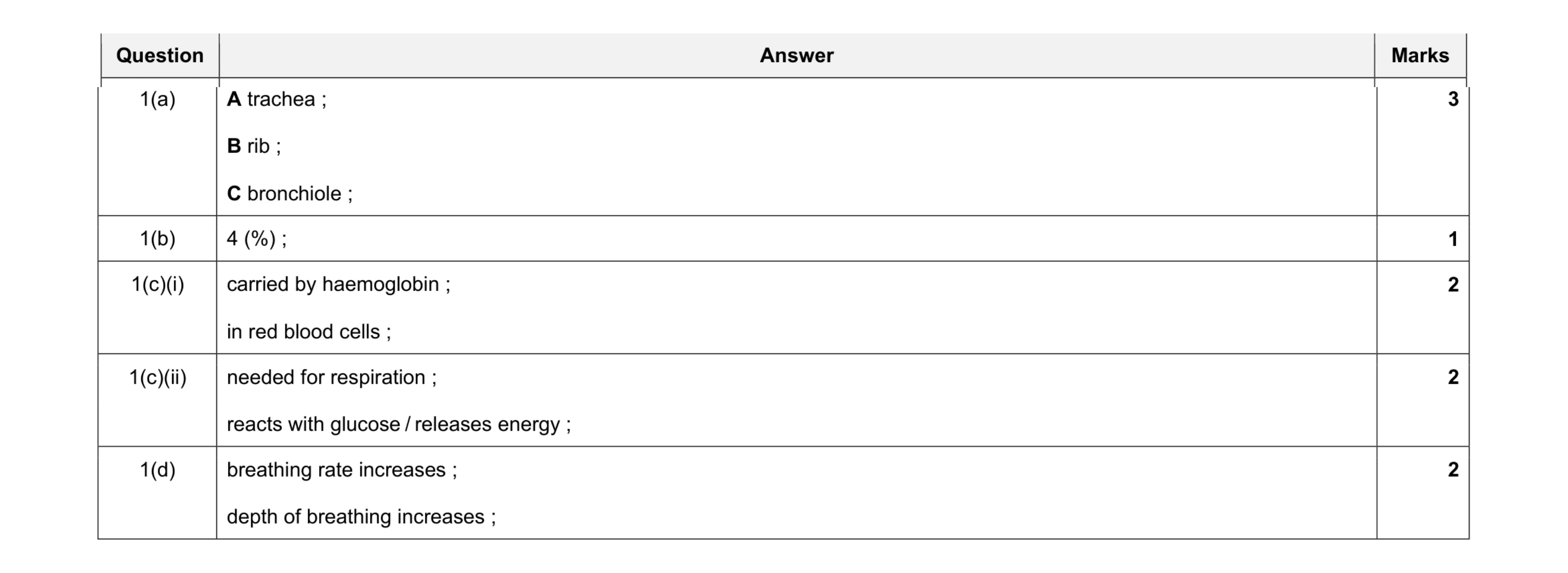 10
10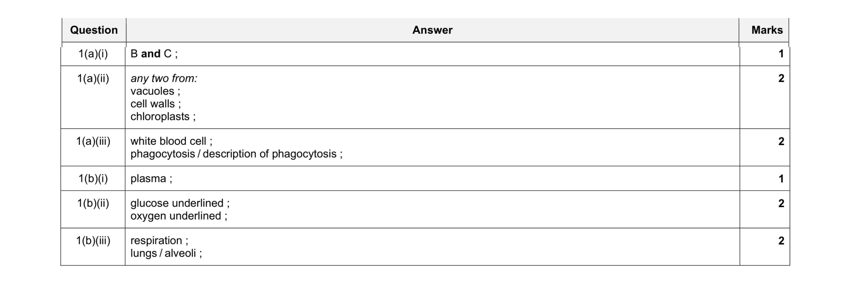 10
10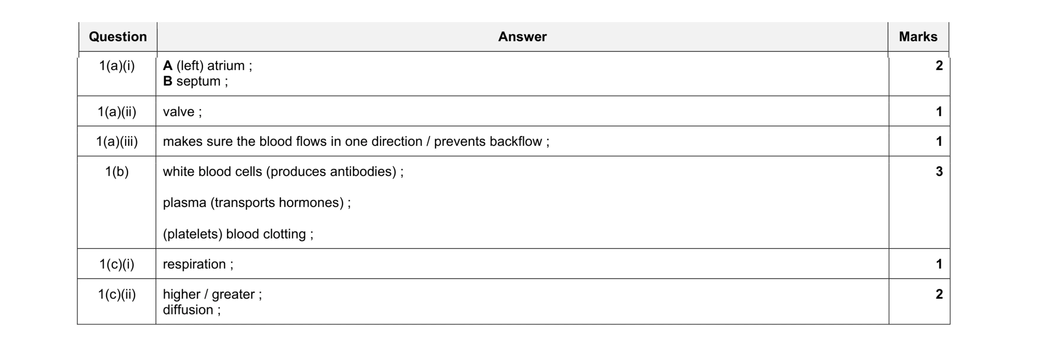 10
10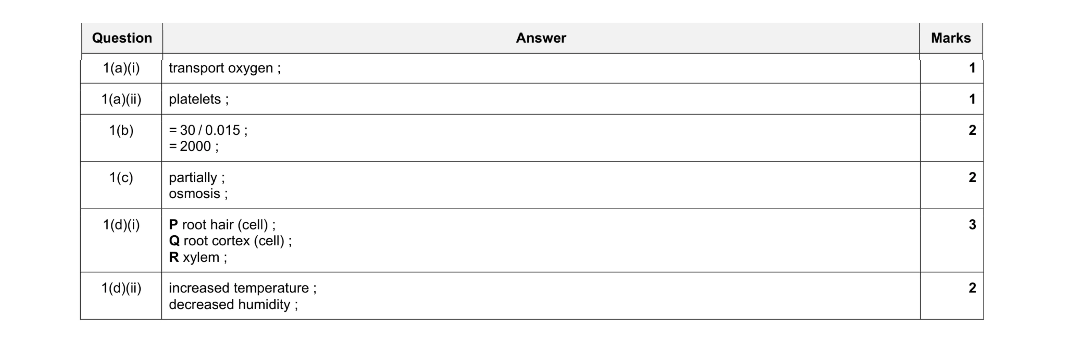 11
11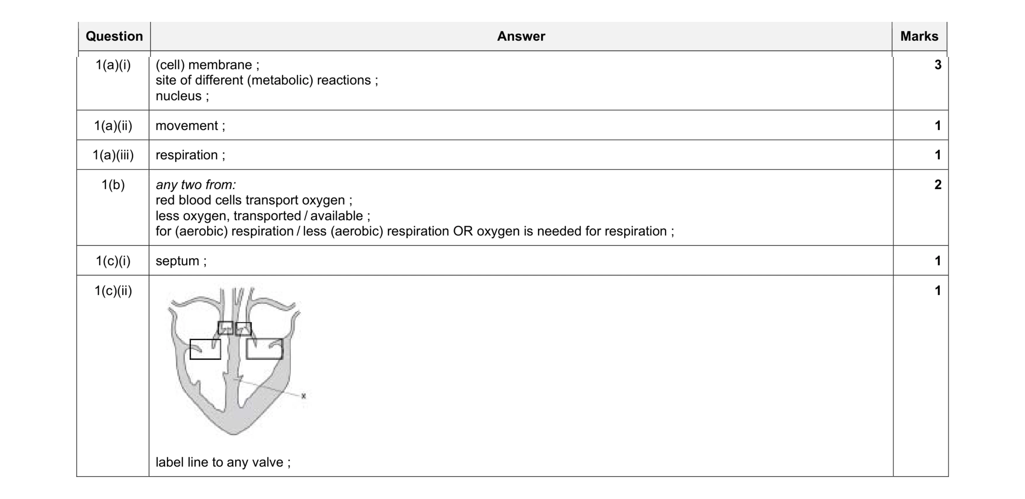 9
9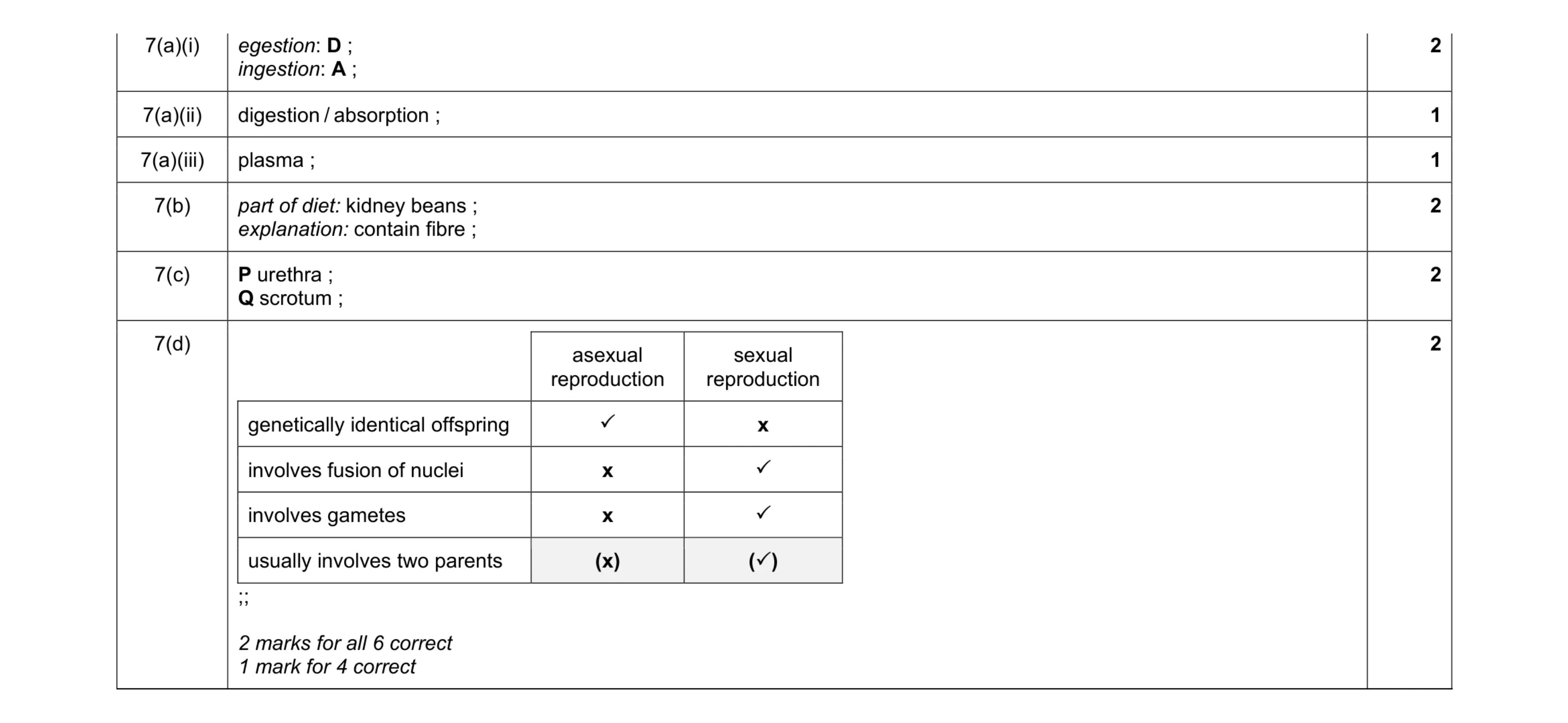 10
10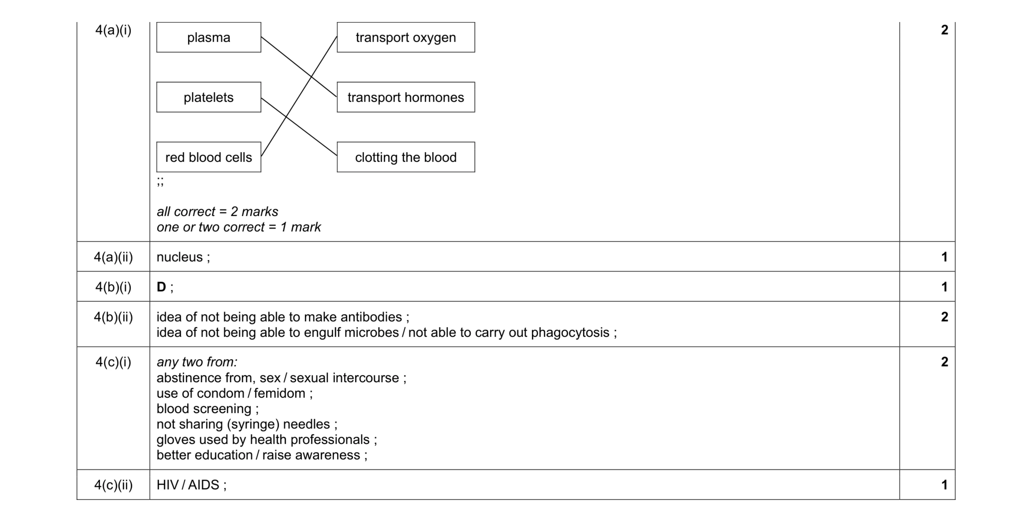 9
9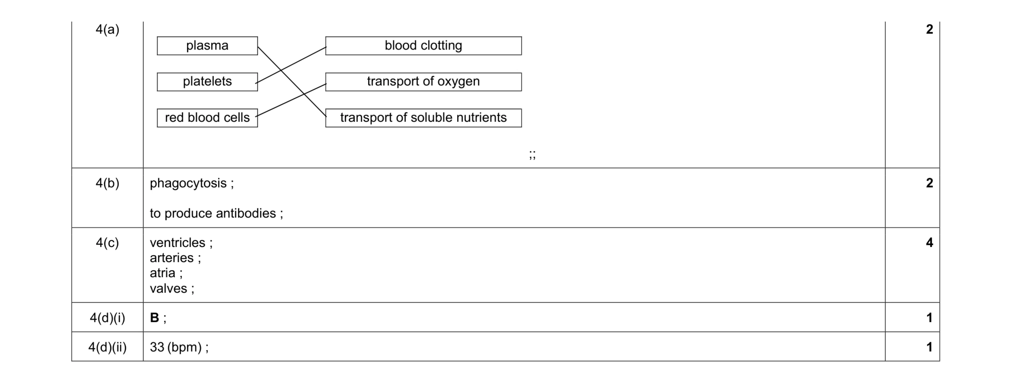 10
10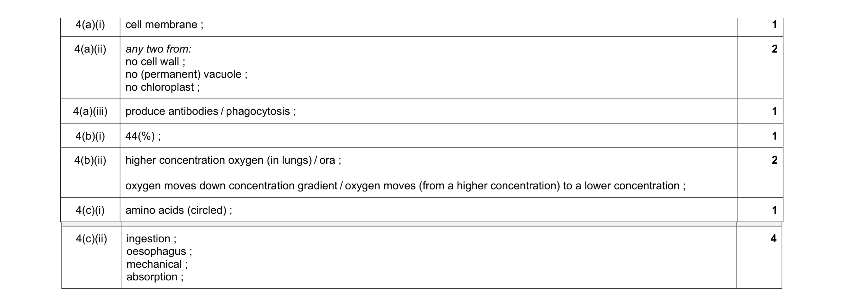 12
12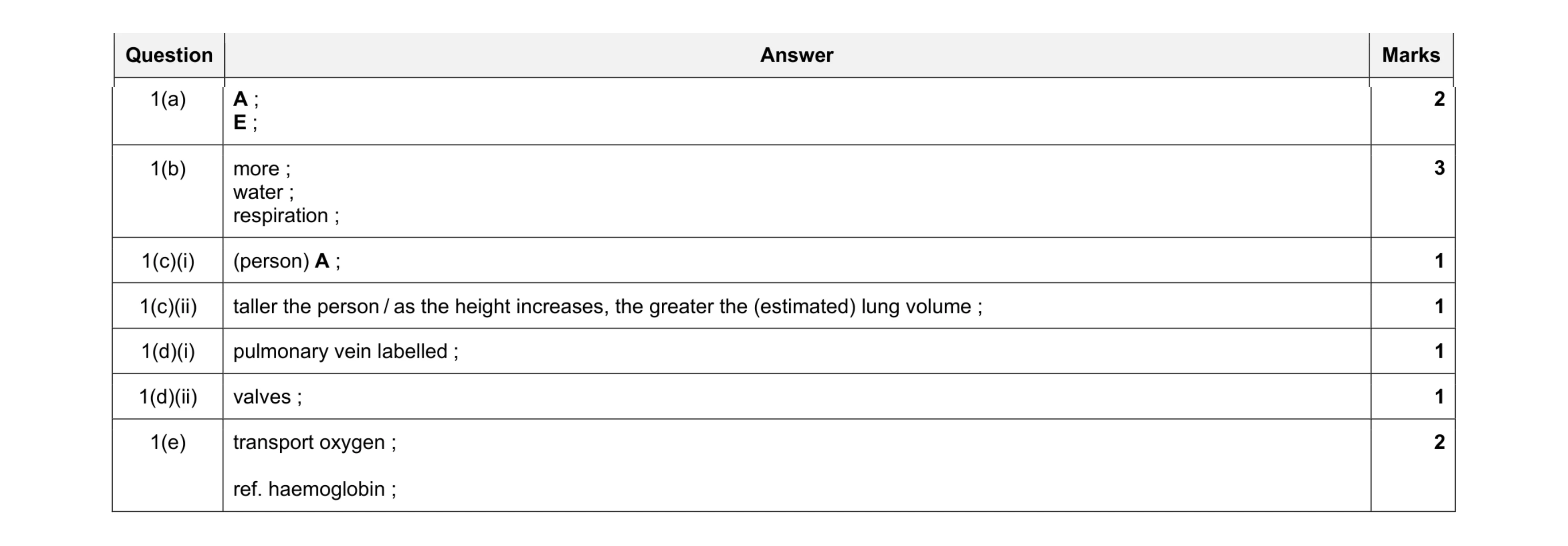 11
11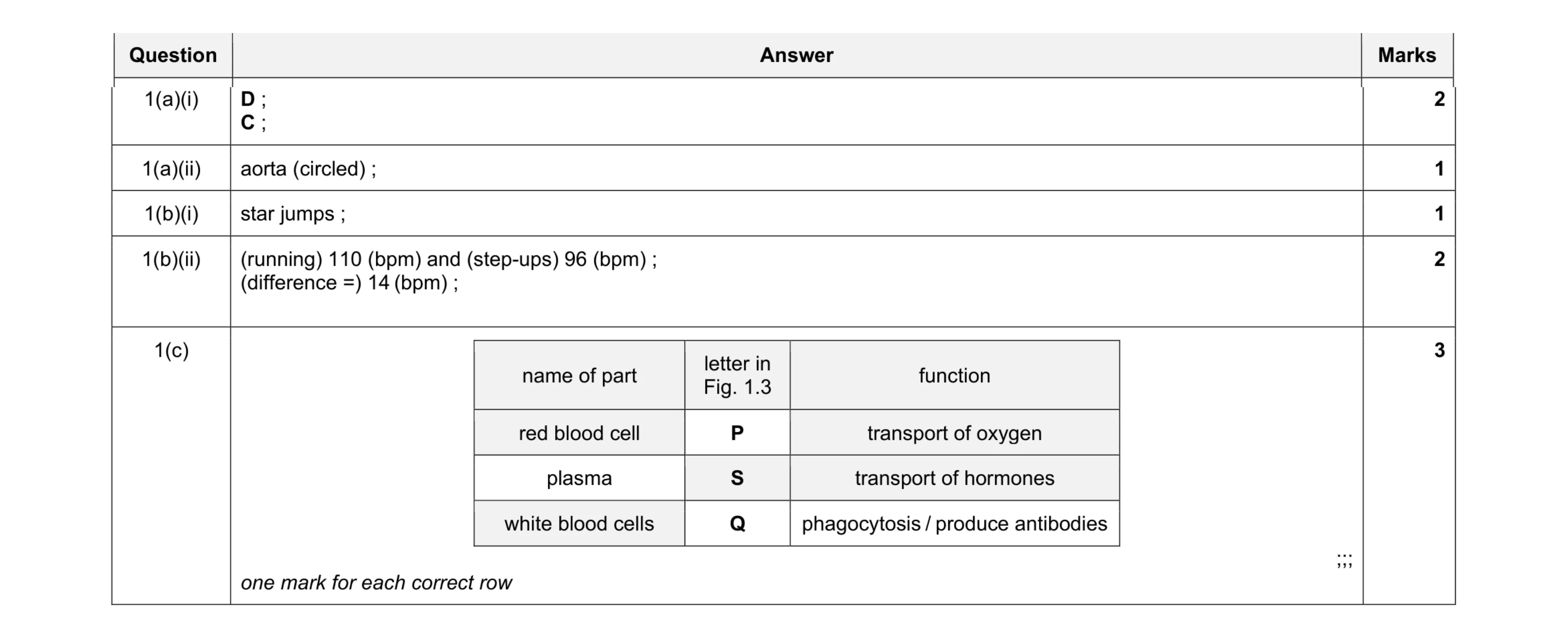 9
9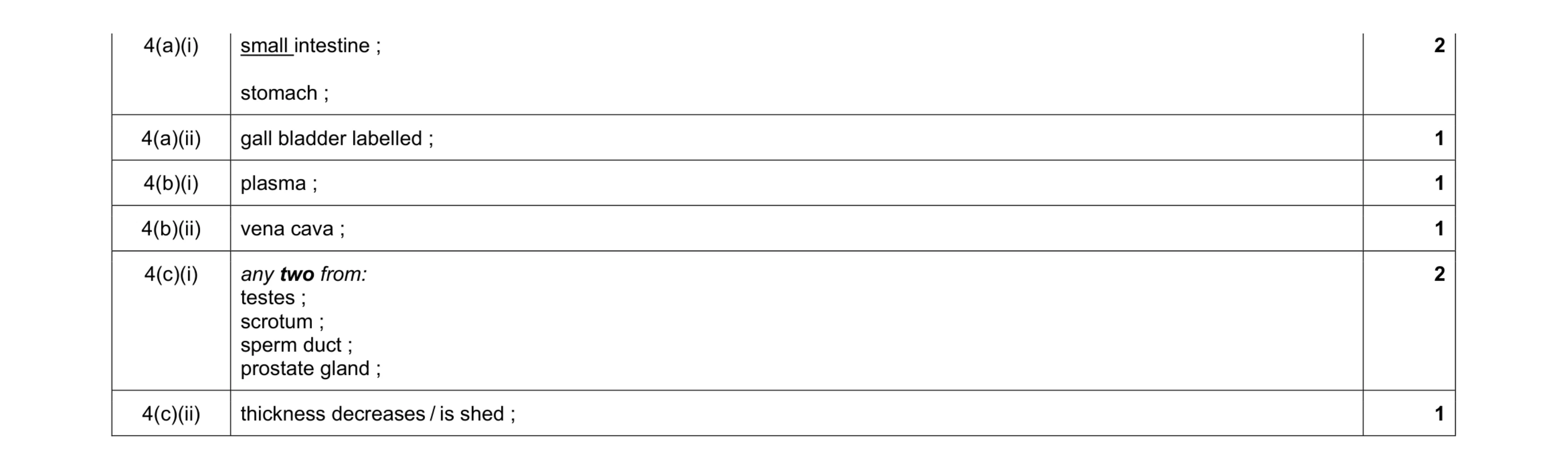 8
8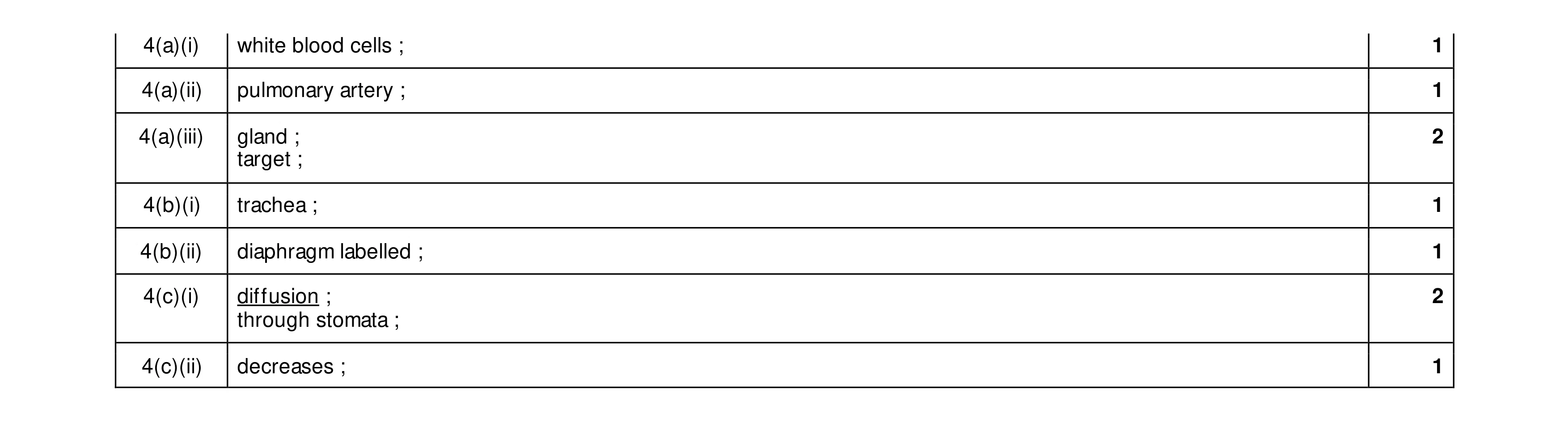 9
9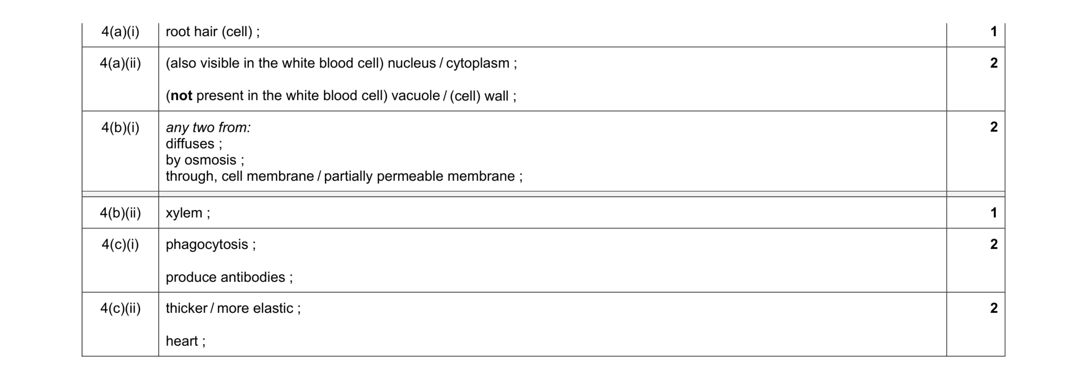 10
10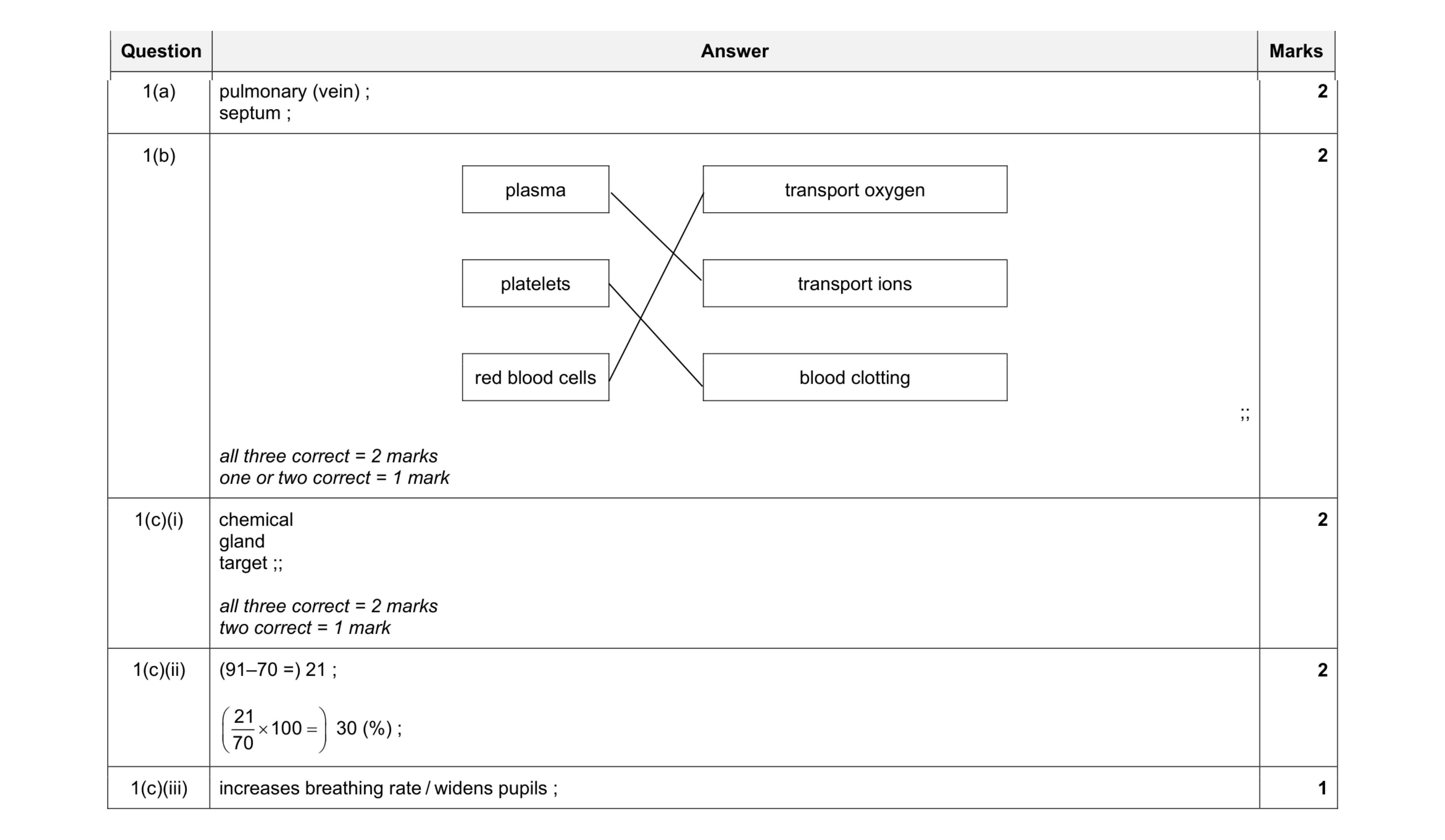 9
9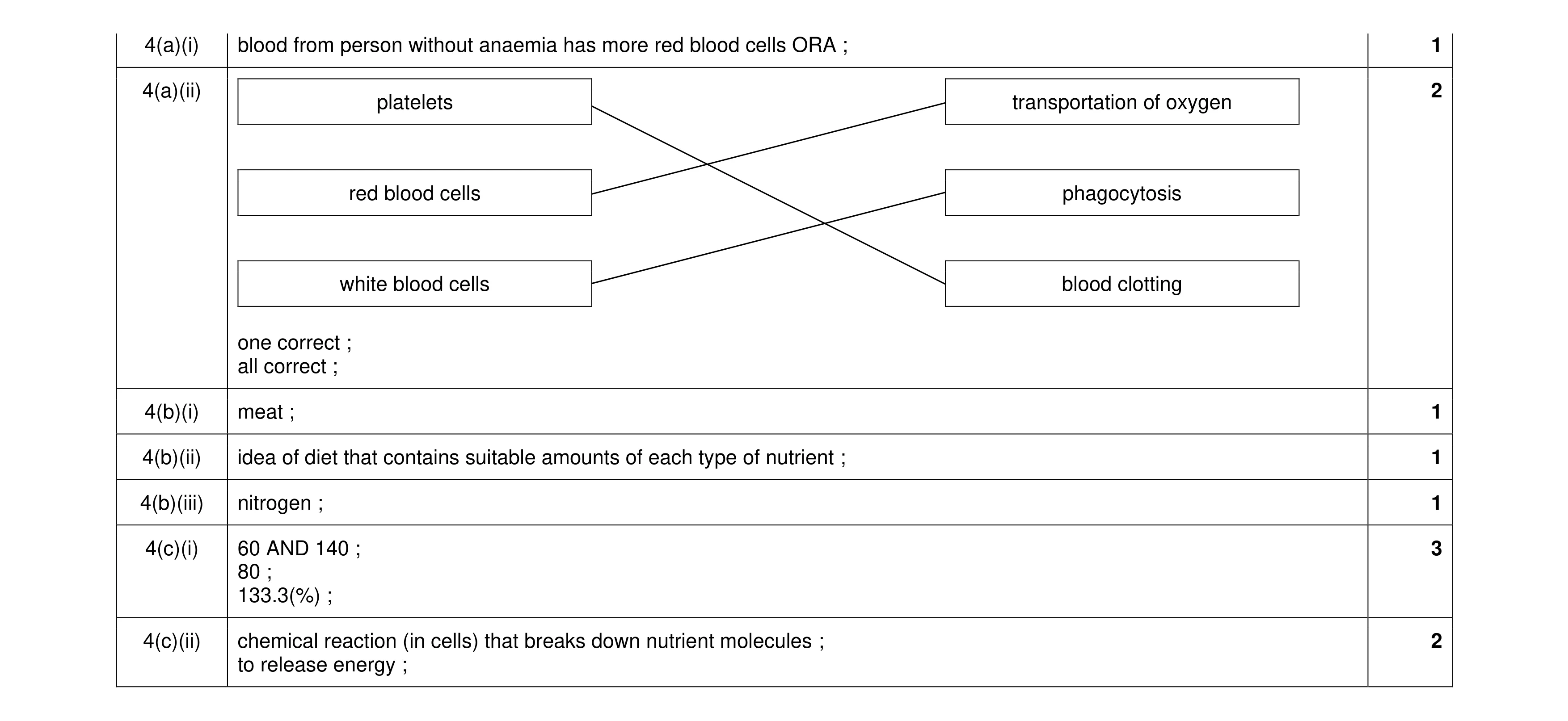 11
11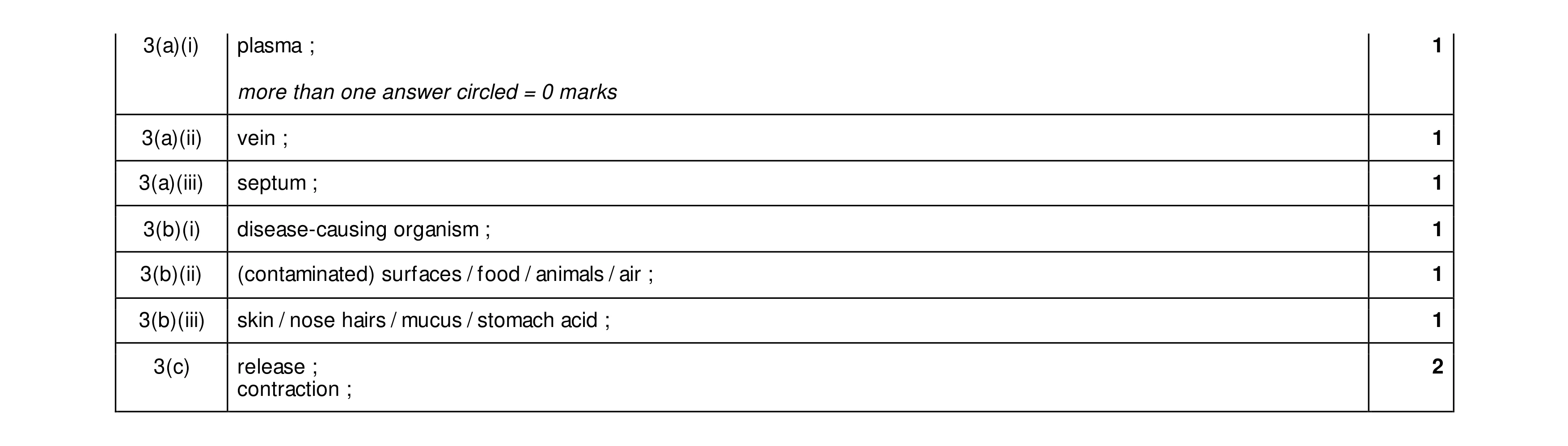 8
8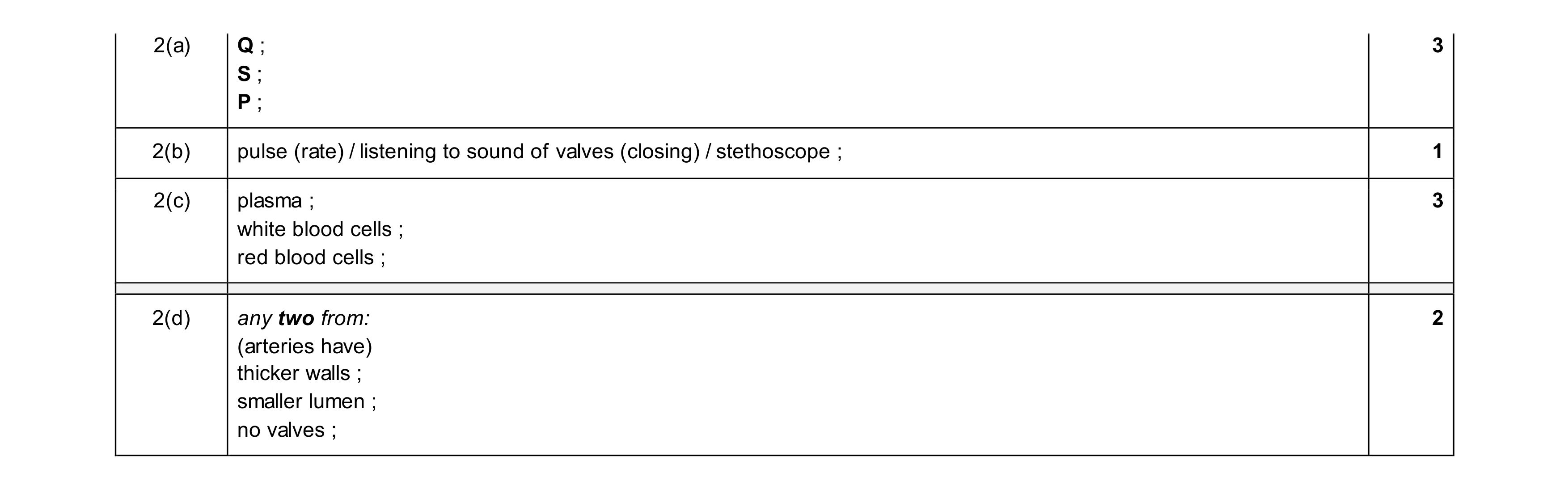 9
9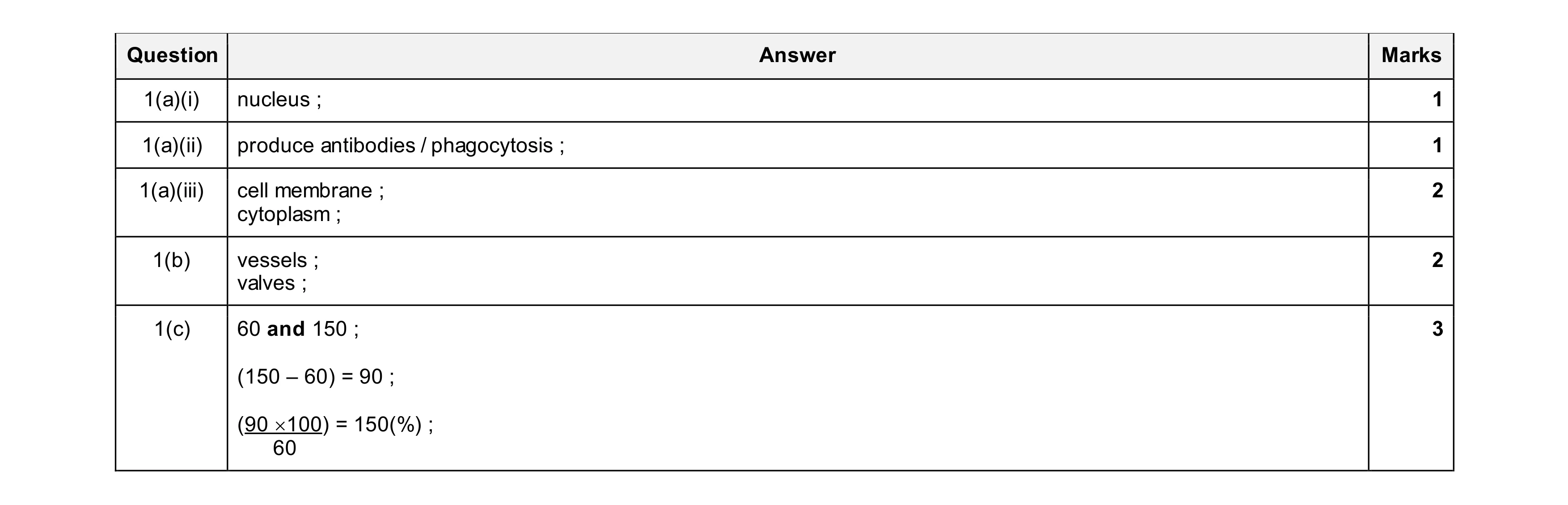 9
9