Questions as text
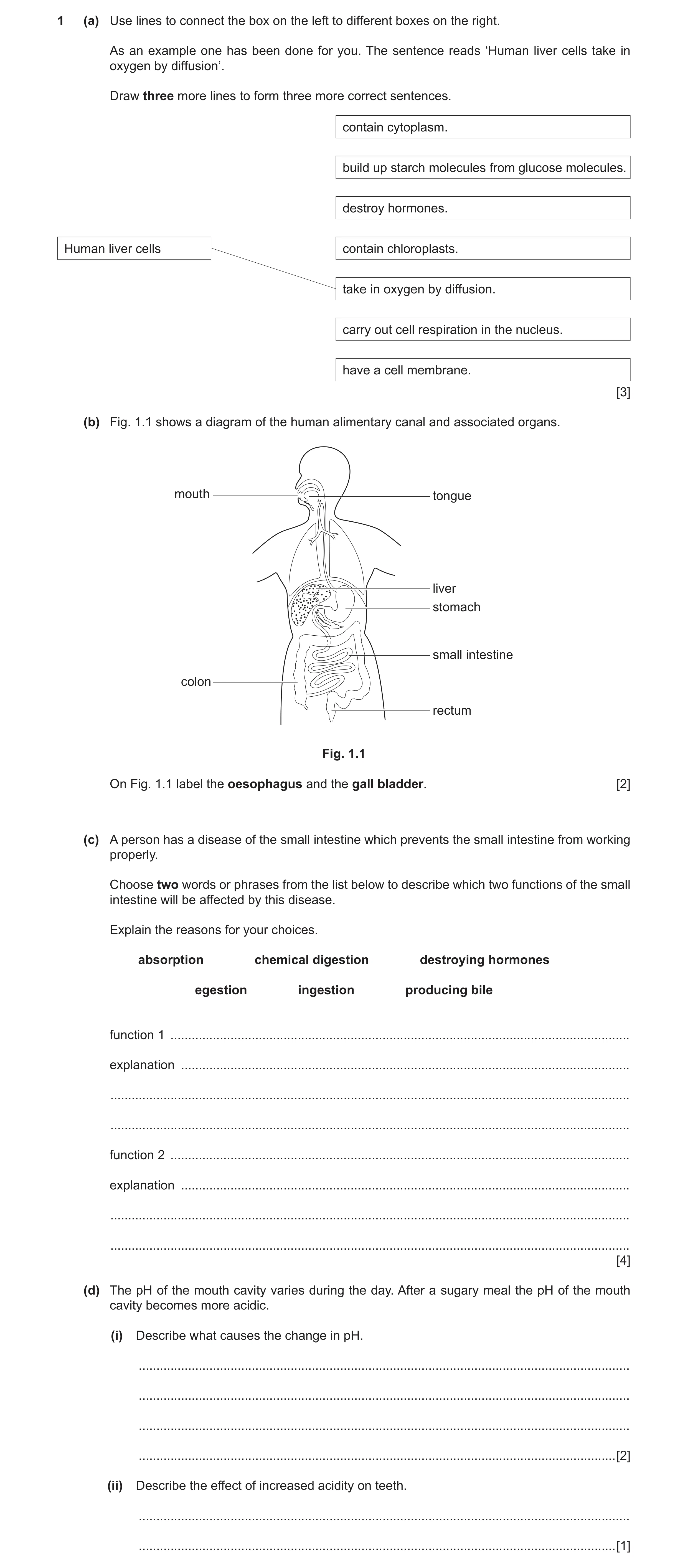
Q1 · Use lines to connect the box on the left to different boxes on the right 0653/32 Feb/March 2017
1 (a) Use lines to connect the box on the left to different boxes on the right. As an example one has been done for you. The sentence reads ‘Human liver cells take in oxygen by diffusion’. Draw three more lines to form three more correct sentences. contain cytoplasm. build up starch molecules from glucose molecules. destroy hormones. Human liver cells contain chloroplasts. take in oxygen by diffusion. carry out cell respiration in the nucleus. have a cell membrane. [3] (b) Fig. 1.1 shows a diagram of the human alimentary canal and associated organs. mouth tongue liver stomach small intestine colon rectum Fig. 1.1 On Fig. 1.1 label the oesophagus and the gall bladder. [2] (c) A person has a disease of the small intestine which prevents the small intestine from working properly. Choose two words or phrases from the list below to describe which two functions of the small intestine will be affected by this disease. Explain the reasons for your choices. absorption chemical digestion destroying hormones egestion ingestion producing bile function 1 … explanation … … … function 2 … explanation … … … [4] (d) The pH of the mouth cavity varies during the day. After a sugary meal the pH of the mouth cavity becomes more acidic. (i) Describe what causes the change in pH. … … … … [2] (ii) Describe the effect of increased acidity on teeth. … … [1]
12 marks
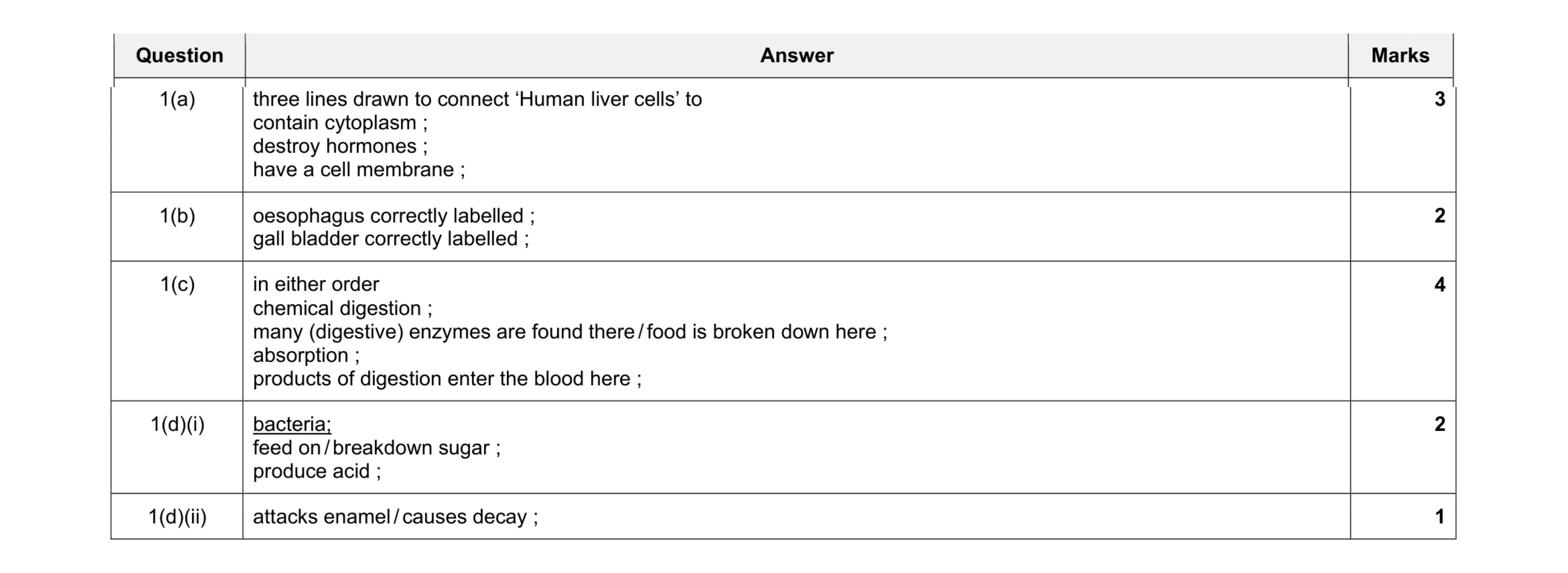
Mark scheme: 1(a) three lines drawn to connect ‘Human liver cells’ to contain cytoplasm ; destroy hormones ; have a cell membrane ; 3 1(b) oesophagus correctly labelled ; gall bladder correctly labelled ; 2 1(c) in either order chemical digestion ; many (digestive) enzymes are found there / food is broken down here ; absorption ; products of digestion enter the blood here ; 4 1(d)(i) bacteria; feed on / breakdown sugar ; produce acid ; 2 1(d)(ii) attacks enamel / causes decay ; 1
This question in 0653/32 Feb/March 2017
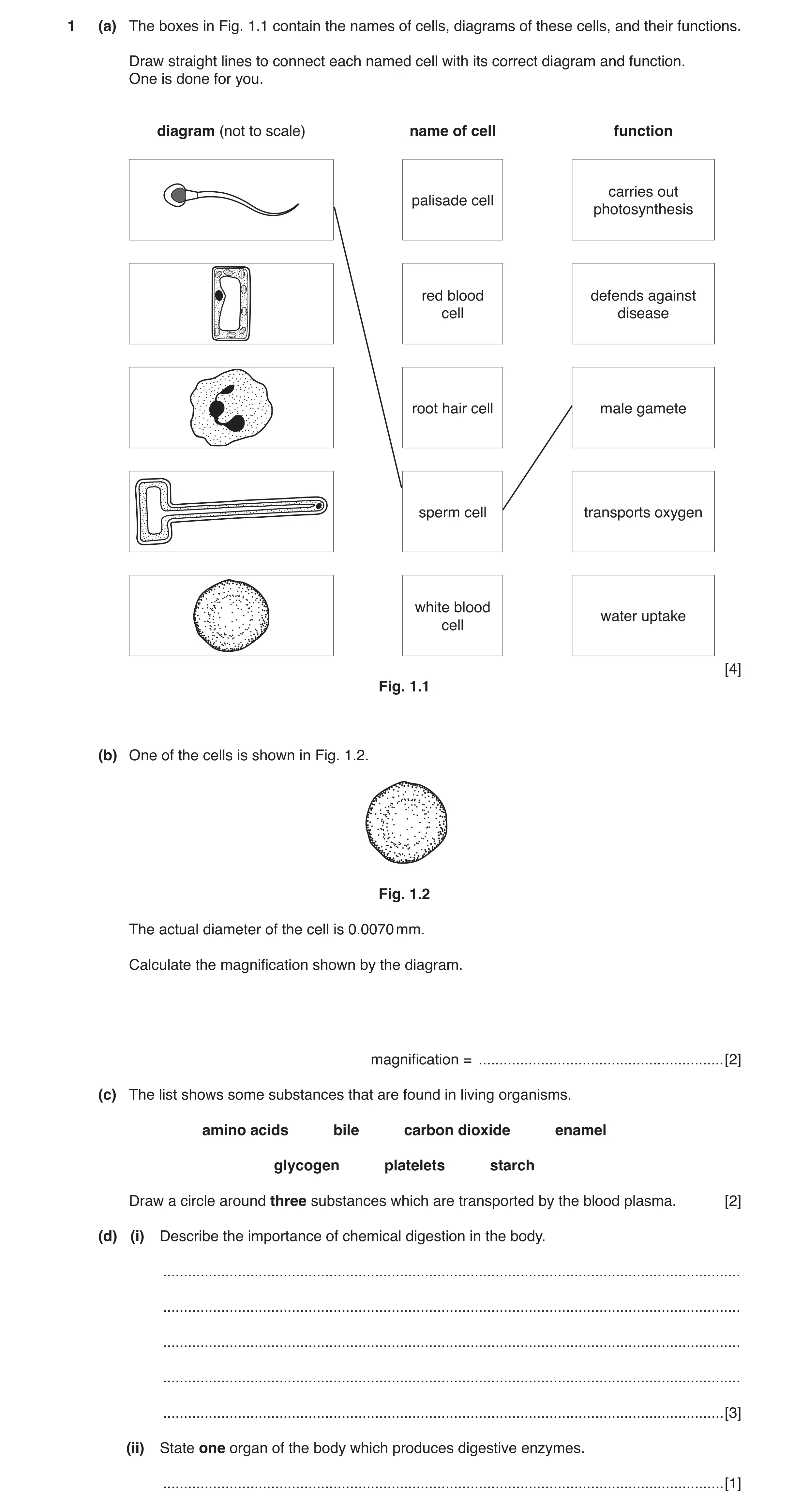
Q2 · The boxes in Fig 0653/32 Oct/Nov 2018
1 (a) The boxes in Fig. 1.1 contain the names of cells, diagrams of these cells, and their functions. Draw straight lines to connect each named cell with its correct diagram and function. One is done for you. diagram (not to scale) name of cell function carries out palisade cell photosynthesis red blood defends against cell disease root hair cell male gamete sperm cell transports oxygen white blood water uptake cell [4] Fig. 1.1 (b) One of the cells is shown in Fig. 1.2. Fig. 1.2 The actual diameter of the cell is 0.0070 mm. Calculate the magnification shown by the diagram. magnification = … [2] (c) The list shows some substances that are found in living organisms. amino acids bile carbon dioxide enamel glycogen platelets starch Draw a circle around three substances which are transported by the blood plasma. [2] (d) (i) Describe the importance of chemical digestion in the body. … … … … … [3] (ii) State one organ of the body which produces digestive enzymes. … [1]
12 marks
Mark scheme: 1(a) palisade cell carries out photosynthesis red blood cell defends against disease root hair cell male gamete sperm cell transports oxygen white blood cell water uptake 1 mark for each correct diagram / name / function ;;;; 4 1(b) diameter of red blood cell = 21 mm ; magnification = 21 / 0.007 = 3000 (times) ; 2 1(c) amino acid ; carbon dioxide ; platelets ; All 3 correct = 2 2 or 1 correct = 1 2 Question Answer Marks 1(d)(i) breaks down ; large / insoluble molecules ; into small / soluble molecules ; that can be absorbed ; Max 3 3 1(d)(ii) salivary glands / stomach / pancreas / ileum / small intestine ; 1
This question in 0653/32 Oct/Nov 2018
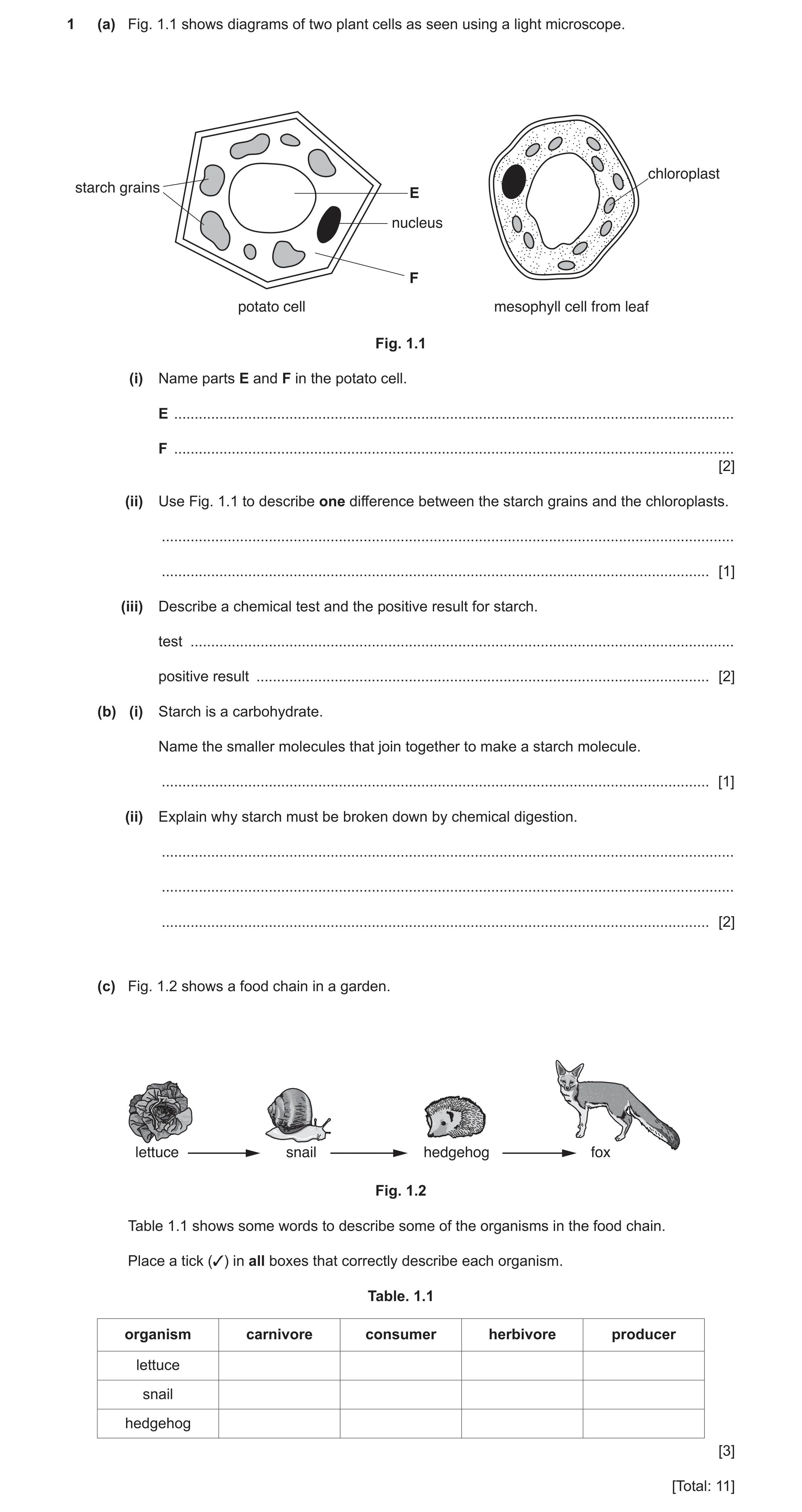
Q3 · Diagrams of two plant cells as seen using a light microscope 0653/32 Feb/March 2019
1 (a) Fig. 1.1 shows diagrams of two plant cells as seen using a light microscope. chloroplast starch grains E nucleus F potato cell mesophyll cell from leaf Fig. 1.1 (i) Name parts E and F in the potato cell. E … F … [2] (ii) Use Fig. 1.1 to describe one difference between the starch grains and the chloroplasts. … … [1] (iii) Describe a chemical test and the positive result for starch. test … positive result … [2] (b) (i) Starch is a carbohydrate. Name the smaller molecules that join together to make a starch molecule. … [1] (ii) Explain why starch must be broken down by chemical digestion. … … … [2] (c) Fig. 1.2 shows a food chain in a garden. lettuce snail hedgehog fox Fig. 1.2 Table 1.1 shows some words to describe some of the organisms in the food chain. Place a tick (✓) in all boxes that correctly describe each organism. Table. 1.1 organism carnivore consumer herbivore producer lettuce snail hedgehog [3] [Total: 11]
11 marks
Mark scheme: 1(a)(i) (E) vacuole ; (F) cytoplasm ; 2 1(a)(ii) starch grains irregular shapes / sizes and chlorophyll all the same shapes / sizes ; 1 1(a)(iii) add iodine solution ; goes blue-black ; 2 1(b)(i) glucose ; 1 1(b)(ii) produces small molecules ; that can be absorbed ; 2 1(c) one mark for each correct line organism carnivore consumer herbivore producer lettuce 3 snail 3 3 hedgehog 3 3 ; ; ; 3
This question in 0653/32 Feb/March 2019
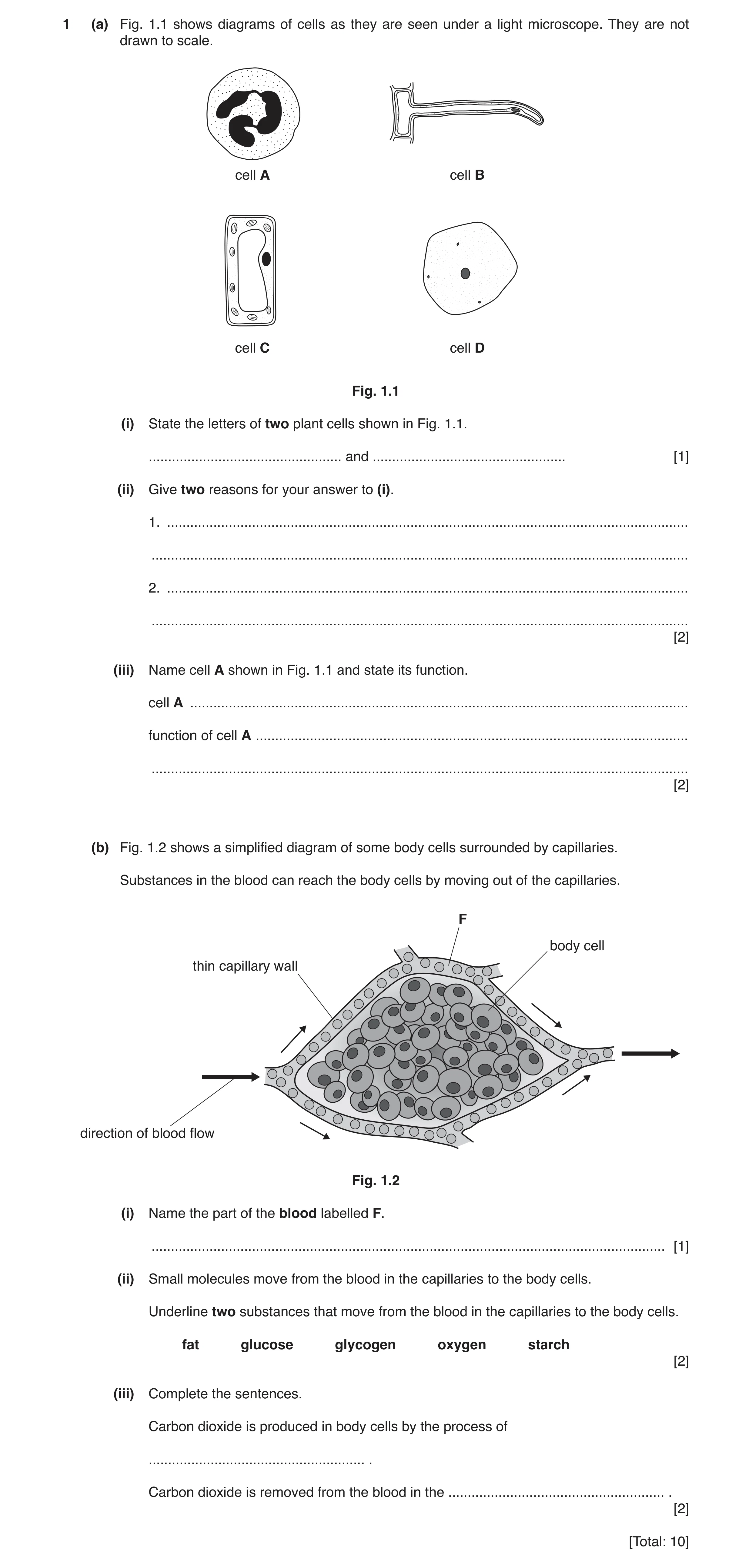
Q4 · Diagrams of cells as they are seen under a light microscope 0653/31 Oct/Nov 2019
1 (a) Fig. 1.1 shows diagrams of cells as they are seen under a light microscope. They are not drawn to scale. cell A cell B cell C cell D Fig. 1.1 (i) State the letters of two plant cells shown in Fig. 1.1. … and … [1] (ii) Give two reasons for your answer to (i). 1. … … 2. … … [2] (iii) Name cell A shown in Fig. 1.1 and state its function. cell A … function of cell A … … [2] (b) Fig. 1.2 shows a simplified diagram of some body cells surrounded by capillaries. Substances in the blood can reach the body cells by moving out of the capillaries. F body cell thin capillary wall direction of blood flow Fig. 1.2 (i) Name the part of the blood labelled F. … [1] (ii) Small molecules move from the blood in the capillaries to the body cells. Underline two substances that move from the blood in the capillaries to the body cells. fat glucose glycogen oxygen starch [2] (iii) Complete the sentences. Carbon dioxide is produced in body cells by the process of … . Carbon dioxide is removed from the blood in the … . [2] [Total: 10]
10 marks
Mark scheme: 1(a)(i) B and C ; 1 1(a)(ii) any two from: vacuoles ; cell walls ; chloroplasts ; 2 1(a)(iii) white blood cell ; phagocytosis / description of phagocytosis ; 2 1(b)(i) plasma ; 1 1(b)(ii) glucose underlined ; oxygen underlined ; 2 1(b)(iii) respiration ; lungs / alveoli ; 2
This question in 0653/31 Oct/Nov 2019
Question 5 0653/32 Oct/Nov 2019
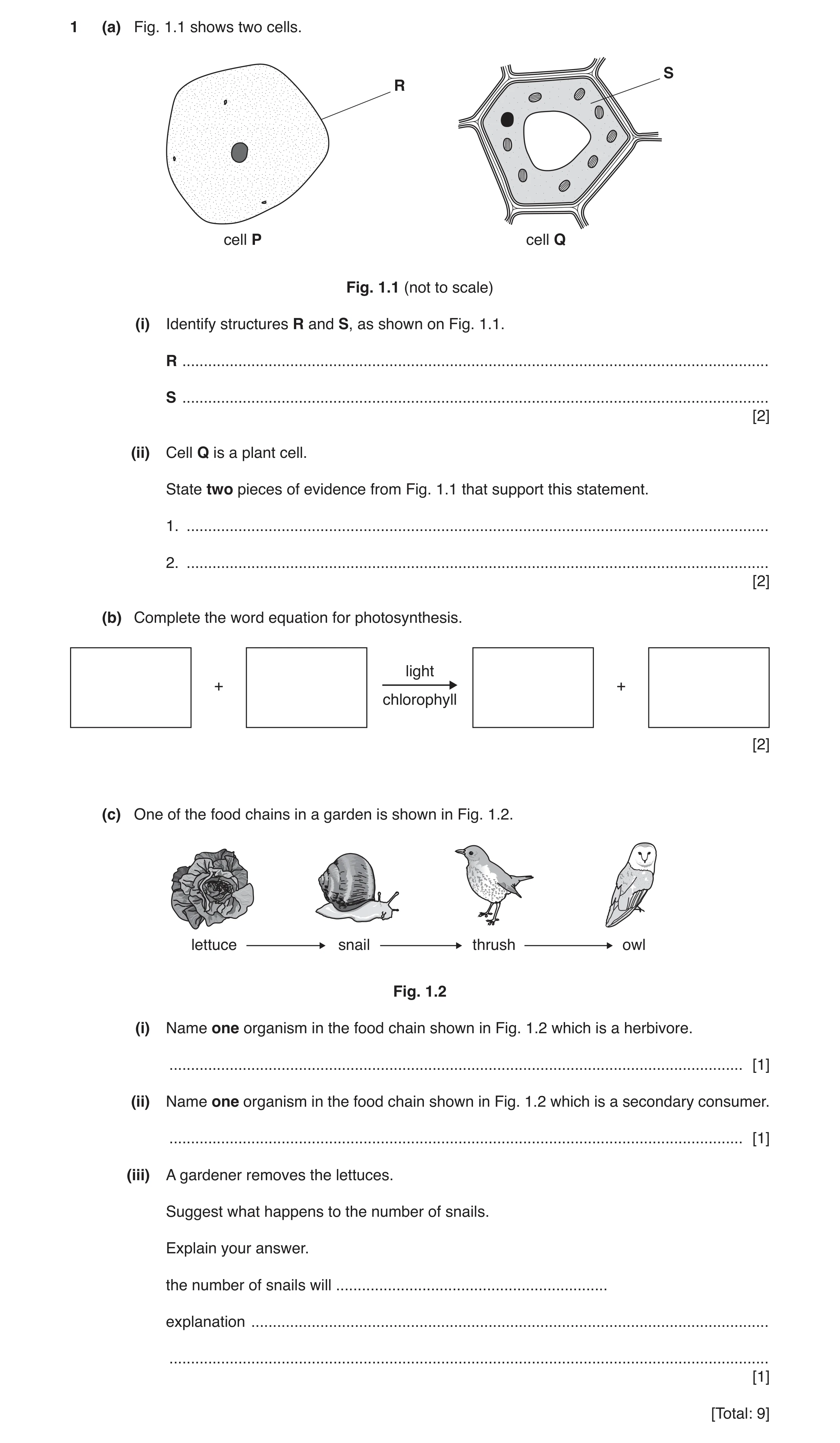
1 (a) Fig. 1.1 shows two cells. S R cell P cell Q Fig. 1.1 (not to scale) (i) Identify structures R and S, as shown on Fig. 1.1. R … S … [2] (ii) Cell Q is a plant cell. State two pieces of evidence from Fig. 1.1 that support this statement. 1. … 2. … [2] (b) Complete the word equation for photosynthesis. light + + chlorophyll [2] (c) One of the food chains in a garden is shown in Fig. 1.2. lettuce snail thrush owl Fig. 1.2 (i) Name one organism in the food chain shown in Fig. 1.2 which is a herbivore. … [1] (ii) Name one organism in the food chain shown in Fig. 1.2 which is a secondary consumer. … [1] (iii) A gardener removes the lettuces. Suggest what happens to the number of snails. Explain your answer. the number of snails will … explanation … … [1] [Total: 9]
9 marks
Mark scheme: 1(a)(i) R cell membrane ; S cytoplasm ; 2 1(a)(ii) any two from: (it has a) cell wall ; (it has a large) vacuole ; (it has) chloroplasts ; 2 1(b) (LHS) carbon dioxide + water ; (RHS) glucose + oxygen ; 2 1(c)(i) snail ; 1 1(c)(ii) thrush ; 1 1(c)(iii) decrease and (they have) less food / no food ; 1
This question in 0653/32 Oct/Nov 2019
Question 6 0653/33 Oct/Nov 2019
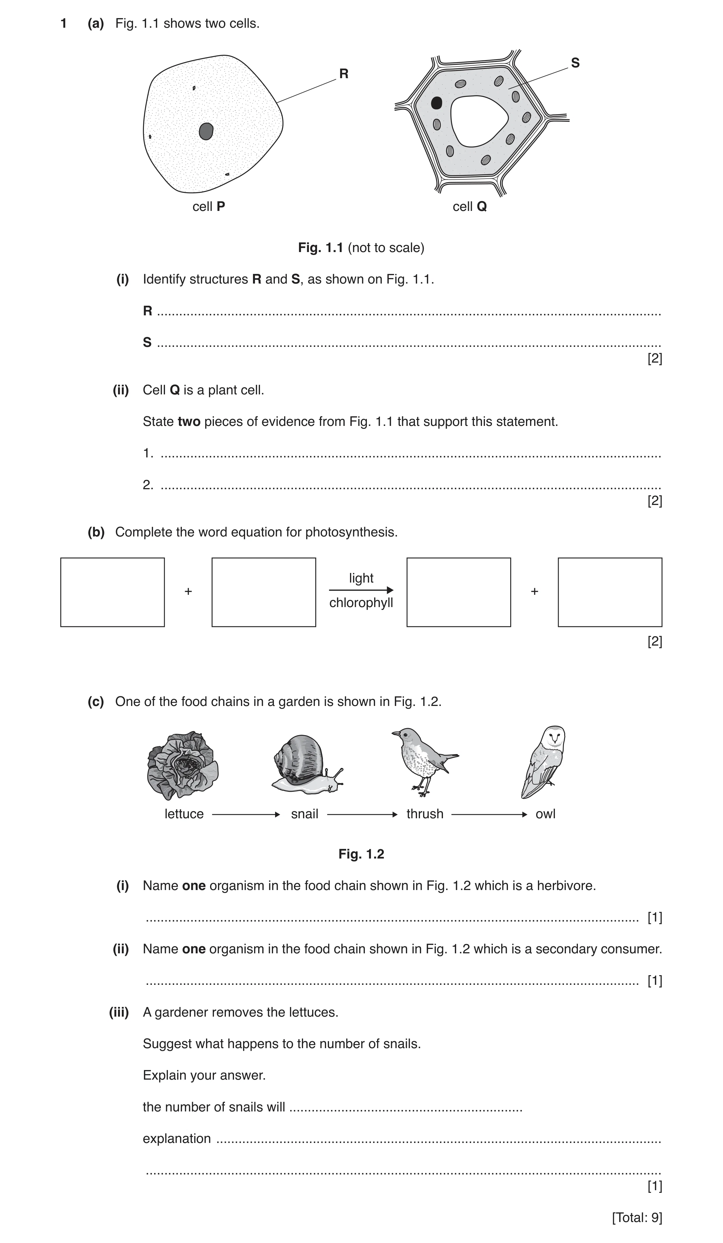
1 (a) Fig. 1.1 shows two cells. S R cell P cell Q Fig. 1.1 (not to scale) (i) Identify structures R and S, as shown on Fig. 1.1. R … S … [2] (ii) Cell Q is a plant cell. State two pieces of evidence from Fig. 1.1 that support this statement. 1. … 2. … [2] (b) Complete the word equation for photosynthesis. light + + chlorophyll [2] (c) One of the food chains in a garden is shown in Fig. 1.2. lettuce snail thrush owl Fig. 1.2 (i) Name one organism in the food chain shown in Fig. 1.2 which is a herbivore. … [1] (ii) Name one organism in the food chain shown in Fig. 1.2 which is a secondary consumer. … [1] (iii) A gardener removes the lettuces. Suggest what happens to the number of snails. Explain your answer. the number of snails will … explanation … … [1] [Total: 9]
9 marks
Mark scheme: 1(a)(i) R cell membrane ; S cytoplasm ; 2 1(a)(ii) any two from: (it has a) cell wall ; (it has a large) vacuole ; (it has) chloroplasts ; 2 1(b) (LHS) carbon dioxide + water ; (RHS) glucose + oxygen ; 2 1(c)(i) snail ; 1 1(c)(ii) thrush ; 1 1(c)(iii) decrease and (they have) less food / no food ; 1
This question in 0653/33 Oct/Nov 2019
Q7 · A diagram of muscle cells 0653/33 Oct/Nov 2020
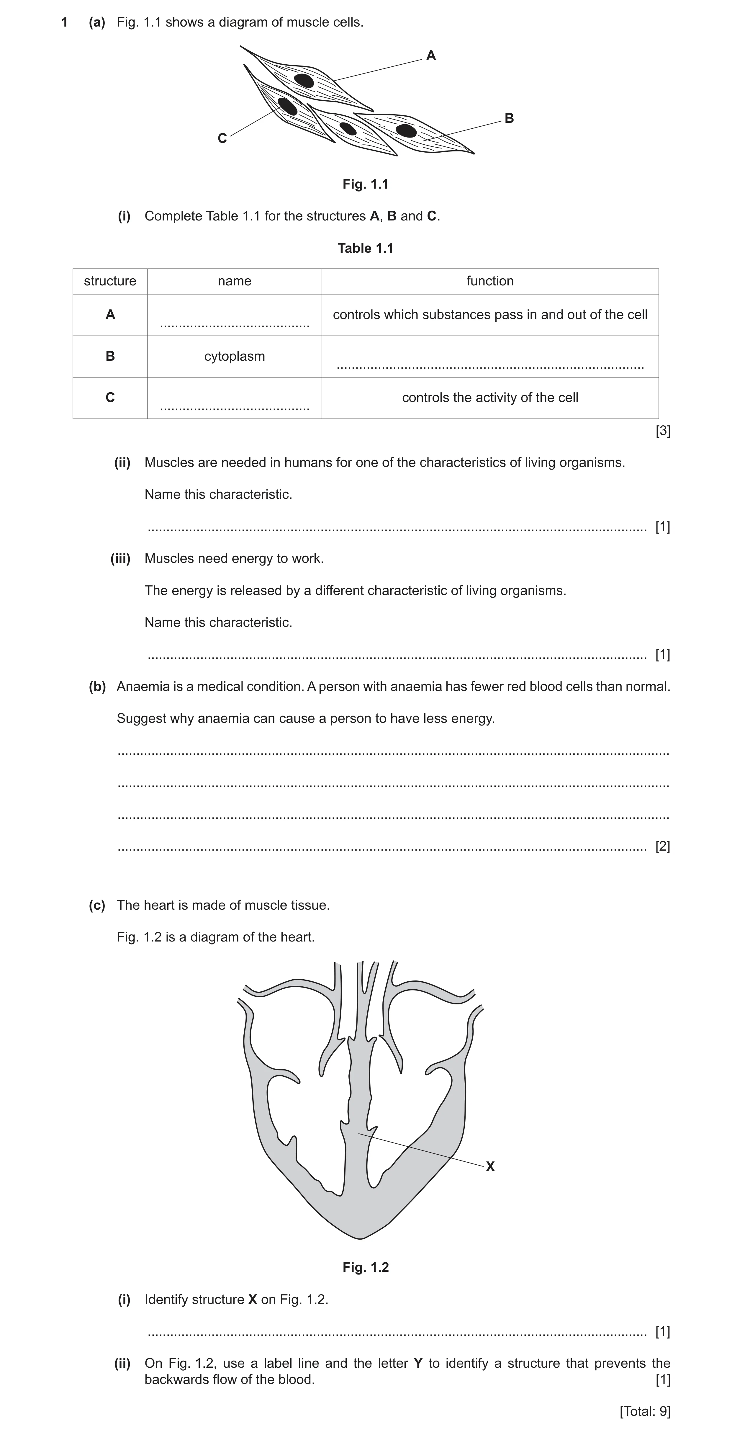
1 (a) Fig. 1.1 shows a diagram of muscle cells. A B C Fig. 1.1 (i) Complete Table 1.1 for the structures A, B and C. Table 1.1 structure name function A controls which substances pass in and out of the cell … B cytoplasm … C controls the activity of the cell … [3] (ii) Muscles are needed in humans for one of the characteristics of living organisms. Name this characteristic. … [1] (iii) Muscles need energy to work. The energy is released by a different characteristic of living organisms. Name this characteristic. … [1] (b) Anaemia is a medical condition. A person with anaemia has fewer red blood cells than normal. Suggest why anaemia can cause a person to have less energy. … … … … [2] (c) The heart is made of muscle tissue. Fig. 1.2 is a diagram of the heart. X Fig. 1.2 (i) Identify structure X on Fig. 1.2. … [1] (ii) On Fig. 1.2, use a label line and the letter Y to identify a structure that prevents the backwards flow of the blood. [1] [Total: 9]
9 marks
Mark scheme: 1(a)(i) (cell) membrane ; site of different (metabolic) reactions ; nucleus ; 3 1(a)(ii) movement ; 1 1(a)(iii) respiration ; 1 1(b) any two from: red blood cells transport oxygen ; less oxygen, transported / available ; for (aerobic) respiration / less (aerobic) respiration OR oxygen is needed for respiration ; 2 1(c)(i) septum ; 1 1(c)(ii) label line to any valve ; 1
This question in 0653/33 Oct/Nov 2020
Q8 · Plant cells as seen with a microscope 0653/33 Oct/Nov 2020
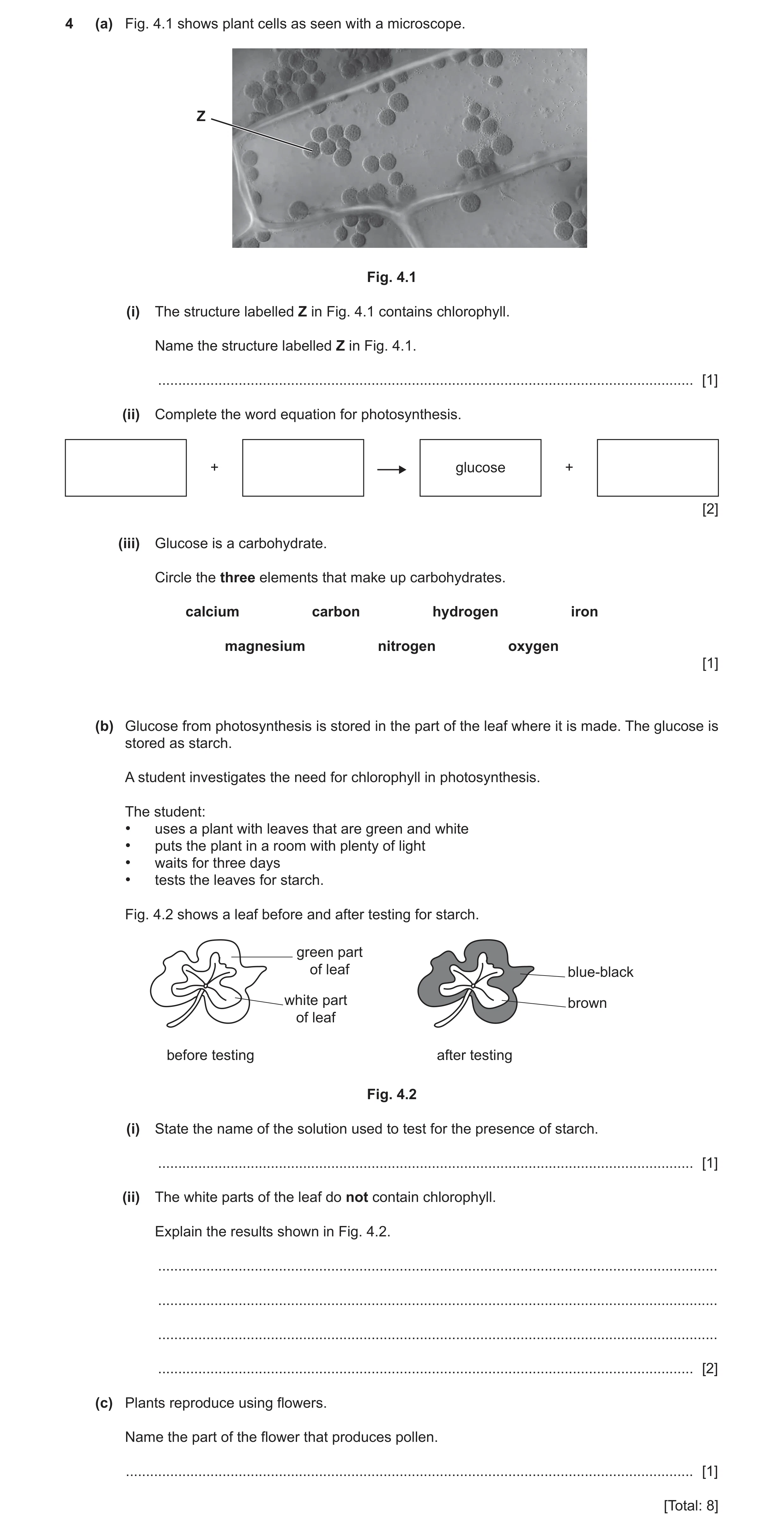
4 (a) Fig. 4.1 shows plant cells as seen with a microscope. Z Fig. 4.1 (i) The structure labelled Z in Fig. 4.1 contains chlorophyll. Name the structure labelled Z in Fig. 4.1. … [1] (ii) Complete the word equation for photosynthesis. + glucose + [2] (iii) Glucose is a carbohydrate. Circle the three elements that make up carbohydrates. calcium carbon hydrogen iron magnesium nitrogen oxygen [1] (b) Glucose from photosynthesis is stored in the part of the leaf where it is made. The glucose is stored as starch. A student investigates the need for chlorophyll in photosynthesis. The student: • uses a plant with leaves that are green and white • puts the plant in a room with plenty of light • waits for three days • tests the leaves for starch. Fig. 4.2 shows a leaf before and after testing for starch. green part of leaf blue-black white part brown of leaf before testing after testing Fig. 4.2 (i) State the name of the solution used to test for the presence of starch. … [1] (ii) The white parts of the leaf do not contain chlorophyll. Explain the results shown in Fig. 4.2. … … … … [2] (c) Plants reproduce using flowers. Name the part of the flower that produces pollen. … [1] [Total: 8]
8 marks
Mark scheme: 4(a)(i) chloroplast ; 1 4(a)(ii) (LHS) carbon dioxide + water ; (RHS) (glucose) + oxygen ; 2 4(a)(iii) carbon AND hydrogen AND oxygen ; 1 4(b)(i) iodine (solution) ; 1 4(b)(ii) no starch in white area OR iodine stays brown so no starch / ORA ; no photosynthesis takes place in white area / ORA OR chlorophyll is needed for photosynthesis ; 2 4(c) anther ; 1
This question in 0653/33 Oct/Nov 2020
Q9 · The boxes on the left show characteristics of living organisms 0653/31 May/June 2021
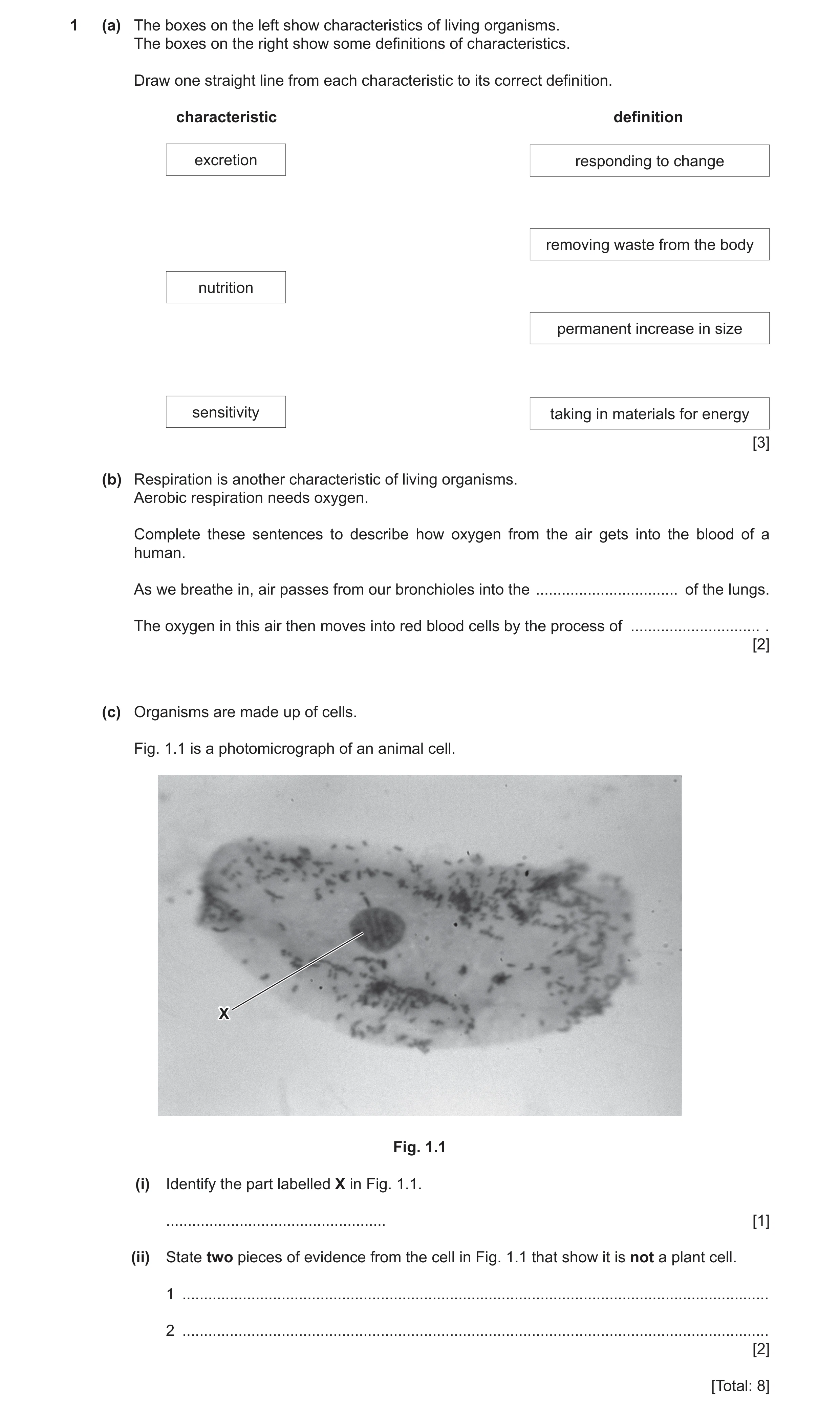
1 (a) The boxes on the left show characteristics of living organisms. The boxes on the right show some definitions of characteristics. Draw one straight line from each characteristic to its correct definition. characteristic definition excretion responding to change removing waste from the body nutrition permanent increase in size sensitivity taking in materials for energy [3] (b) Respiration is another characteristic of living organisms. Aerobic respiration needs oxygen. Complete these sentences to describe how oxygen from the air gets into the blood of a human. As we breathe in, air passes from our bronchioles into the … of the lungs. The oxygen in this air then moves into red blood cells by the process of … . [2] (c) Organisms are made up of cells. Fig. 1.1 is a photomicrograph of an animal cell. X Fig. 1.1 (i) Identify the part labelled X in Fig. 1.1. … [1] (ii) State two pieces of evidence from the cell in Fig. 1.1 that show it is not a plant cell. 1 … 2 … [2] [Total: 8]
8 marks
Mark scheme: 1(a) responding to change excretion removing waste from the body nutrition permanent increases in size sensitivity taking in materials for energy ;;; 1(b) alveoli ; diffusion ; 2 1(c)(i) nucleus ; 1 1(c)(ii) any two from: no cell wall ; no chloroplasts ; no (large) vacuole ; 2
This question in 0653/31 May/June 2021
Question 10 0653/32 Oct/Nov 2021
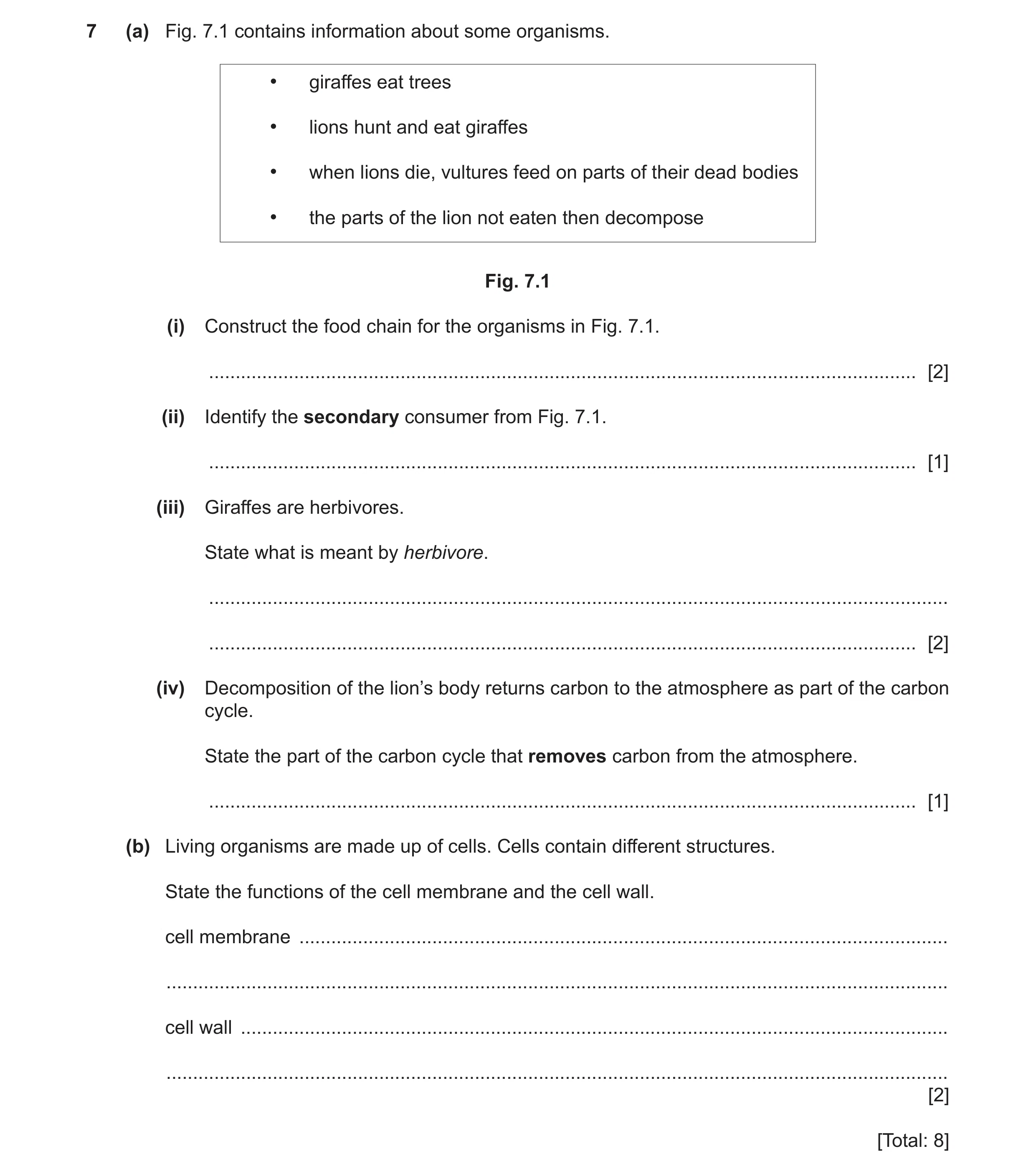
7 (a) Fig. 7.1 contains information about some organisms. • giraffes eat trees • lions hunt and eat giraffes • when lions die, vultures feed on parts of their dead bodies • the parts of the lion not eaten then decompose Fig. 7.1 (i) Construct the food chain for the organisms in Fig. 7.1. … [2] (ii) Identify the secondary consumer from Fig. 7.1. … [1] (iii) Giraffes are herbivores. State what is meant by herbivore. … … [2] (iv) Decomposition of the lion’s body returns carbon to the atmosphere as part of the carbon cycle. State the part of the carbon cycle that removes carbon from the atmosphere. … [1] (b) Living organisms are made up of cells. Cells contain different structures. State the functions of the cell membrane and the cell wall. cell membrane … … cell wall … … [2] [Total: 8]
8 marks
Mark scheme: 7(a)(i) tree(s) → giraffe(s) → lion(s) → vulture(s) all organisms correct ; correct arrows ; 7(a)(ii) lion ; 1 7(a)(iii) (an animal that) gets its energy ; by (only) eating plants ; 2 7(a)(iv) photosynthesis ; 1 7(b) (membrane) controls the movement of substances in and out of the cells ; provides support ; 2
This question in 0653/32 Oct/Nov 2021
Question 11 0653/33 Oct/Nov 2021
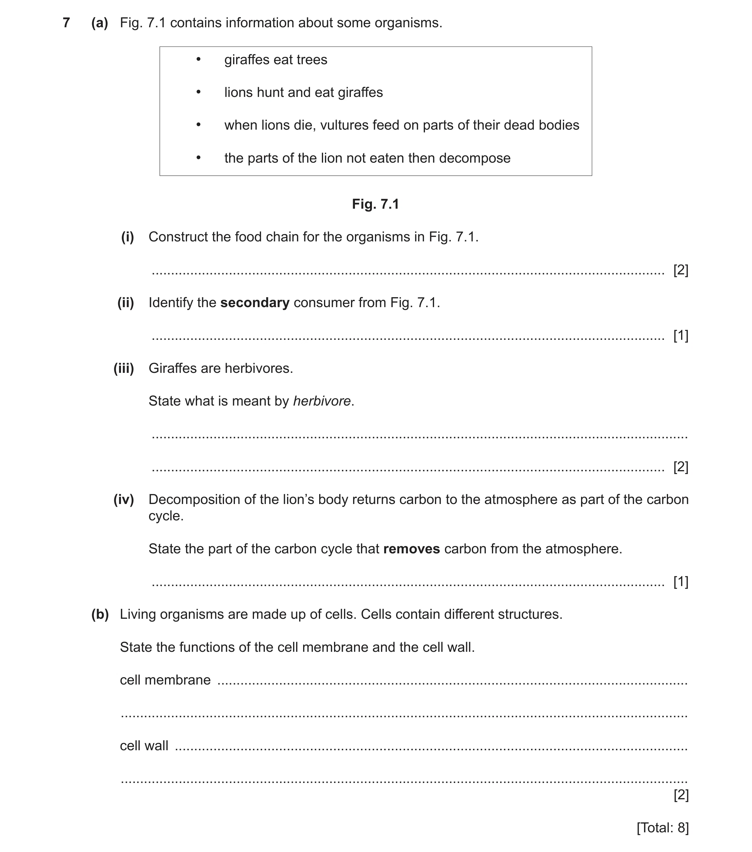
7 (a) Fig. 7.1 contains information about some organisms. • giraffes eat trees • lions hunt and eat giraffes • when lions die, vultures feed on parts of their dead bodies • the parts of the lion not eaten then decompose Fig. 7.1 (i) Construct the food chain for the organisms in Fig. 7.1. … [2] (ii) Identify the secondary consumer from Fig. 7.1. … [1] (iii) Giraffes are herbivores. State what is meant by herbivore. … … [2] (iv) Decomposition of the lion’s body returns carbon to the atmosphere as part of the carbon cycle. State the part of the carbon cycle that removes carbon from the atmosphere. … [1] (b) Living organisms are made up of cells. Cells contain different structures. State the functions of the cell membrane and the cell wall. cell membrane … … cell wall … … [2] [Total: 8]
8 marks
Mark scheme: 7(a)(i) tree(s) → giraffe(s) → lion(s) → vulture(s) all organisms correct ; correct arrows ; 7(a)(ii) lion ; 1 7(a)(iii) (an animal that) gets its energy ; by (only) eating plants ; 2 7(a)(iv) photosynthesis ; 1 7(b) (membrane) controls the movement of substances in and out of the cells ; provides support ; 2
This question in 0653/33 Oct/Nov 2021
Q12 · A diagram of a root hair cell 0653/32 Feb/March 2022
1 (a) Fig. 1.1 is a diagram of a root hair cell. cell membrane cell wall … root hair … … Fig. 1.1 (i) Complete the labels on Fig. 1.1. [3] (ii) State the function of the cell wall. … [1] (iii) Water enters the root hair cell and passes to the leaves of the plant. Complete the flow chart to show the pathway of water through the plant. root hair cell root … cells … vessels mesophyll cells [2] (b) Fig. 1.2 shows a plant cell before and after it is immersed in a concentrated sugar solution. plant cell before immersion plant cell after immersion Fig. 1.2 (i) Describe two visible differences in the cell after immersion compared to the cell before immersion in Fig. 1.2. 1 … … 2 … … [2] (ii) Water has diffused out of the plant cell through the partially permeable membrane. State the name of this type of diffusion. … [1] [Total: 9]
9 marks
Mark scheme: 1(a)(i) vacuole ; cytoplasm ; nucleus ; 3 1(a)(ii) support / strengthen the cell ; 1 1(a)(iii) cortex ; xylem ; 2 1(b)(i) any two from: (whole cell is) smaller / narrower ; smaller vacuole ; membrane pulled away from wall ; 2 1(b)(ii) osmosis ; 1
This question in 0653/32 Feb/March 2022
Q13 · An image of a white blood cell 0653/31 May/June 2022
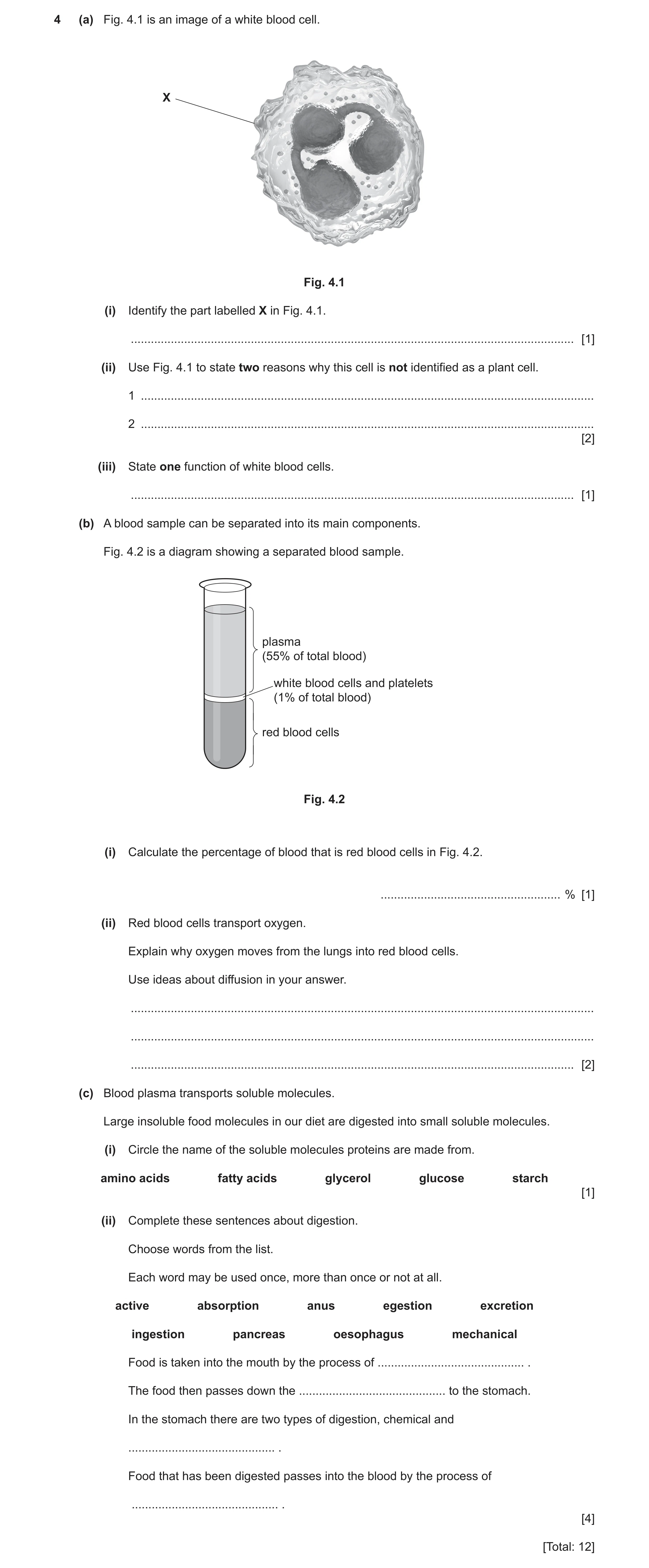
4 (a) Fig. 4.1 is an image of a white blood cell. X Fig. 4.1 (i) Identify the part labelled X in Fig. 4.1. … [1] (ii) Use Fig. 4.1 to state two reasons why this cell is not identified as a plant cell. 1 … 2 … [2] (iii) State one function of white blood cells. … [1] (b) A blood sample can be separated into its main components. Fig. 4.2 is a diagram showing a separated blood sample. plasma (55% of total blood) white blood cells and platelets (1% of total blood) red blood cells Fig. 4.2 (i) Calculate the percentage of blood that is red blood cells in Fig. 4.2. … % [1] (ii) Red blood cells transport oxygen. Explain why oxygen moves from the lungs into red blood cells. Use ideas about diffusion in your answer. … … … [2] (c) Blood plasma transports soluble molecules. Large insoluble food molecules in our diet are digested into small soluble molecules. (i) Circle the name of the soluble molecules proteins are made from. amino acids fatty acids glycerol glucose starch [1] (ii) Complete these sentences about digestion. Choose words from the list. Each word may be used once, more than once or not at all. active absorption anus egestion excretion ingestion pancreas oesophagus mechanical Food is taken into the mouth by the process of … . The food then passes down the … to the stomach. In the stomach there are two types of digestion, chemical and … . Food that has been digested passes into the blood by the process of … . [4] [Total: 12]
12 marks
Mark scheme: 4(a)(i) cell membrane ; 1 4(a)(ii) any two from: no cell wall ; no (permanent) vacuole ; no chloroplast ; 2 4(a)(iii) produce antibodies / phagocytosis ; 1 4(b)(i) 44(%) ; 1 4(b)(ii) higher concentration oxygen (in lungs) / ora ; oxygen moves down concentration gradient / oxygen moves (from a higher concentration) to a lower concentration ; 2 4(c)(i) amino acids (circled) ; 1 Question Answer Marks 4(c)(ii) ingestion ; oesophagus ; mechanical ; absorption ; 4
This question in 0653/31 May/June 2022
Q14 · A cell from the spongy mesophyll of a plant 0653/31 Oct/Nov 2022
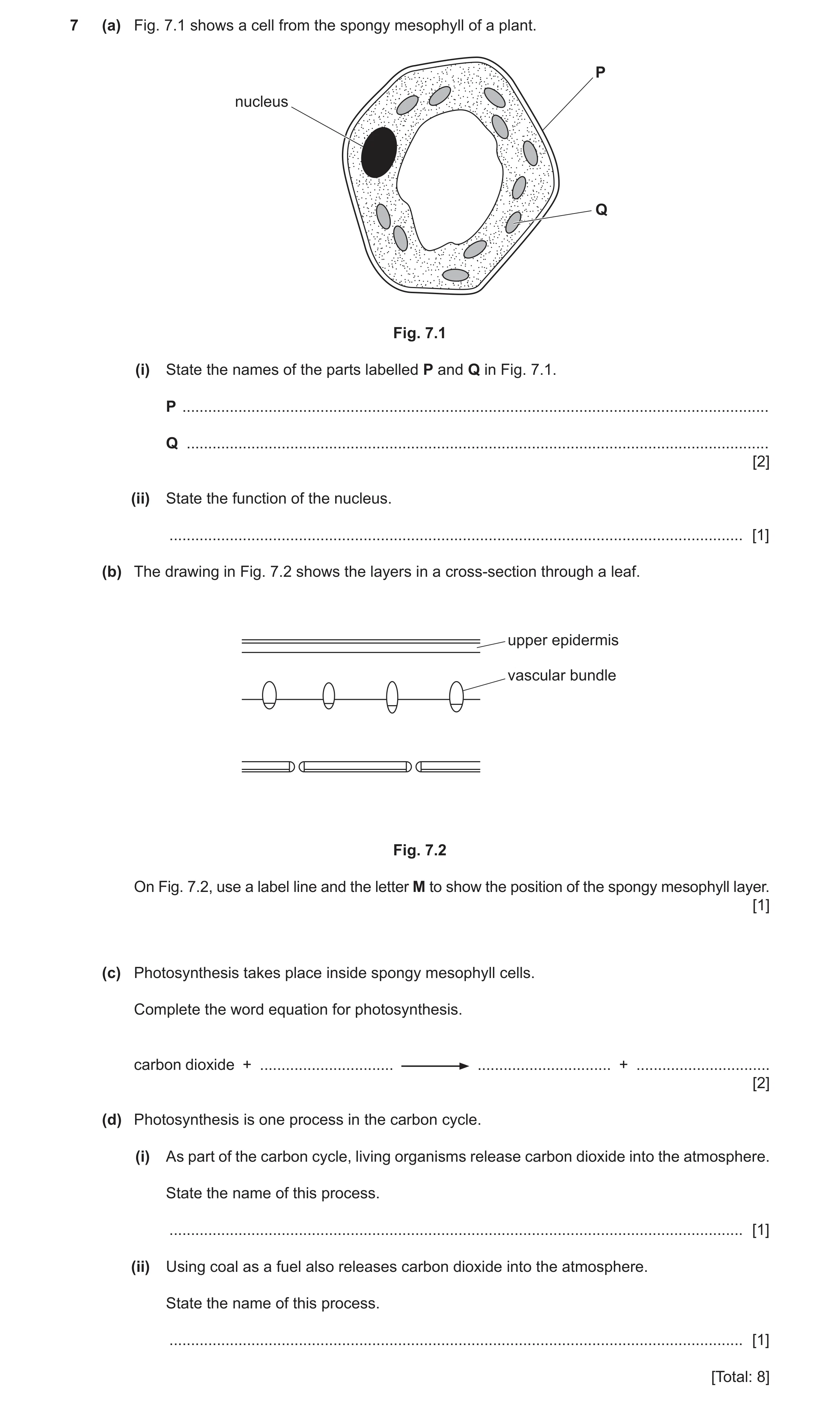
7 (a) Fig. 7.1 shows a cell from the spongy mesophyll of a plant. P nucleus Q Fig. 7.1 (i) State the names of the parts labelled P and Q in Fig. 7.1. P … Q … [2] (ii) State the function of the nucleus. … [1] (b) The drawing in Fig. 7.2 shows the layers in a cross-section through a leaf. upper epidermis vascular bundle Fig. 7.2 On Fig. 7.2, use a label line and the letter M to show the position of the spongy mesophyll layer. [1] (c) Photosynthesis takes place inside spongy mesophyll cells. Complete the word equation for photosynthesis. carbon dioxide + … … + … [2] (d) Photosynthesis is one process in the carbon cycle. (i) As part of the carbon cycle, living organisms release carbon dioxide into the atmosphere. State the name of this process. … [1] (ii) Using coal as a fuel also releases carbon dioxide into the atmosphere. State the name of this process. … [1] [Total: 8]
8 marks
Mark scheme: 7(a)(i) P (cell) wall ; 2 Q chloroplast ; 7(a)(ii) controls the cell (activities) ; 1 7(b) M below line of vascular bundles above lower epidermis ; 1 7(c) water ; 2 glucose + oxygen ; 7(d)(i) respiration ; 1 7(d)(ii) combustion ; 1
This question in 0653/31 Oct/Nov 2022
Q15 · The boxes on the left show some of the characteristics of living organisms 0653/32 Oct/Nov 2022
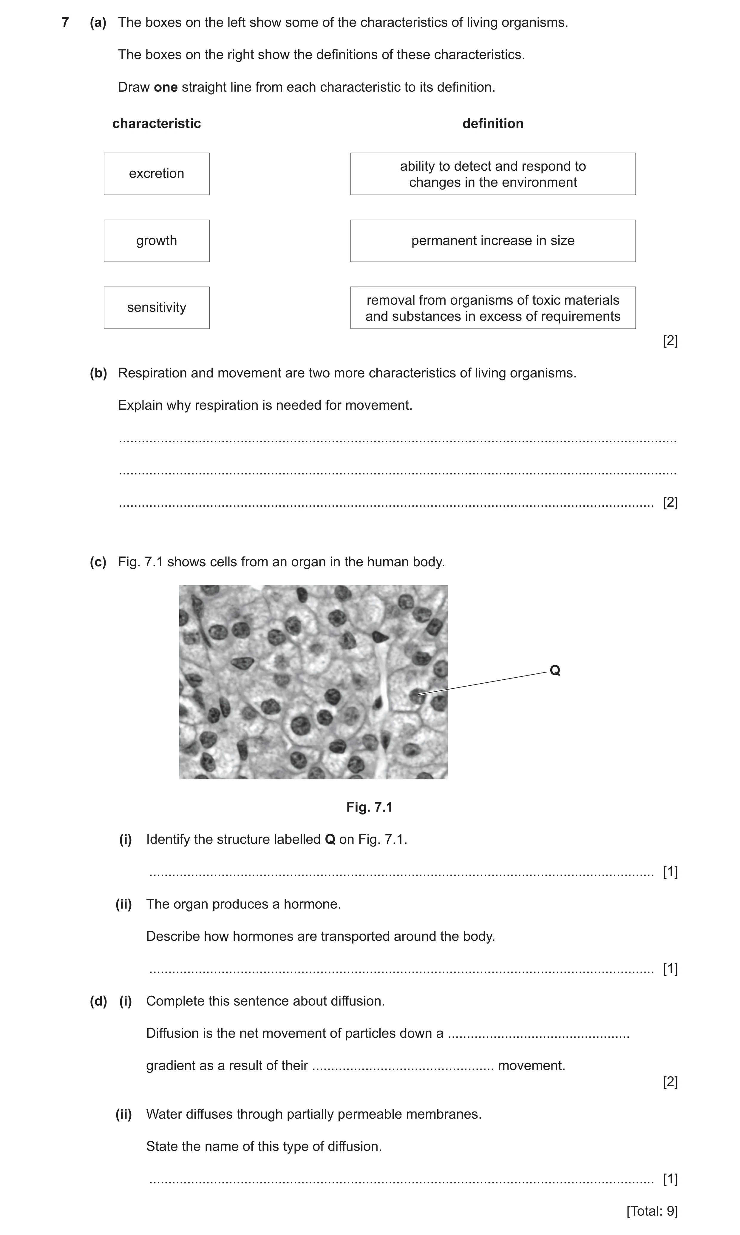
7 (a) The boxes on the left show some of the characteristics of living organisms. The boxes on the right show the definitions of these characteristics. Draw one straight line from each characteristic to its definition. characteristic definition ability to detect and respond to excretion changes in the environment growth permanent increase in size removal from organisms of toxic materials sensitivity and substances in excess of requirements [2] (b) Respiration and movement are two more characteristics of living organisms. Explain why respiration is needed for movement. … … … [2] (c) Fig. 7.1 shows cells from an organ in the human body. Q Fig. 7.1 (i) Identify the structure labelled Q on Fig. 7.1. … [1] (ii) The organ produces a hormone. Describe how hormones are transported around the body. … [1] (d) (i) Complete this sentence about diffusion. Diffusion is the net movement of particles down a … gradient as a result of their … movement. [2] (ii) Water diffuses through partially permeable membranes. State the name of this type of diffusion. … [1] [Total: 9]
9 marks
Mark scheme: 7(a) 2 ability to detect and respond to excretion changes in the environment growth permanent increase in size removal from organisms of toxic sensitivity materials and substances in excess of requirements one correct ; all three correct ; 7(b) respiration releases energy ; 2 energy needed for muscle contraction ; 7(c)(i) nucleus ; 1 7(c)(ii) (in the) blood / plasma ; 1 7(d)(i) concentration ; 2 random ; 7(d)(ii) osmosis ; 1
This question in 0653/32 Oct/Nov 2022
Q16 · The boxes on the left show some of the characteristics of living organisms 0653/33 Oct/Nov 2022
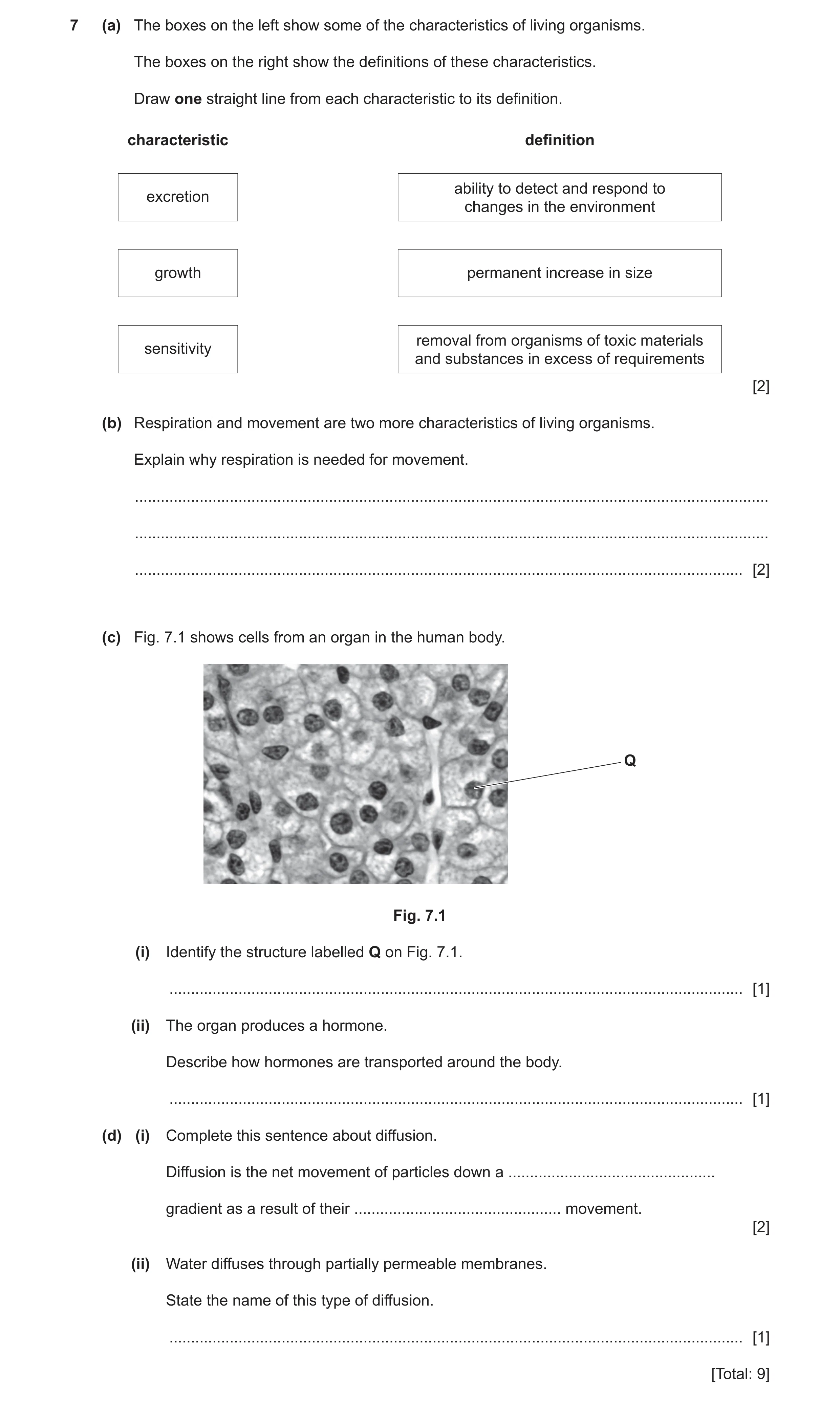
7 (a) The boxes on the left show some of the characteristics of living organisms. The boxes on the right show the definitions of these characteristics. Draw one straight line from each characteristic to its definition. characteristic definition ability to detect and respond to excretion changes in the environment growth permanent increase in size removal from organisms of toxic materials sensitivity and substances in excess of requirements [2] (b) Respiration and movement are two more characteristics of living organisms. Explain why respiration is needed for movement. … … … [2] (c) Fig. 7.1 shows cells from an organ in the human body. Q Fig. 7.1 (i) Identify the structure labelled Q on Fig. 7.1. … [1] (ii) The organ produces a hormone. Describe how hormones are transported around the body. … [1] (d) (i) Complete this sentence about diffusion. Diffusion is the net movement of particles down a … gradient as a result of their … movement. [2] (ii) Water diffuses through partially permeable membranes. State the name of this type of diffusion. … [1] [Total: 9]
9 marks
Mark scheme: 7(a) 2 ability to detect and respond to excretion changes in the environment growth permanent increase in size removal from organisms of toxic sensitivity materials and substances in excess of requirements one correct ; all three correct ; 7(b) respiration releases energy ; 2 energy needed for muscle contraction ; 7(c)(i) nucleus ; 1 7(c)(ii) (in the) blood / plasma ; 1 7(d)(i) concentration ; 2 random ; 7(d)(ii) osmosis ; 1
This question in 0653/33 Oct/Nov 2022
Q17 · The boxes on the left show different characteristics of living organisms 0653/33 May/June 2023
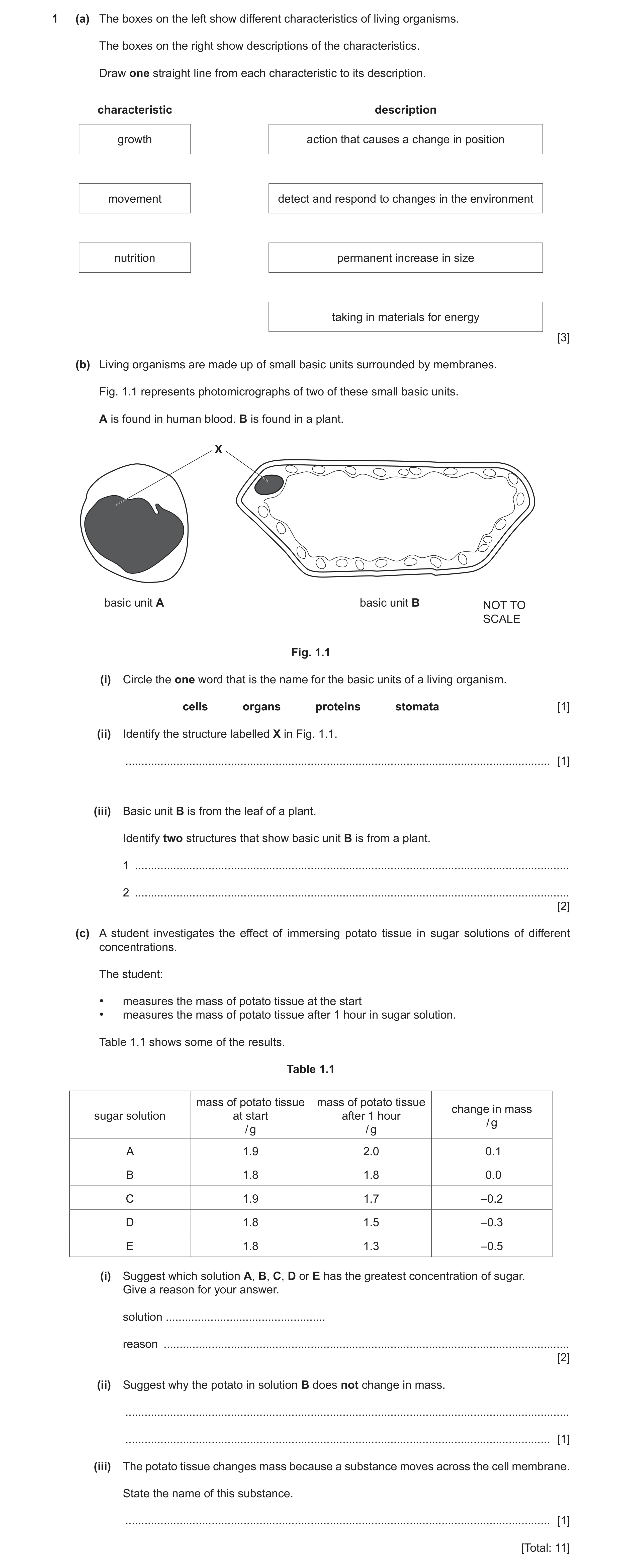
1 (a) The boxes on the left show different characteristics of living organisms. The boxes on the right show descriptions of the characteristics. Draw one straight line from each characteristic to its description. characteristic description growth action that causes a change in position movement detect and respond to changes in the environment nutrition permanent increase in size taking in materials for energy [3] (b) Living organisms are made up of small basic units surrounded by membranes. Fig. 1.1 represents photomicrographs of two of these small basic units. A is found in human blood. B is found in a plant. X basic unit A basic unit B NOT TO SCALE Fig. 1.1 (i) Circle the one word that is the name for the basic units of a living organism. cells organs proteins stomata [1] (ii) Identify the structure labelled X in Fig. 1.1. … [1] (iii) Basic unit B is from the leaf of a plant. Identify two structures that show basic unit B is from a plant. 1 … 2 … [2] (c) A student investigates the effect of immersing potato tissue in sugar solutions of different concentrations. The student: • measures the mass of potato tissue at the start • measures the mass of potato tissue after 1 hour in sugar solution. Table 1.1 shows some of the results. Table 1.1 mass of potato tissue mass of potato tissue change in mass sugar solution at start after 1 hour / g / g / g A 1.9 2.0 0.1 B 1.8 1.8 0.0 C 1.9 1.7 –0.2 D 1.8 1.5 –0.3 E 1.8 1.3 –0.5 (i) Suggest which solution A, B, C, D or E has the greatest concentration of sugar. Give a reason for your answer. solution … reason … [2] (ii) Suggest why the potato in solution B does not change in mass. … … [1] (iii) The potato tissue changes mass because a substance moves across the cell membrane. State the name of this substance. … [1] [Total: 11]
11 marks
Mark scheme: 1(a) growth action that causes a change in position movement detect and respond to changes in the environment nutrition permanent increase in size taking in materials for energy ;;; 3 1(b)(i) cells (circled) ; 1 1(b)(ii) nucleus ; 1 1(b)(iii) any two from: (large) vacuole ; chloroplasts ; cell wall ; 2 1(c)(i) (solution) E ; (reason) it causes the biggest, change / loss in mass ; 2 1(c)(ii) concentration of solution inside cell is the same as the concentration of the sugar solution ; 1 1(c)(iii) water ; 1
This question in 0653/33 May/June 2023
Q18 · The boxes on the left show the names of some structures found in cells 0653/32 Feb/March 2024
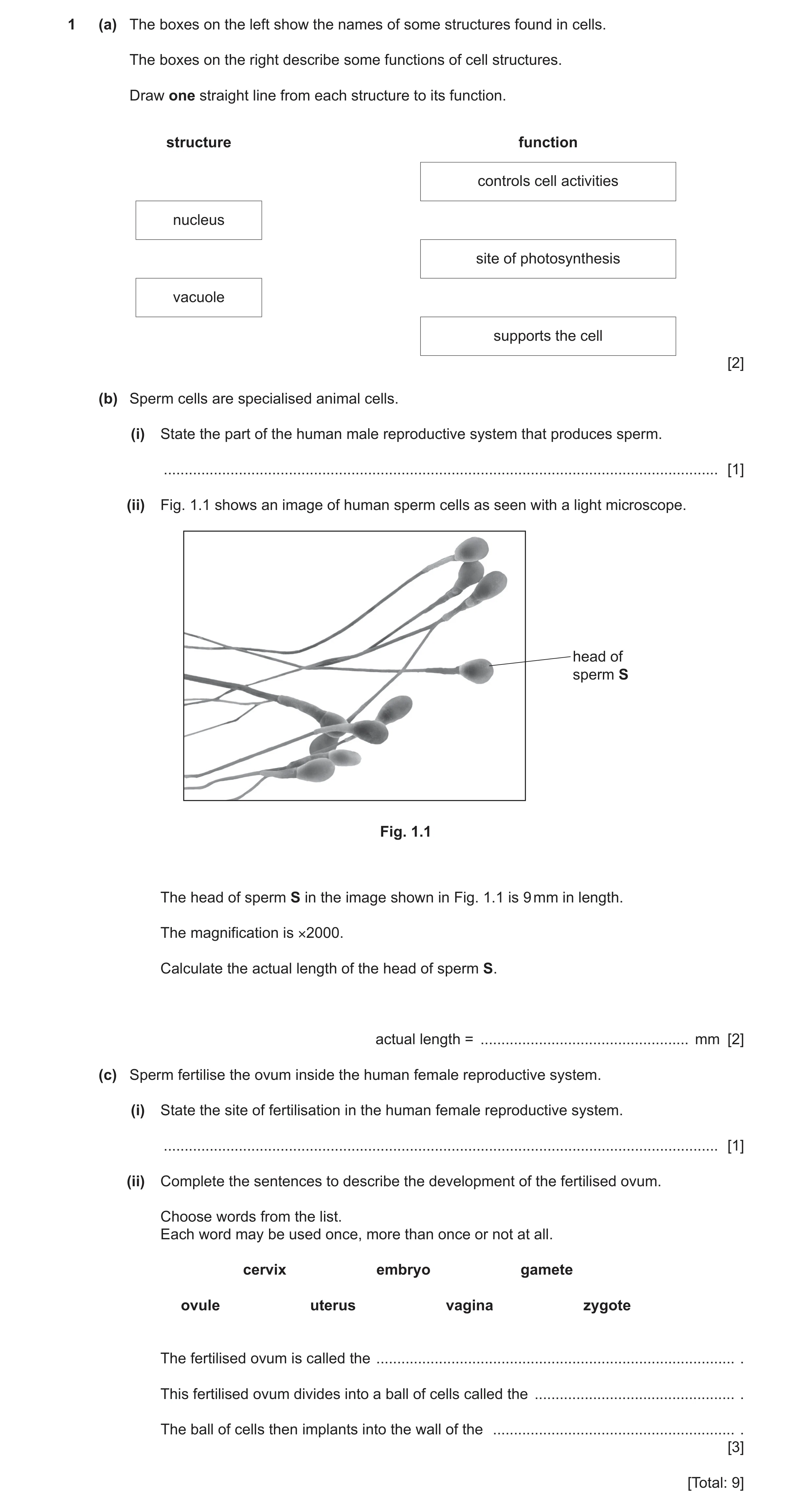
1 (a) The boxes on the left show the names of some structures found in cells. The boxes on the right describe some functions of cell structures. Draw one straight line from each structure to its function. structure function controls cell activities nucleus site of photosynthesis vacuole supports the cell [2] (b) Sperm cells are specialised animal cells. (i) State the part of the human male reproductive system that produces sperm. … [1] (ii) Fig. 1.1 shows an image of human sperm cells as seen with a light microscope. head of sperm S Fig. 1.1 The head of sperm S in the image shown in Fig. 1.1 is 9 mm in length. The magnification is ×2000. Calculate the actual length of the head of sperm S. actual length = … mm [2] (c) Sperm fertilise the ovum inside the human female reproductive system. (i) State the site of fertilisation in the human female reproductive system. … [1] (ii) Complete the sentences to describe the development of the fertilised ovum. Choose words from the list. Each word may be used once, more than once or not at all. cervix embryo gamete ovule uterus vagina zygote The fertilised ovum is called the … . This fertilised ovum divides into a ball of cells called the … . The ball of cells then implants into the wall of the … . [3] [Total: 9]
9 marks
Mark scheme: Question Answer Marks 1(a) 2 controls cell activities nucleus site of photosynthesis vacuole supports the cell ;; one mark for each correct line 1(b)(i) testes ; 1 1(b)(ii) magnification = image length ÷ actual length (in any form) / actual = 9 ÷ 2000 ; 2 = 0.0045 (mm) ; 1(c)(i) oviduct ; 1 1(c)(ii) zygote ; 3 embryo ; uterus ;
This question in 0653/32 Feb/March 2024
Question 19 0653/32 May/June 2024
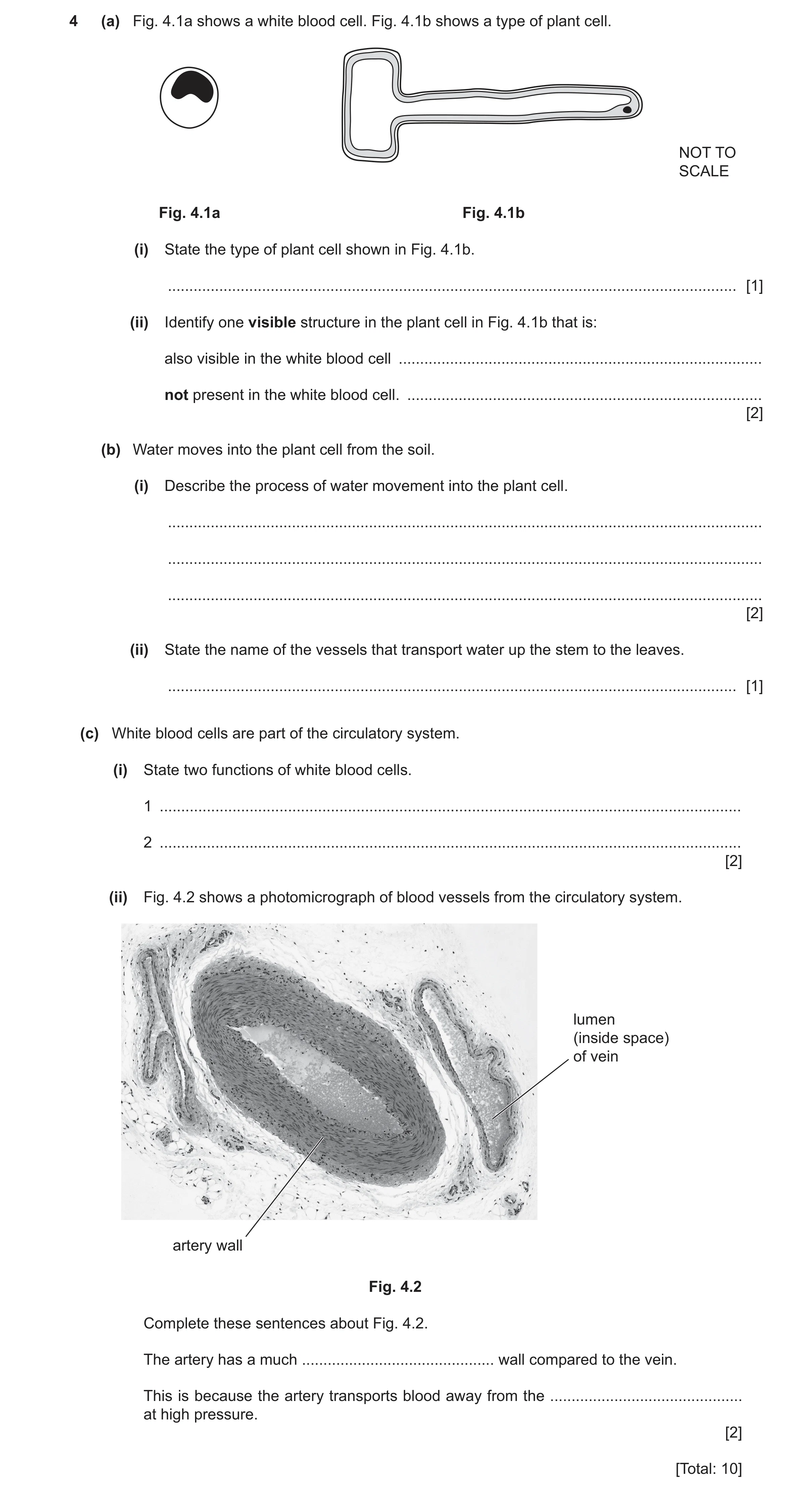
4 (a) Fig. 4.1a shows a white blood cell. Fig. 4.1b shows a type of plant cell. NOT TO SCALE Fig. 4.1a Fig. 4.1b (i) State the type of plant cell shown in Fig. 4.1b. … [1] (ii) Identify one visible structure in the plant cell in Fig. 4.1b that is: also visible in the white blood cell … not present in the white blood cell. … [2] (b) Water moves into the plant cell from the soil. (i) Describe the process of water movement into the plant cell. … … … [2] (ii) State the name of the vessels that transport water up the stem to the leaves. … [1] (c) White blood cells are part of the circulatory system. (i) State two functions of white blood cells. 1 … 2 … [2] (ii) Fig. 4.2 shows a photomicrograph of blood vessels from the circulatory system. lumen (inside space) of vein artery wall Fig. 4.2 Complete these sentences about Fig. 4.2. The artery has a much … wall compared to the vein. This is because the artery transports blood away from the … at high pressure. [2] [Total: 10]
10 marks
Mark scheme: 4(a)(i) root hair (cell) ; 1 4(a)(ii) (also visible in the white blood cell) nucleus / cytoplasm ; (not present in the white blood cell) vacuole / (cell) wall ; 2 4(b)(i) any two from: diffuses ; by osmosis ; through, cell membrane / partially permeable membrane ; 2 Question Answer Marks 4(b)(ii) xylem ; 1 4(c)(i) phagocytosis ; produce antibodies ; 2 4(c)(ii) thicker / more elastic ; heart ; 2
This question in 0653/32 May/June 2024
Q20 · A diagram of a plant root hair cell 0653/31 May/June 2025
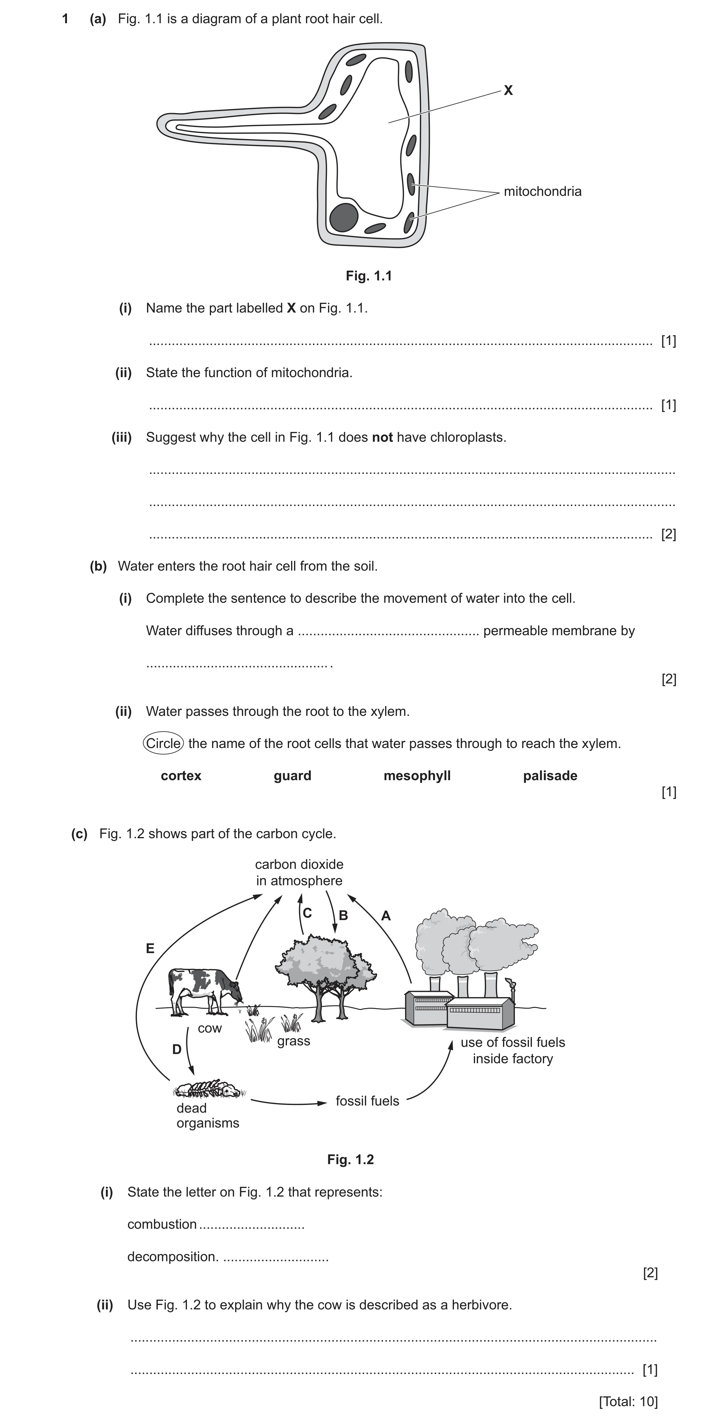
1 (a) Fig. 1.1 is a diagram of a plant root hair cell. X mitochondria Fig. 1.1 (i) Name the part labelled X on Fig. 1.1. … [1] (ii) State the function of mitochondria. … [1] (iii) Suggest why the cell in Fig. 1.1 does not have chloroplasts. … … … [2] (b) Water enters the root hair cell from the soil. (i) Complete the sentence to describe the movement of water into the cell. Water diffuses through a … permeable membrane by … . [2] (ii) Water passes through the root to the xylem. Circle the name of the root cells that water passes through to reach the xylem. cortex guard mesophyll palisade [1] (c) Fig. 1.2 shows part of the carbon cycle. carbon dioxide in atmosphere C B A E cow grass use of fossil fuels D inside factory fossil fuels dead organisms Fig. 1.2 (i) State the letter on Fig. 1.2 that represents: combustion … decomposition. … [2] (ii) Use Fig. 1.2 to explain why the cow is described as a herbivore. … … [1] [Total: 10]
10 marks
Mark scheme: Question Answer Marks 1(a)(i) vacuole ; 1 1(a)(ii) (aerobic) respiration ; 1 1(a)(iii) (root hair cells) do not / cannot, photosynthesise / no photosynthesis ; 2 no light available ; 1(b)(i) partially ; 2 osmosis ; 1(b)(ii) cortex ; 1 1(c)(i) A ; 2 D ; 1(c)(ii) idea that they only feed on plants / only gain energy from plants ; 1
This question in 0653/31 May/June 2025
Q21 · A white blood cell 0653/32 May/June 2025
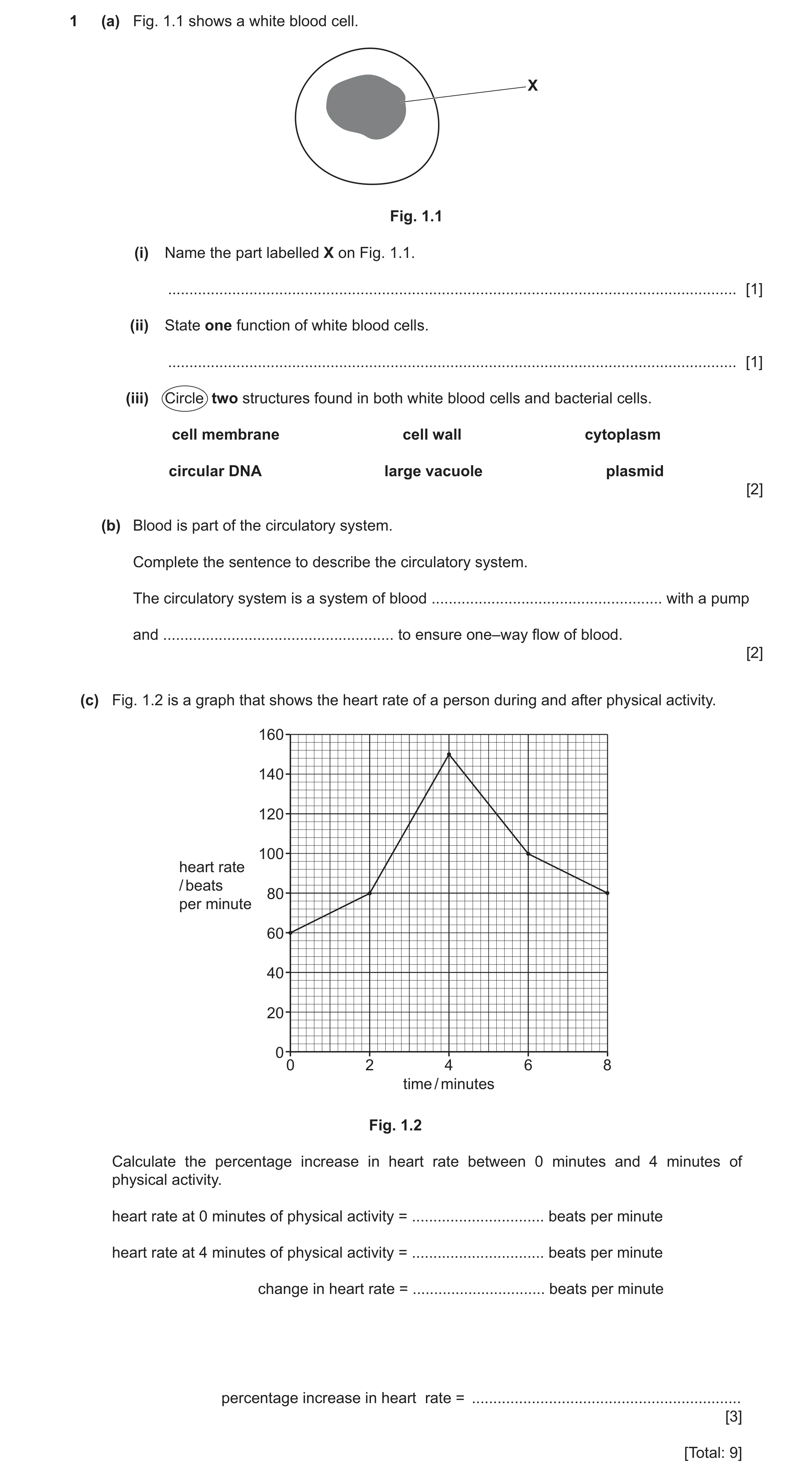
1 (a) Fig. 1.1 shows a white blood cell. X Fig. 1.1 (i) Name the part labelled X on Fig. 1.1. … [1] (ii) State one function of white blood cells. … [1] (iii) Circle two structures found in both white blood cells and bacterial cells. cell membrane cell wall cytoplasm circular DNA large vacuole plasmid [2] (b) Blood is part of the circulatory system. Complete the sentence to describe the circulatory system. The circulatory system is a system of blood … with a pump and … to ensure one–way flow of blood. [2] (c) Fig. 1.2 is a graph that shows the heart rate of a person during and after physical activity. 160 140 120 100 heart rate / beats 80 per minute 60 40 20 00 2 4 6 8 time / minutes Fig. 1.2 Calculate the percentage increase in heart rate between 0 minutes and 4 minutes of physical activity. heart rate at 0 minutes of physical activity = … beats per minute heart rate at 4 minutes of physical activity = … beats per minute change in heart rate = … beats per minute percentage increase in heart rate = … [3] [Total: 9]
9 marks
Mark scheme: Question Answer Marks 1(a)(i) nucleus ; 1 1(a)(ii) produce antibodies / phagocytosis ; 1 1(a)(iii) cell membrane ; 2 cytoplasm ; 1(b) vessels ; 2 valves ; 1(c) 60 and 150 ; 3 (150 – 60) = 90 ; (90 100) = 150(%) ; 60
This question in 0653/32 May/June 2025
Q22 · A diagram of an animal cell and a plant cell 0653/32 Oct/Nov 2025
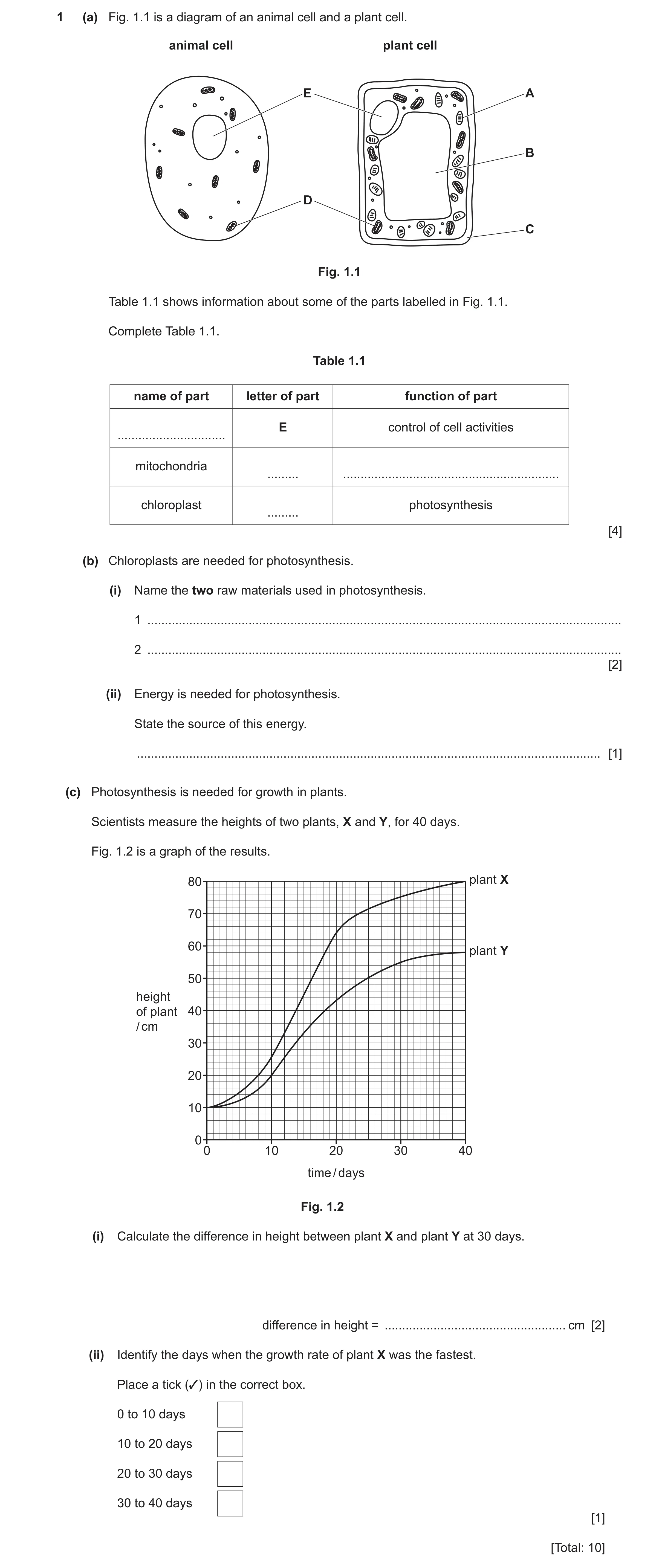
1 (a) Fig. 1.1 is a diagram of an animal cell and a plant cell. animal cell plant cell E A B D C Fig. 1.1 Table 1.1 shows information about some of the parts labelled in Fig. 1.1. Complete Table 1.1. Table 1.1 name of part letter of part function of part E control of cell activities … mitochondria … … chloroplast photosynthesis … [4] (b) Chloroplasts are needed for photosynthesis. (i) Name the two raw materials used in photosynthesis. 1 … 2 … [2] (ii) Energy is needed for photosynthesis. State the source of this energy. … [1] (c) Photosynthesis is needed for growth in plants. Scientists measure the heights of two plants, X and Y, for 40 days. Fig. 1.2 is a graph of the results. 80 plant X 70 60 plant Y 50 height of plant 40 / cm 30 20 10 0 0 10 20 30 40 time / days Fig. 1.2 (i) Calculate the difference in height between plant X and plant Y at 30 days. difference in height = … cm [2] (ii) Identify the days when the growth rate of plant X was the fastest. Place a tick (✓) in the correct box. 0 to 10 days 10 to 20 days 20 to 30 days 30 to 40 days [1] [Total: 10]
10 marks
Mark scheme: Question Answer Marks 1(a) 4 name of part letter function of part nucleus ; (E) (control of cell activities) (mitochondria) D ; respiration ; (chloroplast) A ; (photosynthesis) 1(b)(i) carbon dioxide ; 2 water ; 1(b)(ii) light ; 1 1(c)(i) 75 – 55 ; 2 20 ; 1(c)(ii) 10 to 20 days ; 1
This question in 0653/32 Oct/Nov 2025
Q23 · A diagram of an animal cell and a plant cell 0653/33 Oct/Nov 2025
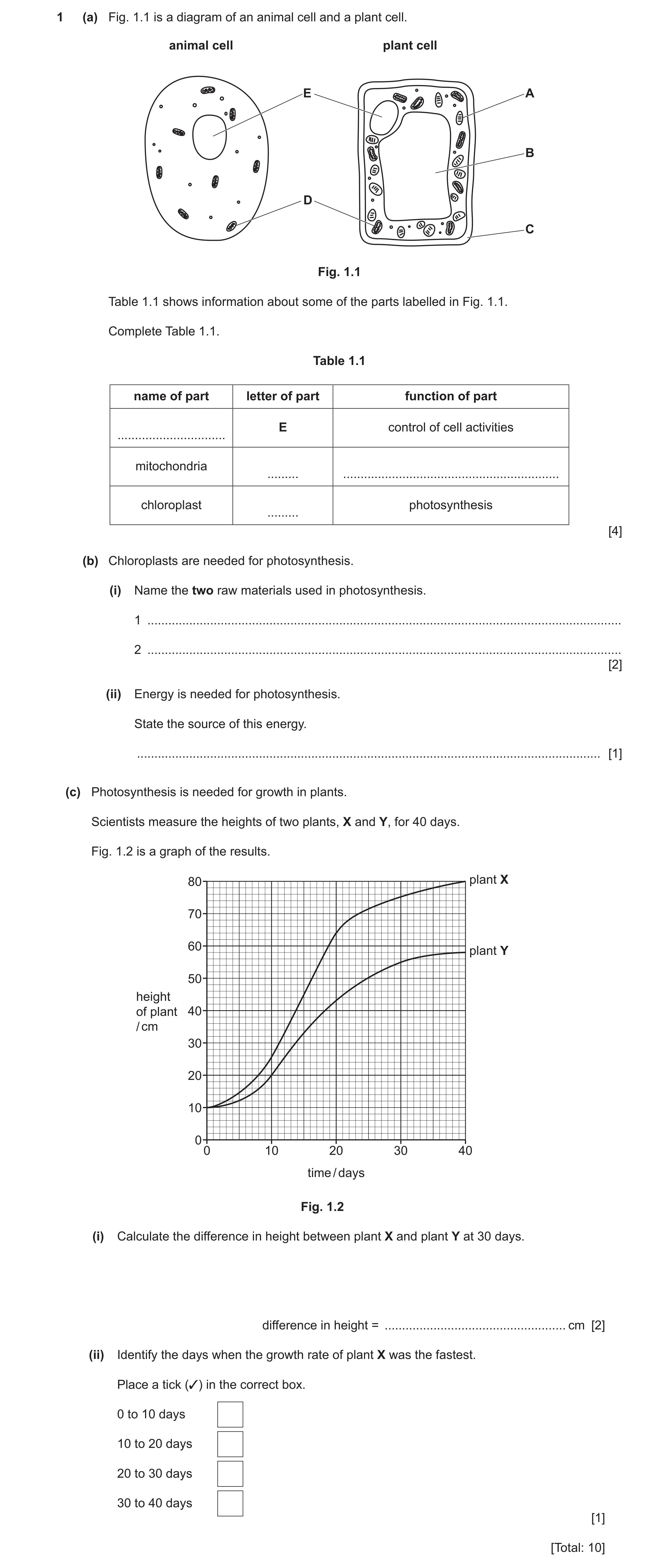
1 (a) Fig. 1.1 is a diagram of an animal cell and a plant cell. animal cell plant cell E A B D C Fig. 1.1 Table 1.1 shows information about some of the parts labelled in Fig. 1.1. Complete Table 1.1. Table 1.1 name of part letter of part function of part E control of cell activities … mitochondria … … chloroplast photosynthesis … [4] (b) Chloroplasts are needed for photosynthesis. (i) Name the two raw materials used in photosynthesis. 1 … 2 … [2] (ii) Energy is needed for photosynthesis. State the source of this energy. … [1] (c) Photosynthesis is needed for growth in plants. Scientists measure the heights of two plants, X and Y, for 40 days. Fig. 1.2 is a graph of the results. 80 plant X 70 60 plant Y 50 height of plant 40 / cm 30 20 10 0 0 10 20 30 40 time / days Fig. 1.2 (i) Calculate the difference in height between plant X and plant Y at 30 days. difference in height = … cm [2] (ii) Identify the days when the growth rate of plant X was the fastest. Place a tick (✓) in the correct box. 0 to 10 days 10 to 20 days 20 to 30 days 30 to 40 days [1] [Total: 10]
10 marks
Mark scheme: Question Answer Marks 1(a) 4 name of part letter function of part nucleus ; (E) (control of cell activities) (mitochondria) D ; respiration ; (chloroplast) A ; (photosynthesis) 1(b)(i) carbon dioxide ; 2 water ; 1(b)(ii) light ; 1 1(c)(i) 75 – 55 ; 2 20 ; 1(c)(ii) 10 to 20 days ; 1
This question in 0653/33 Oct/Nov 2025
 14 / 33
 15 / 33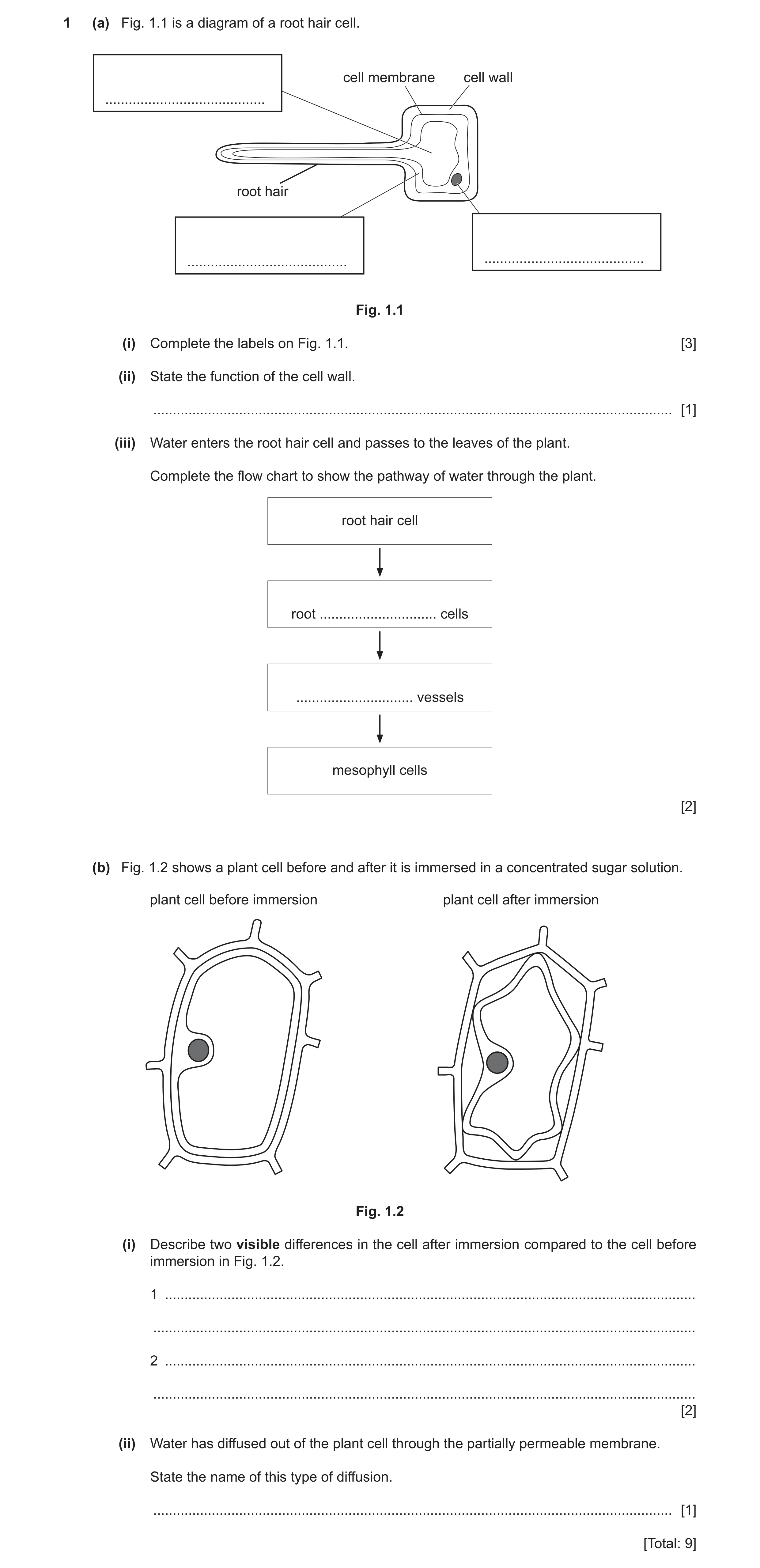
 12
12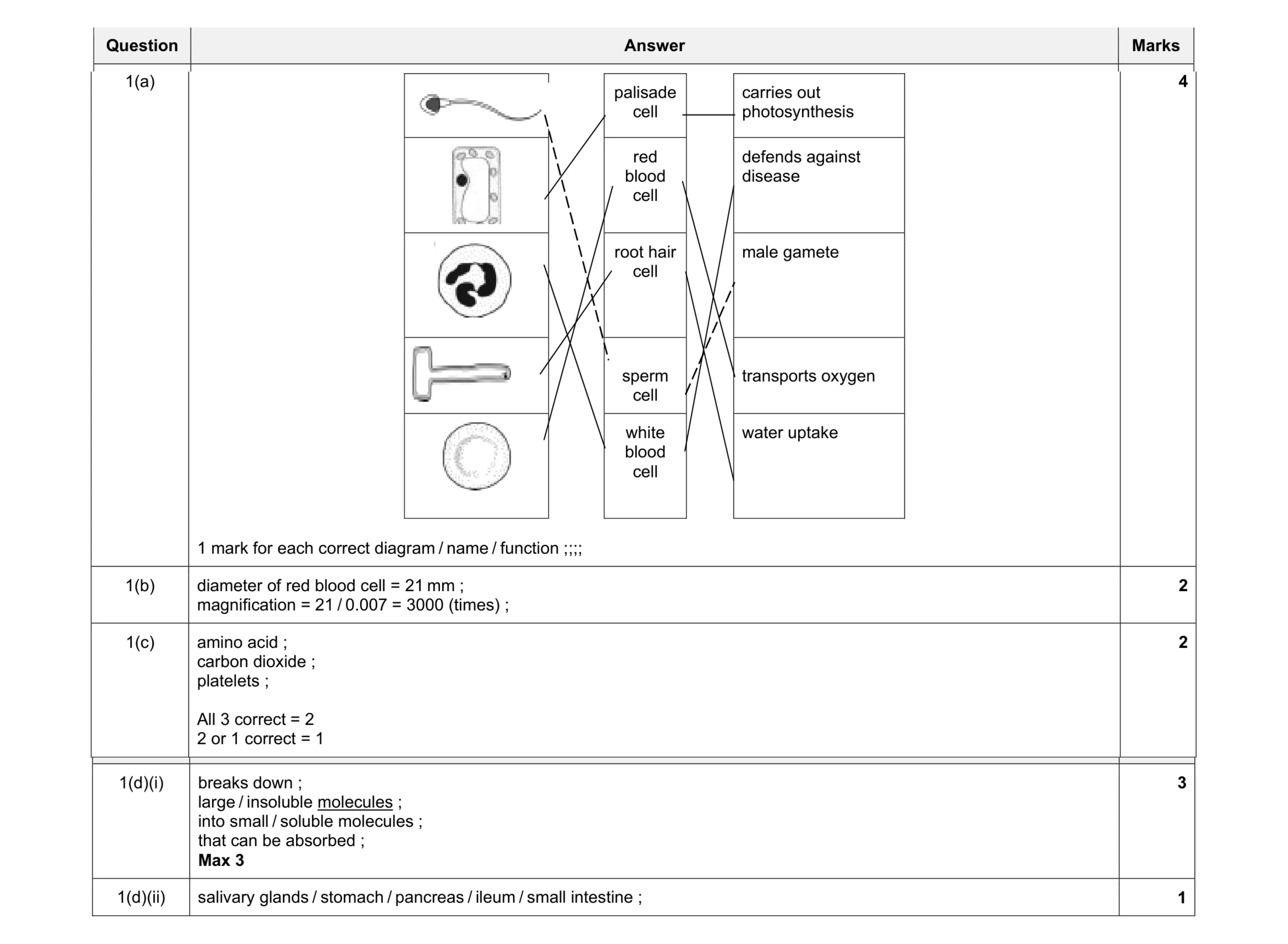 12
12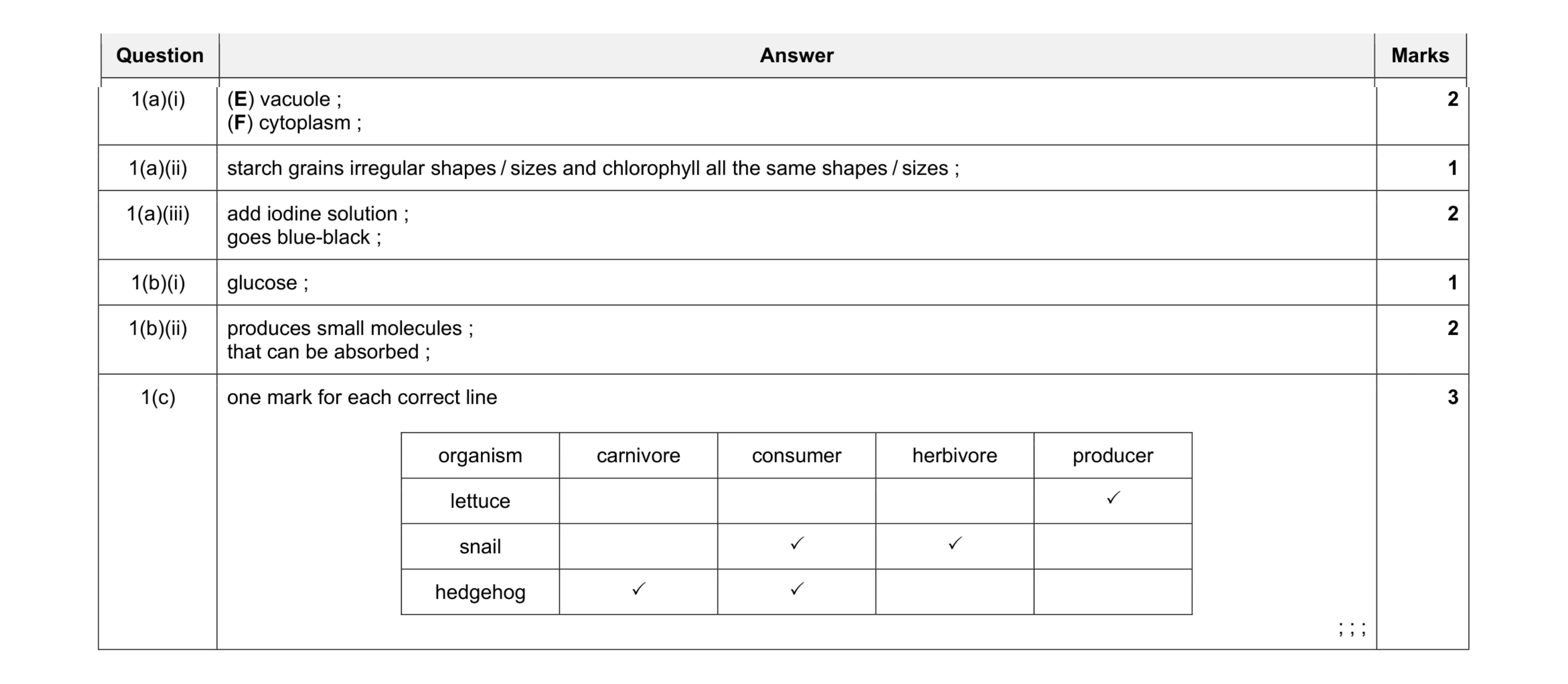 11
11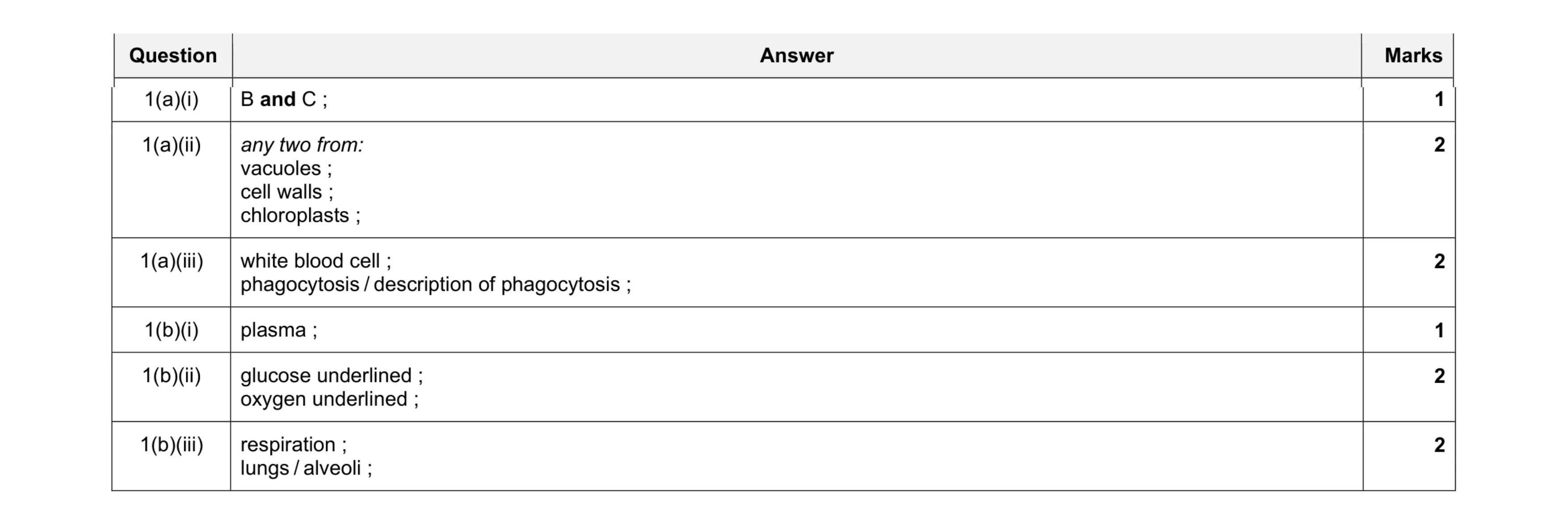 10
10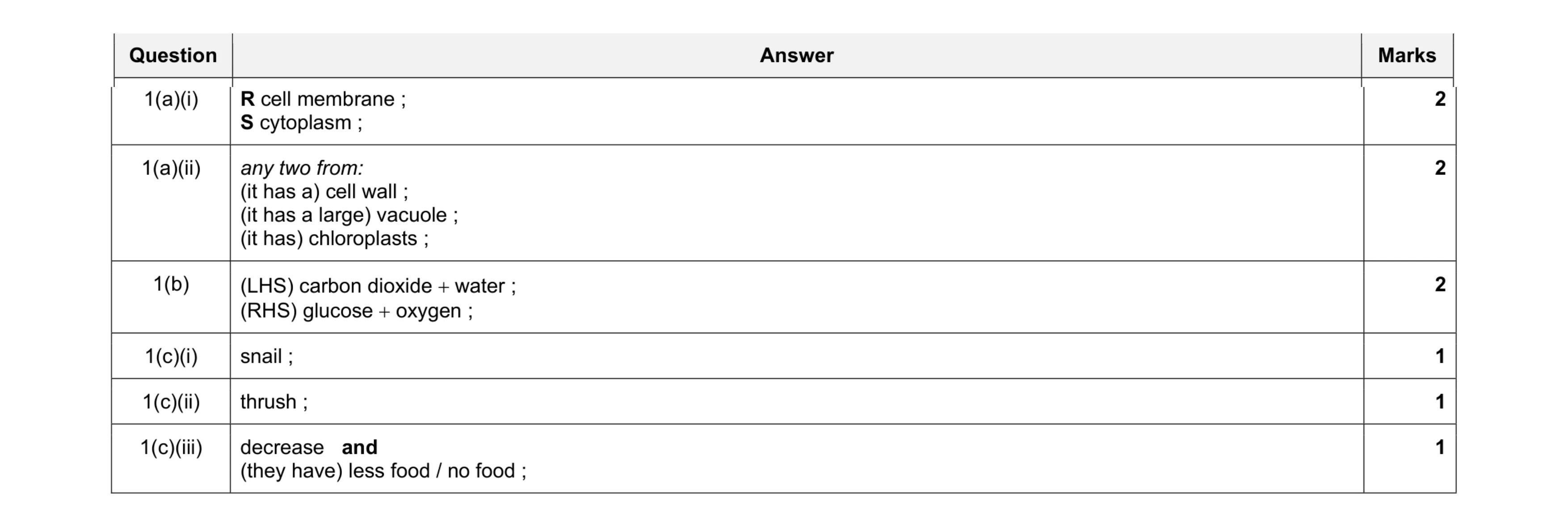 9
9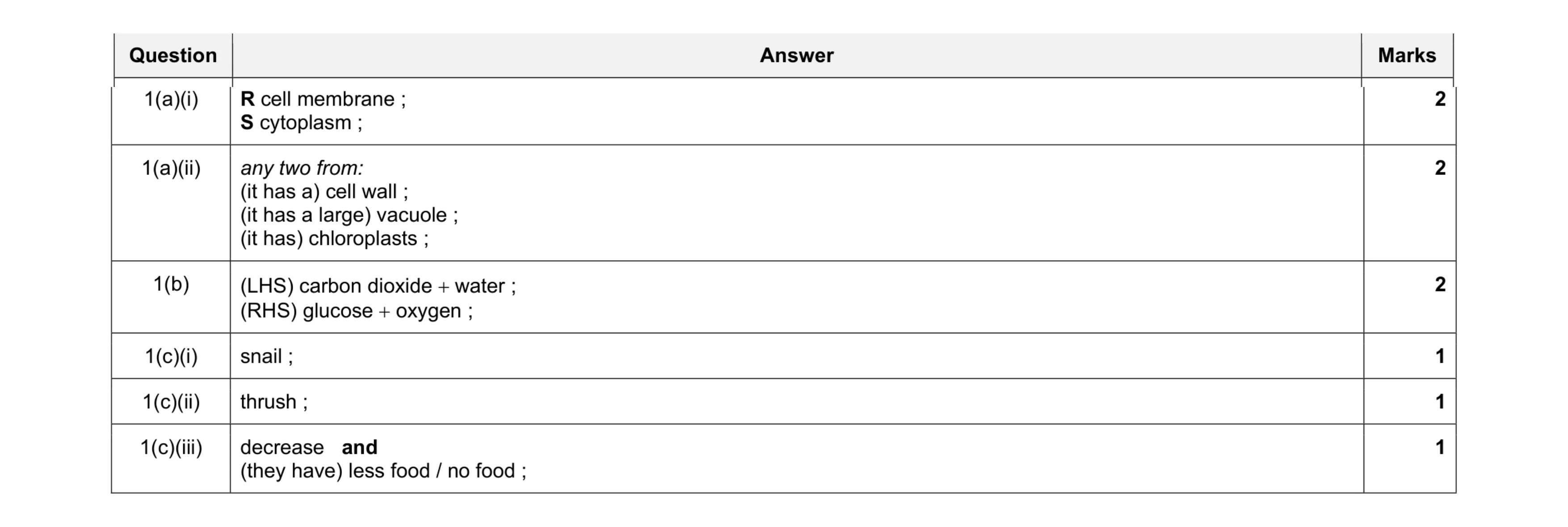 9
9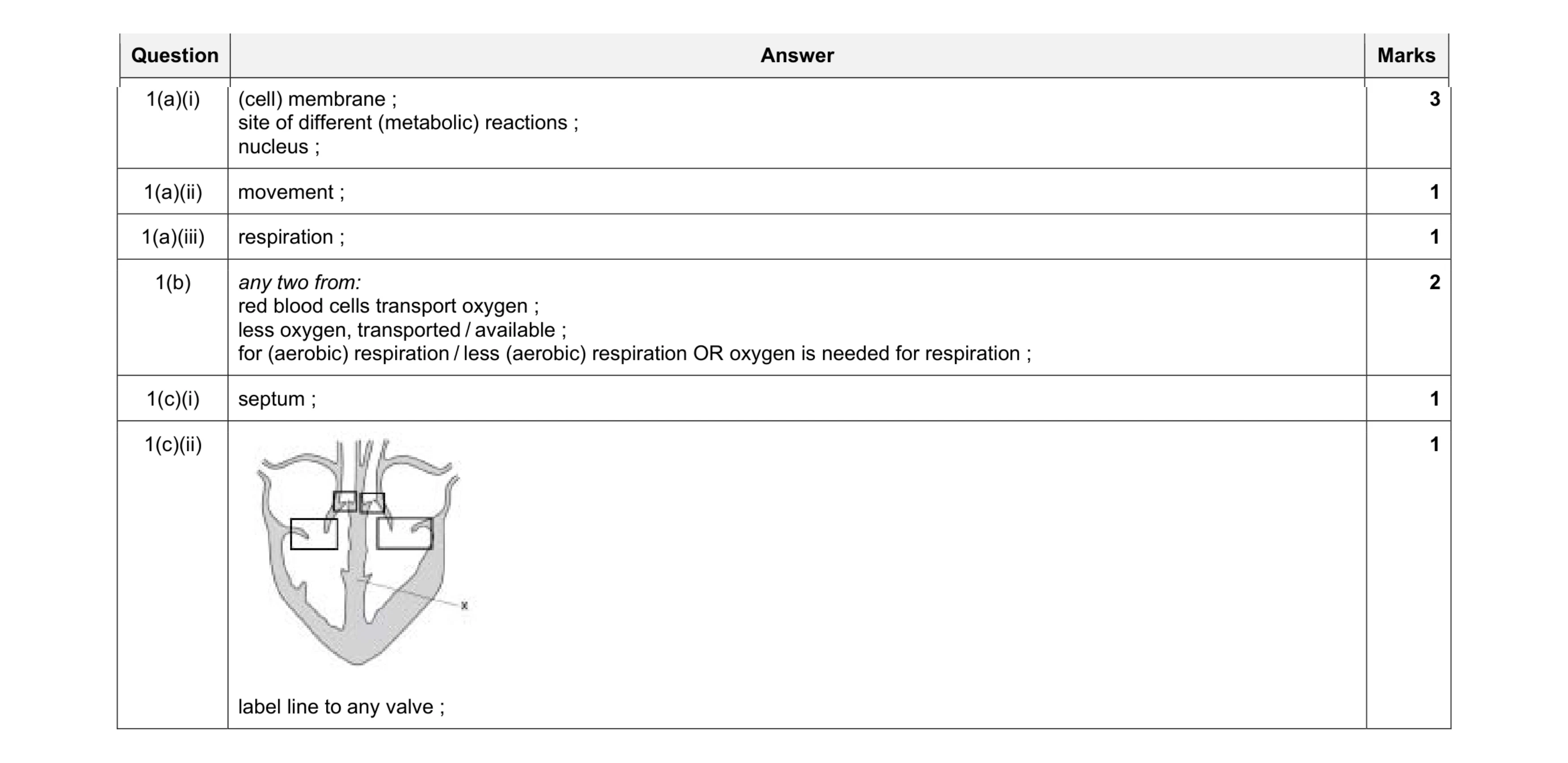 9
9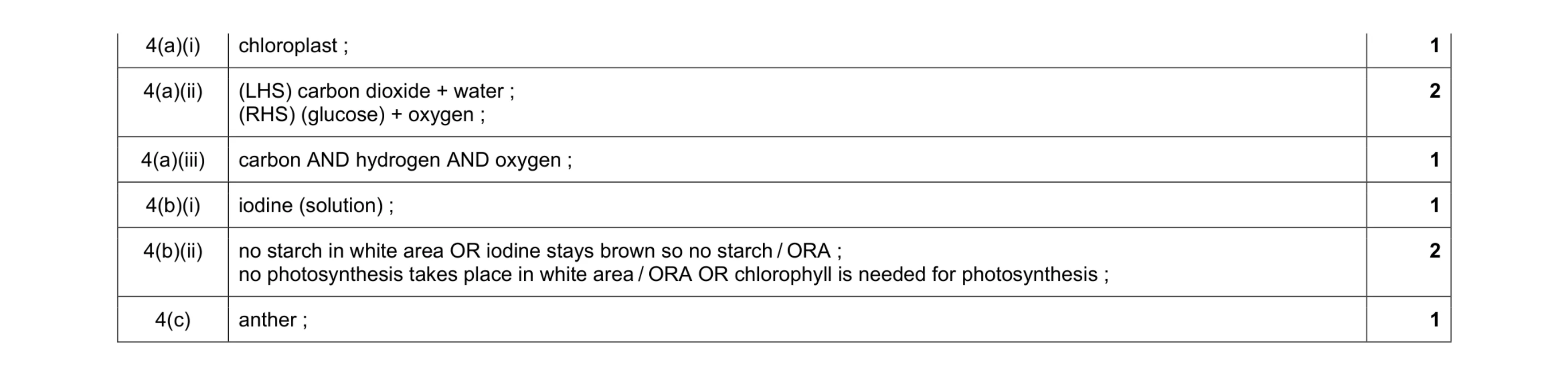 8
8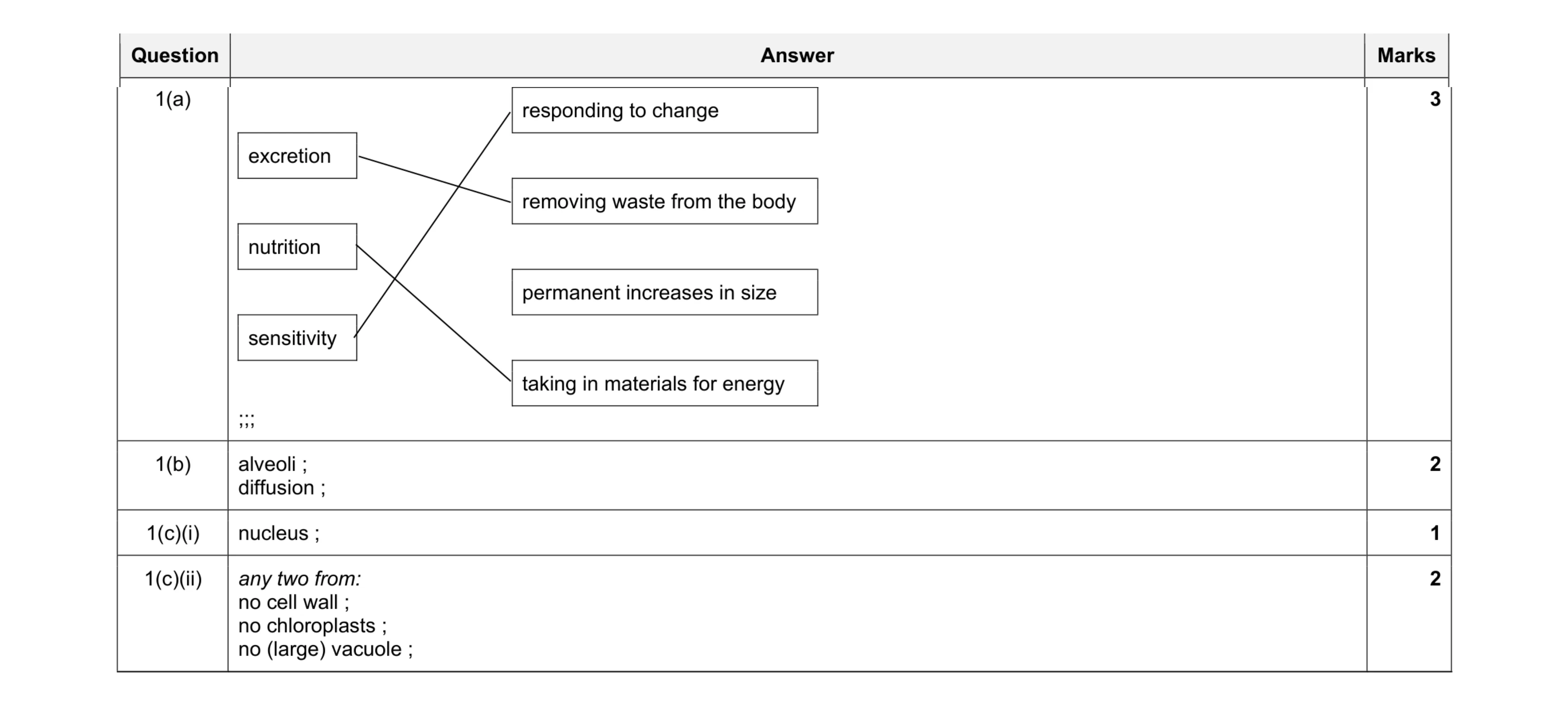 8
8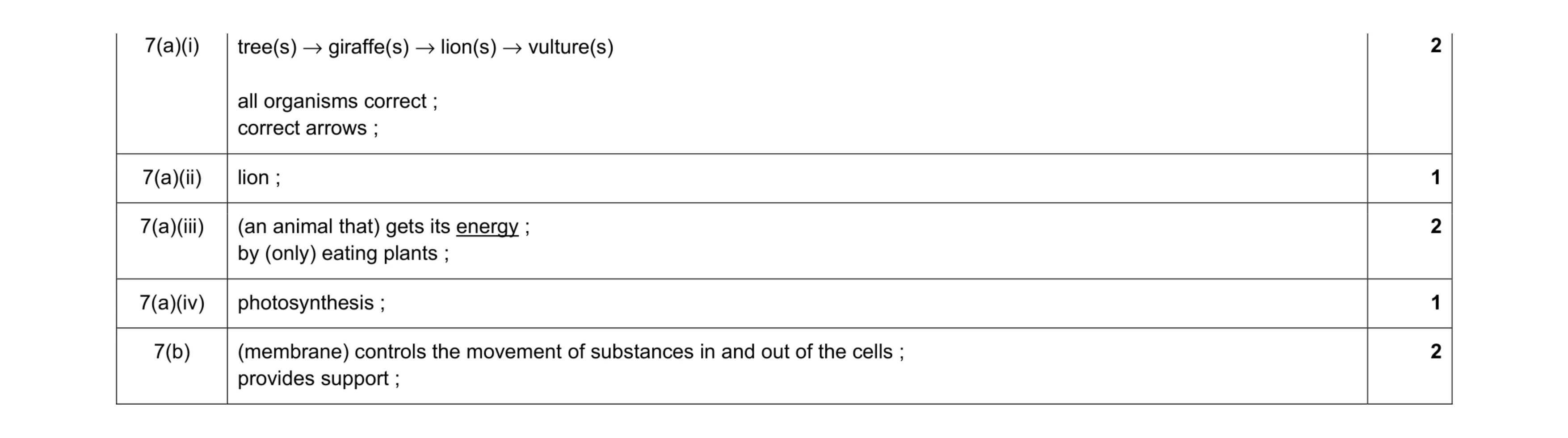 8
8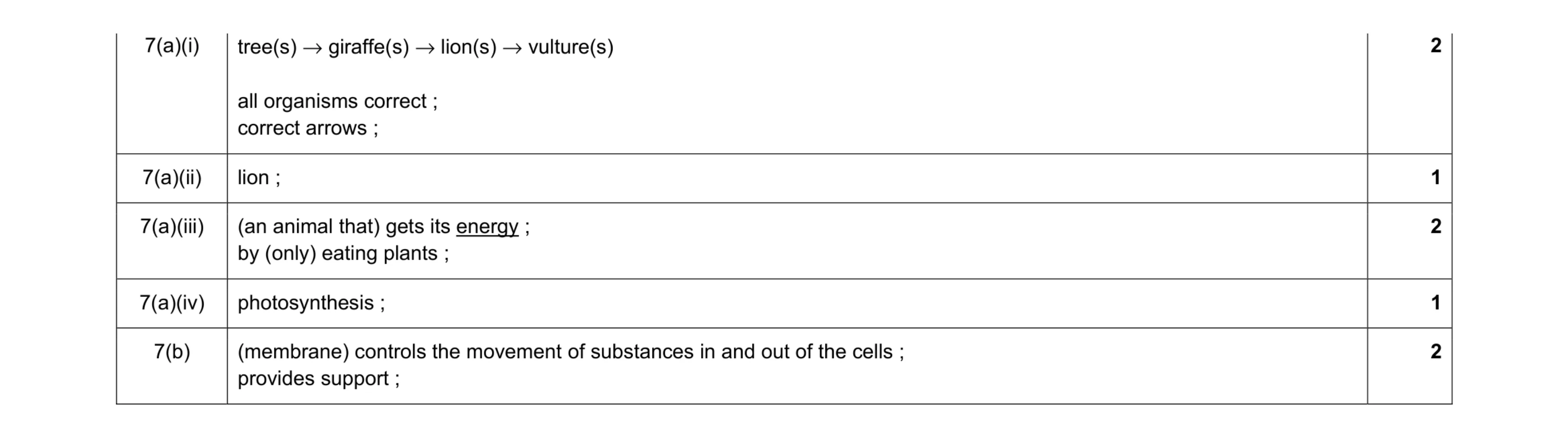 8
8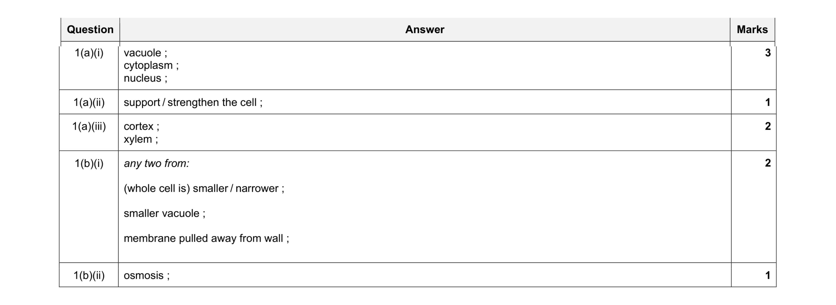 9
9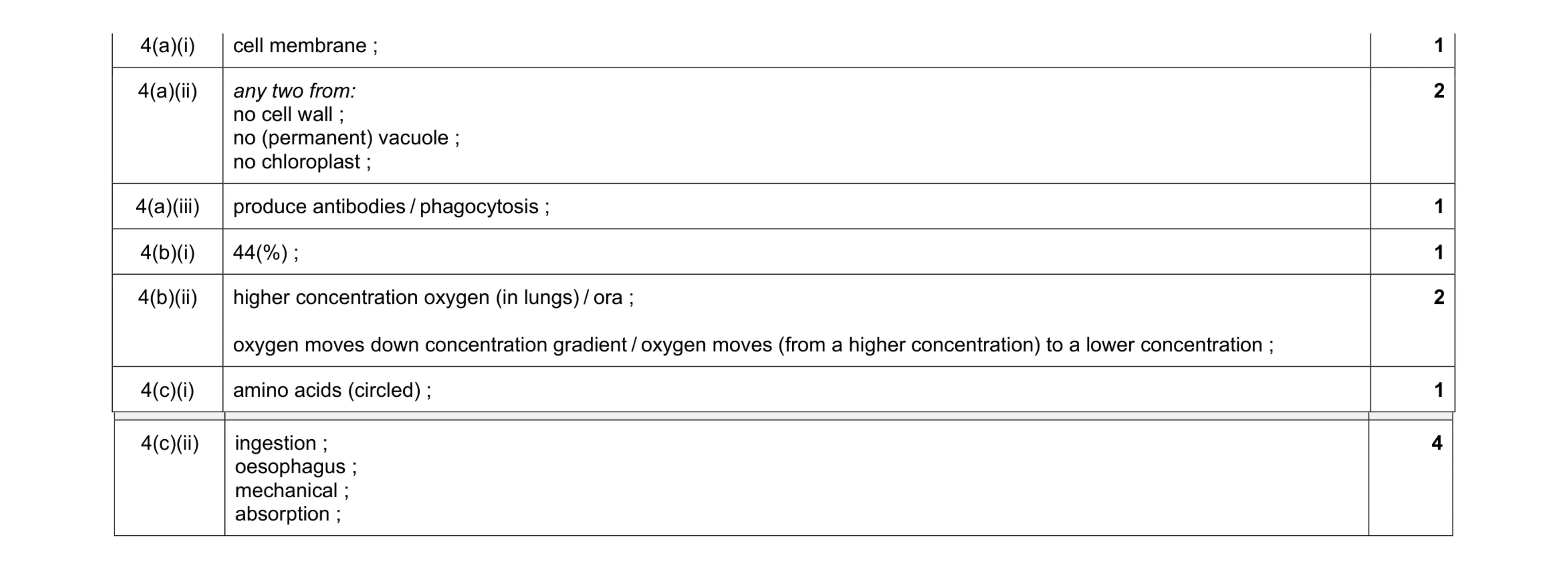 12
12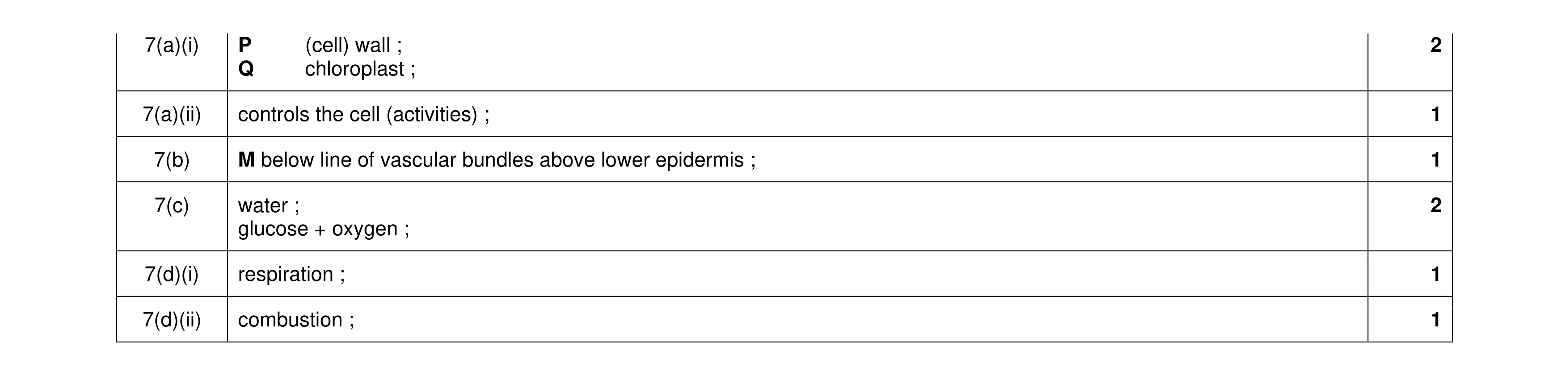 8
8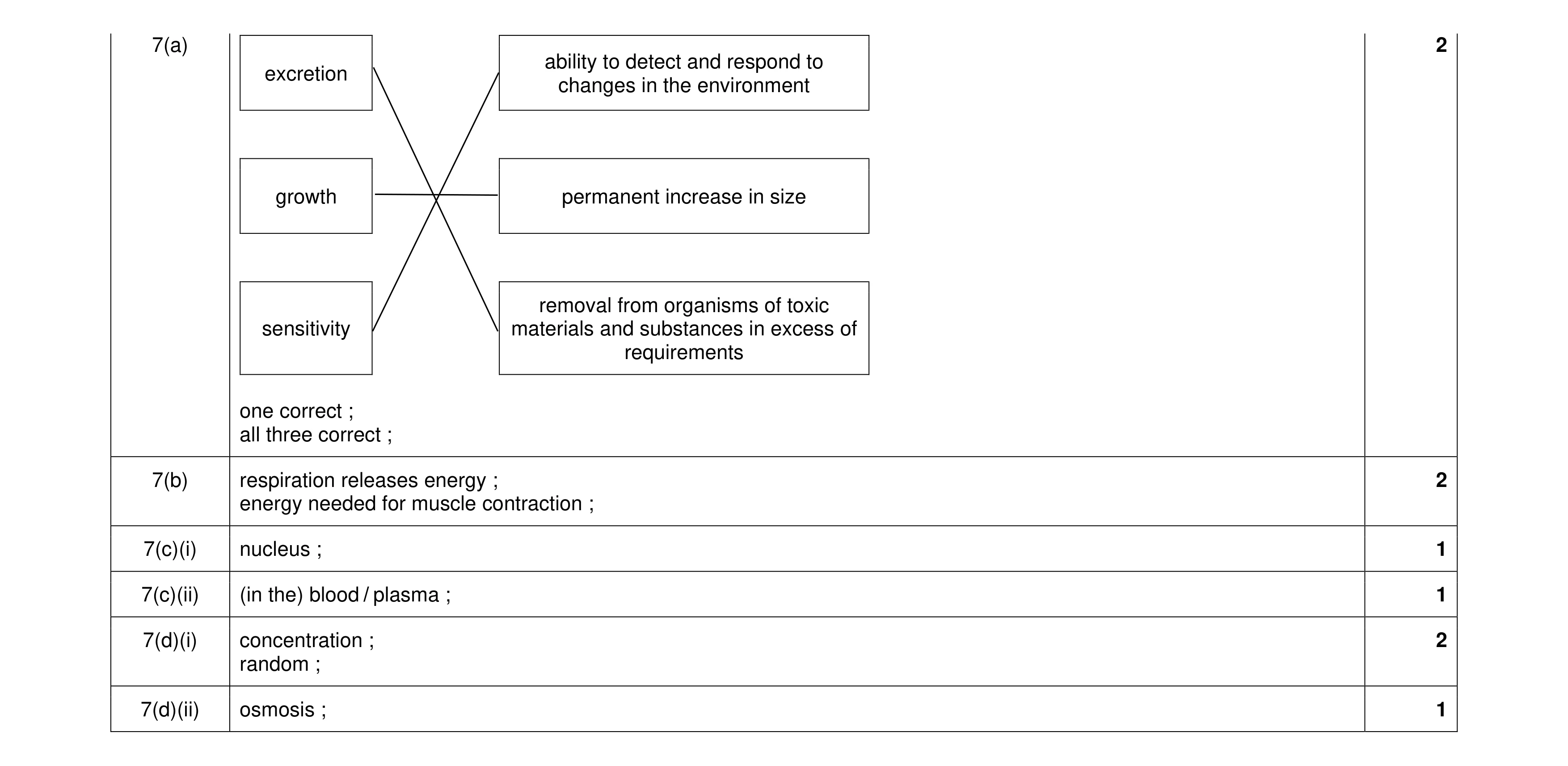 9
9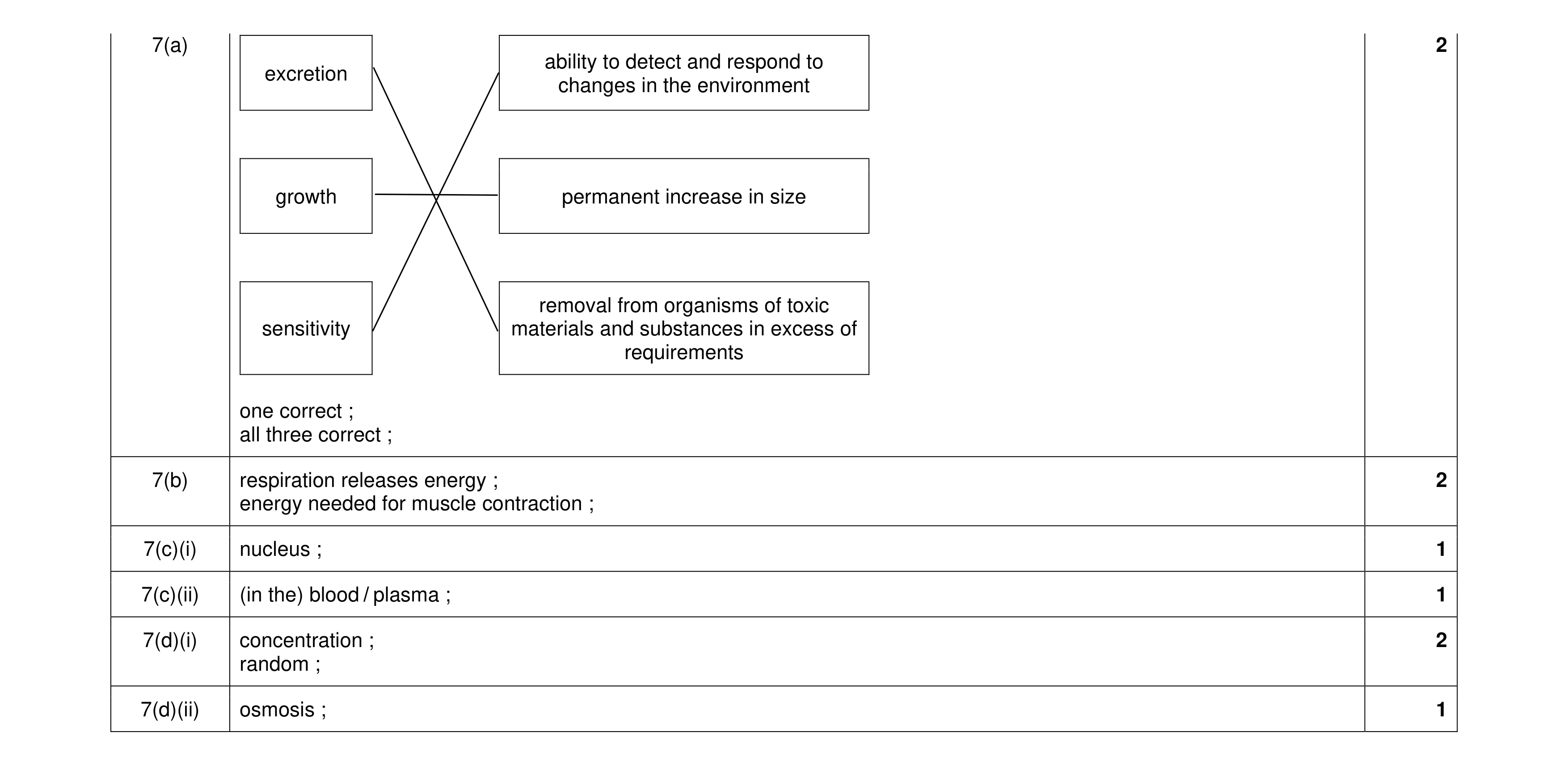 9
9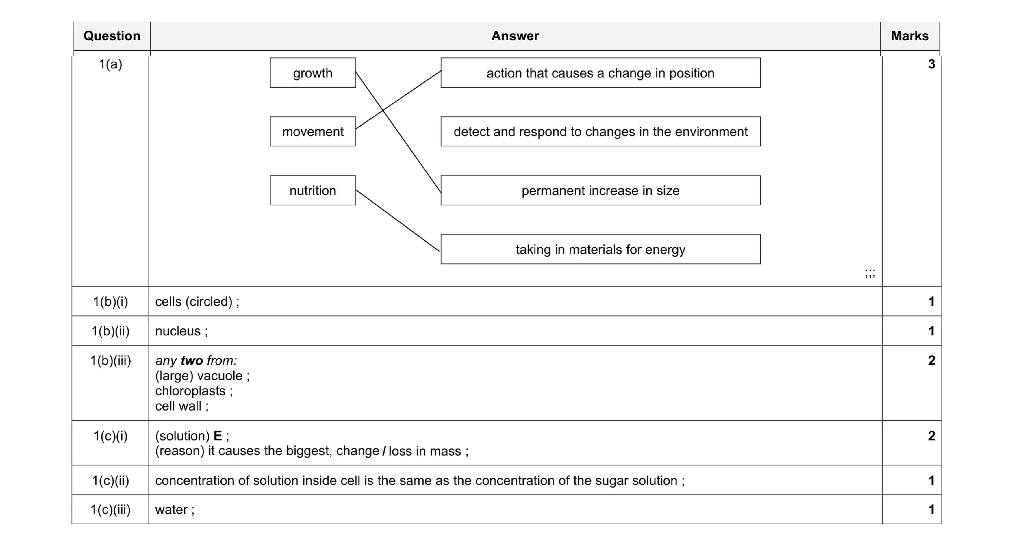 11
11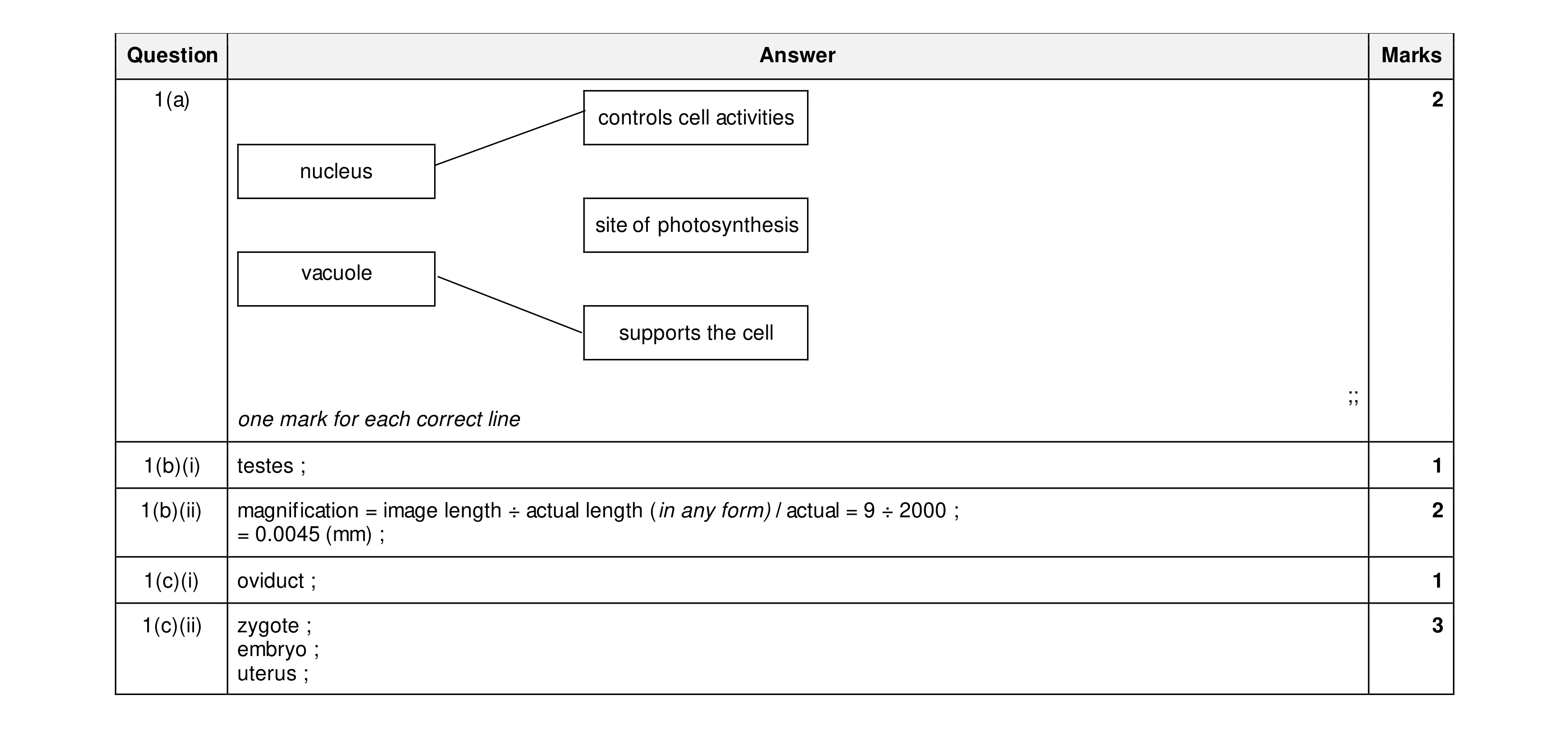 9
9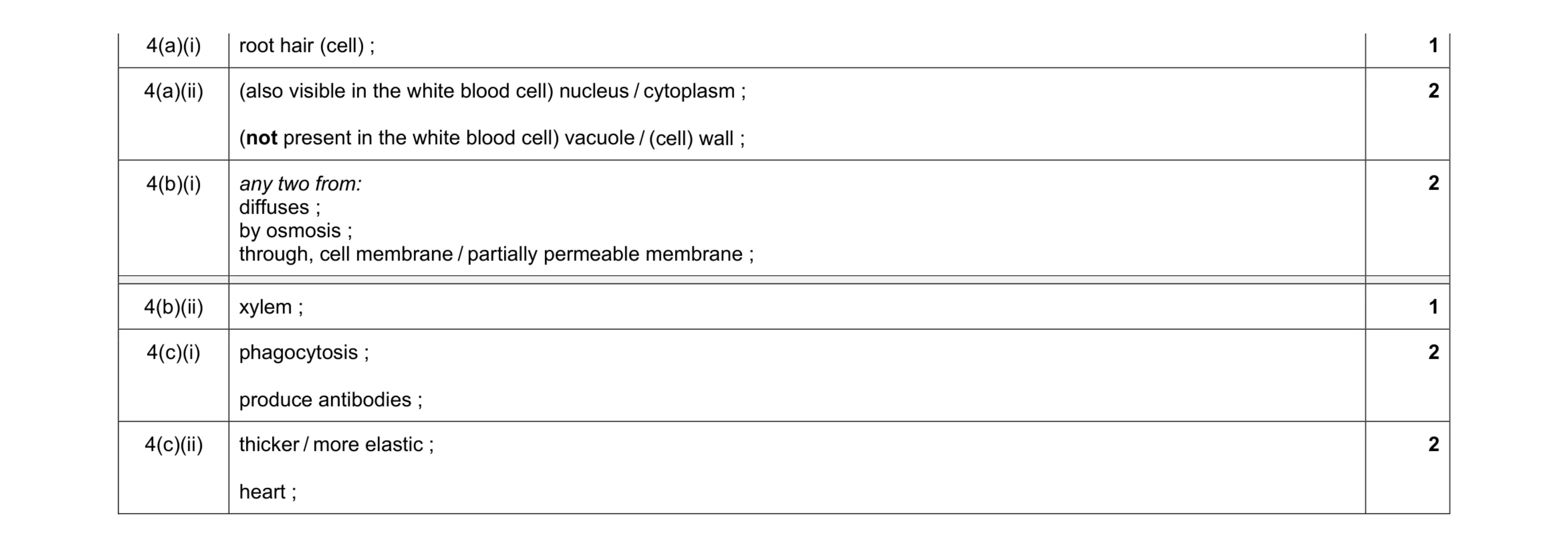 10
10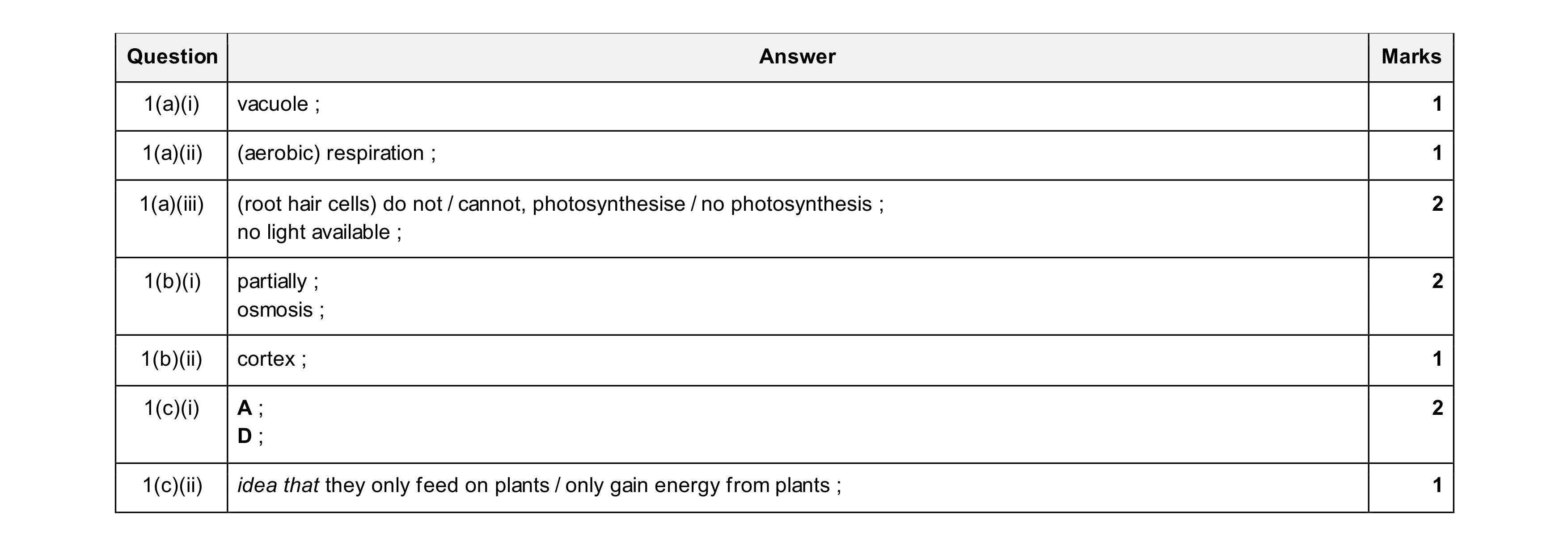 10
10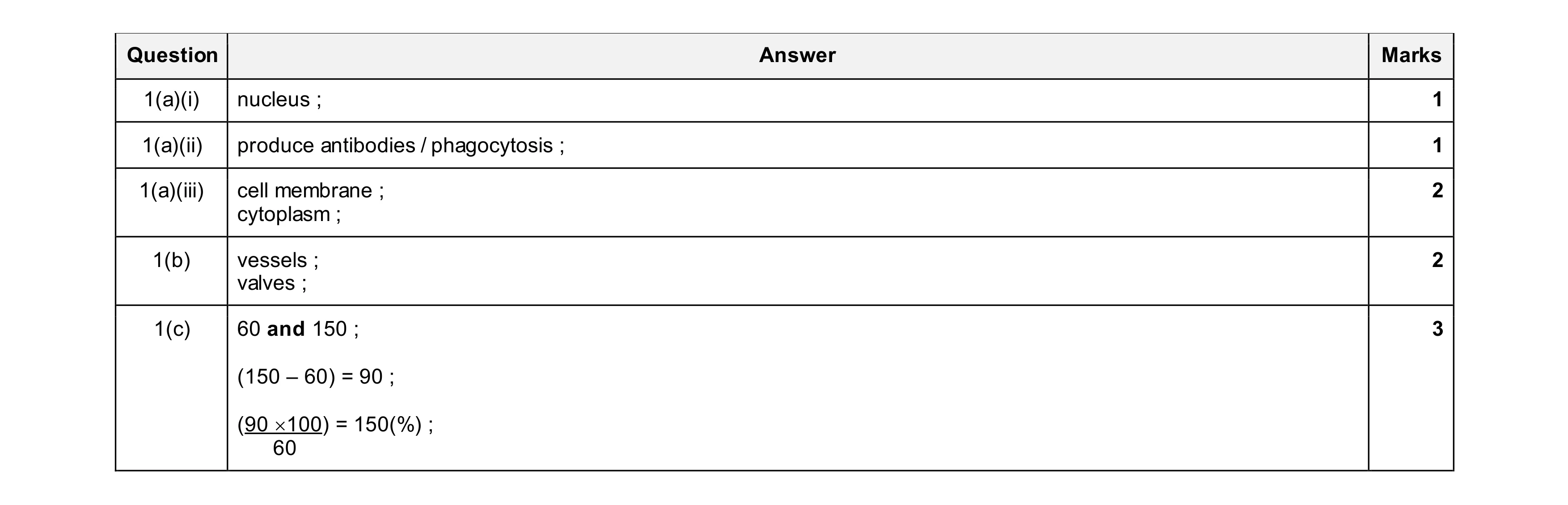 9
9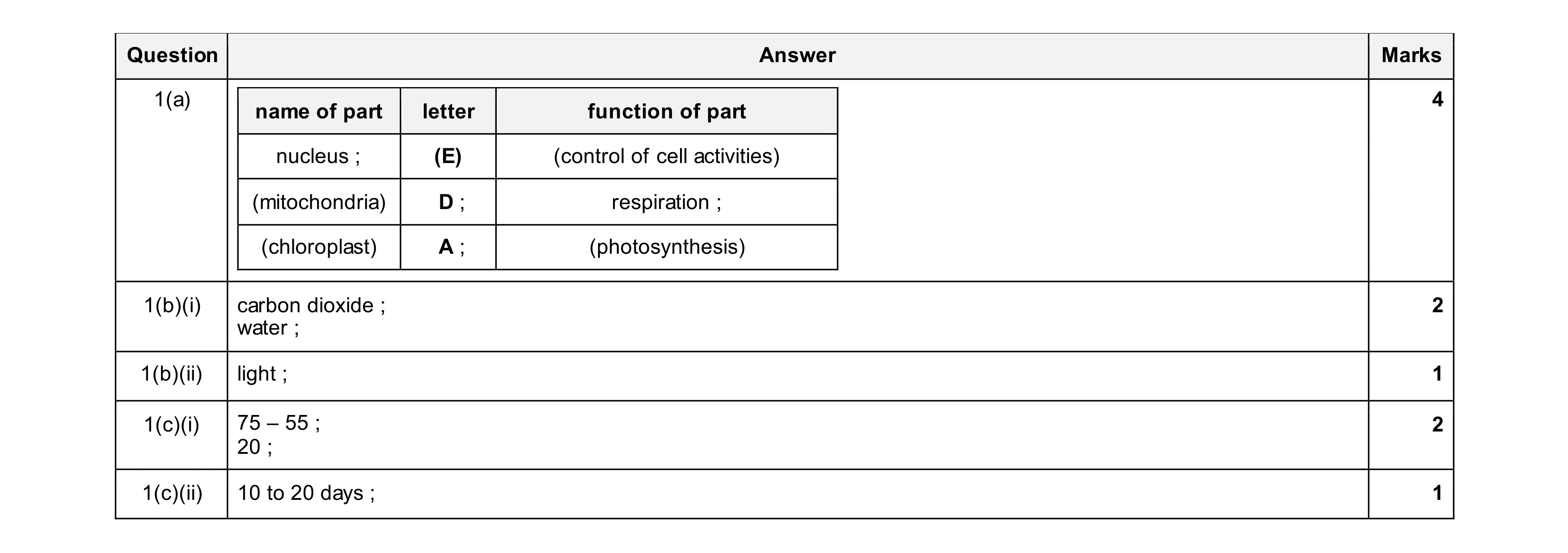 10
10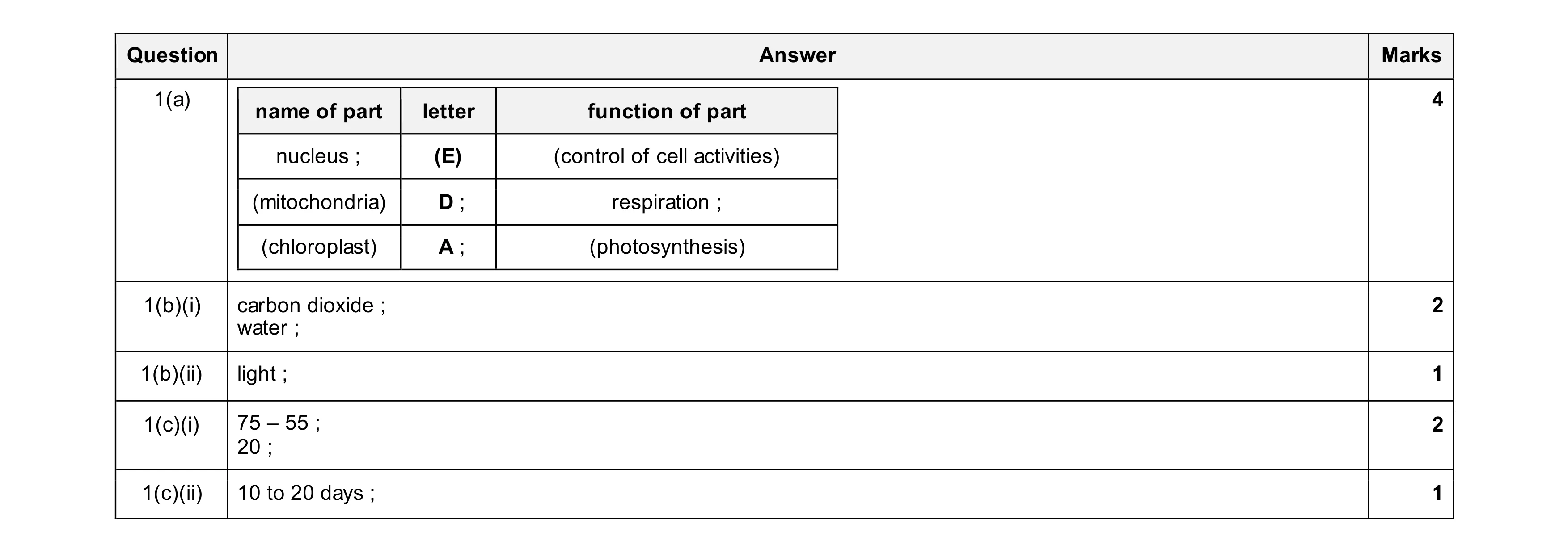 10
10