Questions as text
Q1 · A diagram of the alimentary canal 0653/43 Oct/Nov 2017
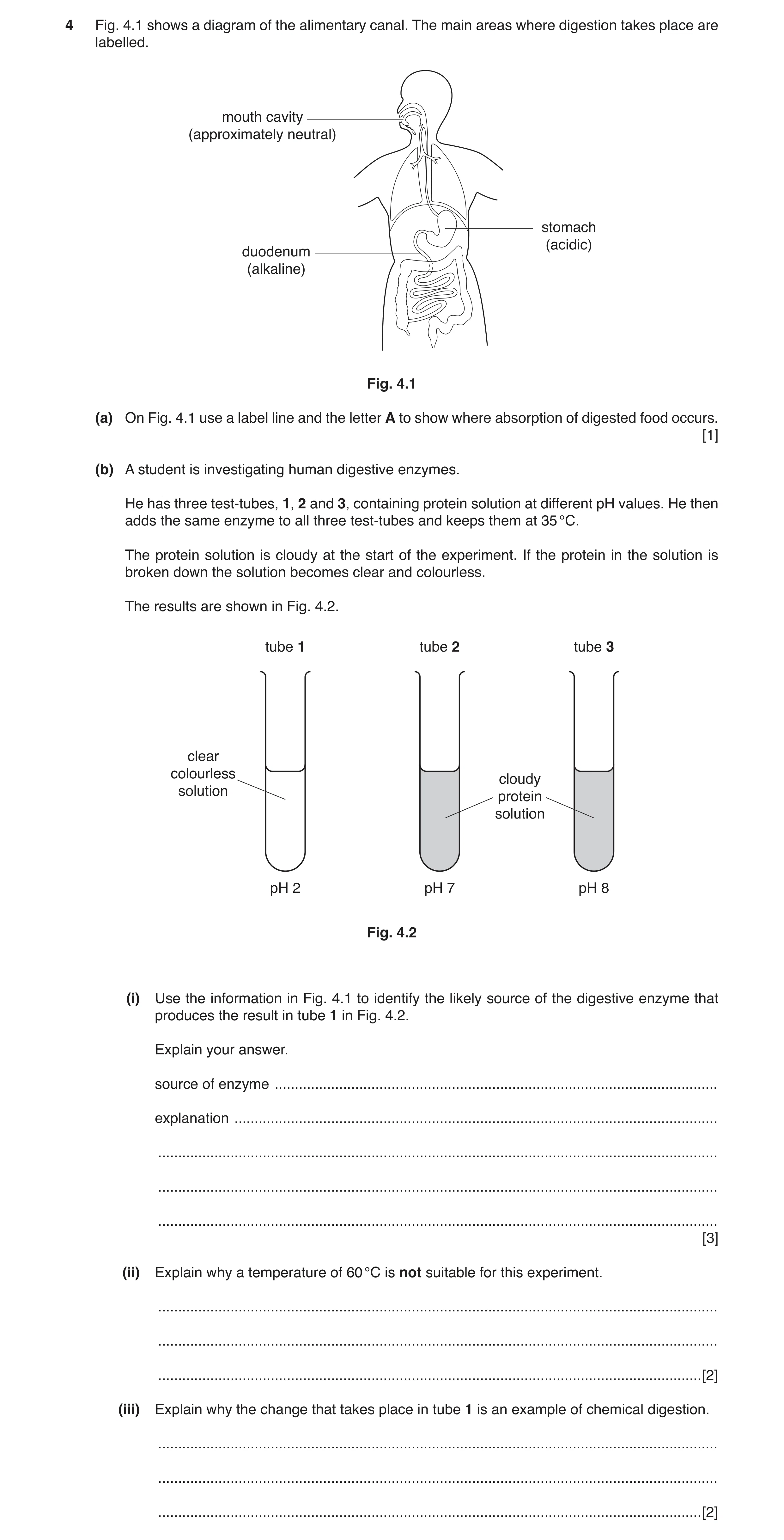
4 Fig. 4.1 shows a diagram of the alimentary canal. The main areas where digestion takes place are labelled. mouth cavity (approximately neutral) stomach (acidic) duodenum (alkaline) Fig. 4.1 (a) On Fig. 4.1 use a label line and the letter A to show where absorption of digested food occurs. [1] (b) A student is investigating human digestive enzymes. He has three test-tubes, 1, 2 and 3, containing protein solution at different pH values. He then adds the same enzyme to all three test-tubes and keeps them at 35 °C. The protein solution is cloudy at the start of the experiment. If the protein in the solution is broken down the solution becomes clear and colourless. The results are shown in Fig. 4.2. tube 1 tube 2 tube 3 clear colourless cloudy solution protein solution pH 2 pH 7 pH 8 Fig. 4.2 (i) Use the information in Fig. 4.1 to identify the likely source of the digestive enzyme that produces the result in tube 1 in Fig. 4.2. Explain your answer. source of enzyme … explanation … … … … [3] (ii) Explain why a temperature of 60 °C is not suitable for this experiment. … … … [2] (iii) Explain why the change that takes place in tube 1 is an example of chemical digestion. … … … [2]
8 marks
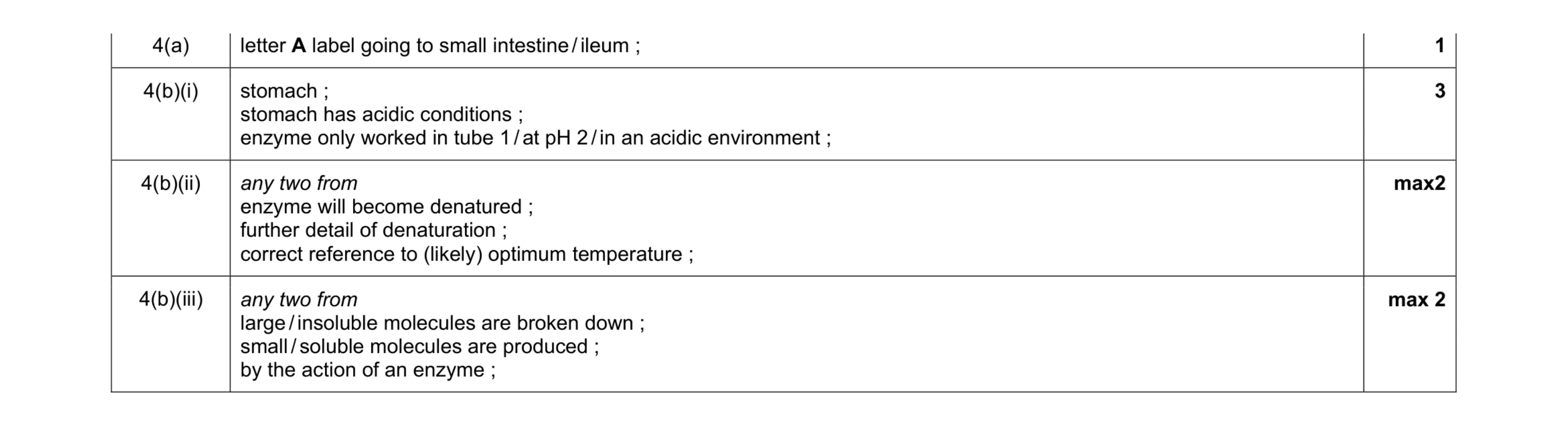
Mark scheme: 4(a) letter A label going to small intestine / ileum ; 1 4(b)(i) stomach ; stomach has acidic conditions ; enzyme only worked in tube 1 / at pH 2 / in an acidic environment ; 3 4(b)(ii) any two from enzyme will become denatured ; further detail of denaturation ; correct reference to (likely) optimum temperature ; max2 4(b)(iii) any two from large / insoluble molecules are broken down ; small / soluble molecules are produced ; by the action of an enzyme ; max 2
This question in 0653/43 Oct/Nov 2017
Q2 · When apples are cut open the exposed surface turns from white to brown 0653/42 May/June 2019
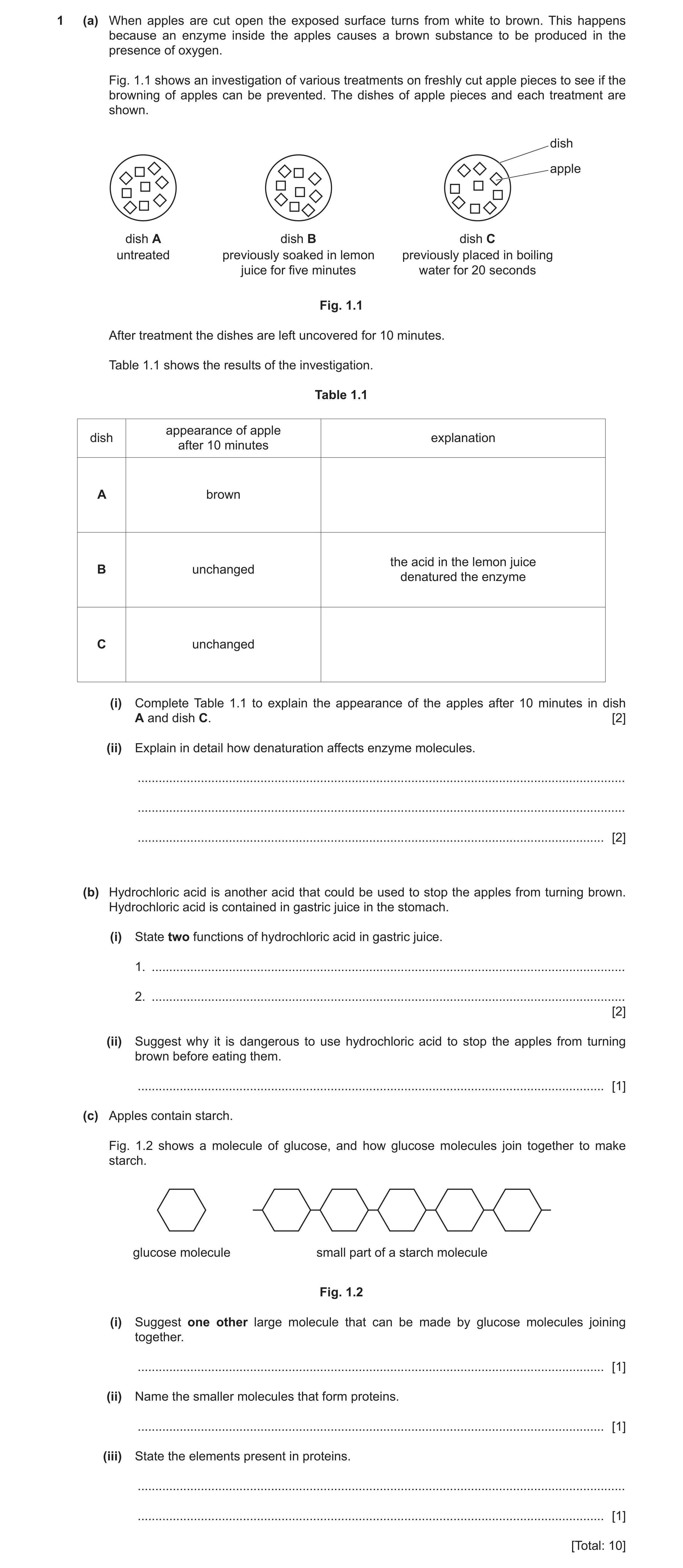
1 (a) When apples are cut open the exposed surface turns from white to brown. This happens because an enzyme inside the apples causes a brown substance to be produced in the presence of oxygen. Fig. 1.1 shows an investigation of various treatments on freshly cut apple pieces to see if the browning of apples can be prevented. The dishes of apple pieces and each treatment are shown. dish apple dish A dish B dish C untreated previously soaked in lemon previously placed in boiling juice for five minutes water for 20 seconds Fig. 1.1 After treatment the dishes are left uncovered for 10 minutes. Table 1.1 shows the results of the investigation. Table 1.1 appearance of apple dish explanation after 10 minutes A brown the acid in the lemon juice B unchanged denatured the enzyme C unchanged (i) Complete Table 1.1 to explain the appearance of the apples after 10 minutes in dish A and dish C. [2] (ii) Explain in detail how denaturation affects enzyme molecules. … … … [2] (b) Hydrochloric acid is another acid that could be used to stop the apples from turning brown. Hydrochloric acid is contained in gastric juice in the stomach. (i) State two functions of hydrochloric acid in gastric juice. 1. … 2. … [2] (ii) Suggest why it is dangerous to use hydrochloric acid to stop the apples from turning brown before eating them. … [1] (c) Apples contain starch. Fig. 1.2 shows a molecule of glucose, and how glucose molecules join together to make starch. glucose molecule small part of a starch molecule Fig. 1.2 (i) Suggest one other large molecule that can be made by glucose molecules joining together. … [1] (ii) Name the smaller molecules that form proteins. … [1] (iii) State the elements present in proteins. … … [1] [Total: 10]
10 marks
Mark scheme: 1(a)(i) A (untreated so the) enzyme caused the brown substance to be produced (in air) ; C (the boiling water) denatured the enzymes ; 2 1(a)(ii) active site / enzyme damaged / destroyed / shape changed ; substrate will no longer fit into it ; 2 1(b)(i) kills bacteria / pathogens / (harmful) microorganisms ; gives an acid pH for enzymes / owtte ; 2 1(b)(ii) hydrochloric acid is a strong acid ; hydrochloric acid can damage mouth, etc. when swallowed ; max 1 1(c)(i) glycogen ; 1 1(c)(ii) amino acids ; 1 1(c)(iii) carbon, hydrogen, oxygen, nitrogen 1
This question in 0653/42 May/June 2019
Q3 · A diagram of parts of the alimentary canal and associated organs 0653/42 Feb/March 2021
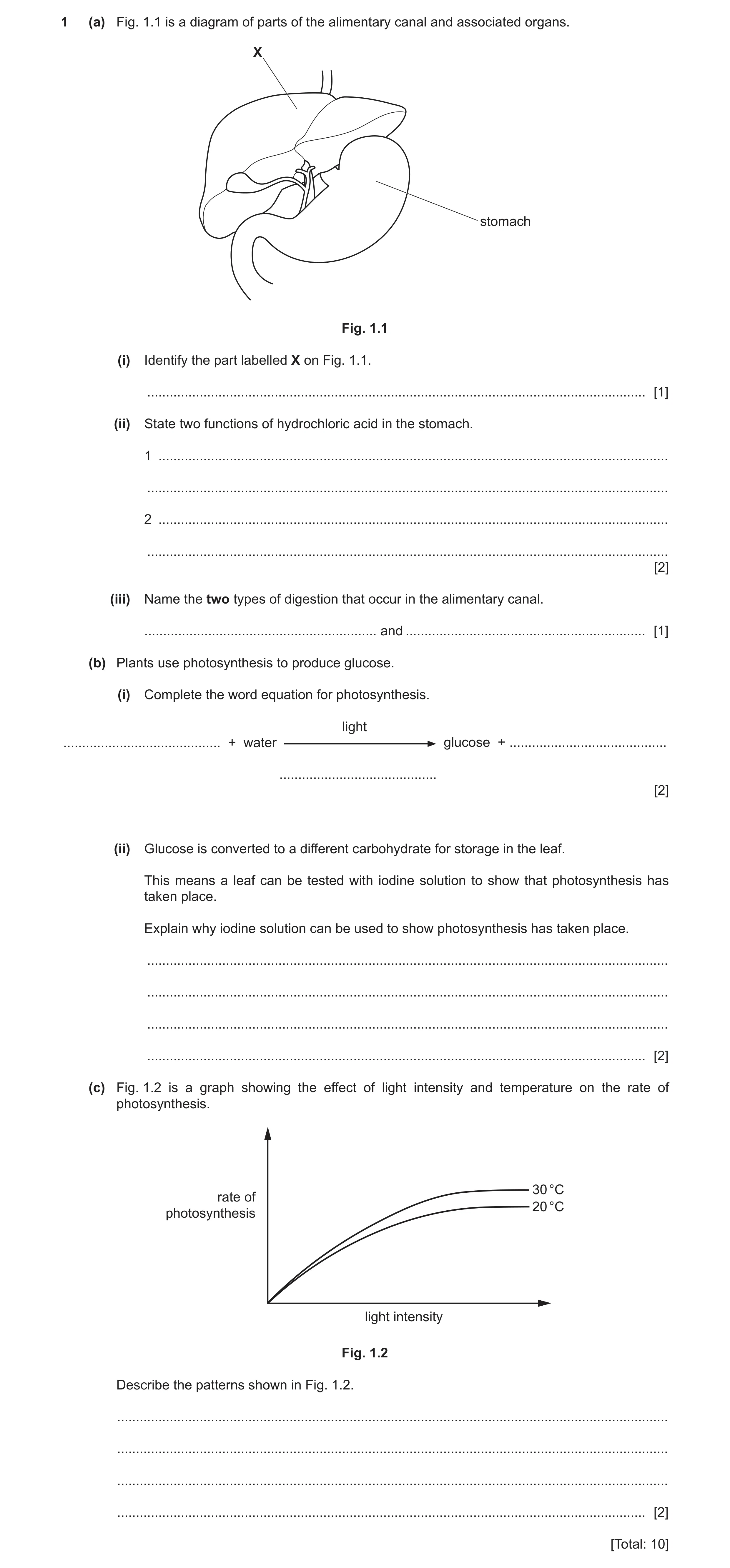
1 (a) Fig. 1.1 is a diagram of parts of the alimentary canal and associated organs. X stomach Fig. 1.1 (i) Identify the part labelled X on Fig. 1.1. … [1] (ii) State two functions of hydrochloric acid in the stomach. 1 … … 2 … … [2] (iii) Name the two types of digestion that occur in the alimentary canal. … and … [1] (b) Plants use photosynthesis to produce glucose. (i) Complete the word equation for photosynthesis. light … + water glucose + … … [2] (ii) Glucose is converted to a different carbohydrate for storage in the leaf. This means a leaf can be tested with iodine solution to show that photosynthesis has taken place. Explain why iodine solution can be used to show photosynthesis has taken place. … … … … [2] (c) Fig. 1.2 is a graph showing the effect of light intensity and temperature on the rate of photosynthesis. 30 °C rate of 20 °C photosynthesis light intensity Fig. 1.2 Describe the patterns shown in Fig. 1.2. … … … … [2] [Total: 10]
10 marks
Mark scheme: 1(a)(i) liver ; 1 1(a)(ii) kill bacteria ; provide acidic pH for enzymes ; 2 1(a)(iii) chemical (digestion) AND mechanical (digestion) ; 1 1(b)(i) (light) carbon dioxide + (water) (glucose) + oxygen chlorophyll ;; one mark for carbon dioxide and oxygen one mark for chlorophyll 2 1(b)(ii) iodine (solution) tests for starch / shows blue-black with starch ; glucose made in photosynthesis is converted to starch ; 2 1(c) as light intensity increases so does (rate of) photosynthesis (then it levels off) ; idea that rate is higher, at 30°C than 20°C / at higher temperature ; 2
This question in 0653/42 Feb/March 2021
Q4 · A diagram of a fetus inside the uterus 0653/42 Oct/Nov 2021
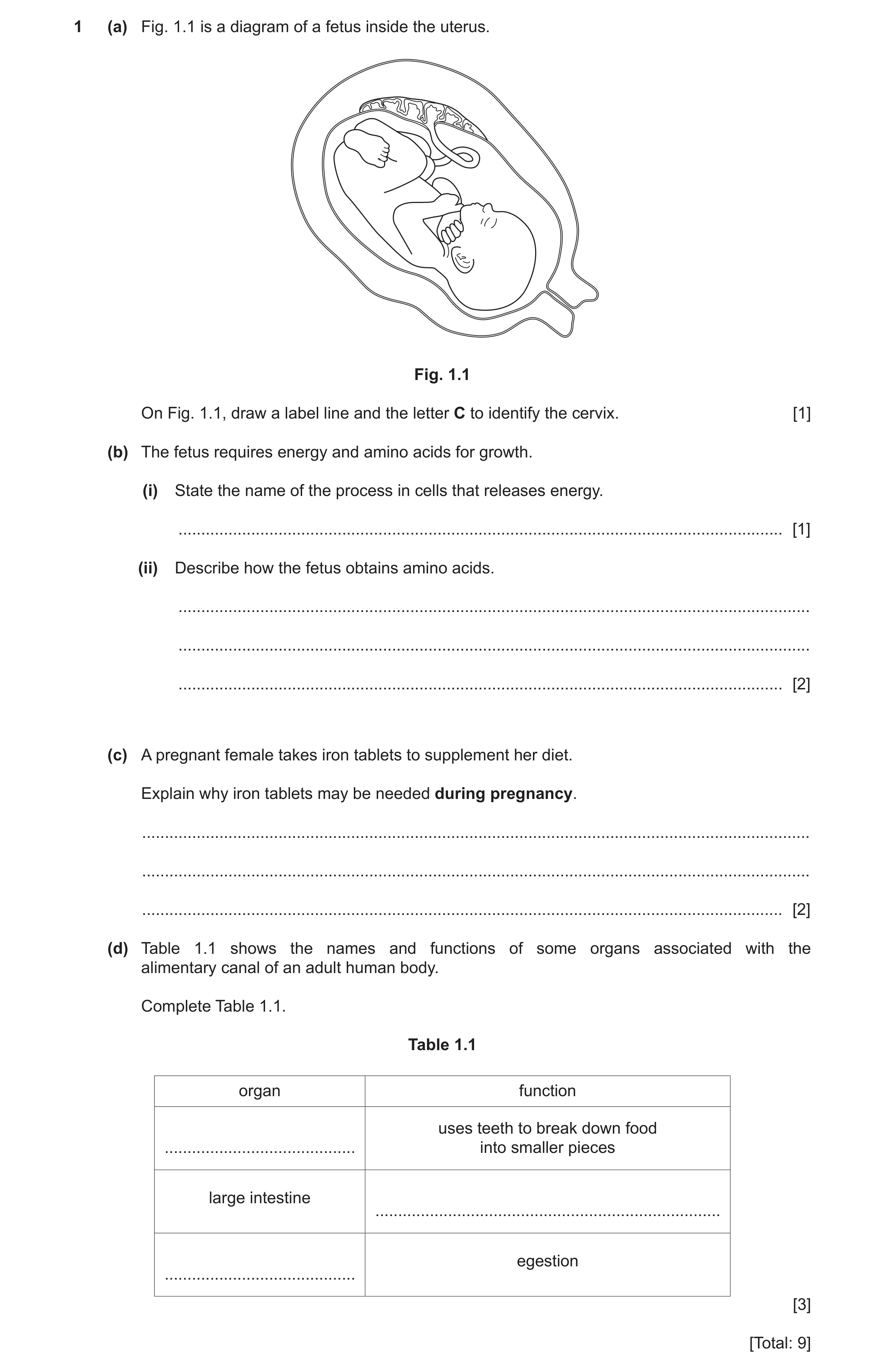
1 (a) Fig. 1.1 is a diagram of a fetus inside the uterus. Fig. 1.1 On Fig. 1.1, draw a label line and the letter C to identify the cervix. [1] (b) The fetus requires energy and amino acids for growth. (i) State the name of the process in cells that releases energy. … [1] (ii) Describe how the fetus obtains amino acids. … … … [2] (c) A pregnant female takes iron tablets to supplement her diet. Explain why iron tablets may be needed during pregnancy. … … … [2] (d) Table 1.1 shows the names and functions of some organs associated with the alimentary canal of an adult human body. Complete Table 1.1. Table 1.1 organ function uses teeth to break down food … into smaller pieces large intestine … egestion … [3] [Total: 9]
9 marks
Mark scheme: 1(a) cervix labelled anywhere in shaded area ; 1 1(b)(i) respiration ; 1 1(b)(ii) amino acids come from mother's blood ; plus any one from: cross the placenta ; brought to fetus by umbilical cord ; via diffusion ; 2 1(c) idea that fetus needs some of the iron ; reference to any named iron deficiency symptom (in mother) / iron is needed for haemoglobin production ; 2 1(d) organ function mouth ; (uses teeth to breakdown food into smaller pieces) (large intestine) absorbs water ; anus ; (egestion) 3
This question in 0653/42 Oct/Nov 2021
Q5 · The first part of the alimentary canal and associated organs 0653/42 Feb/March 2022
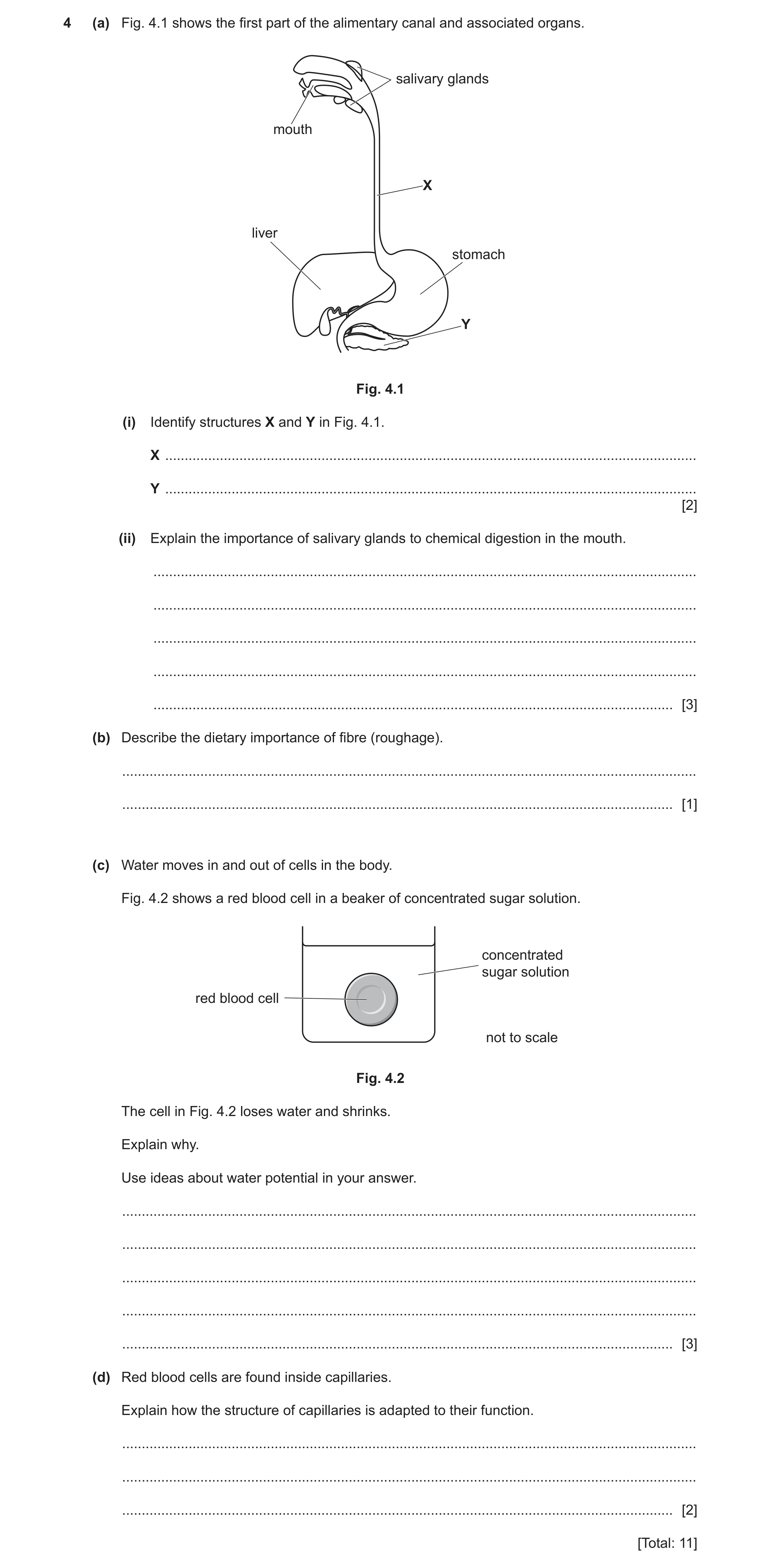
4 (a) Fig. 4.1 shows the first part of the alimentary canal and associated organs. salivary glands mouth X liver stomach Y Fig. 4.1 (i) Identify structures X and Y in Fig. 4.1. X … Y … [2] (ii) Explain the importance of salivary glands to chemical digestion in the mouth. … … … … … [3] (b) Describe the dietary importance of fibre (roughage). … … [1] (c) Water moves in and out of cells in the body. Fig. 4.2 shows a red blood cell in a beaker of concentrated sugar solution. concentrated sugar solution red blood cell not to scale Fig. 4.2 The cell in Fig. 4.2 loses water and shrinks. Explain why. Use ideas about water potential in your answer. … … … … … [3] (d) Red blood cells are found inside capillaries. Explain how the structure of capillaries is adapted to their function. … … … [2] [Total: 11]
11 marks
Mark scheme: 4(a)(i) X – oesophagus ; Y – pancreas ; 2 4(a)(ii) produce / secrete, amylase ; digest starch ; to produce, simpler sugars / glucose ; 3 4(b) prevents constipation ; 1 4(c) water moves from higher to lower water potential ; through a partially permeable membrane ; (by the process of) osmosis ; 3 4(d) thin wall / wall one cell thick ; allow (efficient) exchange of substances ; 2
This question in 0653/42 Feb/March 2022
Q6 · Part of the human alimentary canal and associated organs 0653/41 May/June 2022
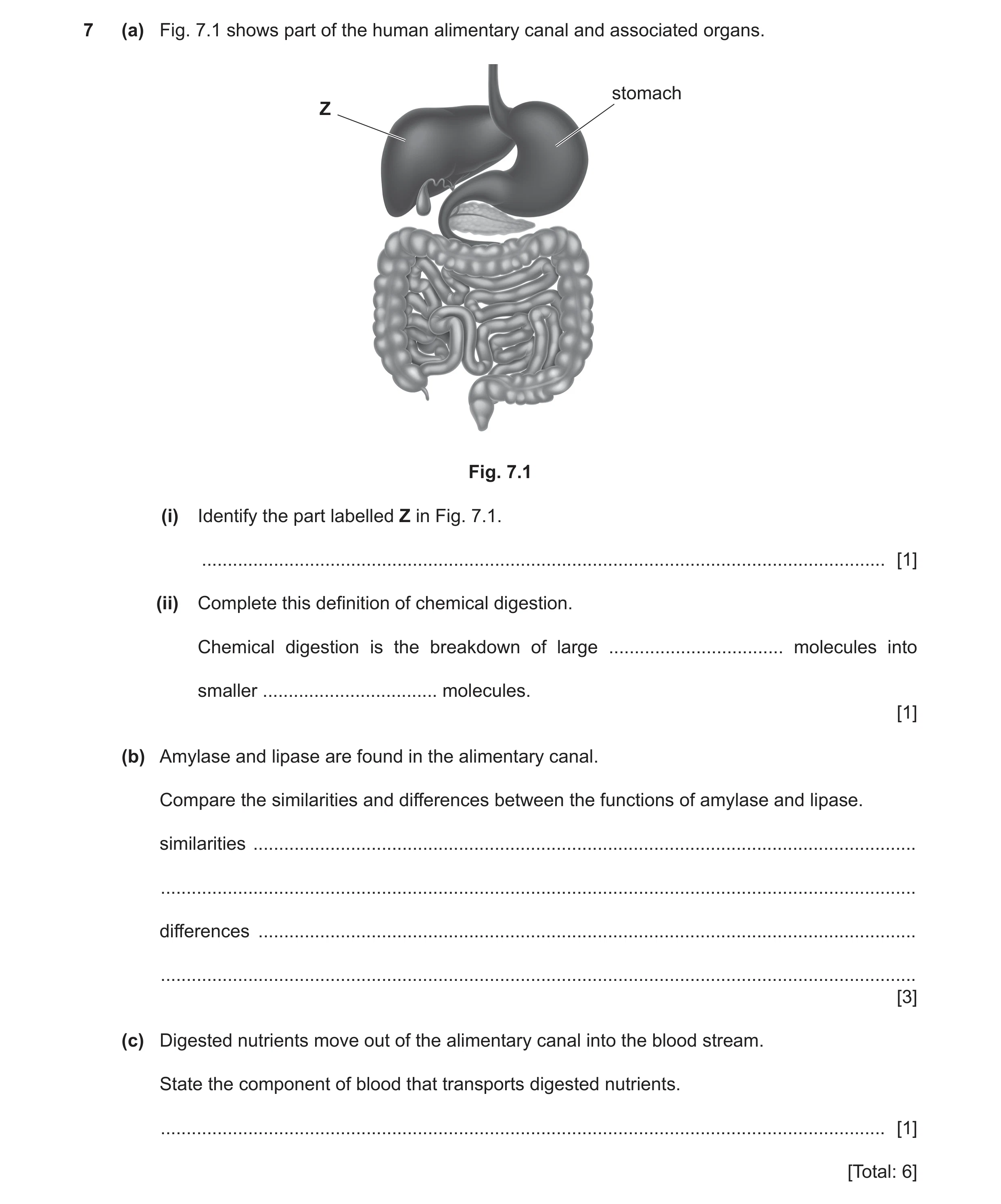
7 (a) Fig. 7.1 shows part of the human alimentary canal and associated organs. stomach Z Fig. 7.1 (i) Identify the part labelled Z in Fig. 7.1. … [1] (ii) Complete this definition of chemical digestion. Chemical digestion is the breakdown of large … molecules into smaller … molecules. [1] (b) Amylase and lipase are found in the alimentary canal. Compare the similarities and differences between the functions of amylase and lipase. similarities … … differences … … [3] (c) Digested nutrients move out of the alimentary canal into the blood stream. State the component of blood that transports digested nutrients. … [1] [Total: 6]
6 marks
Mark scheme: 7(a)(i) liver ; 1 7(a)(ii) insoluble AND soluble ; 1 7(b) any three from: (similarities) are enzymes / speed up digestion ; breakdown large / insoluble, food molecules ; (differences) amylase breaks down / digests starch / carbohydrates / produces sugars ; lipase breaks down / digests fats / oils / lipids / produces fatty acids / glycerol ; 3 7(c) plasma ; 1
This question in 0653/41 May/June 2022
Q7 · A diagram of the alimentary canal and associated organs in humans 0653/43 May/June 2022
1 (a) Fig. 1.1 is a diagram of the alimentary canal and associated organs in humans. B A C E D Fig. 1.1 (i) Table 1.1 shows some of the parts labelled in Fig. 1.1 and their function. Complete Table 1.1. Table 1.1 letter function digests food and uses hydrochloric acid to kill bacteria … ingestion … B secretes the enzyme … [3] (ii) Draw a label line and the letter L to identify the position of the large intestine on Fig. 1.1. [1] (b) Describe the function of protease in the alimentary canal. … … … [2] (c) Digested food is absorbed into the blood and transported through arteries and veins. Describe two ways the structure of an artery is different to the structure of a vein. 1 … … 2 … … [2] (d) Complete these sentences about coronary heart disease. Coronary heart disease is caused by a … in the coronary artery. Possible risk factors of coronary heart disease include poor diet, smoking and … predisposition. [2] [Total: 10]
10 marks
Mark scheme: 1(a)(i) letter function C ; (digests protein and uses hydrochloric acid to kill bacteria) A ; (ingestion) (B) (secretes the enzyme) amylase ; 3 1(a)(ii) label line to large intestine labelled L ; 1 1(b) digestion of protein ; to form amino acids ; 2 1(c) any two from: arteries have a thicker (muscle) wall / ORA ; arteries have a smaller centre / ORA ; only veins have valves ; 2 1(d) blockage ; genetic ; 2
This question in 0653/43 May/June 2022
Q8 · The human alimentary canal and some associated organs 0653/41 Oct/Nov 2023
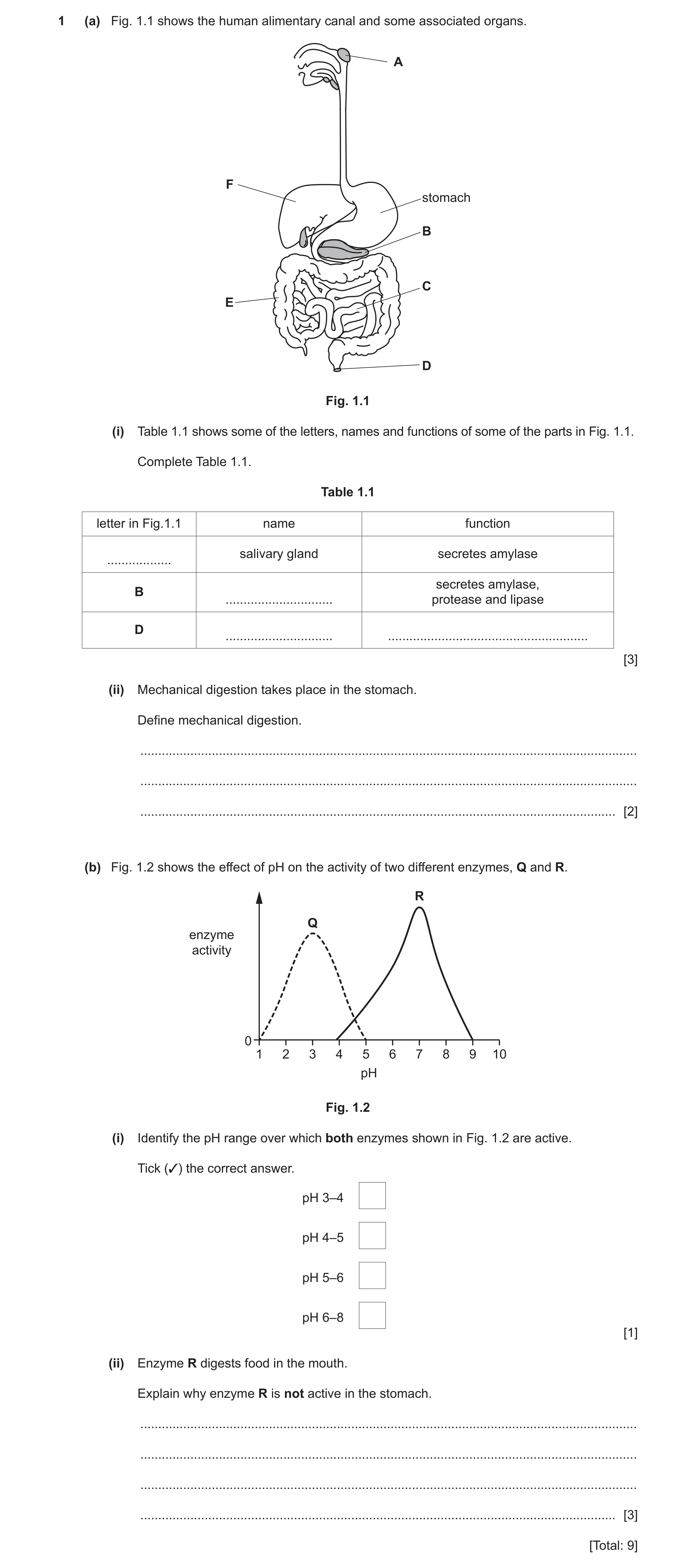
1 (a) Fig. 1.1 shows the human alimentary canal and some associated organs. A F stomach B C E D Fig. 1.1 (i) Table 1.1 shows some of the letters, names and functions of some of the parts in Fig. 1.1. Complete Table 1.1. Table 1.1 letter in Fig.1.1 name function salivary gland secretes amylase … secretes amylase, B … protease and lipase D … … [3] (ii) Mechanical digestion takes place in the stomach. Define mechanical digestion. … … … [2] (b) Fig. 1.2 shows the effect of pH on the activity of two different enzymes, Q and R. R Q enzyme activity 0 1 2 3 4 5 6 7 8 9 10 pH Fig. 1.2 (i) Identify the pH range over which both enzymes shown in Fig. 1.2 are active. Tick (✓) the correct answer. pH 3–4 pH 4–5 pH 5–6 pH 6–8 [1] (ii) Enzyme R digests food in the mouth. Explain why enzyme R is not active in the stomach. … … … … [3] [Total: 9]
9 marks
Mark scheme: Question Answer Marks 1(a)(i) one mark for each correct row 3 letter in Fig. 1.1. name function A (salivary gland) (secretes amylase) ; (B) pancreas (secretes amylase, ; protease and lipase) (D) anus egestion ; 1(a)(ii) (mechanical) breakdown of food into smaller pieces / AW ; 2 then any one from: to increase surface area ; without chemical change (to molecules) ; 1(b)(i) pH 4–5 ; 1 1(b)(ii) any three from: 3 reference to, strongly acidic / low pH conditions (in stomach) ; R is denatured ; active site changes shape ; substrate no longer fits into active site ;
This question in 0653/41 Oct/Nov 2023
Q9 · Part of the human alimentary canal and associated organs 0653/43 Oct/Nov 2023
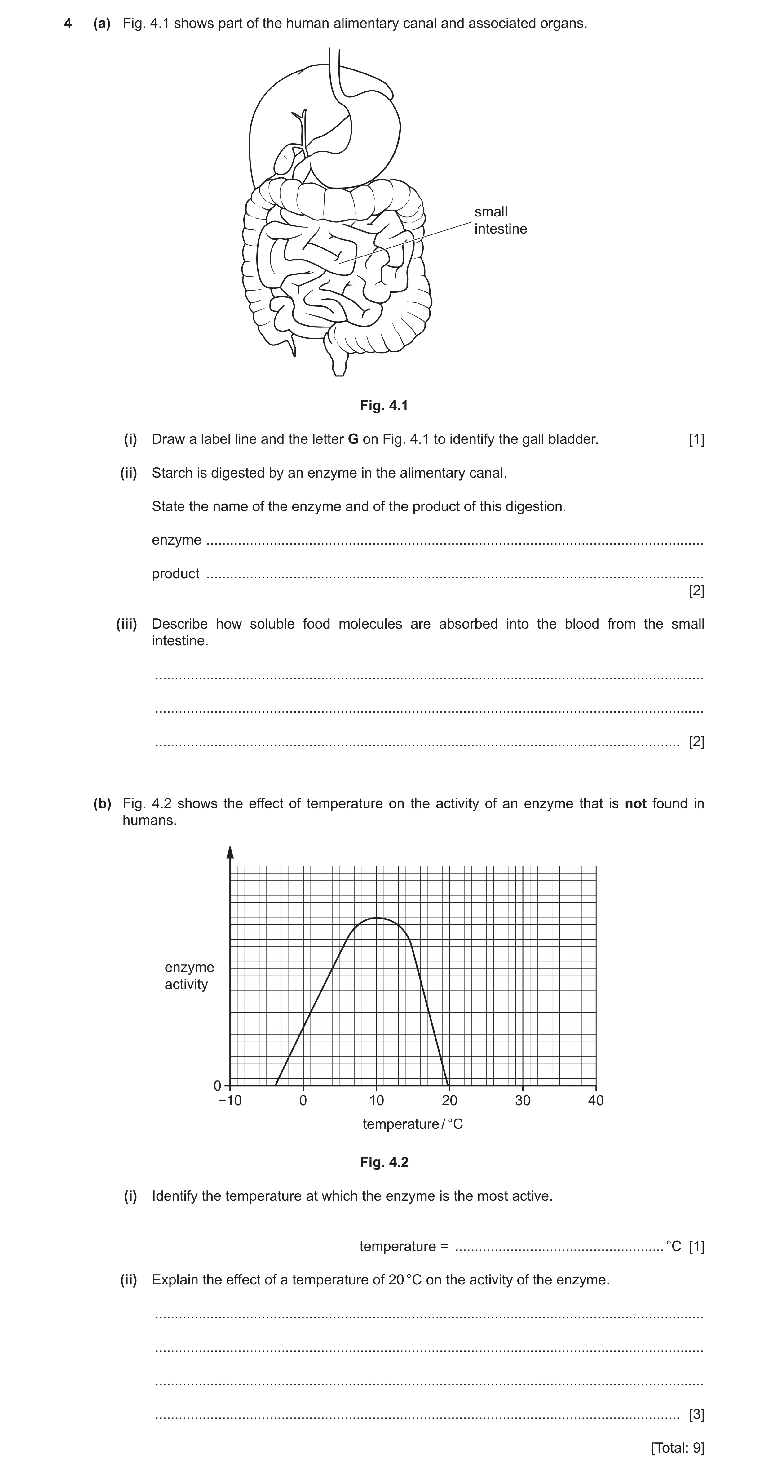
4 (a) Fig. 4.1 shows part of the human alimentary canal and associated organs. small intestine Fig. 4.1 (i) Draw a label line and the letter G on Fig. 4.1 to identify the gall bladder. [1] (ii) Starch is digested by an enzyme in the alimentary canal. State the name of the enzyme and of the product of this digestion. enzyme … product … [2] (iii) Describe how soluble food molecules are absorbed into the blood from the small intestine. … … … [2] (b) Fig. 4.2 shows the effect of temperature on the activity of an enzyme that is not found in humans. enzyme activity 0 −10 0 10 20 30 40 temperature / °C Fig. 4.2 (i) Identify the temperature at which the enzyme is the most active. temperature = … °C [1] (ii) Explain the effect of a temperature of 20 °C on the activity of the enzyme. … … … … [3] [Total: 9]
9 marks
Mark scheme: 4(a)(i) gall bladder labelled ; 1 4(a)(ii) amylase ; 2 (simpler) sugars ; 4(a)(iii) any two from: 2 diffuses ; through, cell membrane / capillary wall ; (dissolves) into the plasma ; 4(b)(i) 10 (°C) ; 1 4(b)(ii) enzyme is denatured ; 3 active site changes shape ; substrate no longer fits (into active site) ;
This question in 0653/43 Oct/Nov 2023
Q10 · Part of the human alimentary canal and associated organs 0653/42 Feb/March 2024
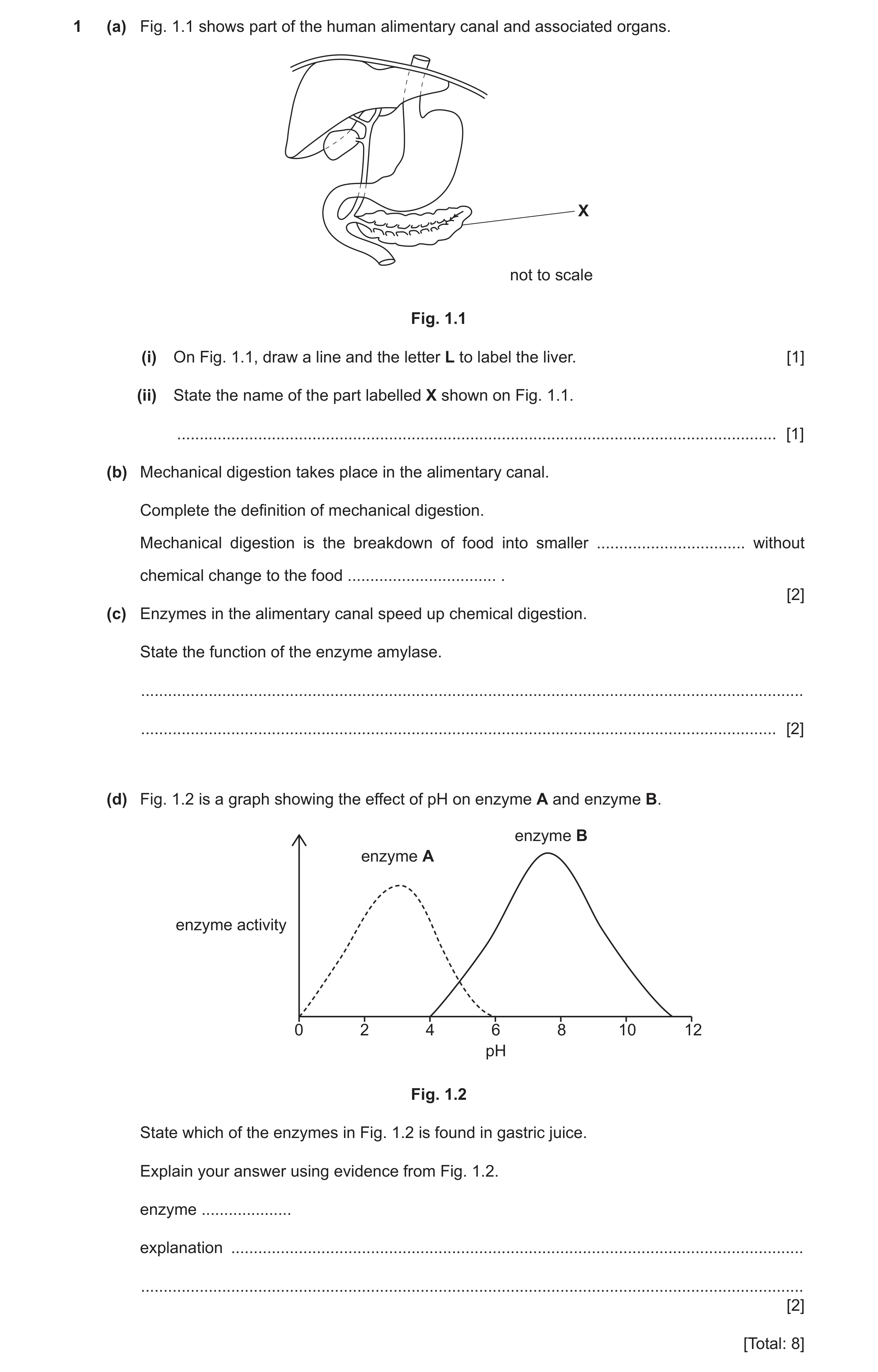
1 (a) Fig. 1.1 shows part of the human alimentary canal and associated organs. X not to scale Fig. 1.1 (i) On Fig. 1.1, draw a line and the letter L to label the liver. [1] (ii) State the name of the part labelled X shown on Fig. 1.1. … [1] (b) Mechanical digestion takes place in the alimentary canal. Complete the definition of mechanical digestion. Mechanical digestion is the breakdown of food into smaller … without chemical change to the food … . [2] (c) Enzymes in the alimentary canal speed up chemical digestion. State the function of the enzyme amylase. … … [2] (d) Fig. 1.2 is a graph showing the effect of pH on enzyme A and enzyme B. enzyme B enzyme A enzyme activity 0 2 4 6 8 10 12 pH Fig. 1.2 State which of the enzymes in Fig. 1.2 is found in gastric juice. Explain your answer using evidence from Fig. 1.2. enzyme … explanation … … [2] [Total: 8]
8 marks
Mark scheme: Question Answer Marks 1(a)(i) liver labelled ; 1 1(a)(ii) pancreas ; 1 1(b) pieces ; 2 molecules ; 1(c) digest / breakdown, starch ; 2 into (simpler) sugars ; 1(d) enzyme: A 2 AND works (best) at low pH / acidic conditions ; gastric juice, contains hydrochloric acid / is acidic ;
This question in 0653/42 Feb/March 2024
Q11 · The alimentary canal and associated organs in humans 0653/42 May/June 2024
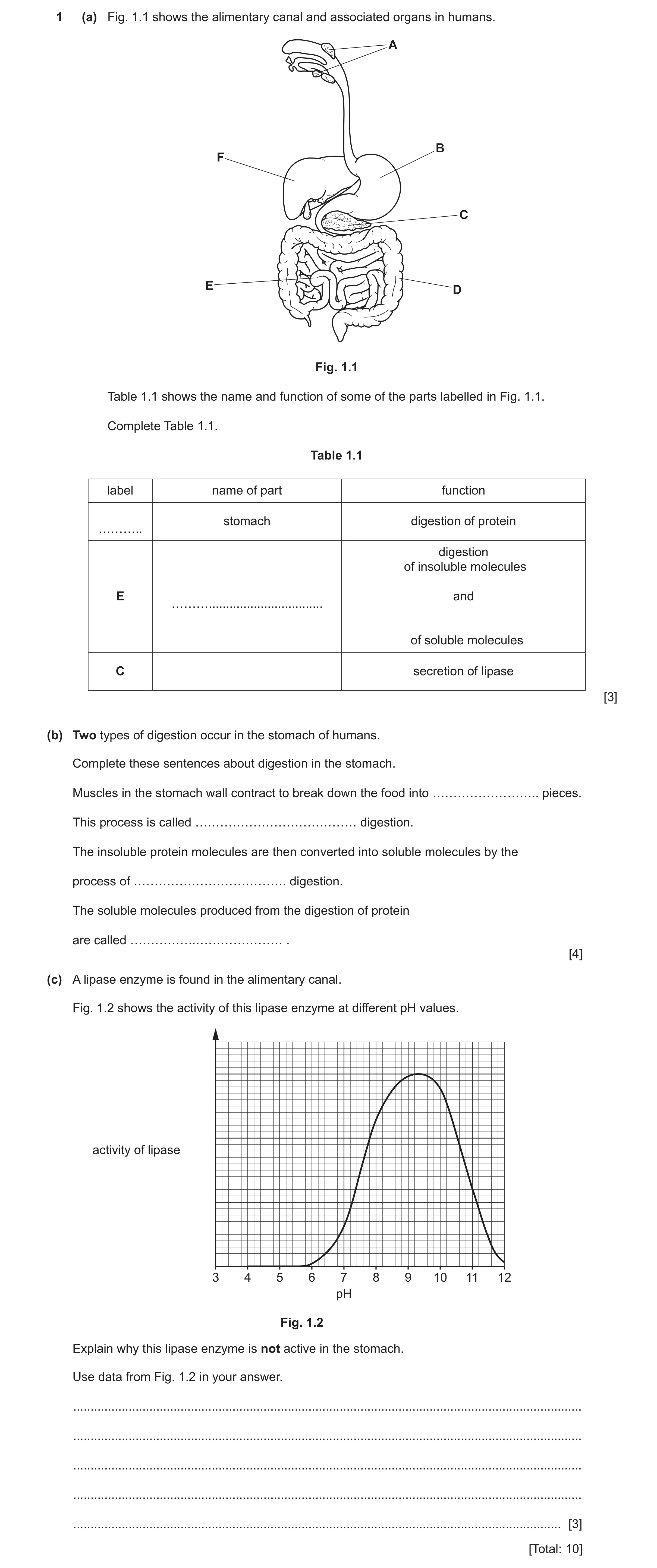
1 (a) Fig. 1.1 shows the alimentary canal and associated organs in humans. A B F C E D Fig. 1.1 Table 1.1 shows the name and function of some of the parts labelled in Fig. 1.1. Complete Table 1.1. Table 1.1 label name of part function stomach digestion of protein ……….. digestion of insoluble molecules E and ……… … of soluble molecules C secretion of lipase [3] (b) Two types of digestion occur in the stomach of humans. Complete these sentences about digestion in the stomach. Muscles in the stomach wall contract to break down the food into …………………….. pieces. This process is called ………………………………… digestion. The insoluble protein molecules are then converted into soluble molecules by the process of ………………………………. digestion. The soluble molecules produced from the digestion of protein are called …………….………………… . [4] (c) A lipase enzyme is found in the alimentary canal. Fig. 1.2 shows the activity of this lipase enzyme at different pH values. activity of lipase 3 4 5 6 7 8 9 10 11 12 pH Fig. 1.2 Explain why this lipase enzyme is not active in the stomach. Use data from Fig. 1.2 in your answer. … … … … … [3] [Total: 10]
10 marks
Mark scheme: 1(a) label name of part function B (stomach) (digestion of protein) (E) small intestine (digestion of insoluble molecules and) absorption (of soluble molecules) (C) pancreas (secretion of lipase) ;;; one mark for each correct row 1(b) small(er) ; mechanical ; chemical ; amino acids ; 4 1(c) any three from: lipase activity between pH6–pH12 / lipase inactive below pH6 / lipase inactive in acidic conditions / stomach pH low / stomach is (very) acidic / stomach pH from 1.5 to 3.5 ; (so) lipase would denature (in stomach) ; active site, changes shape / is no longer complementary (to the substrate) ; lipase is unable to, bind to / fit, substrate ; 3
This question in 0653/42 May/June 2024
Q12 · A model of the alimentary canal and associated organs of humans 0653/43 May/June 2024
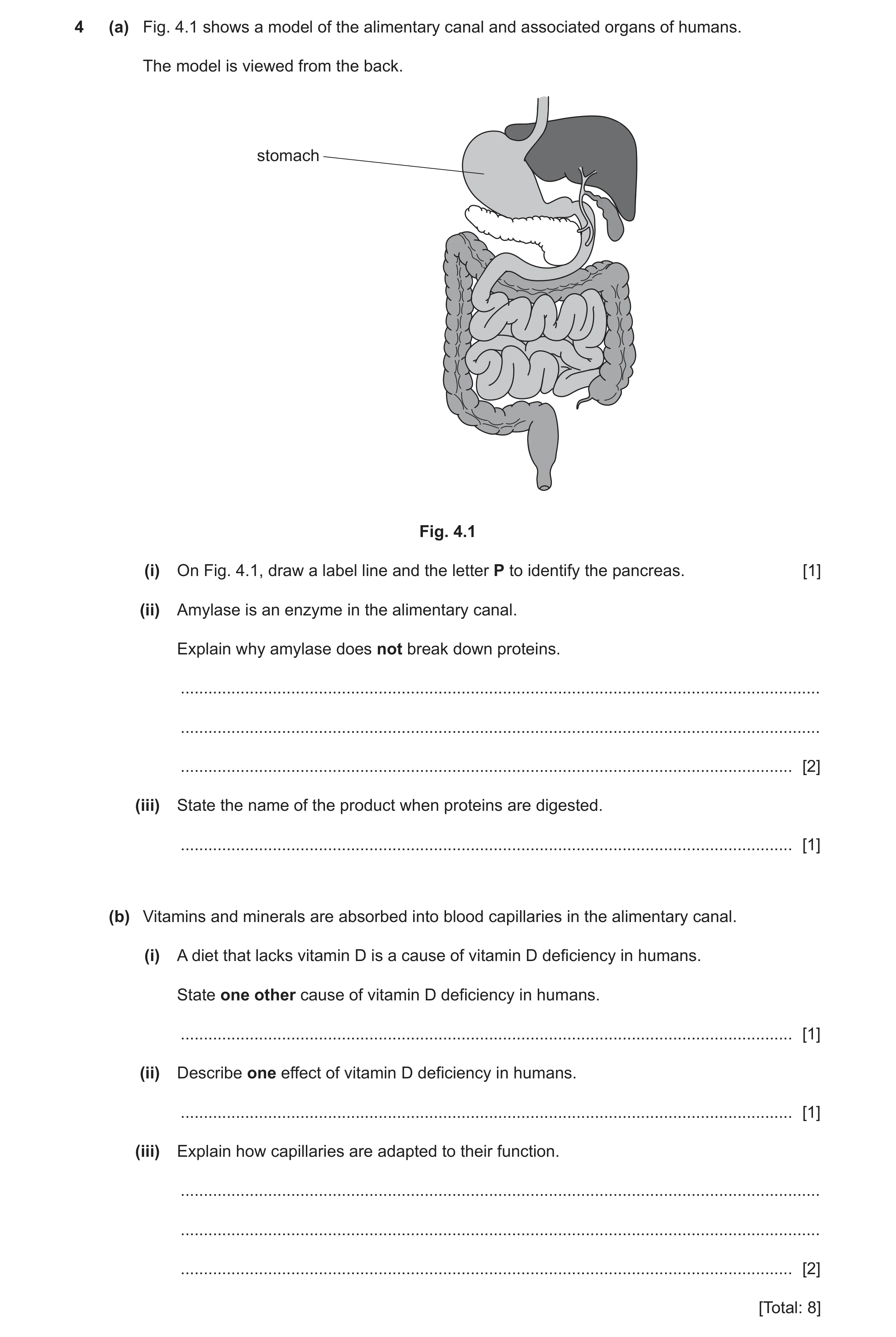
4 (a) Fig. 4.1 shows a model of the alimentary canal and associated organs of humans. The model is viewed from the back. stomach Fig. 4.1 (i) On Fig. 4.1, draw a label line and the letter P to identify the pancreas. [1] (ii) Amylase is an enzyme in the alimentary canal. Explain why amylase does not break down proteins. … … … [2] (iii) State the name of the product when proteins are digested. … [1] (b) Vitamins and minerals are absorbed into blood capillaries in the alimentary canal. (i) A diet that lacks vitamin D is a cause of vitamin D deficiency in humans. State one other cause of vitamin D deficiency in humans. … [1] (ii) Describe one effect of vitamin D deficiency in humans. … [1] (iii) Explain how capillaries are adapted to their function. … … … [2] [Total: 8]
8 marks
Mark scheme: 4(a)(i) pancreas labelled ; 1 4(a)(ii) amylase breaks down or digests, starch / carbohydrate, or amylase has an active site complementary to starch ; protein (shape), is not complementary to / will not fit active site ; 2 4(a)(iii) amino acids ; 1 4(b)(i) lack of sunlight ; 1 4(b)(ii) soft, bones / teeth ; 1 4(b)(iii) thin walls / permeable walls / short diffusion distance ; to allow exchange of material / to allow substances to, diffuse / pass through the walls (between cells and blood) ; 2
This question in 0653/43 May/June 2024
Q13 · A diagram of the digestive system and associated organs 0653/42 May/June 2025
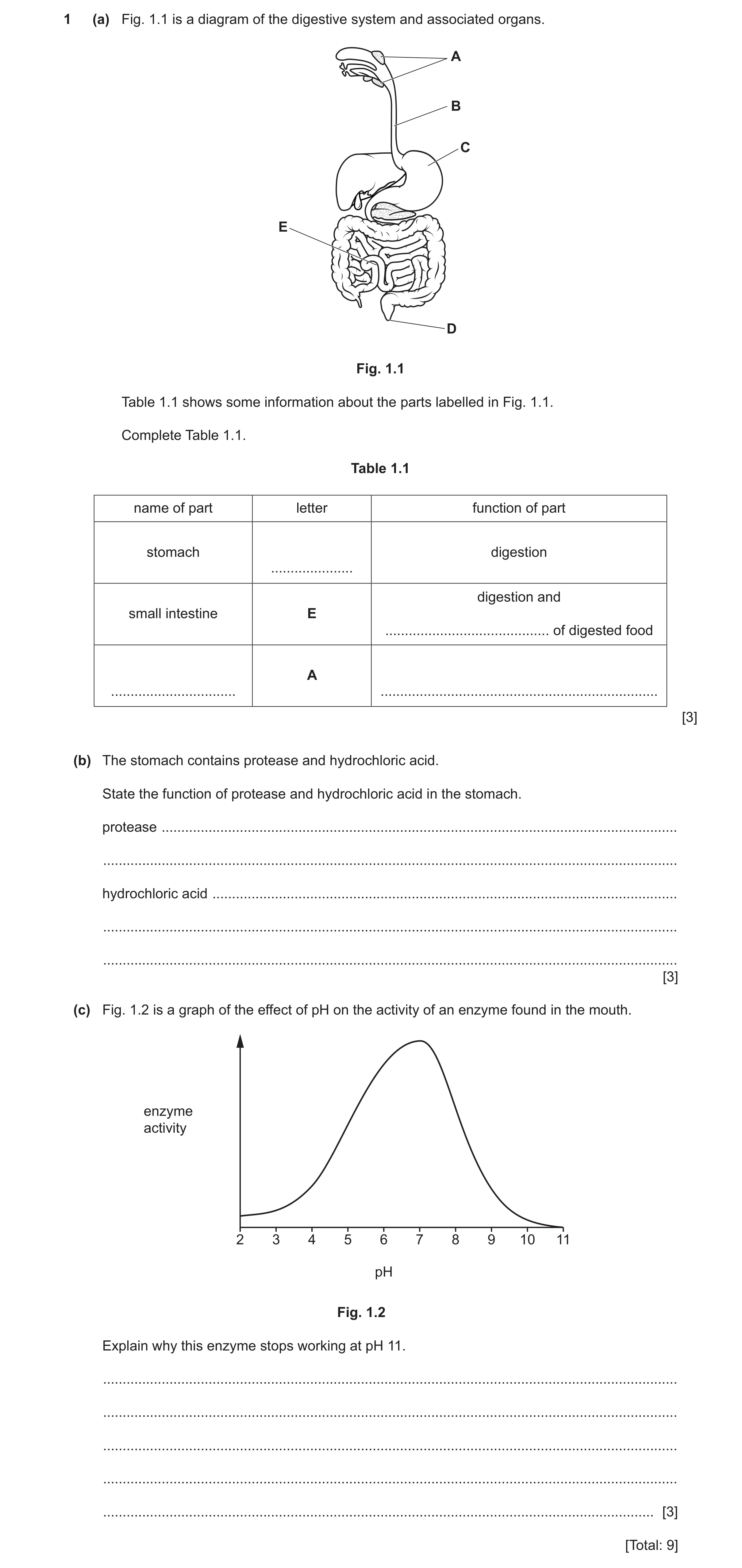
1 (a) Fig. 1.1 is a diagram of the digestive system and associated organs. A B C E D Fig. 1.1 Table 1.1 shows some information about the parts labelled in Fig. 1.1. Complete Table 1.1. Table 1.1 name of part letter function of part stomach digestion … digestion and small intestine E … of digested food A … … [3] (b) The stomach contains protease and hydrochloric acid. State the function of protease and hydrochloric acid in the stomach. protease … … hydrochloric acid … … … [3] (c) Fig. 1.2 is a graph of the effect of pH on the activity of an enzyme found in the mouth. enzyme activity 2 3 4 5 6 7 8 9 10 11 pH Fig. 1.2 Explain why this enzyme stops working at pH 11. … … … … … [3] [Total: 9]
9 marks
Mark scheme: Question Answer Marks 1(a) 3 name of part letter function of part stomach C digestion small intestine E (digestion and) absorption (of digested food) salivary gland A produce, saliva / amylase ;;; one mark for each correct row 1(b) protease: 3 digests / breaks down, proteins (into amino acids) ; hydrochloric acid: kills, (harmful) microorganisms / pathogens / bacteria ; provides optimum / acidic / low pH, for enzyme / protease ; 1(c) enzyme will denature ; 3 active site changes shape ; active site no longer complementary to substrate / substrate no longer fits active site ;
This question in 0653/42 May/June 2025
Q14 · Part of the human alimentary canal and associated organs 0653/43 Oct/Nov 2025
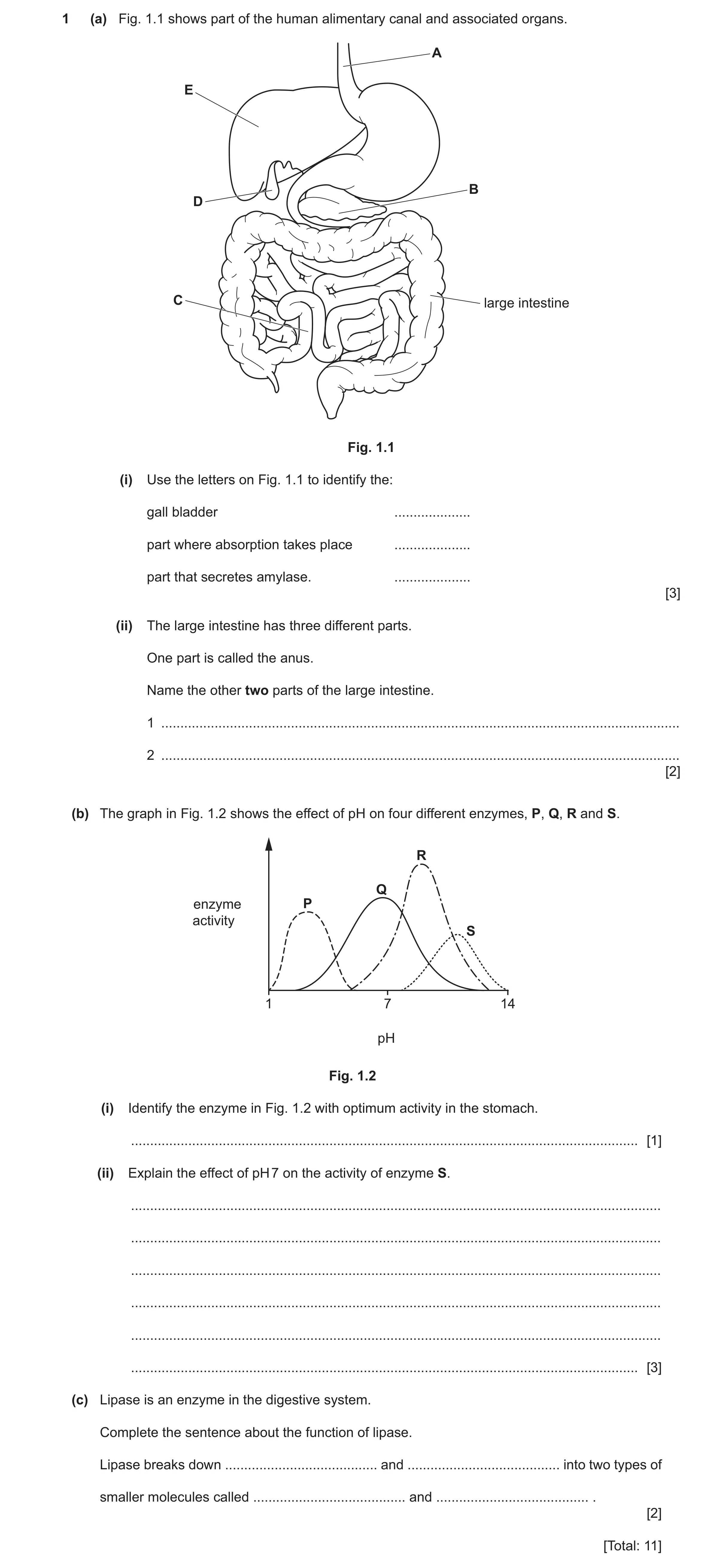
1 (a) Fig. 1.1 shows part of the human alimentary canal and associated organs. A E B D C large intestine Fig. 1.1 (i) Use the letters on Fig. 1.1 to identify the: gall bladder … part where absorption takes place … part that secretes amylase. … [3] (ii) The large intestine has three different parts. One part is called the anus. Name the other two parts of the large intestine. 1 … 2 … [2] (b) The graph in Fig. 1.2 shows the effect of pH on four different enzymes, P, Q, R and S. R Q enzyme P activity S 1 7 14 pH Fig. 1.2 (i) Identify the enzyme in Fig. 1.2 with optimum activity in the stomach. … [1] (ii) Explain the effect of pH 7 on the activity of enzyme S. … … … … … … [3] (c) Lipase is an enzyme in the digestive system. Complete the sentence about the function of lipase. Lipase breaks down … and … into two types of smaller molecules called … and … . [2] [Total: 11]
11 marks
Mark scheme: Question Answer Marks 1(a)(i) D ; 3 C ; B ; 1(a)(ii) colon ; 2 rectum ; 1(b)(i) P ; 1 1(b)(ii) enzyme will denature (at pH 7) ; 3 active site changes shape ; (active site) no longer complementary to substrate / substrate no longer fits active site / enzyme-substrate complexes cannot form ; 1(c) fats AND oils; 2 fatty acids AND glycerol ;
This question in 0653/43 Oct/Nov 2025
 9 / 22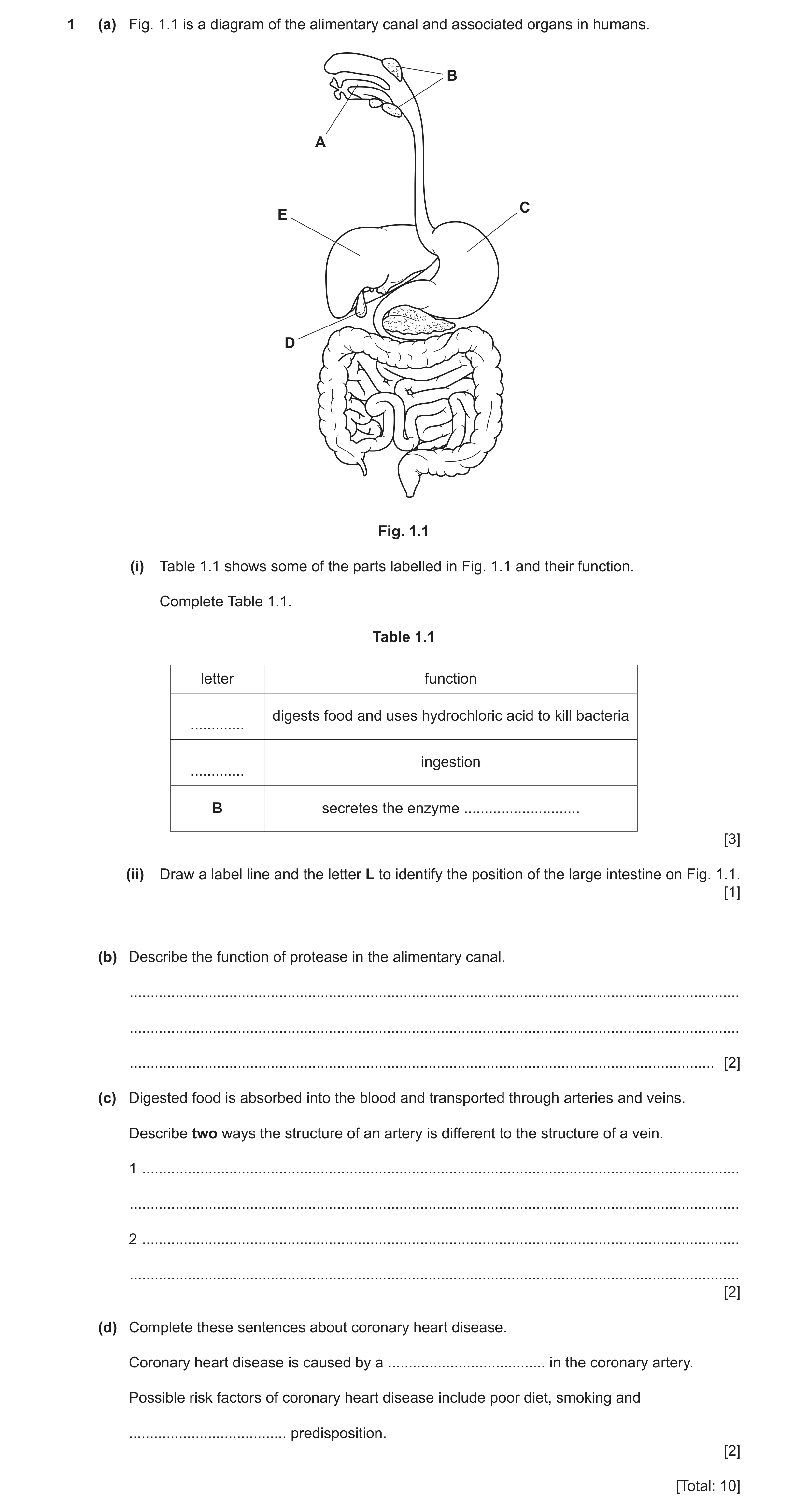
 8
8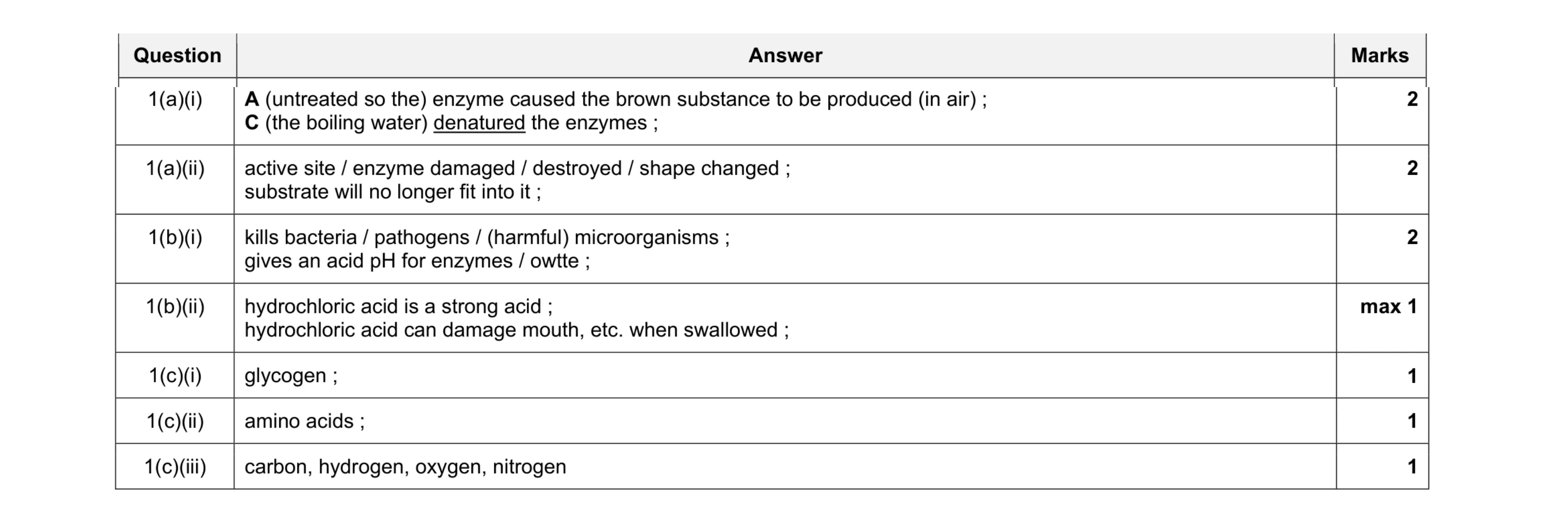 10
10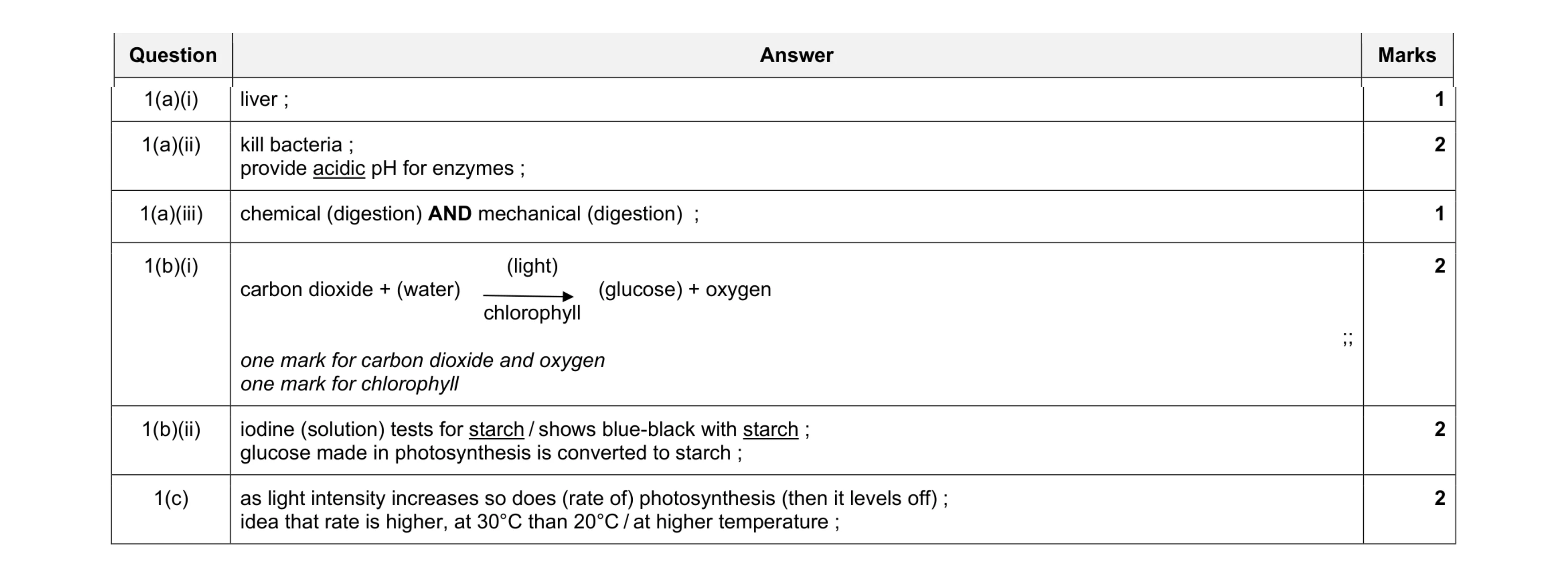 10
10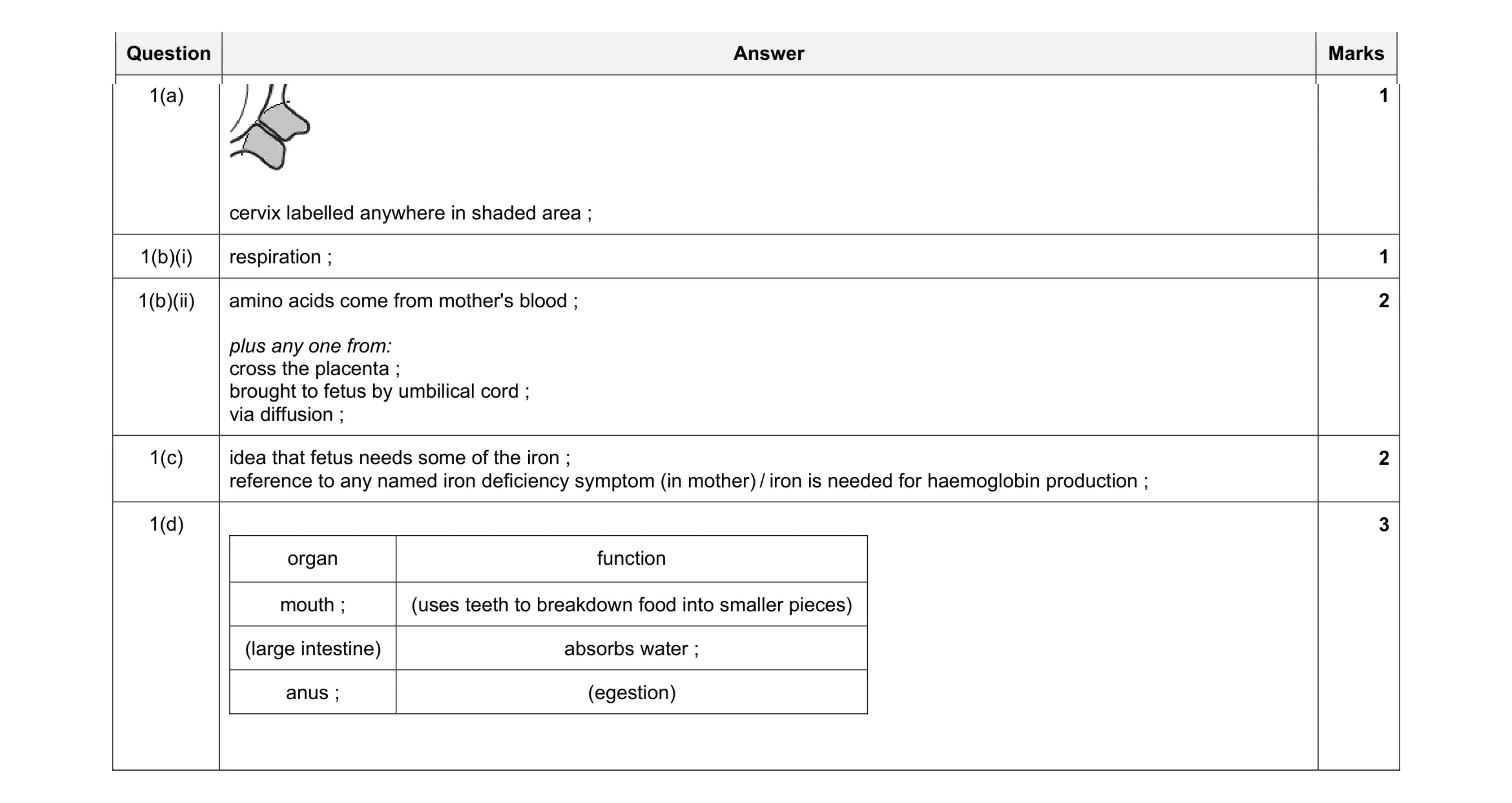 9
9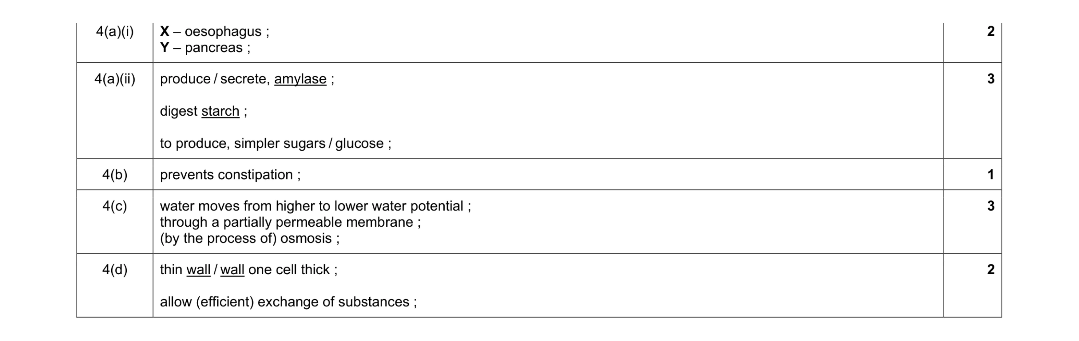 11
11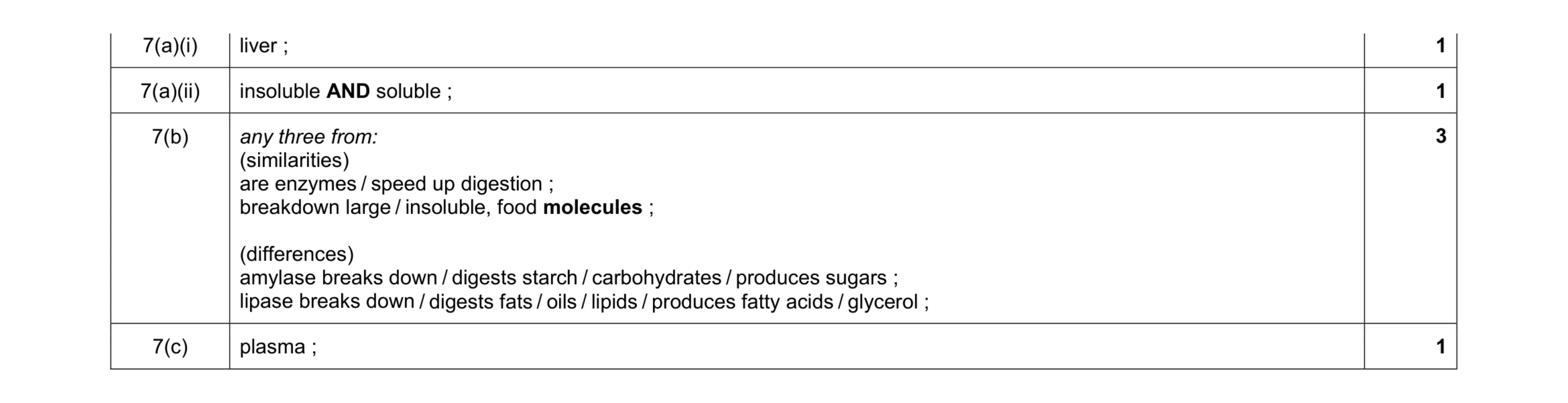 6
6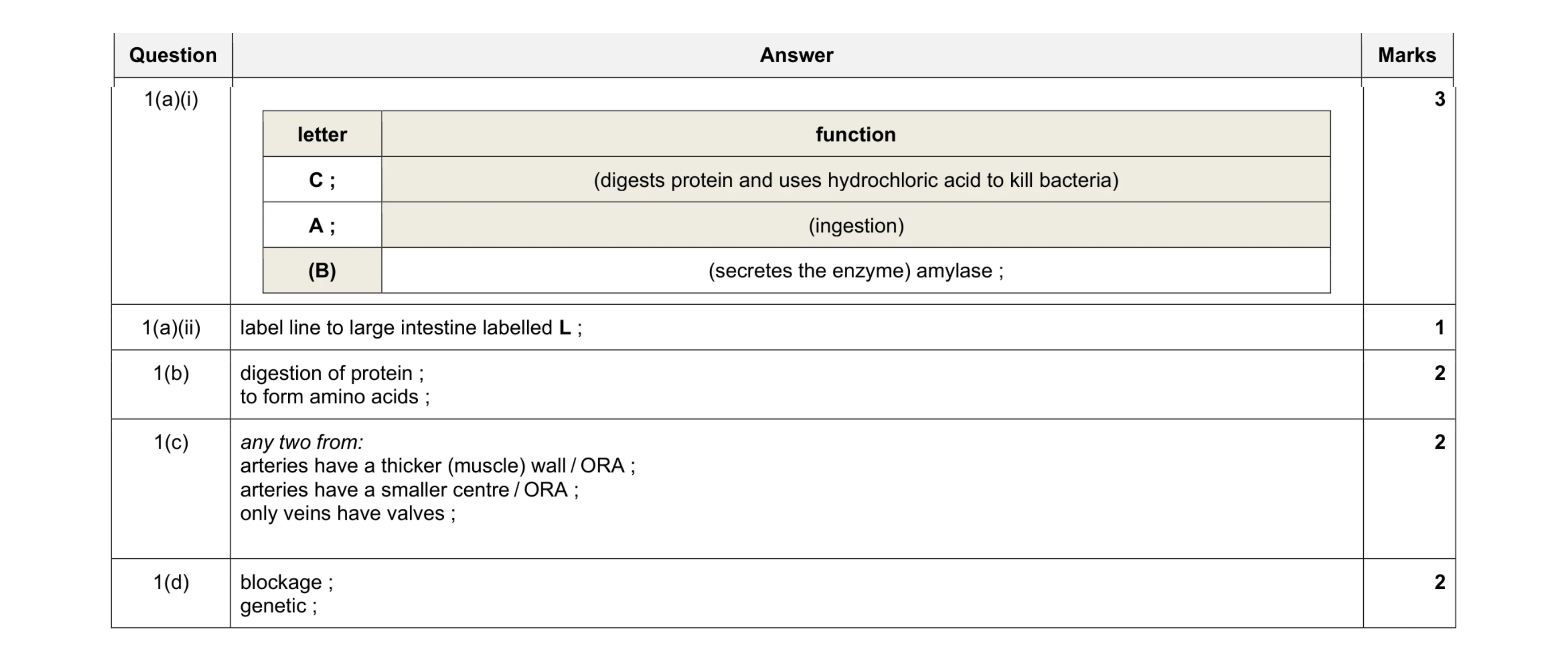 10
10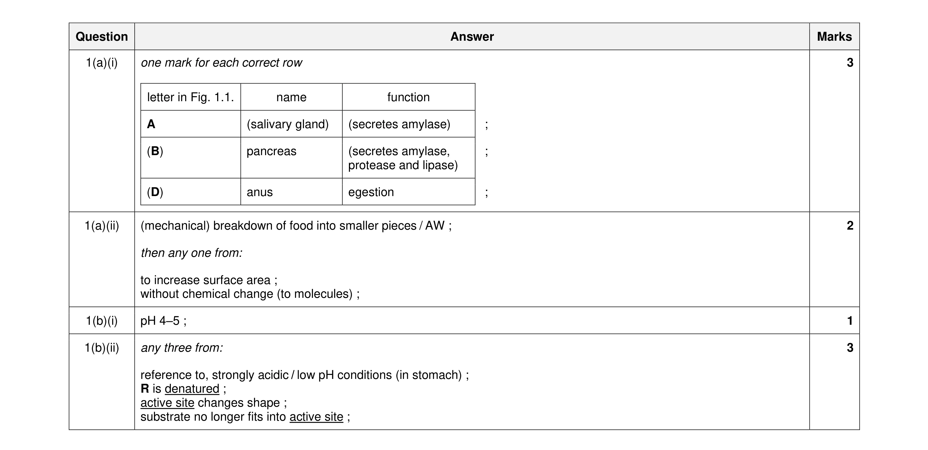 9
9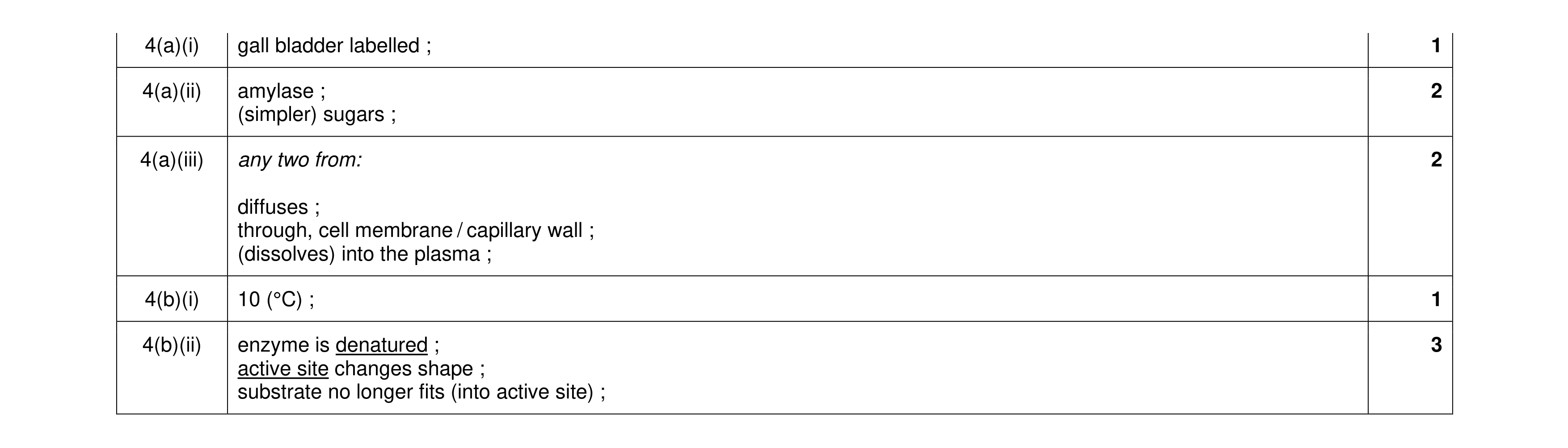 9
9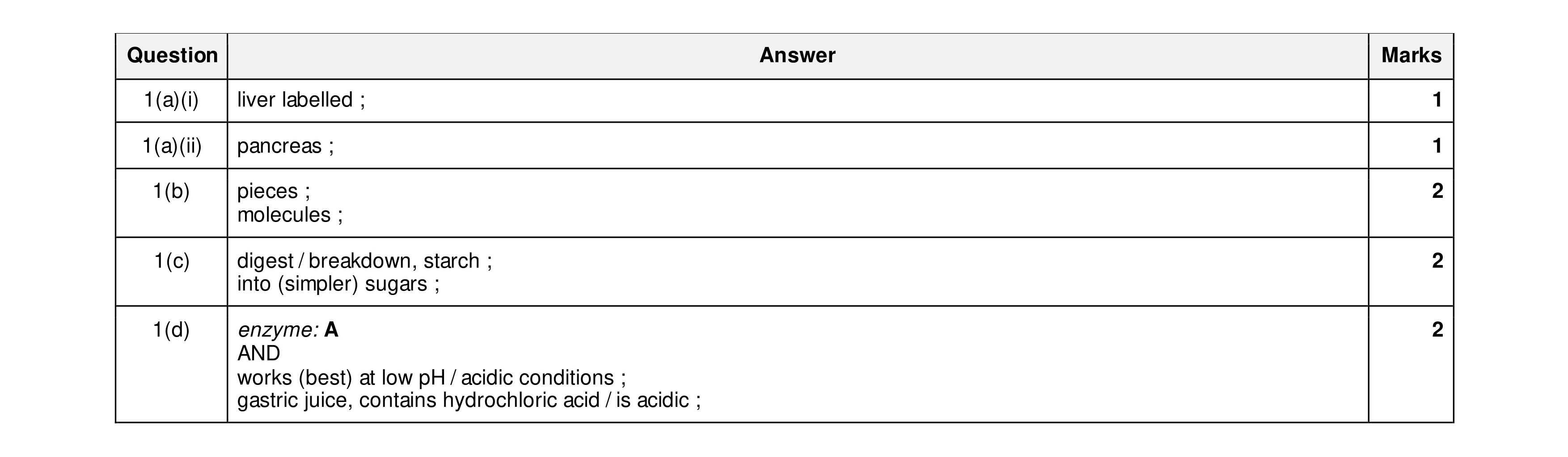 8
8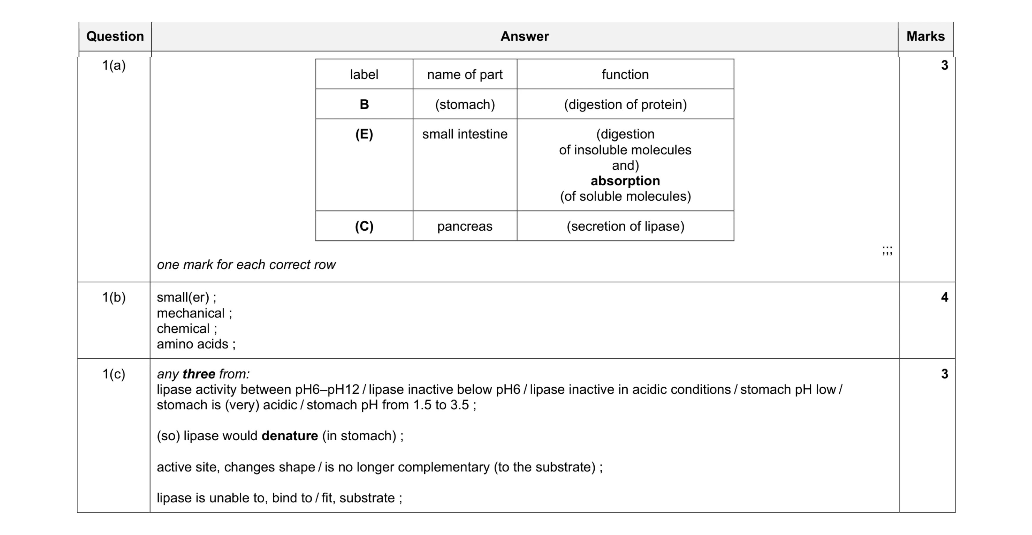 10
10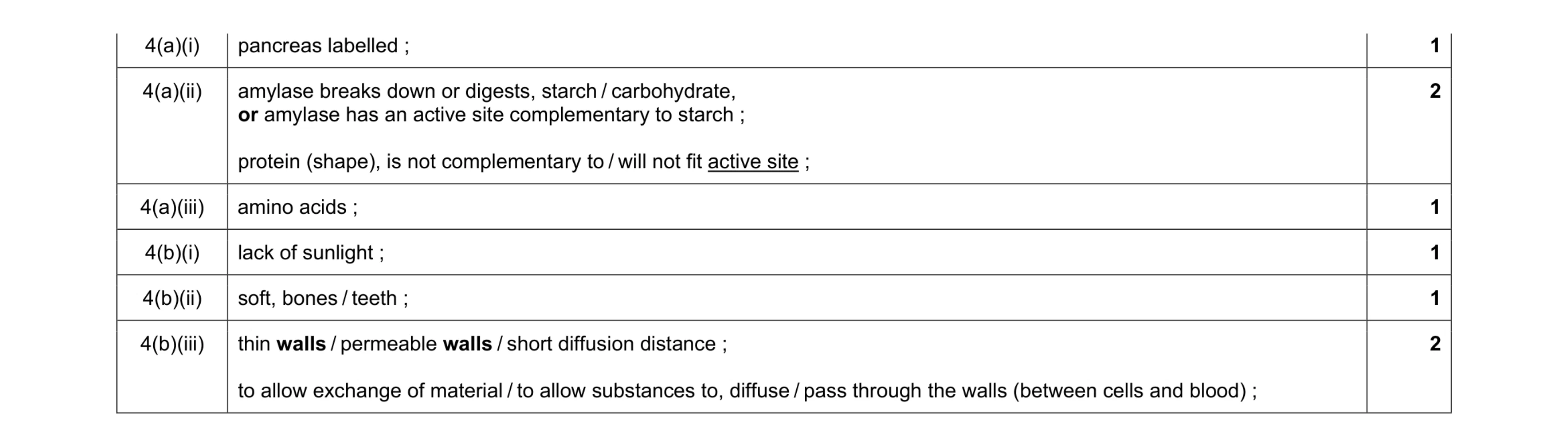 8
8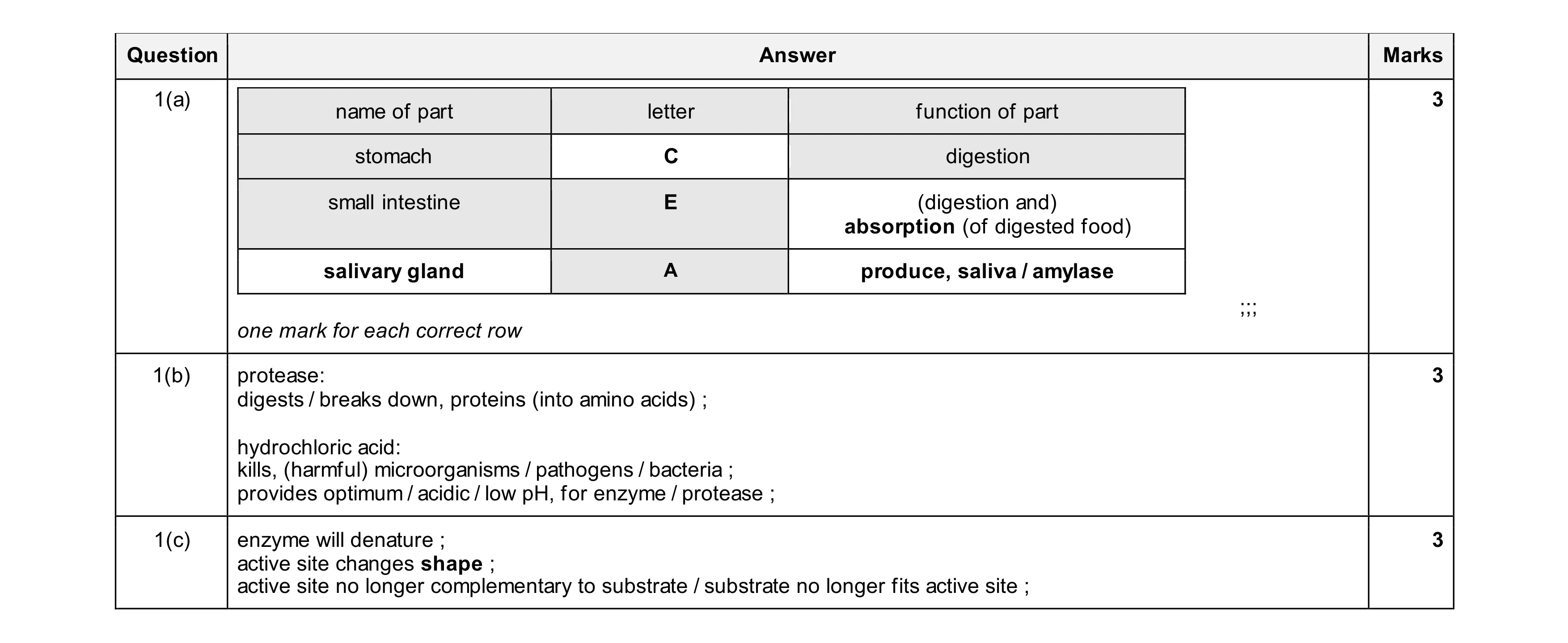 9
9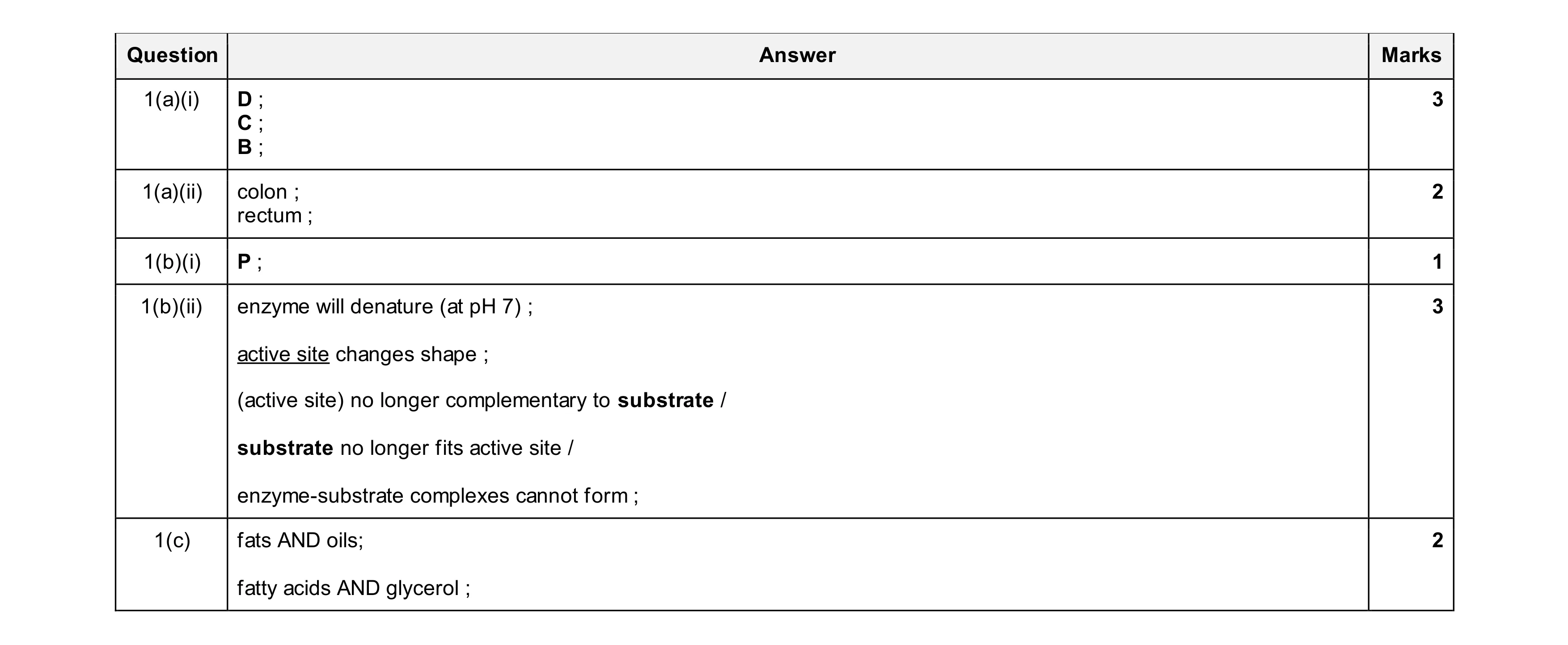 11
11