Questions as text
Q1 · Explain the main principles of the generation of ultrasound waves for medical use 9702/42 Feb/March 2017
4 Explain the main principles of the generation of ultrasound waves for medical use. … … … … … … … … … … [4] [Total: 4]
4 marks
Mark scheme: 4 piezo-electric / quartz crystal / transducer B1 alternating p.d. applied across crystal / transducer B1 causes crystal to vibrate / resonate B1 crystal resonates at ultrasound frequencies / crystal’s natural frequency is in the ultrasound range / alternating p.d. is in ultrasound frequency range B1
This question in 9702/42 Feb/March 2017
Q2 · Explain the main principles behind the use of ultrasound to obtain diagnostic information… 9702/42 May/June 2017
4 (a) Explain the main principles behind the use of ultrasound to obtain diagnostic information about internal body structures. … … … … … … … … … … … … … … … [6] (b) A parallel beam of ultrasound has intensity I0 as it enters a muscle of thickness 4.6 cm, as illustrated in Fig. 4.1. 4.6 cm muscle beam of I0 IT ultrasound Fig. 4.1 The intensity of the beam just before it leaves the muscle is IT. The ratio I0 / IT is found to be 2.9. Calculate the linear attenuation (absorption) coefficient μ of the ultrasound in the layer of muscle. μ = … cm–1 [3] [Total: 9]
9 marks
Mark scheme: 4(a) pulse (of ultrasound) B1 * produced by quartz crystal/piezo-electric crystal * gel/coupling medium (on skin) used to reduce reflection at skin reflected from boundaries (between media) B1 reflected pulse/wave detected by (ultrasound) transmitter B1 reflected wave processed and displayed B1 * intensity of reflected pulse/wave gives information about boundary * time delay gives information about depth of boundary max. 2 of additional detail points marked * B2 4(b) IT = I0 exp (–µx) C1 2.9 = exp (4.6µ) C1 µ = 0.23 cm–1 A1
This question in 9702/42 May/June 2017
Q3 · State two advantages of the transmission of data in digital form rather than in analogue… 9702/42 May/June 2017
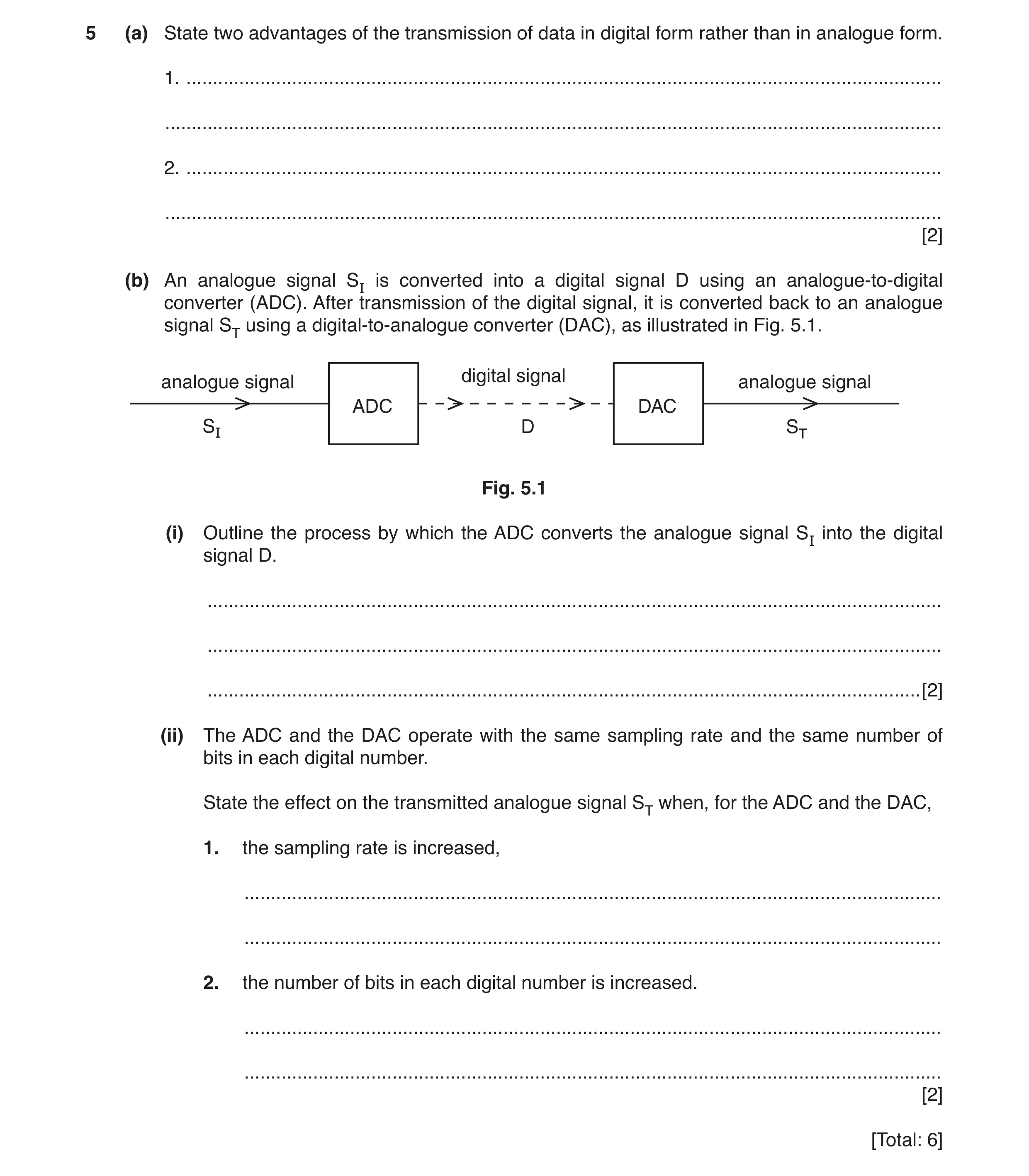
5 (a) State two advantages of the transmission of data in digital form rather than in analogue form. 1. … … 2. … … [2] (b) An analogue signal SI is converted into a digital signal D using an analogue-to-digital converter (ADC). After transmission of the digital signal, it is converted back to an analogue signal ST using a digital-to-analogue converter (DAC), as illustrated in Fig. 5.1. digital signal analogue signal analogue signal ADC DAC SI D ST Fig. 5.1 (i) Outline the process by which the ADC converts the analogue signal SI into the digital signal D. … … … [2] (ii) The ADC and the DAC operate with the same sampling rate and the same number of bits in each digital number. State the effect on the transmitted analogue signal ST when, for the ADC and the DAC, 1. the sampling rate is increased, … … 2. the number of bits in each digital number is increased. … … [2] [Total: 6]
6 marks
Mark scheme: 5(a) any two reasonable suggestions e.g. • signal can be regenerated/noise removed (not “no noise”) • circuits more reliable • circuits cheaper to produce • multiplexing (is possible) • error correction/checking • easier encryption/better security B2 5(b)(i) samples the analogue signal M1 at regular intervals and converts it (to a digital number) A1 5(b)(ii) 1. smaller step depth B1 2. smaller step height B1
This question in 9702/42 May/June 2017
Q4 · The digital transmission of speech may be illustrated using the block diagram of Fig 9702/43 May/June 2017
3 The digital transmission of speech may be illustrated using the block diagram of Fig. 3.1. serial -to - ADC X parallel Y optic fibre converter Fig. 3.1 (a) (i) State what is meant by a digital signal. … … [1] (ii) State the names of the components labelled X and Y on Fig. 3.1. X: … Y: … [2] (iii) Describe the function of the ADC. … … … [2] (b) The optic fibre has length 84 km and the attenuation per unit length in the fibre is 0.19 dB km–1. The input power to the optic fibre is 9.7 mW. At the output from the optic fibre, the signal-to- noise ratio is 28 dB. Calculate (i) in dB, the ratio input power to optic fibre noise power at output of optic fibre, ratio = … dB [2] (ii) the noise power at the output of the optic fibre. noise power = … W [3] [Total: 10]
10 marks
Mark scheme: 3(a)(i) signal consists of (a series of) 1s and 0s or offs and ons or highs and lows B1 3(a)(ii) component X: parallel-to-serial converter B1 component Y: DAC/digital-to-analogue converter B1 3(a)(iii) sample the (analogue) signal M1 at regular intervals and converts the analogue number to a digital number A1 3(b)(i) attenuation in fibre = 84 × 0.19 (= 16 dB) C1 ratio = 16 + 28 = 44 dB A1 3(b)(ii) ratio / dB = 10 lg (P2 / P1) C1 44 = 10 lg ({9.7 × 10⁻³} / P) or –44 = 10 lg (P / {9.7 × 10⁻³}) C1 power = 3.9 × 10⁻⁷ W A1
This question in 9702/43 May/June 2017
Q5 · Explain the principles behind the generation of ultrasound waves for diagnosis in medicine 9702/42 Oct/Nov 2017
4 (a) Explain the principles behind the generation of ultrasound waves for diagnosis in medicine. … … … … … … … … … … … … … … … [5] (b) Ultrasound frequencies as high as 10 MHz are used in medical diagnosis. Suggest one advantage of the use of high-frequency ultrasound rather than lower-frequency ultrasound. … … … [1] [Total: 6]
6 marks
Mark scheme: 4(a) quartz/piezo-electric and crystal/transducer B1 p.d. across crystal causes it to distort B1 applying alternating p.d. causes oscillations/vibrations B1 when applied frequency is natural frequency, crystal resonates B1 natural frequency of crystal is in ultrasound range B1 4(b) small(er) structures can be resolved/observed/identified B1
This question in 9702/42 Oct/Nov 2017
Q6 · Explain the main principles behind the use of ultrasound to obtain diagnostic information… 9702/42 Feb/March 2018
5 (a) Explain the main principles behind the use of ultrasound to obtain diagnostic information about internal body structures. … … … … … … … … … … … … [6] (b) (i) Define specific acoustic impedance. … … … [2] (ii) Two media have specific acoustic impedances of Z1 and Z2. The magnitudes of the acoustic impedances may be almost equal or very different. State how these differences affect the intensity reflection coefficient at the boundary between the two media. Z1 ≈ Z2 … … Z1 » Z2 or Z1 « Z2 … … [2] [Total: 10]
10 marks
Mark scheme: 5(a) pulses of ultrasound B1 reflected at boundaries (between media) B1 reflected pulses detected by (ultrasound) generator B1 Any three from: (reflected signal) processed and displayed (B1) time delay (between transmission and receipt) gives information about depth (of boundary) (B1) intensity of reflected pulse gives information about (nature of) boundary (B1) gel used to minimise reflection at skin / maximise transmission into skin (B1) degree of reflection depends upon impedances of two media (at boundary) (B1) B3 5(b)(i) product of density and speed M1 of sound in the medium A1 5(b)(ii) (Z1 about equal to Z2,) coefficient very small / nearly 0 B1 (Z1 very different to Z2,) coefficient nearly 1 B1
This question in 9702/42 Feb/March 2018
Q7 · Piezo-electric transducers are used for the generation of ultrasonic waves 9702/41 May/June 2018
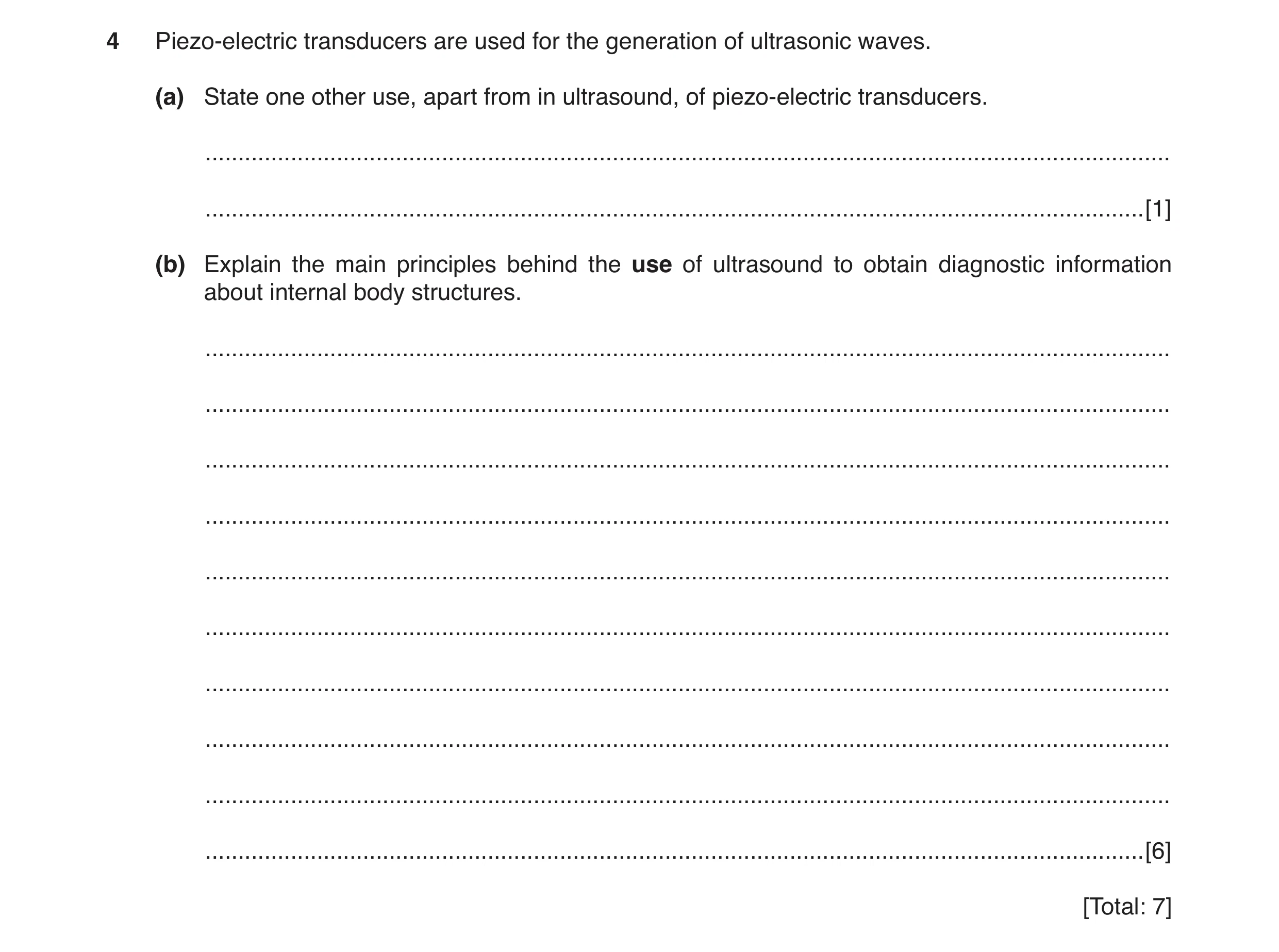
4 Piezo-electric transducers are used for the generation of ultrasonic waves. (a) State one other use, apart from in ultrasound, of piezo-electric transducers. … … [1] (b) Explain the main principles behind the use of ultrasound to obtain diagnostic information about internal body structures. … … … … … … … … … … [6] [Total: 7]
7 marks
Mark scheme: 4(a) e.g. microphone weighing scales/pressure sensor lighters/spark generation watches/clocks/regulation of time B1 4(b) pulses (of ultrasound) B1 reflected at boundaries (between media) B1 (reflected pulses) detected by (ultrasound) generator B1 Any three from: • time delay (between transmission and receipt) gives information about depth (of boundary) • intensity of reflected pulse gives information about (nature of) boundary • gel used to minimise reflection at skin/maximise transmission into skin • degree of reflection depends upon impedances of two media (at boundary) B3
This question in 9702/41 May/June 2018
Q8 · A geostationary satellite orbits the Earth with a period of 24 hours 9702/41 May/June 2018
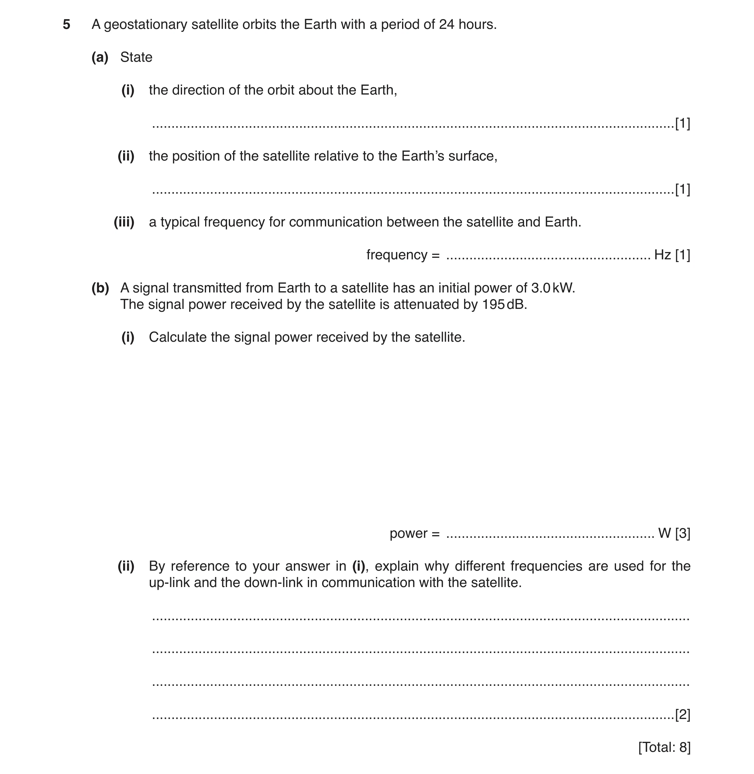
5 A geostationary satellite orbits the Earth with a period of 24 hours. (a) State (i) the direction of the orbit about the Earth, … [1] (ii) the position of the satellite relative to the Earth’s surface, … [1] (iii) a typical frequency for communication between the satellite and Earth. frequency = … Hz [1] (b) A signal transmitted from Earth to a satellite has an initial power of 3.0 kW. The signal power received by the satellite is attenuated by 195 dB. (i) Calculate the signal power received by the satellite. power = … W [3] (ii) By reference to your answer in (i), explain why different frequencies are used for the up-link and the down-link in communication with the satellite. … … … … [2] [Total: 8]
8 marks
Mark scheme: 5(a)(i) west to east B1 5(a)(ii) above the Equator B1 5(a)(iii) value in range (1–300) × 109 Hz A1 5(b)(i) gain / dB = 10 lg (P2 / P1) C1 –195 = 10 lg (P / 3000) or 195 = 10 lg (3000 / P) C1 power = 9.5 × 10⁻¹⁷ W A1 5(b)(ii) up-link has been (greatly) attenuated (before reaching satellite) or down-link signal must be (greatly) amplified (before transmission back to Earth) or up-link has (much) smaller intensity/power than down-link B1 (different frequency) prevents down-link (signal) swamping up-link (signal) B1
This question in 9702/41 May/June 2018
Q9 · Piezo-electric transducers are used for the generation of ultrasonic waves 9702/43 May/June 2018
4 Piezo-electric transducers are used for the generation of ultrasonic waves. (a) State one other use, apart from in ultrasound, of piezo-electric transducers. … … [1] (b) Explain the main principles behind the use of ultrasound to obtain diagnostic information about internal body structures. … … … … … … … … … … [6] [Total: 7]
7 marks
Mark scheme: 4(a) e.g. microphone weighing scales/pressure sensor lighters/spark generation watches/clocks/regulation of time B1 4(b) pulses (of ultrasound) B1 reflected at boundaries (between media) B1 (reflected pulses) detected by (ultrasound) generator B1 Any three from: • time delay (between transmission and receipt) gives information about depth (of boundary) • intensity of reflected pulse gives information about (nature of) boundary • gel used to minimise reflection at skin/maximise transmission into skin • degree of reflection depends upon impedances of two media (at boundary) B3
This question in 9702/43 May/June 2018
Q10 · A geostationary satellite orbits the Earth with a period of 24 hours 9702/43 May/June 2018
5 A geostationary satellite orbits the Earth with a period of 24 hours. (a) State (i) the direction of the orbit about the Earth, … [1] (ii) the position of the satellite relative to the Earth’s surface, … [1] (iii) a typical frequency for communication between the satellite and Earth. frequency = … Hz [1] (b) A signal transmitted from Earth to a satellite has an initial power of 3.0 kW. The signal power received by the satellite is attenuated by 195 dB. (i) Calculate the signal power received by the satellite. power = … W [3] (ii) By reference to your answer in (i), explain why different frequencies are used for the up-link and the down-link in communication with the satellite. … … … … [2] [Total: 8]
8 marks
Mark scheme: 5(a)(i) west to east B1 5(a)(ii) above the Equator B1 5(a)(iii) value in range (1–300) × 109 Hz A1 5(b)(i) gain / dB = 10 lg (P2 / P1) C1 –195 = 10 lg (P / 3000) or 195 = 10 lg (3000 / P) C1 power = 9.5 × 10⁻¹⁷ W A1 5(b)(ii) up-link has been (greatly) attenuated (before reaching satellite) or down-link signal must be (greatly) amplified (before transmission back to Earth) or up-link has (much) smaller intensity/power than down-link B1 (different frequency) prevents down-link (signal) swamping up-link (signal) B1
This question in 9702/43 May/June 2018
Q11 · Explain the main principles behind the use of ultrasound to obtain diagnostic information… 9702/41 Oct/Nov 2018
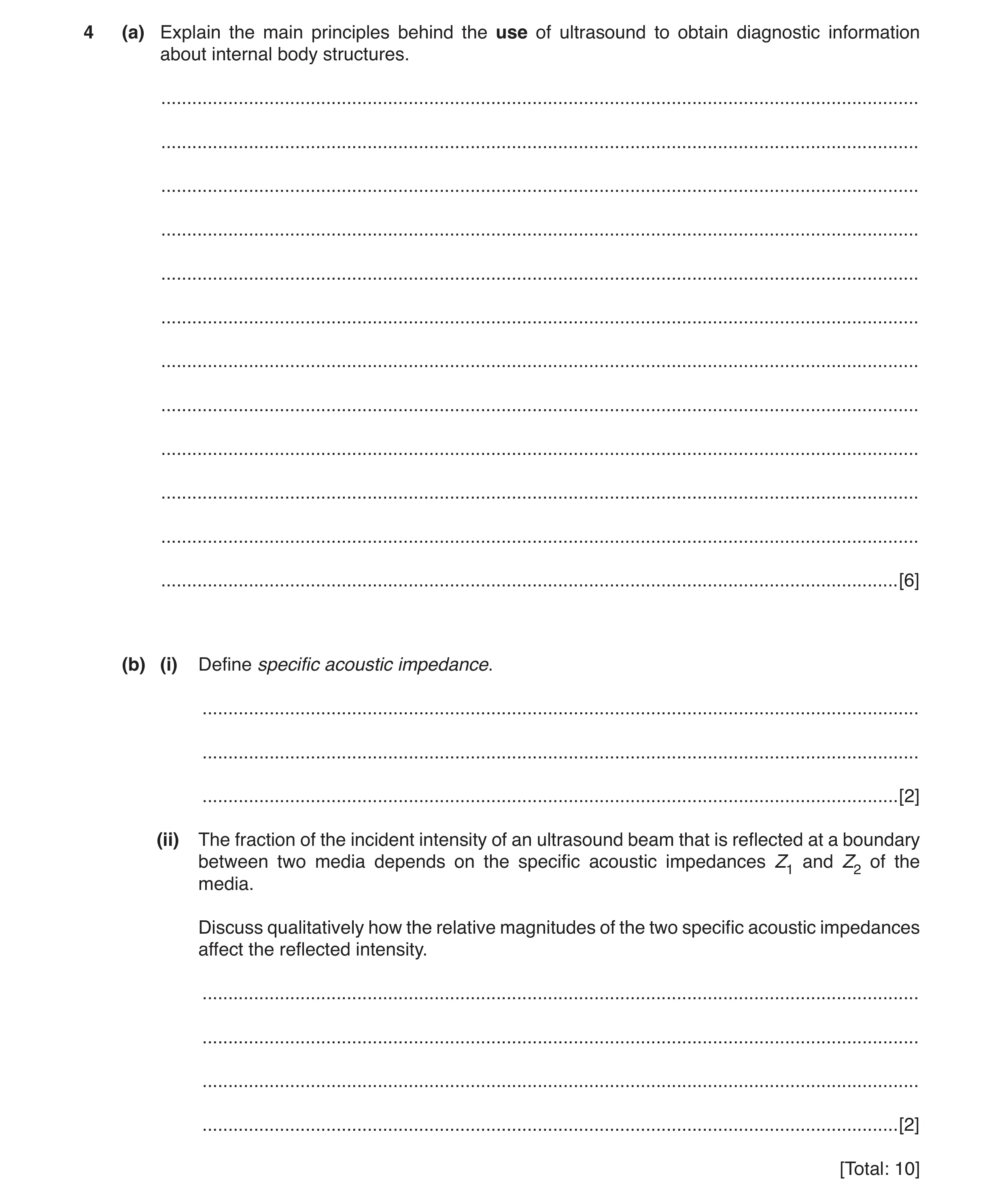
4 (a) Explain the main principles behind the use of ultrasound to obtain diagnostic information about internal body structures. … … … … … … … … … … … … [6] (b) (i) Define specific acoustic impedance. … … … [2] (ii) The fraction of the incident intensity of an ultrasound beam that is reflected at a boundary between two media depends on the specific acoustic impedances Z1 and Z2 of the media. Discuss qualitatively how the relative magnitudes of the two specific acoustic impedances affect the reflected intensity. … … … … [2] [Total: 10]
10 marks
Mark scheme: 4(a) pulses (of ultrasound from generator) B1 reflected at boundaries (between media) B1 time delay (between transmission and receipt) gives information about depth B1 intensity of reflected pulse gives information about nature (of tissues)/type (of tissues)/boundary B1 Any two from: • (reflected pulses) detected by the (ultrasound) generator • gel used to minimise reflection at skin/maximise transmission into skin • degree of reflection depends upon impedances of two media (at boundary) B2 4(b)(i) product of density and speed M1 speed of ultrasound in medium A1 4(b)(ii) Z1 about equal to Z2 results in negligible/no reflection B1 Z1 ≫ Z2 (or Z1 ≪ Z2) results in mostly reflection B1
This question in 9702/41 Oct/Nov 2018
Q12 · Explain the main principles behind the use of ultrasound to obtain diagnostic information… 9702/43 Oct/Nov 2018
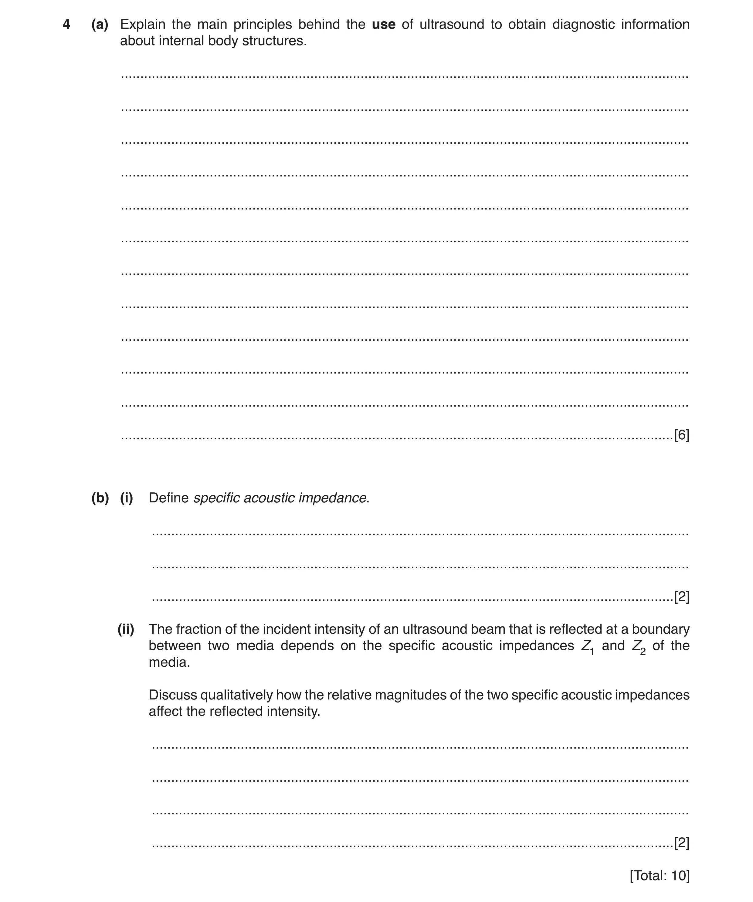
4 (a) Explain the main principles behind the use of ultrasound to obtain diagnostic information about internal body structures. … … … … … … … … … … … … [6] (b) (i) Define specific acoustic impedance. … … … [2] (ii) The fraction of the incident intensity of an ultrasound beam that is reflected at a boundary between two media depends on the specific acoustic impedances Z1 and Z2 of the media. Discuss qualitatively how the relative magnitudes of the two specific acoustic impedances affect the reflected intensity. … … … … [2] [Total: 10]
10 marks
Mark scheme: 4(a) pulses (of ultrasound from generator) B1 reflected at boundaries (between media) B1 time delay (between transmission and receipt) gives information about depth B1 intensity of reflected pulse gives information about nature (of tissues)/type (of tissues)/boundary B1 Any two from: • (reflected pulses) detected by the (ultrasound) generator • gel used to minimise reflection at skin/maximise transmission into skin • degree of reflection depends upon impedances of two media (at boundary) B2 4(b)(i) product of density and speed M1 speed of ultrasound in medium A1 4(b)(ii) Z1 about equal to Z2 results in negligible/no reflection B1 Z1 ≫ Z2 (or Z1 ≪ Z2) results in mostly reflection B1
This question in 9702/43 Oct/Nov 2018
Q13 · State what is meant by the specific acoustic impedance of a medium 9702/42 May/June 2019
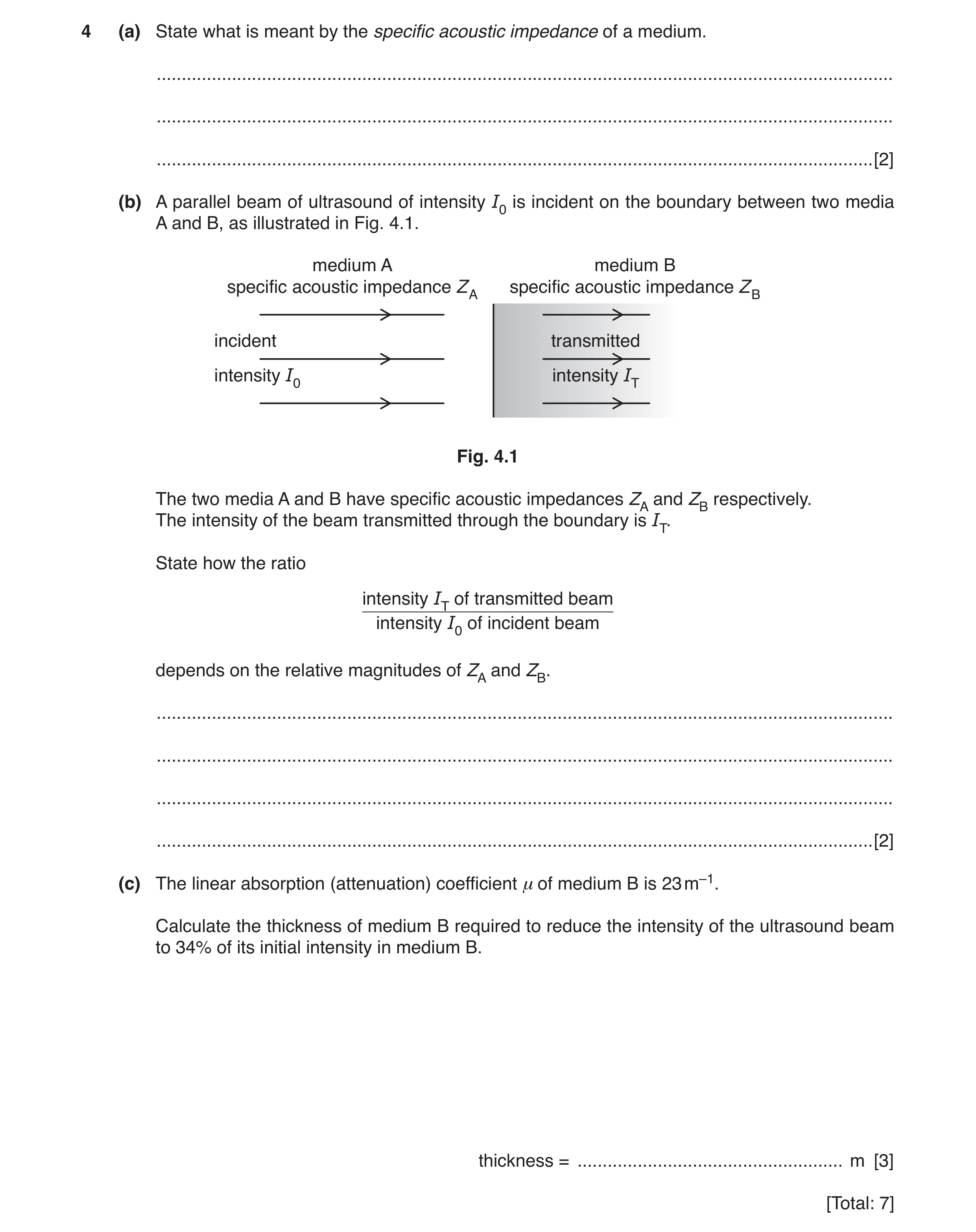
4 (a) State what is meant by the specific acoustic impedance of a medium. … … … [2] (b) A parallel beam of ultrasound of intensity I0 is incident on the boundary between two media A and B, as illustrated in Fig. 4.1. medium A medium B specific acoustic impedance ZA specific acoustic impedance ZB incident transmitted intensity I0 intensity IT Fig. 4.1 The two media A and B have specific acoustic impedances ZA and ZB respectively. The intensity of the beam transmitted through the boundary is IT. State how the ratio intensity IT of transmitted beam intensity I0 of incident beam depends on the relative magnitudes of ZA and ZB. … … … … [2] (c) The linear absorption (attenuation) coefficient μ of medium B is 23 m–1. Calculate the thickness of medium B required to reduce the intensity of the ultrasound beam to 34% of its initial intensity in medium B. thickness = … m [3] [Total: 7]
7 marks
Mark scheme: 4(a) product of density and speed M1 speed of sound in medium A1 4(b) Any two from: • if ZA ≫ ZB then ratio is (nearly) zero or if ZB ≫ ZA then ratio is (nearly) zero or if ZB and ZA are very different then ratio is (nearly) zero or the greater the difference the lower the ratio • if ZA ≈ ZB then ratio is (nearly) 1 or if ZA = ZB then ratio is 1 or the smaller the difference the closer the ratio to 1 (not ‘large’) • IT / I0 = 1 – [(ZA – ZB)2 / (ZA + ZB)2] B2 4(c) I = I0e–µx C1 0.34 = exp(–23 × x) C1 x = 0.047 m A1
This question in 9702/42 May/June 2019
Q14 · State what is meant by the specific acoustic impedance of a medium 9702/42 Oct/Nov 2019
5 (a) (i) State what is meant by the specific acoustic impedance of a medium. … … … [2] (ii) The density of a sample of bone is 1.8 g cm–3 and the speed of ultrasound in the bone is 4.1 × 10³ m s–1. Calculate the specific acoustic impedance ZB of the sample of bone. ZB = … kg m–2 s–1 [1] (b) A parallel beam of ultrasound passes normally through a layer of fat and of muscle, as illustrated in Fig. 5.1. fat muscle beam transmitted beam of ultrasound of ultrasound 0.45 cm 2.1 cm Fig. 5.1 (not to scale) The fat has thickness 0.45 cm and the muscle has thickness 2.1 cm. Data for fat and for muscle are given in Fig. 5.2. specific acoustic impedance linear attenuation (absorption) Z / 106 kg m–2 s–1 coefficient μ/ cm–1 fat 1.3 0.24 muscle 1.7 0.23 Fig. 5.2 The intensity reflection coefficient α at a boundary between two media of specific acoustic impedances Z1 and Z2 is given by the expression - (Z 2 Z 1) 2 α = . + (Z 2 Z 1) 2 Calculate the fraction of the intensity of the ultrasound that is transmitted through the boundary between the fat and the muscle. fraction transmitted = … [1] (c) (i) State what is meant by attenuation of an ultrasound wave. … … … [2] (ii) Data for linear attenuation coefficients are given in Fig. 5.2. Determine the ratio intensity of ultrasound transmitted through the medium intensity of ultrasound entering the medium for: 1. the layer of fat of thickness 0.45 cm ratio = … 2. the layer of muscle of thickness 2.1 cm. ratio = … [3] (d) Use your answers in (b) and (c)(ii) to determine the fraction of the intensity entering the layer of fat that is transmitted through the layer of muscle. fraction transmitted = … [1] [Total: 10]
10 marks
Mark scheme: 5(a)(i) product of density and speed M1 speed of sound in the medium A1 5(a)(ii) ZB = 1.8 × 103 × 4.1 × 103 = 7.4 × 10⁶ kg m2 s–1 A1 5(b) α = (1.7 – 1.3)2 / (1.7 + 1.3)2 = 0.018 fraction = 0.98 A1 5(c)(i) reduction in power/intensity (of wave) M1 as the wave passes through the medium A1 5(c)(ii) 1. ratio = e–µx C1 = 0.90 A1 2. ratio = 0.62 A1 5(d) fraction = 0.898 × 0.617 × 0.98 = 0.54 A1
This question in 9702/42 Oct/Nov 2019
Q15 · Explain why ultrasound used in medical diagnosis is emitted in pulses 9702/42 Feb/March 2020
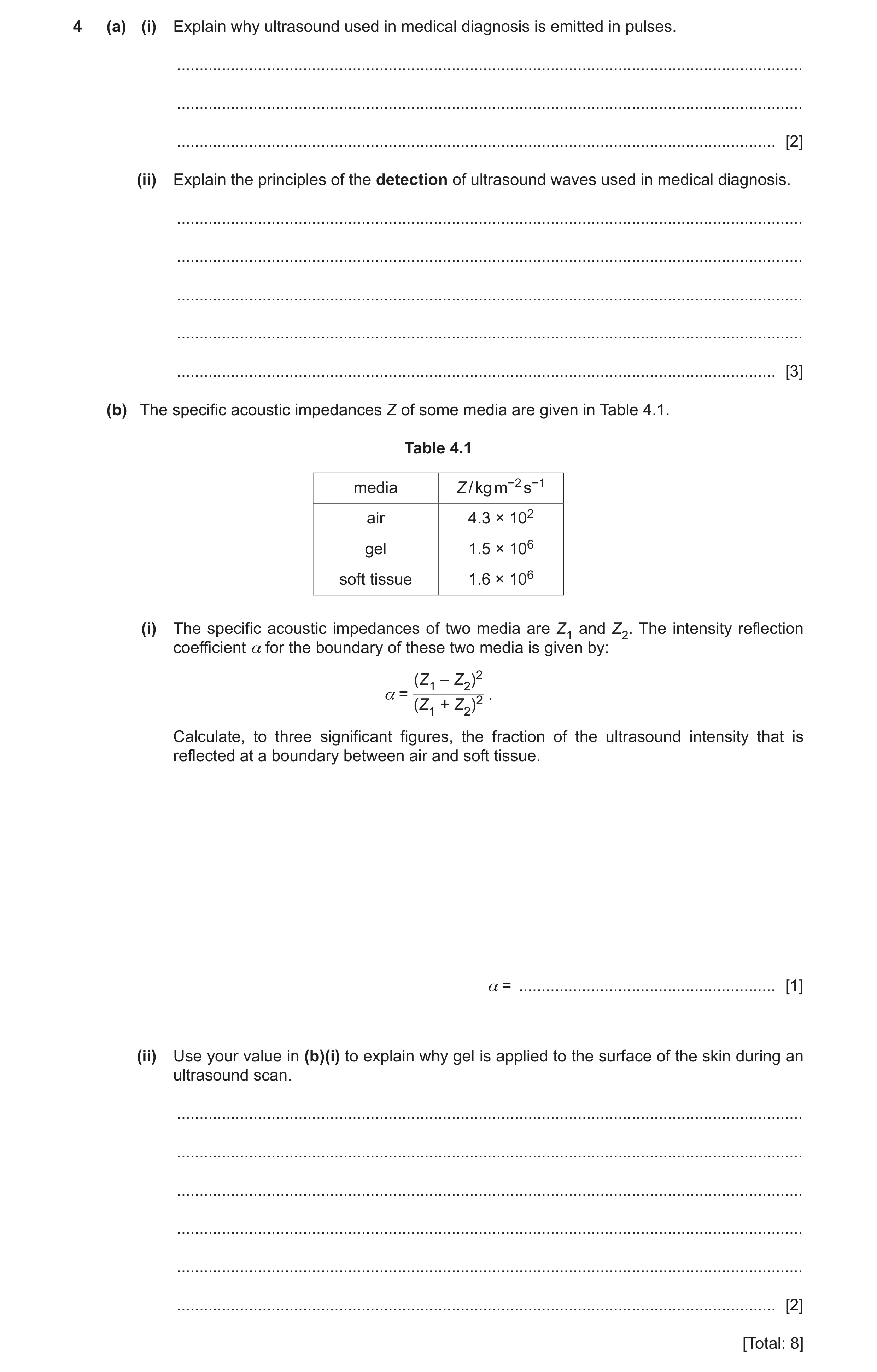
4 (a) (i) Explain why ultrasound used in medical diagnosis is emitted in pulses. … … … [2] (ii) Explain the principles of the detection of ultrasound waves used in medical diagnosis. … … … … … [3] (b) The specific acoustic impedances Z of some media are given in Table 4.1. Table 4.1 media Z / kg m−2 s−1 air 4.3 × 10² gel 1.5 × 10⁶ soft tissue 1.6 × 10⁶ (i) The specific acoustic impedances of two media are Z1 and Z2. The intensity reflection coefficient α for the boundary of these two media is given by: (Z1 – Z2)2 α = . (Z1 + Z2)2 Calculate, to three significant figures, the fraction of the ultrasound intensity that is reflected at a boundary between air and soft tissue. α = … [1] (ii) Use your value in (b)(i) to explain why gel is applied to the surface of the skin during an ultrasound scan. … … … … … … [2] [Total: 8]
8 marks
Mark scheme: 4(a)(i) Any 2 from: • allows the reflected signal to be distinguished from the emitted signal • detection occurs in the time between emitted pulses • (reflection of ultrasound) detected by same probe / transducer / crystal • cannot emit and detect at same time (hence pulses) B2 4(a)(ii) piezo-electric crystal B1 ultrasound makes crystal vibrate / resonate B1 vibration produces (alternating) e.m.f. / p.d. across crystal B1 4(b)(i) = (1.6 × 10⁶ – 4.3 × 10²)2 / (1.6 × 10⁶ + 4.3 × 10²)2 = 0.999 B1 4(b)(ii) without the gel most of the ultrasound is reflected B1 Z values more similar / α reduces so less (ultrasound) is reflected / more (ultrasound) is transmitted B1
This question in 9702/42 Feb/March 2020
Q16 · By reference to an ultrasound wave, explain what is meant by specific acoustic impedance 9702/41 May/June 2020
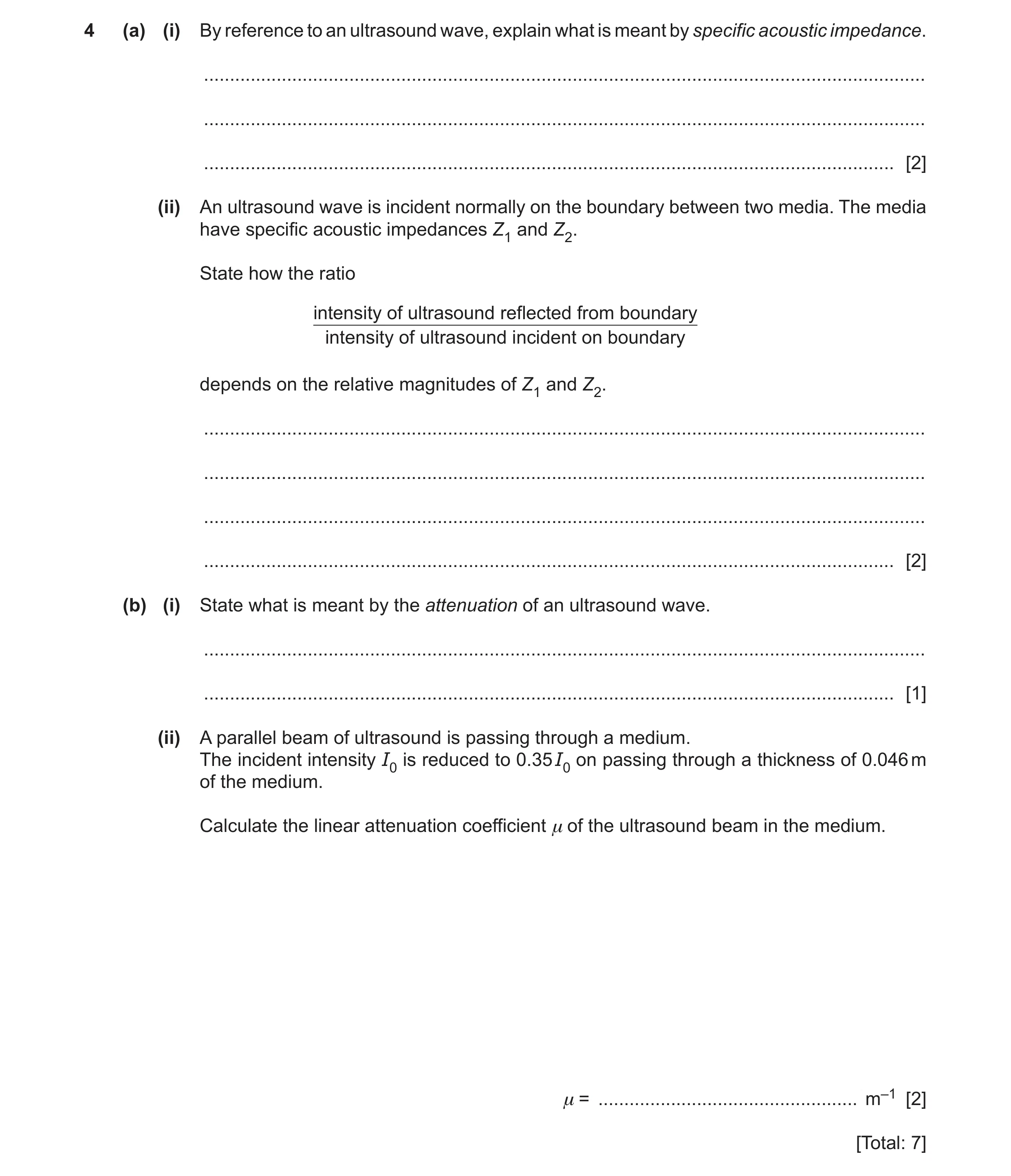
4 (a) (i) By reference to an ultrasound wave, explain what is meant by specific acoustic impedance. … … … [2] (ii) An ultrasound wave is incident normally on the boundary between two media. The media have specific acoustic impedances Z1 and Z2. State how the ratio intensity of ultrasound reflected from boundary intensity of ultrasound incident on boundary depends on the relative magnitudes of Z1 and Z2. … … … … [2] (b) (i) State what is meant by the attenuation of an ultrasound wave. … … [1] (ii) A parallel beam of ultrasound is passing through a medium. The incident intensity I0 is reduced to 0.35 I0 on passing through a thickness of 0.046 m of the medium. Calculate the linear attenuation coefficient μ of the ultrasound beam in the medium. μ = … m–1 [2] [Total: 7]
7 marks
Mark scheme: 4(a)(i) product of density and speed M1 speed of ultrasound in medium A1 4(a)(ii) the greater the difference between Z1 and Z2, the closer the ratio is to 1 or if difference between Z1 and Z2 large, ratio is close to 1 B1 the closer together Z1 and Z2, the closer the ratio is to 0 or if difference between Z1 and Z2 small, ratio close to 0 B1 4(b)(i) loss of intensity/amplitude/power (of the wave) B1 4(b)(ii) I = I0 e–μx C1 0.35 = e–0.046μ μ = 23 m–1 A1
This question in 9702/41 May/June 2020
Q17 · Explain the principles of the detection of ultrasound waves for medical diagnosis 9702/42 May/June 2020
5 (a) Explain the principles of the detection of ultrasound waves for medical diagnosis. … … … … … … … … [4] (b) By reference to specific acoustic impedance, explain why there is very little transmission of ultrasound waves from air into skin. … … … … … … [3] [Total: 7]
7 marks
Mark scheme: 5(a) pulses of ultrasound B1 ultrasound incident on quartz crystal B1 waves make crystal oscillate B1 oscillations (of crystal) generates an e.m.f. (across the crystal) B1 5(b) specific acoustic impedances of air and skin are very different B1 intensity reflection coefficient depends on difference between acoustic impedance B1 most ultrasound reflected so little transmission B1
This question in 9702/42 May/June 2020
Q18 · By reference to an ultrasound wave, explain what is meant by specific acoustic impedance 9702/43 May/June 2020
4 (a) (i) By reference to an ultrasound wave, explain what is meant by specific acoustic impedance. … … … [2] (ii) An ultrasound wave is incident normally on the boundary between two media. The media have specific acoustic impedances Z1 and Z2. State how the ratio intensity of ultrasound reflected from boundary intensity of ultrasound incident on boundary depends on the relative magnitudes of Z1 and Z2. … … … … [2] (b) (i) State what is meant by the attenuation of an ultrasound wave. … … [1] (ii) A parallel beam of ultrasound is passing through a medium. The incident intensity I0 is reduced to 0.35 I0 on passing through a thickness of 0.046 m of the medium. Calculate the linear attenuation coefficient μ of the ultrasound beam in the medium. μ = … m–1 [2] [Total: 7]
7 marks
Mark scheme: 4(a)(i) product of density and speed M1 speed of ultrasound in medium A1 4(a)(ii) the greater the difference between Z1 and Z2, the closer the ratio is to 1 or if difference between Z1 and Z2 large, ratio is close to 1 B1 the closer together Z1 and Z2, the closer the ratio is to 0 or if difference between Z1 and Z2 small, ratio close to 0 B1 4(b)(i) loss of intensity/amplitude/power (of the wave) B1 4(b)(ii) I = I0 e–μx C1 0.35 = e–0.046μ μ = 23 m–1 A1
This question in 9702/43 May/June 2020
Q19 · Explain the principles of the generation of ultrasound waves for use in medical diagnosis 9702/41 Oct/Nov 2020
4 (a) Explain the principles of the generation of ultrasound waves for use in medical diagnosis. … … … … … … … … [4] (b) The linear attenuation (absorption) coefficient for a parallel beam of ultrasound waves in air is 1.2 cm–1. The parallel beam passes through a layer of air of thickness 3.5 cm. Calculate the ratio, in dB, intensity of beam after passing through the layer of air . intensity of beam entering the layer of air ratio = … dB [4] [Total: 8]
8 marks
Mark scheme: 4(a) quartz crystal B1 alternating p.d. across crystal causes it to vibrate B1 resonance occurs when frequency of p.d. matches natural frequency of crystal B1 natural frequency of crystal is in ultrasound range B1 4(b) I = I0 e–μx C1 I / I0 = e–1.2 × 3.5 = 0.015 C1 ratio / dB = –10 lg (1 / 0.015) or 10 lg (0.015) C1 = –18 dB A1
This question in 9702/41 Oct/Nov 2020
Q20 · Explain the principles of the generation of ultrasound waves for use in medical diagnosis 9702/43 Oct/Nov 2020
4 (a) Explain the principles of the generation of ultrasound waves for use in medical diagnosis. … … … … … … … … [4] (b) The linear attenuation (absorption) coefficient for a parallel beam of ultrasound waves in air is 1.2 cm–1. The parallel beam passes through a layer of air of thickness 3.5 cm. Calculate the ratio, in dB, intensity of beam after passing through the layer of air . intensity of beam entering the layer of air ratio = … dB [4] [Total: 8]
8 marks
Mark scheme: 4(a) quartz crystal B1 alternating p.d. across crystal causes it to vibrate B1 resonance occurs when frequency of p.d. matches natural frequency of crystal B1 natural frequency of crystal is in ultrasound range B1 4(b) I = I0 e–μx C1 I / I0 = e–1.2 × 3.5 = 0.015 C1 ratio / dB = –10 lg (1 / 0.015) or 10 lg (0.015) C1 = –18 dB A1
This question in 9702/43 Oct/Nov 2020
Q21 · Outline the use of ultrasound to obtain diagnostic information about internal body… 9702/41 May/June 2021
4 Outline the use of ultrasound to obtain diagnostic information about internal body structures. … … … … … … … … … … [5]
5 marks
Mark scheme: 4 (ultrasound) pulse B1 reflected at boundaries B1 gel is used to minimise reflection at skin or generated and detected by quartz crystal B1 time delay between generation and detection gives information about depth B1 intensity (of reflected wave) gives information about nature of boundary B1
This question in 9702/41 May/June 2021
Q22 · Outline the use of ultrasound to obtain diagnostic information about internal body… 9702/43 May/June 2021
4 Outline the use of ultrasound to obtain diagnostic information about internal body structures. … … … … … … … … … … [5]
5 marks
Mark scheme: 4 (ultrasound) pulse B1 reflected at boundaries B1 gel is used to minimise reflection at skin or generated and detected by quartz crystal B1 time delay between generation and detection gives information about depth B1 intensity (of reflected wave) gives information about nature of boundary B1
This question in 9702/43 May/June 2021
Q23 · A piezoelectric transducer containing a quartz crystal is used to obtain diagnostic… 9702/42 Oct/Nov 2021
11 (a) A piezoelectric transducer containing a quartz crystal is used to obtain diagnostic information about internal structures. Describe the function of the quartz crystal. … … … … [3] (b) (i) Define specific acoustic impedance. … … … [2] (ii) Describe, qualitatively, how the specific acoustic impedances of two materials affect the intensity reflection coefficient at a boundary between the materials. … … … … [2] [Total: 7]
7 marks
Mark scheme: 11(a) generates ultrasound B1 detects reflected ultrasound B1 applied p.d. causes crystal to vibrate or vibrations cause crystal to generate an e.m.f. B1 11(b)(i) product of density and speed M1 speed of ultrasound in medium A1 11(b)(ii) difference between (the specific acoustic impedances) C1 • if similar/same then reflection coefficient is zero/very low • if very different then reflection coefficient is (nearly) 1 • the lower the difference means lower the reflection coefficient (any one point) A1
This question in 9702/42 Oct/Nov 2021
Q24 · A sinusoidal alternating voltage has a root-mean-square (r.m.s.) potential difference… 9702/41 Oct/Nov 2022
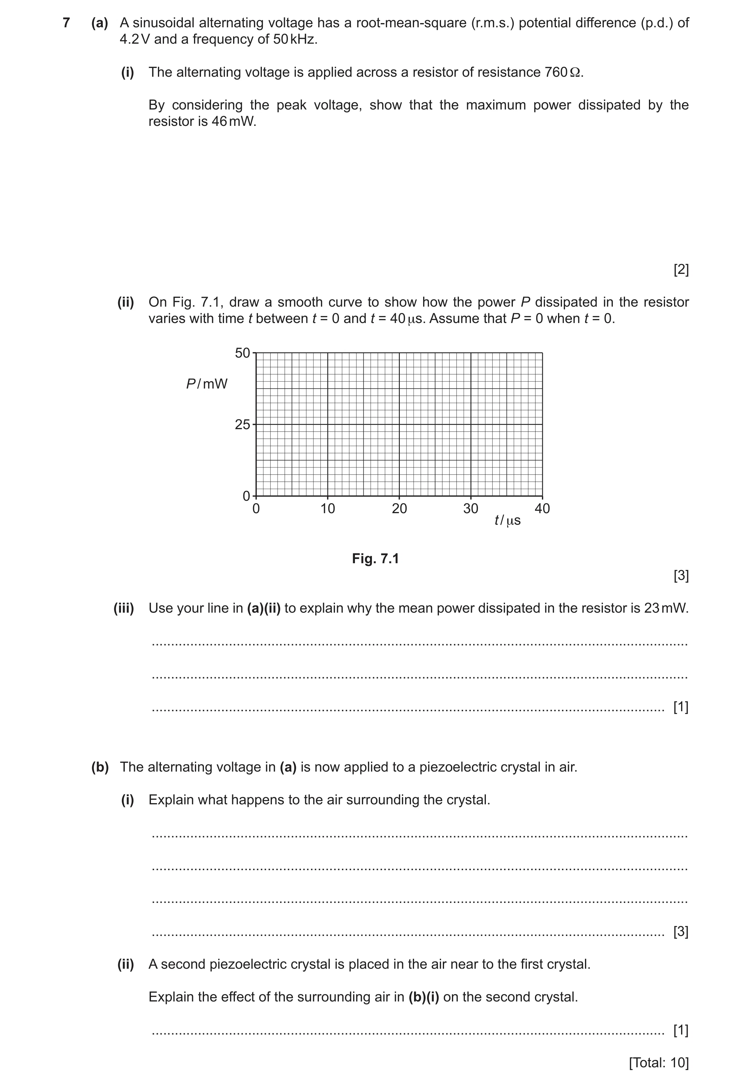
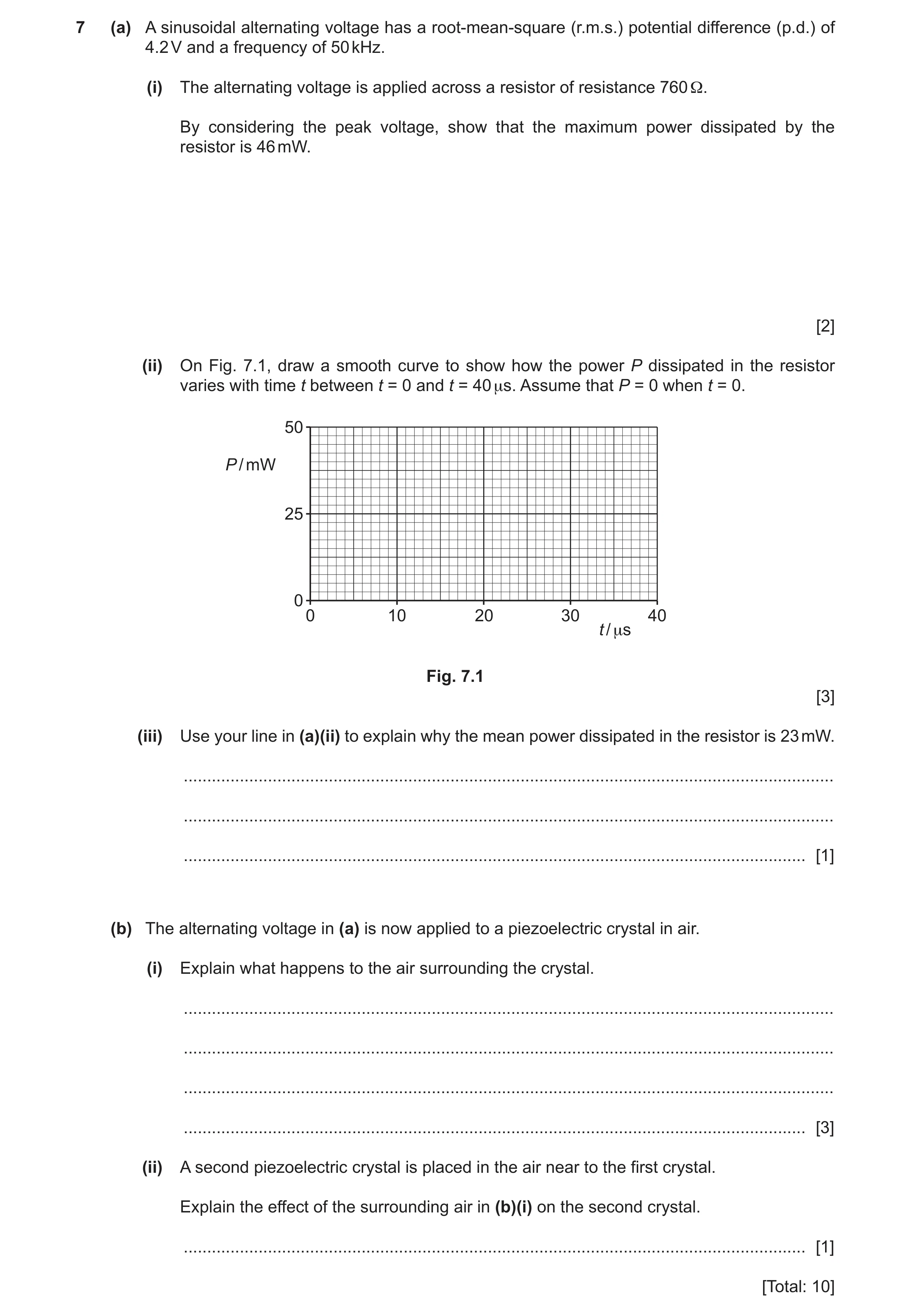
7 (a) A sinusoidal alternating voltage has a root-mean-square (r.m.s.) potential difference (p.d.) of 4.2 V and a frequency of 50 kHz. (i) The alternating voltage is applied across a resistor of resistance 760 Ω. By considering the peak voltage, show that the maximum power dissipated by the resistor is 46 mW. [2] (ii) On Fig. 7.1, draw a smooth curve to show how the power P dissipated in the resistor varies with time t between t = 0 and t = 40 μs. Assume that P = 0 when t = 0. 50 P / mW 25 0 0 10 20 30 40 t / μs Fig. 7.1 [3] (iii) Use your line in (a)(ii) to explain why the mean power dissipated in the resistor is 23 mW. … … … [1] (b) The alternating voltage in (a) is now applied to a piezoelectric crystal in air. (i) Explain what happens to the air surrounding the crystal. … … … … [3] (ii) A second piezoelectric crystal is placed in the air near to the first crystal. Explain the effect of the surrounding air in (b)(i) on the second crystal. … [1] [Total: 10]
10 marks
Mark scheme: 7(a)(i) peak voltage = 4.2 2 B1 ( = 5.9 V) power = V2 / R A1 = 5.92 / 760 = 0.046 W or 46 mW 7(a)(ii) sketch shows peak(s) in power at 46 mW B1 correct shape (sinusoidal wave sitting on t-axis) B1 four cycles of repeating pattern shown, with P = 0 at 0, 10, 20, 30, 40 s B1 7(a)(iii) line is symmetrical about 23 mW B1 7(b)(i) (alternating p.d. makes) the crystal vibrate B1 vibrations (of crystal) causes air to vibrate B1 frequency is in ultrasound range B1 7(b)(ii) (air makes) crystal vibrate, which causes an e.m.f. to be generated across the (second) crystal B1
This question in 9702/41 Oct/Nov 2022
Q25 · A sinusoidal alternating voltage has a root-mean-square (r.m.s.) potential difference… 9702/43 Oct/Nov 2022
7 (a) A sinusoidal alternating voltage has a root-mean-square (r.m.s.) potential difference (p.d.) of 4.2 V and a frequency of 50 kHz. (i) The alternating voltage is applied across a resistor of resistance 760 Ω. By considering the peak voltage, show that the maximum power dissipated by the resistor is 46 mW. [2] (ii) On Fig. 7.1, draw a smooth curve to show how the power P dissipated in the resistor varies with time t between t = 0 and t = 40 μs. Assume that P = 0 when t = 0. 50 P / mW 25 0 0 10 20 30 40 t / μs Fig. 7.1 [3] (iii) Use your line in (a)(ii) to explain why the mean power dissipated in the resistor is 23 mW. … … … [1] (b) The alternating voltage in (a) is now applied to a piezoelectric crystal in air. (i) Explain what happens to the air surrounding the crystal. … … … … [3] (ii) A second piezoelectric crystal is placed in the air near to the first crystal. Explain the effect of the surrounding air in (b)(i) on the second crystal. … [1] [Total: 10]
10 marks
Mark scheme: 7(a)(i) peak voltage = 4.2 2 B1 ( = 5.9 V) power = V2 / R A1 = 5.92 / 760 = 0.046 W or 46 mW 7(a)(ii) sketch shows peak(s) in power at 46 mW B1 correct shape (sinusoidal wave sitting on t-axis) B1 four cycles of repeating pattern shown, with P = 0 at 0, 10, 20, 30, 40 s B1 7(a)(iii) line is symmetrical about 23 mW B1 7(b)(i) (alternating p.d. makes) the crystal vibrate B1 vibrations (of crystal) causes air to vibrate B1 frequency is in ultrasound range B1 7(b)(ii) (air makes) crystal vibrate, which causes an e.m.f. to be generated across the (second) crystal B1
This question in 9702/43 Oct/Nov 2022
Q26 · Ultrasound is used to produce diagnostic information about internal body structures 9702/42 Feb/March 2023
9 Ultrasound is used to produce diagnostic information about internal body structures. (a) Explain how ultrasound waves are detected. … … … … [3] (b) An alternating voltage V varies with time t according to V = Vo sin ωt. The voltage is applied to an ultrasound probe. The root-mean-square (r.m.s.) voltage is 66 V. The frequency of the ultrasound generated by the probe is 4.3 MHz. Determine the values of (i) Vo Vo = … V [1] (ii) ω. ω = … rad s–1 [1] (c) Table 9.1 contains information about air and soft tissue. Table 9.1 density / kg m–3 speed of ultrasound specific acoustic / m s–1 impedance / … air 1.30 330 4.3 × 10² soft tissue 1600 1.7 × 10⁶ (i) Determine the unit for the specific acoustic impedance values shown in Table 9.1. [1] (ii) Calculate the density of soft tissue. density = … kg m–3 [1] (iii) Use data from Table 9.1 to explain why ultrasound cannot be used to produce an image inside an air-filled cavity such as the lungs. … … … … [2] [Total: 9]
9 marks
Mark scheme: 9(a) piezo-electric crystal B1 (ultrasound) wave causes shape change / vibrations (of crystal) B1 shape change / vibrations causes e.m.f. (which is detected) B1 9(b)(i) 93 V A1 9(b)(ii) 2.7 107 rad s–1 A1 9(c)(i) kg m–2 s–1 B1 9(c)(ii) = Z / c = 1.7 106 / 1600 A1 = 1100 kg m–3 9(c)(iii) intensity reflection coefficient ≈ 1 or Z1 and Z2 are very different B1 almost no / no ultrasound transmitted (into air filled cavity) B1
This question in 9702/42 Feb/March 2023
Q27 · Some data relating to the properties of air, gel and body tissue 9702/41 May/June 2023
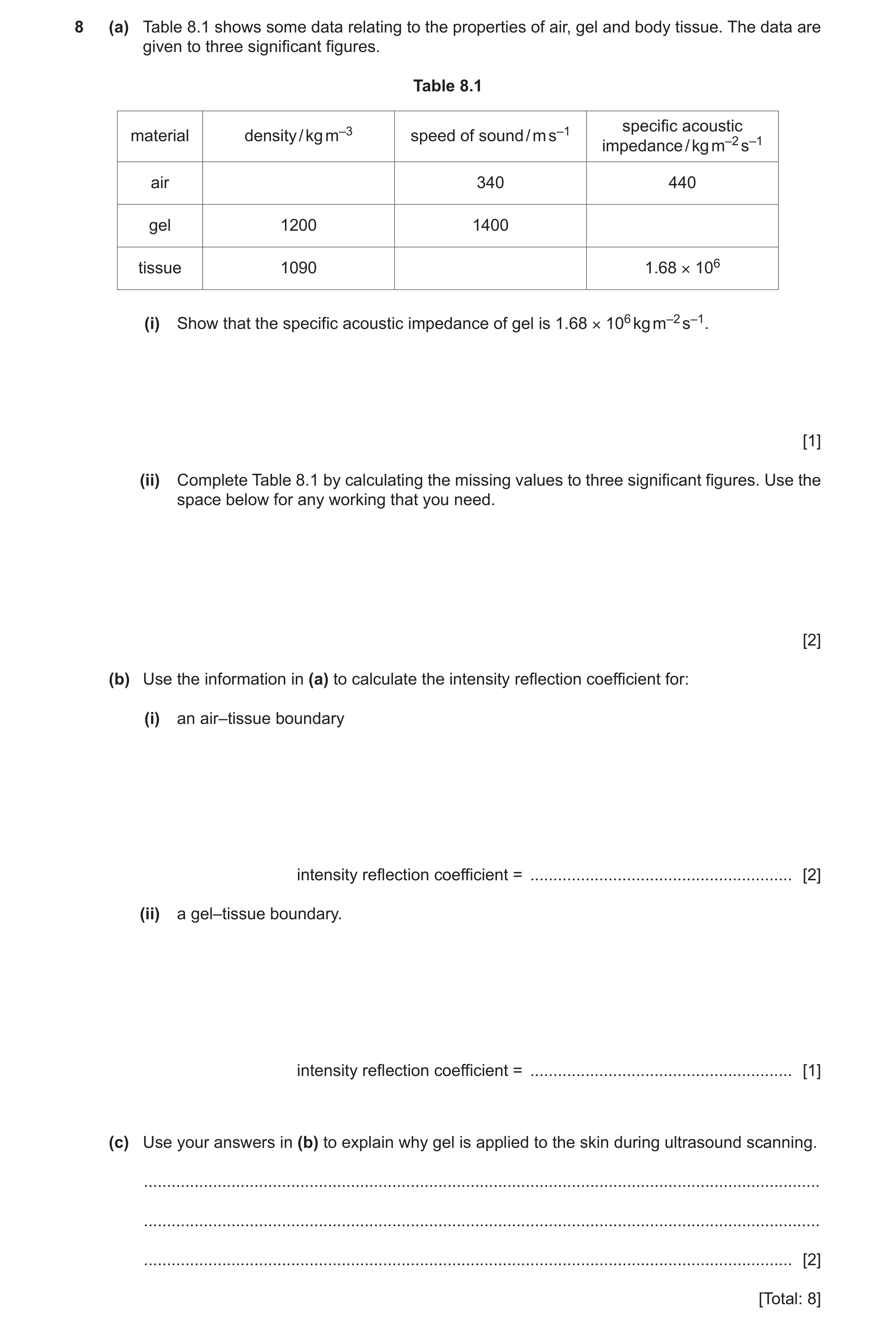
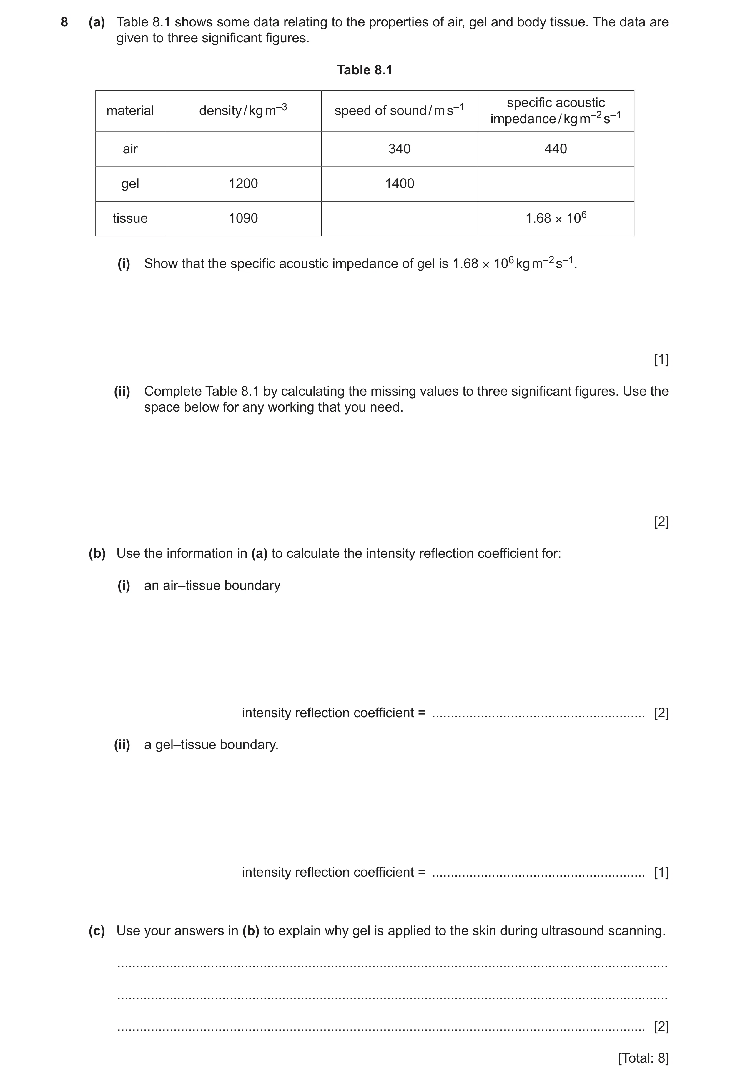
8 (a) Table 8.1 shows some data relating to the properties of air, gel and body tissue. The data are given to three significant figures. Table 8.1 specific acoustic material density / kg m–3 speed of sound / m s–1 impedance / kg m–2 s–1 air 340 440 gel 1200 1400 tissue 1090 1.68 × 10⁶ (i) Show that the specific acoustic impedance of gel is 1.68 × 10⁶ kg m–2 s–1. [1] (ii) Complete Table 8.1 by calculating the missing values to three significant figures. Use the space below for any working that you need. [2] (b) Use the information in (a) to calculate the intensity reflection coefficient for: (i) an air–tissue boundary intensity reflection coefficient = … [2] (ii) a gel–tissue boundary. intensity reflection coefficient = … [1] (c) Use your answers in (b) to explain why gel is applied to the skin during ultrasound scanning. … … … [2] [Total: 8]
8 marks
Mark scheme: 8(a)(i) A1 8(a)(ii) density of air shown in table as 1.29 A1 speed of sound in tissue shown in table as 1540 A1 8(b)(i) intensity reflection coefficient = (Z1 – Z2)2 / (Z1 + Z2)2 = (1680000 – 440)2 / (1680000 + 440)2 C1 = 0.999 A1 8(b)(ii) intensity reflection coefficient = (Z1 – Z2)2 / (Z1 + Z2)2 = (1680000 – 1680000)2 / (1680000 + 1680000)2 = 0 A1 8(c) without gel, (almost) all of the (incident) ultrasound is reflected (from skin) B1 with gel, (almost) all of the (incident) ultrasound is transmitted (into the body) B1
This question in 9702/41 May/June 2023
Q28 · Some data relating to the properties of air, gel and body tissue 9702/43 May/June 2023
8 (a) Table 8.1 shows some data relating to the properties of air, gel and body tissue. The data are given to three significant figures. Table 8.1 specific acoustic material density / kg m–3 speed of sound / m s–1 impedance / kg m–2 s–1 air 340 440 gel 1200 1400 tissue 1090 1.68 × 10⁶ (i) Show that the specific acoustic impedance of gel is 1.68 × 10⁶ kg m–2 s–1. [1] (ii) Complete Table 8.1 by calculating the missing values to three significant figures. Use the space below for any working that you need. [2] (b) Use the information in (a) to calculate the intensity reflection coefficient for: (i) an air–tissue boundary intensity reflection coefficient = … [2] (ii) a gel–tissue boundary. intensity reflection coefficient = … [1] (c) Use your answers in (b) to explain why gel is applied to the skin during ultrasound scanning. … … … [2] [Total: 8]
8 marks
Mark scheme: 8(a)(i) A1 8(a)(ii) density of air shown in table as 1.29 A1 speed of sound in tissue shown in table as 1540 A1 8(b)(i) intensity reflection coefficient = (Z1 – Z2)2 / (Z1 + Z2)2 = (1680000 – 440)2 / (1680000 + 440)2 C1 = 0.999 A1 8(b)(ii) intensity reflection coefficient = (Z1 – Z2)2 / (Z1 + Z2)2 = (1680000 – 1680000)2 / (1680000 + 1680000)2 = 0 A1 8(c) without gel, (almost) all of the (incident) ultrasound is reflected (from skin) B1 with gel, (almost) all of the (incident) ultrasound is transmitted (into the body) B1
This question in 9702/43 May/June 2023
Q29 · Ultrasound and X-rays are both types of wave that are used in medical diagnosis to form… 9702/41 Oct/Nov 2023
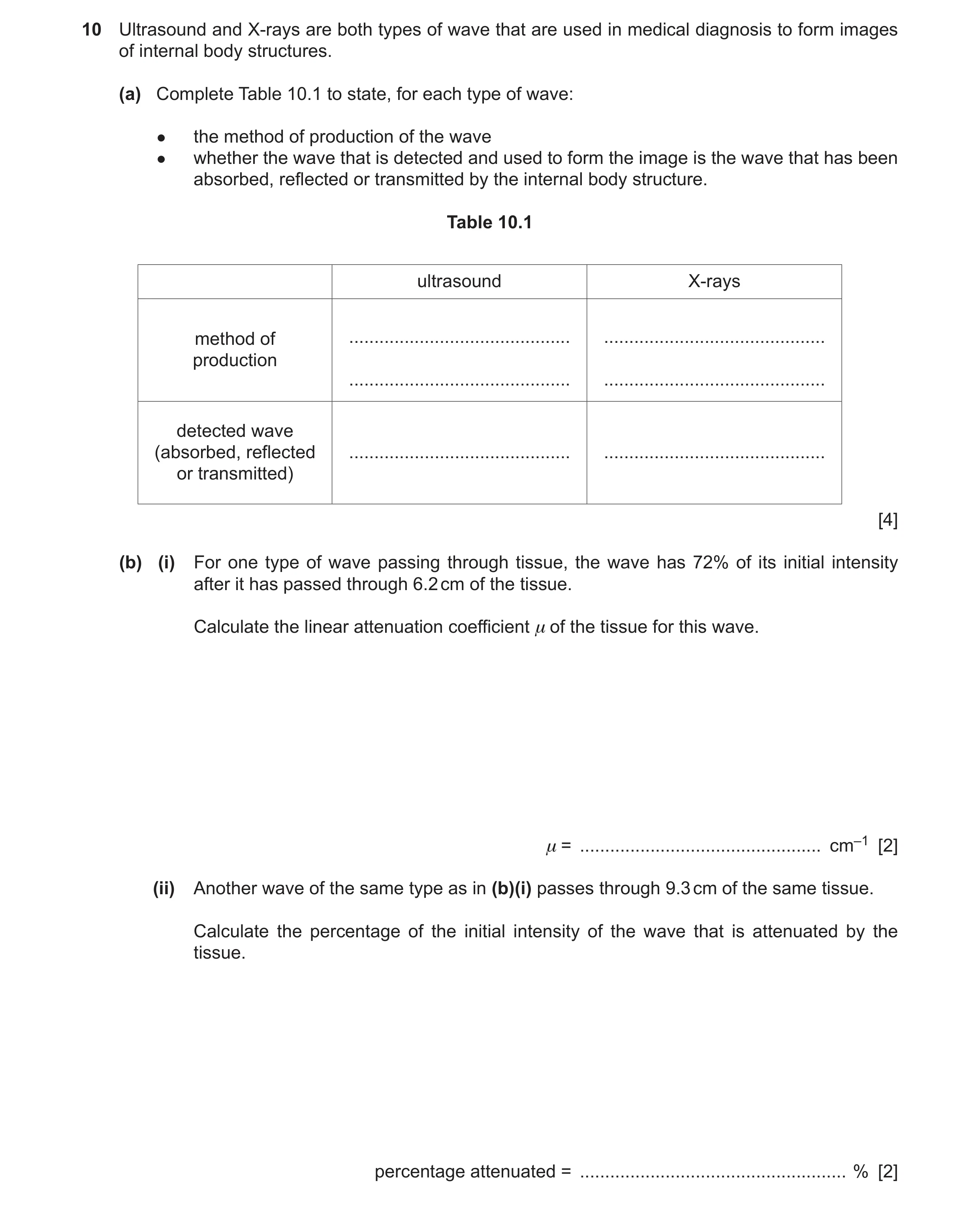
10 Ultrasound and X-rays are both types of wave that are used in medical diagnosis to form images of internal body structures. (a) Complete Table 10.1 to state, for each type of wave: ● the method of production of the wave ● whether the wave that is detected and used to form the image is the wave that has been absorbed, reflected or transmitted by the internal body structure. Table 10.1 ultrasound X-rays method of … … production … … detected wave (absorbed, reflected … … or transmitted) [4] (b) (i) For one type of wave passing through tissue, the wave has 72% of its initial intensity after it has passed through 6.2 cm of the tissue. Calculate the linear attenuation coefficient μ of the tissue for this wave. μ = … cm–1 [2] (ii) Another wave of the same type as in (b)(i) passes through 9.3 cm of the same tissue. Calculate the percentage of the initial intensity of the wave that is attenuated by the tissue. percentage attenuated = … % [2]
8 marks
Mark scheme: 10(a) ultrasound production: vibrating quartz crystal B1 X-ray production: electrons hitting metal target B1 ultrasound detected wave: reflected B1 X-ray detected wave: transmitted B1 10(b)(i) I = I0 exp (–x) C1 ln (0.72) = –6.2 A1 = 0.053 cm–1 10(b)(ii) I / I0 = exp (–9.3 0.053) C1 ( = 0.61) percentage attenuated = 100 (1.00 – 0.61) A1 = 39%
This question in 9702/41 Oct/Nov 2023
Q30 · Ultrasound and X-rays are both types of wave that are used in medical diagnosis to form… 9702/43 Oct/Nov 2023
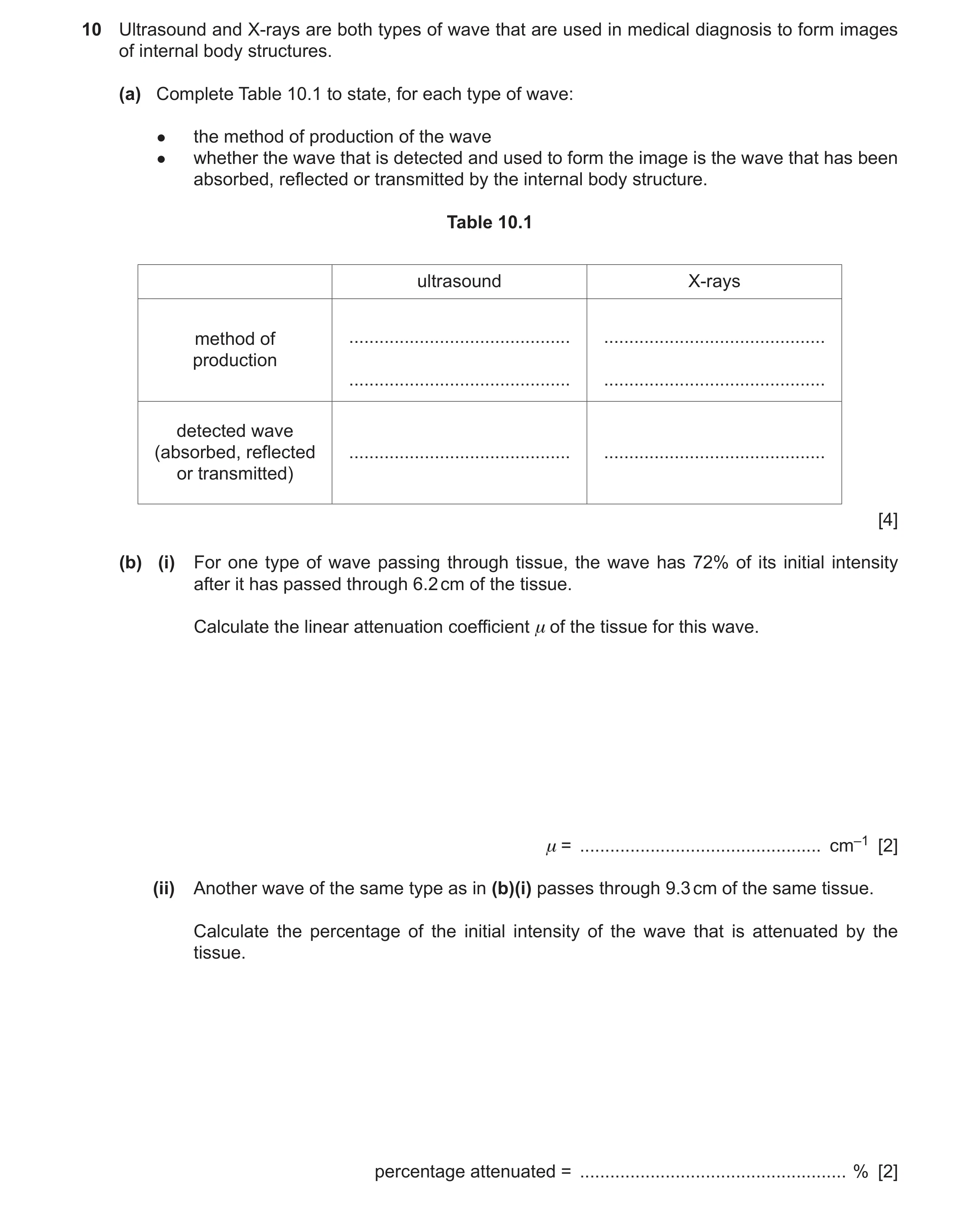
10 Ultrasound and X-rays are both types of wave that are used in medical diagnosis to form images of internal body structures. (a) Complete Table 10.1 to state, for each type of wave: ● the method of production of the wave ● whether the wave that is detected and used to form the image is the wave that has been absorbed, reflected or transmitted by the internal body structure. Table 10.1 ultrasound X-rays method of … … production … … detected wave (absorbed, reflected … … or transmitted) [4] (b) (i) For one type of wave passing through tissue, the wave has 72% of its initial intensity after it has passed through 6.2 cm of the tissue. Calculate the linear attenuation coefficient μ of the tissue for this wave. μ = … cm–1 [2] (ii) Another wave of the same type as in (b)(i) passes through 9.3 cm of the same tissue. Calculate the percentage of the initial intensity of the wave that is attenuated by the tissue. percentage attenuated = … % [2]
8 marks
Mark scheme: 10(a) ultrasound production: vibrating quartz crystal B1 X-ray production: electrons hitting metal target B1 ultrasound detected wave: reflected B1 X-ray detected wave: transmitted B1 10(b)(i) I = I0 exp (–x) C1 ln (0.72) = –6.2 A1 = 0.053 cm–1 10(b)(ii) I / I0 = exp (–9.3 0.053) C1 ( = 0.61) percentage attenuated = 100 (1.00 – 0.61) A1 = 39%
This question in 9702/43 Oct/Nov 2023
Q31 · Describe how reflected ultrasound pulses may be used to obtain diagnostic information… 9702/42 May/June 2024
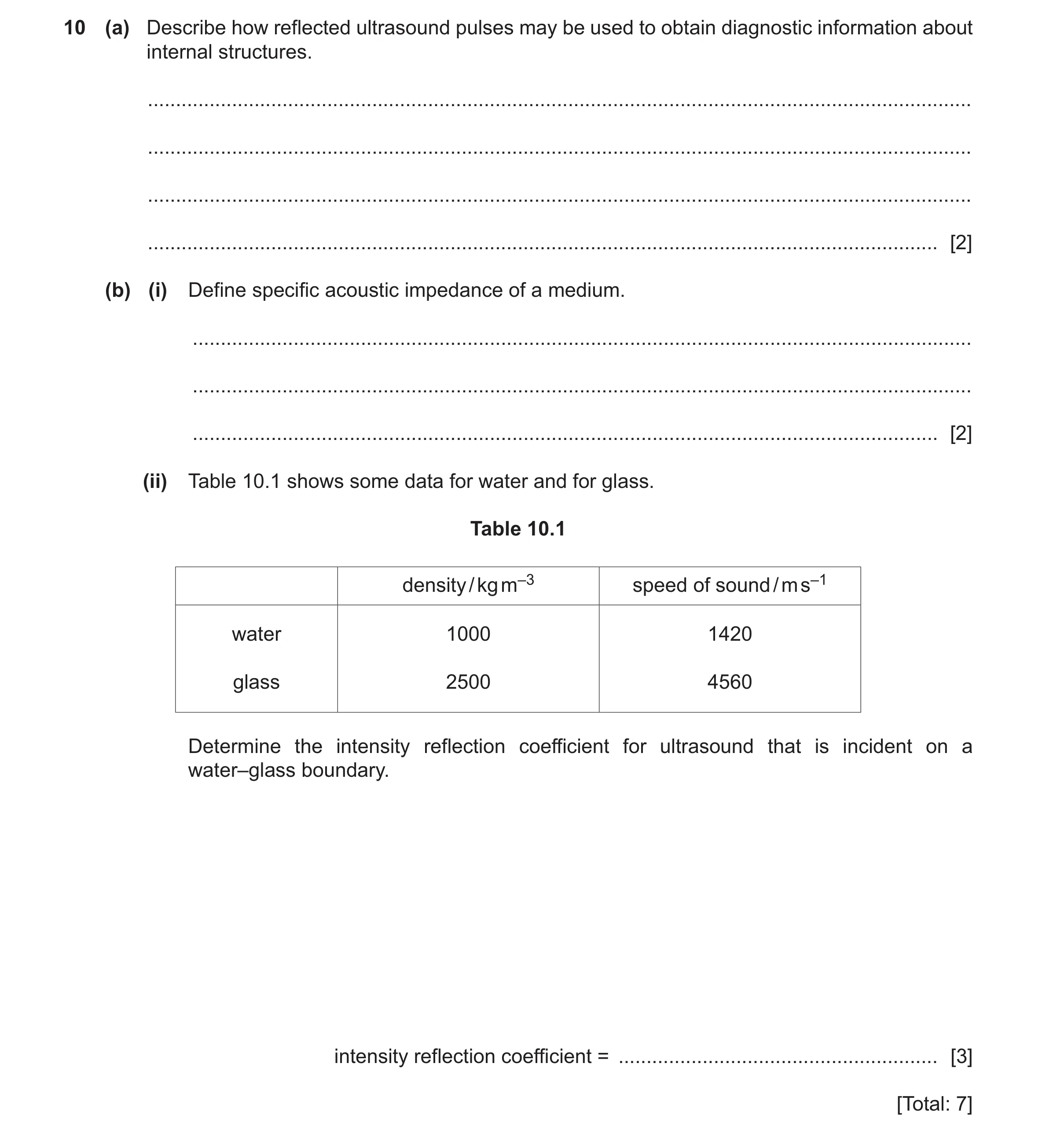
10 (a) Describe how reflected ultrasound pulses may be used to obtain diagnostic information about internal structures. … … … … [2] (b) (i) Define specific acoustic impedance of a medium. … … … [2] (ii) Table 10.1 shows some data for water and for glass. Table 10.1 density / kg m–3 speed of sound / m s–1 water 1000 1420 glass 2500 4560 Determine the intensity reflection coefficient for ultrasound that is incident on a water–glass boundary. intensity reflection coefficient = … [3] [Total: 7]
7 marks
Mark scheme: 10(a) time gives information about depth (of boundary) B1 intensity gives information about nature of boundary B1 10(b)(i) product of density and speed M1 speed of ultrasound in medium (and density of medium) A1 10(b)(ii) Zwater = 1000 1420 (= 1.42 106 kg m–2 s–1) and Zglass = 2500 4560 (= 11.4 106 kg m–2 s–1) C1 intensity reflection coefficient = (11.4 – 1.42)2 / (11.4 + 1.42)2 C1 = 0.61 A1
This question in 9702/42 May/June 2024
Q32 · A small crystal is made to vibrate with simple harmonic motion 9702/42 Feb/March 2025
4 A small crystal is made to vibrate with simple harmonic motion. The variation with time t of the displacement x of one surface of the crystal from its equilibrium position is shown in Fig. 4.1. 50 x / 10−6 m t / 10−6 s 0 0 0.1 0.2 0.3 0.4 0.5 0.6 –50 Fig. 4.1 (a) Show that the angular frequency of the vibration of the surface is 4.2 × 10⁷ rad s–1. [2] (b) Determine the maximum acceleration a0 of the vibration of the surface. a0 = … m s–2 [2] (c) The crystal may be modelled as a single mass of 2.4 × 10– 4 kg that vibrates as shown in Fig. 4.1. Calculate the total energy E of the vibrations. E = … J [3] (d) The crystal generates ultrasound waves that are used to obtain diagnostic information about internal structures. (i) The crystal is made from piezoelectric material. Explain how the crystal is made to vibrate. … … … … [2] (ii) A parallel beam of ultrasound waves is incident on a muscle‑bone boundary. Data for muscle and bone are given in Table 4.1. Table 4.1 material density / kg m–3 speed of sound / m s–1 muscle 1100 1600 bone 1900 4100 Calculate the percentage of the intensity of the ultrasound beam that is transmitted at this boundary. percentage transmitted = … % [3] [Total: 12]
12 marks
Mark scheme: 4(a) = 2 / T C1 = 2 / (0.15 10–6) = 4.2 107 rad s–1 A1 4(b) a0 = 2x0 C1 = (4.2 107)2 40 10–6 A1 = 7.1 1010 m s–2 4(c) E = ½m2xo2 C1 = ½ 2.4 10–4 (4.2 107)2 (40 10–6)2 C1 = 340 J A1 4(d)(i) apply alternating p.d. (to / across crystal) B1 applying p.d. to / across crystal causes it to distort B1 4(d)(ii) Z = c C1 Zm = 1100 1600 (= 1.76 106) Zb = 1900 4100 (= 7.79 106) intensity reflection co-efficient= [(7.79 – 1.76) / (7.79 + 1.76)]2 C1 = 0.40 or 40% percentage transmitted = 60% A1
This question in 9702/42 Feb/March 2025
Q33 · Describe how the piezoelectric crystal in a transducer generates ultrasound waves for use… 9702/44 May/June 2025
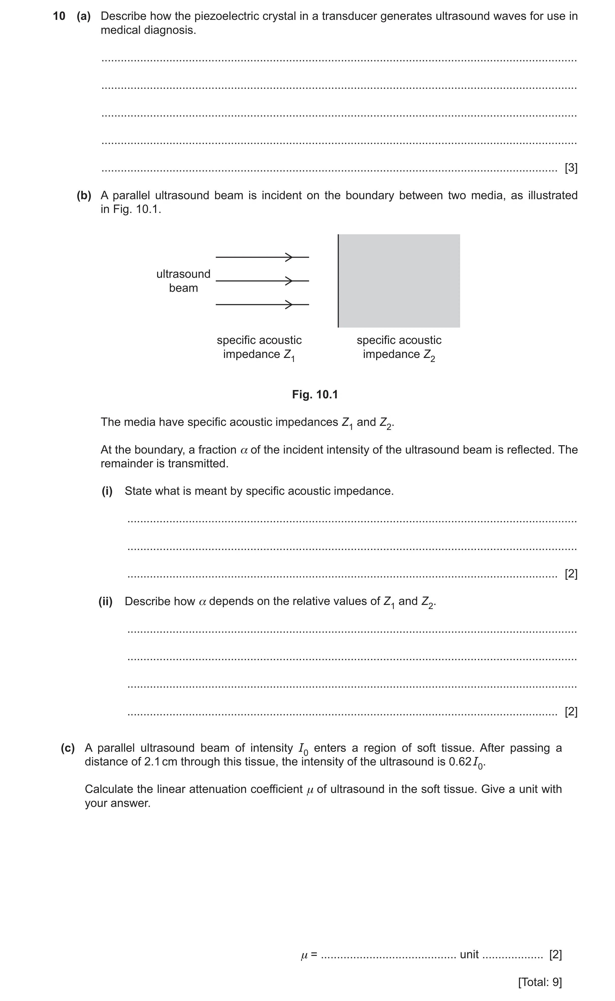
10 (a) Describe how the piezoelectric crystal in a transducer generates ultrasound waves for use in medical diagnosis. … … … … … [3] (b) A parallel ultrasound beam is incident on the boundary between two media, as illustrated in Fig. 10.1. ultrasound beam specific acoustic specific acoustic impedance Z1 impedance Z2 Fig. 10.1 The media have specific acoustic impedances Z1 and Z2. At the boundary, a fraction α of the incident intensity of the ultrasound beam is reflected. The remainder is transmitted. (i) State what is meant by specific acoustic impedance. … … … [2] (ii) Describe how α depends on the relative values of Z1 and Z2. … … … … [2] (c) A parallel ultrasound beam of intensity I0 enters a region of soft tissue. After passing a distance of 2.1 cm through this tissue, the intensity of the ultrasound is 0.62 I0. Calculate the linear attenuation coefficient μ of ultrasound in the soft tissue. Give a unit with your answer. μ = … unit … [2] [Total: 9]
9 marks
Mark scheme: 10(a) alternating p.d. (applied to crystal) makes crystal vibrate B1 when frequency of applied p.d. equals natural frequency of crystal, crystal resonates B1 natural frequency of crystal is in ultrasound range B1 10(b)(i) product of density and speed M1 speed is speed of ultrasound in medium (and density of medium) A1 10(b)(ii) (is close to) 0 when Z1 and Z2 are equal B1 (is close to) 1 when Z1 and Z2 are very different B1 10(c) I = I0 exp (–x) C1 ln (I / I0) = – x A1 ln 0.62 = – 2.1 = 0.23 cm–1
This question in 9702/44 May/June 2025
Q34 · Define specific acoustic impedance 9702/41 Oct/Nov 2025
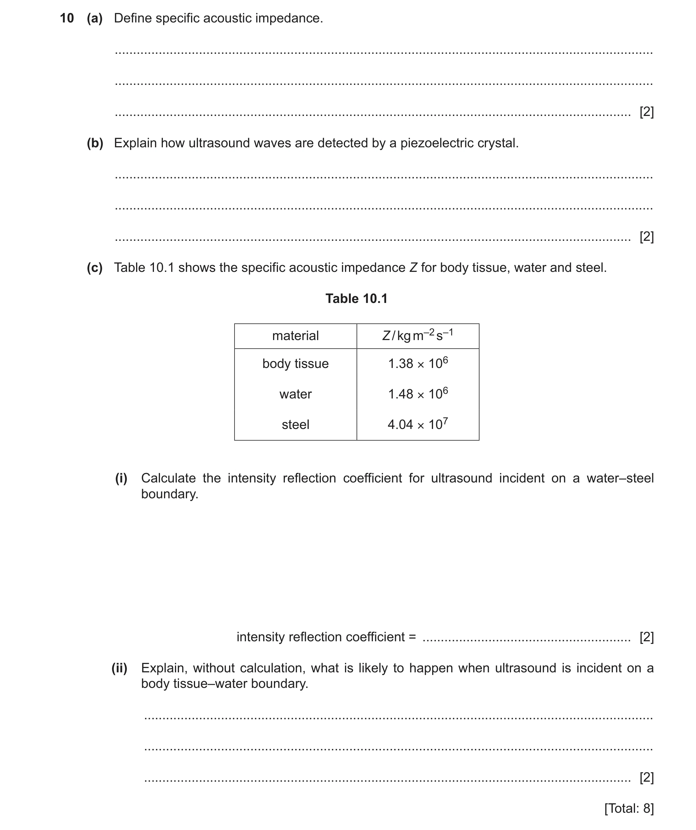
10 (a) Define specific acoustic impedance. … … … [2] (b) Explain how ultrasound waves are detected by a piezoelectric crystal. … … … [2] (c) Table 10.1 shows the specific acoustic impedance Z for body tissue, water and steel. Table 10.1 material Z / kg m–2 s–1 body tissue 1.38 × 10⁶ water 1.48 × 10⁶ steel 4.04 × 10⁷ (i) Calculate the intensity reflection coefficient for ultrasound incident on a water–steel boundary. intensity reflection coefficient = … [2] (ii) Explain, without calculation, what is likely to happen when ultrasound is incident on a body tissue–water boundary. … … … [2] [Total: 8]
8 marks
Mark scheme: 10(a) product of density and speed M1 speed of sound in medium (and density of the medium) A1 10(b) ultrasound waves cause crystal to vibrate B1 vibrations (of crystal) cause induced e.m.f. (across crystal) B1 10(c)(i) intensity reflection coefficient= (40.4 – 1.48)2 / (40.4 + 1.48)2 C1 = 0.86 A1 10(c)(ii) Z values are very similar B1 (almost) all the ultrasound will be transmitted B1 or (almost) none of the ultrasound will be reflected
This question in 9702/41 Oct/Nov 2025
Q35 · Define specific acoustic impedance 9702/43 Oct/Nov 2025
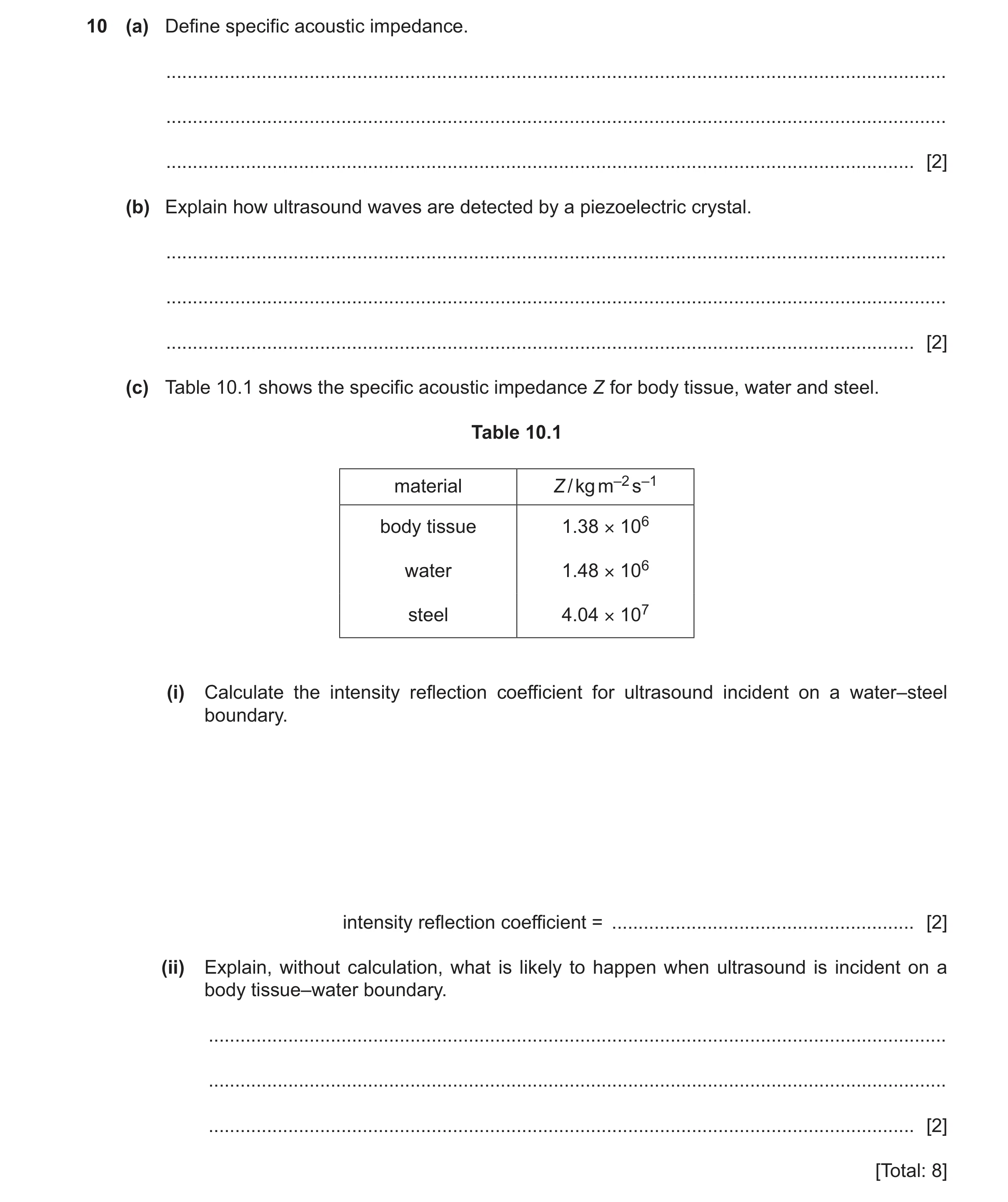
10 (a) Define specific acoustic impedance. … … … [2] (b) Explain how ultrasound waves are detected by a piezoelectric crystal. … … … [2] (c) Table 10.1 shows the specific acoustic impedance Z for body tissue, water and steel. Table 10.1 material Z / kg m–2 s–1 body tissue 1.38 × 10⁶ water 1.48 × 10⁶ steel 4.04 × 10⁷ (i) Calculate the intensity reflection coefficient for ultrasound incident on a water–steel boundary. intensity reflection coefficient = … [2] (ii) Explain, without calculation, what is likely to happen when ultrasound is incident on a body tissue–water boundary. … … … [2] [Total: 8]
8 marks
Mark scheme: 10(a) product of density and speed M1 speed of sound in medium (and density of the medium) A1 10(b) ultrasound waves cause crystal to vibrate B1 vibrations (of crystal) cause induced e.m.f. (across crystal) B1 10(c)(i) intensity reflection coefficient= (40.4 – 1.48)2 / (40.4 + 1.48)2 C1 = 0.86 A1 10(c)(ii) Z values are very similar B1 (almost) all the ultrasound will be transmitted B1 or (almost) none of the ultrasound will be reflected
This question in 9702/43 Oct/Nov 2025
 1 / 38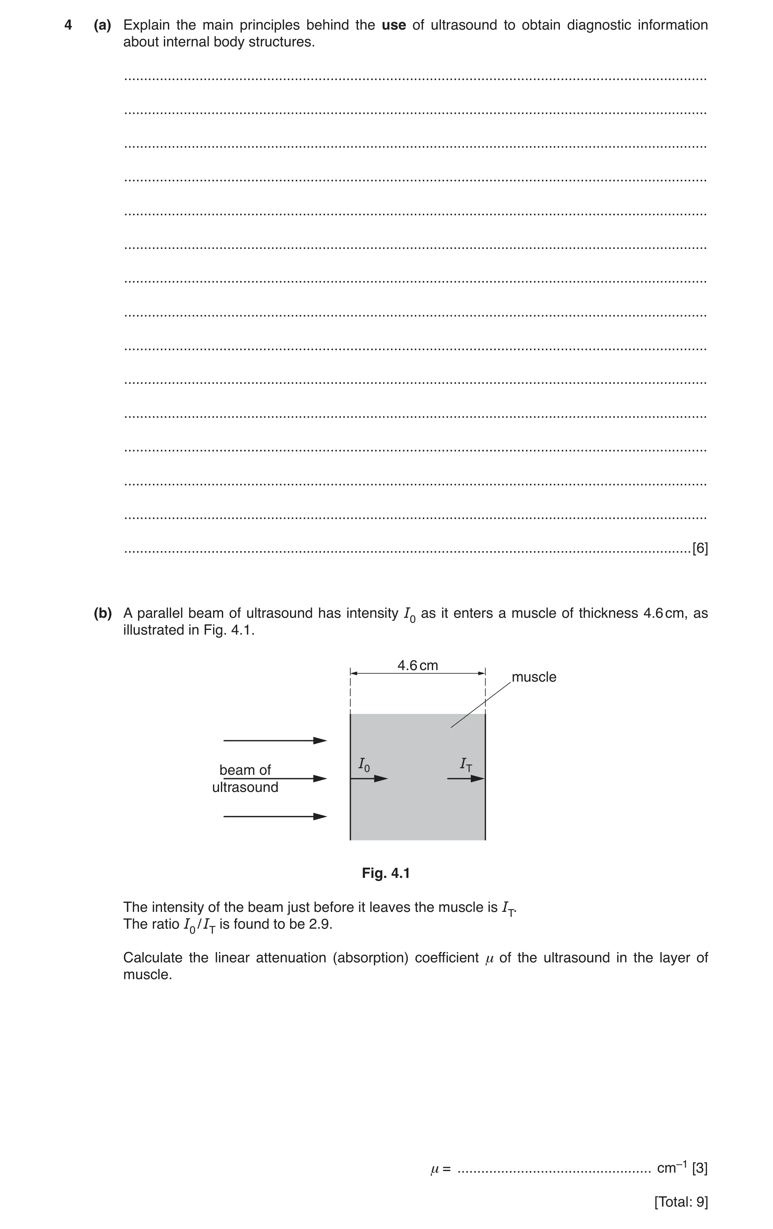
 3 / 38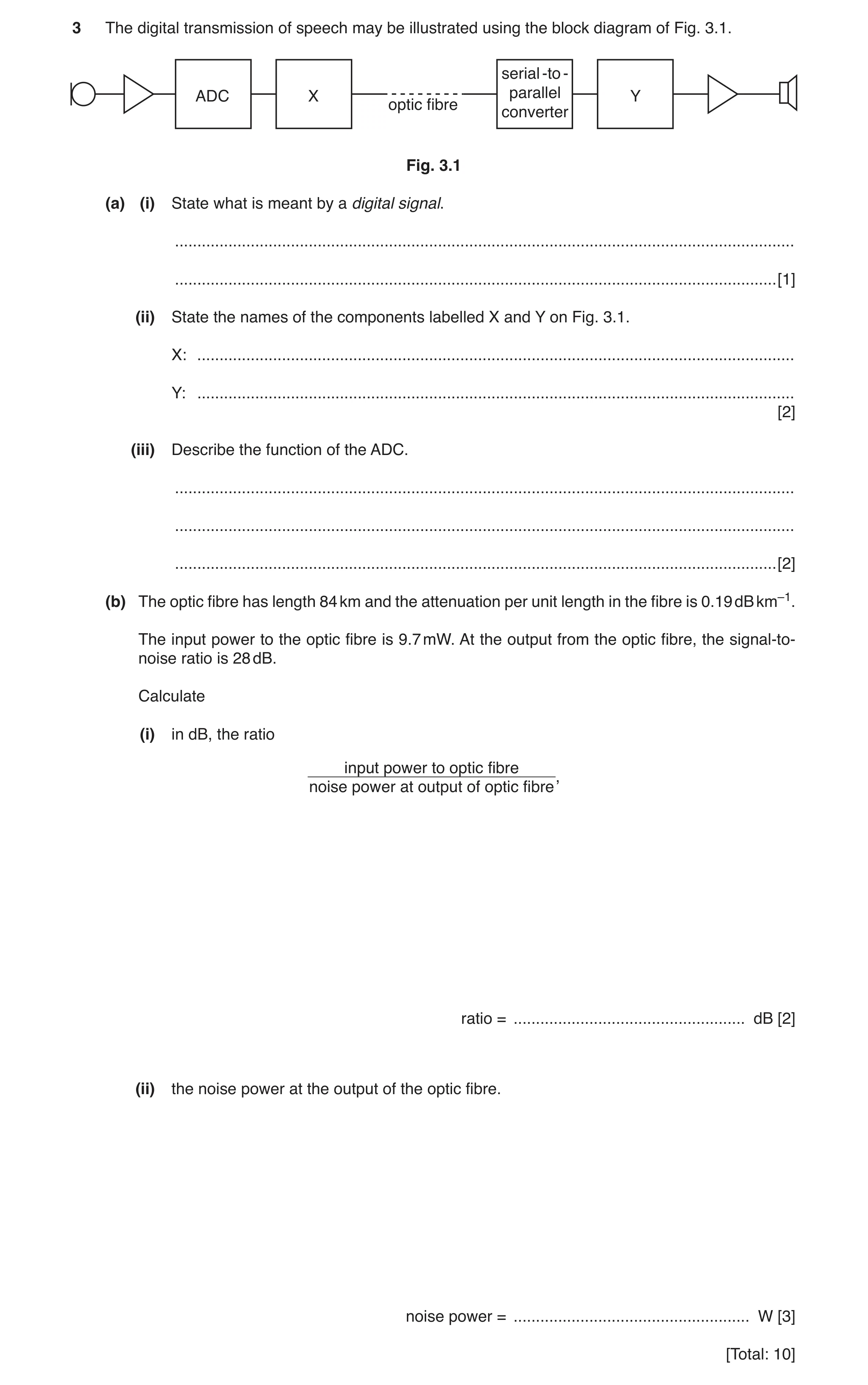
 5 / 38
 6 / 38
 7 / 38
 8 / 38
 9 / 38
 10 / 38
 11 / 38
 12 / 38
 13 / 38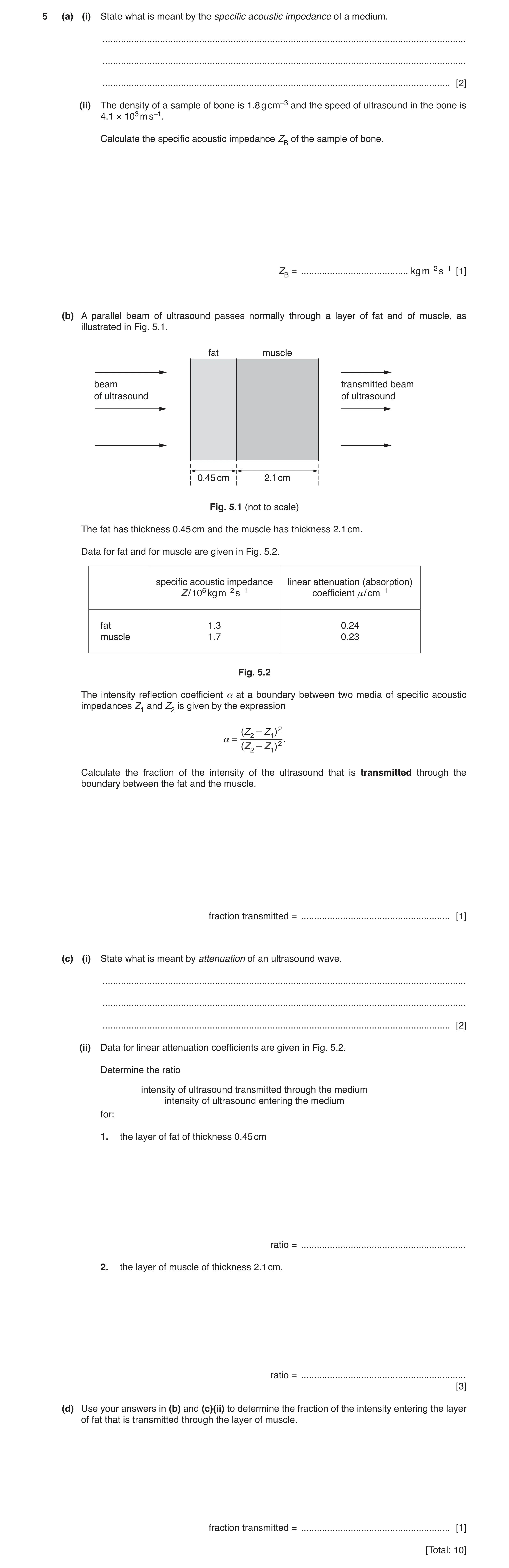
 18 / 38
 19 / 38
 20 / 38
 21 / 38
 22 / 38
 23 / 38
 24 / 38
 25 / 38
 26 / 38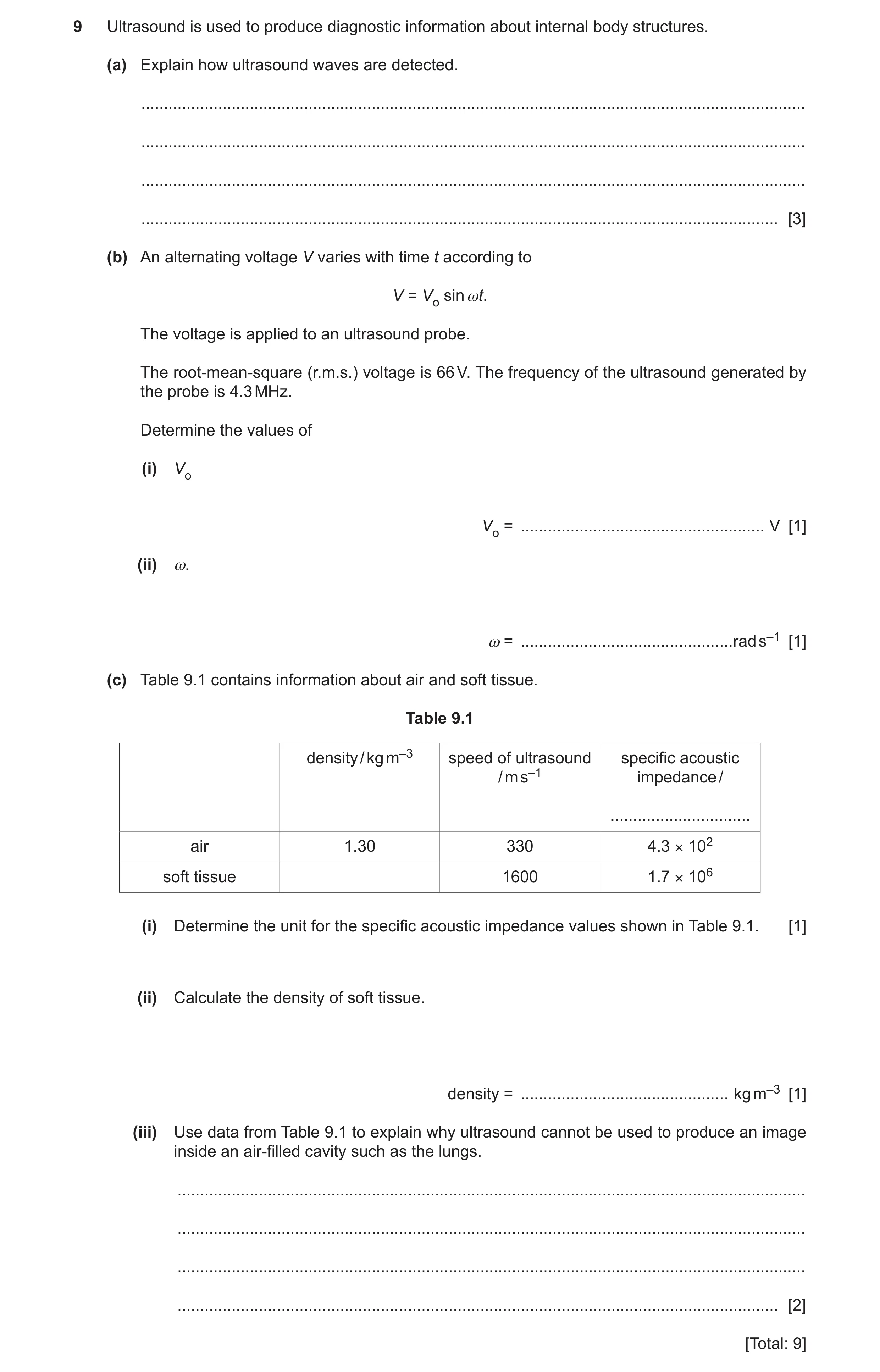
 31 / 38
 32 / 38
 33 / 38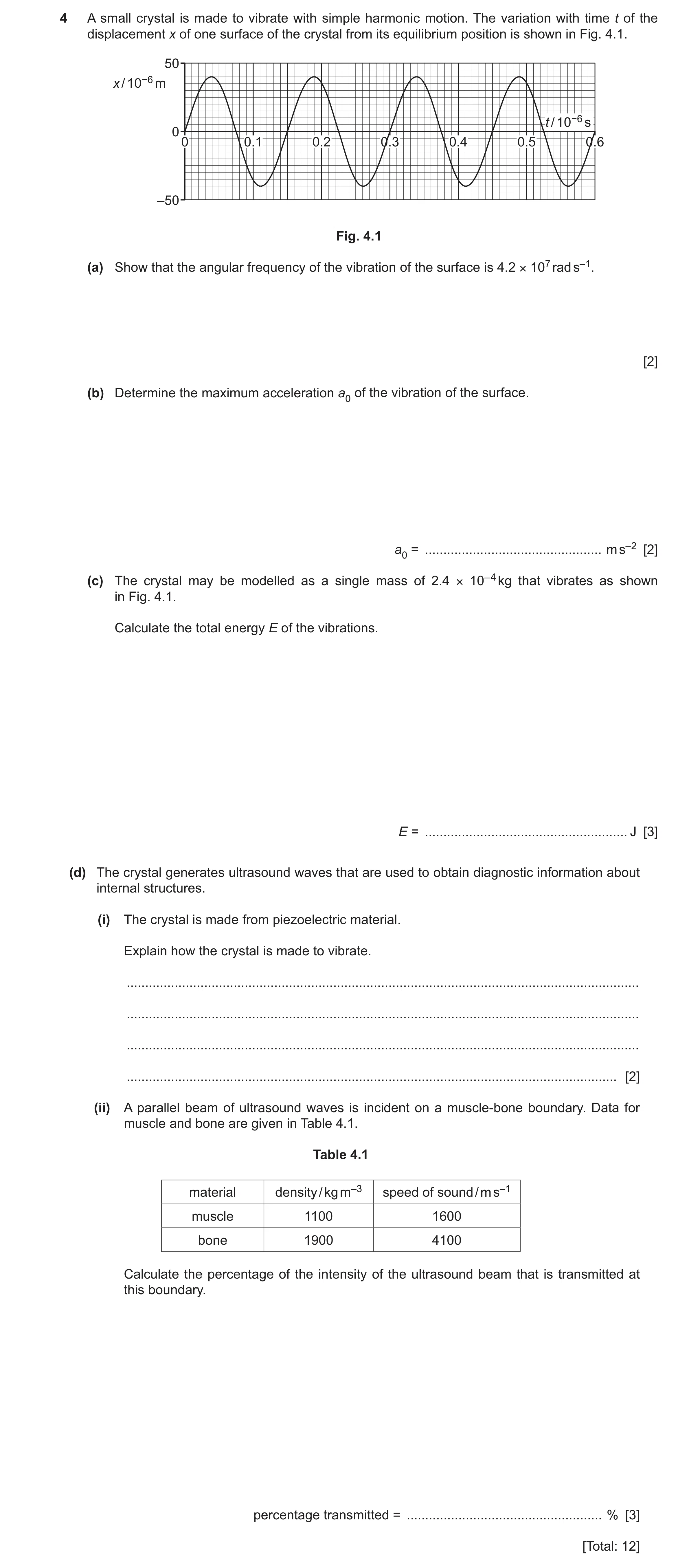
 37 / 38
 38 / 38
 4
4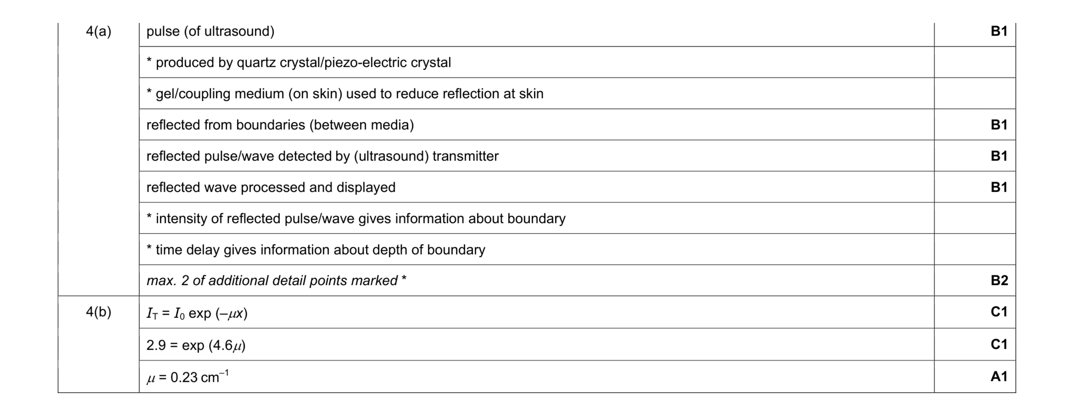 9
9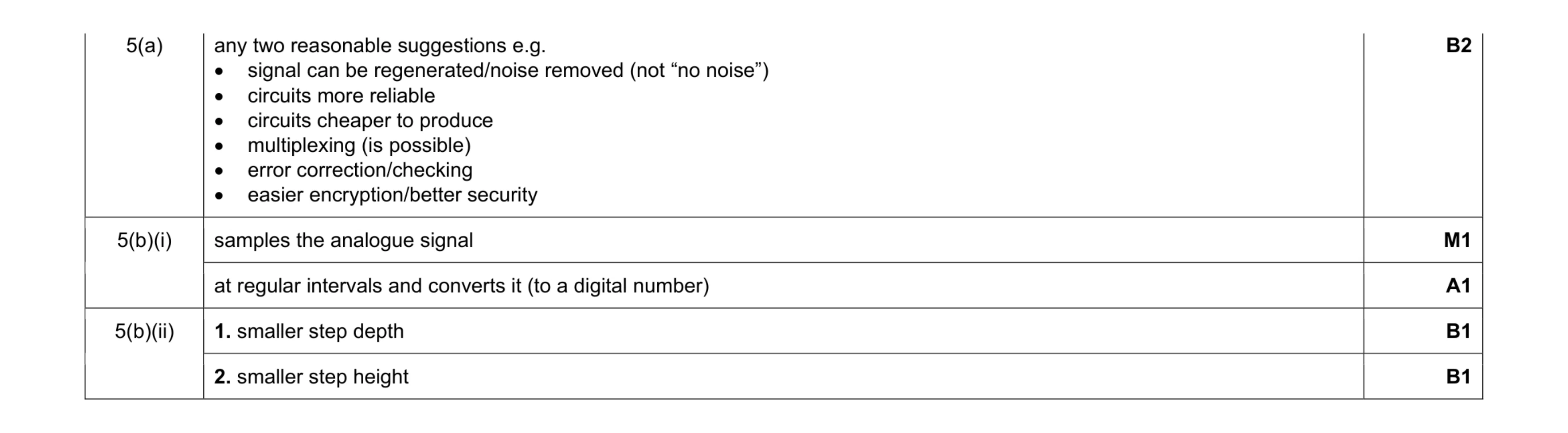 6
6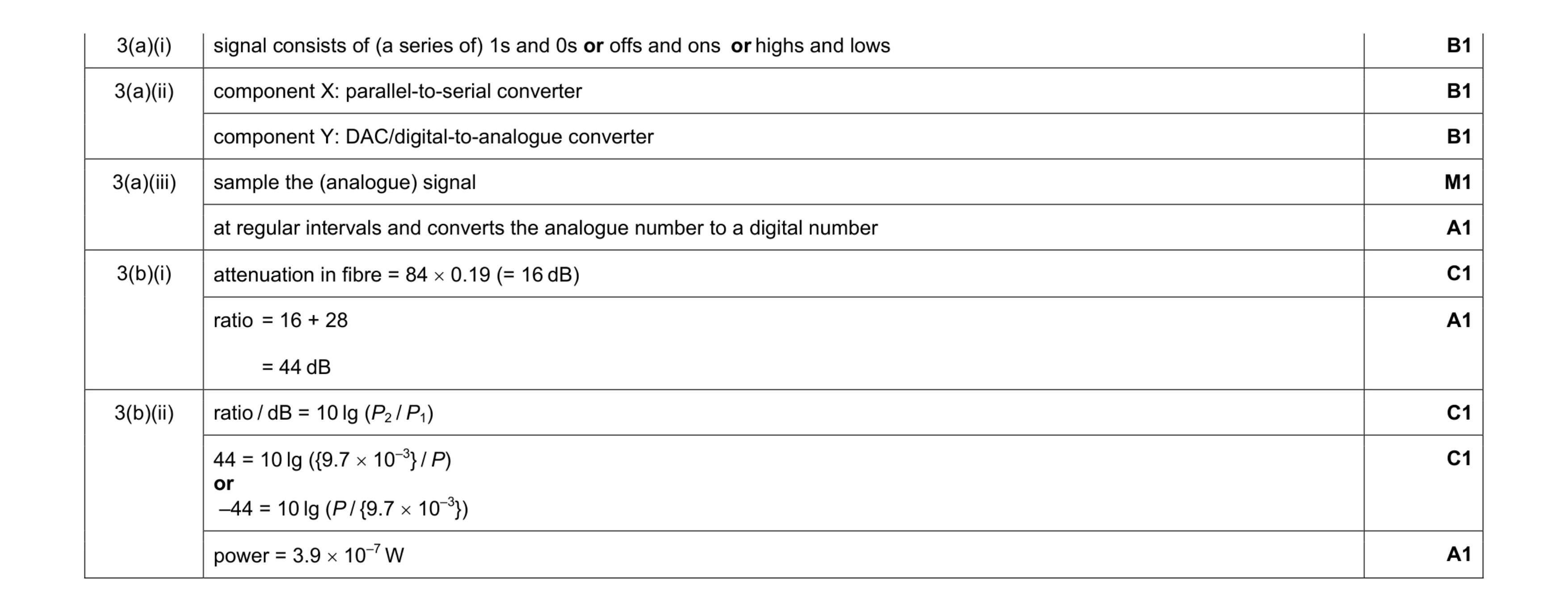 10
10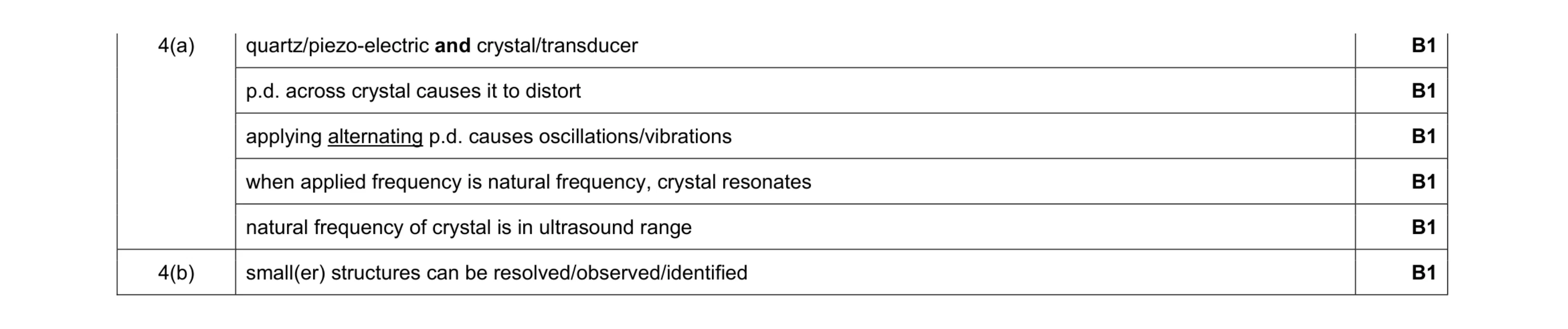 6
6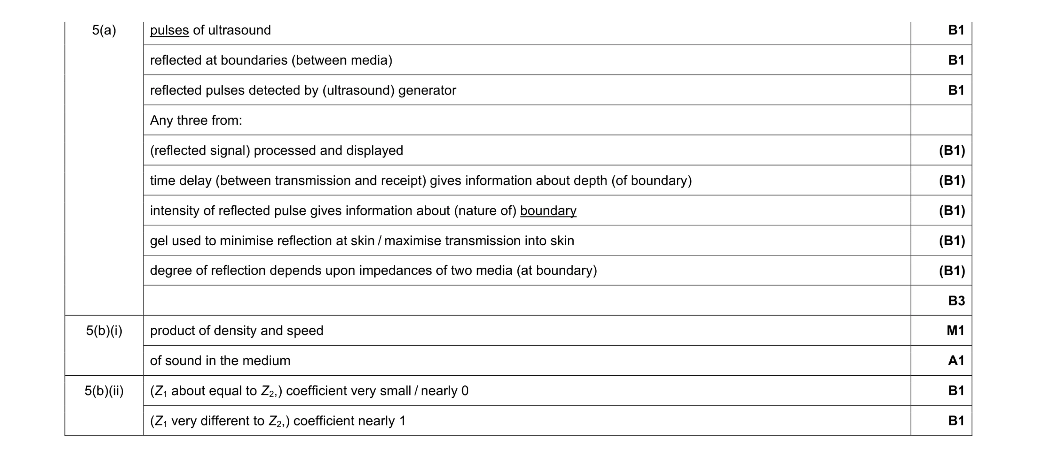 10
10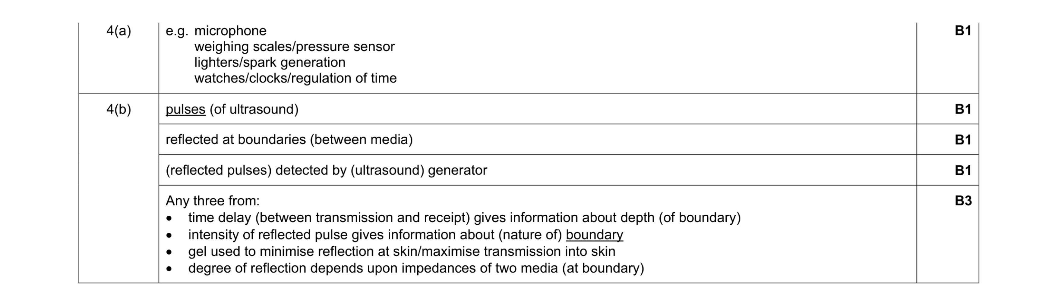 7
7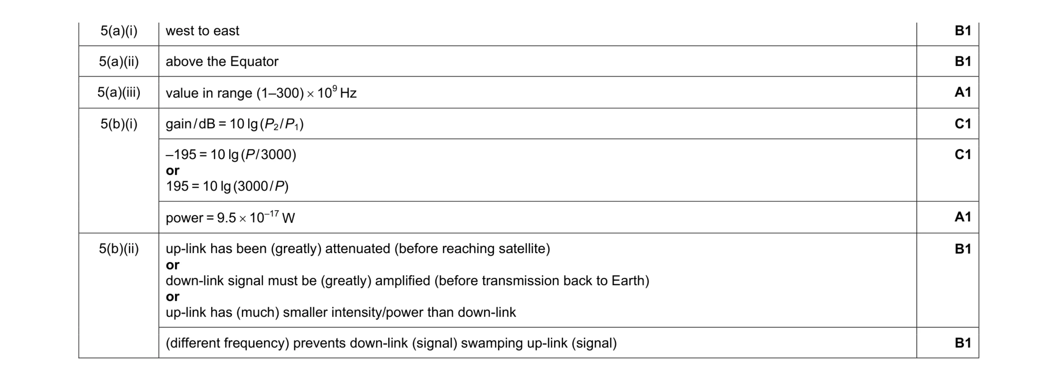 8
8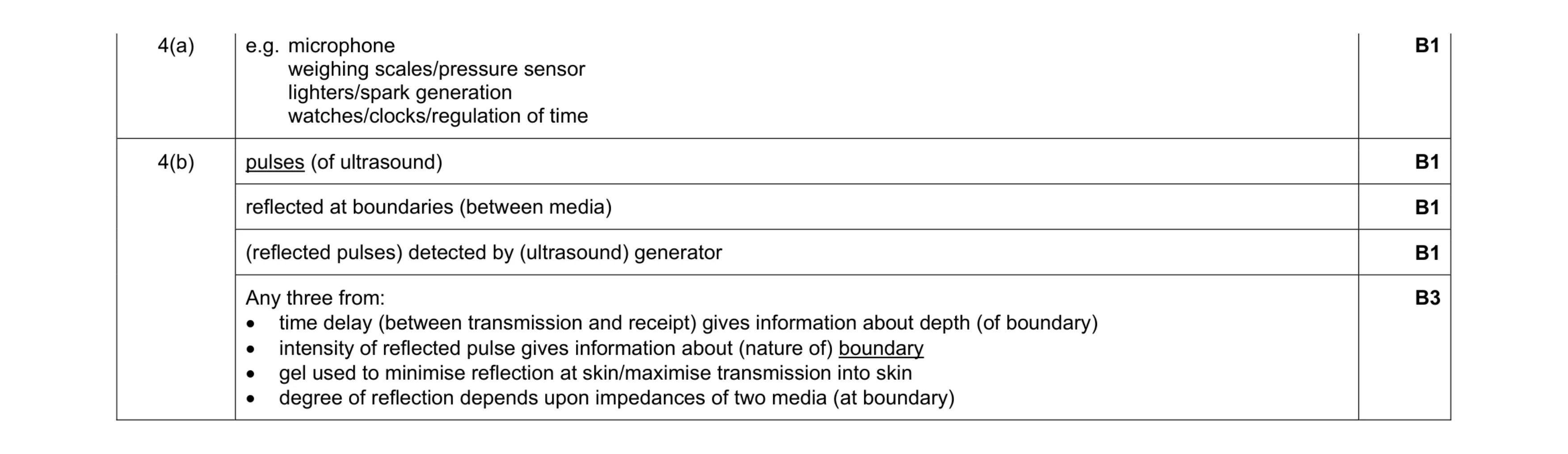 7
7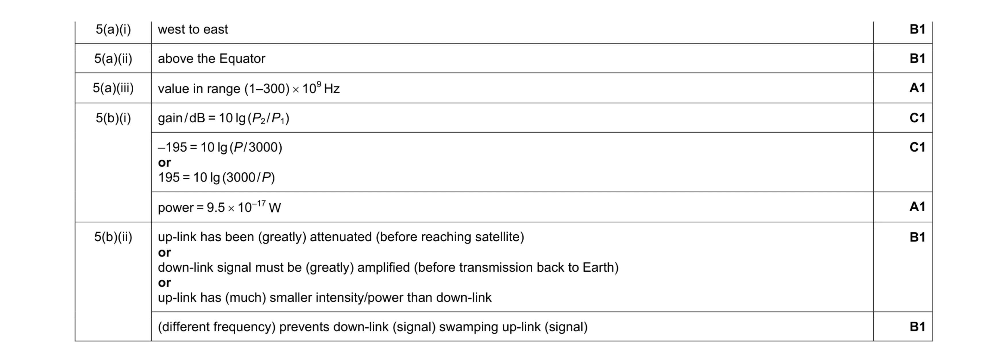 8
8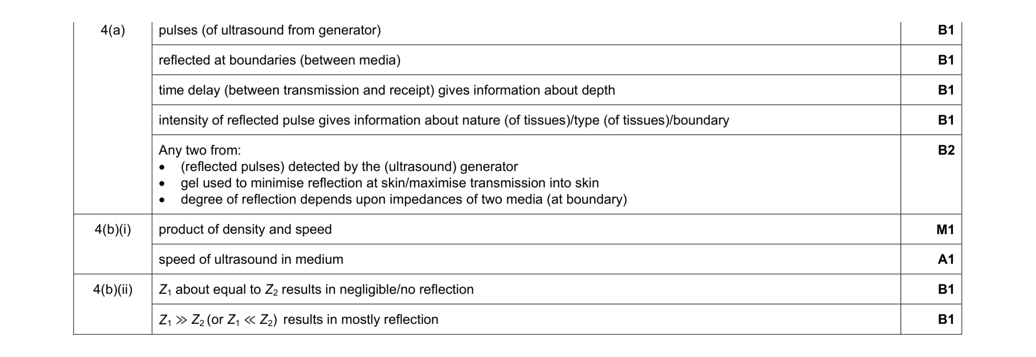 10
10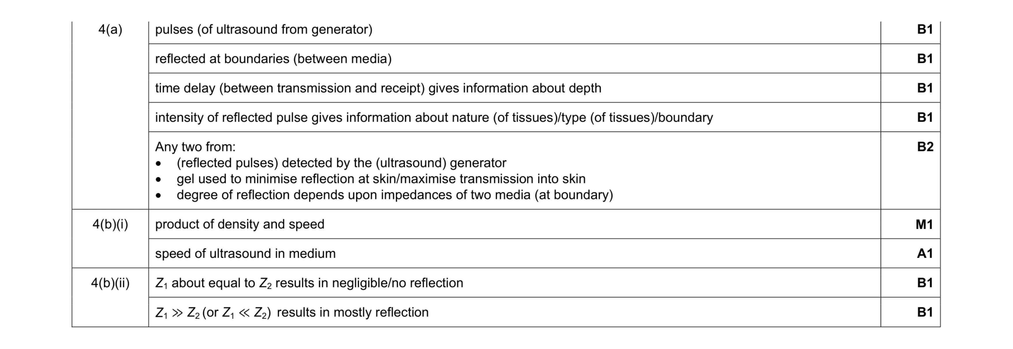 10
10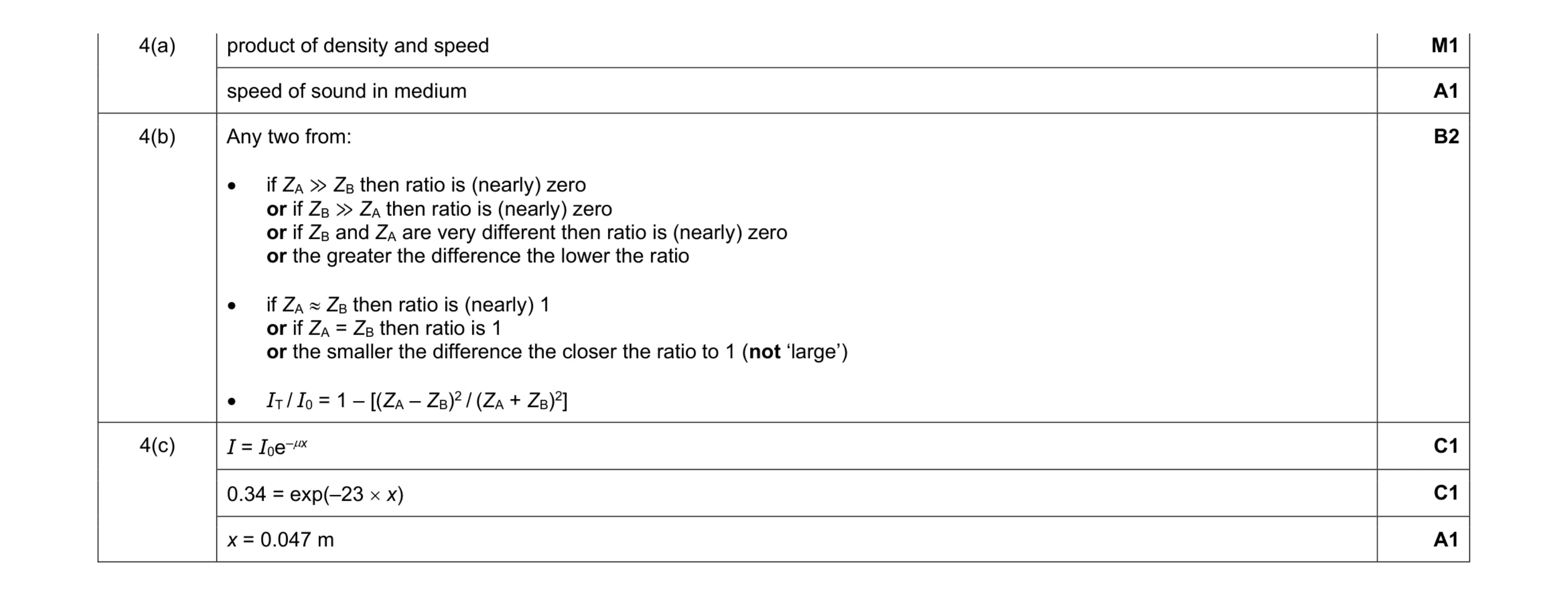 7
7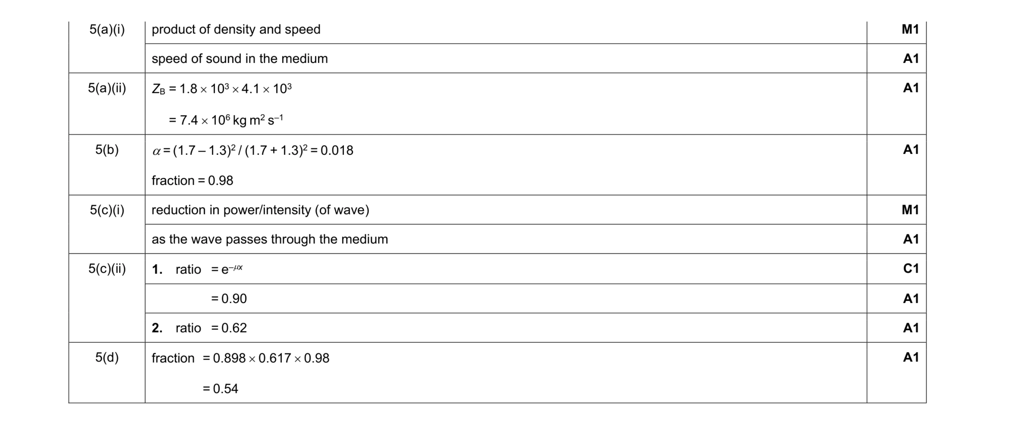 10
10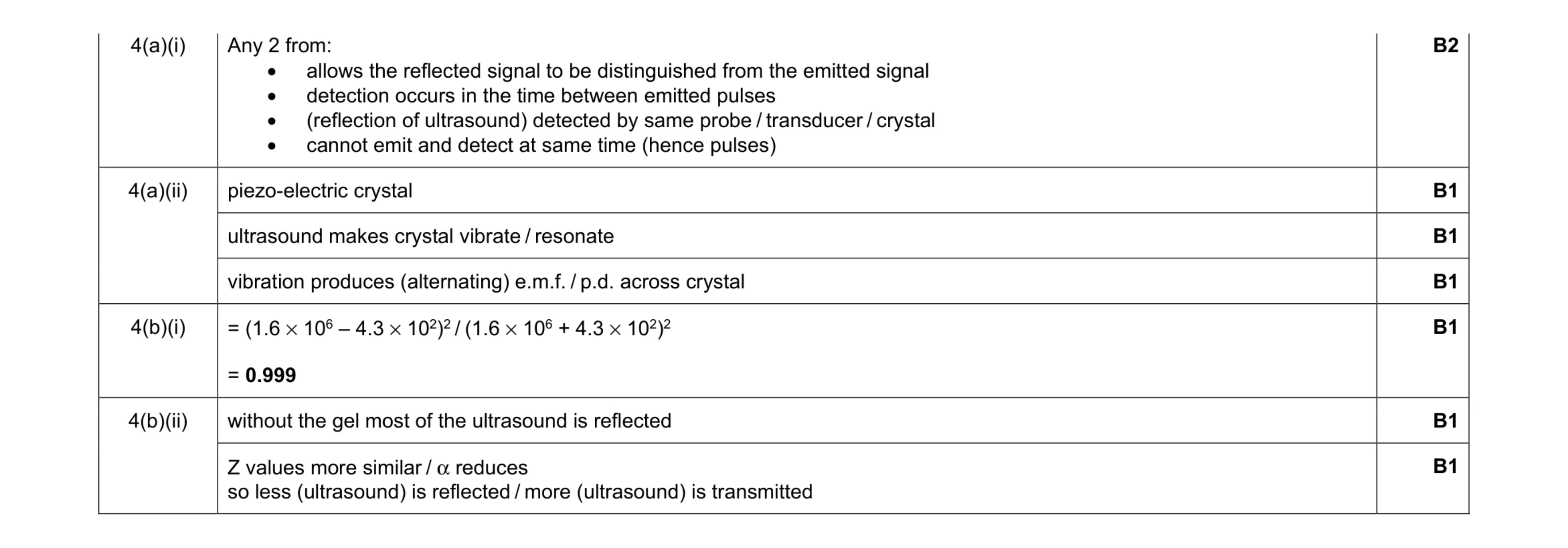 8
8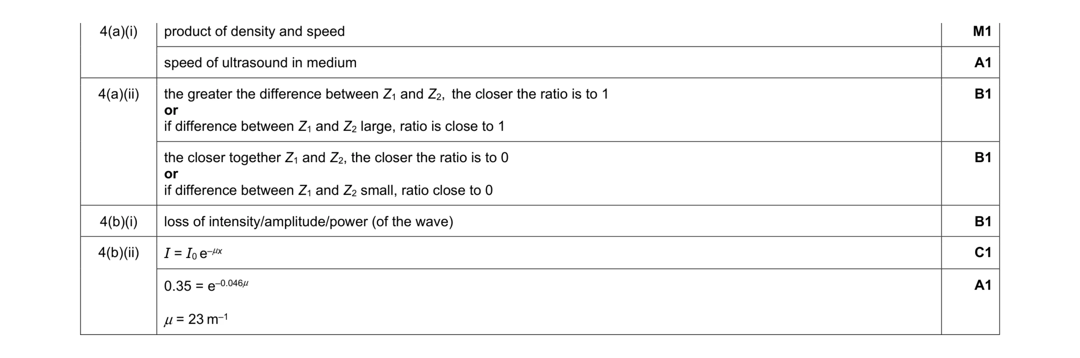 7
7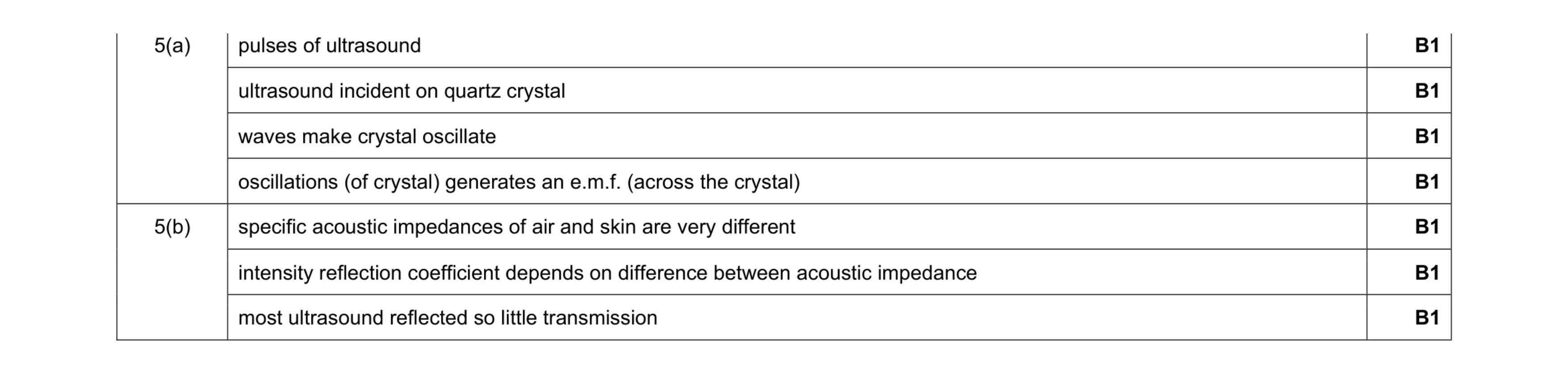 7
7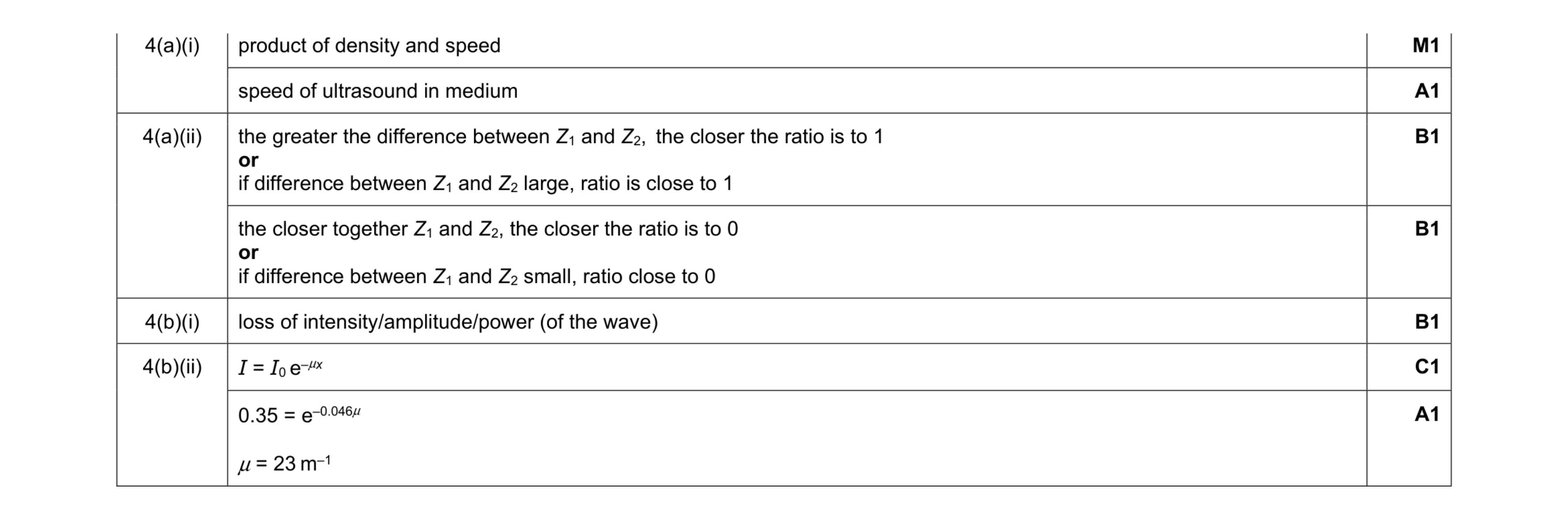 7
7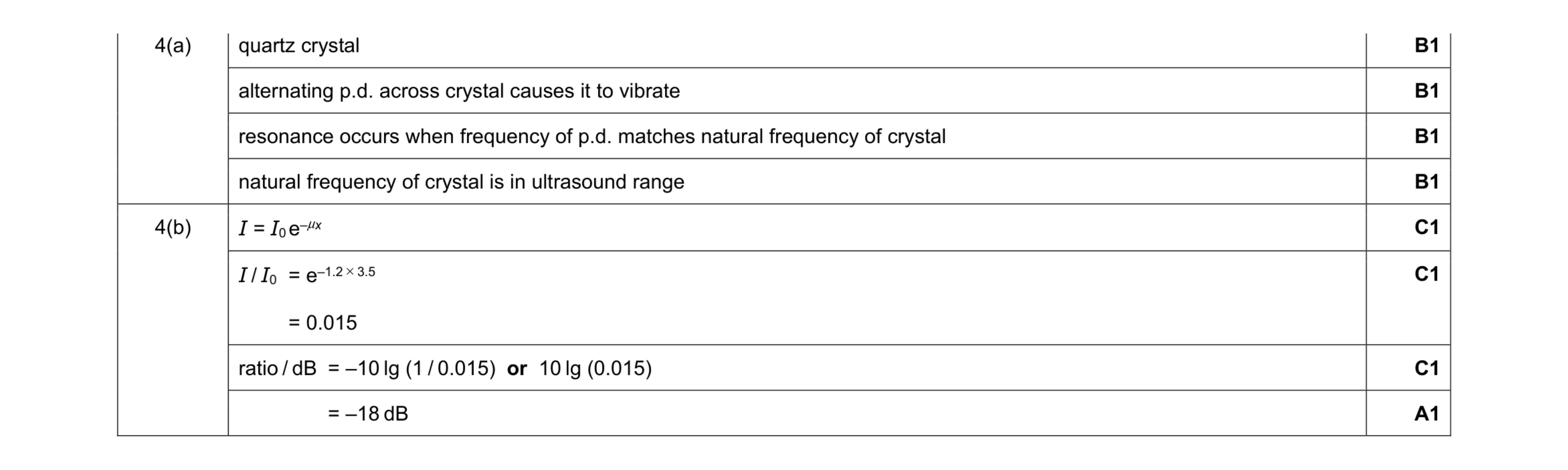 8
8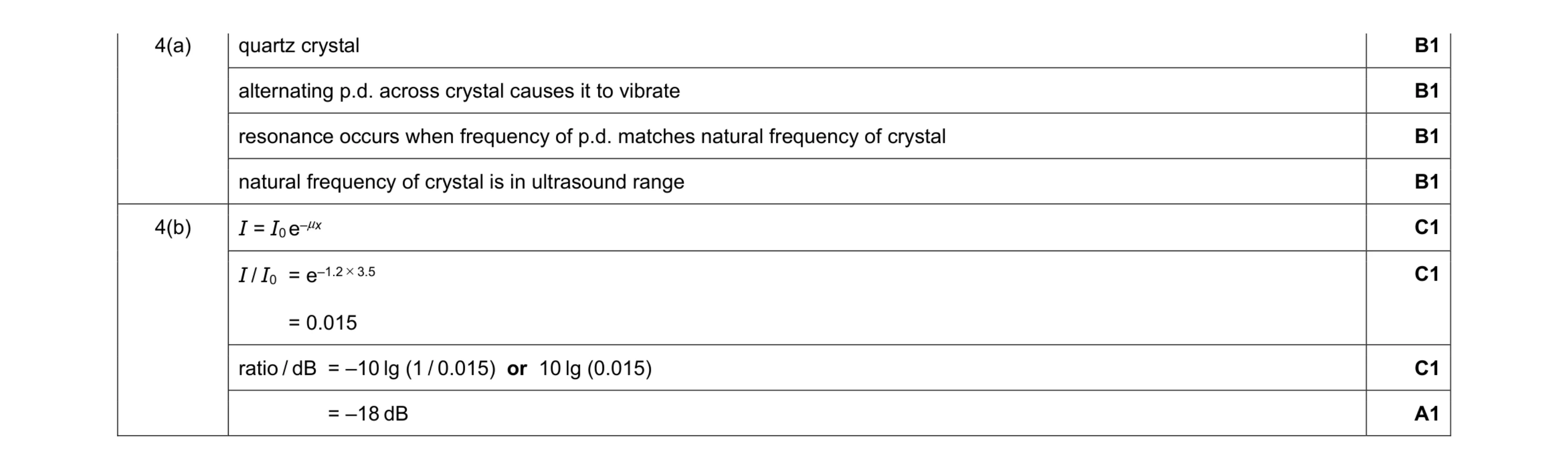 8
8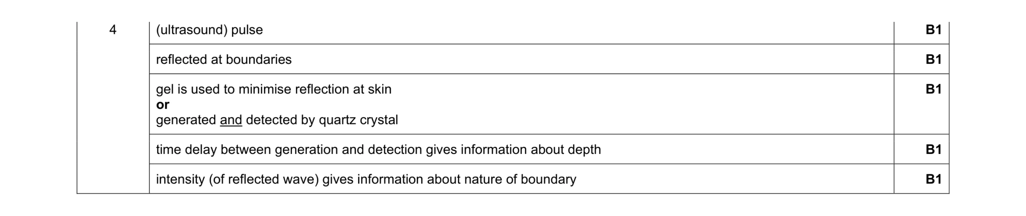 5
5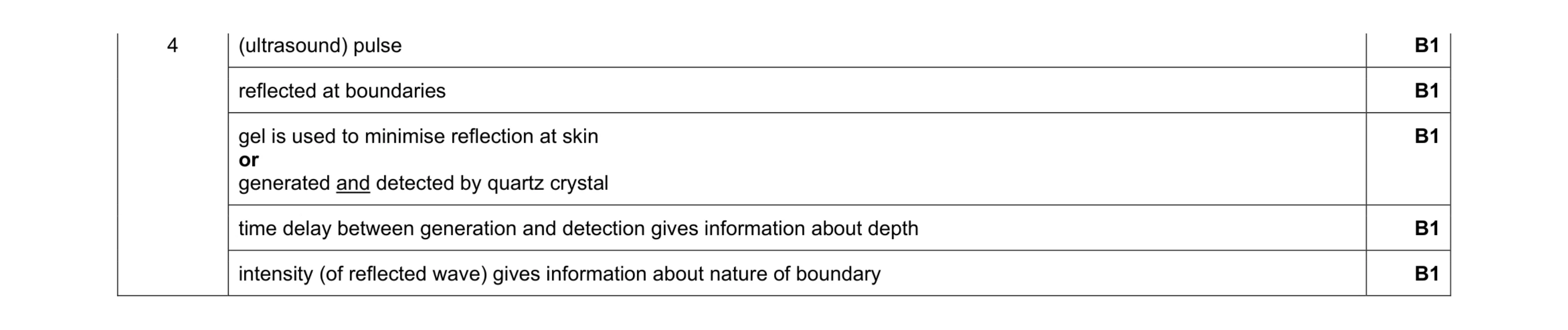 5
5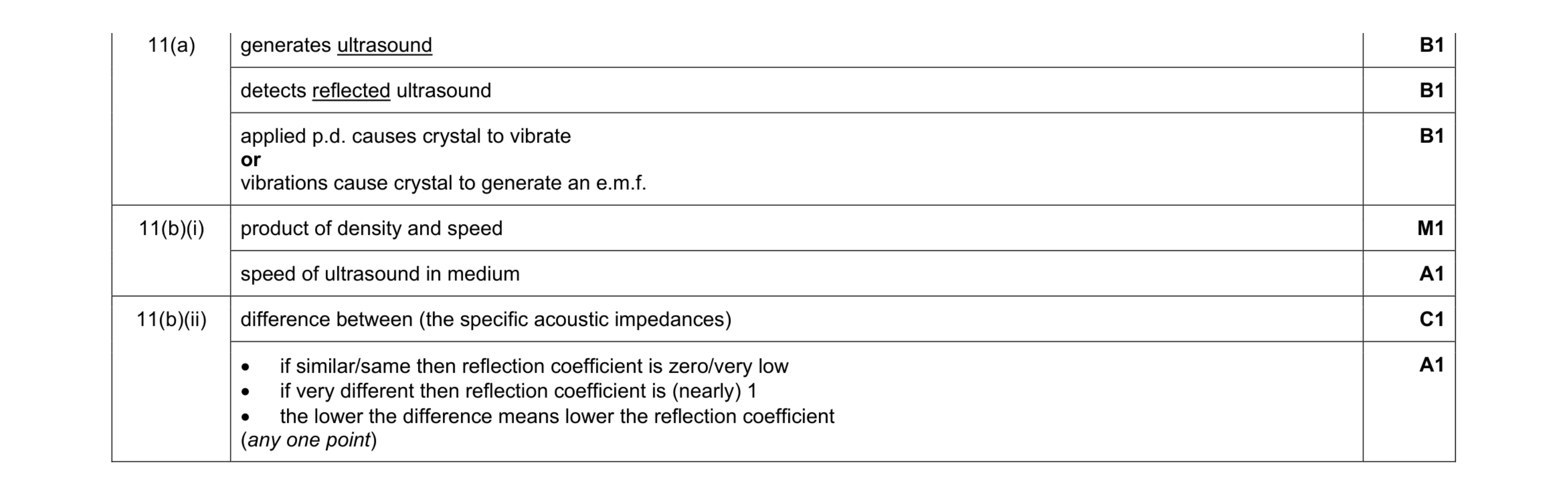 7
7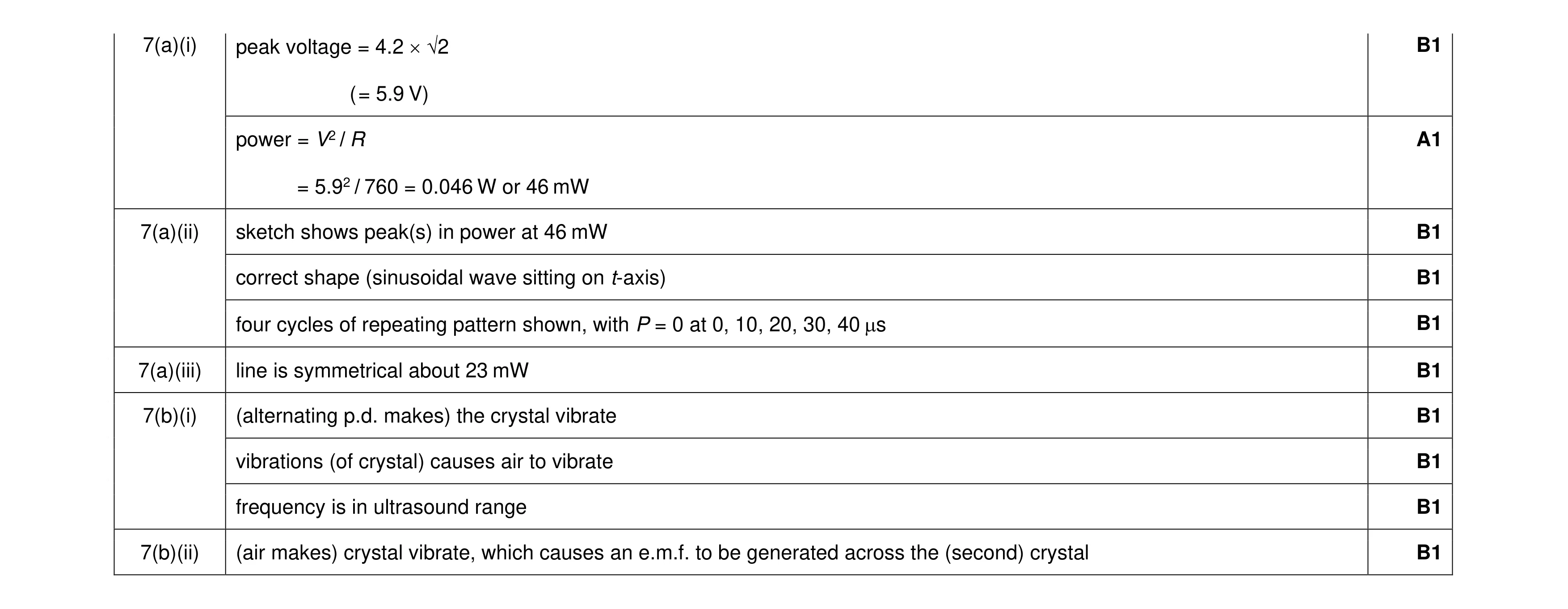 10
10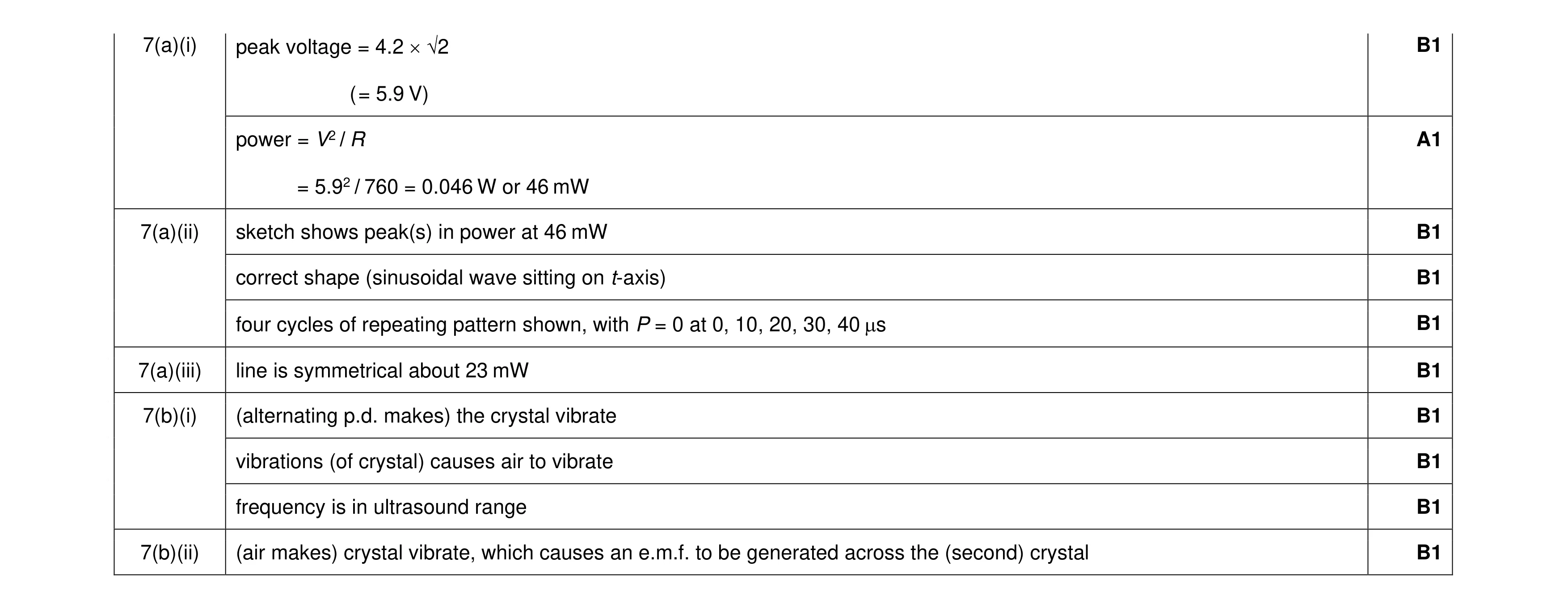 10
10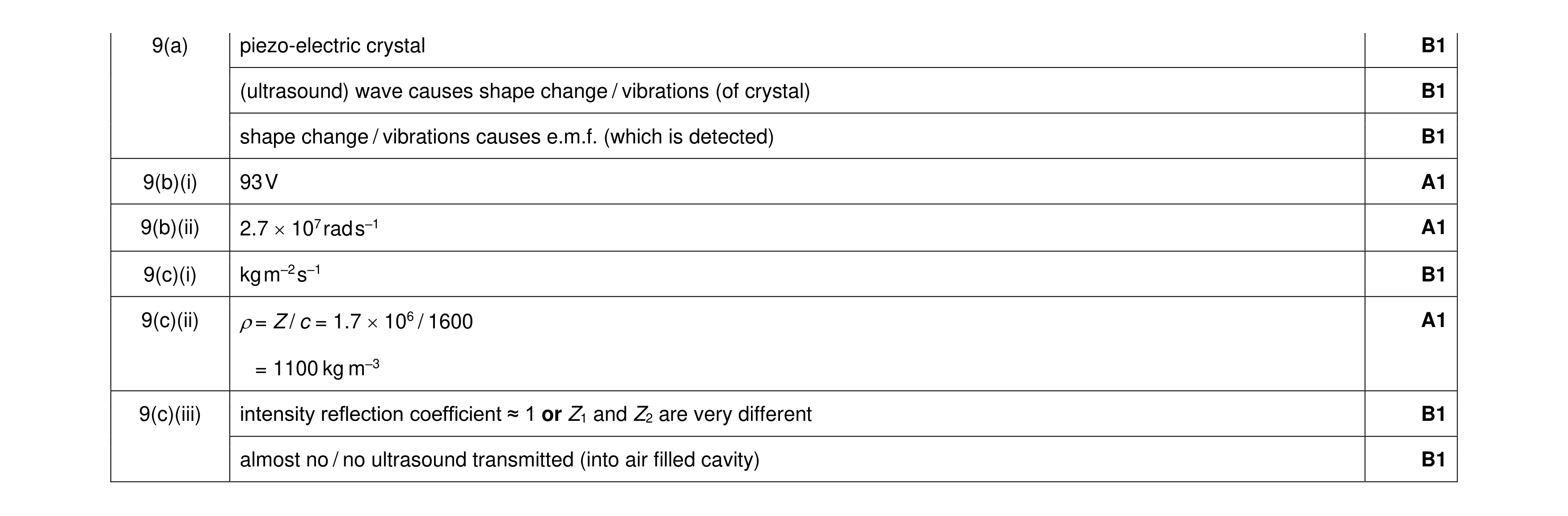 9
9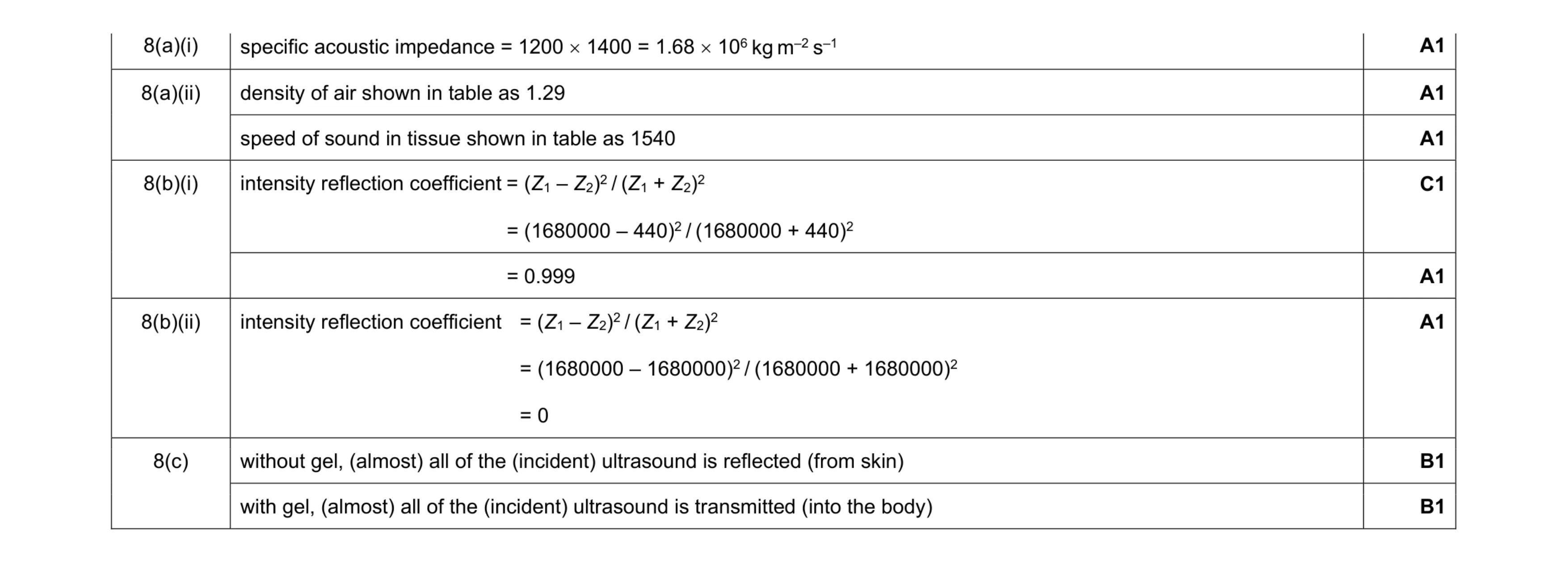 8
8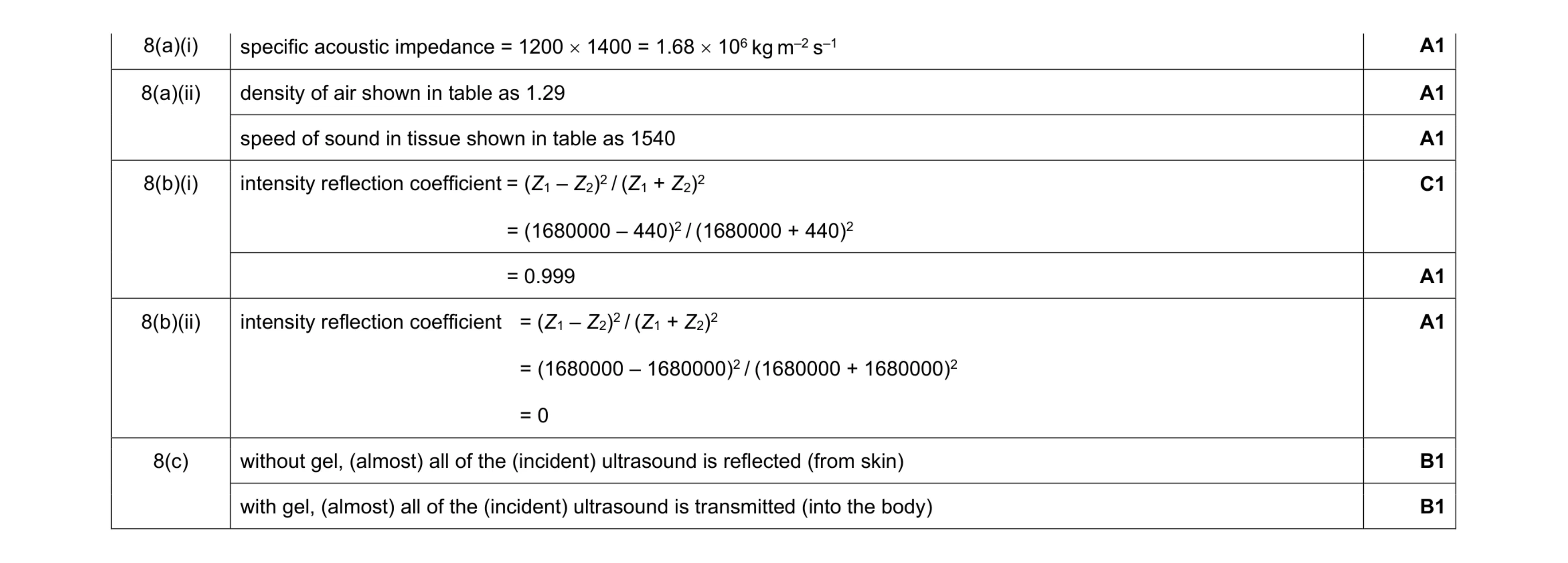 8
8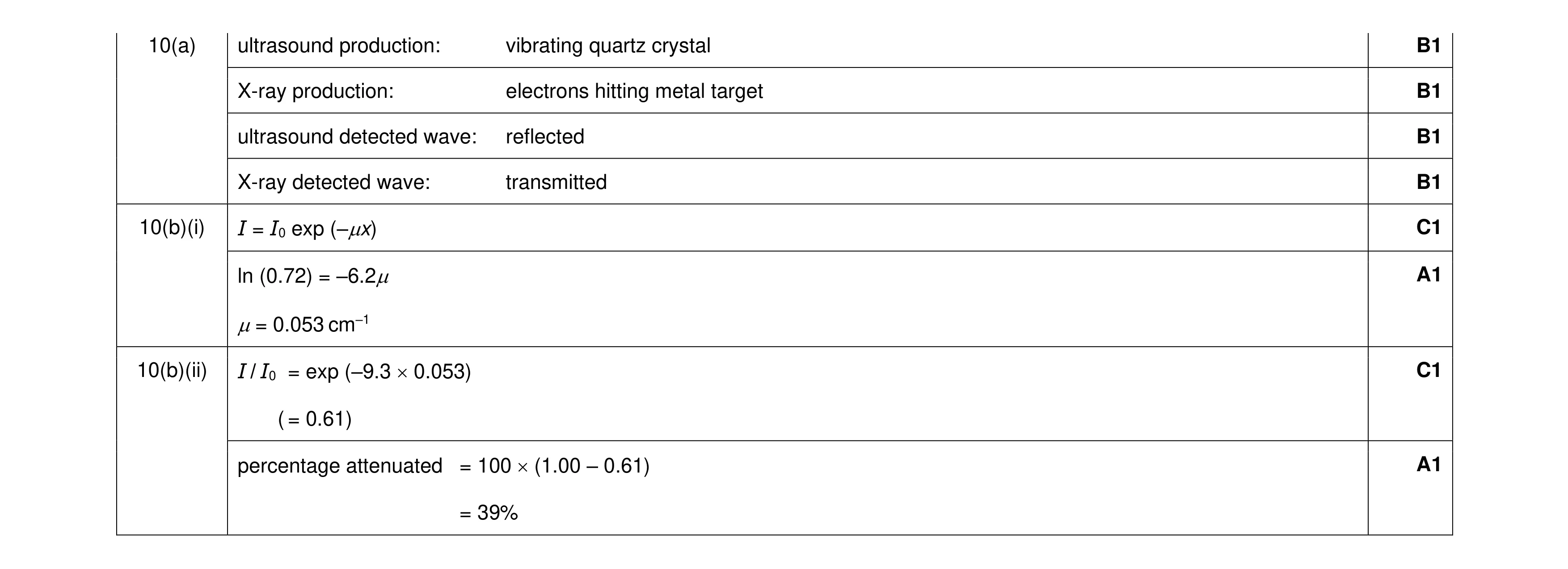 8
8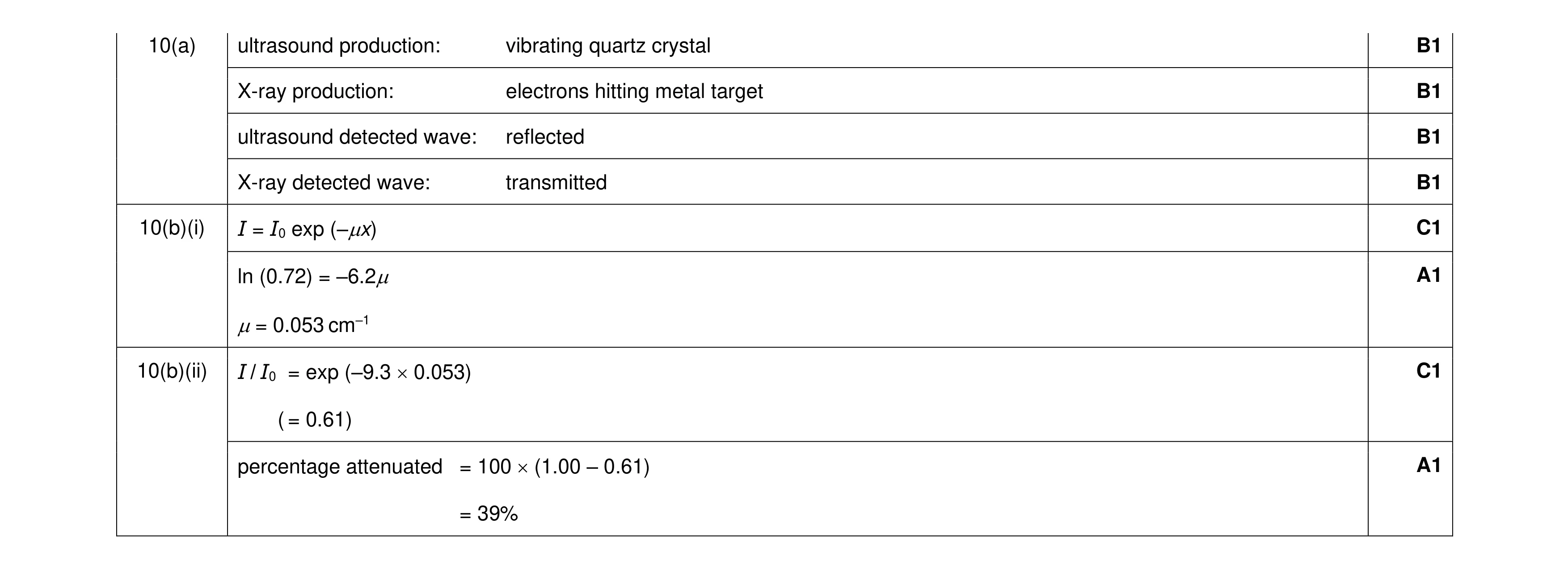 8
8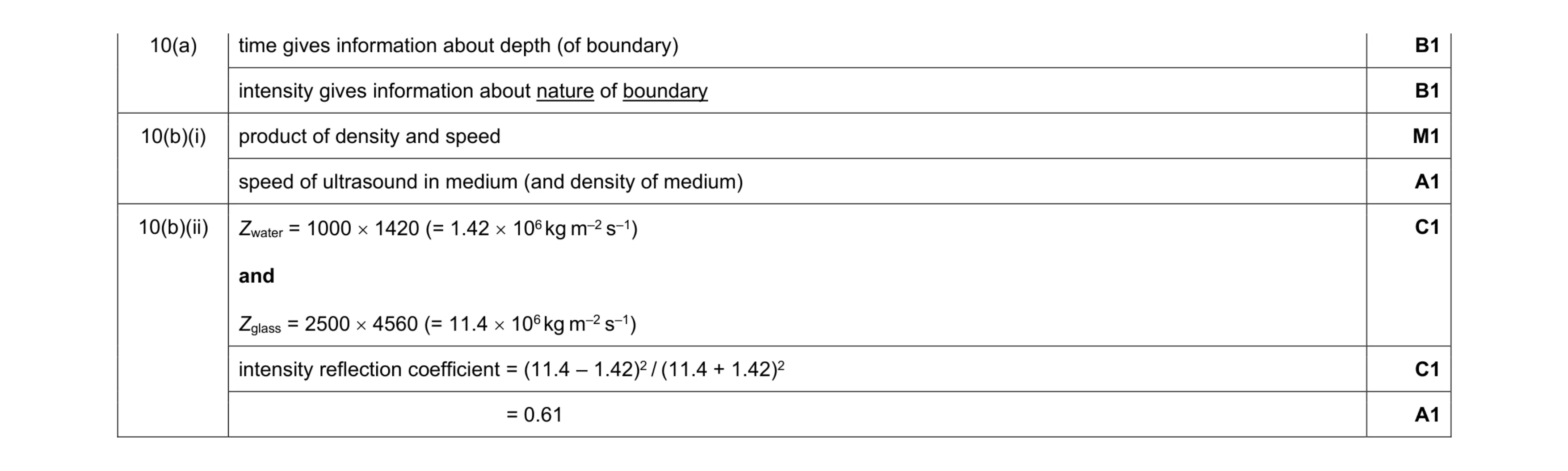 7
7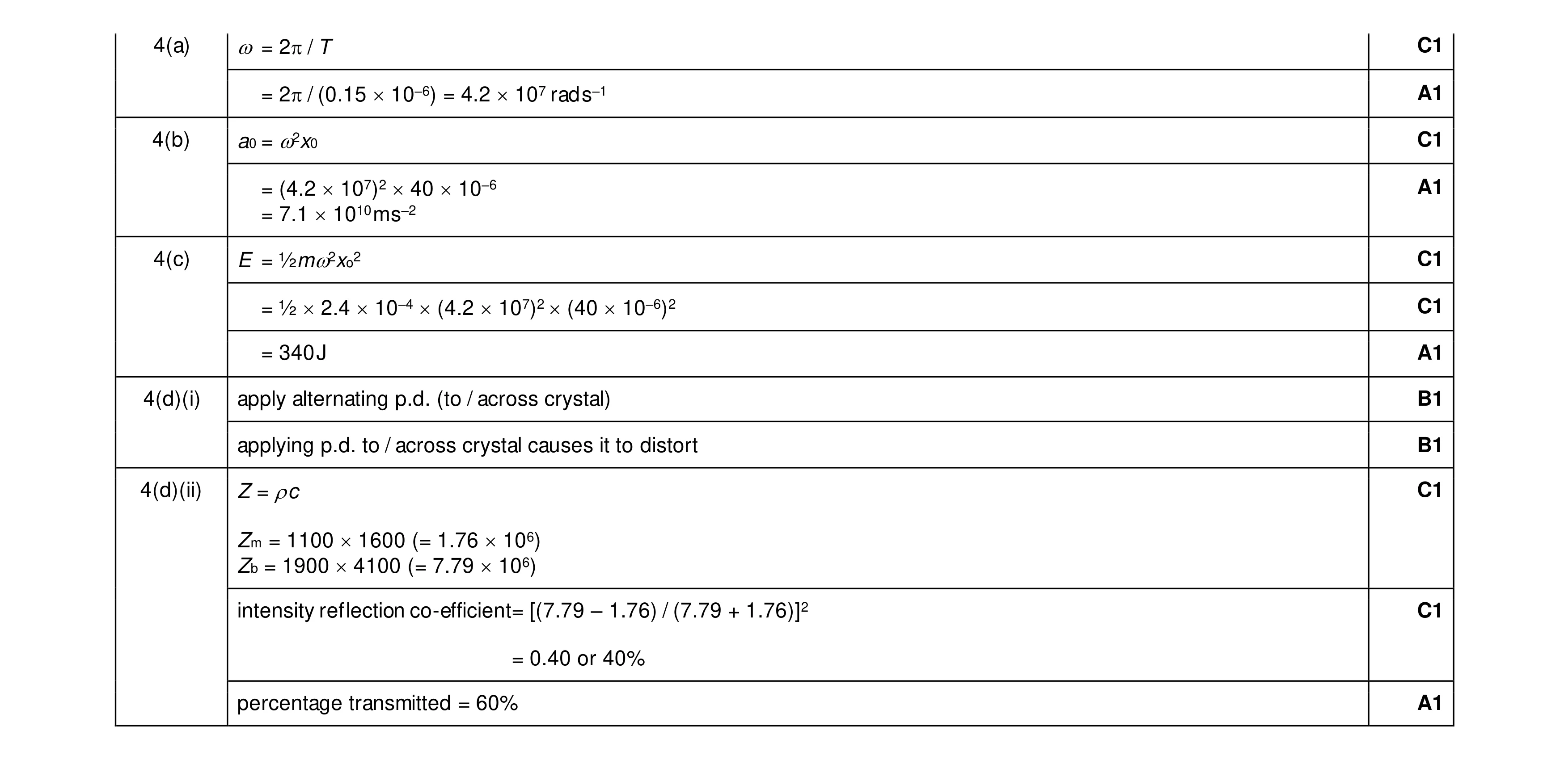 12
12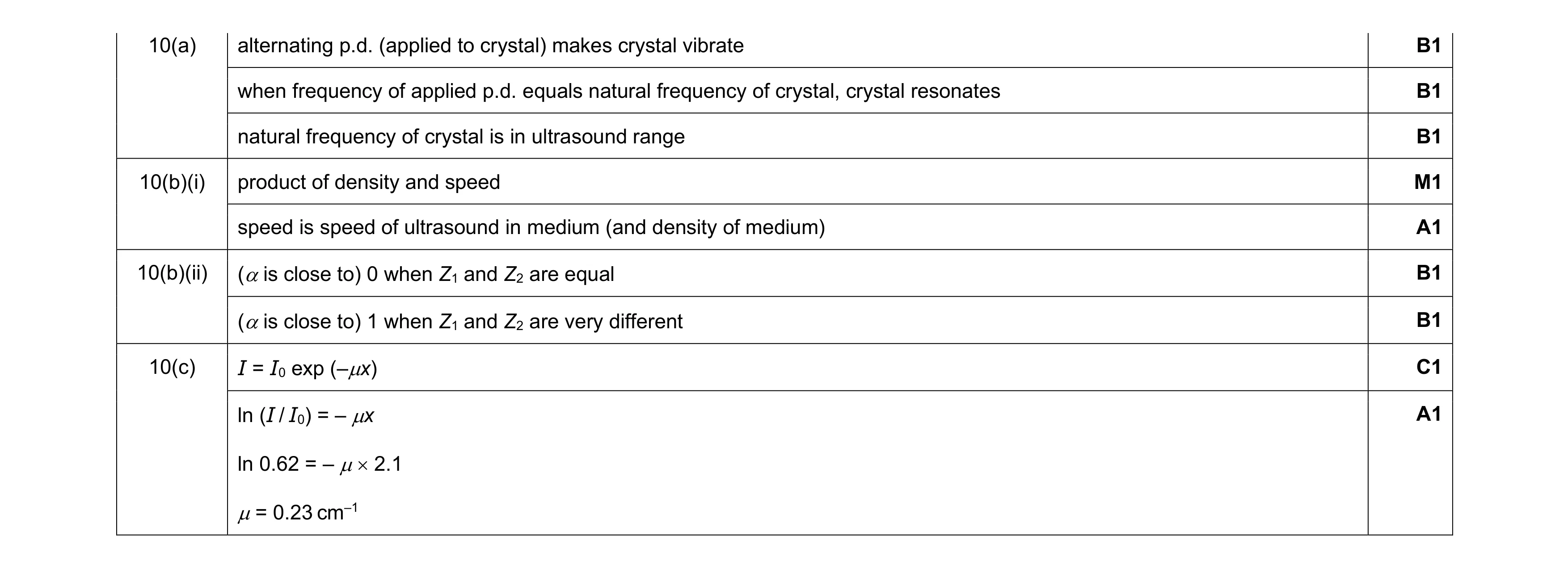 9
9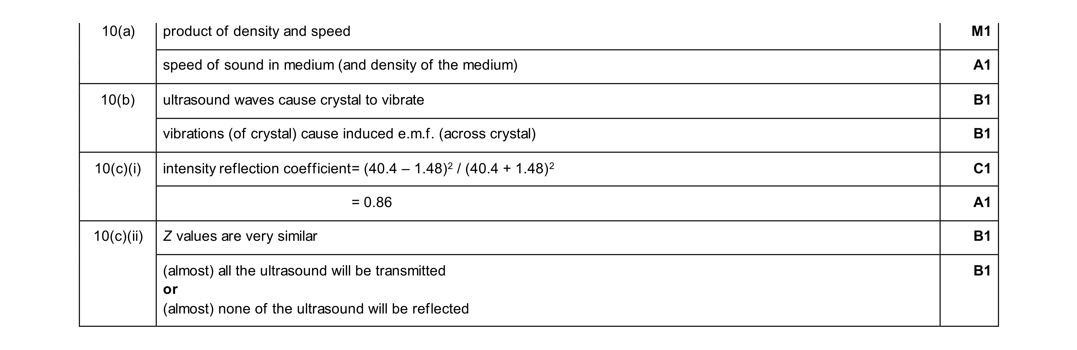 8
8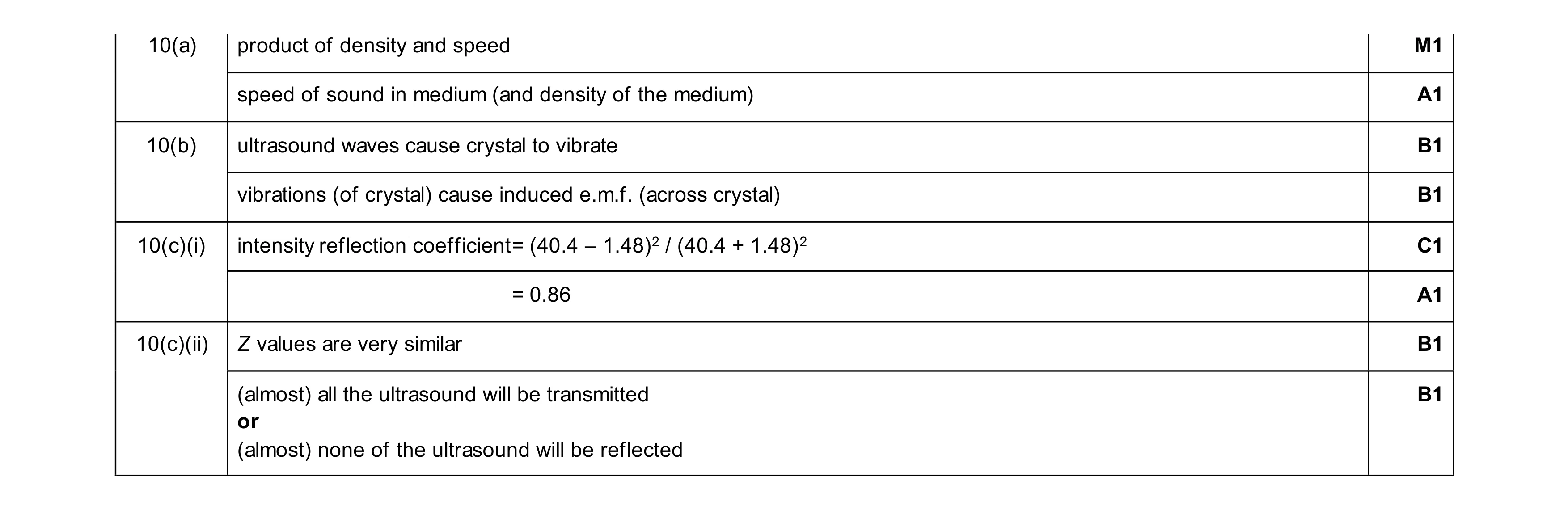 8
8