Questions as text
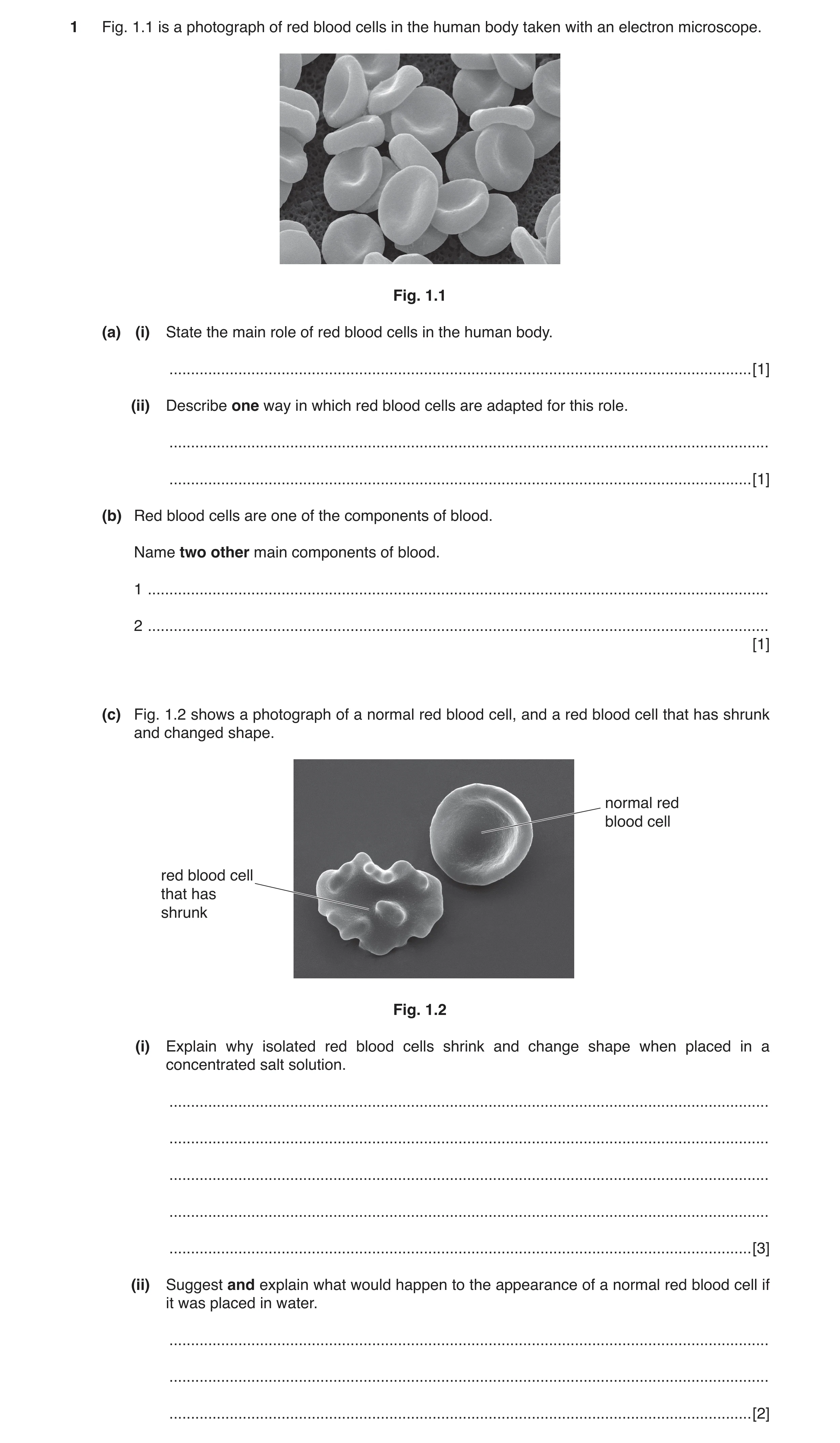
Q1 · A photograph of red blood cells in the human body taken with an electron microscope 0654/42 Oct/Nov 2017
1 Fig. 1.1 is a photograph of red blood cells in the human body taken with an electron microscope. Fig. 1.1 (a) (i) State the main role of red blood cells in the human body. … [1] (ii) Describe one way in which red blood cells are adapted for this role. … … [1] (b) Red blood cells are one of the components of blood. Name two other main components of blood. 1 … 2 … [1] (c) Fig. 1.2 shows a photograph of a normal red blood cell, and a red blood cell that has shrunk and changed shape. normal red blood cell red blood cell that has shrunk Fig. 1.2 (i) Explain why isolated red blood cells shrink and change shape when placed in a concentrated salt solution. … … … … … [3] (ii) Suggest and explain what would happen to the appearance of a normal red blood cell if it was placed in water. … … … [2]
8 marks
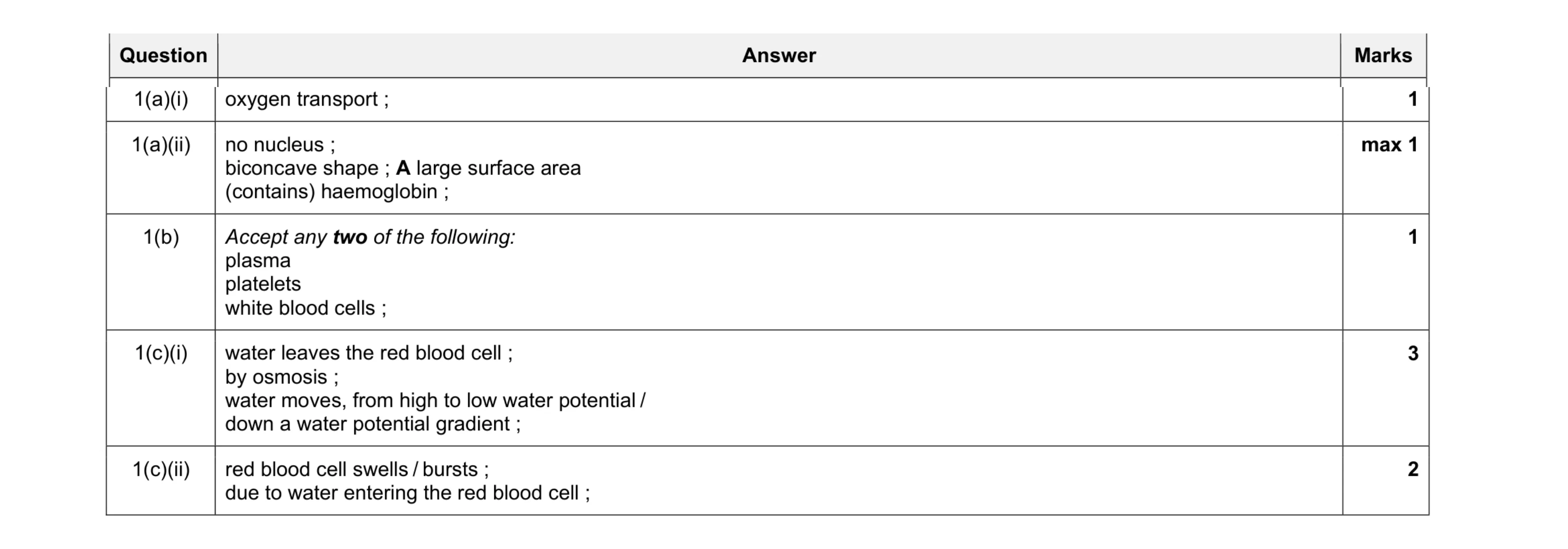
Mark scheme: 1(a)(i) oxygen transport ; 1 1(a)(ii) no nucleus ; biconcave shape ; A large surface area (contains) haemoglobin ; max 1 1(b) Accept any two of the following: plasma platelets white blood cells ; 1 1(c)(i) water leaves the red blood cell ; by osmosis ; water moves, from high to low water potential / down a water potential gradient ; 3 1(c)(ii) red blood cell swells / bursts ; due to water entering the red blood cell ; 2
This question in 0654/42 Oct/Nov 2017
Q2 · HIV is a sexually transmitted infection 0654/41 Oct/Nov 2018
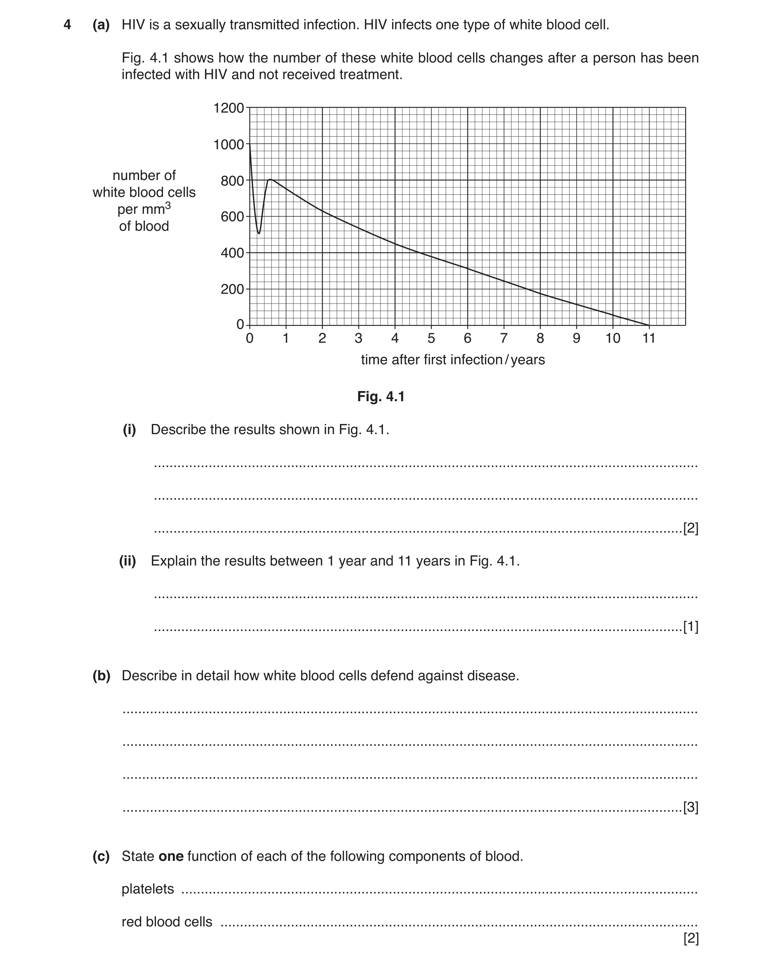
4 (a) HIV is a sexually transmitted infection. HIV infects one type of white blood cell. Fig. 4.1 shows how the number of these white blood cells changes after a person has been infected with HIV and not received treatment. 1200 1000 number of 800 white blood cells per mm3 600 of blood 400 200 0 0 1 2 3 4 5 6 7 8 9 10 11 time after first infection / years Fig. 4.1 (i) Describe the results shown in Fig. 4.1. … … … [2] (ii) Explain the results between 1 year and 11 years in Fig. 4.1. … … [1] (b) Describe in detail how white blood cells defend against disease. … … … … [3] (c) State one function of each of the following components of blood. platelets … red blood cells … [2]
8 marks
Mark scheme: 4(a)(i) (rapid) decrease and increase (in first 6 months) ; decrease after 0.5 years / 6 months / AVP ; 2 4(a)(ii) HIV virus kills / destroys white blood cells ; 1 4(b) antibodies / wbc, destroy, pathogens / bacteria / microorganisms ; (white blood cells) produce / release antibodies ; (white blood cells) responsible for phagocytosis ; AVP ; max 3 4(c) platelets – blood clotting ; red blood cells – transport of oxygen ; 2
This question in 0654/41 Oct/Nov 2018
Q3 · Mammals have a double circulatory system 0654/41 Oct/Nov 2020
1 (a) Mammals have a double circulatory system. Fish have a single circulatory system. Fig. 1.1 shows the circulatory system of a fish and the circulatory system of a mammal. gill capillaries lung capillaries ventricle atria atrium ventricles body capillaries body capillaries fish mammal Fig. 1.1 (i) Use Fig. 1.1 to describe two ways the circulatory system of a fish is different from the circulatory system of a mammal. 1 … … 2 … … [2] (ii) Explain one advantage of having a double circulatory system. … … … … [2] (b) Table 1.1 shows the diameter and the thickness of the wall of two different blood vessels in humans. Table 1.1 type of blood vessel aorta vena cava diameter of blood vessel 25 30 / mm thickness of wall 2.0 1.5 / mm (i) Calculate the difference in thickness of the wall of the aorta and the vena cava. … mm [1] (ii) Explain why the walls of the aorta and the vena cava need to be of different thicknesses. … … … … [2] (c) Red blood cells transport oxygen around the body. (i) Describe two ways red blood cells are adapted for their function. 1 … … 2 … … [2] (ii) State two other major components of blood. 1 … 2 … [2] [Total: 11]
11 marks
Mark scheme: 1(a)(i) single circulatory system only has one, atria / ventricle ; blood flows through heart only once on one circuit of the body ; heart only has two major blood vessels ; AVP ; max 2 1(a)(ii) higher blood pressure to body / greater blood flow (to tissues) ; more efficient delivery of oxygen (for respiration) / more efficient removal of excretory / waste products ; or lower blood pressure to lungs ; delicate lung tissues are not damaged ; max 2 2 1(b)(i) 2 – 1.5 = 0.5 (mm) ; 1 1(b)(ii) (aorta is thicker) so that it doesn’t burst / AW ; because blood is under higher pressure ; 2 1(c)(i) any two from biconcave shape / large surface area ; contain haemoglobin ; no nucleus ; 2 1(c)(ii) any two from white blood cells ; platelets ; plasma ; 2
This question in 0654/41 Oct/Nov 2020
Q4 · The blood groups of some people are recorded 0654/42 Feb/March 2021
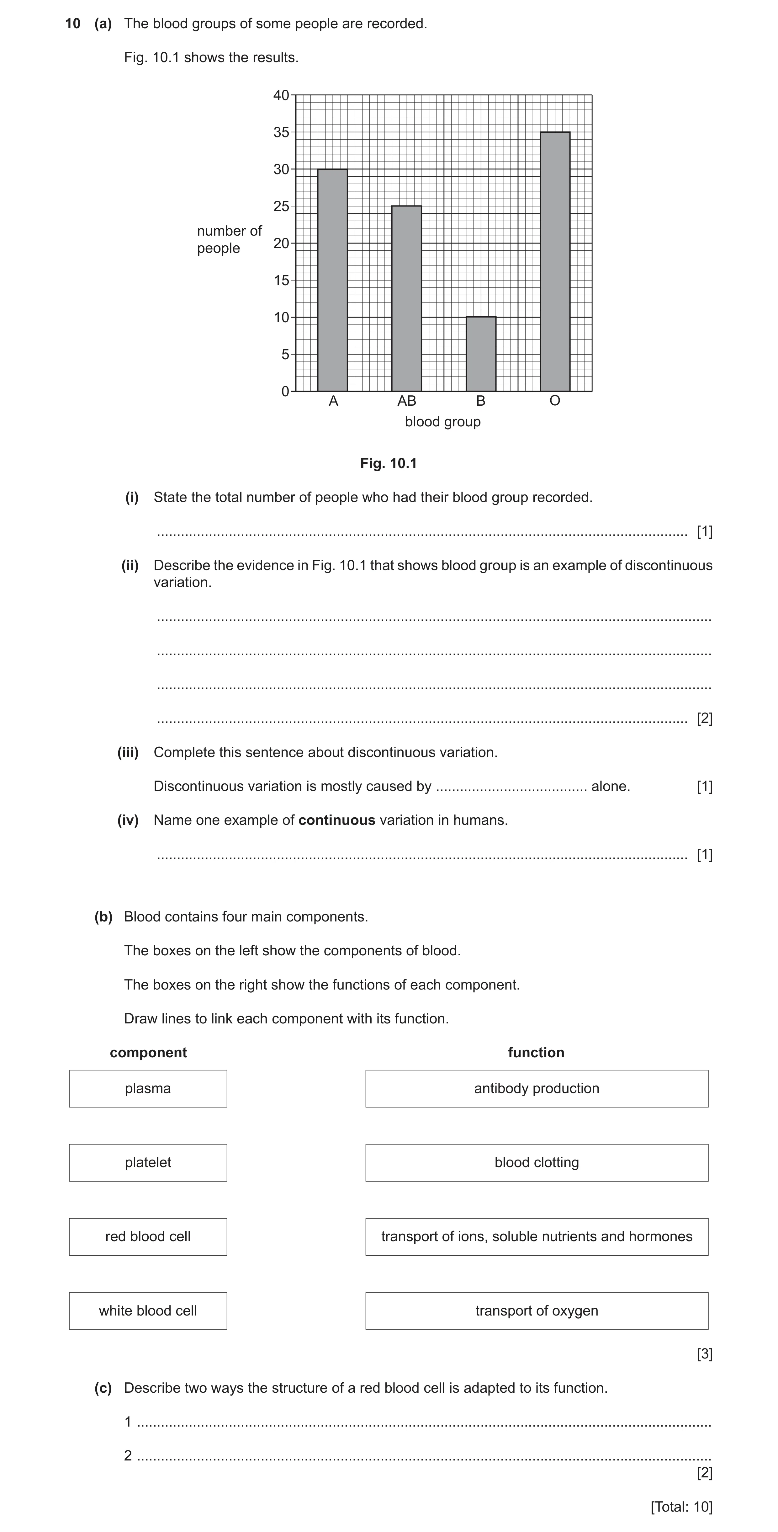
10 (a) The blood groups of some people are recorded. Fig. 10.1 shows the results. 40 35 30 25 number of 20 people 15 10 5 0 A AB B O blood group Fig. 10.1 (i) State the total number of people who had their blood group recorded. … [1] (ii) Describe the evidence in Fig. 10.1 that shows blood group is an example of discontinuous variation. … … … … [2] (iii) Complete this sentence about discontinuous variation. Discontinuous variation is mostly caused by … alone. [1] (iv) Name one example of continuous variation in humans. … [1] (b) Blood contains four main components. The boxes on the left show the components of blood. The boxes on the right show the functions of each component. Draw lines to link each component with its function. component function plasma antibody production platelet blood clotting red blood cell transport of ions, soluble nutrients and hormones white blood cell transport of oxygen [3] (c) Describe two ways the structure of a red blood cell is adapted to its function. 1 … 2 … [2] [Total: 10]
10 marks
Mark scheme: 10(a)(i) 100 ; 1 10(a)(ii) limited number of phenotypes ; no intermediates ; 2 10(a)(iii) genes ; 1 10(a)(iv) height / AVP ; 1 10(b) plasma antibody production platelets blood clotting red blood cell transport of ions, soluble nutrients and hormones white blood cell transport of oxygen ;;; 3 10(c) any two from: no nucleus ; contains haemoglobin ; biconcave shape / large surface area ; 2
This question in 0654/42 Feb/March 2021
Q5 · Some plant cells are immersed in a concentrated salt solution 0654/42 May/June 2021
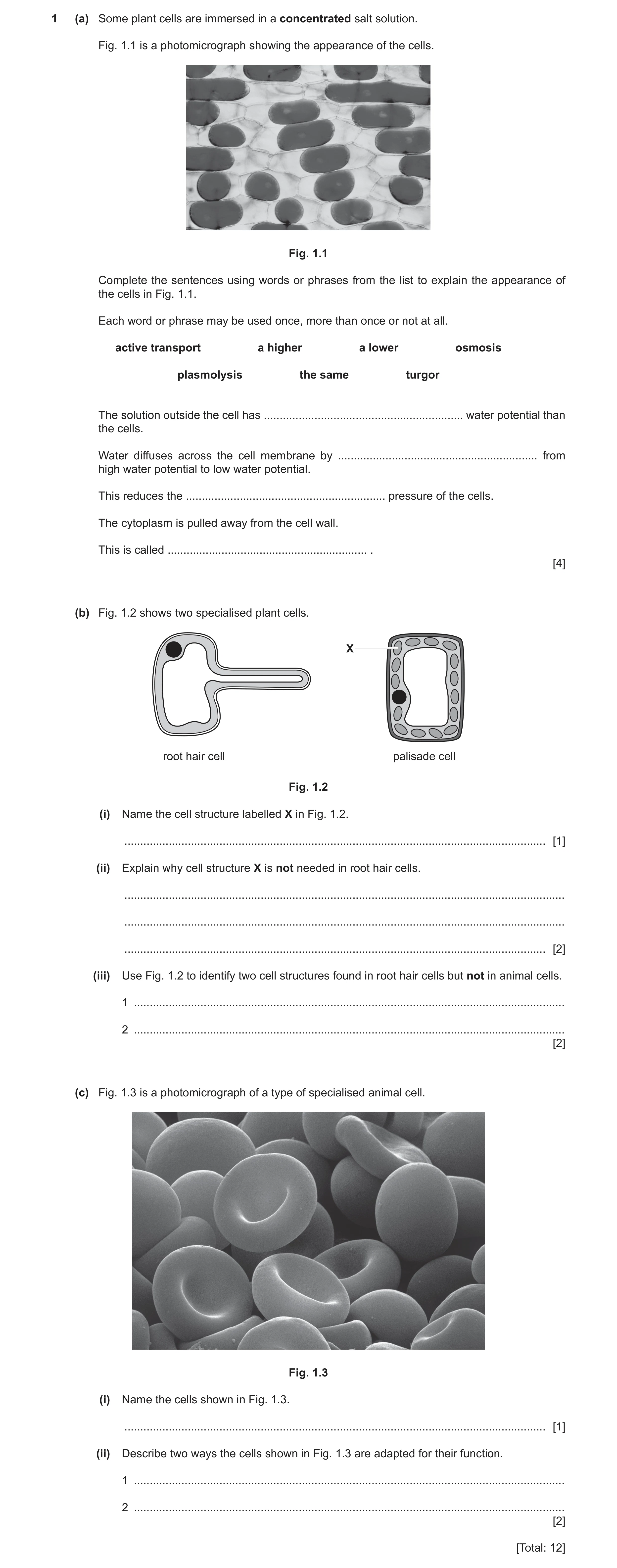
1 (a) Some plant cells are immersed in a concentrated salt solution. Fig. 1.1 is a photomicrograph showing the appearance of the cells. Fig. 1.1 Complete the sentences using words or phrases from the list to explain the appearance of the cells in Fig. 1.1. Each word or phrase may be used once, more than once or not at all. active transport a higher a lower osmosis plasmolysis the same turgor The solution outside the cell has … water potential than the cells. Water diffuses across the cell membrane by … from high water potential to low water potential. This reduces the … pressure of the cells. The cytoplasm is pulled away from the cell wall. This is called … . [4] (b) Fig. 1.2 shows two specialised plant cells. X root hair cell palisade cell Fig. 1.2 (i) Name the cell structure labelled X in Fig. 1.2. … [1] (ii) Explain why cell structure X is not needed in root hair cells. … … … [2] (iii) Use Fig. 1.2 to identify two cell structures found in root hair cells but not in animal cells. 1 … 2 … [2] (c) Fig. 1.3 is a photomicrograph of a type of specialised animal cell. Fig. 1.3 (i) Name the cells shown in Fig. 1.3. … [1] (ii) Describe two ways the cells shown in Fig. 1.3 are adapted for their function. 1 … 2 … [2] [Total: 12]
12 marks
Mark scheme: 1(a) a lower ; osmosis ; turgor ; plasmolysis ; 4 1(b)(i) chloroplast ; 1 1(b)(ii) any two from: root hair cells absorb, water / mineral ions ; root hair cells, are underground / do not receive light ; correct reference to photosynthesis ; 2 1(b)(iii) vacuole ; cell wall ; 2 1(c)(i) red blood cells ; 1 1(c)(ii) any two from: contain haemoglobin ; no nucleus ; biconcave shape / large surface area ; AVP ; 2
This question in 0654/42 May/June 2021
Q6 · Hormones control blood glucose concentration 0654/41 Oct/Nov 2021
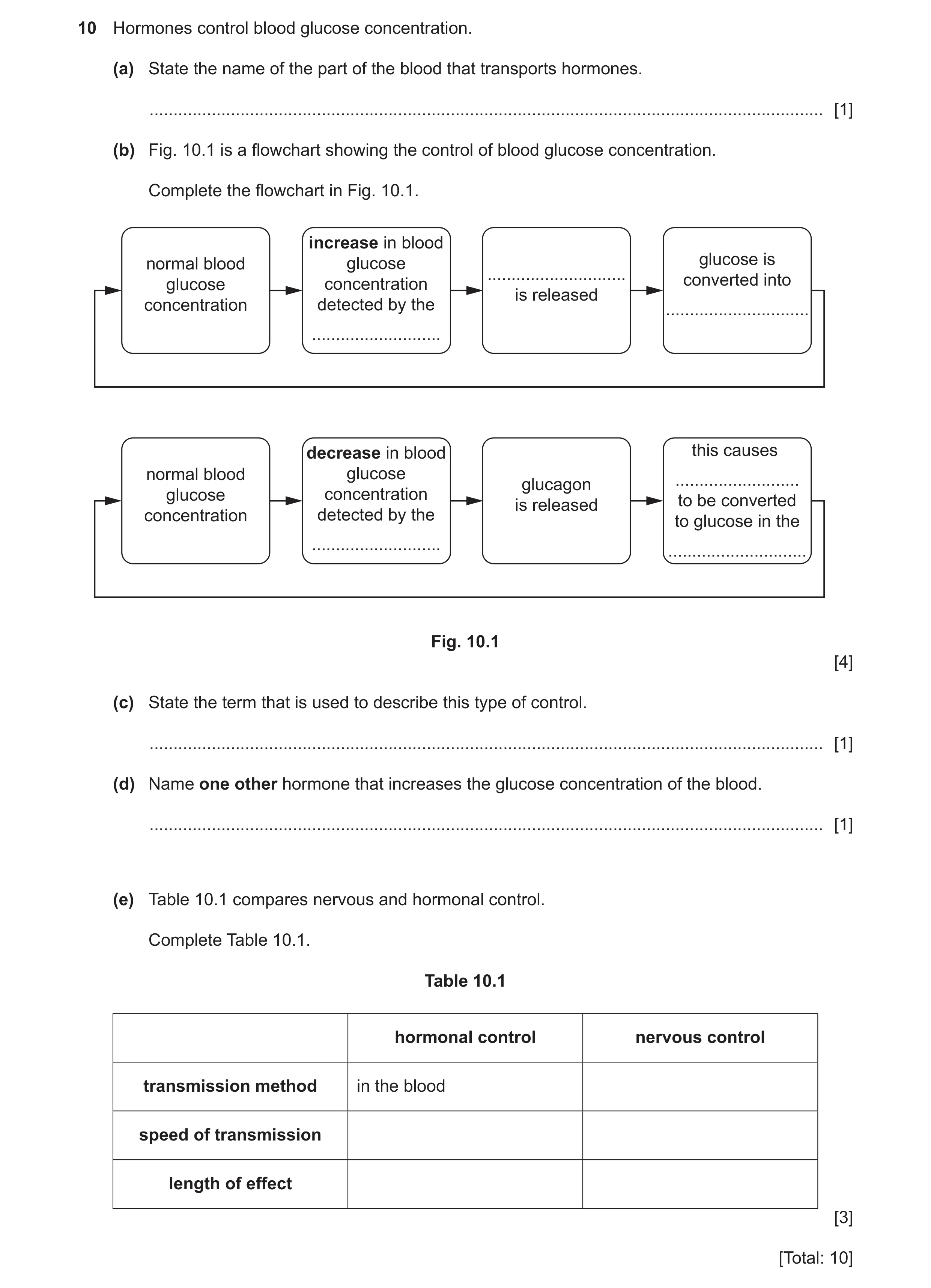
10 Hormones control blood glucose concentration. (a) State the name of the part of the blood that transports hormones. … [1] (b) Fig. 10.1 is a flowchart showing the control of blood glucose concentration. Complete the flowchart in Fig. 10.1. increase in blood normal blood glucose glucose is … glucose concentration converted into is released concentration detected by the … … decrease in blood this causes normal blood glucose glucagon … glucose concentration is released to be converted concentration detected by the to glucose in the … … Fig. 10.1 [4] (c) State the term that is used to describe this type of control. … [1] (d) Name one other hormone that increases the glucose concentration of the blood. … [1] (e) Table 10.1 compares nervous and hormonal control. Complete Table 10.1. Table 10.1 hormonal control nervous control transmission method in the blood speed of transmission length of effect [3] [Total: 10]
10 marks
Mark scheme: 10(a) plasma ; 1 10(b) pancreas in boxes 2 and 6 ; insulin in box 3 ; glycogen in box 4 and 8 ; liver in box 8 ; 4 10(c) negative feedback ; 1 10(d) adrenaline ; 1 10(e) hormonal control nervous control transmission method in the blood by neurones / electrical impulses ; relative speed of transmission slow fast ; relative length of effect long lasting short-lived ; 3
This question in 0654/41 Oct/Nov 2021
Q7 · Gills are the gas exchange surface in fish 0654/43 Oct/Nov 2022
1 (a) Gills are the gas exchange surface in fish. Fig. 1.1 is a photograph of gills in fish. Fig. 1.1 Gills have adaptive features for gas exchange. Use your scientific knowledge and Fig. 1.1 to suggest two of these adaptive features. 1 … 2 … [2] (b) Table 1.1 shows the composition of some gases in inspired and expired air. Table 1.1 gas percentage of oxygen percentage of carbon dioxide inspired air 21 0.004 expired air 16 4 (i) Use Table 1.1 to calculate the difference in percentage of oxygen between inspired and expired air. … % [1] (ii) Explain the difference in percentage of oxygen between inspired and expired air that is shown in Table 1.1. … … … [2] (iii) State one difference in composition between inspired and expired air that is not shown in Table 1.1. … … [1] (c) Red blood cells have adaptive features for the efficient transport of oxygen. State two of these features. 1 … 2 … [2] (d) Lung cancer is a disease caused by smoking. (i) Place ticks (3) to show two other diseases caused by smoking. chronic obstructive pulmonary disease (COPD) coronary heart disease kwashiorkor marasmus scurvy [2] (ii) State the name of the component in tobacco smoke that causes cancer. … [1] (iii) Describe how the goblet cells, mucus and ciliated cells protect the gas exchange system from some of the particles in tobacco smoke. … … … … … [3] [Total: 14]
14 marks
Mark scheme: Question Answer Marks 1(a) any two from: 2 large surface area ; thin (surface) ; good blood supply ; any valid point ; 1(b)(i) 5 (%) ; 1 1(b)(ii) oxygen is required for (aerobic) respiration ; 2 to release energy (for metabolic processes) ; 1(b)(iii) greater percentage of water (vapour) in expired air ; 1 1(c) any two from: 2 large surface area / biconcave shape ; contain haemoglobin ; no nucleus ; 1(d)(i) COPD ticked ; 2 CHD ticked ; 1(d)(ii) tar ; 1 1(d)(iii) goblet cells produce mucus ; 3 mucus traps particles/pathogens ; cilia move mucus/particles/pathogens (away from gas exchange surface) ;
This question in 0654/43 Oct/Nov 2022
Q8 · A diagram showing the development of a strain of antibiotic resistant bacteria 0654/41 May/June 2023
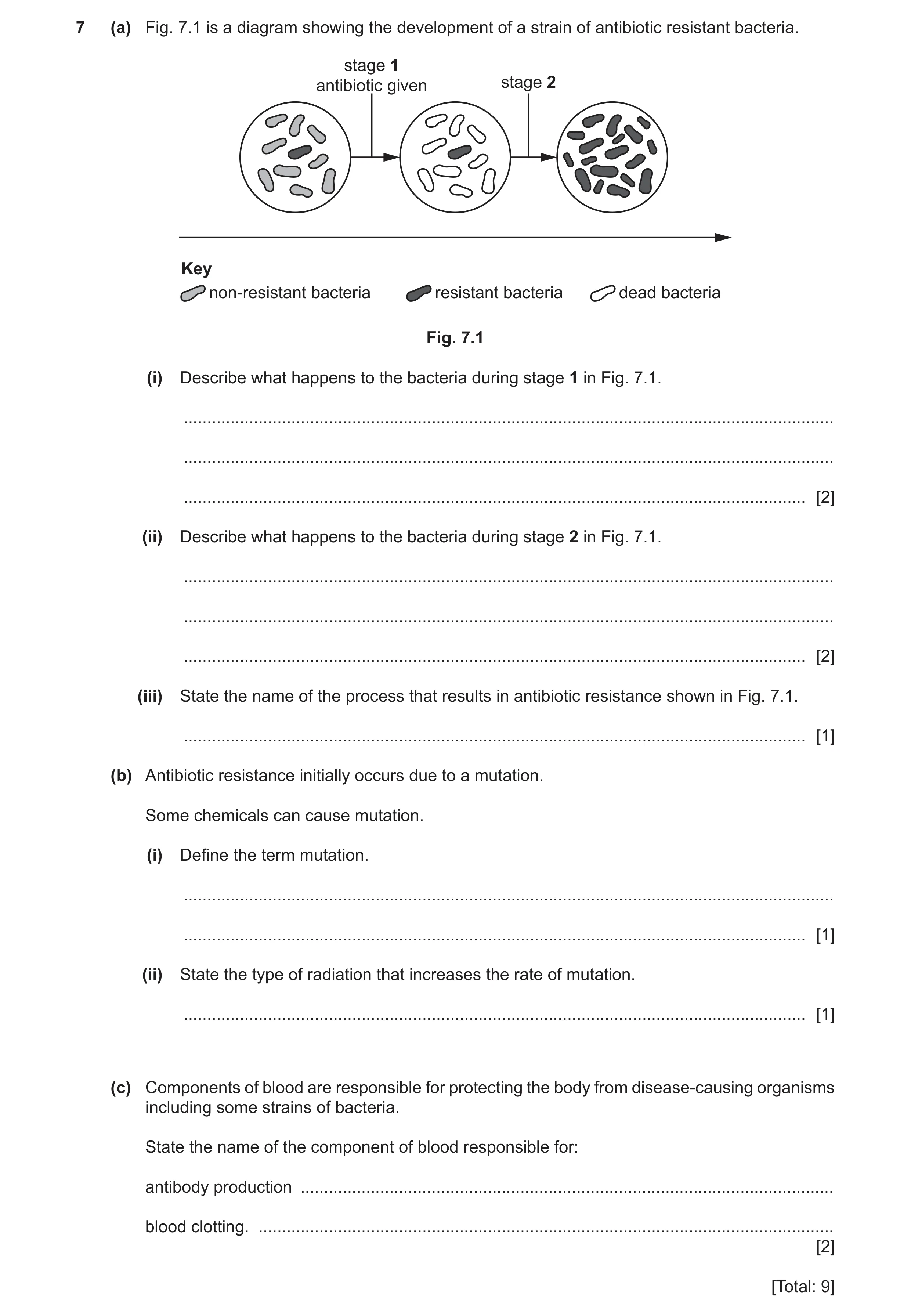
7 (a) Fig. 7.1 is a diagram showing the development of a strain of antibiotic resistant bacteria. stage 1 antibiotic given stage 2 Key non-resistant bacteria resistant bacteria dead bacteria Fig. 7.1 (i) Describe what happens to the bacteria during stage 1 in Fig. 7.1. … … … [2] (ii) Describe what happens to the bacteria during stage 2 in Fig. 7.1. … … … [2] (iii) State the name of the process that results in antibiotic resistance shown in Fig. 7.1. … [1] (b) Antibiotic resistance initially occurs due to a mutation. Some chemicals can cause mutation. (i) Define the term mutation. … … [1] (ii) State the type of radiation that increases the rate of mutation. … [1] (c) Components of blood are responsible for protecting the body from disease‑causing organisms including some strains of bacteria. State the name of the component of blood responsible for: antibody production … blood clotting. … [2] [Total: 9]
9 marks
Mark scheme: 7(a)(i) the bacteria with (antibiotic) resistance survive ; antibiotics kill the bacteria without (antibiotic) resistance ; 2 7(a)(ii) the bacteria with (antibiotic) resistance, multiplies / divides ; passing on alleles / genes / (mutated) DNA to the next generation / the offspring all gain resistance (to antibiotics) ; 2 7(a)(iii) natural selection ; 1 7(b)(i) change in, gene / chromosome ; 1 7(b)(ii) ionising ; 1 7(c) white blood cells ; platelets ; 2
This question in 0654/41 May/June 2023
Q9 · Two red blood cells after they have been immersed in different solutions for an hour 0654/41 May/June 2023
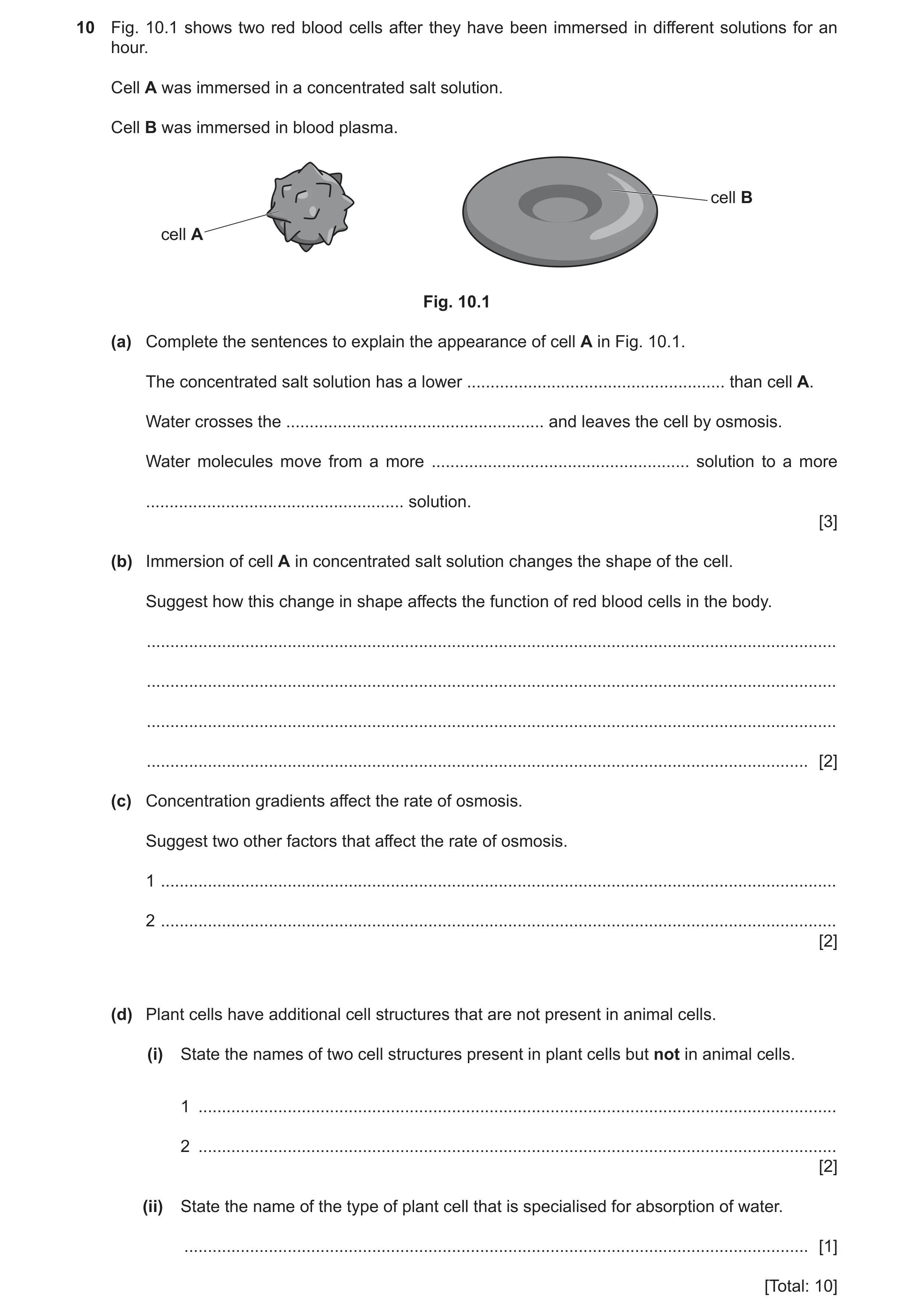
10 Fig. 10.1 shows two red blood cells after they have been immersed in different solutions for an hour. Cell A was immersed in a concentrated salt solution. Cell B was immersed in blood plasma. cell B cell A Fig. 10.1 (a) Complete the sentences to explain the appearance of cell A in Fig. 10.1. The concentrated salt solution has a lower … than cell A. Water crosses the … and leaves the cell by osmosis. Water molecules move from a more … solution to a more … solution. [3] (b) Immersion of cell A in concentrated salt solution changes the shape of the cell. Suggest how this change in shape affects the function of red blood cells in the body. … … … … [2] (c) Concentration gradients affect the rate of osmosis. Suggest two other factors that affect the rate of osmosis. 1 … 2 … [2] (d) Plant cells have additional cell structures that are not present in animal cells. (i) State the names of two cell structures present in plant cells but not in animal cells. 1 … 2 … [2] (ii) State the name of the type of plant cell that is specialised for absorption of water. … [1] [Total: 10]
10 marks
Mark scheme: 10(a) water potential ; (cell) membrane ; dilute, concentrated ; 3 10(b) any two from: less, surface area / (internal) volume ; so less oxygen transported ; AVP ; 2 Question Answer Marks 10(c) any two from: surface area ; temperature ; diffusion distance/thickness of cell membrane ; 2 10(d)(i) any two from: chloroplast ; (permanent) vacuole ; cell wall ; 2 10(d)(ii) root hair (cell) ; 1
This question in 0654/41 May/June 2023
Q10 · A diagram of the male reproductive system in humans 0654/42 Feb/March 2024
1 (a) Fig. 1.1 is a diagram of the male reproductive system in humans. A E B D C Fig. 1.1 State the letter from Fig. 1.1 that identifies the part: that is a tube transporting excretory products … that secretes fluid for the formation of semen … where meiosis occurs. … [3] (b) Fig. 1.2 is a diagram of a sperm cell. Y X Fig. 1.2 (i) State how the arrangement of the chromosomes in the part labelled X in Fig. 1.2 is different to that of a human body cell. … … [1] (ii) State the name and function of the adaptive feature labelled Y in Fig. 1.2. name … function … … [2] (c) The list shows the names of some specialised cells. ciliated cell egg cell palisade mesophyll cell red blood cell root hair cell white blood cell Choose one cell from the list that: contains a haploid nucleus … does not contain a nucleus … is found in the bronchi … is responsible for phagocytosis. … [4] (d) Red blood cells and white blood cells are components of blood. State the name of one other major component of blood. … [1] [Total: 11]
11 marks
Mark scheme: Question Answer Marks 1(a) B ; 3 E ; D ; 1(b)(i) unpaired ; 1 1(b)(ii) flagellum ; 2 movement / locomotion ; 1(c) egg (cell) ; 4 red blood (cell) ; ciliated (cell) ; white blood (cell) ; 1(d) plasma / platelets ; 1
This question in 0654/42 Feb/March 2024
Q11 · Blood glucose concentration is controlled so that it remains within set limits 0654/42 May/June 2024
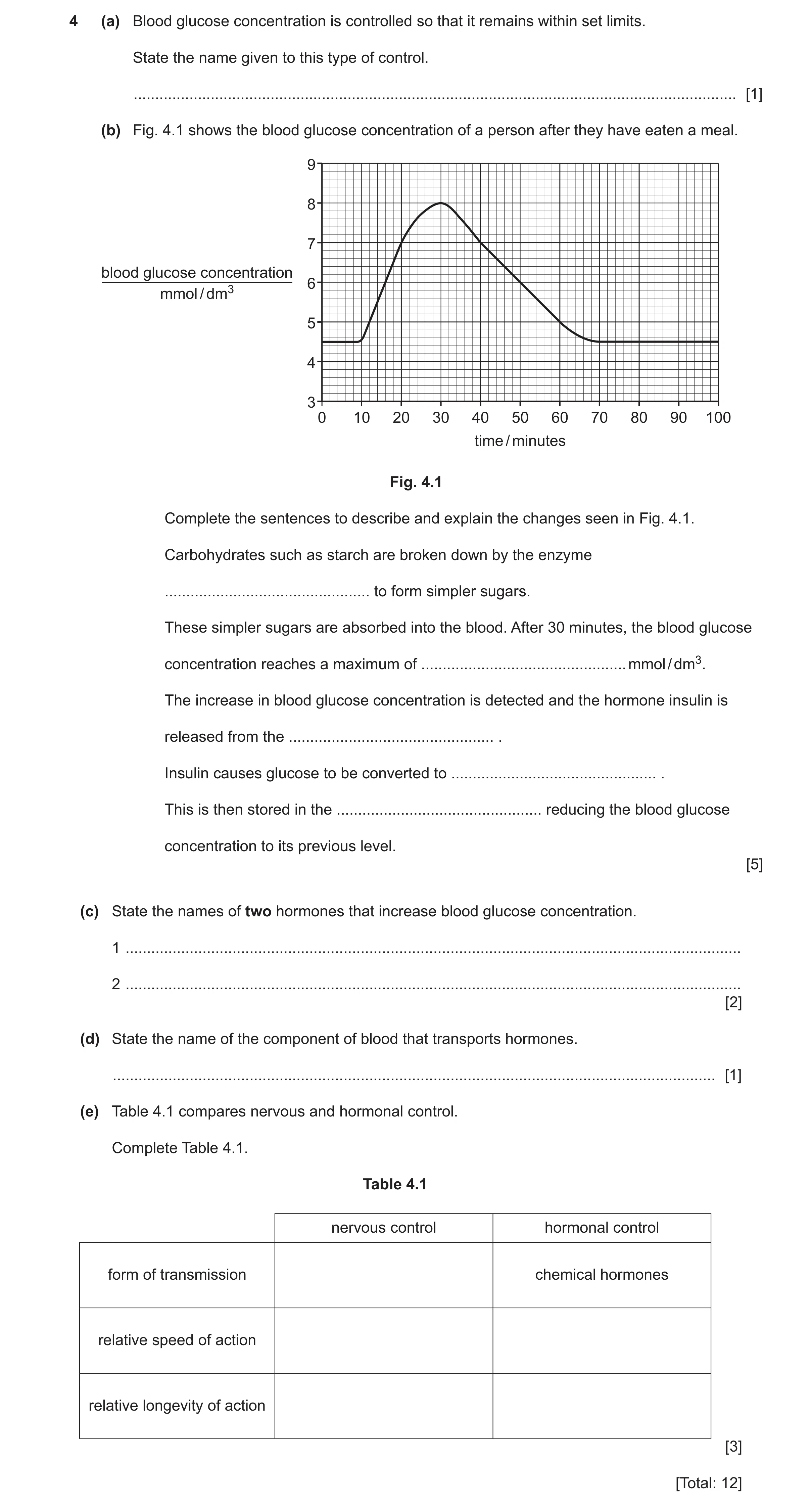
4 (a) Blood glucose concentration is controlled so that it remains within set limits. State the name given to this type of control. … [1] (b) Fig. 4.1 shows the blood glucose concentration of a person after they have eaten a meal. 9 8 7 blood glucose concentration 6 mmol / dm3 5 4 3 0 10 20 30 40 50 60 70 80 90 100 time / minutes Fig. 4.1 Complete the sentences to describe and explain the changes seen in Fig. 4.1. Carbohydrates such as starch are broken down by the enzyme … to form simpler sugars. These simpler sugars are absorbed into the blood. After 30 minutes, the blood glucose concentration reaches a maximum of … mmol / dm3. The increase in blood glucose concentration is detected and the hormone insulin is released from the … . Insulin causes glucose to be converted to … . This is then stored in the … reducing the blood glucose concentration to its previous level. [5] (c) State the names of two hormones that increase blood glucose concentration. 1 … 2 … [2] (d) State the name of the component of blood that transports hormones. … [1] (e) Table 4.1 compares nervous and hormonal control. Complete Table 4.1. Table 4.1 nervous control hormonal control form of transmission chemical hormones relative speed of action relative longevity of action [3] [Total: 12]
12 marks
Mark scheme: 4(a) homeostasis ; 1 4(b) amylase ; 8.0 (mmol per dm3) ; pancreas ; glycogen ; liver / muscles ; 5 4(c) adrenaline ; glucagon ; 2 4(d) plasma ; 1 4(e) nervous control hormonal control form of transmission electrical impulse chemical hormones relative speed of action fast slow relative longevity of action short-lived / AW long-lasting / AW ;;; 3
This question in 0654/42 May/June 2024
Q12 · Red blood cells are specialised to transport oxygen 0654/42 May/June 2024
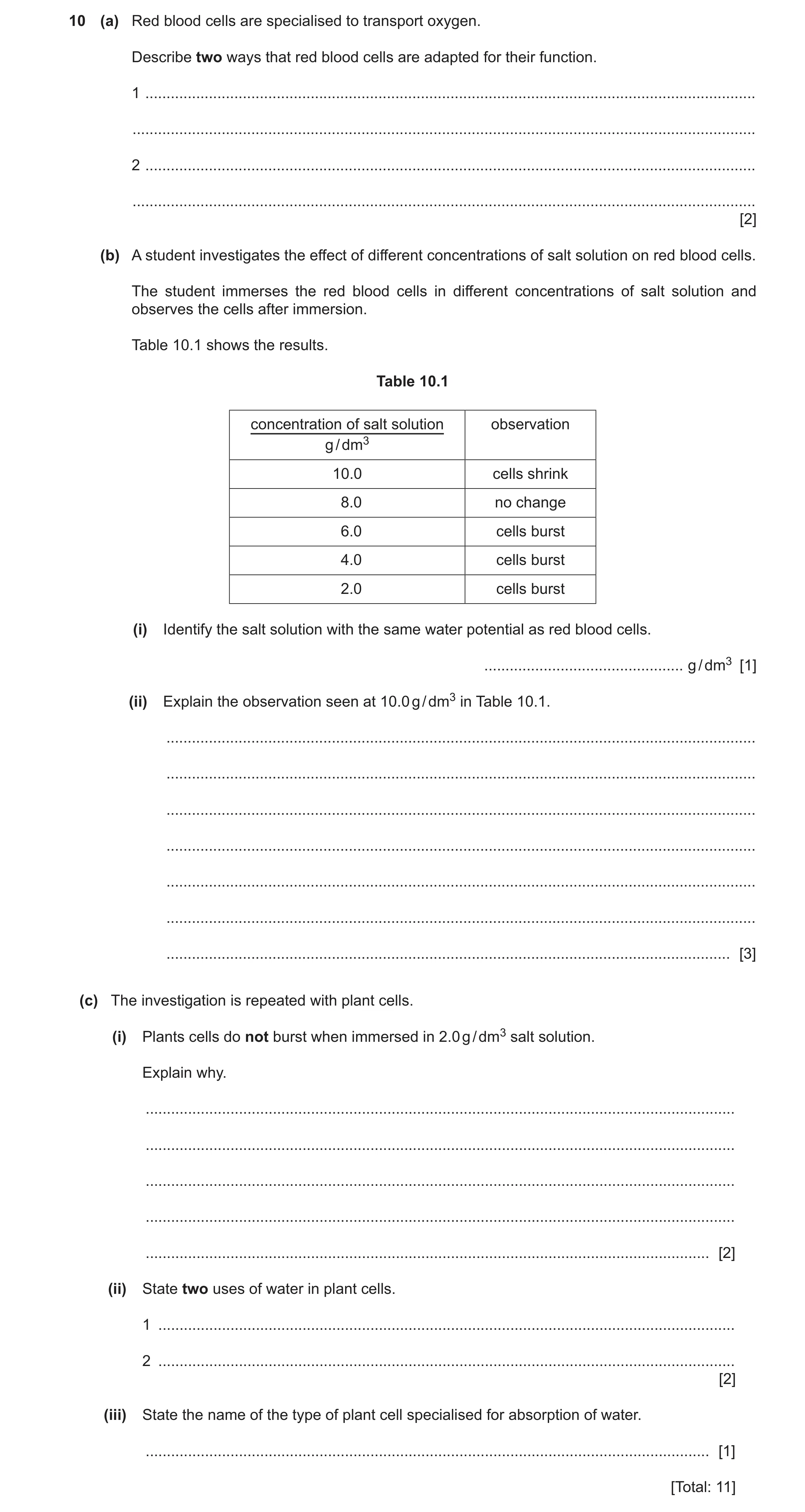
10 (a) Red blood cells are specialised to transport oxygen. Describe two ways that red blood cells are adapted for their function. 1 … … 2 … … [2] (b) A student investigates the effect of different concentrations of salt solution on red blood cells. The student immerses the red blood cells in different concentrations of salt solution and observes the cells after immersion. Table 10.1 shows the results. Table 10.1 concentration of salt solution observation g / dm3 10.0 cells shrink 8.0 no change 6.0 cells burst 4.0 cells burst 2.0 cells burst (i) Identify the salt solution with the same water potential as red blood cells. … g / dm3 [1] (ii) Explain the observation seen at 10.0 g / dm3 in Table 10.1. … … … … … … … [3] (c) The investigation is repeated with plant cells. (i) Plants cells do not burst when immersed in 2.0 g / dm3 salt solution. Explain why. … … … … … [2] (ii) State two uses of water in plant cells. 1 … 2 … [2] (iii) State the name of the type of plant cell specialised for absorption of water. … [1] [Total: 11]
11 marks
Mark scheme: 10(a) any two from: no nucleus ; large surface area / biconcave (shape) ; contains haemoglobin ; 2 10(b)(i) 8.0 (g per dm3) ; 1 Question Answer Marks 10(b)(ii) water leaves the cell ; by osmosis ; the salt solution has a lower water potential (than the red blood cell) / water moves from an area of high water potential to low water potential ; 3 10(c)(i) presence of cell wall ; strengthens the cell ; 2 10(c)(ii) any two from: solvent ; photosynthesis ; support / ref to turgidity / prevent wilting ; transport of, minerals / mineral ions / ions ; AVP ; 2 10(c)(iii) root hair (cell) ; 1
This question in 0654/42 May/June 2024
Q13 · The circulatory system in horses is similar to in humans 0654/42 Oct/Nov 2024
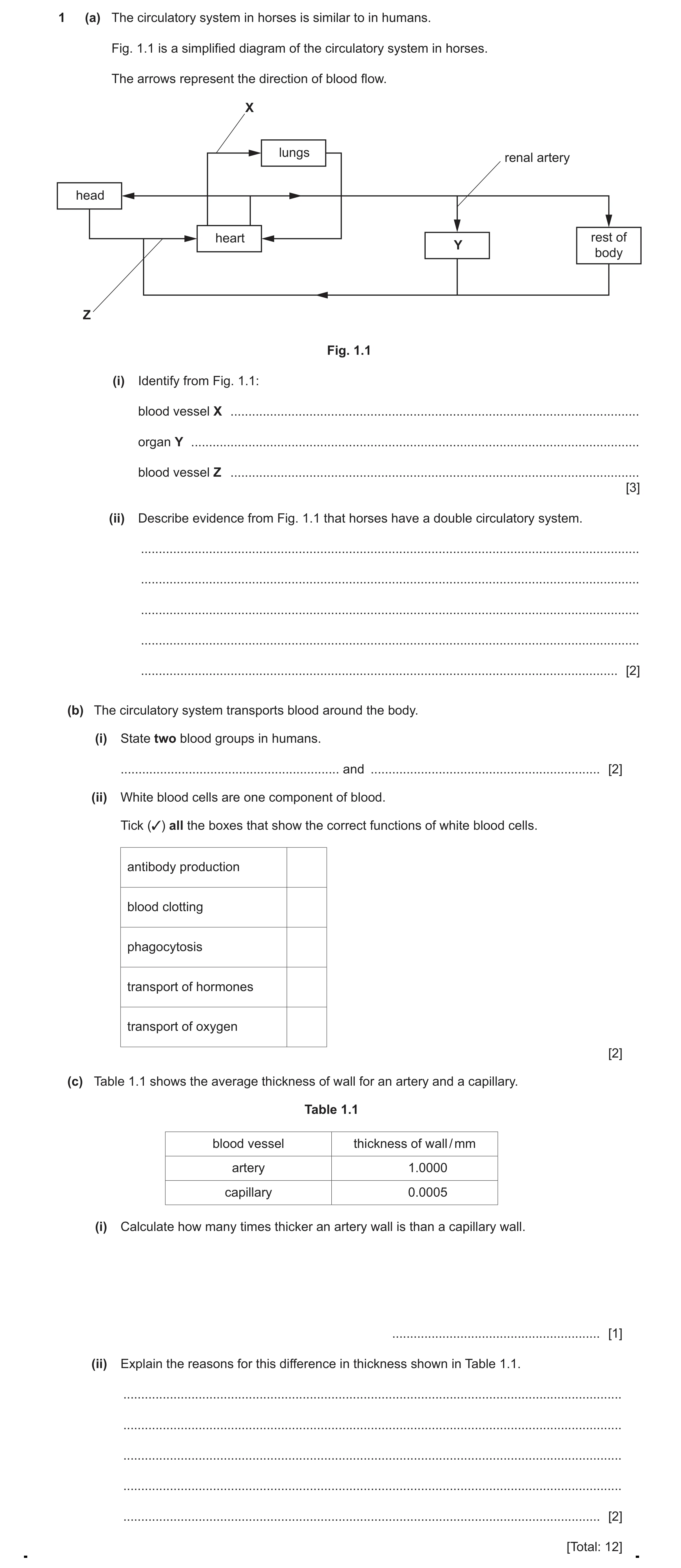
1 (a) The circulatory system in horses is similar to in humans. Fig. 1.1 is a simplified diagram of the circulatory system in horses. The arrows represent the direction of blood flow. X lungs renal artery head heart rest of Y body Z Fig. 1.1 (i) Identify from Fig. 1.1: blood vessel X … organ Y … blood vessel Z … [3] (ii) Describe evidence from Fig. 1.1 that horses have a double circulatory system. … … … … … [2] (b) The circulatory system transports blood around the body. (i) State two blood groups in humans. … and … [2] (ii) White blood cells are one component of blood. Tick (✓) all the boxes that show the correct functions of white blood cells. antibody production blood clotting phagocytosis transport of hormones transport of oxygen [2] (c) Table 1.1 shows the average thickness of wall for an artery and a capillary. Table 1.1 blood vessel thickness of wall / mm artery 1.0000 capillary 0.0005 (i) Calculate how many times thicker an artery wall is than a capillary wall. … [1] (ii) Explain the reasons for this difference in thickness shown in Table 1.1. … … … … … [2] [Total: 12]
12 marks
Mark scheme: Question Answer Marks 1(a)(i) X pulmonary artery ; 3 Y kidney; Z vena cava ; 1(a)(ii) (one circulation) to the lungs ; 2 (one circulation) to the (rest of) body (tissues) ; 1(b)(i) any two from: 2 A ; B ; AB ; O ; 1(b)(ii) 2 antibody production blood clotting phagocytosis transport of hormones transport of oxygen 1 mark for each correct tick ;; 1(c)(i) (1 / 0.0005 =) 2000 ; 1 1(c)(ii) (artery wall is thick to withstand / maintain) high (blood) pressure ; 2 (capillary wall is thin) to provide a short diffusion distance / AW ;
This question in 0654/42 Oct/Nov 2024
Q14 · Blood plasma transports blood cells and platelets 0654/42 Feb/March 2025
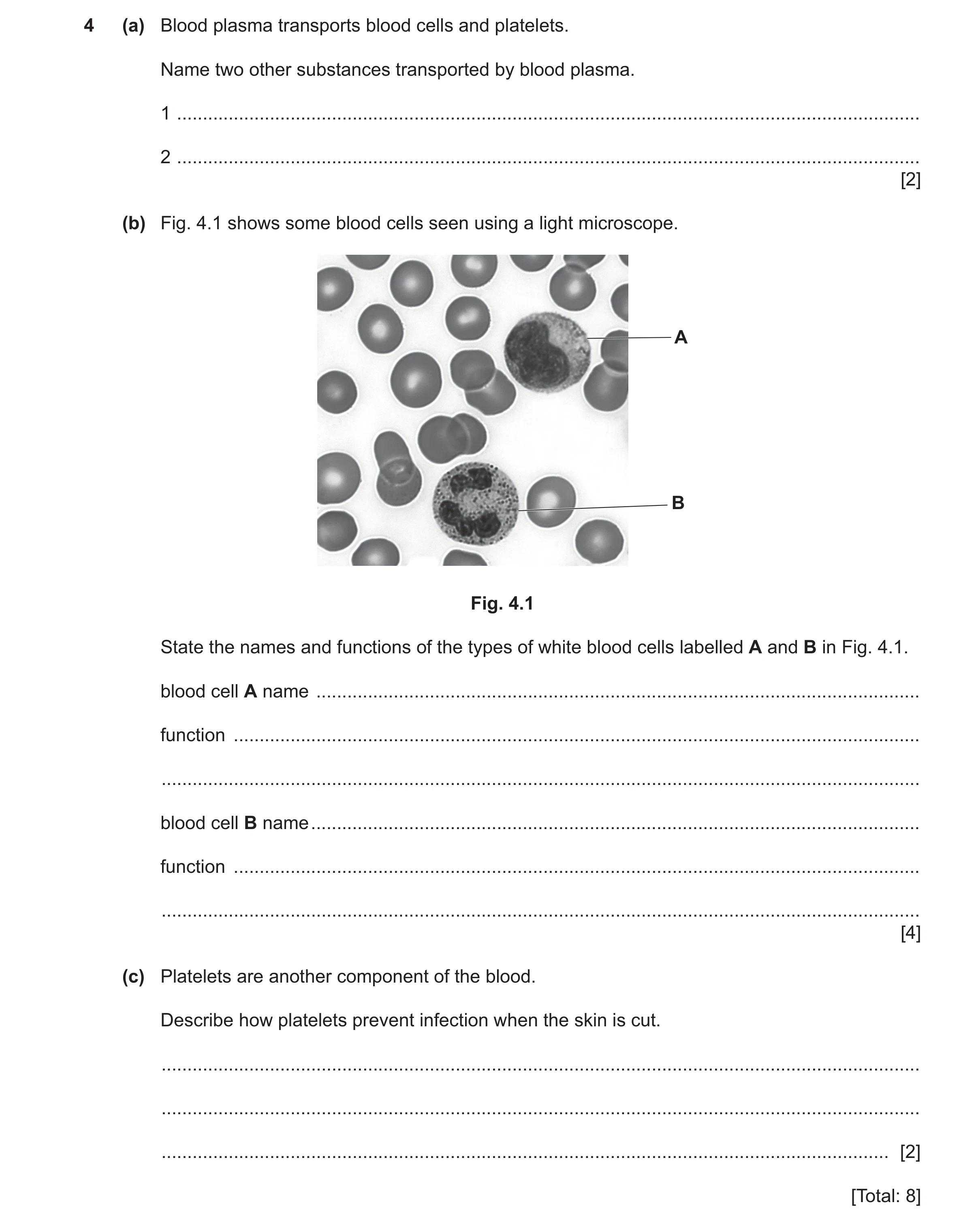
4 (a) Blood plasma transports blood cells and platelets. Name two other substances transported by blood plasma. 1 … 2 … [2] (b) Fig. 4.1 shows some blood cells seen using a light microscope. A B Fig. 4.1 State the names and functions of the types of white blood cells labelled A and B in Fig. 4.1. blood cell A name … function … … blood cell B name … function … … [4] (c) Platelets are another component of the blood. Describe how platelets prevent infection when the skin is cut. … … … [2] [Total: 8]
8 marks
Mark scheme: 4(a) any two from: 2 ions (named) nutrients urea hormones carbon dioxide AVP ; ; 4(b) A - lymphocyte ; 4 antibody production ; B- phagocyte ; engulfing pathogens / phagocytosis ; 4(c) clot blood ; 2 prevent entry of pathogens ;
This question in 0654/42 Feb/March 2025
Q15 · The circulatory system of a fish 0654/43 May/June 2025
1 Fig. 1.1 shows the circulatory system of a fish. capillaries in gills heart capillaries in organs Fig. 1.1 (a) Put an X on Fig. 1.1 to show one area where oxygenated blood is flowing. [1] (b) Mammals have a double circulation of blood. Explain the advantages of a double circulation compared to the circulation in a fish. … … … … … [3] (c) Capillaries have thin walls. (i) Tick (3) one box to identify which statement explains why capillaries have thin walls. to allow blood to flow out to allow movement of substances in and out of blood to make space for more tissues in organs to increase the rate of blood flow [1] (ii) Fish gills have a good blood supply as they have many capillaries with thin walls. Suggest one other feature that makes the gills a good gas exchange surface. … … [1] (d) The healthy range for platelets in human blood is between 150 000 and 400 000 platelets per mm3. An 8 mm3 sample is taken from a patient. The sample contains 1.022 × 10⁶ platelets. (i) Calculate the number of platelets per mm3 in the sample. … platelets per mm3 [1] (ii) Suggest how this number of platelets may affect the patient if they cut themselves. … … … … … [3] [Total: 10]
10 marks
Mark scheme: Question Answer Marks 1(a) X on right hand side of circulatory system; 1 1(b) any three from: 3 blood circulated more quickly (in mammalian system); high blood pressure (around body); low blood pressure to lungs; more effective delivery of oxygen / nutrients (in mammalian system); oxygenated and deoxygenated blood kept separate; 1(c)(i) to allow movement of substances in and out of blood; 1 1(c)(ii) large surface area / good ventilation (with water); 1 1(d)(i) 127 750 / 1.278 105 (platelets per mm3) ; 1 1(d)(ii) (platelets) below healthy range; 3 AND any 2 from less clotting; (more) entry of pathogens into wound; increased blood loss;
This question in 0654/43 May/June 2025
Q16 · A diagram of the heart and associated blood vessels 0654/43 Oct/Nov 2025
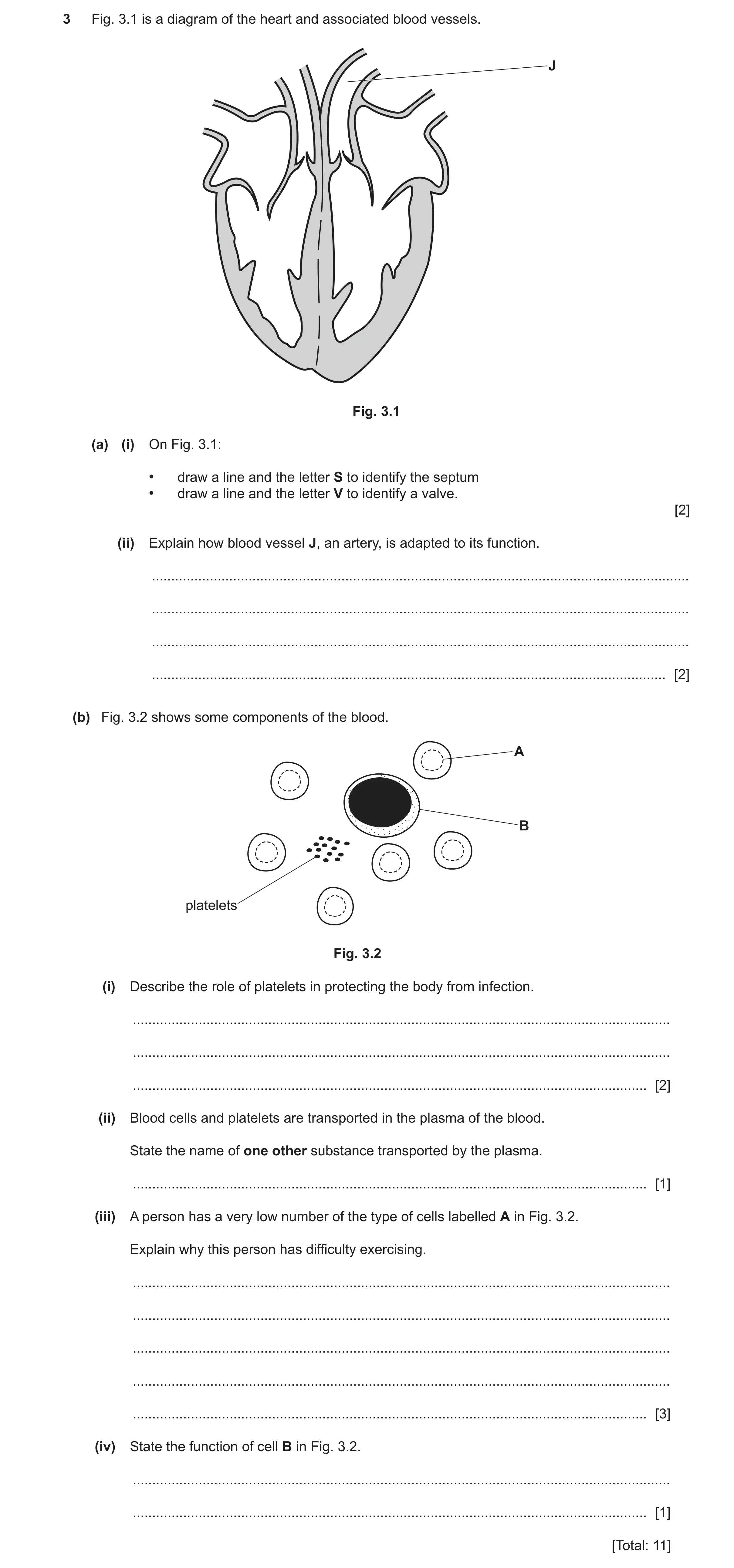
3 Fig. 3.1 is a diagram of the heart and associated blood vessels. J Fig. 3.1 (a) (i) On Fig. 3.1: • draw a line and the letter S to identify the septum • draw a line and the letter V to identify a valve. [2] (ii) Explain how blood vessel J, an artery, is adapted to its function. … … … … [2] (b) Fig. 3.2 shows some components of the blood. A B platelets Fig. 3.2 (i) Describe the role of platelets in protecting the body from infection. … … … [2] (ii) Blood cells and platelets are transported in the plasma of the blood. State the name of one other substance transported by the plasma. … [1] (iii) A person has a very low number of the type of cells labelled A in Fig. 3.2. Explain why this person has difficulty exercising. … … … … … [3] (iv) State the function of cell B in Fig. 3.2. … … [1] [Total: 11]
11 marks
Mark scheme: 3(a)(i) septum labelled S ; 2 any valve labelled V ; 3(a)(ii) has a small lumen / has muscular wall / has thick muscle wall ; 2 (to maintain or withstand the) high pressure of the blood ; 3(b)(i) clots (blood) ; 2 prevents entry of pathogens / seals wounds ; 3(b)(ii) any one from: 1 • ions ; • nutrients ; • urea ; • hormones ; • carbon dioxide ; • antibodies ; 3(b)(iii) any three from: 3 • (fewer) red blood cells ; • idea of insufficient (transport of) oxygen ; • (idea oxygen is needed) for aerobic respiration ; • idea of less energy for muscle contraction / exercise ; • increase in lactic acid / oxygen debt ; 3(b)(iv) (lymphocytes) antibody production ; 1
This question in 0654/43 Oct/Nov 2025
 3 / 24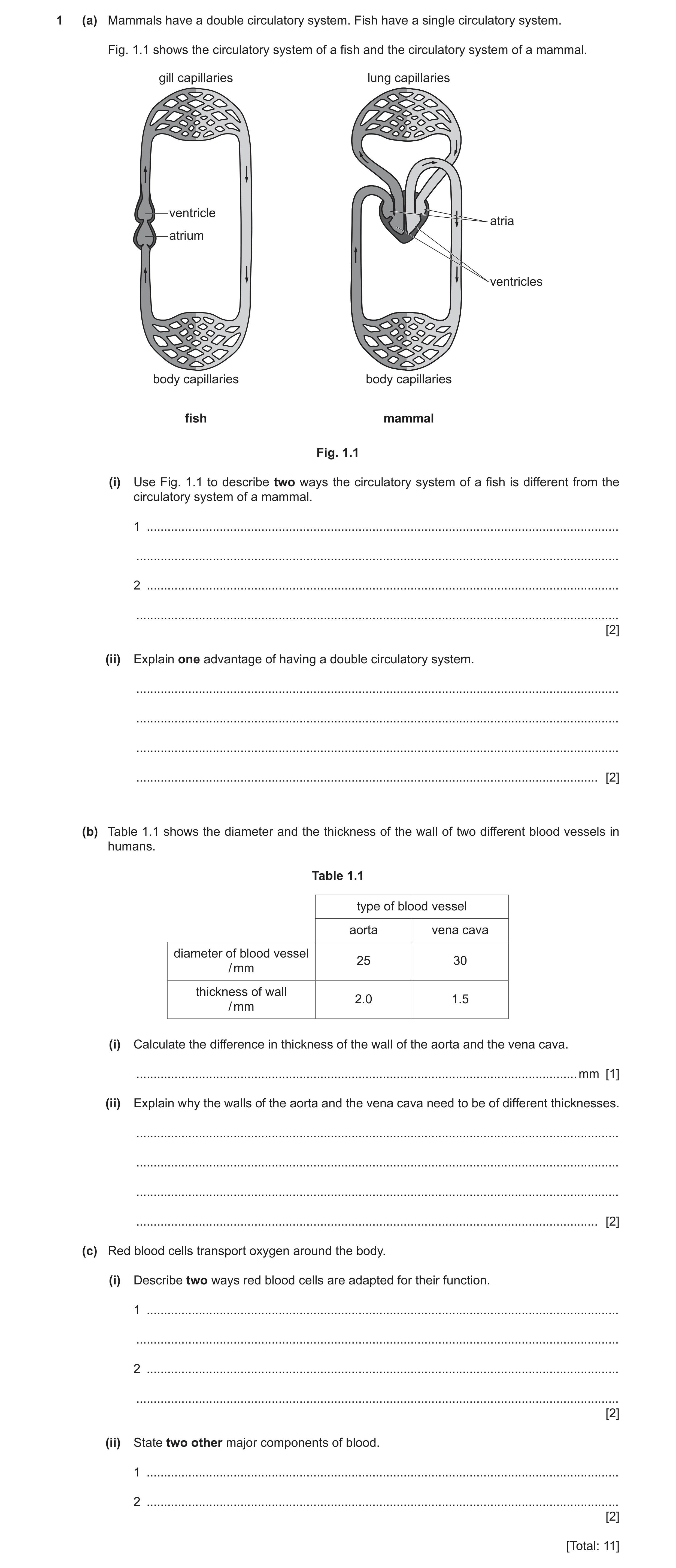
 10 / 24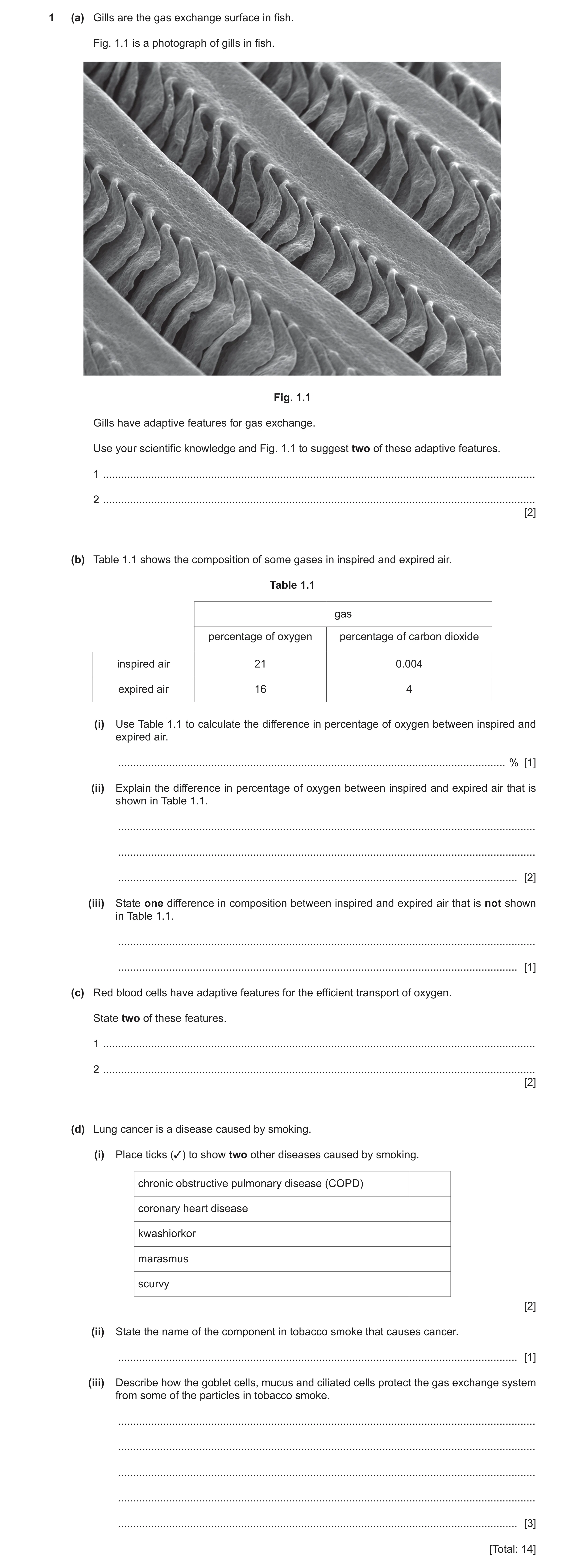
 13 / 24
 14 / 24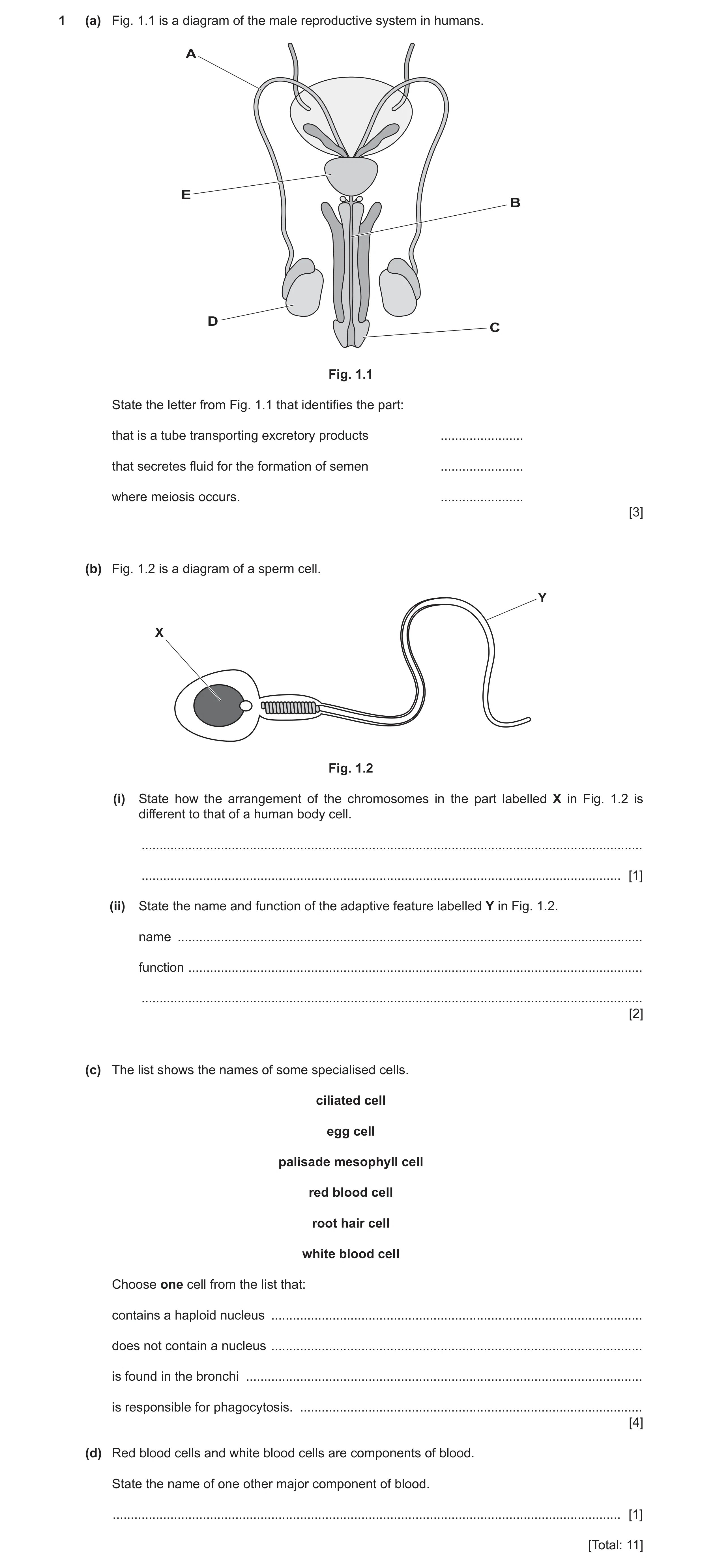
 21 / 24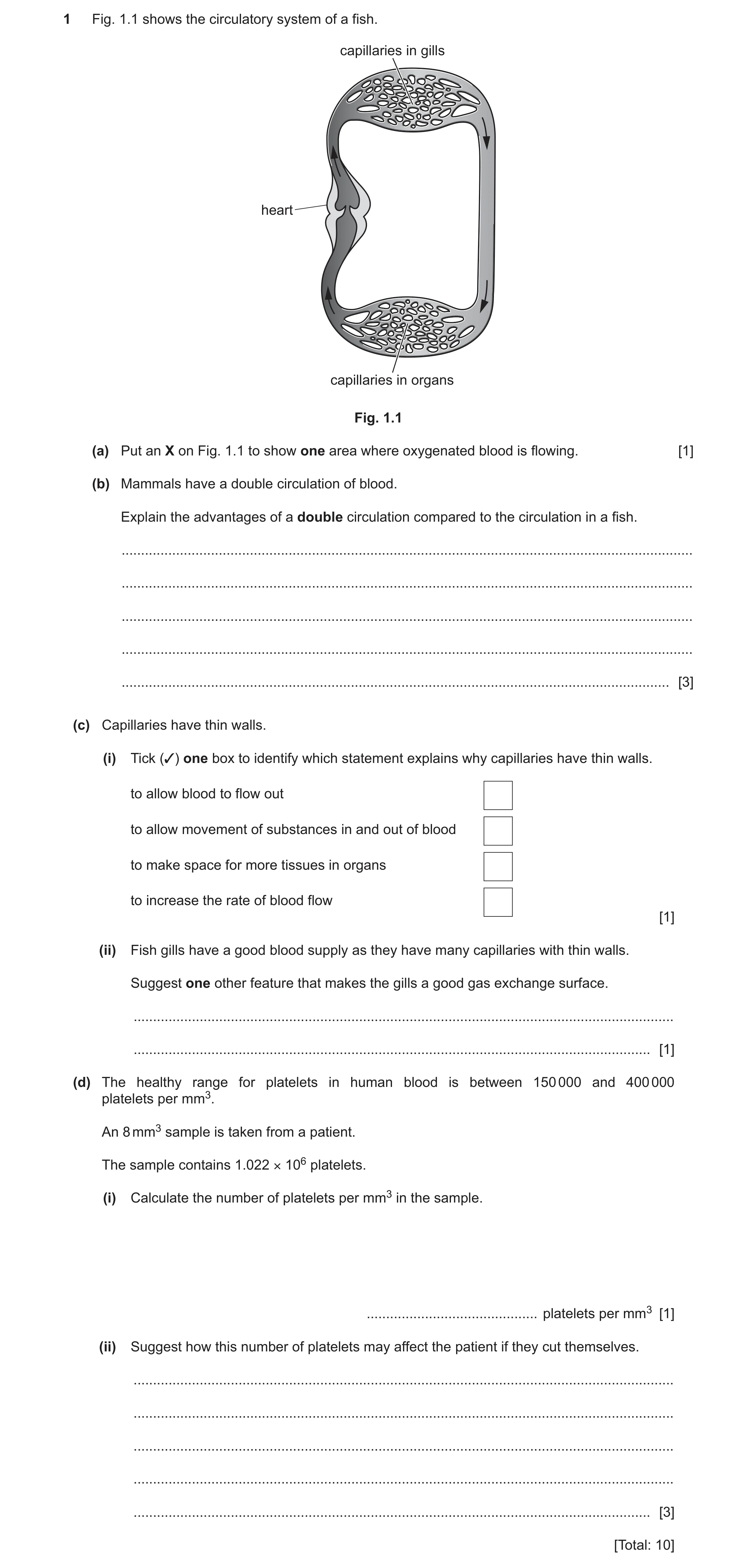
 8
8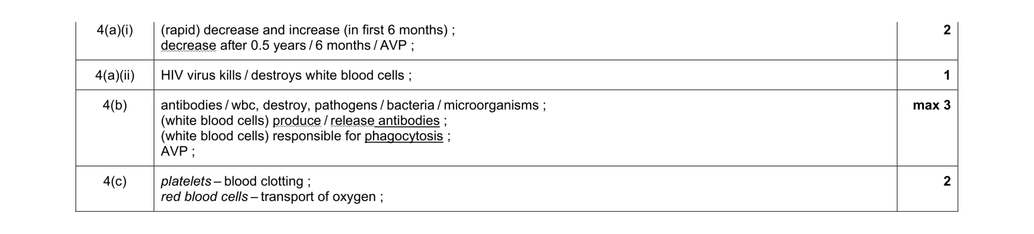 8
8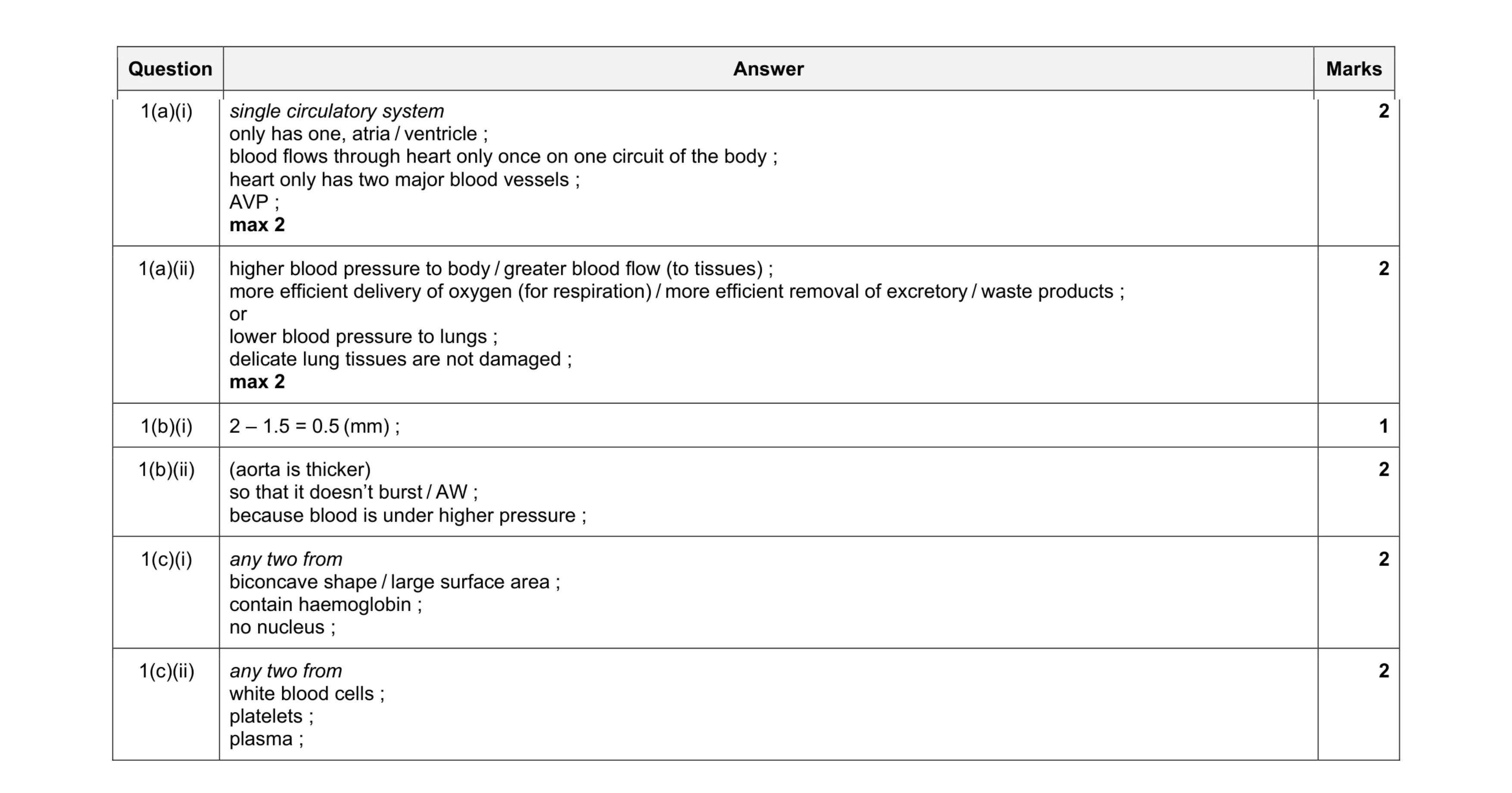 11
11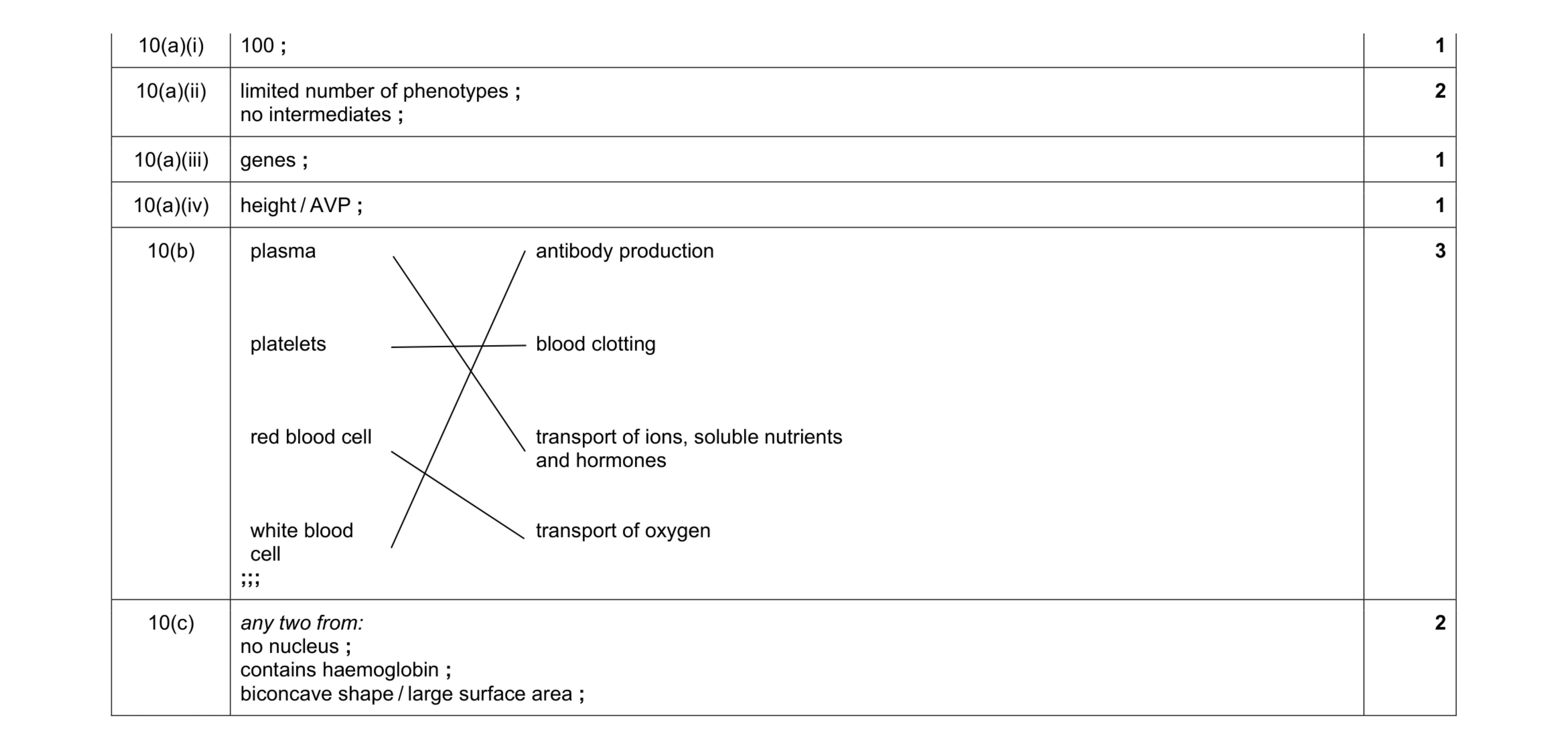 10
10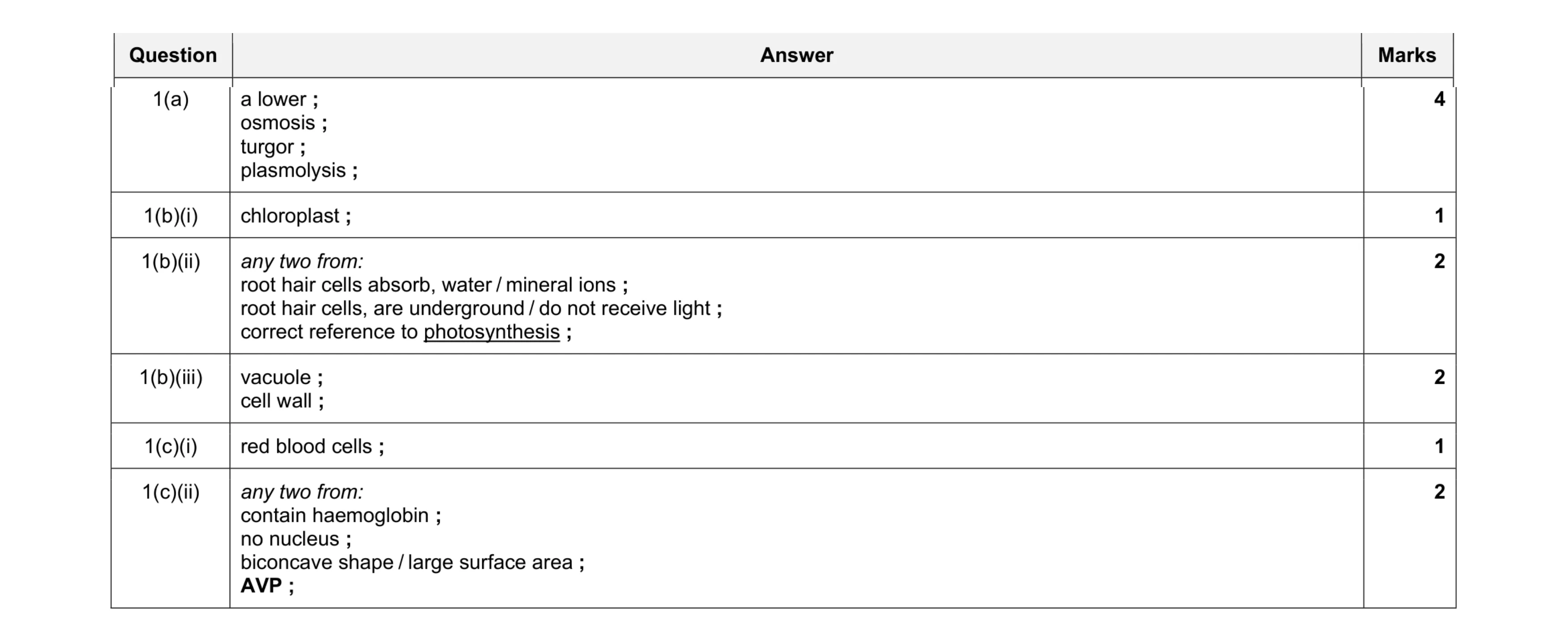 12
12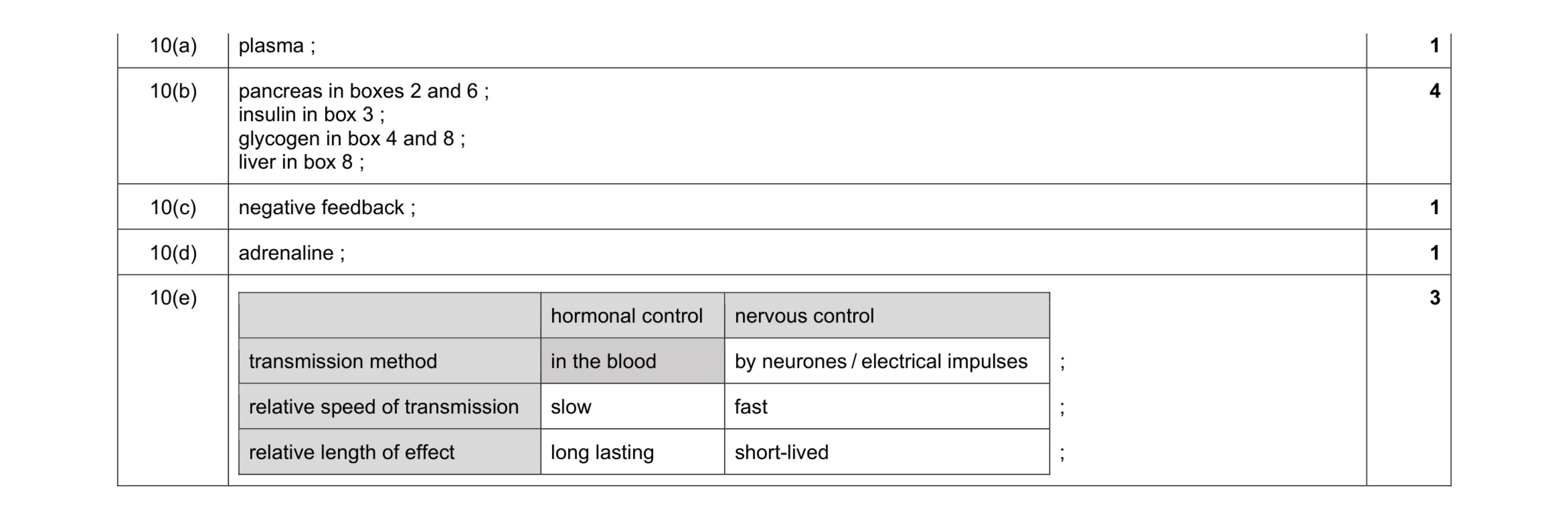 10
10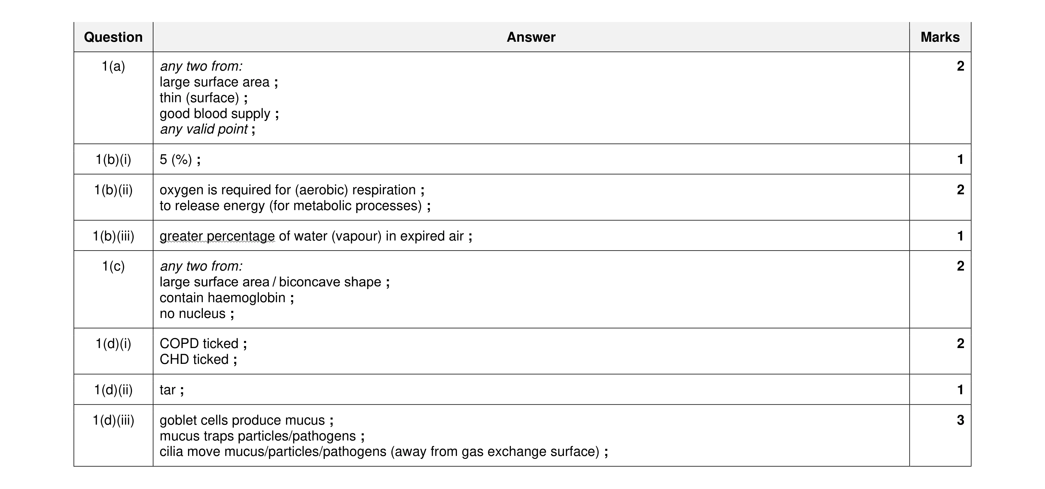 14
14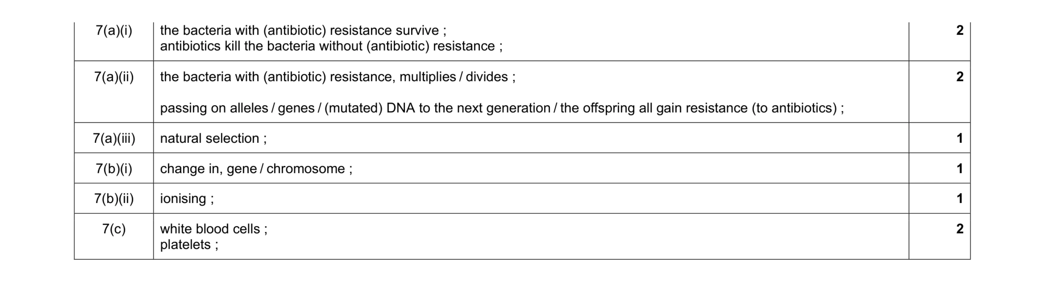 9
9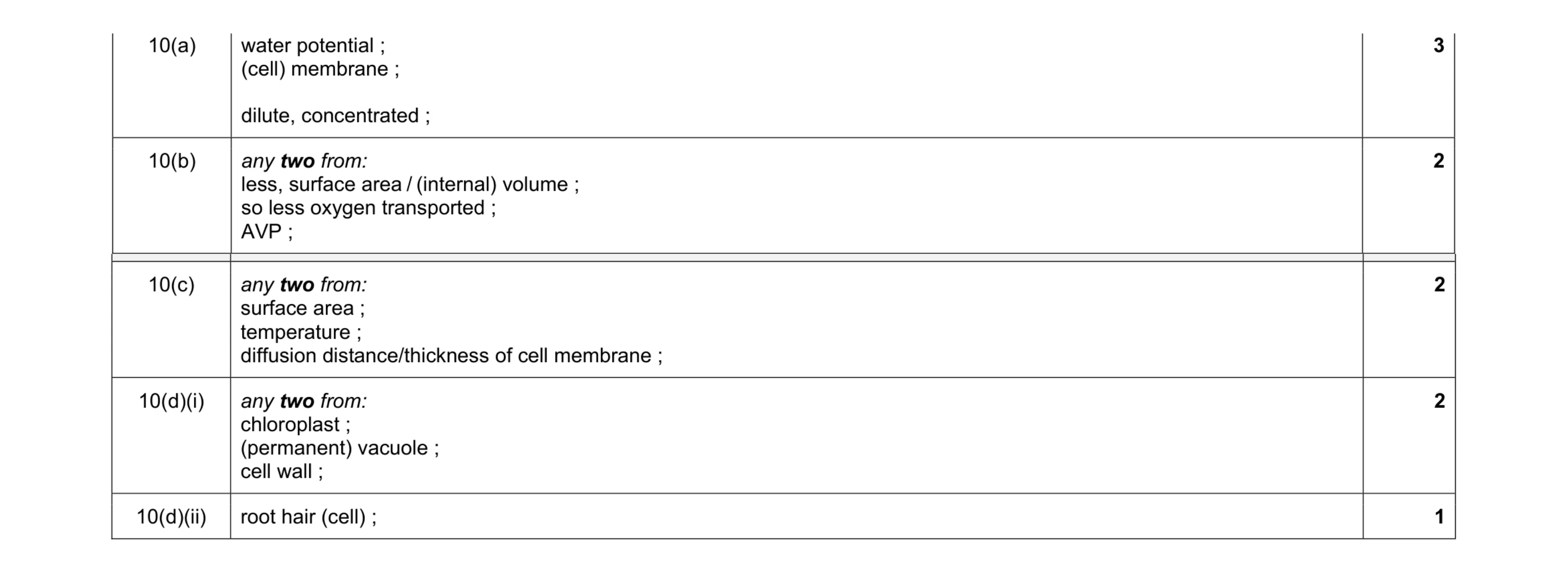 10
10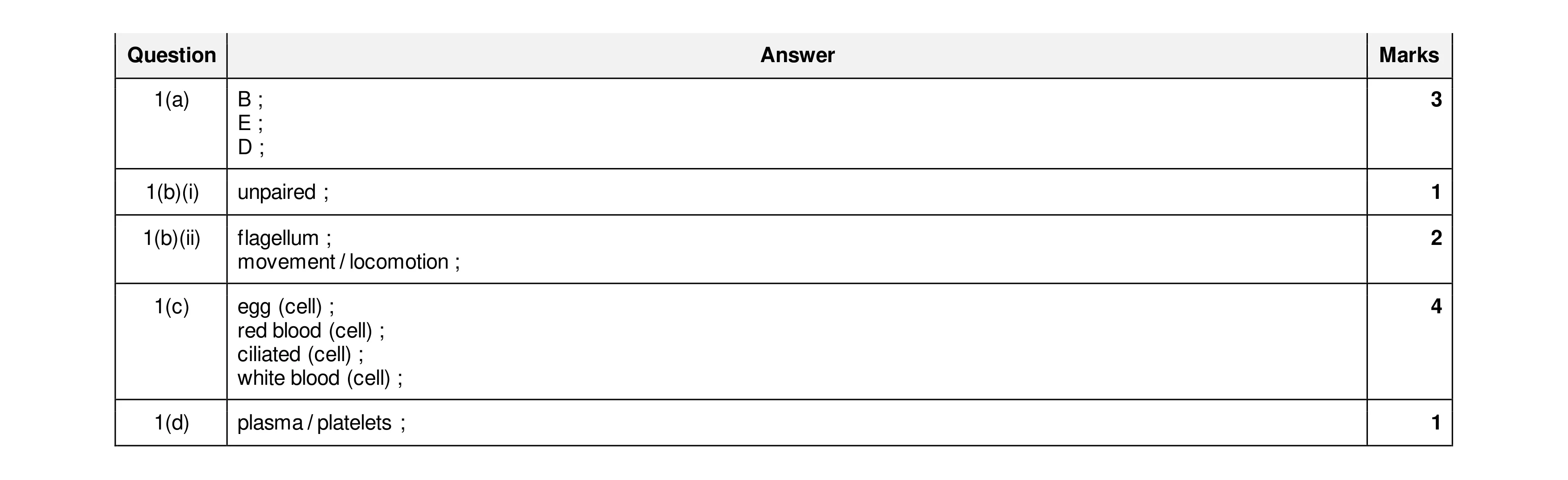 11
11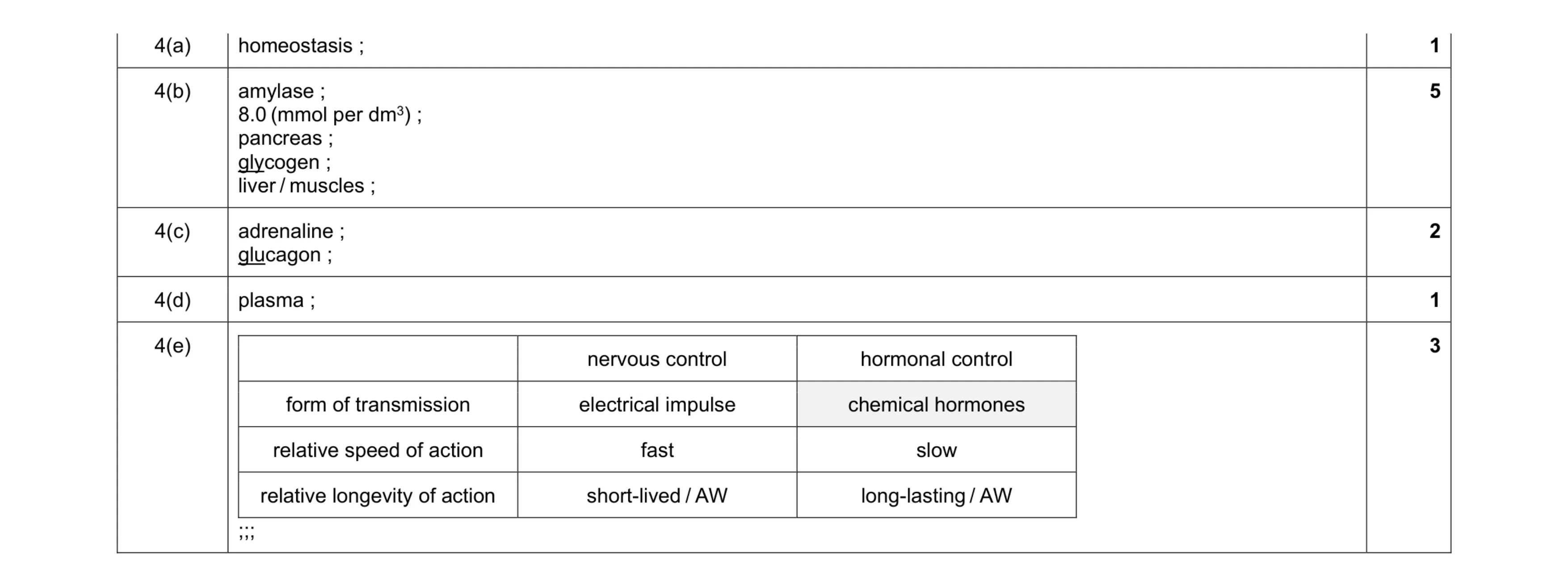 12
12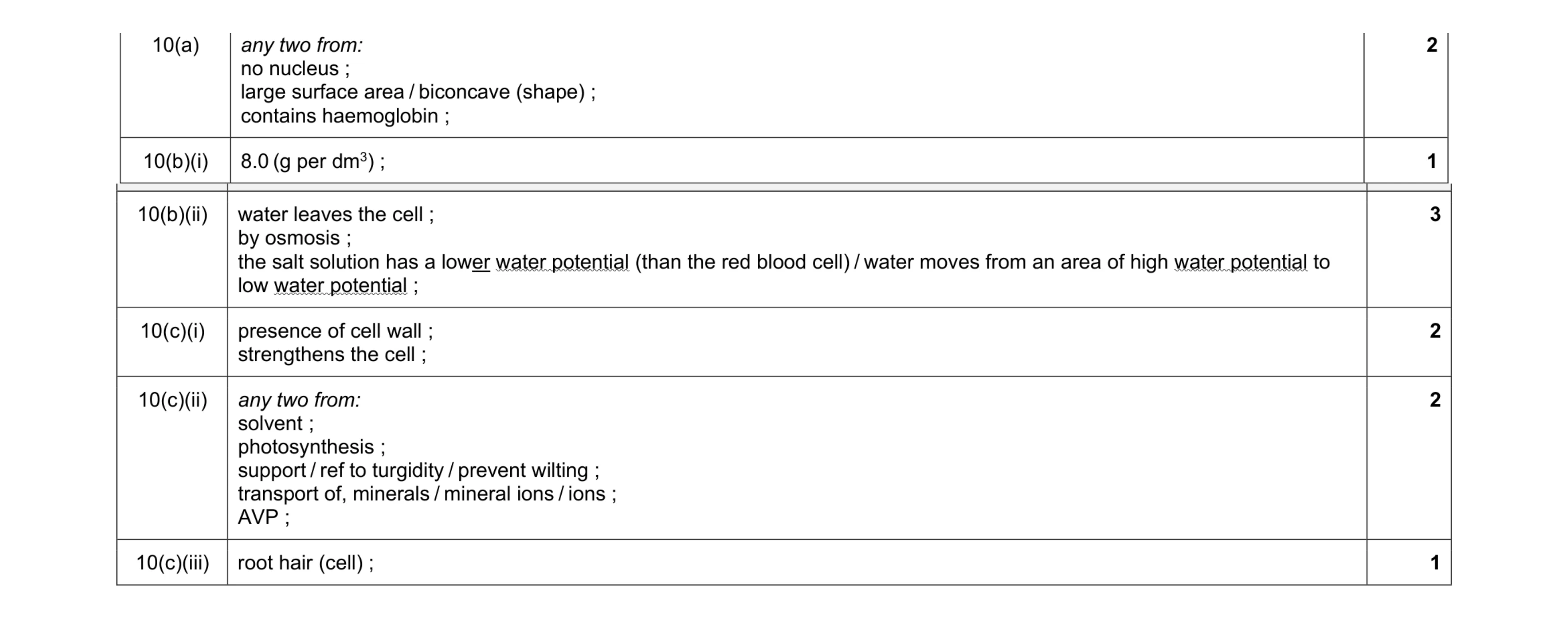 11
11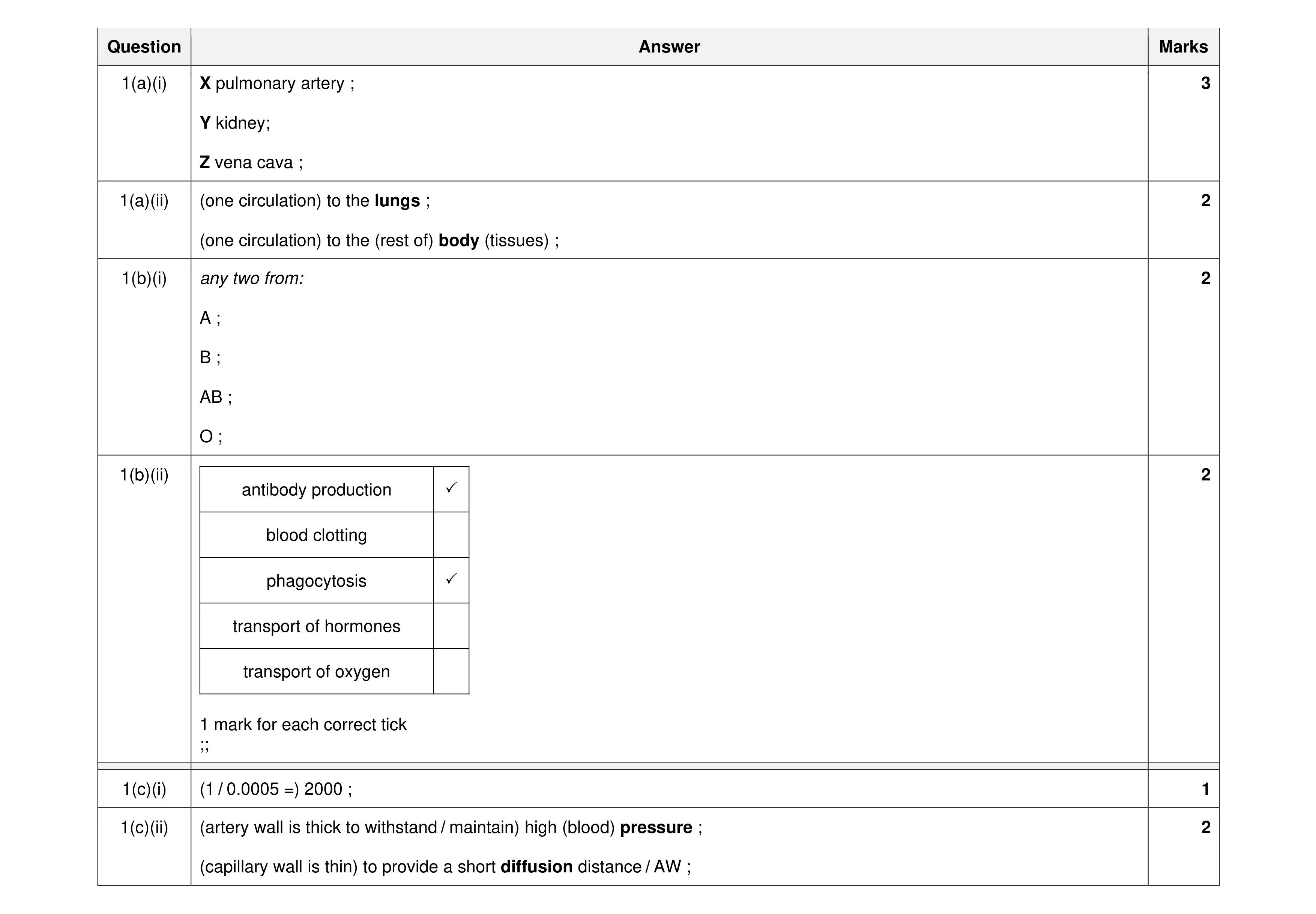 12
12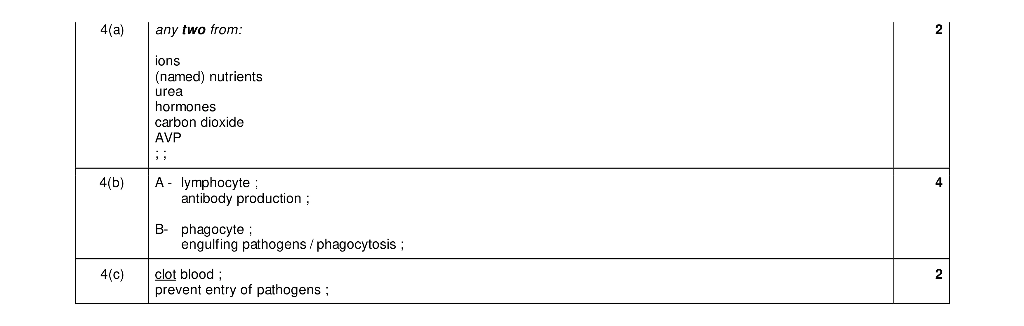 8
8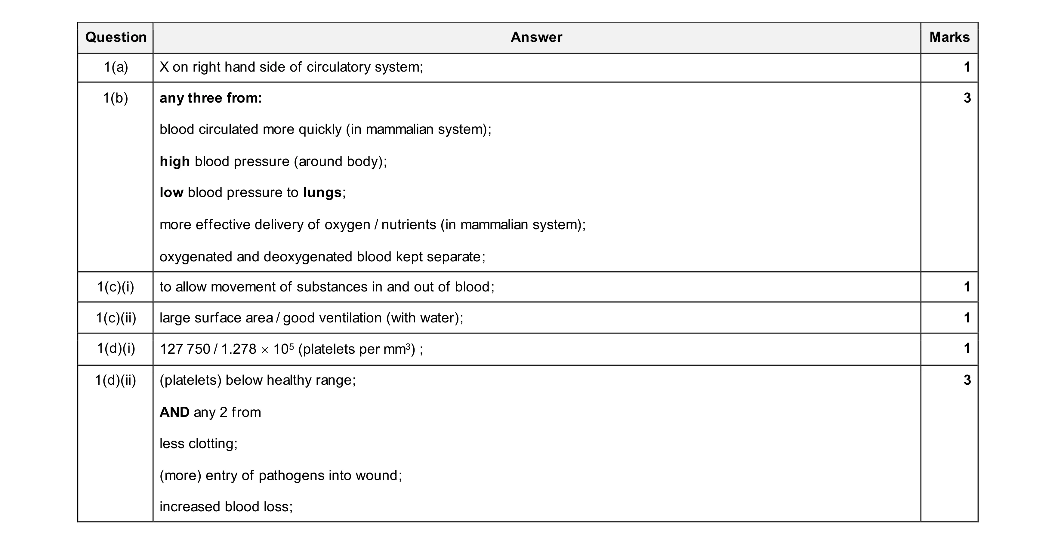 10
10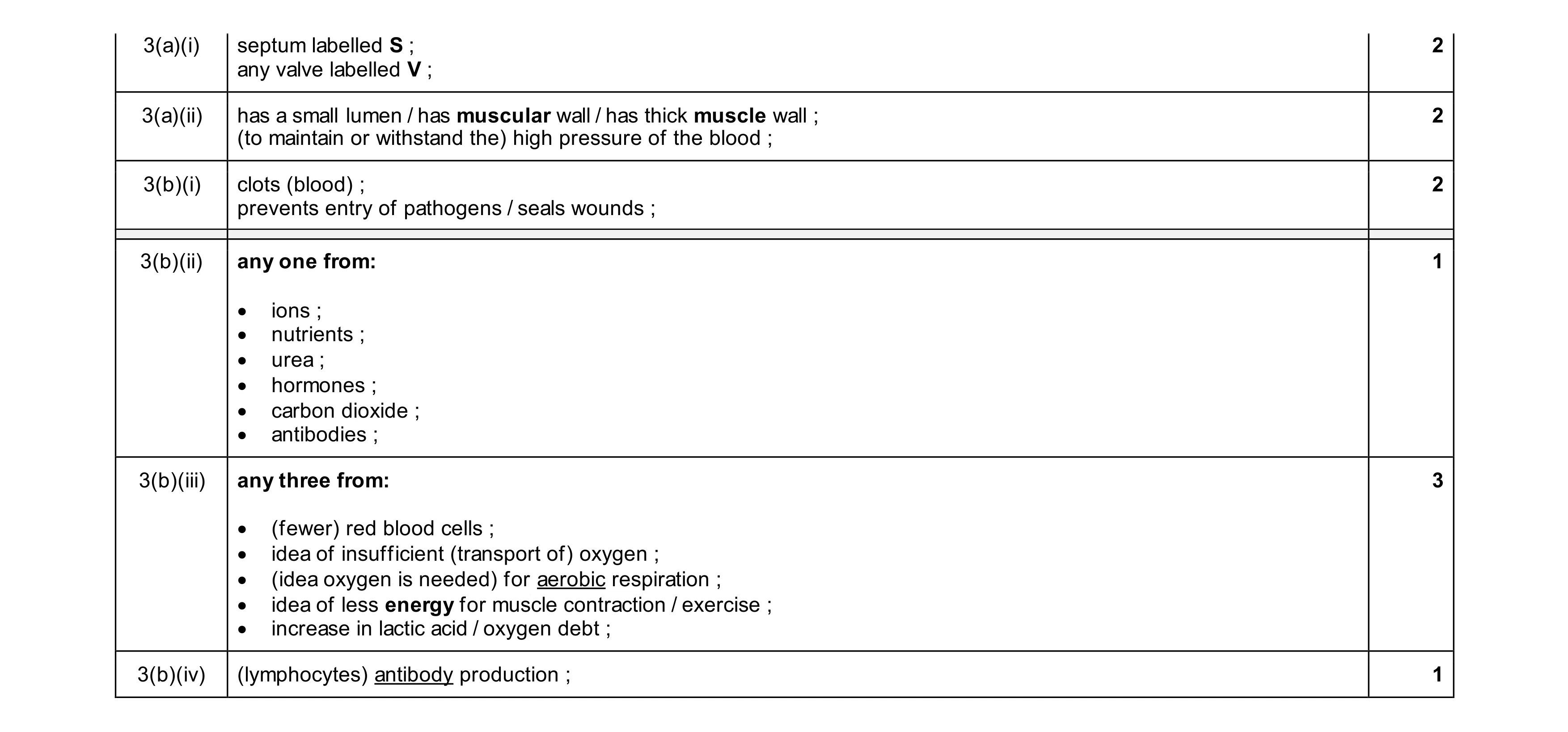 11
11