Questions as text
Q1 · The circulatory system transports blood to organs in the body 0654/31 Oct/Nov 2017
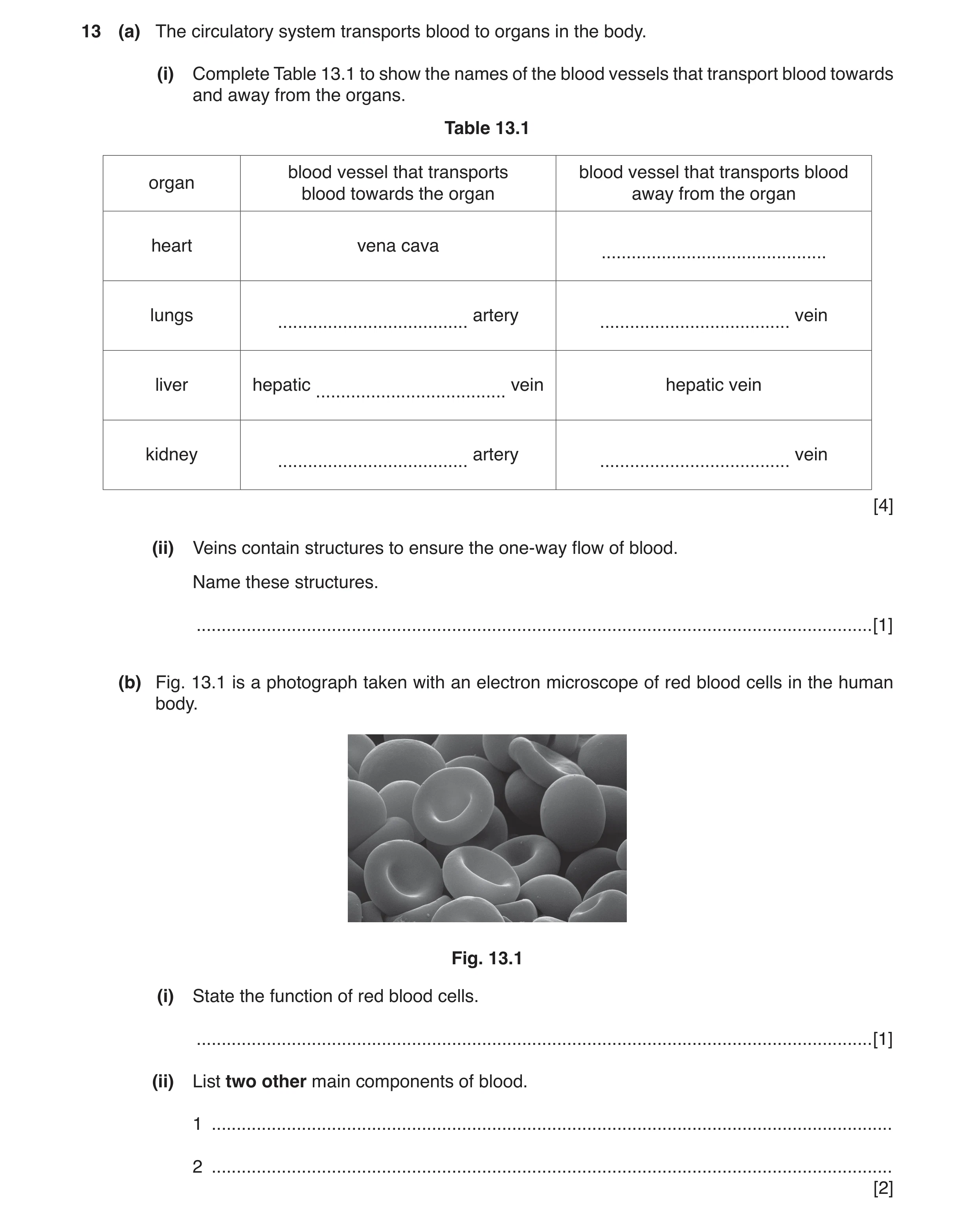
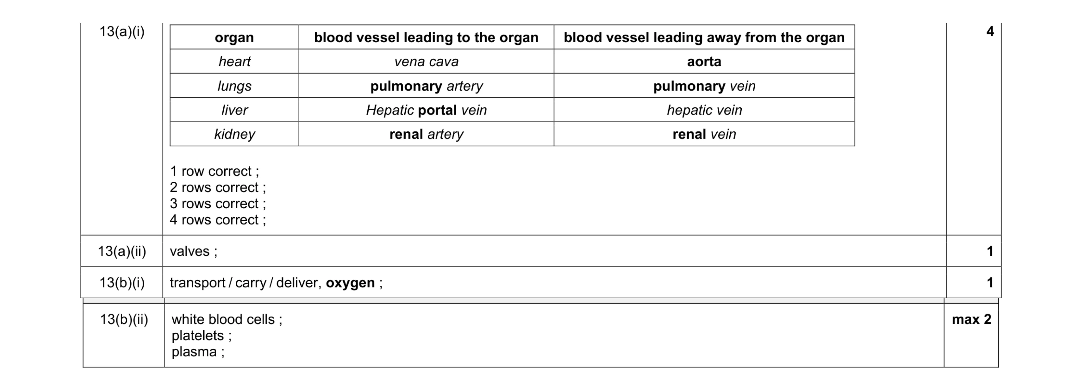
13 (a) The circulatory system transports blood to organs in the body. (i) Complete Table 13.1 to show the names of the blood vessels that transport blood towards and away from the organs. Table 13.1 blood vessel that transports blood vessel that transports blood organ blood towards the organ away from the organ heart vena cava … lungs artery vein … … liver hepatic vein hepatic vein … kidney artery vein … … [4] (ii) Veins contain structures to ensure the one-way flow of blood. Name these structures. … [1] (b) Fig. 13.1 is a photograph taken with an electron microscope of red blood cells in the human body. Fig. 13.1 (i) State the function of red blood cells. … [1] (ii) List two other main components of blood. 1 … 2 … [2]
8 marks
Mark scheme: 13(a)(i) organ blood vessel leading to the organ blood vessel leading away from the organ heart vena cava aorta lungs pulmonary artery pulmonary vein liver Hepatic portal vein hepatic vein kidney renal artery renal vein 1 row correct ; 2 rows correct ; 3 rows correct ; 4 rows correct ; 4 13(a)(ii) valves ; 1 13(b)(i) transport / carry / deliver, oxygen ; 1 Question Answer Marks 13(b)(ii) white blood cells ; platelets ; plasma ; max 2
This question in 0654/31 Oct/Nov 2017
Q2 · A cross-section through an artery and a vein 0654/33 Oct/Nov 2018
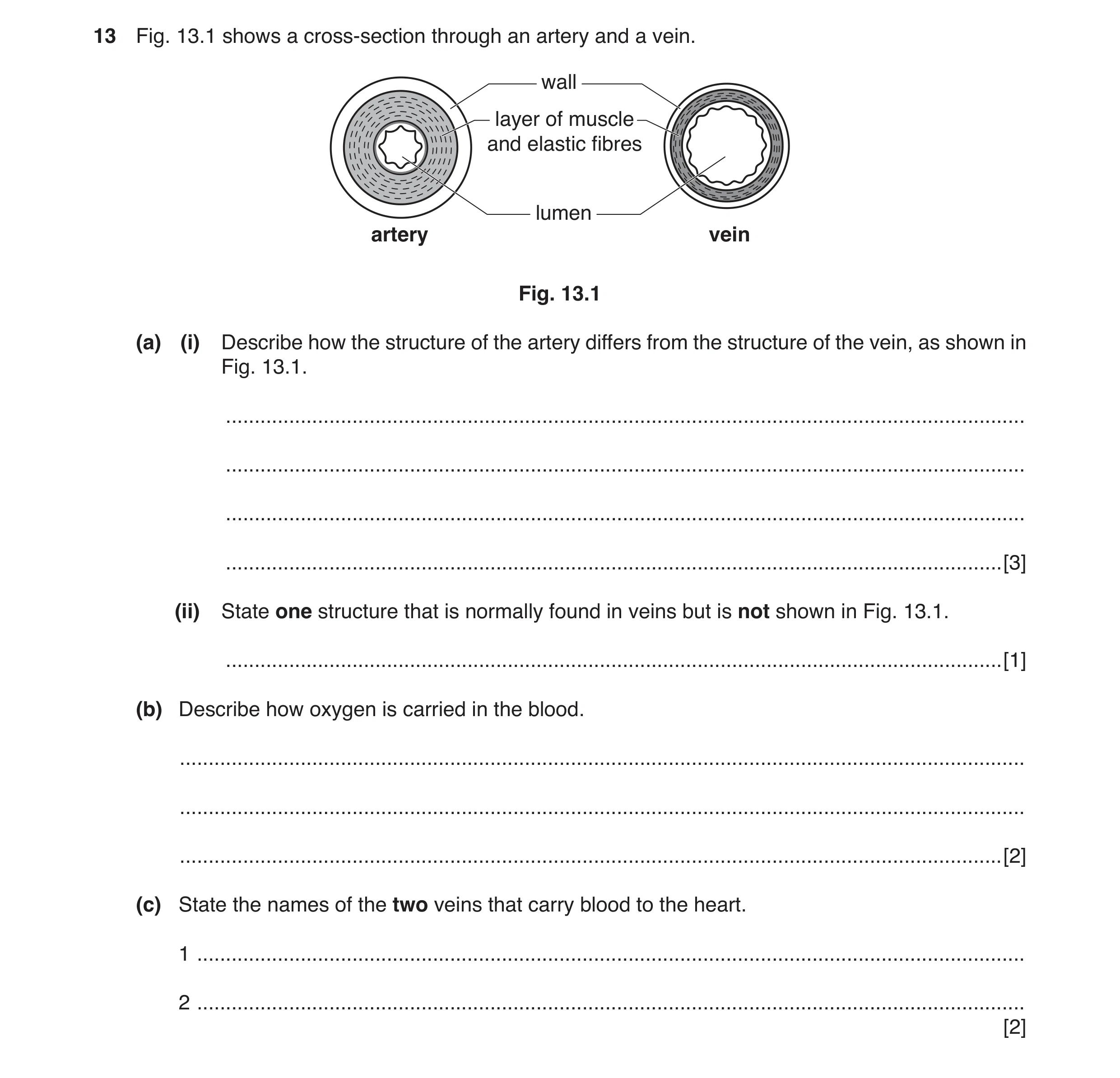
13 Fig. 13.1 shows a cross-section through an artery and a vein. wall layer of muscle and elastic fibres lumen artery vein Fig. 13.1 (a) (i) Describe how the structure of the artery differs from the structure of the vein, as shown in Fig. 13.1. … … … … [3] (ii) State one structure that is normally found in veins but is not shown in Fig. 13.1. … [1] (b) Describe how oxygen is carried in the blood. … … … [2] (c) State the names of the two veins that carry blood to the heart. 1 … 2 … [2]
8 marks
Mark scheme: 13(a)(i) thicker wall ; thicker muscle / elastic layer ; narrower lumen ; 3 13(a)(ii) valves ; 1 13(b) combined / AW, to haemoglobin ; in red blood cells ; 2 13(c) vena cava ; pulmonary vein ; 2
This question in 0654/33 Oct/Nov 2018
Q3 · A photograph of blood as seen under a microscope 0654/32 Oct/Nov 2019
1 (a) Fig. 1.1 is a photograph of blood as seen under a microscope. Fig. 1.1 (i) Use a label and a label line on Fig. 1.1 to identify a white blood cell. [1] (ii) Name one other type of cell visible in Fig. 1.1. … [1] (b) White blood cells produce antibodies that fight infection in the body. The graph in Fig. 1.2 shows how the number of antibodies in the blood changes for 15 days after a person is infected by a pathogen. number of antibodies in the blood 0 5 10 15 20 25 30 35 40 time after infection / days Fig. 1.2 (i) Use Fig. 1.2 to identify on which day there are the most antibodies in the blood. … [1] (ii) Suggest one reason why there is a decrease in the number of antibodies in the blood as shown in Fig. 1.2. … … [1] (c) Twenty days after the first infection, the same person is infected with a different pathogen. Draw a line on Fig. 1.2 to suggest how the number of antibodies in the blood changes from day 15. [2] (d) Platelets are also found in the blood. State the function of platelets. … [1] [Total: 7]
7 marks
Mark scheme: 1(a)(i) white blood cell labelled ; 1 1(a)(ii) red blood cell ; 1 1(b)(i) 12 ; 1 1(b)(ii) pathogen was destroyed ; 1 1(c) line decreasing / flat from 15–20 days ; line shown increasing (and decreasing) ; 2 1(d) blood clotting ; 1
This question in 0654/32 Oct/Nov 2019
Q4 · A diagram of a section through the heart and associated blood vessels 0654/33 Oct/Nov 2019
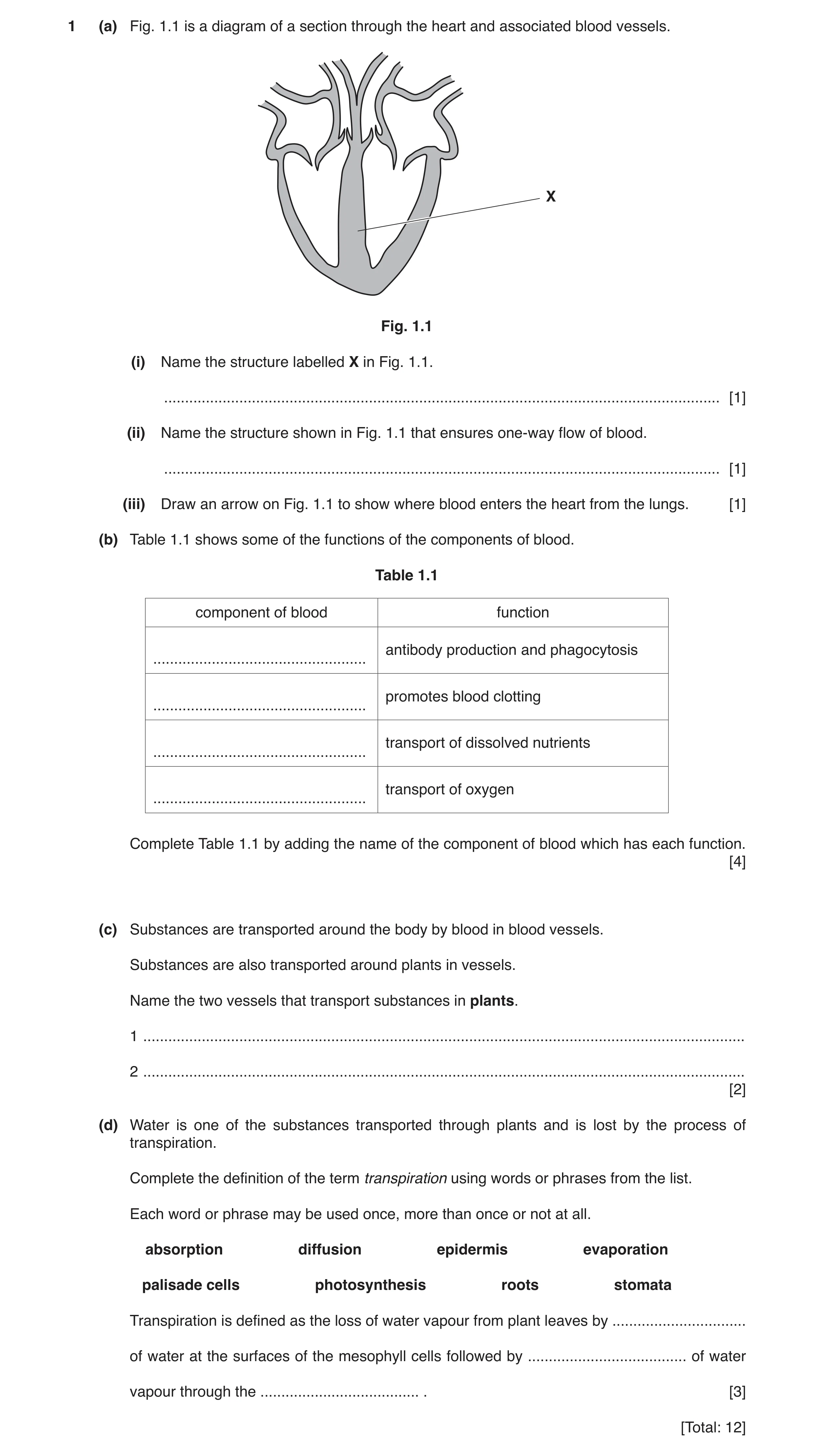
1 (a) Fig. 1.1 is a diagram of a section through the heart and associated blood vessels. X Fig. 1.1 (i) Name the structure labelled X in Fig. 1.1. … [1] (ii) Name the structure shown in Fig. 1.1 that ensures one-way flow of blood. … [1] (iii) Draw an arrow on Fig. 1.1 to show where blood enters the heart from the lungs. [1] (b) Table 1.1 shows some of the functions of the components of blood. Table 1.1 component of blood function antibody production and phagocytosis … promotes blood clotting … transport of dissolved nutrients … transport of oxygen … Complete Table 1.1 by adding the name of the component of blood which has each function. [4] (c) Substances are transported around the body by blood in blood vessels. Substances are also transported around plants in vessels. Name the two vessels that transport substances in plants. 1 … 2 … [2] (d) Water is one of the substances transported through plants and is lost by the process of transpiration. Complete the definition of the term transpiration using words or phrases from the list. Each word or phrase may be used once, more than once or not at all. absorption diffusion epidermis evaporation palisade cells photosynthesis roots stomata Transpiration is defined as the loss of water vapour from plant leaves by … of water at the surfaces of the mesophyll cells followed by … of water vapour through the … . [3] [Total: 12]
12 marks
Mark scheme: 1(a)(i) septum ; 1 1(a)(ii) valve; 1 1(a)(iii) arrow pointing into the heart from the pulmonary vein ; (furthest right blood vessel) 1 1(b) component of blood function white blood cell ; antibody production and phagocytosis platelets ; promotes blood clotting plasma ; transport of dissolved nutrients red blood cell ; transport of oxygen 4 1(c) xylem ; phloem ; 2 1(d) evaporation ; diffusion ; stomata ; 3
This question in 0654/33 Oct/Nov 2019
Q5 · Some of the functions of the main components of blood 0654/31 May/June 2021
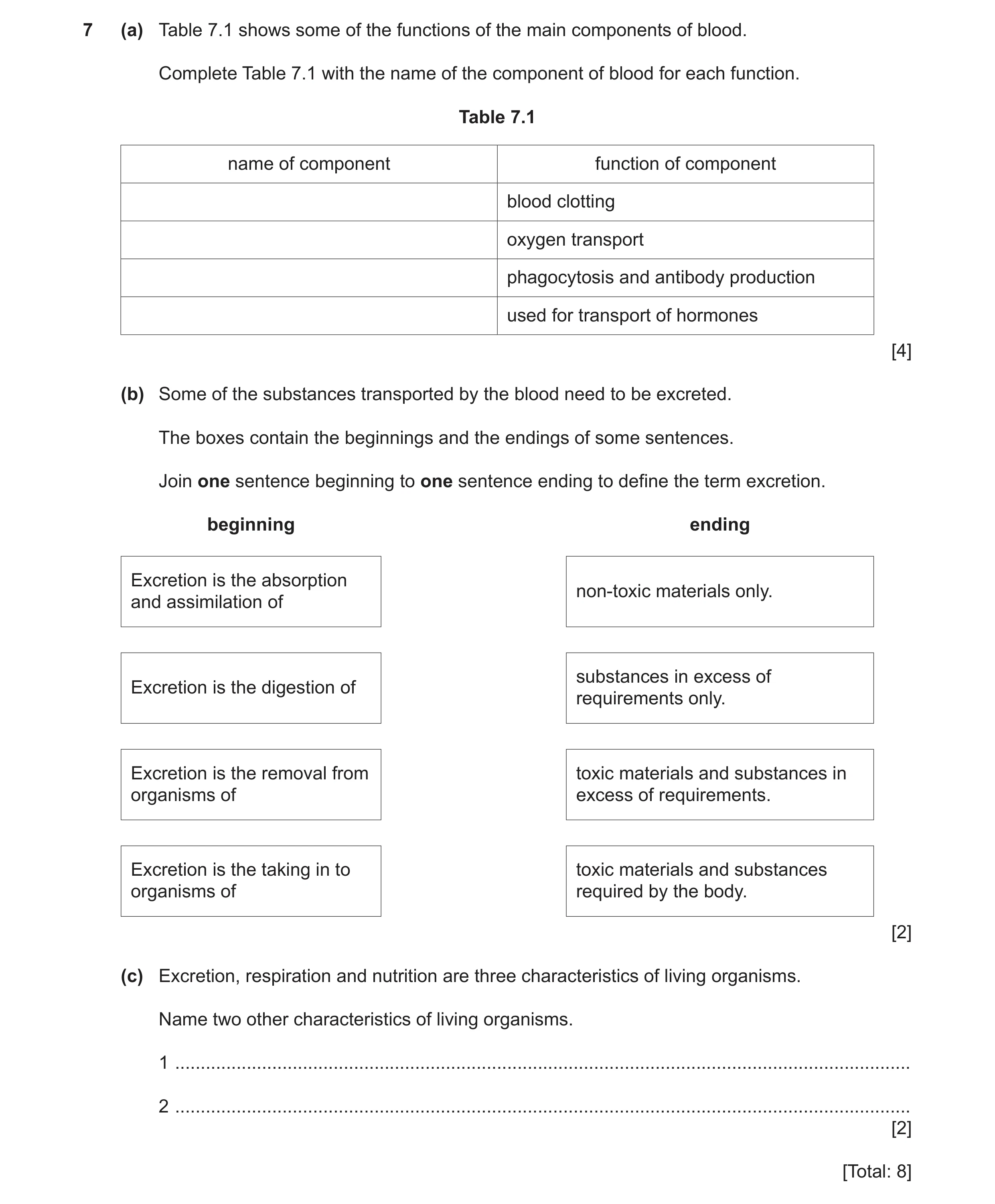
7 (a) Table 7.1 shows some of the functions of the main components of blood. Complete Table 7.1 with the name of the component of blood for each function. Table 7.1 name of component function of component blood clotting oxygen transport phagocytosis and antibody production used for transport of hormones [4] (b) Some of the substances transported by the blood need to be excreted. The boxes contain the beginnings and the endings of some sentences. Join one sentence beginning to one sentence ending to define the term excretion. beginning ending Excretion is the absorption non‑toxic materials only. and assimilation of substances in excess of Excretion is the digestion of requirements only. Excretion is the removal from toxic materials and substances in organisms of excess of requirements. Excretion is the taking in to toxic materials and substances organisms of required by the body. [2] (c) Excretion, respiration and nutrition are three characteristics of living organisms. Name two other characteristics of living organisms. 1 … 2 … [2] [Total: 8]
8 marks
Mark scheme: 7(a) platelets ; red blood cells ; white blood cells ; plasma ; 4 7(b) ;; 2 7(c) any two from: reproduction ; movement ; sensitivity ; growth ; 2
This question in 0654/31 May/June 2021
Q6 · A student measures their pulse rate at rest, during and after exercise 0654/33 Oct/Nov 2021
1 (a) A student measures their pulse rate at rest, during and after exercise. Fig. 1.1 shows the results. 120 110 100 90 pulse rate / beats per minute 80 70 atat duringduring afterafter 60 restrest exerciseexercise exerciseexercise 0 0 5 10 15 20 25 30 35 40 45 50 time / minutes Fig. 1.1 (i) Calculate the difference in pulse rate between the pulse rate at rest and the maximum pulse rate of the student. pulse rate at rest … beats per minute maximum pulse rate … beats per minute difference … beats per minute [2] (ii) Calculate the length of time taken for the student’s pulse rate to return to the resting value once exercise had stopped. … min [1] (b) Fig. 1.2 is a photomicrograph of a cross section of a human vein. Fig. 1.2 (i) Name a structure present in veins but not visible in Fig. 1.2. … [1] (ii) Describe two ways in which the structure of arteries is different from the vein shown in Fig. 1.2. 1 … … 2 … … [2] (iii) Describe the function of capillaries. … … [1] (c) Table 1.1 shows some blood vessels and some organs. Place ticks (3) in the boxes to show which blood vessels transport blood to these organs. One row has been done for you. Table 1.1 organ heart kidney lung coronary artery pulmonary artery renal artery vena cava 3 [3] (d) List two of the main components of blood. 1 … 2 … [2] [Total: 12]
12 marks
Mark scheme: 1(a)(i) pulse rate at rest = 72 (bpm) and maximum pulse rate = 115 (bpm) ; (115 – 72) = 43 (bpm) ; 2 1(a)(ii) 20 (mins) ; 1 1(b)(i) valves ; 1 1(b)(ii) any two from: thicker wall ; muscle / elastic fibres ; no valves ; narrower lumen ; 2 1(b)(iii) transfer of (named) substances (to / from, tissues / cells) ; 1 1(c) heart kidney lung coronary artery pulmonary artery renal artery vena cava ;;; 3 1(d) any two from: red blood cells ; white blood cells ; plasma ; platelets ; 2
This question in 0654/33 Oct/Nov 2021
Q7 · The total number of teeth of different animals 0654/32 Feb/March 2022
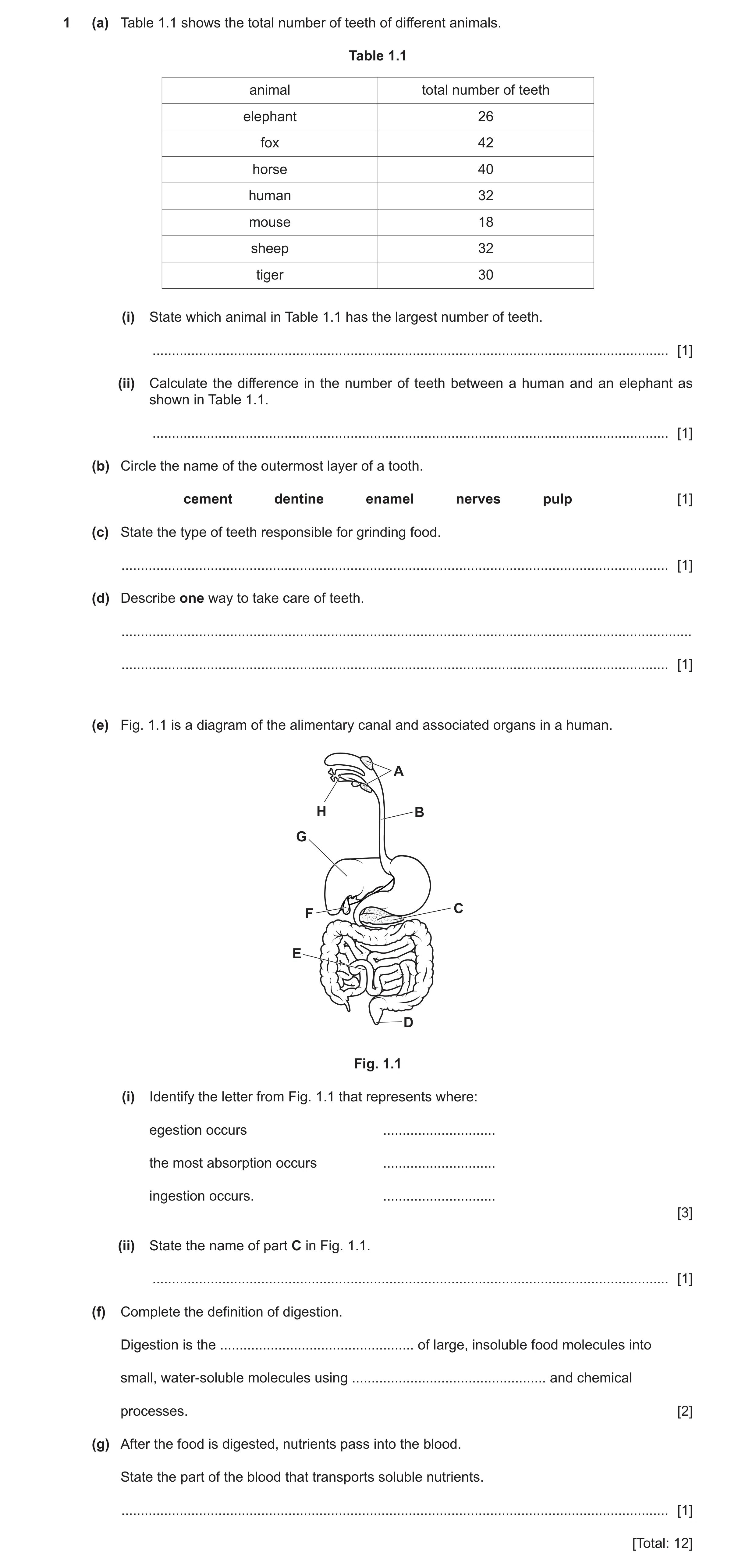
1 (a) Table 1.1 shows the total number of teeth of different animals. Table 1.1 animal total number of teeth elephant 26 fox 42 horse 40 human 32 mouse 18 sheep 32 tiger 30 (i) State which animal in Table 1.1 has the largest number of teeth. … [1] (ii) Calculate the difference in the number of teeth between a human and an elephant as shown in Table 1.1. … [1] (b) Circle the name of the outermost layer of a tooth. cement dentine enamel nerves pulp [1] (c) State the type of teeth responsible for grinding food. … [1] (d) Describe one way to take care of teeth. … … [1] (e) Fig. 1.1 is a diagram of the alimentary canal and associated organs in a human. A H B G C F E D Fig. 1.1 (i) Identify the letter from Fig. 1.1 that represents where: egestion occurs … the most absorption occurs … ingestion occurs. … [3] (ii) State the name of part C in Fig. 1.1. … [1] (f) Complete the definition of digestion. Digestion is the … of large, insoluble food molecules into small, water-soluble molecules using … and chemical processes. [2] (g) After the food is digested, nutrients pass into the blood. State the part of the blood that transports soluble nutrients. … [1] [Total: 12]
12 marks
Mark scheme: 1(a)(i) fox ; 1 1(a)(ii) 6 ; 1 1(b) enamel ; 1 1(c) molar / pre molar ; 1 1(d) brushing / using toothpaste / (regular) visits to dentist / consume less sugary food or drink ; 1 1(e)(i) D ; E ; H ; 3 1(e)(ii) pancreas ; 1 1(f) break(ing) down ; mechanical ; 2 1(g) plasma ; 1
This question in 0654/32 Feb/March 2022
Q8 · A diagram of a plant cell 0654/32 Feb/March 2022
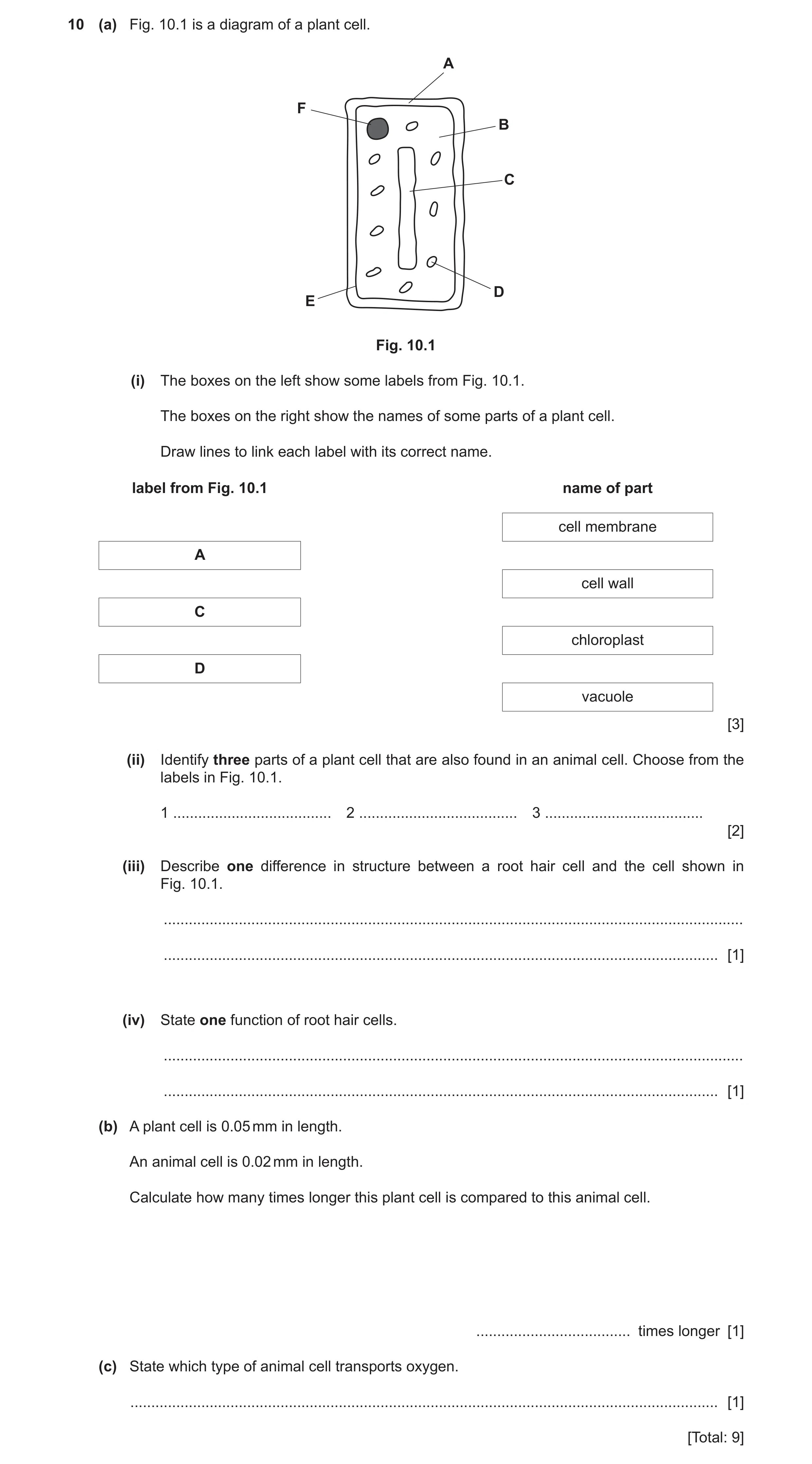
10 (a) Fig. 10.1 is a diagram of a plant cell. A F B C D E Fig. 10.1 (i) The boxes on the left show some labels from Fig. 10.1. The boxes on the right show the names of some parts of a plant cell. Draw lines to link each label with its correct name. label from Fig. 10.1 name of part cell membrane A cell wall C chloroplast D vacuole [3] (ii) Identify three parts of a plant cell that are also found in an animal cell. Choose from the labels in Fig. 10.1. 1 … 2 … 3 … [2] (iii) Describe one difference in structure between a root hair cell and the cell shown in Fig. 10.1. … … [1] (iv) State one function of root hair cells. … … [1] (b) A plant cell is 0.05 mm in length. An animal cell is 0.02 mm in length. Calculate how many times longer this plant cell is compared to this animal cell. … times longer [1] (c) State which type of animal cell transports oxygen. … [1] [Total: 9]
9 marks
Mark scheme: 10(a)(i) cell membrane A cell wall C chloroplast D vacuole ;;; 10(a)(ii) B, E and F ;; 2 10(a)(iii) elongated shape / no chloroplasts ; 1 10(a)(iv) absorption (of water / mineral ions) ; 1 10(b) 2.5 ; 1 Question Answer Marks 10(c) red blood cell ; 1
This question in 0654/32 Feb/March 2022
Q9 · A diagram of the gas exchange system in humans 0654/32 May/June 2022
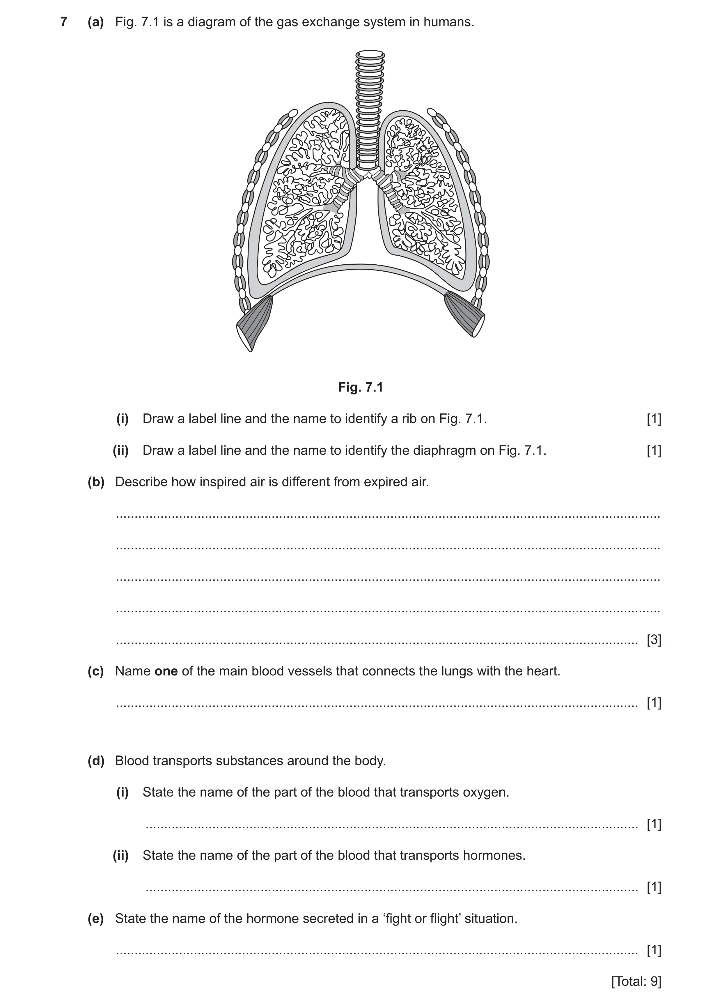
7 (a) Fig. 7.1 is a diagram of the gas exchange system in humans. Fig. 7.1 (i) Draw a label line and the name to identify a rib on Fig. 7.1. [1] (ii) Draw a label line and the name to identify the diaphragm on Fig. 7.1. [1] (b) Describe how inspired air is different from expired air. … … … … … [3] (c) Name one of the main blood vessels that connects the lungs with the heart. … [1] (d) Blood transports substances around the body. (i) State the name of the part of the blood that transports oxygen. … [1] (ii) State the name of the part of the blood that transports hormones. … [1] (e) State the name of the hormone secreted in a ‘fight or flight’ situation. … [1] [Total: 9]
9 marks
Mark scheme: 7(a)(i) rib labelled correctly ; 1 7(a)(ii) diaphragm labelled correctly ; 1 7(b) contains more oxygen ; contains less water vapour ; contains less carbon dioxide ; AVP ; Max 3 marks 3 7(c) pulmonary vein / pulmonary artery ; 1 7(d)(i) red blood cell ; 1 Question Answer Marks 7(d)(ii) plasma ; 1 7(e) adrenaline ; 1
This question in 0654/32 May/June 2022
Q10 · A diagram of the gas exchange system in humans 0654/33 May/June 2022
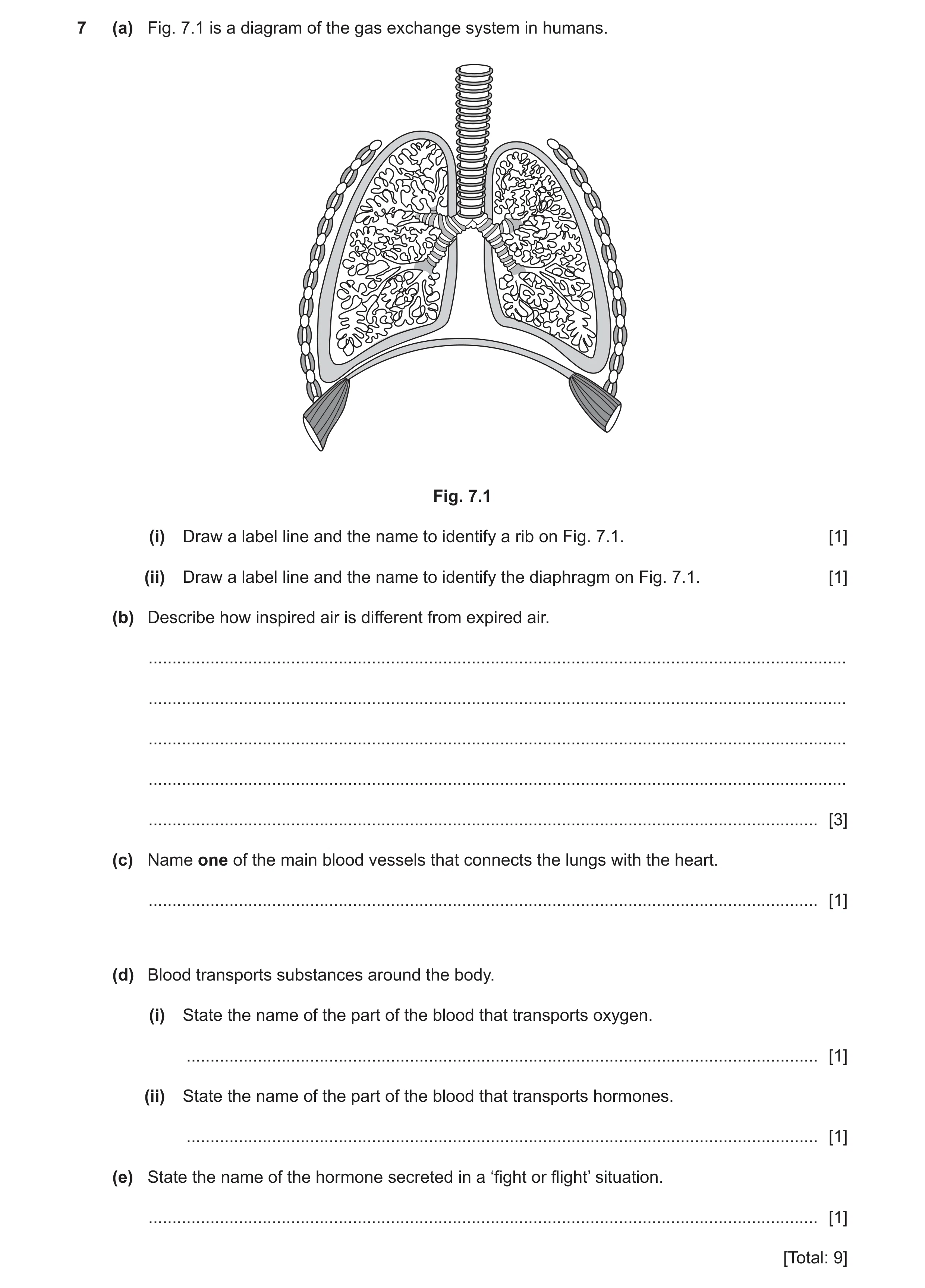
7 (a) Fig. 7.1 is a diagram of the gas exchange system in humans. Fig. 7.1 (i) Draw a label line and the name to identify a rib on Fig. 7.1. [1] (ii) Draw a label line and the name to identify the diaphragm on Fig. 7.1. [1] (b) Describe how inspired air is different from expired air. … … … … … [3] (c) Name one of the main blood vessels that connects the lungs with the heart. … [1] (d) Blood transports substances around the body. (i) State the name of the part of the blood that transports oxygen. … [1] (ii) State the name of the part of the blood that transports hormones. … [1] (e) State the name of the hormone secreted in a ‘fight or flight’ situation. … [1] [Total: 9]
9 marks
Mark scheme: 7(a)(i) rib labelled correctly ; 1 7(a)(ii) diaphragm labelled correctly ; 1 7(b) contains more oxygen ; contains less water vapour ; contains less carbon dioxide ; AVP ; Max 3 marks 3 7(c) pulmonary vein / pulmonary artery ; 1 7(d)(i) red blood cell ; 1 Question Answer Marks 7(d)(ii) plasma ; 1 7(e) adrenaline ; 1
This question in 0654/33 May/June 2022
Q11 · Complete the definition of the term hormone 0654/31 Oct/Nov 2022
4 (a) Complete the definition of the term hormone. A hormone is a chemical substance, produced by a … and carried by the blood. It alters the activity of one or more specific target … . [2] (b) State the name of the component of blood that transports hormones. … [1] (c) A person experiences a scary event whilst wearing a heart rate monitor. Fig. 4.1 shows her heart rate before, during and after the event. 120 110 100 heart 90 rate / bpm 80 70 60 50 0 0 1 2 3 4 5 6 7 8 9 10 11 12 13 14 time / minutes Fig. 4.1 (i) Use Fig. 4.1 to suggest the time that the scary event takes place. time = … minutes [1] (ii) Use Fig. 4.1 to identify the maximum heart rate of this person. heart rate = … bpm [1] (iii) State the name of the hormone that causes the increase in heart rate seen in Fig. 4.1. … [1] (iv) State two other effects of this hormone. 1 … 2 … [2] (d) Complete these sentences about the human nervous system. The nervous system consists of the central nervous system and the … nervous system. The central nervous system has two parts: the … and the … . [3] [Total: 11]
11 marks
Mark scheme: 4(a) gland ; 2 organs ; 4(b) plasma ; 1 4(c)(i) 4 (minutes) ; 1 4(c)(ii) 109 (bpm) ; 1 4(c)(iii) adrenaline ; 1 4(c)(iv) widened pupils ; 2 increased breathing rate ; any valid point ;; 4(d) peripheral ; 3 brain ; spinal cord ;
This question in 0654/31 Oct/Nov 2022
Q12 · A photomicrograph of a cross-section of a human vein 0654/32 Oct/Nov 2022
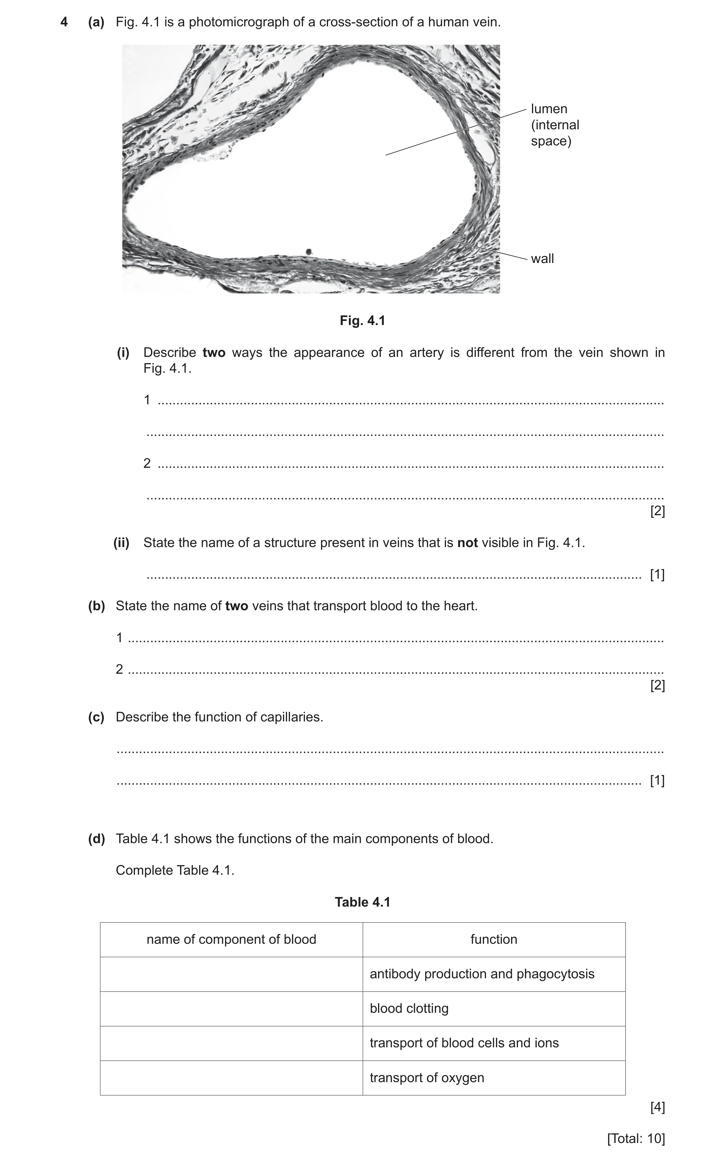
4 (a) Fig. 4.1 is a photomicrograph of a cross-section of a human vein. lumen (internal space) wall Fig. 4.1 (i) Describe two ways the appearance of an artery is different from the vein shown in Fig. 4.1. 1 … … 2 … … [2] (ii) State the name of a structure present in veins that is not visible in Fig. 4.1. … [1] (b) State the name of two veins that transport blood to the heart. 1 … 2 … [2] (c) Describe the function of capillaries. … … [1] (d) Table 4.1 shows the functions of the main components of blood. Complete Table 4.1. Table 4.1 name of component of blood function antibody production and phagocytosis blood clotting transport of blood cells and ions transport of oxygen [4] [Total: 10]
10 marks
Mark scheme: 4(a)(i) thicker wall ; 2 narrower lumen ; 4(a)(ii) valves ; 1 4(b) pulmonary (vein) ; 2 vena cava ; 4(c) transfer, oxygen/nutrients, to, tissues/cells ; 1 or transfer carbon dioxide/urea/(named) waste product, away from tissues/cells ; 4(d) white blood cell ; 4 platelets ; plasma ; red blood cell ;
This question in 0654/32 Oct/Nov 2022
Q13 · A diagram of the circulatory system in humans 0654/32 Feb/March 2023
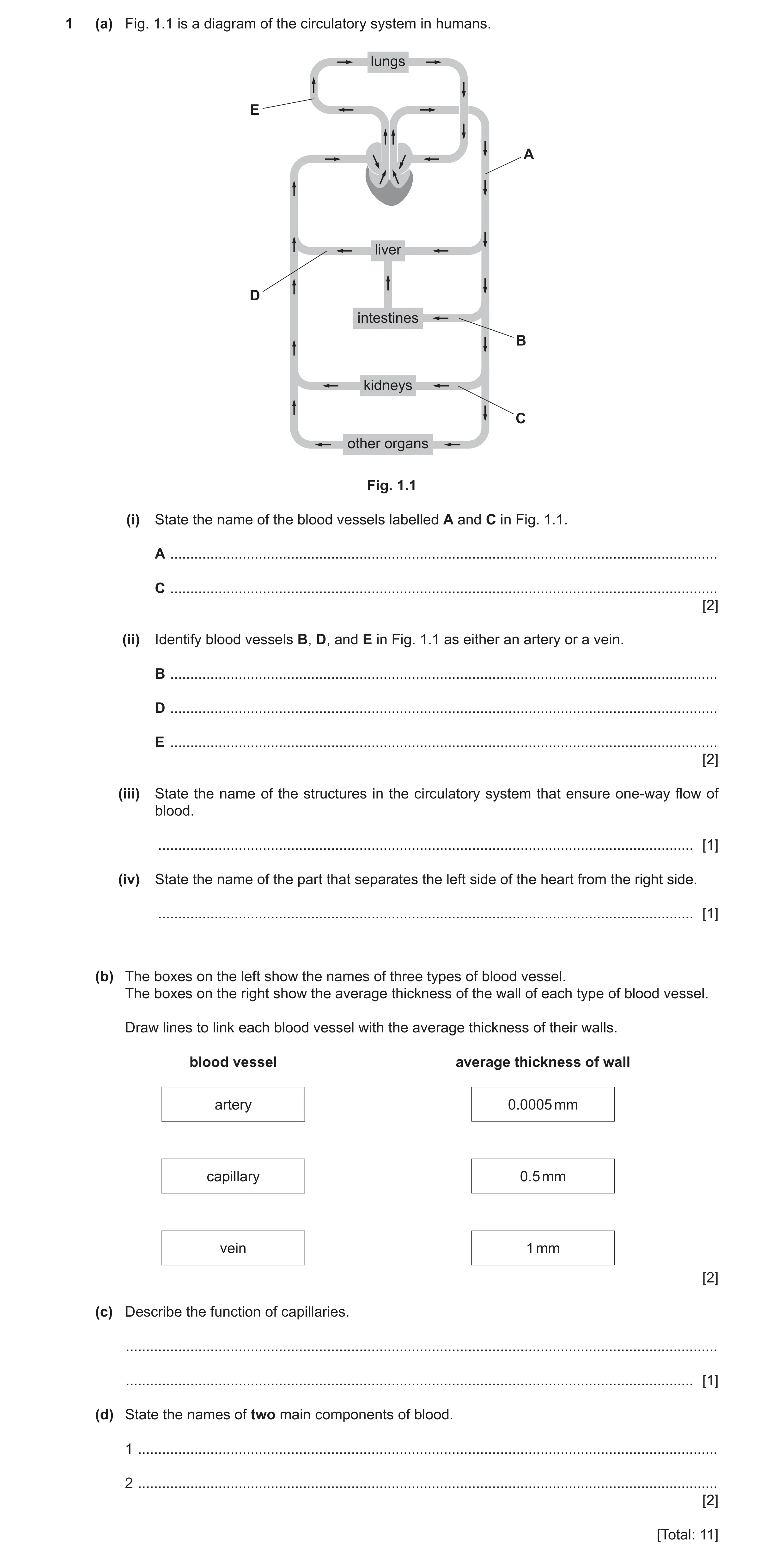
1 (a) Fig. 1.1 is a diagram of the circulatory system in humans. lungs E A liver D intestines B kidneys C other organs Fig. 1.1 (i) State the name of the blood vessels labelled A and C in Fig. 1.1. A … C … [2] (ii) Identify blood vessels B, D, and E in Fig. 1.1 as either an artery or a vein. B … D … E … [2] (iii) State the name of the structures in the circulatory system that ensure one-way flow of blood. … [1] (iv) State the name of the part that separates the left side of the heart from the right side. … [1] (b) The boxes on the left show the names of three types of blood vessel. The boxes on the right show the average thickness of the wall of each type of blood vessel. Draw lines to link each blood vessel with the average thickness of their walls. blood vessel average thickness of wall artery 0.0005 mm capillary 0.5 mm vein 1 mm [2] (c) Describe the function of capillaries. … … [1] (d) State the names of two main components of blood. 1 … 2 … [2] [Total: 11]
11 marks
Mark scheme: Question Answer Marks 1(a)(i) A – aorta ; 2 C – renal artery ; 1(a)(ii) B – artery 2 D – vein E – artery ;; 1(a)(iii) valves ; 1 1(a)(iv) septum ; 1 1(b) 2 artery 0.0005 mm capillary 0.5 mm vein 1 mm ;; 1(c) exchange of (named) material between blood and tissue (cells) / AW ; 1 1(d) any two from: 2 red blood cells ; white blood cells ; plasma ; platelets ;
This question in 0654/32 Feb/March 2023
Q14 · A diagram of a cross-section through a leaf 0654/33 Oct/Nov 2023
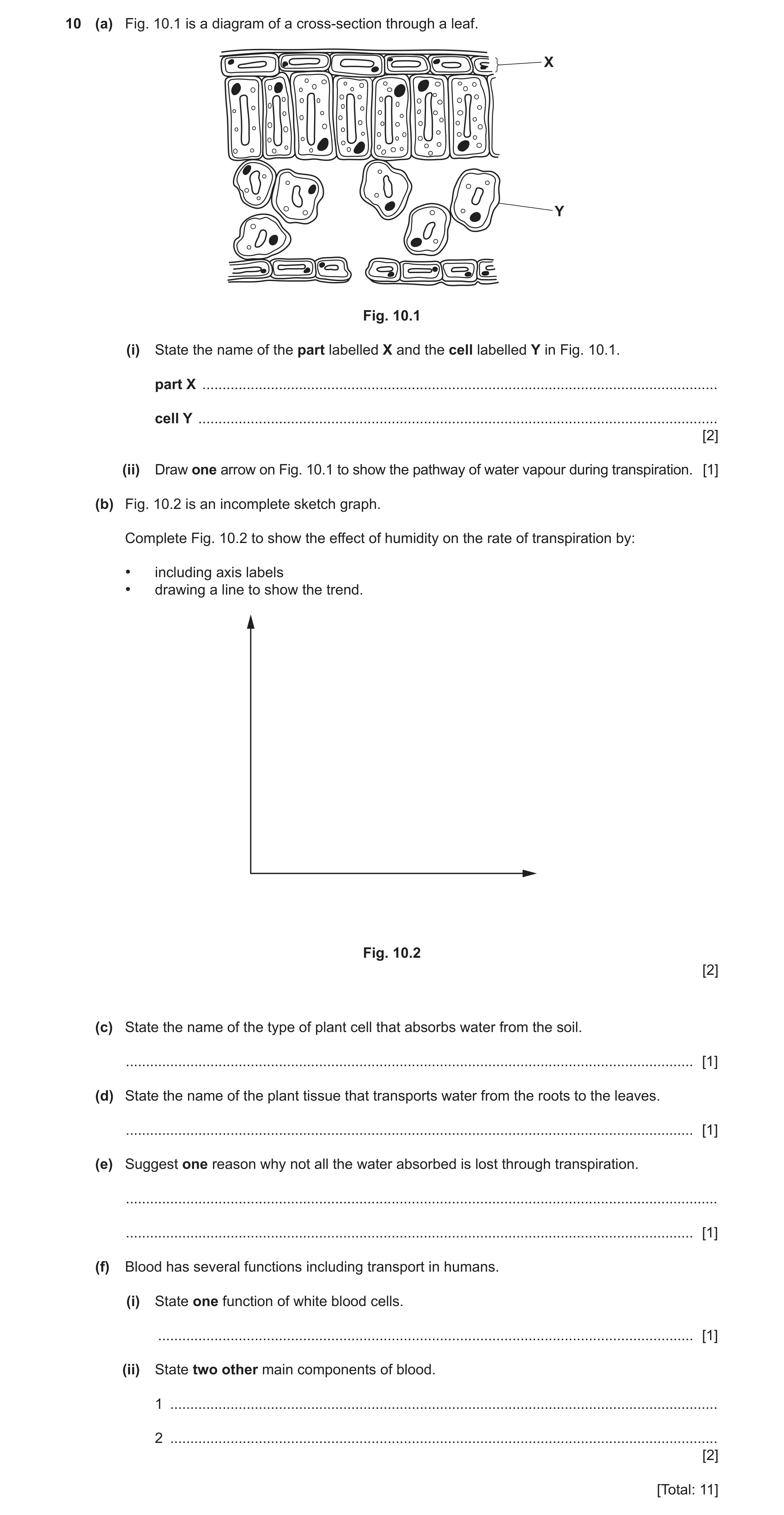
10 (a) Fig. 10.1 is a diagram of a cross-section through a leaf. X Y Fig. 10.1 (i) State the name of the part labelled X and the cell labelled Y in Fig. 10.1. part X … cell Y … [2] (ii) Draw one arrow on Fig. 10.1 to show the pathway of water vapour during transpiration. [1] (b) Fig. 10.2 is an incomplete sketch graph. Complete Fig. 10.2 to show the effect of humidity on the rate of transpiration by: • including axis labels • drawing a line to show the trend. Fig. 10.2 [2] (c) State the name of the type of plant cell that absorbs water from the soil. … [1] (d) State the name of the plant tissue that transports water from the roots to the leaves. … [1] (e) Suggest one reason why not all the water absorbed is lost through transpiration. … … [1] (f) Blood has several functions including transport in humans. (i) State one function of white blood cells. … [1] (ii) State two other main components of blood. 1 … 2 … [2] [Total: 11]
11 marks
Mark scheme: 10(a)(i) X – (upper) epidermis ; 2 Y – spongy mesophyll ; 10(a)(ii) arrow drawn from leaf going out of stoma ; 1 10(b) axis labels ; 2 negative correlation ; 10(c) root hair (cell) ; 1 10(d) xylem ; 1 10(e) some used in, photosynthesis / support ; 1 10(f)(i) phagocytosis / production of antibodies ; 1 10(f)(ii) any two from: 2 red blood cells ; plasma ; platelets ;
This question in 0654/33 Oct/Nov 2023
Q15 · A diagram of the gas exchange system in humans 0654/32 Feb/March 2024
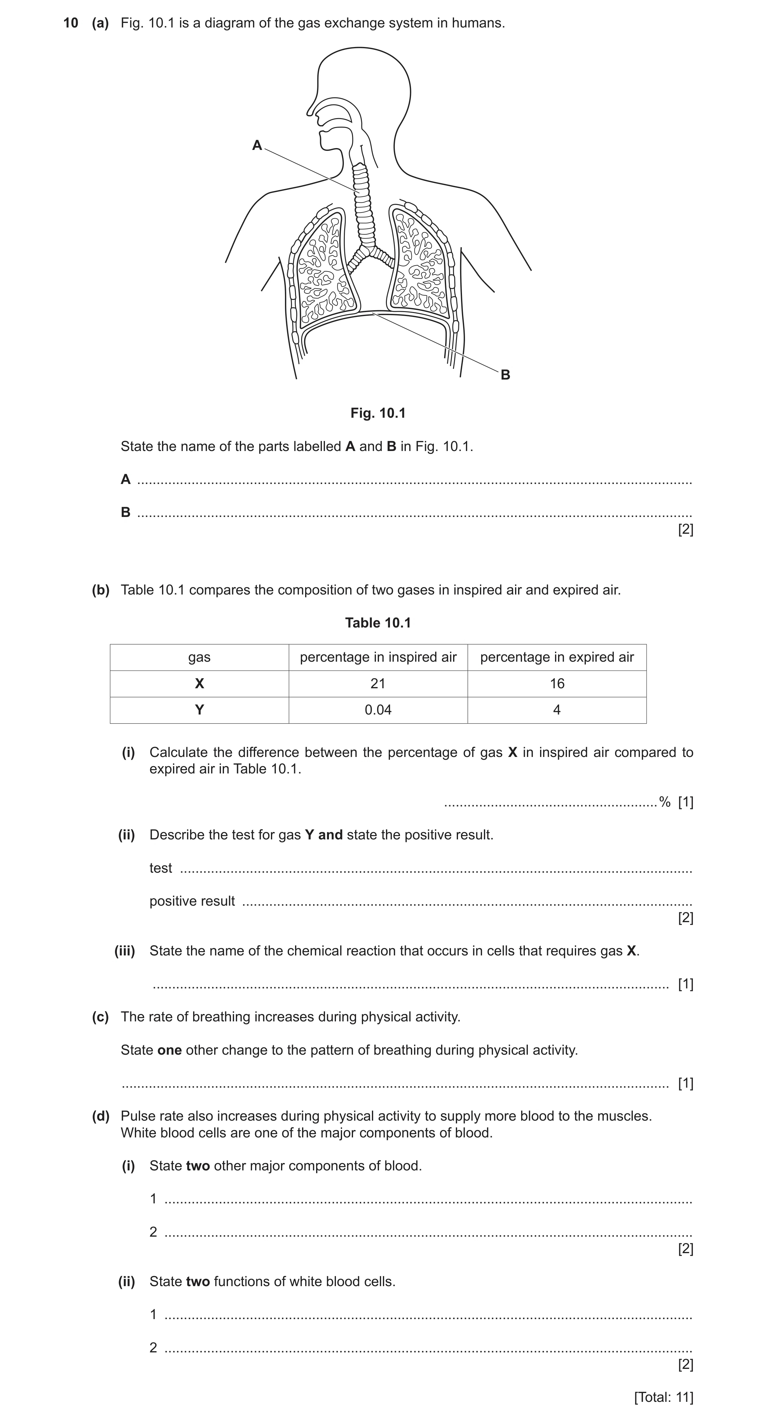
10 (a) Fig. 10.1 is a diagram of the gas exchange system in humans. A B Fig. 10.1 State the name of the parts labelled A and B in Fig. 10.1. A … B … [2] (b) Table 10.1 compares the composition of two gases in inspired air and expired air. Table 10.1 gas percentage in inspired air percentage in expired air X 21 16 Y 0.04 4 (i) Calculate the difference between the percentage of gas X in inspired air compared to expired air in Table 10.1. … % [1] (ii) Describe the test for gas Y and state the positive result. test … positive result … [2] (iii) State the name of the chemical reaction that occurs in cells that requires gas X. … [1] (c) The rate of breathing increases during physical activity. State one other change to the pattern of breathing during physical activity. … [1] (d) Pulse rate also increases during physical activity to supply more blood to the muscles. White blood cells are one of the major components of blood. (i) State two other major components of blood. 1 … 2 … [2] (ii) State two functions of white blood cells. 1 … 2 … [2] [Total: 11]
11 marks
Mark scheme: 10(a) A – trachea ; 2 B – diaphragm ; 10(b)(i) 5 (%) ; 1 10(b)(ii) limewater ; 2 cloudy ; 10(b)(iii) (aerobic) respiration ; 1 10(c) increased depth of breathing ; 1 10(d)(i) any two from: 2 red blood cells ; platelets ; plasma ; 10(d)(ii) phagocytosis ; 2 antibody production ;
This question in 0654/32 Feb/March 2024
Q16 · The parts of a human tooth 0654/31 May/June 2024
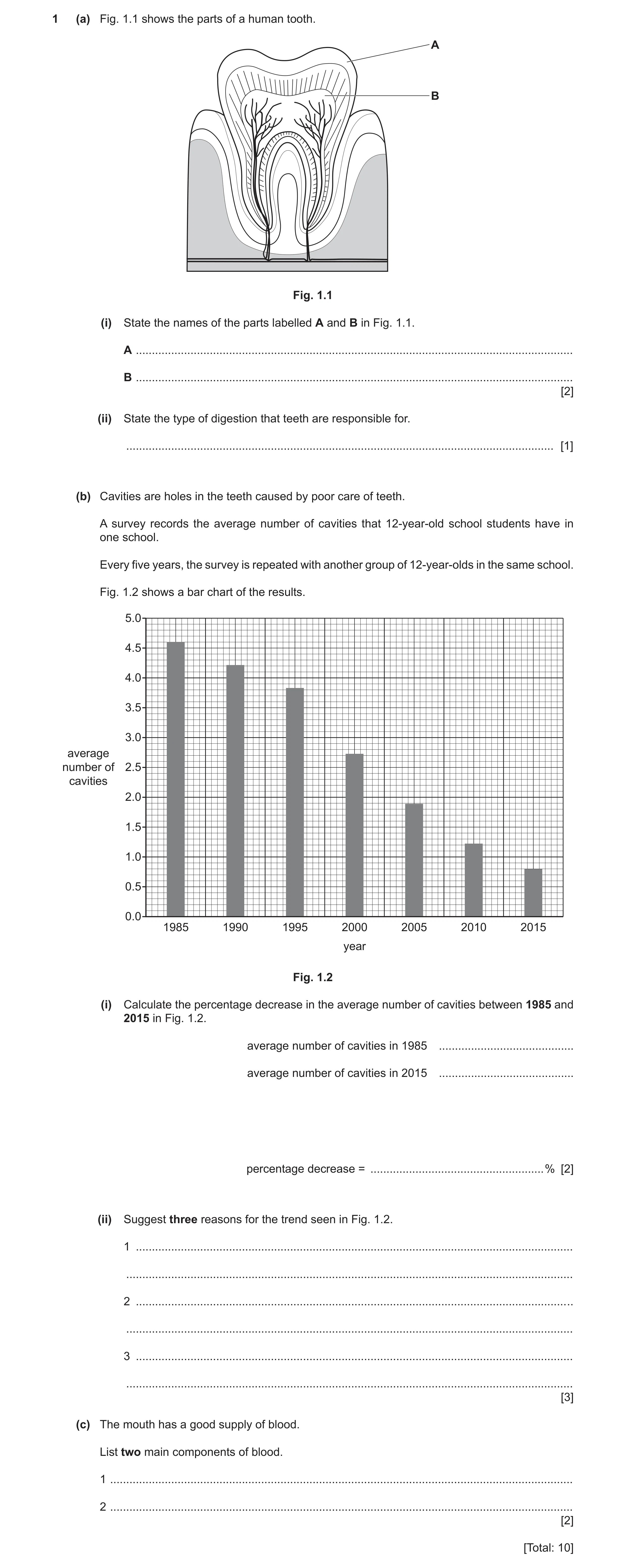
1 (a) Fig. 1.1 shows the parts of a human tooth. A B Fig. 1.1 (i) State the names of the parts labelled A and B in Fig. 1.1. A … B … [2] (ii) State the type of digestion that teeth are responsible for. … [1] (b) Cavities are holes in the teeth caused by poor care of teeth. A survey records the average number of cavities that 12‑year‑old school students have in one school. Every five years, the survey is repeated with another group of 12‑year‑olds in the same school. Fig. 1.2 shows a bar chart of the results. 5.0 4.5 4.0 3.5 3.0 average number of 2.5 cavities 2.0 1.5 1.0 0.5 0.0 1985 1990 1995 2000 2005 2010 2015 year Fig. 1.2 (i) Calculate the percentage decrease in the average number of cavities between 1985 and 2015 in Fig. 1.2. average number of cavities in 1985 … average number of cavities in 2015 … percentage decrease = … % [2] (ii) Suggest three reasons for the trend seen in Fig. 1.2. 1 … … 2 … … 3 … … [3] (c) The mouth has a good supply of blood. List two main components of blood. 1 … 2 … [2] [Total: 10]
10 marks
Mark scheme: 1(a)(i) A – enamel ; B – pulp (cavity) ; 2 1(a)(ii) mechanical ; 1 1(b)(i) 4.6 and 0.8 ; ((3.8 / 4.6) 100) = 83 (%) ; 2 1(b)(ii) any three from: improvement in dental care by people ; improvement in dental, care / technology ; reduction in consumption of sugar / more calcium in diet ; ref to education ; addition of fluoride to drinking water / toothpaste ; 3 1(c) any two from: red blood cells ; white blood cells ; plasma ; platelets ; 2
This question in 0654/31 May/June 2024
Q17 · The number of new infections of HIV in one country was estimated in 2000 and 2018 0654/31 Oct/Nov 2024
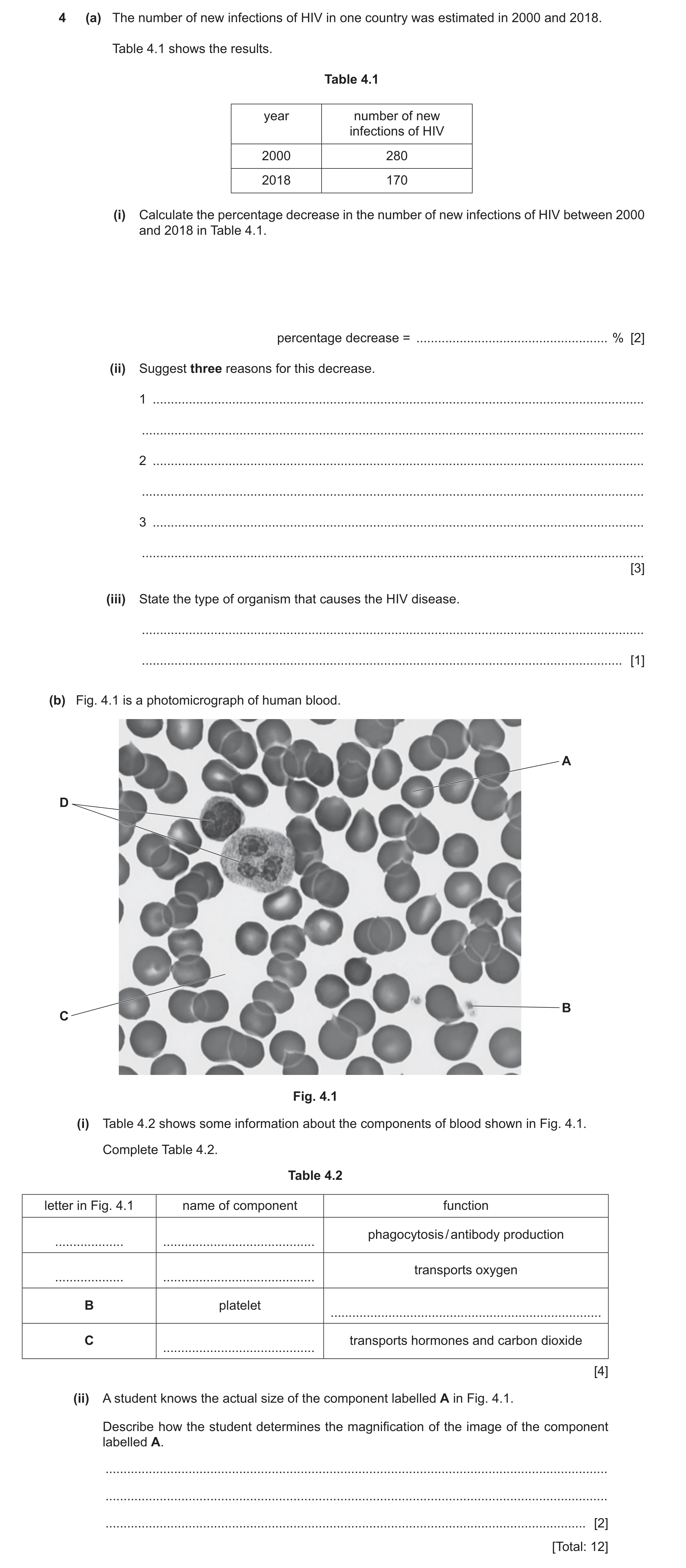
4 (a) The number of new infections of HIV in one country was estimated in 2000 and 2018. Table 4.1 shows the results. Table 4.1 year number of new infections of HIV 2000 280 2018 170 (i) Calculate the percentage decrease in the number of new infections of HIV between 2000 and 2018 in Table 4.1. percentage decrease = … % [2] (ii) Suggest three reasons for this decrease. 1 … … 2 … … 3 … … [3] (iii) State the type of organism that causes the HIV disease. … … [1] (b) Fig. 4.1 is a photomicrograph of human blood. A D B C Fig. 4.1 (i) Table 4.2 shows some information about the components of blood shown in Fig. 4.1. Complete Table 4.2. Table 4.2 letter in Fig. 4.1 name of component function phagocytosis / antibody production … … transports oxygen … … B platelet … C transports hormones and carbon dioxide … [4] (ii) A student knows the actual size of the component labelled A in Fig. 4.1. Describe how the student determines the magnification of the image of the component labelled A. … … … [2] [Total: 12]
12 marks
Mark scheme: 4(a)(i) 280 – 170 / 110 ; 2 ((110 / 280) 100) = 39(.3) (%) ; 4(a)(ii) any three from: 3 ref to education / campaigns ; increased, monitoring / testing ; use of condoms / barrier contraception ; increased abstinence ; screening of blood transfusions ; provision of clean needles / needle banks ; reduction in sharing needles ; reduced breast feeding by infected mothers AVP ; 4(a)(iii) virus ; 1 4(b)(i) 4 D white blood cell phagocytosis / antibody production A red blood cell transport oxygen B platelet (blood) clotting C plasma transports hormones and carbon dioxide ;;;; one mark for each row 4(b)(ii) measure (with a ruler the image length of one of the red blood cells) ; 2 divide this length by the actual size / magnification = image size / actual size ;
This question in 0654/31 Oct/Nov 2024
 1 / 25
 2 / 25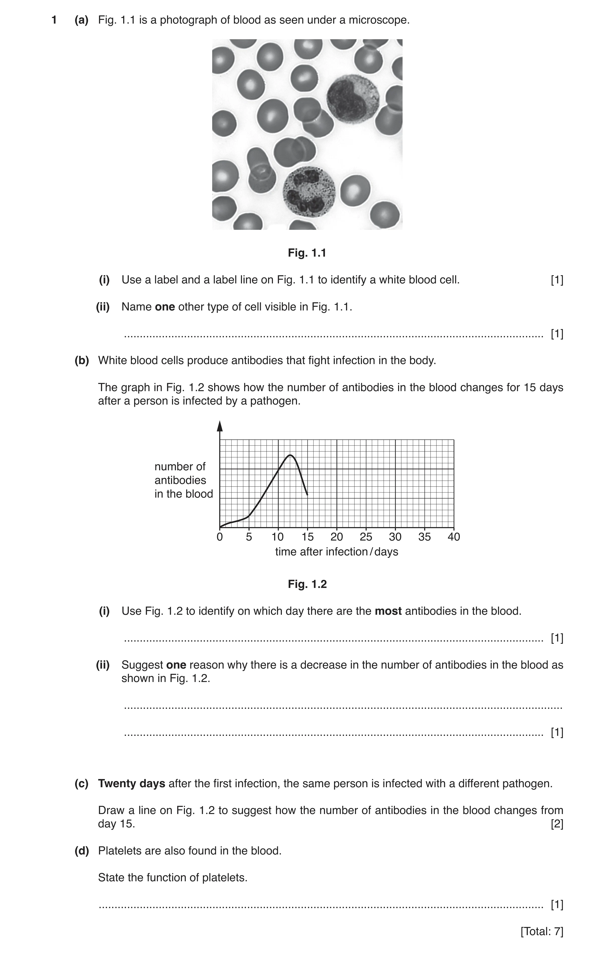
 6 / 25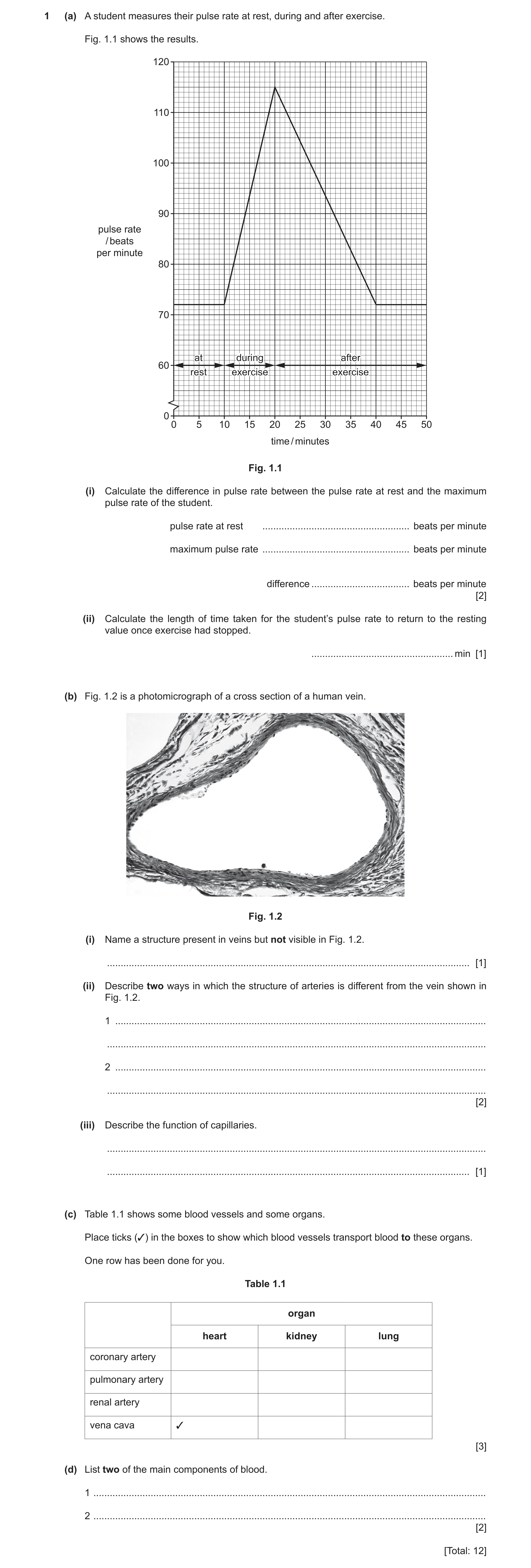
 13 / 25
 14 / 25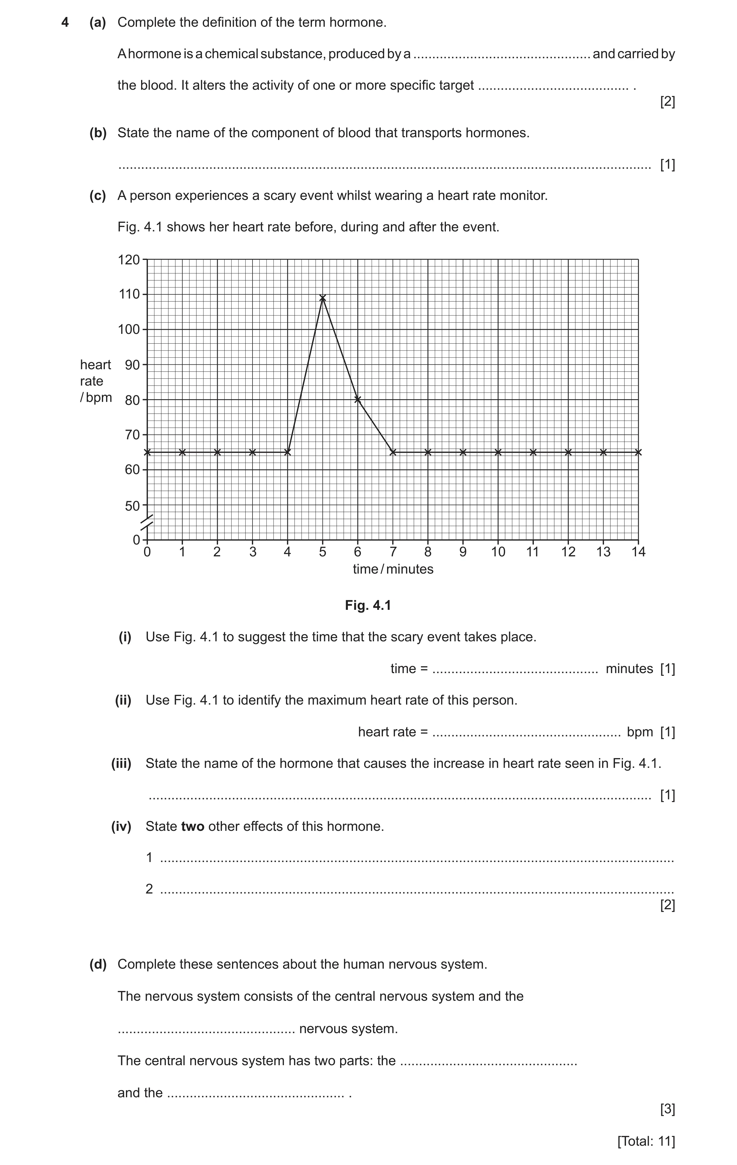
 8
8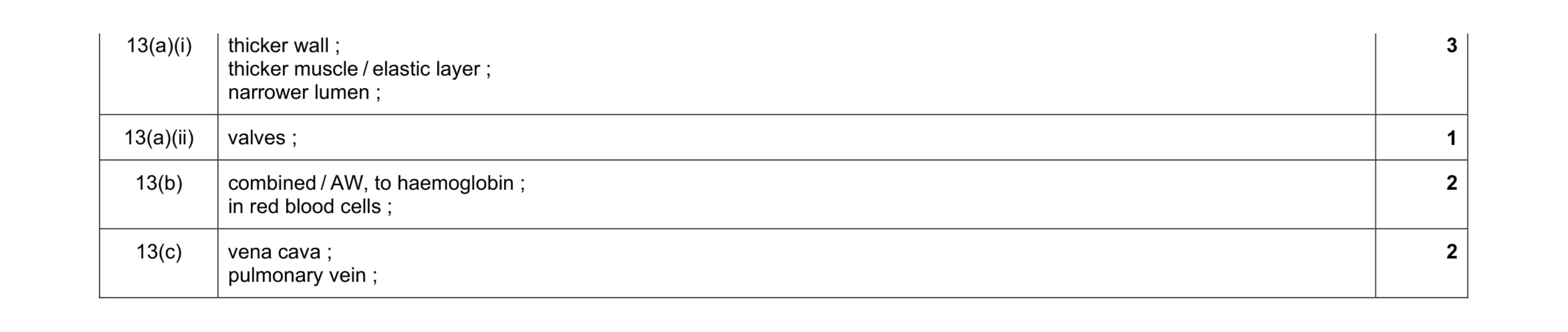 8
8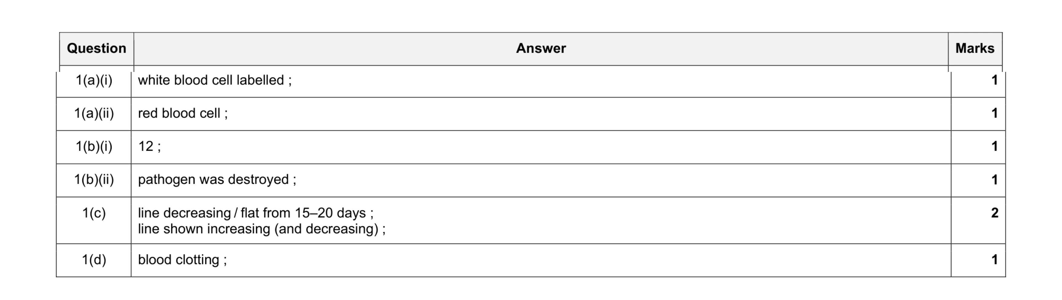 7
7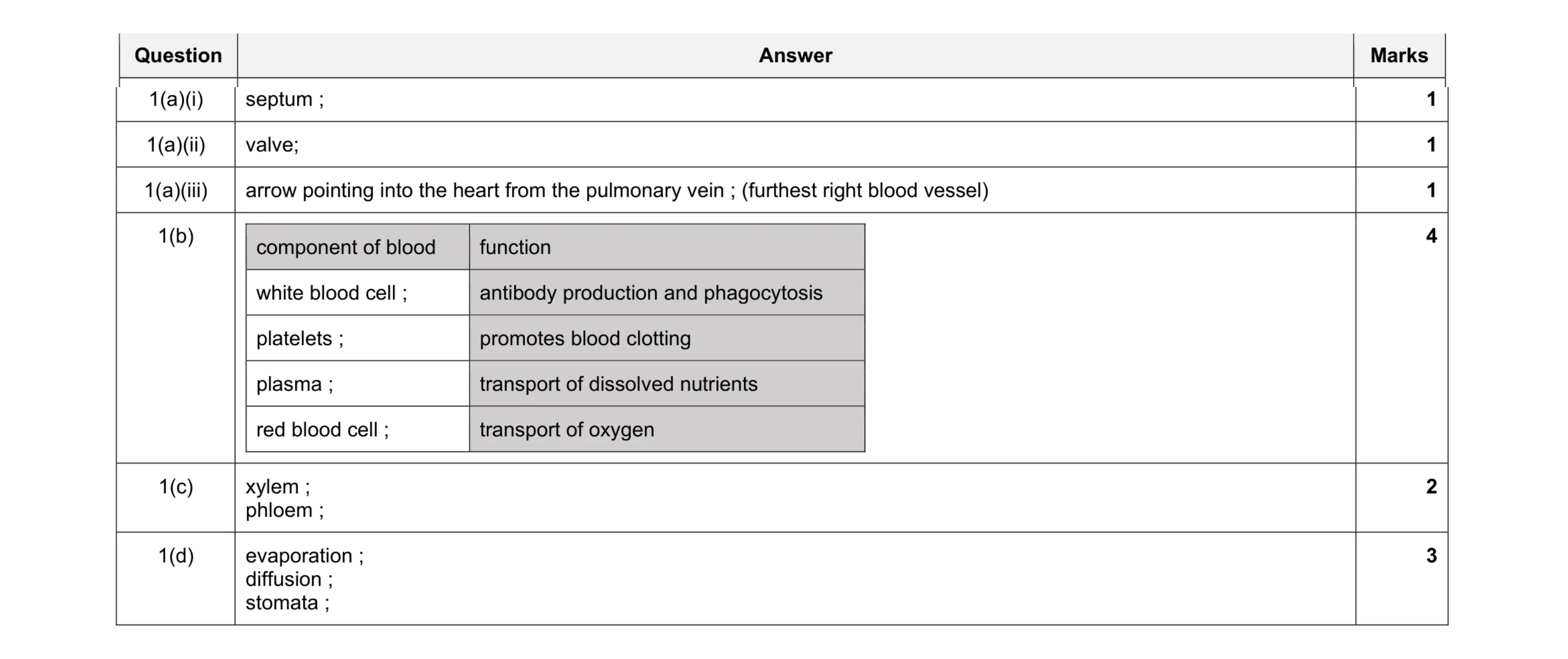 12
12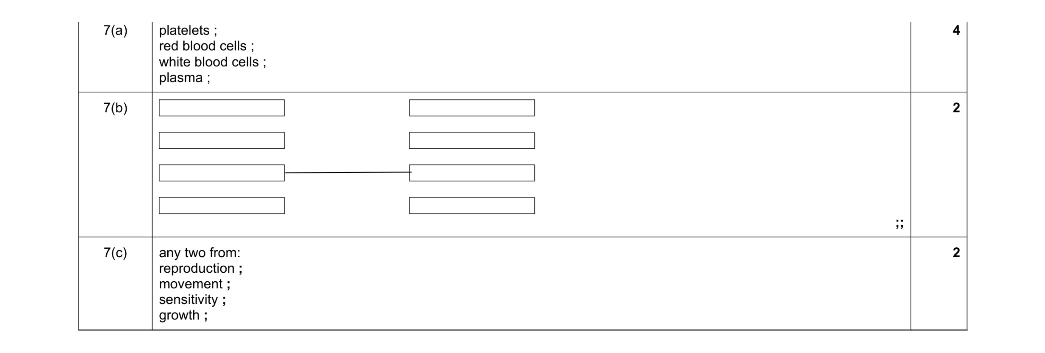 8
8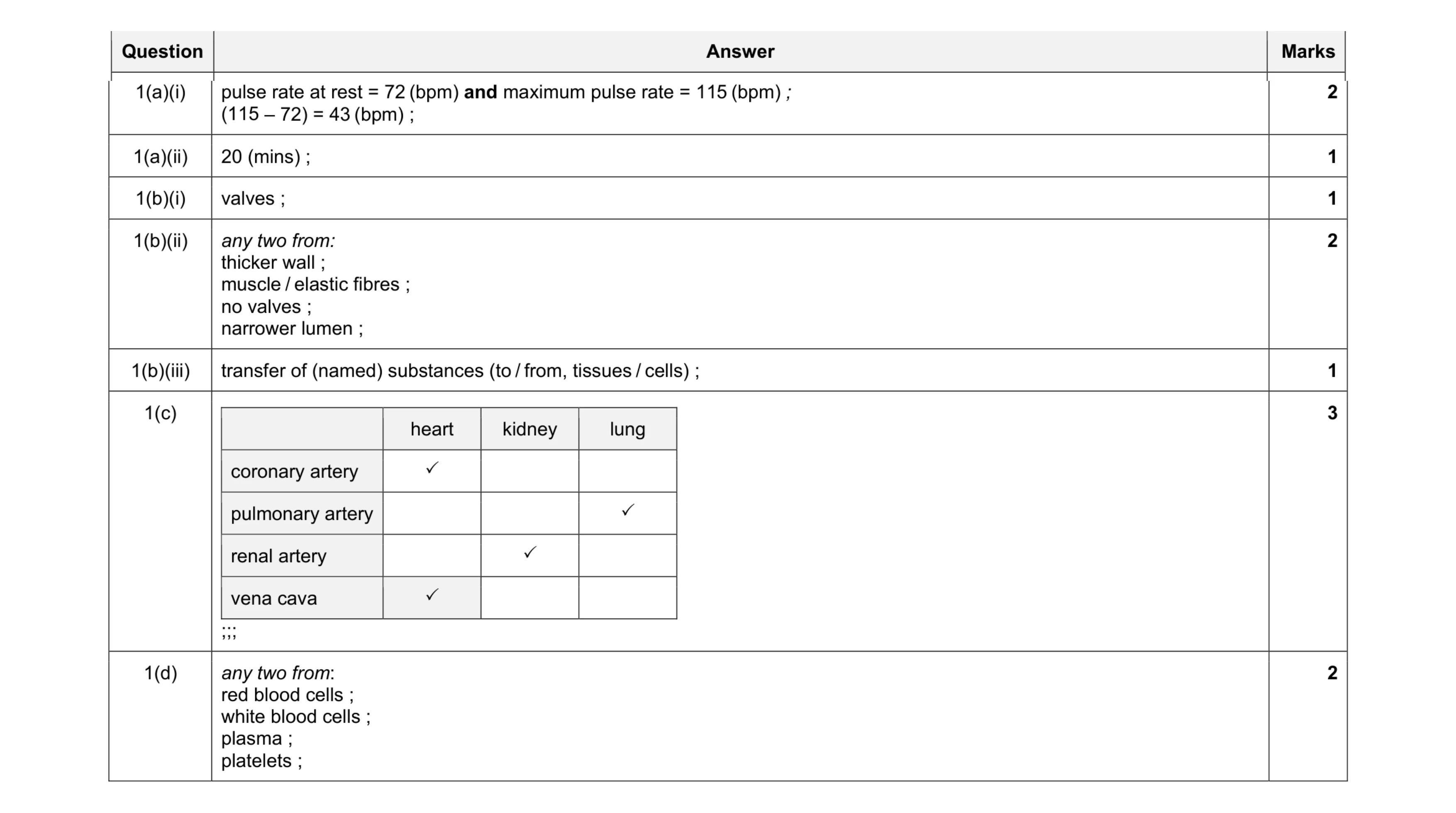 12
12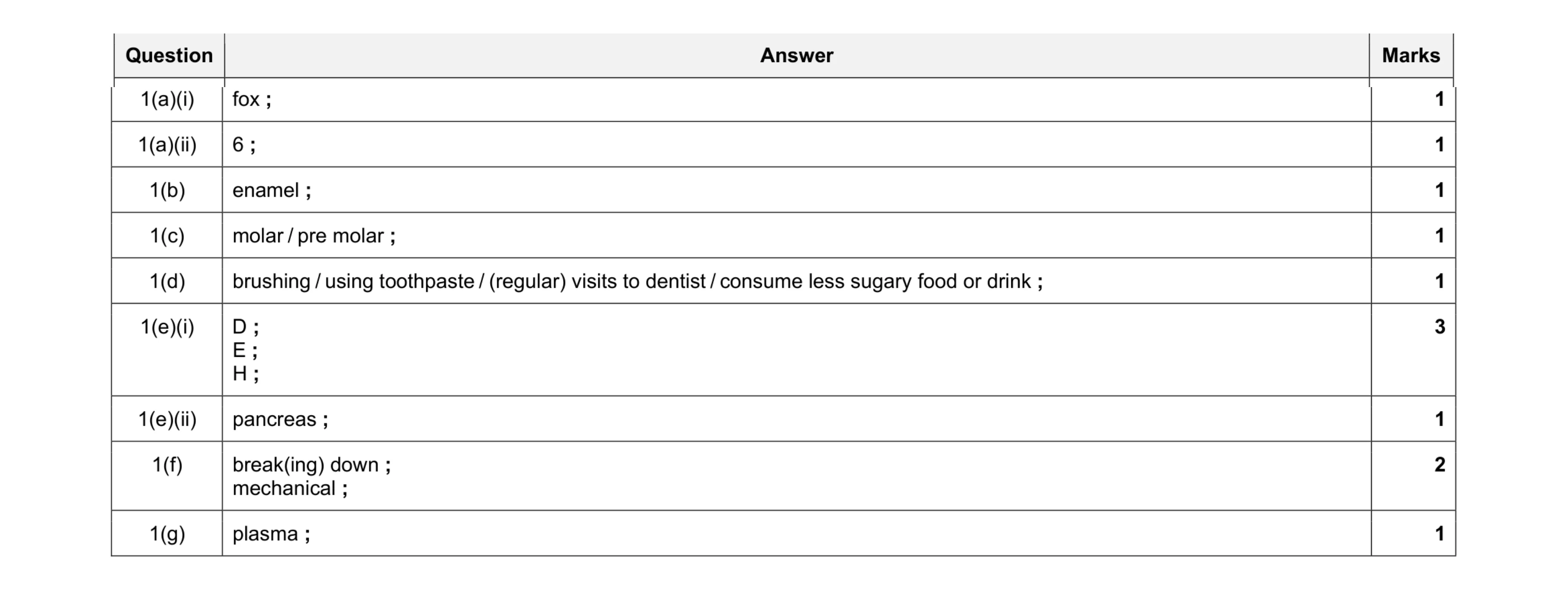 12
12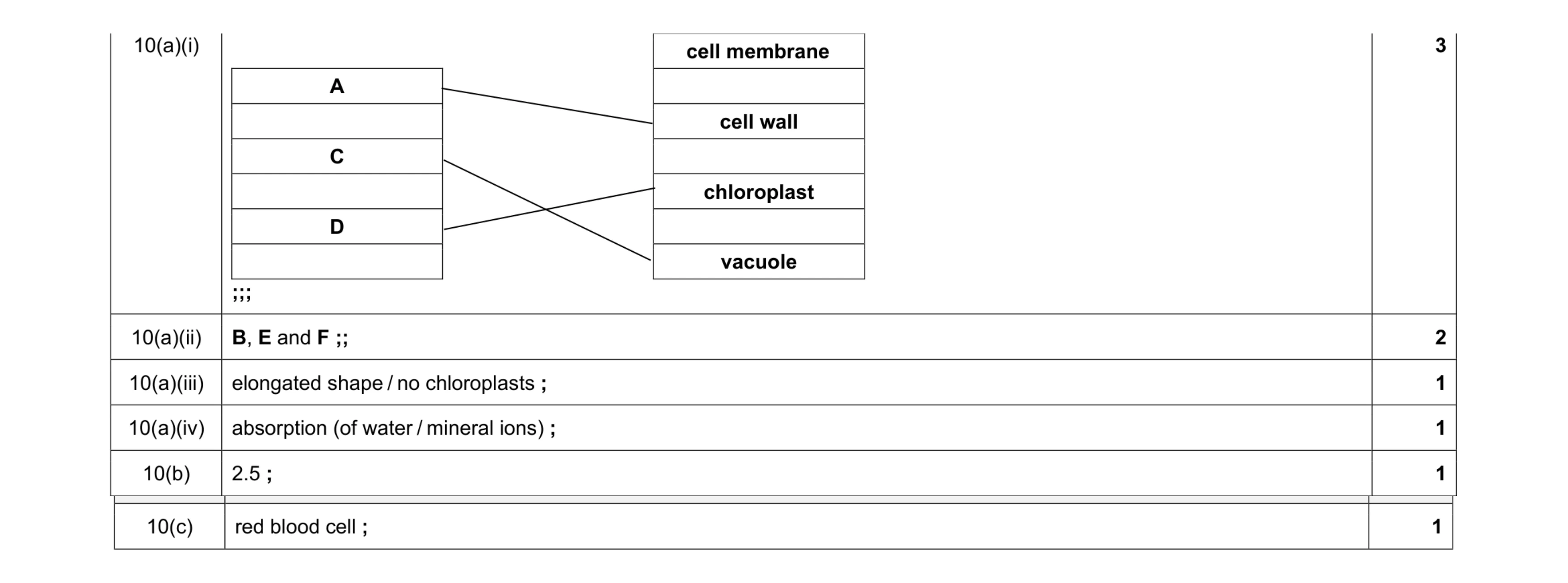 9
9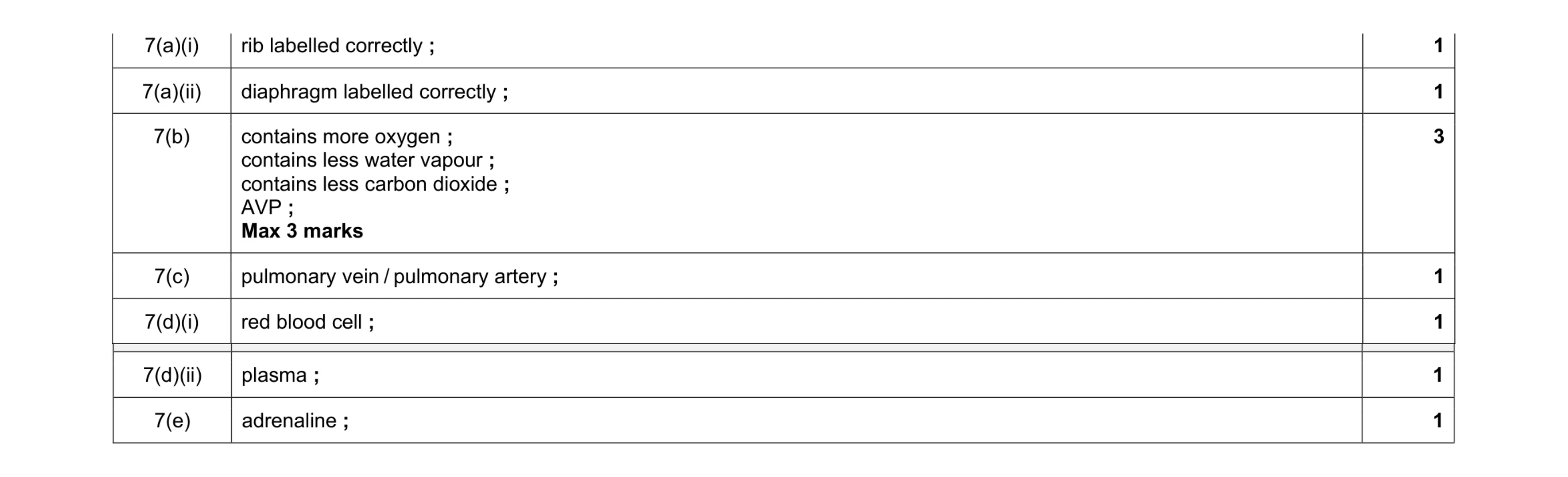 9
9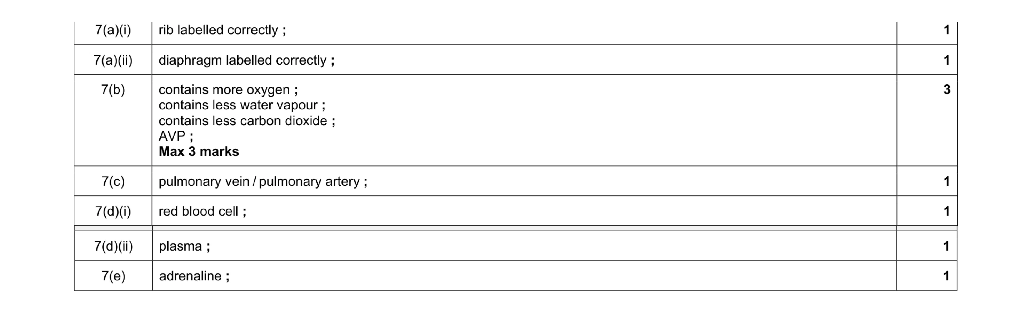 9
9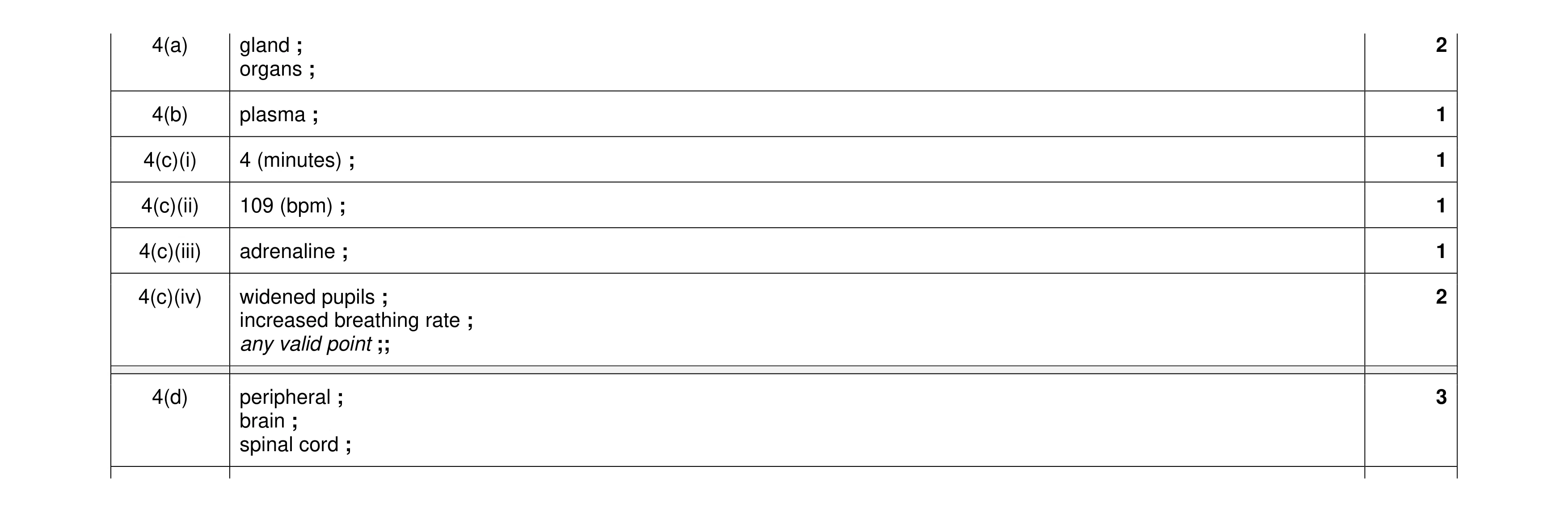 11
11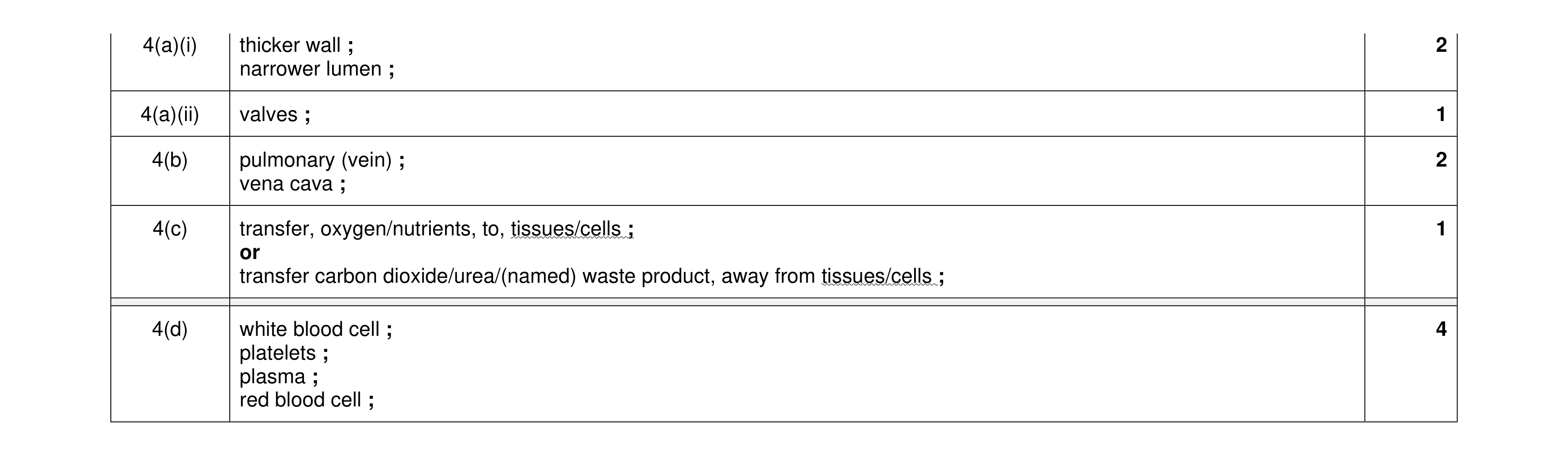 10
10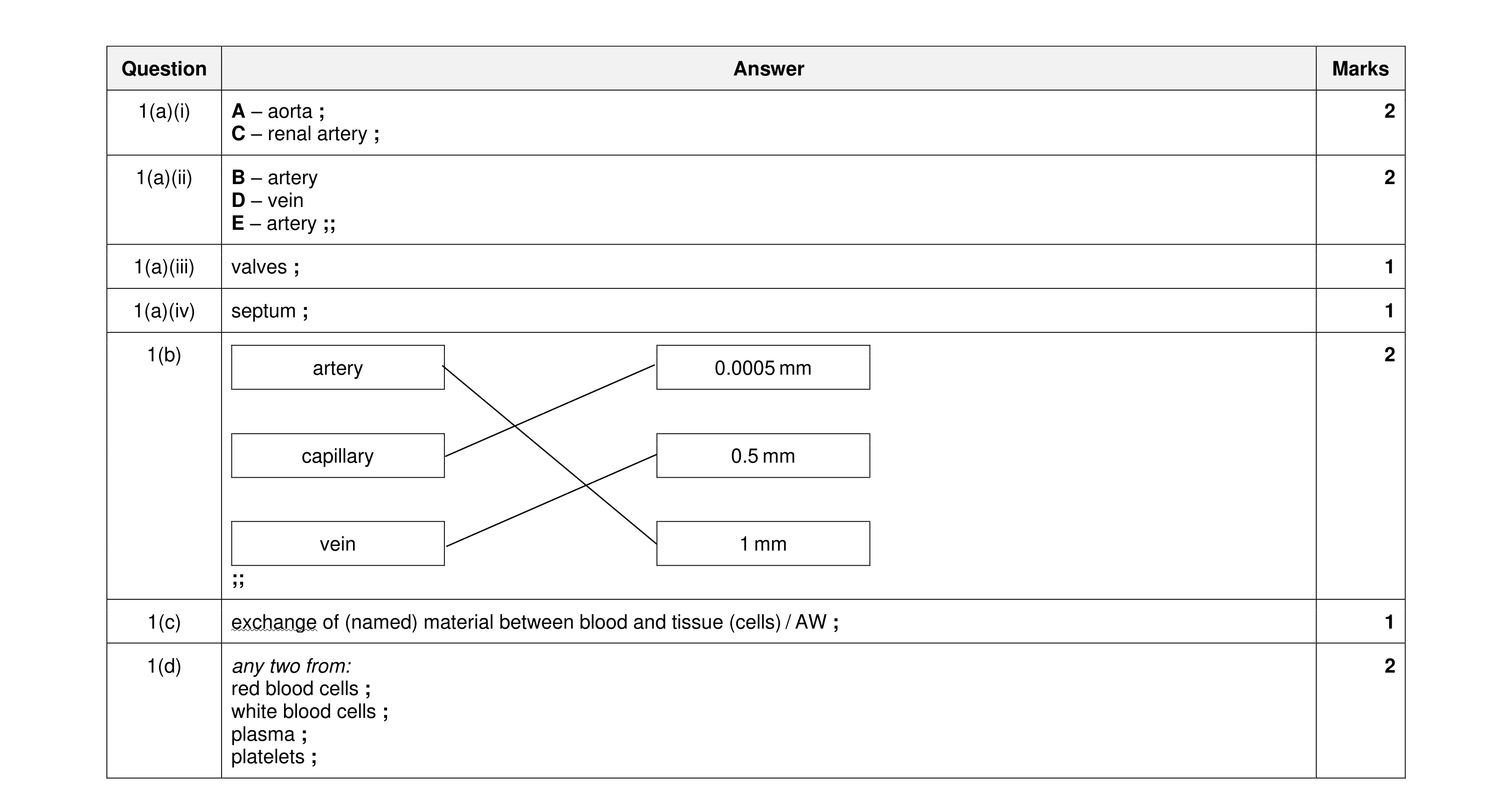 11
11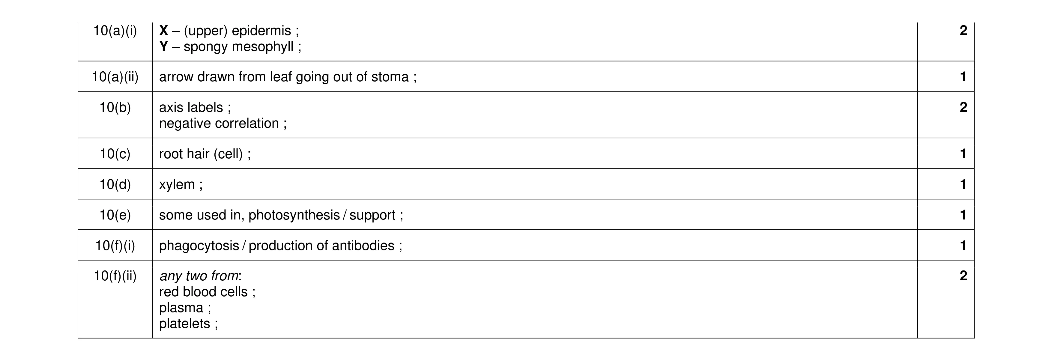 11
11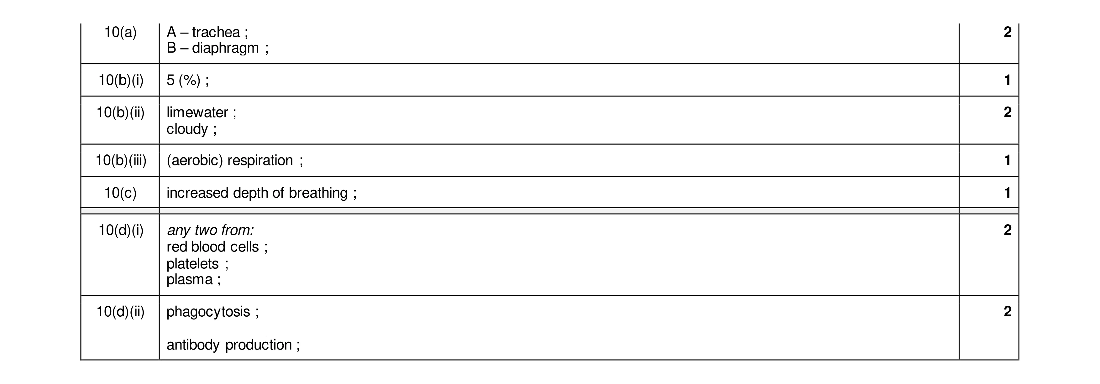 11
11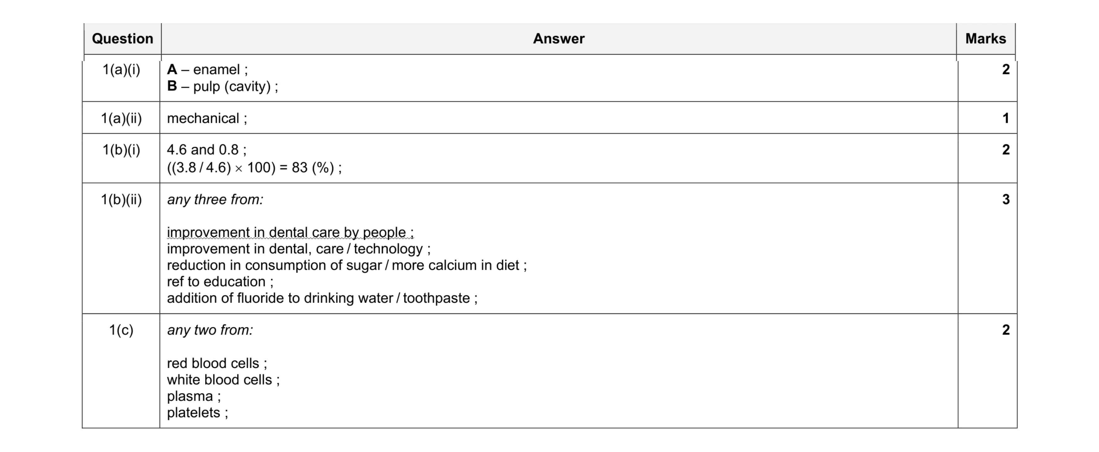 10
10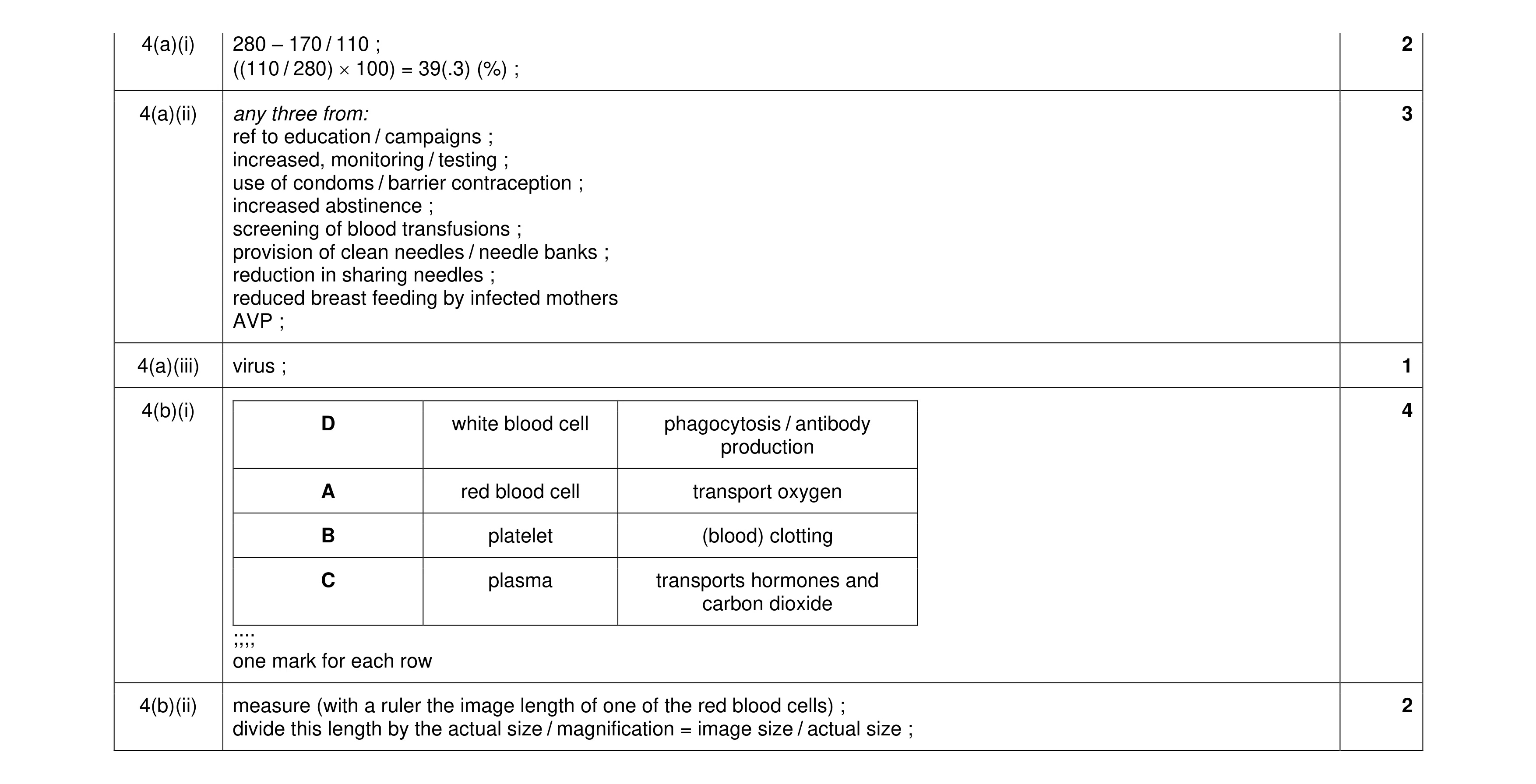 12
12