Questions as text
Q1 · Use the following words or phrases to complete the flow chart about the transport of… 0653/41 Oct/Nov 2017
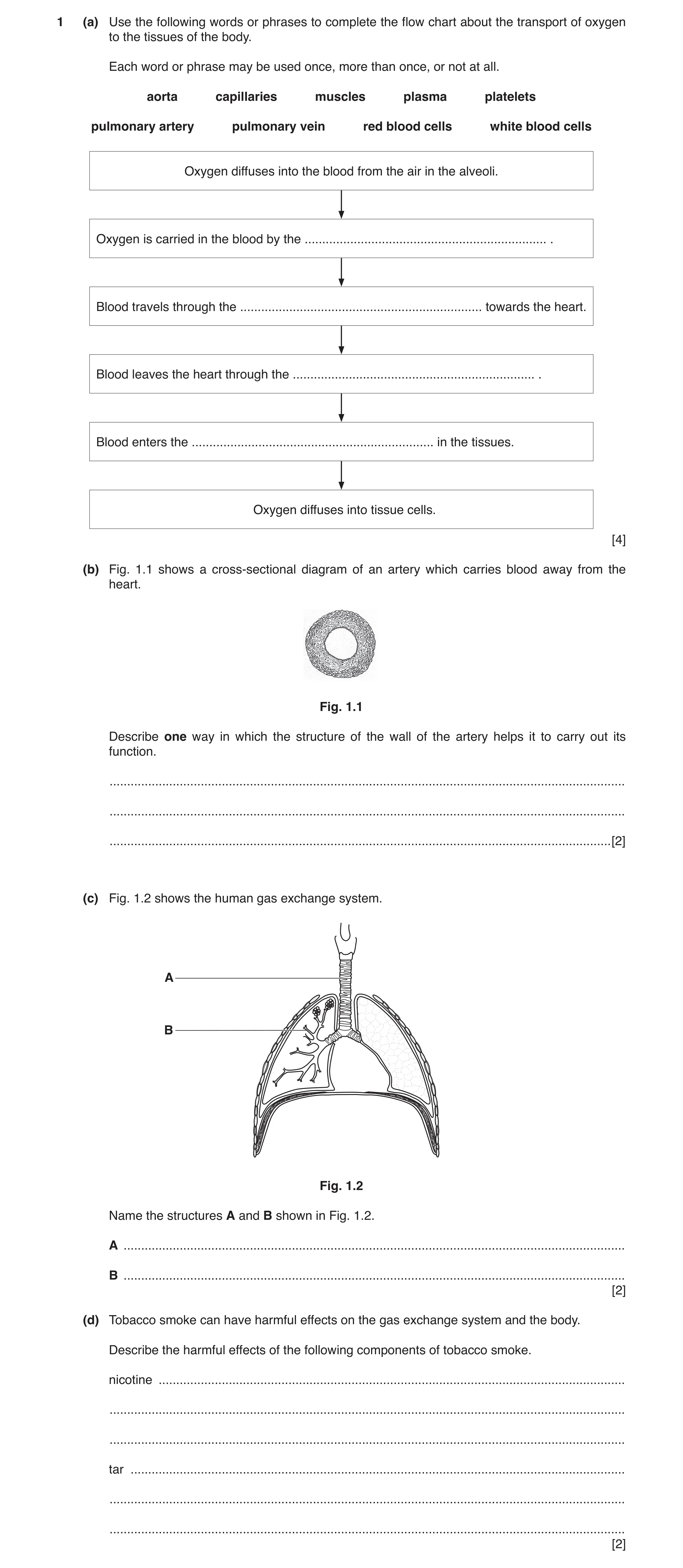
1 (a) Use the following words or phrases to complete the flow chart about the transport of oxygen to the tissues of the body. Each word or phrase may be used once, more than once, or not at all. aorta capillaries muscles plasma platelets pulmonary artery pulmonary vein red blood cells white blood cells Oxygen diffuses into the blood from the air in the alveoli. Oxygen is carried in the blood by the … . Blood travels through the … towards the heart. Blood leaves the heart through the … . Blood enters the … in the tissues. Oxygen diffuses into tissue cells. [4] (b) Fig. 1.1 shows a cross-sectional diagram of an artery which carries blood away from the heart. Fig. 1.1 Describe one way in which the structure of the wall of the artery helps it to carry out its function. … … … [2] (c) Fig. 1.2 shows the human gas exchange system. A B Fig. 1.2 Name the structures A and B shown in Fig. 1.2. A … B … [2] (d) Tobacco smoke can have harmful effects on the gas exchange system and the body. Describe the harmful effects of the following components of tobacco smoke. nicotine … … … tar … … … [2]
10 marks
Mark scheme: 1(a) red blood cells ; pulmonary vein ; aorta ; capillaries ; 4 1(b) thick (wall) ; to resist bursting / withstand high blood pressure ; 2 1(c) A trachea ; B bronchiole ; 2 1(d) (nicotine) causes addiction / increases blood pressure / leads to heart disease ; (tar) increases mucus / (tar) builds up in lungs / paralyses / destroys cilia / can cause cancer / avp ; 2
This question in 0653/41 Oct/Nov 2017
Q2 · A diagram of the internal structure of the heart 0653/42 Oct/Nov 2018
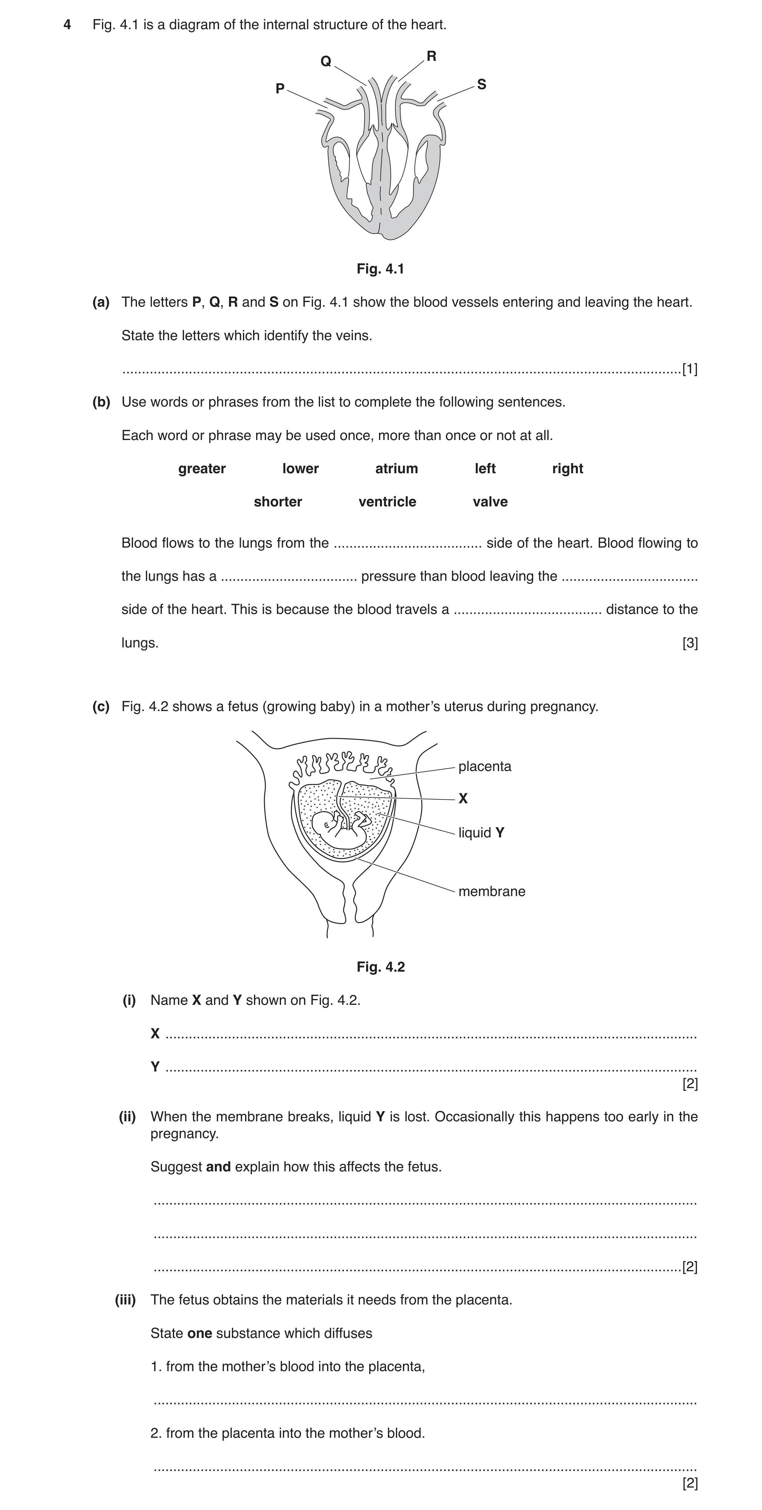
4 Fig. 4.1 is a diagram of the internal structure of the heart. R Q S P Fig. 4.1 (a) The letters P, Q, R and S on Fig. 4.1 show the blood vessels entering and leaving the heart. State the letters which identify the veins. … [1] (b) Use words or phrases from the list to complete the following sentences. Each word or phrase may be used once, more than once or not at all. greater lower atrium left right shorter ventricle valve Blood flows to the lungs from the … side of the heart. Blood flowing to the lungs has a … pressure than blood leaving the … side of the heart. This is because the blood travels a … distance to the lungs. [3] (c) Fig. 4.2 shows a fetus (growing baby) in a mother’s uterus during pregnancy. placenta X liquid Y membrane Fig. 4.2 (i) Name X and Y shown on Fig. 4.2. X … Y … [2] (ii) When the membrane breaks, liquid Y is lost. Occasionally this happens too early in the pregnancy. Suggest and explain how this affects the fetus. … … … [2] (iii) The fetus obtains the materials it needs from the placenta. State one substance which diffuses 1. from the mother’s blood into the placenta, … 2. from the placenta into the mother’s blood. … [2]
10 marks
Mark scheme: 4(a) P and S ; 1 Question Answer Marks 4(b) right ; lower ; left ; shorter ; 3 4(c)(i) X umbilical cord ; Y amniotic fluid ; 2 4(c)(ii) reference to the loss of protection (of fetus) ; higher risk of physical damage / infection / premature birth ; 2 4(c)(iii) oxygen / glucose / any named dissolved nutrient ; carbon dioxide ; 2
This question in 0653/42 Oct/Nov 2018
Q3 · A diagram of a cross-section through an artery 0653/42 Oct/Nov 2020
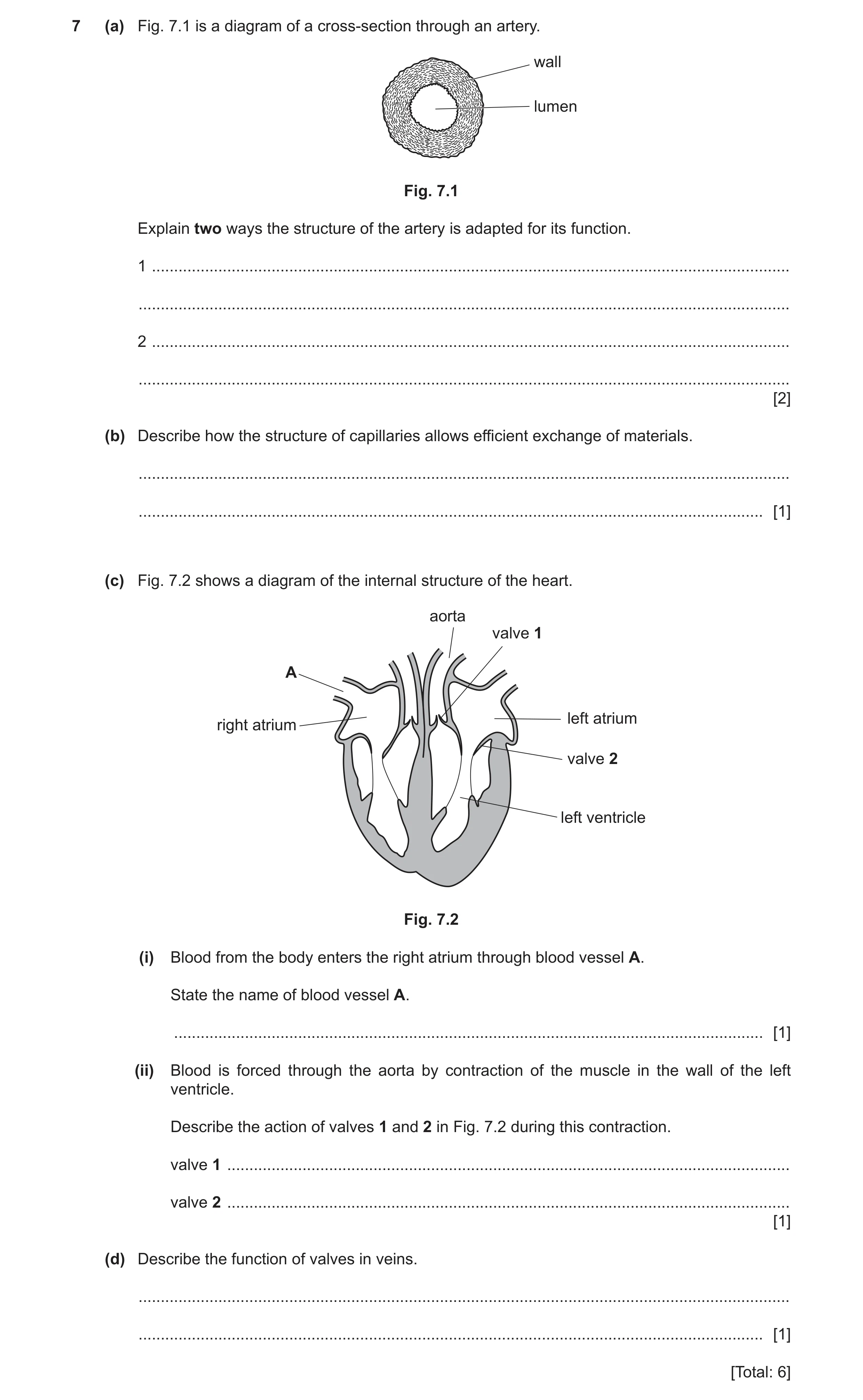
7 (a) Fig. 7.1 is a diagram of a cross-section through an artery. wall lumen Fig. 7.1 Explain two ways the structure of the artery is adapted for its function. 1 … … 2 … … [2] (b) Describe how the structure of capillaries allows efficient exchange of materials. … … [1] (c) Fig. 7.2 shows a diagram of the internal structure of the heart. aorta valve 1 A left atrium right atrium valve 2 left ventricle Fig. 7.2 (i) Blood from the body enters the right atrium through blood vessel A. State the name of blood vessel A. … [1] (ii) Blood is forced through the aorta by contraction of the muscle in the wall of the left ventricle. Describe the action of valves 1 and 2 in Fig. 7.2 during this contraction. valve 1 … valve 2 … [1] (d) Describe the function of valves in veins. … … [1] [Total: 6]
6 marks
Mark scheme: 7(a) any two paired responses: strong / thick wall, prevents bursting / withstands high pressure ; elastic fibres in wall, prevent bursting / stretch to accommodate high pressure of blood / enable wall to recoil and maintain pressure ; narrow lumen, maintains high pressure ; 7(b) (capillaries have) thin walls ; 1 7(c)(i) vena cava ; 1 7(c)(ii) valve 1 opens AND valve 2 closes ; 1 7(d) prevent backflow of blood / ensure that blood only travels in one direction ; 1
This question in 0653/42 Oct/Nov 2020
Q4 · A heart with coronary heart disease 0653/42 Feb/March 2021
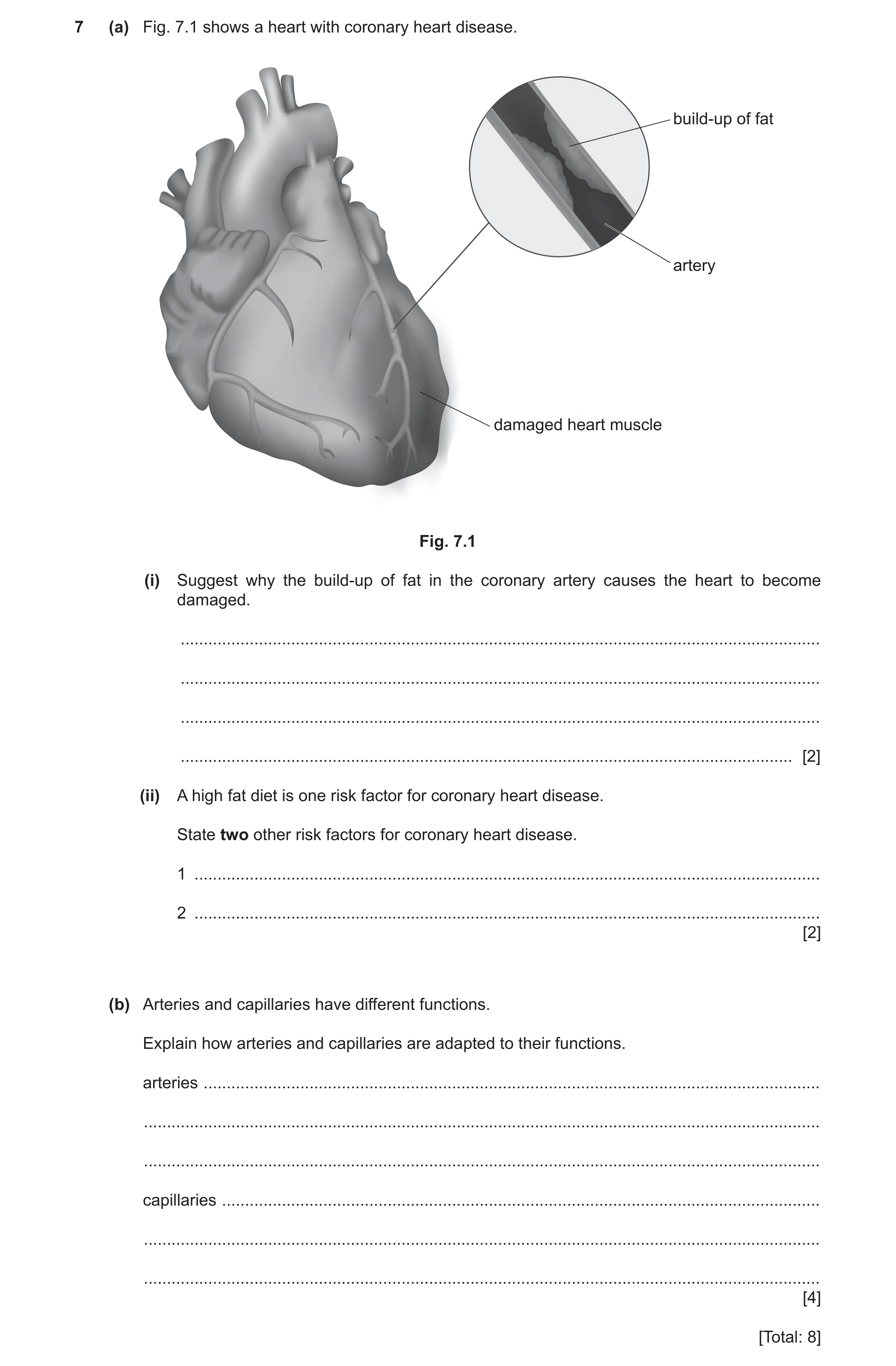
7 (a) Fig. 7.1 shows a heart with coronary heart disease. build-up of fat artery damaged heart muscle Fig. 7.1 (i) Suggest why the build-up of fat in the coronary artery causes the heart to become damaged. … … … … [2] (ii) A high fat diet is one risk factor for coronary heart disease. State two other risk factors for coronary heart disease. 1 … 2 … [2] (b) Arteries and capillaries have different functions. Explain how arteries and capillaries are adapted to their functions. arteries … … … capillaries … … … [4] [Total: 8]
8 marks
Mark scheme: 7(a)(i) less, blood / oxygen transported to heart (muscle) ; (heart) muscle unable to respire effectively ; 2 7(a)(ii) any two from: gender / sex ; stress ; smoking ; age ; genetic predisposition ; AVP, e.g. obesity / lack of exercise ; 2 7(b) any one adaptation and explanation pairing for arteries from: thick muscular wall ; to withstand high pressure ; OR elastic (muscle fibres in walls) ; to allow, contraction / relaxation / maintain blood flow rate ; OR narrow lumen ; to maintain, blood pressure / flow rate ; adaptation and explanation for capillaries: thin walls / walls only one cell thick ; to allow for exchange of materials / to allow substances to diffuse in and out ; 4
This question in 0653/42 Feb/March 2021
Q5 · A photomicrograph of a cross-section of an artery 0653/41 May/June 2021
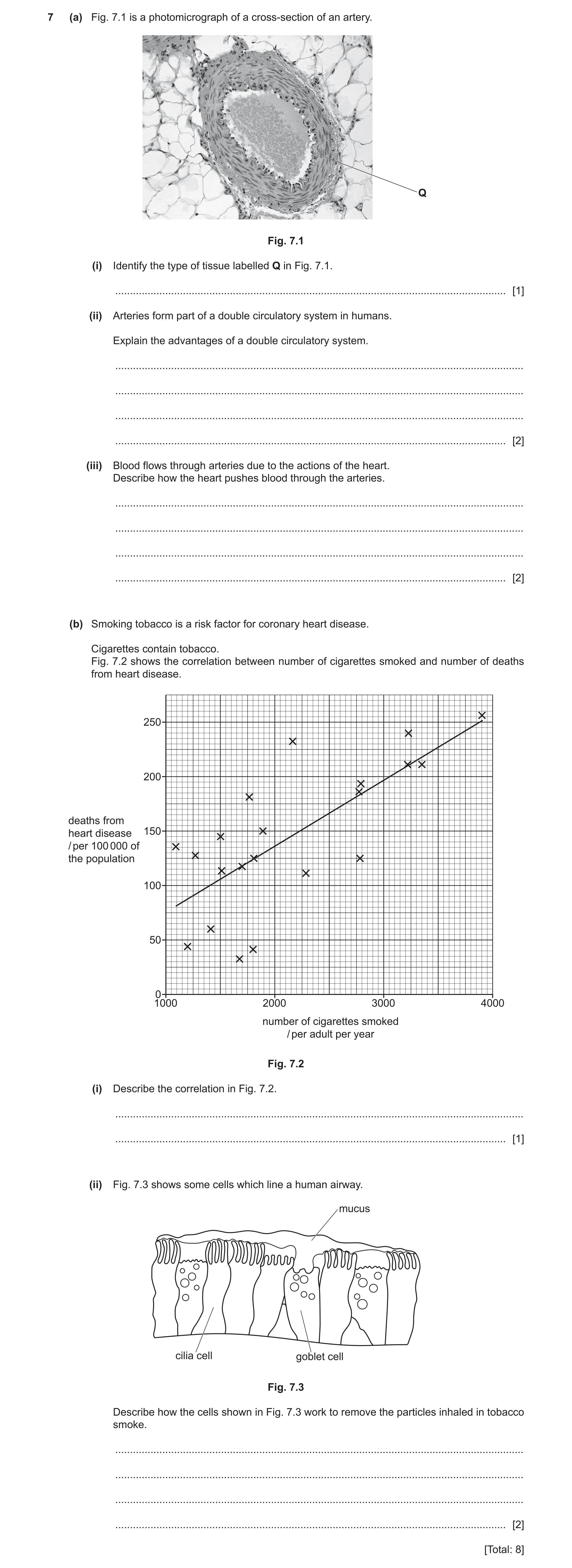
7 (a) Fig. 7.1 is a photomicrograph of a cross-section of an artery. Q Fig. 7.1 (i) Identify the type of tissue labelled Q in Fig. 7.1. … [1] (ii) Arteries form part of a double circulatory system in humans. Explain the advantages of a double circulatory system. … … … … [2] (iii) Blood flows through arteries due to the actions of the heart. Describe how the heart pushes blood through the arteries. … … … … [2] (b) Smoking tobacco is a risk factor for coronary heart disease. Cigarettes contain tobacco. Fig. 7.2 shows the correlation between number of cigarettes smoked and number of deaths from heart disease. 250 200 deaths from heart disease 150 / per 100 000 of the population 100 50 0 1000 2000 3000 4000 number of cigarettes smoked / per adult per year Fig. 7.2 (i) Describe the correlation in Fig. 7.2. … … [1] (ii) Fig. 7.3 shows some cells which line a human airway. mucus cilia cell goblet cell Fig. 7.3 Describe how the cells shown in Fig. 7.3 work to remove the particles inhaled in tobacco smoke. … … … … [2] [Total: 8]
8 marks
Mark scheme: 7(a)(i) muscle / elastic tissue ; 1 7(a)(ii) any two from: oxygenated blood is kept separate from deoxygenated blood ; blood pressure to body is higher ; blood pressure to lungs is lower ; 2 7(a)(iii) reference to contraction ; (contraction of) muscles in ventricle (wall) ; 2 7(b)(i) as the number of cigarettes smoked increases so do the number of deaths ; 1 7(b)(ii) any two from: the goblet cells, secrete / produce (sticky) mucus ; the mucus traps, particles / microbes / dust ; cilia move (to remove) the mucus (containing particles) ; 2
This question in 0653/41 May/June 2021
Q6 · A diagram of the alimentary canal and associated organs in humans 0653/43 May/June 2022
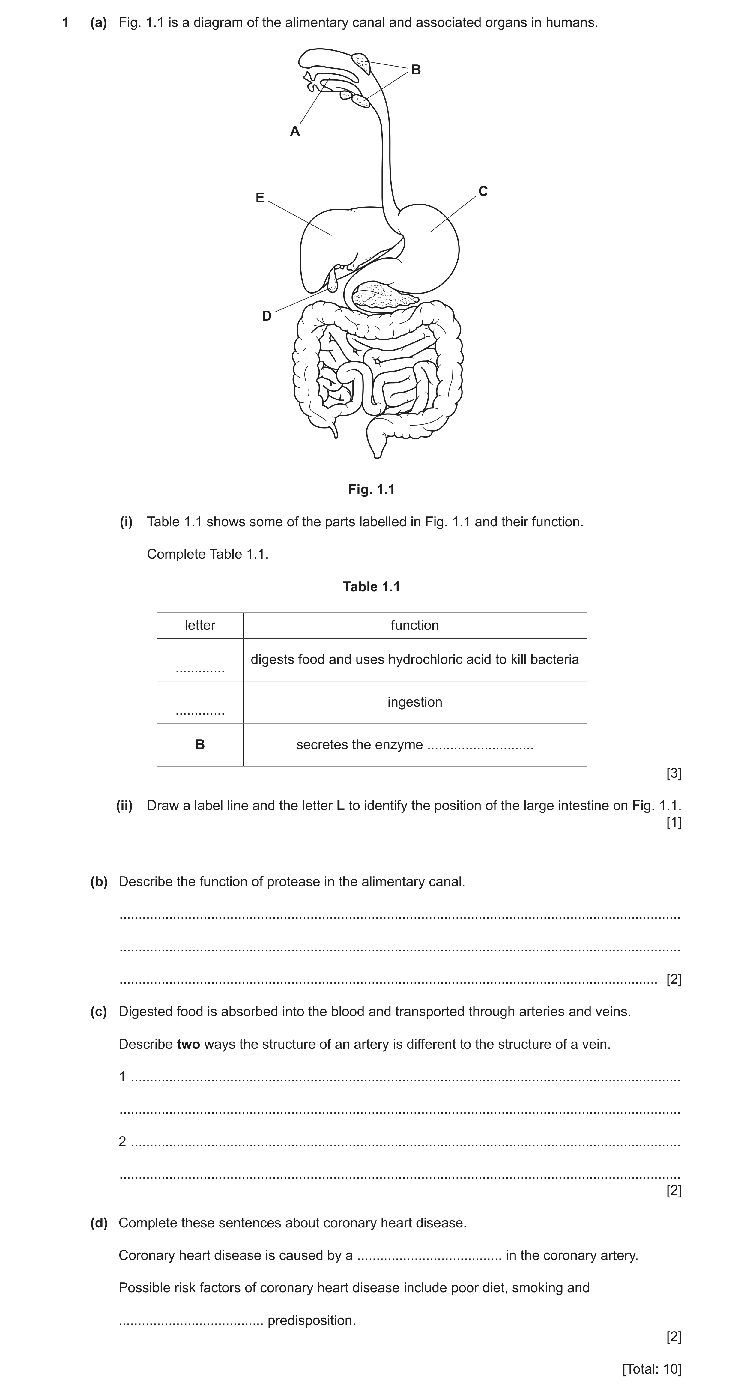
1 (a) Fig. 1.1 is a diagram of the alimentary canal and associated organs in humans. B A C E D Fig. 1.1 (i) Table 1.1 shows some of the parts labelled in Fig. 1.1 and their function. Complete Table 1.1. Table 1.1 letter function digests food and uses hydrochloric acid to kill bacteria … ingestion … B secretes the enzyme … [3] (ii) Draw a label line and the letter L to identify the position of the large intestine on Fig. 1.1. [1] (b) Describe the function of protease in the alimentary canal. … … … [2] (c) Digested food is absorbed into the blood and transported through arteries and veins. Describe two ways the structure of an artery is different to the structure of a vein. 1 … … 2 … … [2] (d) Complete these sentences about coronary heart disease. Coronary heart disease is caused by a … in the coronary artery. Possible risk factors of coronary heart disease include poor diet, smoking and … predisposition. [2] [Total: 10]
10 marks
Mark scheme: 1(a)(i) letter function C ; (digests protein and uses hydrochloric acid to kill bacteria) A ; (ingestion) (B) (secretes the enzyme) amylase ; 3 1(a)(ii) label line to large intestine labelled L ; 1 1(b) digestion of protein ; to form amino acids ; 2 1(c) any two from: arteries have a thicker (muscle) wall / ORA ; arteries have a smaller centre / ORA ; only veins have valves ; 2 1(d) blockage ; genetic ; 2
This question in 0653/43 May/June 2022
Q7 · The circulatory system of frogs and the circulatory system of humans 0653/41 May/June 2023
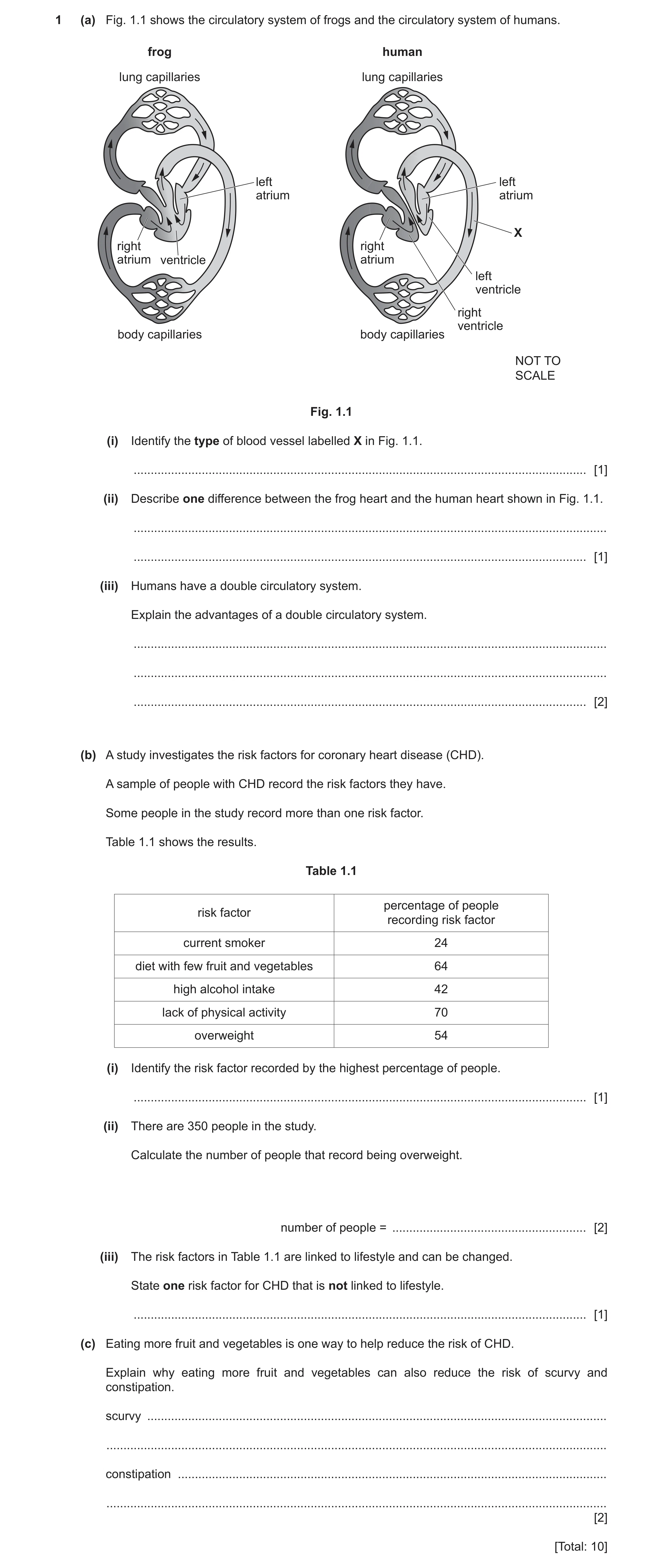
1 (a) Fig. 1.1 shows the circulatory system of frogs and the circulatory system of humans. frog human lung capillaries lung capillaries left left atrium atrium X right right atrium ventricle atrium left ventricle right ventricle body capillaries body capillaries NOT TO SCALE Fig. 1.1 (i) Identify the type of blood vessel labelled X in Fig. 1.1. … [1] (ii) Describe one difference between the frog heart and the human heart shown in Fig. 1.1. … … [1] (iii) Humans have a double circulatory system. Explain the advantages of a double circulatory system. … … … [2] (b) A study investigates the risk factors for coronary heart disease (CHD). A sample of people with CHD record the risk factors they have. Some people in the study record more than one risk factor. Table 1.1 shows the results. Table 1.1 percentage of people risk factor recording risk factor current smoker 24 diet with few fruit and vegetables 64 high alcohol intake 42 lack of physical activity 70 overweight 54 (i) Identify the risk factor recorded by the highest percentage of people. … [1] (ii) There are 350 people in the study. Calculate the number of people that record being overweight. number of people = … [2] (iii) The risk factors in Table 1.1 are linked to lifestyle and can be changed. State one risk factor for CHD that is not linked to lifestyle. … [1] (c) Eating more fruit and vegetables is one way to help reduce the risk of CHD. Explain why eating more fruit and vegetables can also reduce the risk of scurvy and constipation. scurvy … … constipation … … [2] [Total: 10]
10 marks
Mark scheme: 1(a)(i) artery ; 1 1(a)(ii) any one from: frog has only 3 chambers / frog has one ventricle ; humans have four chambers / humans have two ventricles ; no septum in frog / AW ; 1 1(a)(iii) oxygenated and deoxygenated blood kept separate ; high blood pressure around body / low blood pressure to lungs ; 2 1(b)(i) (lack of) physical activity ; 1 1(b)(ii) (54 350) ÷ 100 ; 189 ; 2 1(b)(iii) age / genetics / inheritance ; 1 1(c) scurvy – (fruit and vegetables) are a source of Vit C / Vit C prevents scurvy ; constipation – (vegetables) are a source of fibre / fibre needed for bulk ; 2
This question in 0653/41 May/June 2023
Q8 · Explain one way the structure of an artery is adapted to its function 0653/42 Oct/Nov 2023
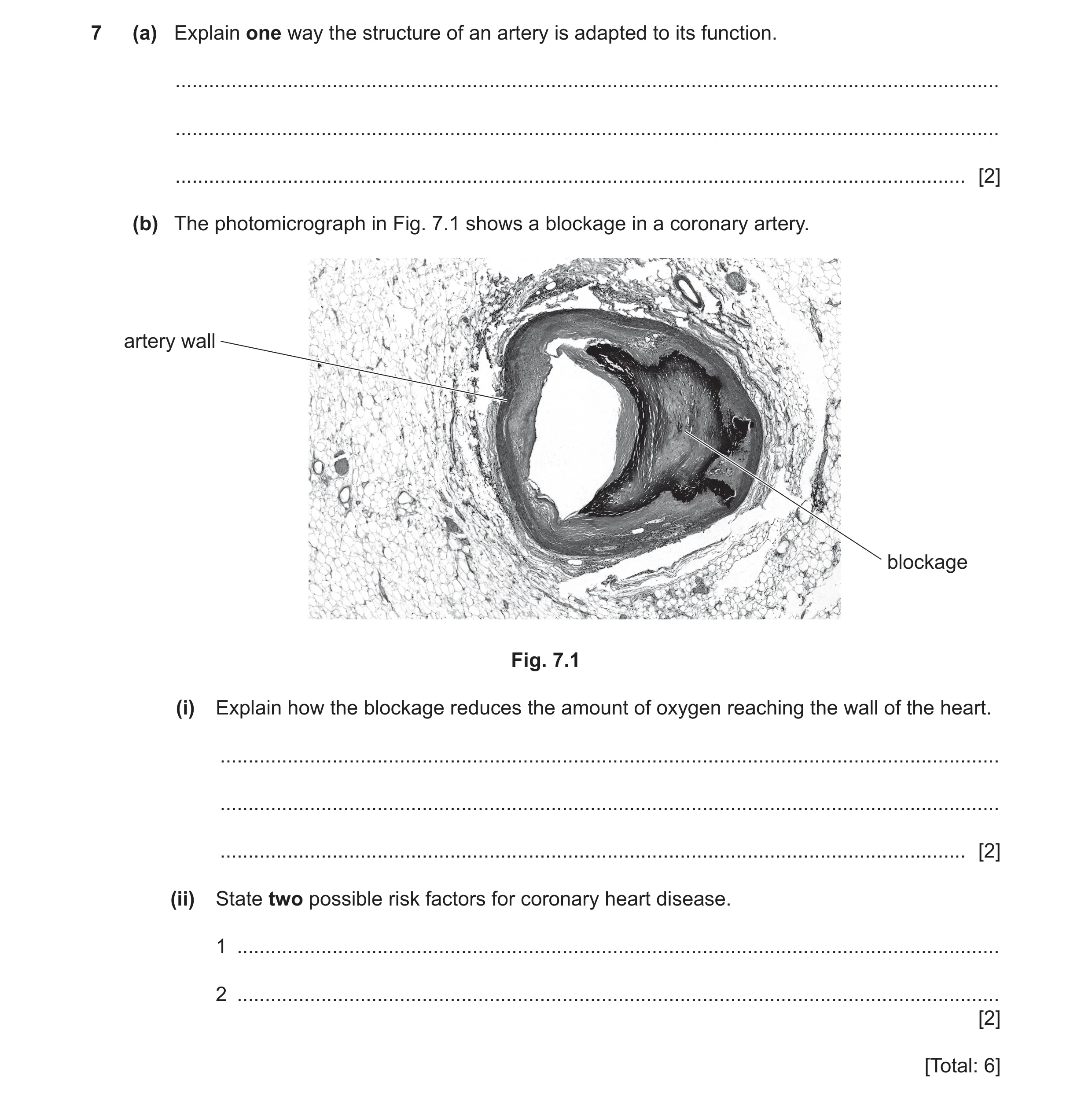
7 (a) Explain one way the structure of an artery is adapted to its function. … … … [2] (b) The photomicrograph in Fig. 7.1 shows a blockage in a coronary artery. artery wall blockage Fig. 7.1 (i) Explain how the blockage reduces the amount of oxygen reaching the wall of the heart. … … … [2] (ii) State two possible risk factors for coronary heart disease. 1 … 2 … [2] [Total: 6]
6 marks
Mark scheme: 7(a) thick (muscular) wall ; 2 to withstand pressure ; OR small lumen ; to maintain pressure ; OR elastic fibres ; to allow stretch and recoil ; 7(b)(i) less blood passes through ; 2 reference to transport of oxygen in blood ; 7(b)(ii) any two from: 2 smoking ; genetic predisposition ; age ; gender / sex ; diet ; stress ; AVP ;
This question in 0653/42 Oct/Nov 2023
Q9 · Drawings of three different blood vessels, W, X and Y 0653/42 Oct/Nov 2024
4 (a) Fig. 4.1 shows drawings of three different blood vessels, W, X and Y. W X Y Fig. 4.1 Each blood vessel in Fig. 4.1 has an adaptation for a particular function. Draw one straight line from each blood vessel to its adaptation and one straight line from each adaptation to its function. blood vessel adaptation function for diffusion W thick elastic walls of materials to withstand X permeable walls high blood pressure to reduce resistance Y large lumen to blood flow [3] (b) (i) Describe coronary heart disease. … … … [2] (ii) A scientist records the number of people that die from coronary heart disease in one country in one year. Table 4.1 shows the results. Table 4.1 population of country number of people that die from coronary heart disease 329 million 659 000 Calculate the percentage of the population that die from coronary heart disease. percentage of population = … % [2] (iii) Smoking is a risk factor for coronary heart disease. State two types of disease of the gas exchange system caused by smoking. 1 … 2 … [2] [Total: 9]
9 marks
Mark scheme: 4(a) 3 two lines correct ; four lines correct ; six lines correct ; 4(b)(i) idea of blockage of, a blood vessel / blood flow ; 2 coronary artery ; 4(b)(ii) 329 000 000 / 0.659 million / 0.002 / ratio of N:P 100 ; 2 0.200 (%) ; 4(b)(iii) any two from: 2 chronic obstructive pulmonary disease / COPD ; lung cancer ; AVP ;
This question in 0653/42 Oct/Nov 2024
Q10 · A photomicrograph of human blood 0653/43 May/June 2025
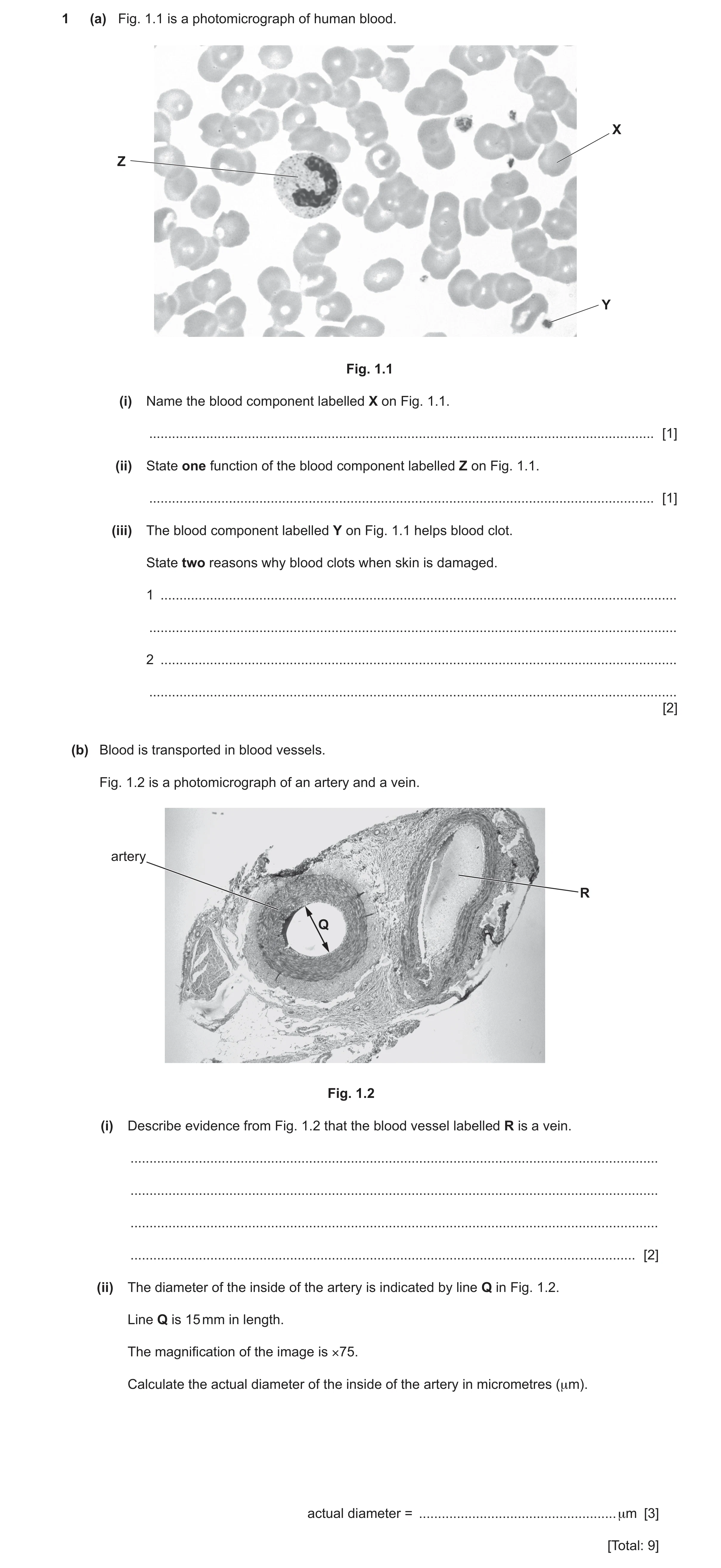
1 (a) Fig. 1.1 is a photomicrograph of human blood. X Z Y Fig. 1.1 (i) Name the blood component labelled X on Fig. 1.1. … [1] (ii) State one function of the blood component labelled Z on Fig. 1.1. … [1] (iii) The blood component labelled Y on Fig. 1.1 helps blood clot. State two reasons why blood clots when skin is damaged. 1 … … 2 … … [2] (b) Blood is transported in blood vessels. Fig. 1.2 is a photomicrograph of an artery and a vein. artery R Q Fig. 1.2 (i) Describe evidence from Fig. 1.2 that the blood vessel labelled R is a vein. … … … … [2] (ii) The diameter of the inside of the artery is indicated by line Q in Fig. 1.2. Line Q is 15 mm in length. The magnification of the image is ×75. Calculate the actual diameter of the inside of the artery in micrometres (μm). actual diameter = … μm [3] [Total: 9]
9 marks
Mark scheme: Question Answer Marks 1(a)(i) red blood cell ; 1 1(a)(ii) produce antibodies / phagocytosis ; 1 1(a)(iii) prevent blood loss ; 2 prevent entry of pathogens ; 1(b)(i) larger / wider, lumen (than artery) ; 2 thinner (muscular) wall (than artery) / less muscular wall ; 1(b)(ii) magnification = image size ÷ actual size in any form or 15 ÷ 75 or 0.2 ; 3 0.2 × 1000 ; 200 (µm) ;
This question in 0653/43 May/June 2025
Q11 · A diagram of a human heart 0653/41 Oct/Nov 2025
3 (a) Fig. 3.1 is a diagram of a human heart. Fig. 3.1 (i) Draw a label line and the letter C to identify a coronary artery in Fig. 3.1. [1] (ii) Describe what happens to the coronary artery when a person has coronary heart disease. … … [1] (iii) Diet is one risk factor for coronary heart disease. State two other risk factors for coronary heart disease. 1 … 2 … [2] (b) Arteries transport blood away from the heart. Explain how the structure of an artery is adapted to its function. … … … … … … [3] [Total: 7]
7 marks
Mark scheme: 3(a)(i) either coronary artery labelled ; 1 3(a)(ii) blocked / AW ; 1 3(a)(iii) any two from: 2 lack of exercise / AW ; stress ; smoking ; genetic predisposition ; age ; sex ; high blood pressure ; AVP;; 3(b) Any two from 3 (wall) is muscular (wall) is elastic; narrow lumen ; and then to withstand high (blood) pressure / maintain high (blood) pressure ;
This question in 0653/41 Oct/Nov 2025
 12 / 16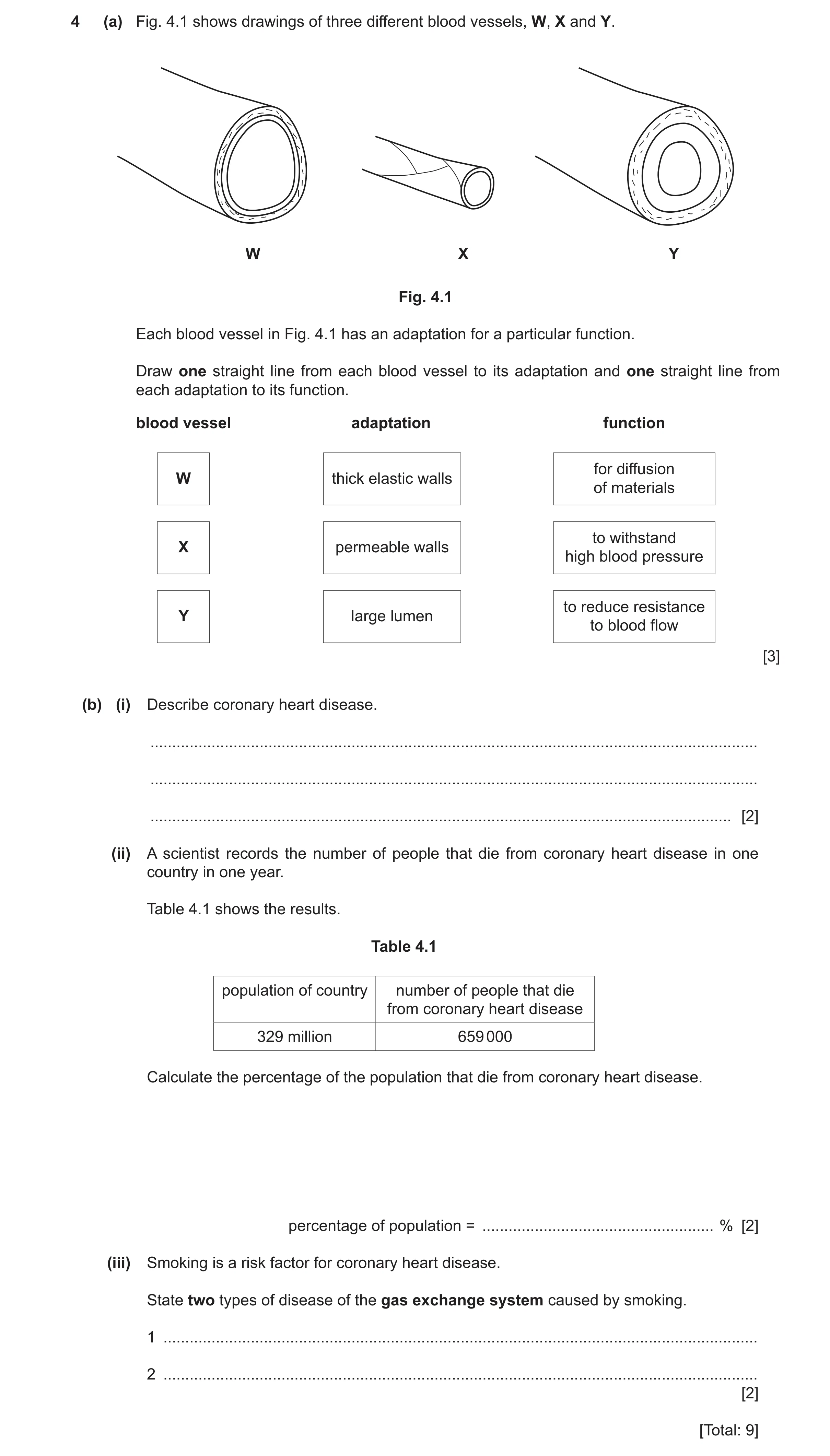
![Question 11: (a) Fig. 3.1 is a diagram of a human heart. Fig. 3.1 (i) Draw a label line and the letter C to identify a coronary artery in Fig. 3.1. [1] …](https://img.pastlit.com/crops/6fc12538-e60d-48ee-a0ed-7796e6999497/q3.webp) 16 / 16
16 / 16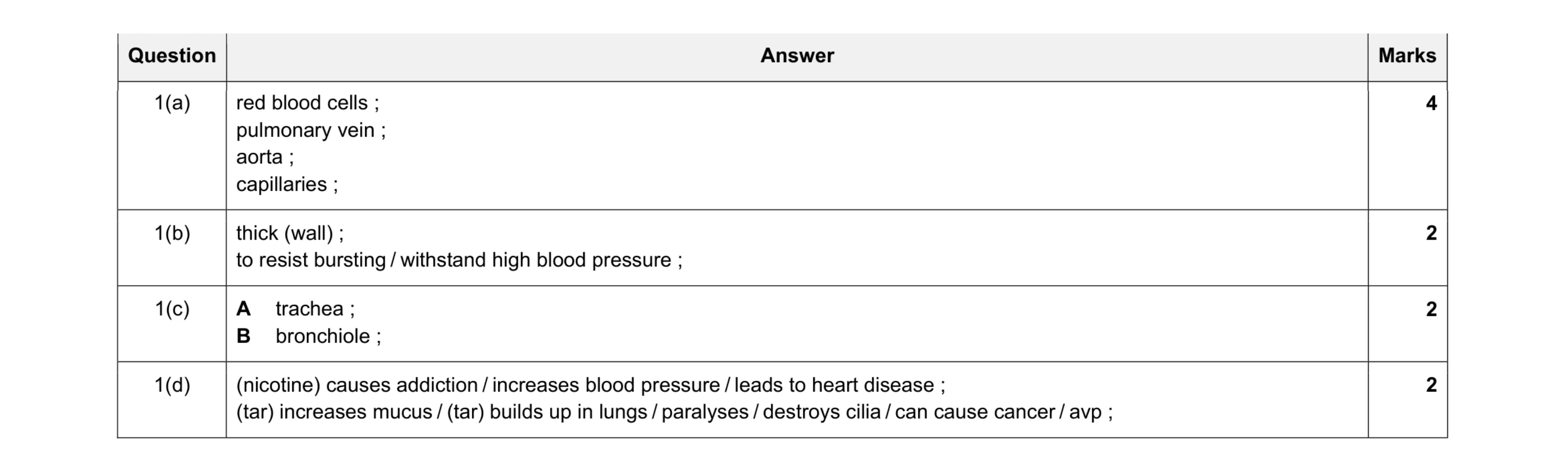 10
10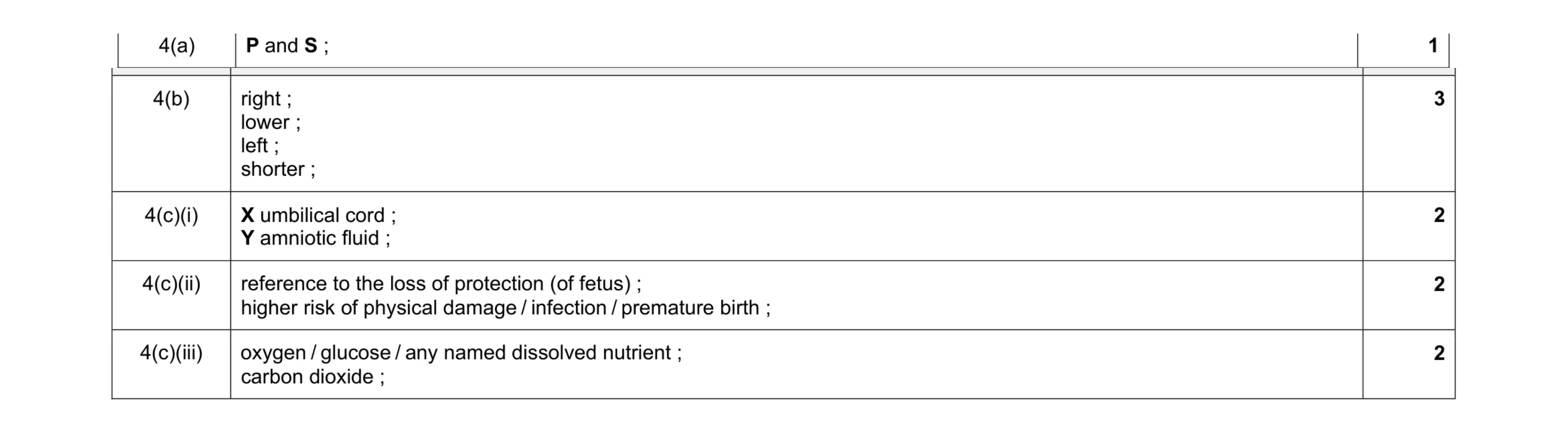 10
10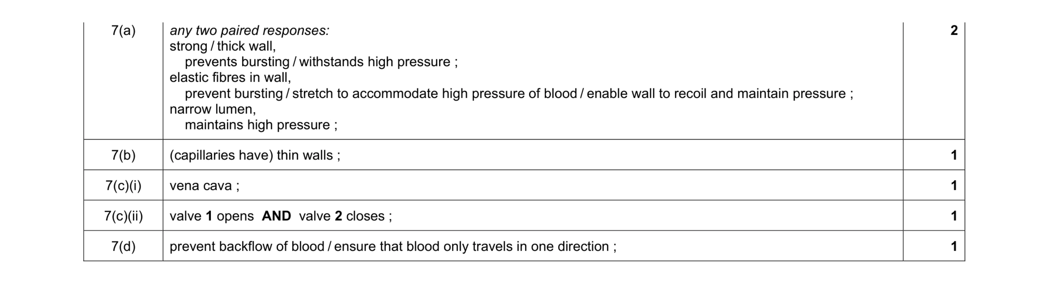 6
6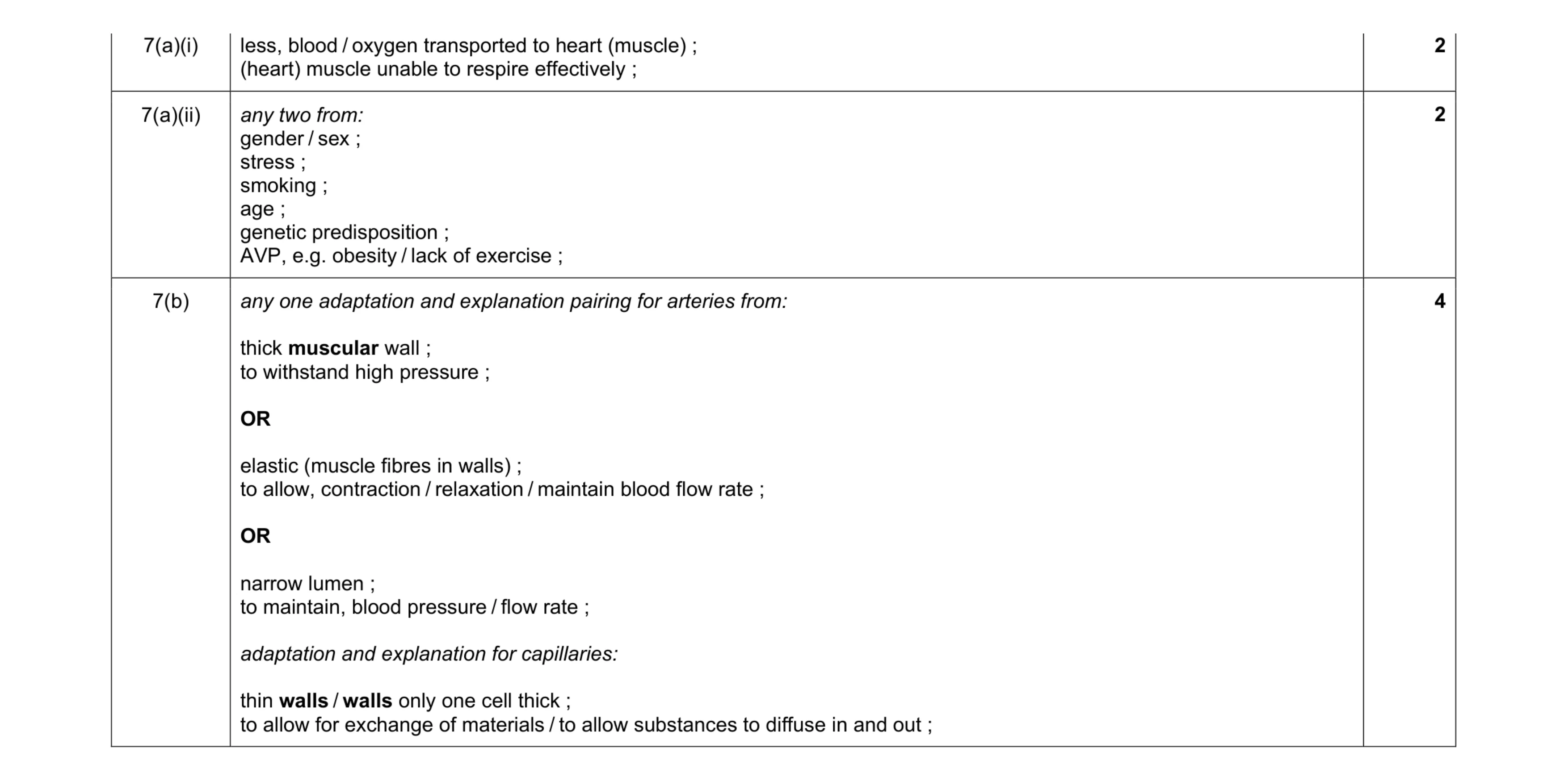 8
8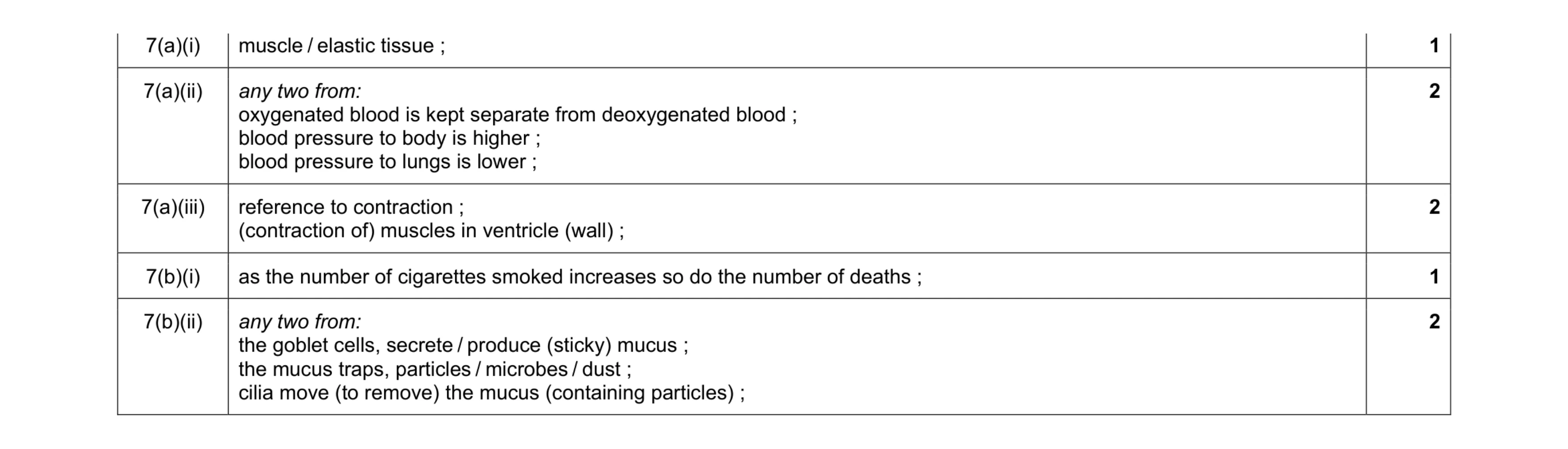 8
8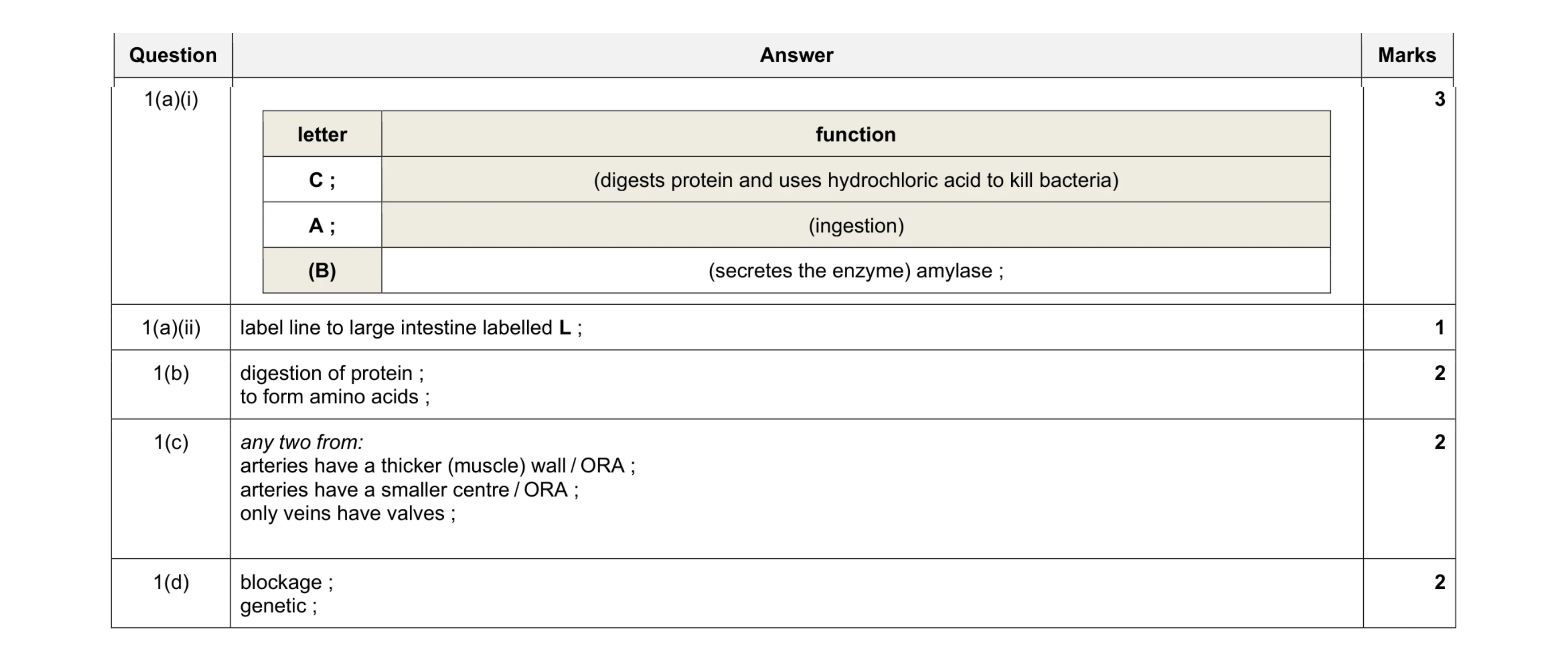 10
10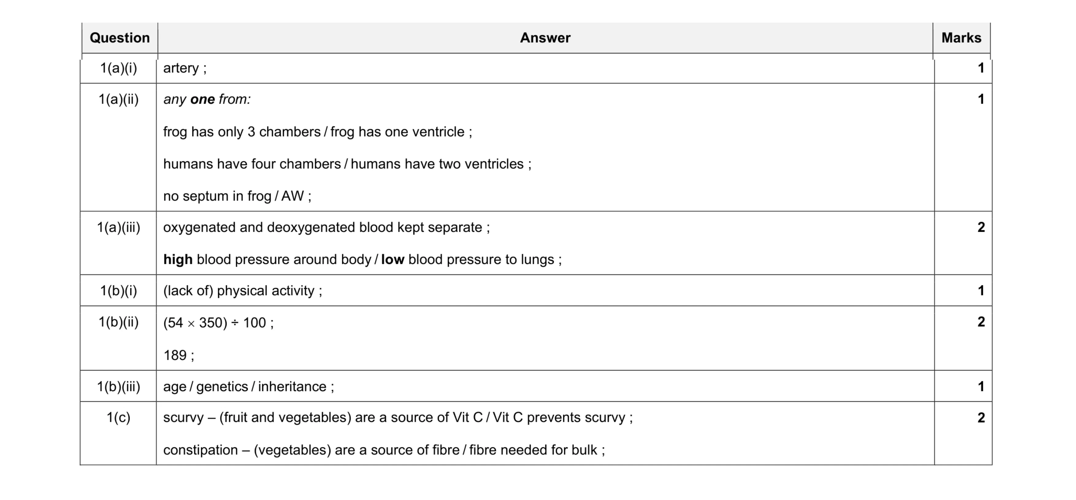 10
10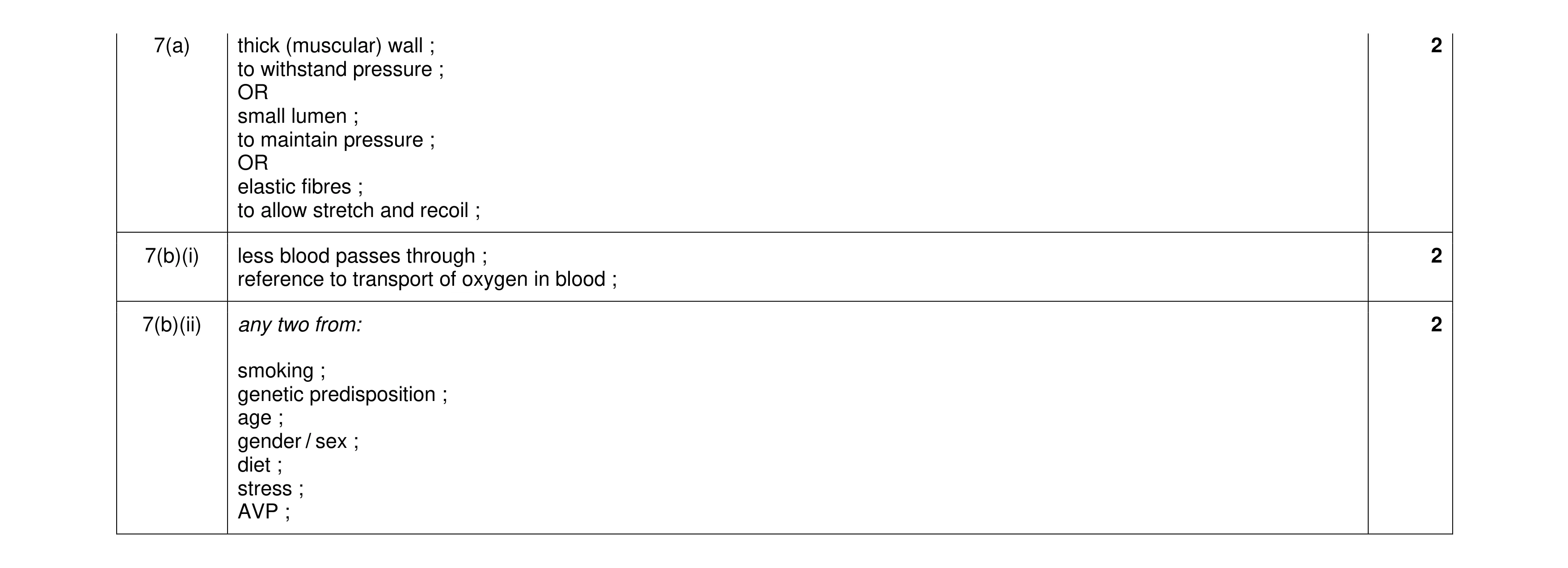 6
6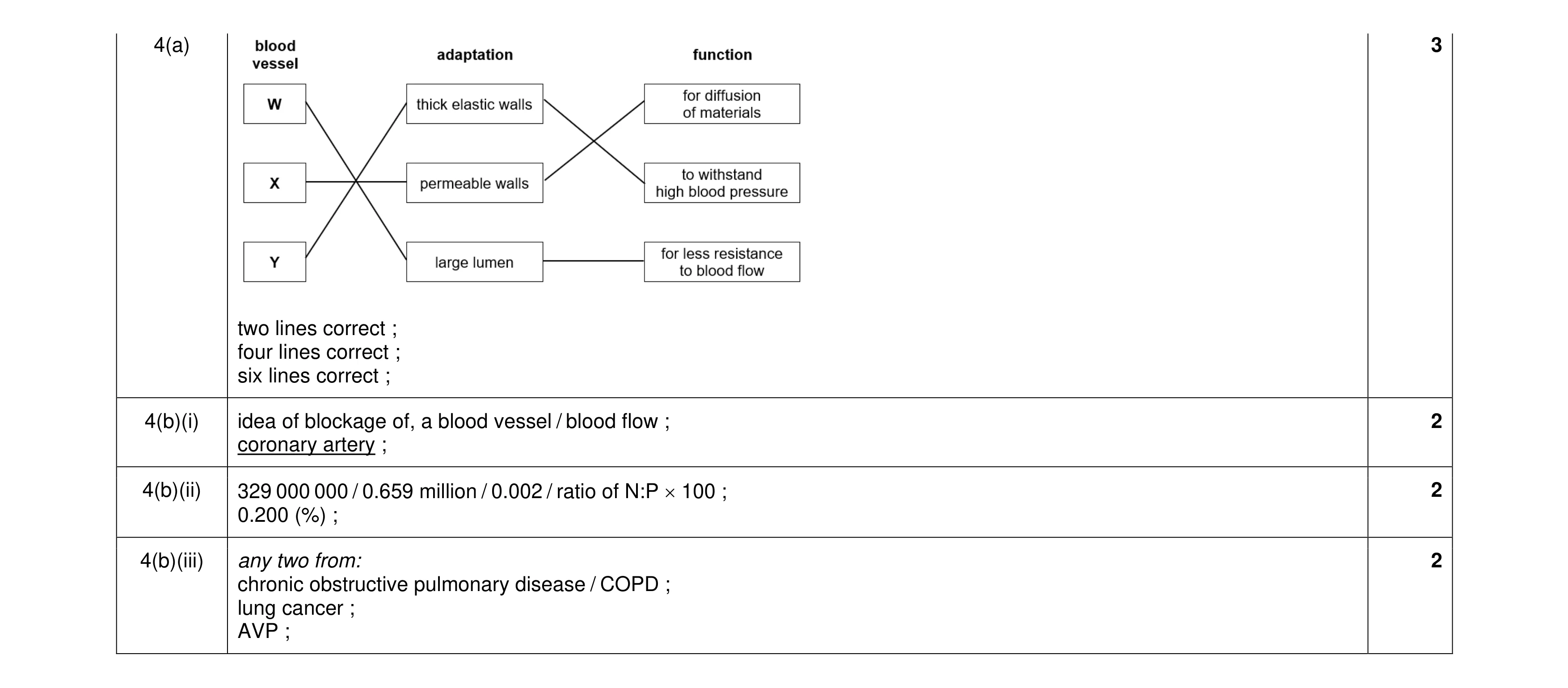 9
9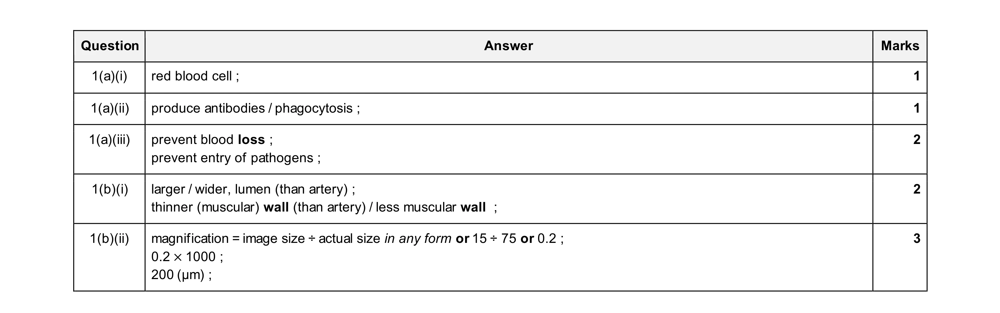 9
9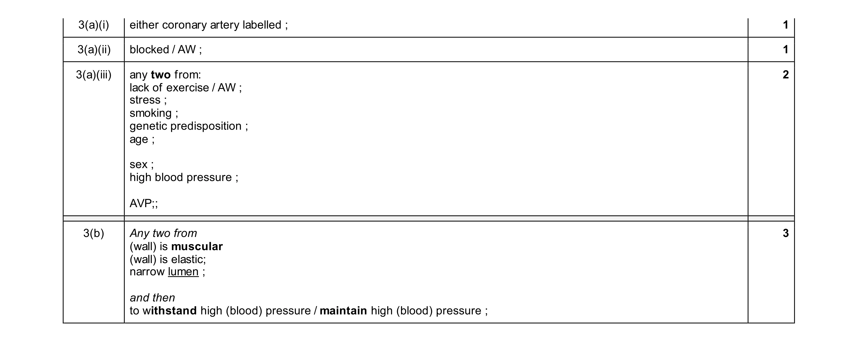 7
7