Questions as text
Q1 · The circulatory system transports blood to organs in the body 0654/31 Oct/Nov 2017
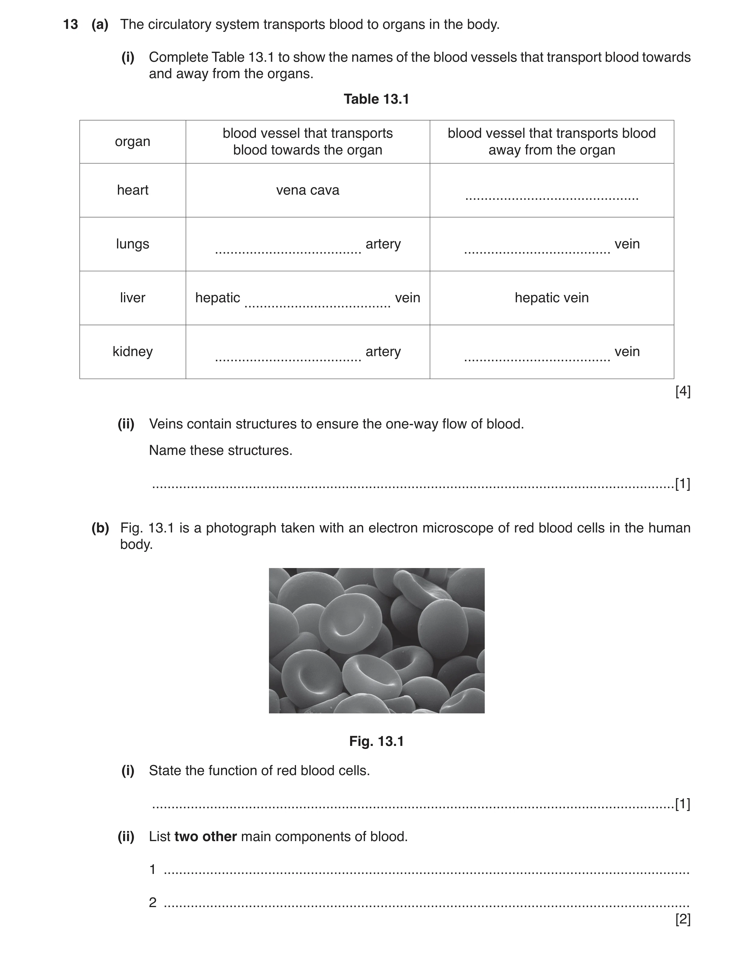
13 (a) The circulatory system transports blood to organs in the body. (i) Complete Table 13.1 to show the names of the blood vessels that transport blood towards and away from the organs. Table 13.1 blood vessel that transports blood vessel that transports blood organ blood towards the organ away from the organ heart vena cava … lungs artery vein … … liver hepatic vein hepatic vein … kidney artery vein … … [4] (ii) Veins contain structures to ensure the one-way flow of blood. Name these structures. … [1] (b) Fig. 13.1 is a photograph taken with an electron microscope of red blood cells in the human body. Fig. 13.1 (i) State the function of red blood cells. … [1] (ii) List two other main components of blood. 1 … 2 … [2]
8 marks
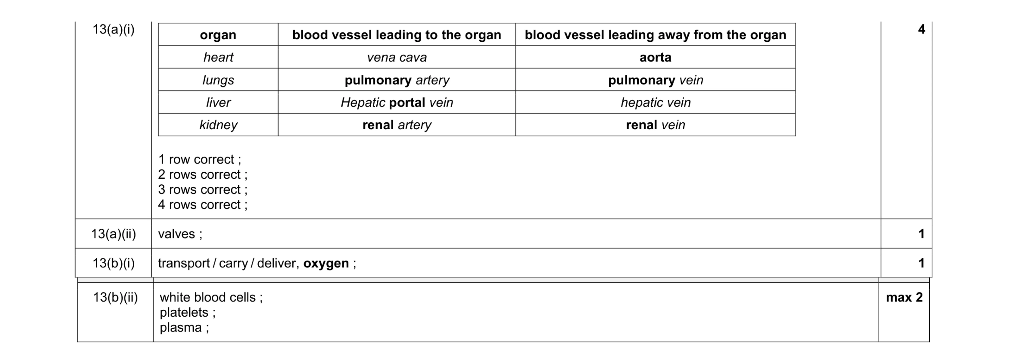
Mark scheme: 13(a)(i) organ blood vessel leading to the organ blood vessel leading away from the organ heart vena cava aorta lungs pulmonary artery pulmonary vein liver Hepatic portal vein hepatic vein kidney renal artery renal vein 1 row correct ; 2 rows correct ; 3 rows correct ; 4 rows correct ; 4 13(a)(ii) valves ; 1 13(b)(i) transport / carry / deliver, oxygen ; 1 Question Answer Marks 13(b)(ii) white blood cells ; platelets ; plasma ; max 2
This question in 0654/31 Oct/Nov 2017
Q2 · A cross-section through an artery and a vein 0654/33 Oct/Nov 2018
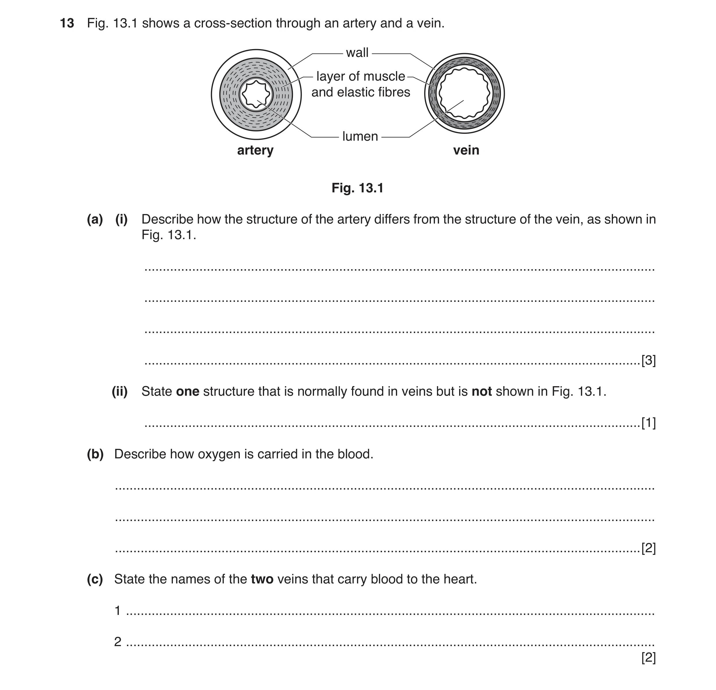
13 Fig. 13.1 shows a cross-section through an artery and a vein. wall layer of muscle and elastic fibres lumen artery vein Fig. 13.1 (a) (i) Describe how the structure of the artery differs from the structure of the vein, as shown in Fig. 13.1. … … … … [3] (ii) State one structure that is normally found in veins but is not shown in Fig. 13.1. … [1] (b) Describe how oxygen is carried in the blood. … … … [2] (c) State the names of the two veins that carry blood to the heart. 1 … 2 … [2]
8 marks
Mark scheme: 13(a)(i) thicker wall ; thicker muscle / elastic layer ; narrower lumen ; 3 13(a)(ii) valves ; 1 13(b) combined / AW, to haemoglobin ; in red blood cells ; 2 13(c) vena cava ; pulmonary vein ; 2
This question in 0654/33 Oct/Nov 2018
Q3 · A diagram of a cross-section through a vein 0654/31 Oct/Nov 2019
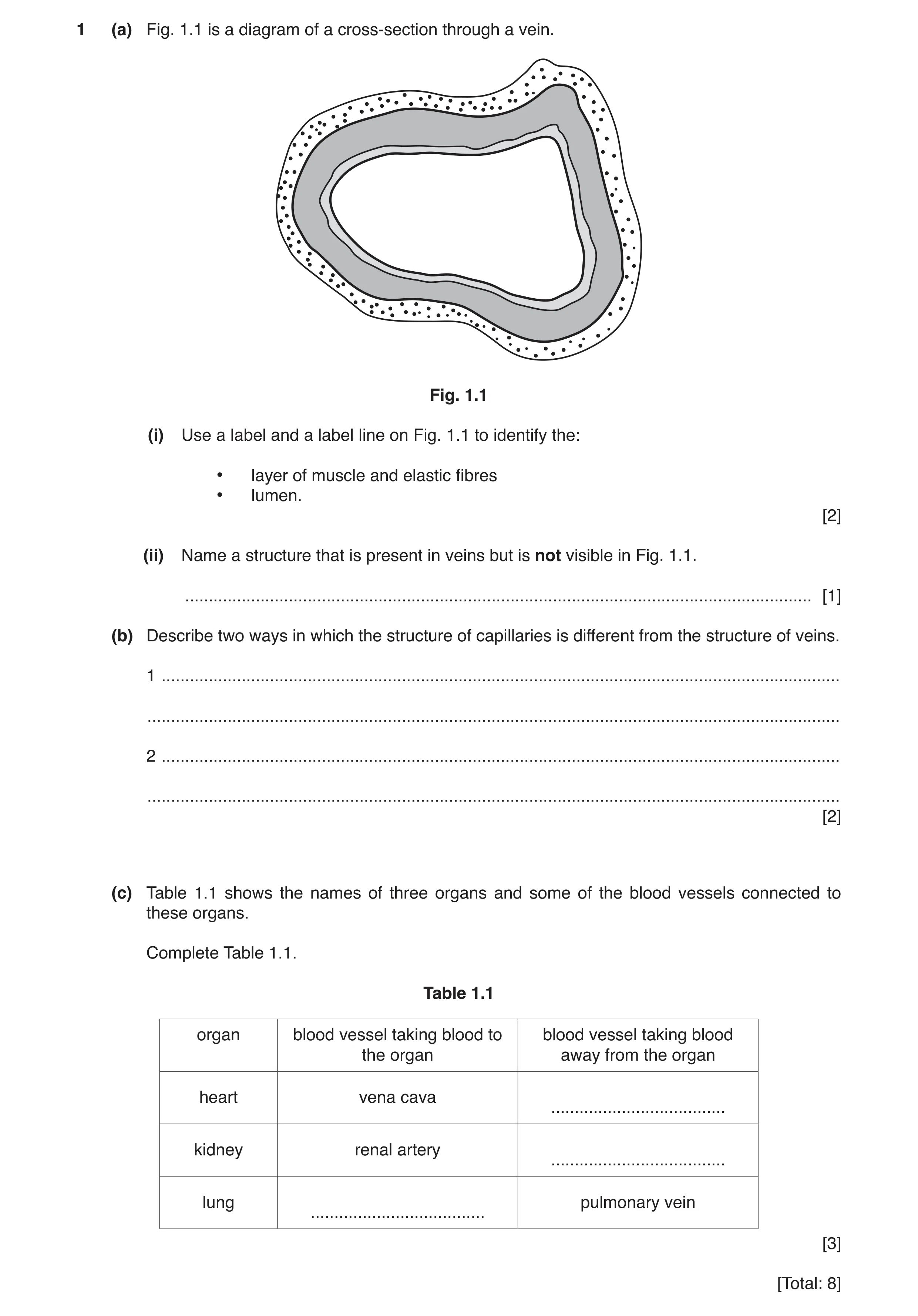
1 (a) Fig. 1.1 is a diagram of a cross-section through a vein. Fig. 1.1 (i) Use a label and a label line on Fig. 1.1 to identify the: • layer of muscle and elastic fibres • lumen. [2] (ii) Name a structure that is present in veins but is not visible in Fig. 1.1. … [1] (b) Describe two ways in which the structure of capillaries is different from the structure of veins. 1 … … 2 … … [2] (c) Table 1.1 shows the names of three organs and some of the blood vessels connected to these organs. Complete Table 1.1. Table 1.1 organ blood vessel taking blood to blood vessel taking blood the organ away from the organ heart vena cava … kidney renal artery … lung pulmonary vein … [3] [Total: 8]
8 marks
Mark scheme: 1(a)(i) layer of muscle and elastic tissue labelled ; lumen labelled ; 2 1(a)(ii) valve ; 1 1(b) narrower (lumen) ; thin wall / wall only one cell thick ; does not contain muscular tissue / valves ; max 2 2 1(c) organ blood vessel bringing blood to the organ blood vessel taking blood away from the organ heart vena cava aorta / pulmonary artery ; kidney renal artery renal vein ; lungs pulmonary artery ; pulmonary vein 3
This question in 0654/31 Oct/Nov 2019
Q4 · Simplified cross-sections of three different types of blood vessel 0654/32 May/June 2020
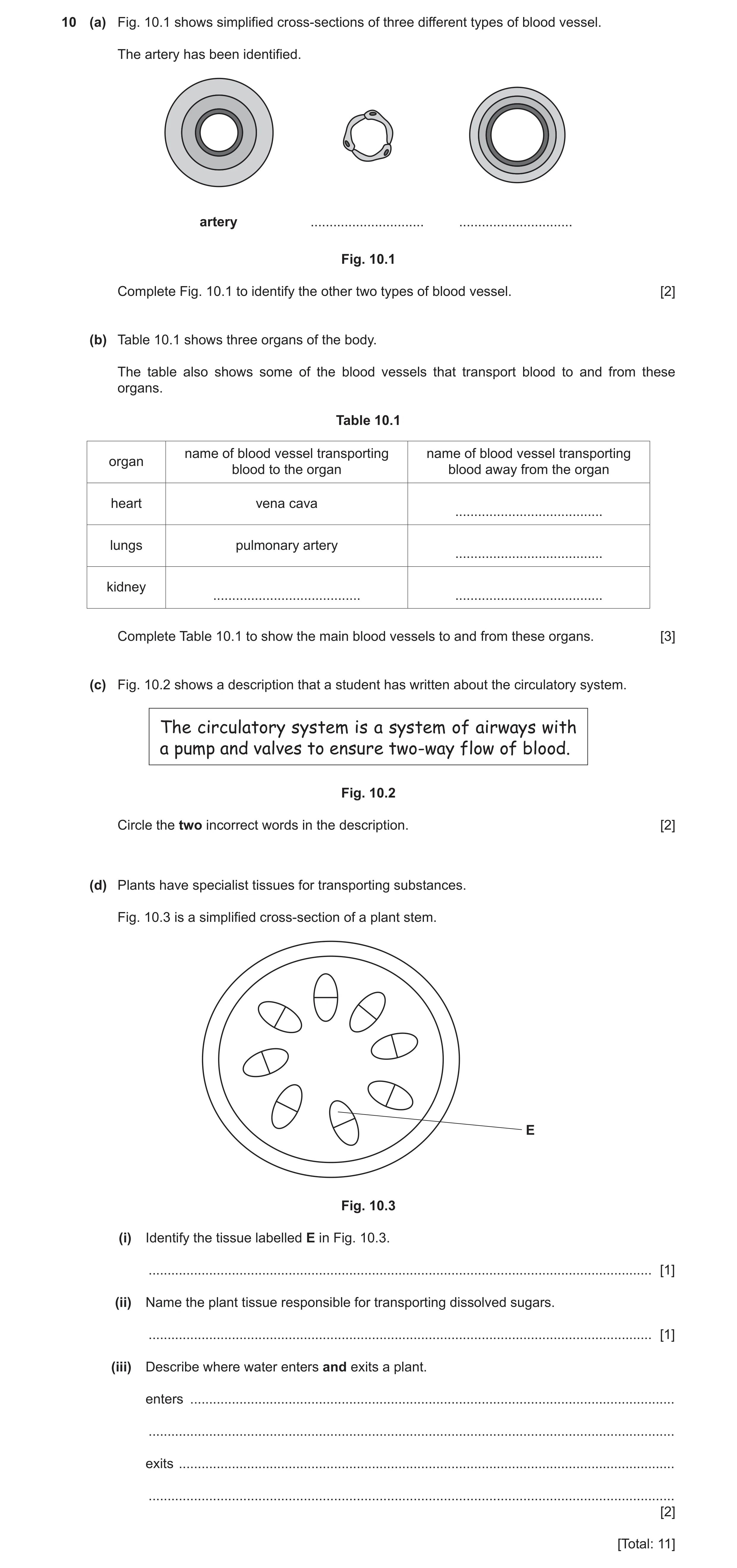
10 (a) Fig. 10.1 shows simplified cross-sections of three different types of blood vessel. The artery has been identified. artery … … Fig. 10.1 Complete Fig. 10.1 to identify the other two types of blood vessel. [2] (b) Table 10.1 shows three organs of the body. The table also shows some of the blood vessels that transport blood to and from these organs. Table 10.1 name of blood vessel transporting name of blood vessel transporting organ blood to the organ blood away from the organ heart vena cava … lungs pulmonary artery … kidney … … Complete Table 10.1 to show the main blood vessels to and from these organs. [3] (c) Fig. 10.2 shows a description that a student has written about the circulatory system. The circulatory system is a system of airways with a pump and valves to ensure two-way flow of blood. Fig. 10.2 Circle the two incorrect words in the description. [2] (d) Plants have specialist tissues for transporting substances. Fig. 10.3 is a simplified cross-section of a plant stem. E Fig. 10.3 (i) Identify the tissue labelled E in Fig. 10.3. … [1] (ii) Name the plant tissue responsible for transporting dissolved sugars. … [1] (iii) Describe where water enters and exits a plant. enters … … exits … … [2] [Total: 11]
11 marks
Mark scheme: 10(a) capillaries ; veins ; 2 10(b) heart vena cava aorta / pulmonary artery lungs pulmonary artery pulmonary vein kidney renal artery renal vein ;;; each correct row for 1 mark 3 10(c) airways circled ; two / two-way circled ; 2 10(d)(i) xylem ; 1 10(d)(ii) phloem ; 1 10(d)(iii) enter root hair (cell) ; leave through stomata / leaf ; 2
This question in 0654/32 May/June 2020
Q5 · Simplified cross-sections of three different types of blood vessel 0654/33 May/June 2020
10 (a) Fig. 10.1 shows simplified cross-sections of three different types of blood vessel. The artery has been identified. artery … … Fig. 10.1 Complete Fig. 10.1 to identify the other two types of blood vessel. [2] (b) Table 10.1 shows three organs of the body. The table also shows some of the blood vessels that transport blood to and from these organs. Table 10.1 name of blood vessel transporting name of blood vessel transporting organ blood to the organ blood away from the organ heart vena cava … lungs pulmonary artery … kidney … … Complete Table 10.1 to show the main blood vessels to and from these organs. [3] (c) Fig. 10.2 shows a description that a student has written about the circulatory system. The circulatory system is a system of airways with a pump and valves to ensure two-way flow of blood. Fig. 10.2 Circle the two incorrect words in the description. [2] (d) Plants have specialist tissues for transporting substances. Fig. 10.3 is a simplified cross-section of a plant stem. E Fig. 10.3 (i) Identify the tissue labelled E in Fig. 10.3. … [1] (ii) Name the plant tissue responsible for transporting dissolved sugars. … [1] (iii) Describe where water enters and exits a plant. enters … … exits … … [2] [Total: 11]
11 marks
Mark scheme: 10(a) capillaries ; veins ; 2 10(b) heart vena cava aorta / pulmonary artery lungs pulmonary artery pulmonary vein kidney renal artery renal vein ;;; each correct row for 1 mark 3 10(c) airways circled ; two / two-way circled ; 2 10(d)(i) xylem ; 1 10(d)(ii) phloem ; 1 10(d)(iii) enter root hair (cell) ; leave through stomata / leaf ; 2
This question in 0654/33 May/June 2020
Q6 · A scientist investigates the recovery time of an athlete and a non‑athlete after exercise 0654/31 Oct/Nov 2020
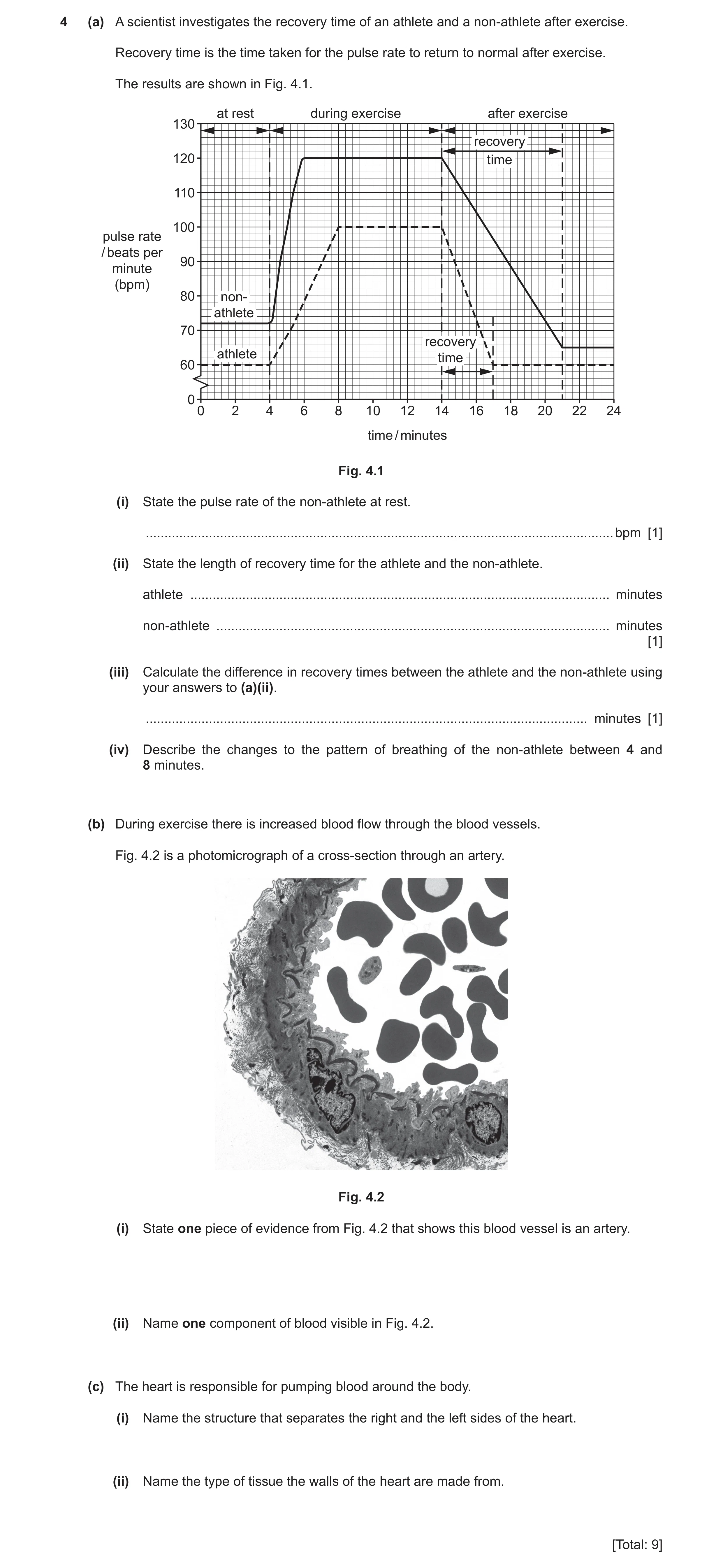
4 (a) A scientist investigates the recovery time of an athlete and a non‑athlete after exercise. Recovery time is the time taken for the pulse rate to return to normal after exercise. The results are shown in Fig. 4.1. at rest during exercise after exercise 130 recovery 120 time 110 100 pulse rate / beats per 90 minute (bpm) 80 non- athlete 70 recovery athlete time 60 0 0 2 4 6 8 10 12 14 16 18 20 22 24 time / minutes Fig. 4.1 (i) State the pulse rate of the non‑athlete at rest. ������������������������������������������������������������������������������������������������������������������������������ bpm [1] (ii) State the length of recovery time for the athlete and the non‑athlete. athlete ����������������������������������������������������������������������������������������������������������������� minutes non‑athlete ���������������������������������������������������������������������������������������������������������� minutes [1] (iii) Calculate the difference in recovery times between the athlete and the non‑athlete using your answers to (a)(ii). ����������������������������������������������������������������������������������������������������������������������� minutes [1] (iv) Describe the changes to the pattern of breathing of the non‑athlete between 4 and 8 minutes. (b) During exercise there is increased blood flow through the blood vessels. Fig. 4.2 is a photomicrograph of a cross‑section through an artery. Fig. 4.2 (i) State one piece of evidence from Fig. 4.2 that shows this blood vessel is an artery. (ii) Name one component of blood visible in Fig. 4.2. (c) The heart is responsible for pumping blood around the body. (i) Name the structure that separates the right and the left sides of the heart. (ii) Name the type of tissue the walls of the heart are made from. [Total: 9]
9 marks
Mark scheme: 4(a)(i) 72 (bpm) 1 4(a)(ii) athlete 3 (minutes) and non-athlete 7 (minutes) ; 1 4(a)(iii) 7 – 3 = 4 (minutes) ; 1 4(a)(iv) breathing rate would increase ; breathing depth would increase ; 2 4(b)(i) thick wall ; 1 4(b)(ii) red blood cell ; 1 4(c)(i) septum ; 1 4(c)(ii) muscular (tissue) ; 1
This question in 0654/31 Oct/Nov 2020
Q7 · A scientist investigates the recovery time of an athlete and a non‑athlete after exercise 0654/33 Oct/Nov 2020
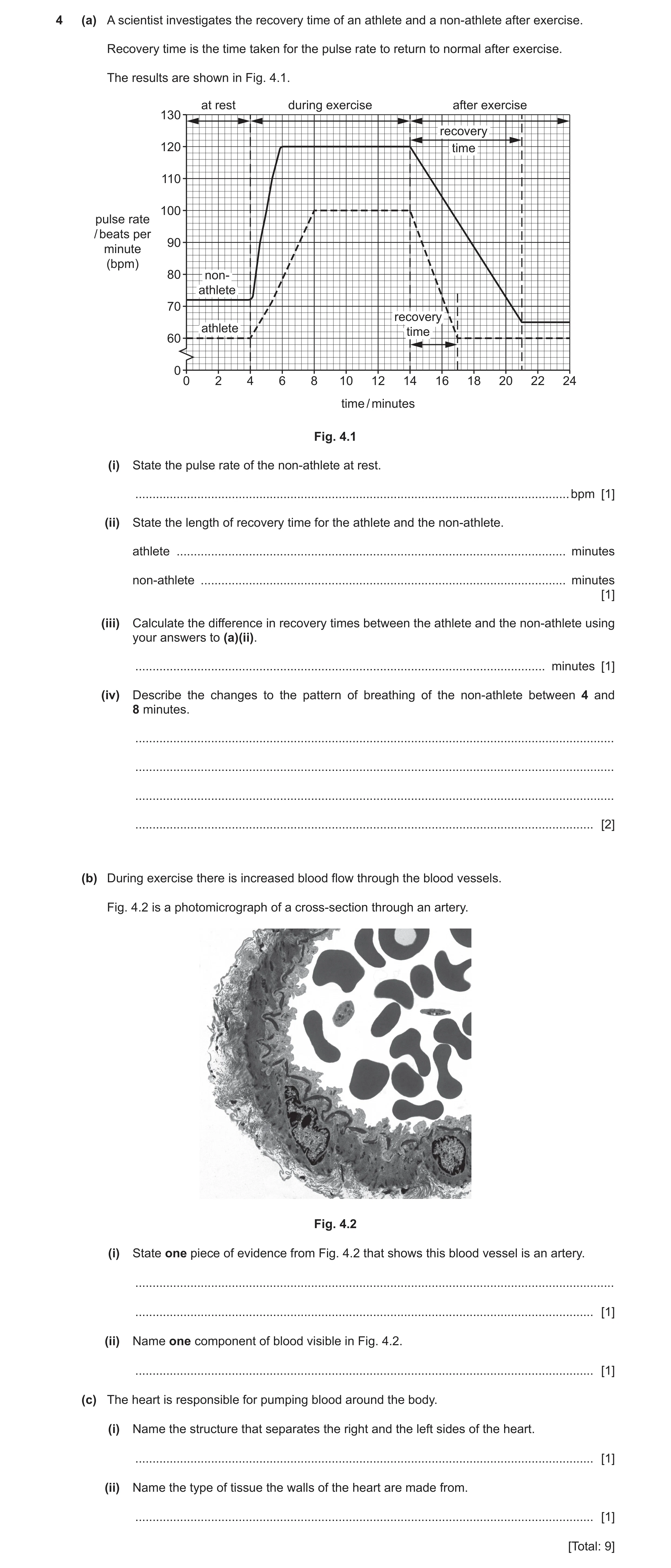
4 (a) A scientist investigates the recovery time of an athlete and a non‑athlete after exercise. Recovery time is the time taken for the pulse rate to return to normal after exercise. The results are shown in Fig. 4.1. at rest during exercise after exercise 130 recovery 120 time 110 100 pulse rate / beats per 90 minute (bpm) 80 non- athlete 70 recovery athlete time 60 0 0 2 4 6 8 10 12 14 16 18 20 22 24 time / minutes Fig. 4.1 (i) State the pulse rate of the non‑athlete at rest. … bpm [1] (ii) State the length of recovery time for the athlete and the non‑athlete. athlete … minutes non‑athlete … minutes [1] (iii) Calculate the difference in recovery times between the athlete and the non‑athlete using your answers to (a)(ii). … minutes [1] (iv) Describe the changes to the pattern of breathing of the non‑athlete between 4 and 8 minutes. … … … … [2] (b) During exercise there is increased blood flow through the blood vessels. Fig. 4.2 is a photomicrograph of a cross‑section through an artery. Fig. 4.2 (i) State one piece of evidence from Fig. 4.2 that shows this blood vessel is an artery. … … [1] (ii) Name one component of blood visible in Fig. 4.2. … [1] (c) The heart is responsible for pumping blood around the body. (i) Name the structure that separates the right and the left sides of the heart. … [1] (ii) Name the type of tissue the walls of the heart are made from. … [1] [Total: 9]
9 marks
Mark scheme: 4(a)(i) 72 (bpm) 1 4(a)(ii) athlete 3 (minutes) and non-athlete 7 (minutes) ; 1 4(a)(iii) 7 – 3 = 4 (minutes) ; 1 4(a)(iv) breathing rate would increase ; breathing depth would increase ; 2 4(b)(i) thick wall ; 1 4(b)(ii) red blood cell ; 1 4(c)(i) septum ; 1 4(c)(ii) muscular (tissue) ; 1
This question in 0654/33 Oct/Nov 2020
Q8 · A student measures their pulse rate at rest, during and after exercise 0654/33 Oct/Nov 2021
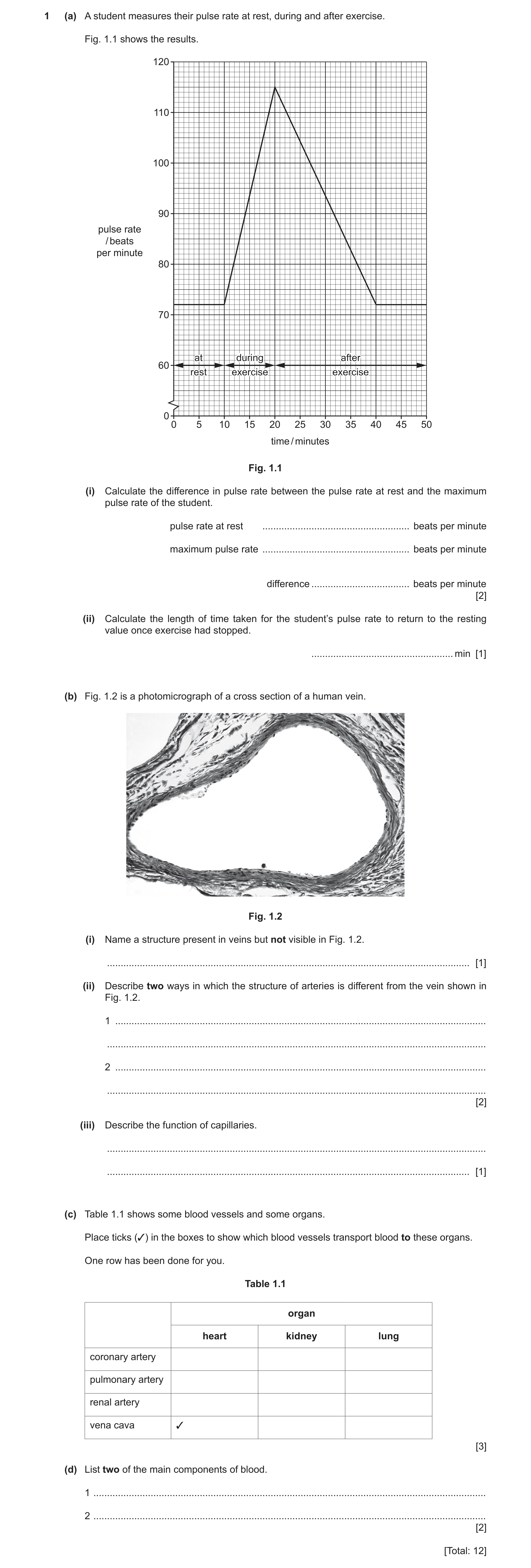
1 (a) A student measures their pulse rate at rest, during and after exercise. Fig. 1.1 shows the results. 120 110 100 90 pulse rate / beats per minute 80 70 atat duringduring afterafter 60 restrest exerciseexercise exerciseexercise 0 0 5 10 15 20 25 30 35 40 45 50 time / minutes Fig. 1.1 (i) Calculate the difference in pulse rate between the pulse rate at rest and the maximum pulse rate of the student. pulse rate at rest … beats per minute maximum pulse rate … beats per minute difference … beats per minute [2] (ii) Calculate the length of time taken for the student’s pulse rate to return to the resting value once exercise had stopped. … min [1] (b) Fig. 1.2 is a photomicrograph of a cross section of a human vein. Fig. 1.2 (i) Name a structure present in veins but not visible in Fig. 1.2. … [1] (ii) Describe two ways in which the structure of arteries is different from the vein shown in Fig. 1.2. 1 … … 2 … … [2] (iii) Describe the function of capillaries. … … [1] (c) Table 1.1 shows some blood vessels and some organs. Place ticks (3) in the boxes to show which blood vessels transport blood to these organs. One row has been done for you. Table 1.1 organ heart kidney lung coronary artery pulmonary artery renal artery vena cava 3 [3] (d) List two of the main components of blood. 1 … 2 … [2] [Total: 12]
12 marks
Mark scheme: 1(a)(i) pulse rate at rest = 72 (bpm) and maximum pulse rate = 115 (bpm) ; (115 – 72) = 43 (bpm) ; 2 1(a)(ii) 20 (mins) ; 1 1(b)(i) valves ; 1 1(b)(ii) any two from: thicker wall ; muscle / elastic fibres ; no valves ; narrower lumen ; 2 1(b)(iii) transfer of (named) substances (to / from, tissues / cells) ; 1 1(c) heart kidney lung coronary artery pulmonary artery renal artery vena cava ;;; 3 1(d) any two from: red blood cells ; white blood cells ; plasma ; platelets ; 2
This question in 0654/33 Oct/Nov 2021
Q9 · A diagram of the gas exchange system in humans 0654/33 May/June 2022
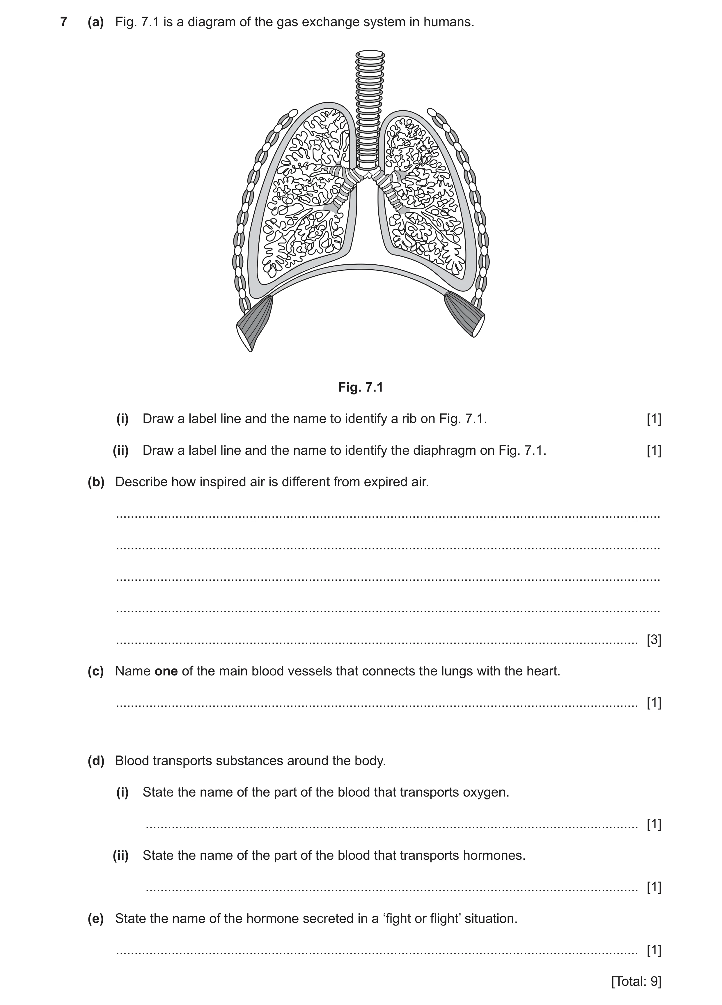
7 (a) Fig. 7.1 is a diagram of the gas exchange system in humans. Fig. 7.1 (i) Draw a label line and the name to identify a rib on Fig. 7.1. [1] (ii) Draw a label line and the name to identify the diaphragm on Fig. 7.1. [1] (b) Describe how inspired air is different from expired air. … … … … … [3] (c) Name one of the main blood vessels that connects the lungs with the heart. … [1] (d) Blood transports substances around the body. (i) State the name of the part of the blood that transports oxygen. … [1] (ii) State the name of the part of the blood that transports hormones. … [1] (e) State the name of the hormone secreted in a ‘fight or flight’ situation. … [1] [Total: 9]
9 marks
Mark scheme: 7(a)(i) rib labelled correctly ; 1 7(a)(ii) diaphragm labelled correctly ; 1 7(b) contains more oxygen ; contains less water vapour ; contains less carbon dioxide ; AVP ; Max 3 marks 3 7(c) pulmonary vein / pulmonary artery ; 1 7(d)(i) red blood cell ; 1 Question Answer Marks 7(d)(ii) plasma ; 1 7(e) adrenaline ; 1
This question in 0654/33 May/June 2022
Q10 · A photomicrograph of a cross-section of a human vein 0654/32 Oct/Nov 2022
4 (a) Fig. 4.1 is a photomicrograph of a cross-section of a human vein. lumen (internal space) wall Fig. 4.1 (i) Describe two ways the appearance of an artery is different from the vein shown in Fig. 4.1. 1 … … 2 … … [2] (ii) State the name of a structure present in veins that is not visible in Fig. 4.1. … [1] (b) State the name of two veins that transport blood to the heart. 1 … 2 … [2] (c) Describe the function of capillaries. … … [1] (d) Table 4.1 shows the functions of the main components of blood. Complete Table 4.1. Table 4.1 name of component of blood function antibody production and phagocytosis blood clotting transport of blood cells and ions transport of oxygen [4] [Total: 10]
10 marks
Mark scheme: 4(a)(i) thicker wall ; 2 narrower lumen ; 4(a)(ii) valves ; 1 4(b) pulmonary (vein) ; 2 vena cava ; 4(c) transfer, oxygen/nutrients, to, tissues/cells ; 1 or transfer carbon dioxide/urea/(named) waste product, away from tissues/cells ; 4(d) white blood cell ; 4 platelets ; plasma ; red blood cell ;
This question in 0654/32 Oct/Nov 2022
Q11 · A diagram of the circulatory system in humans 0654/32 Feb/March 2023
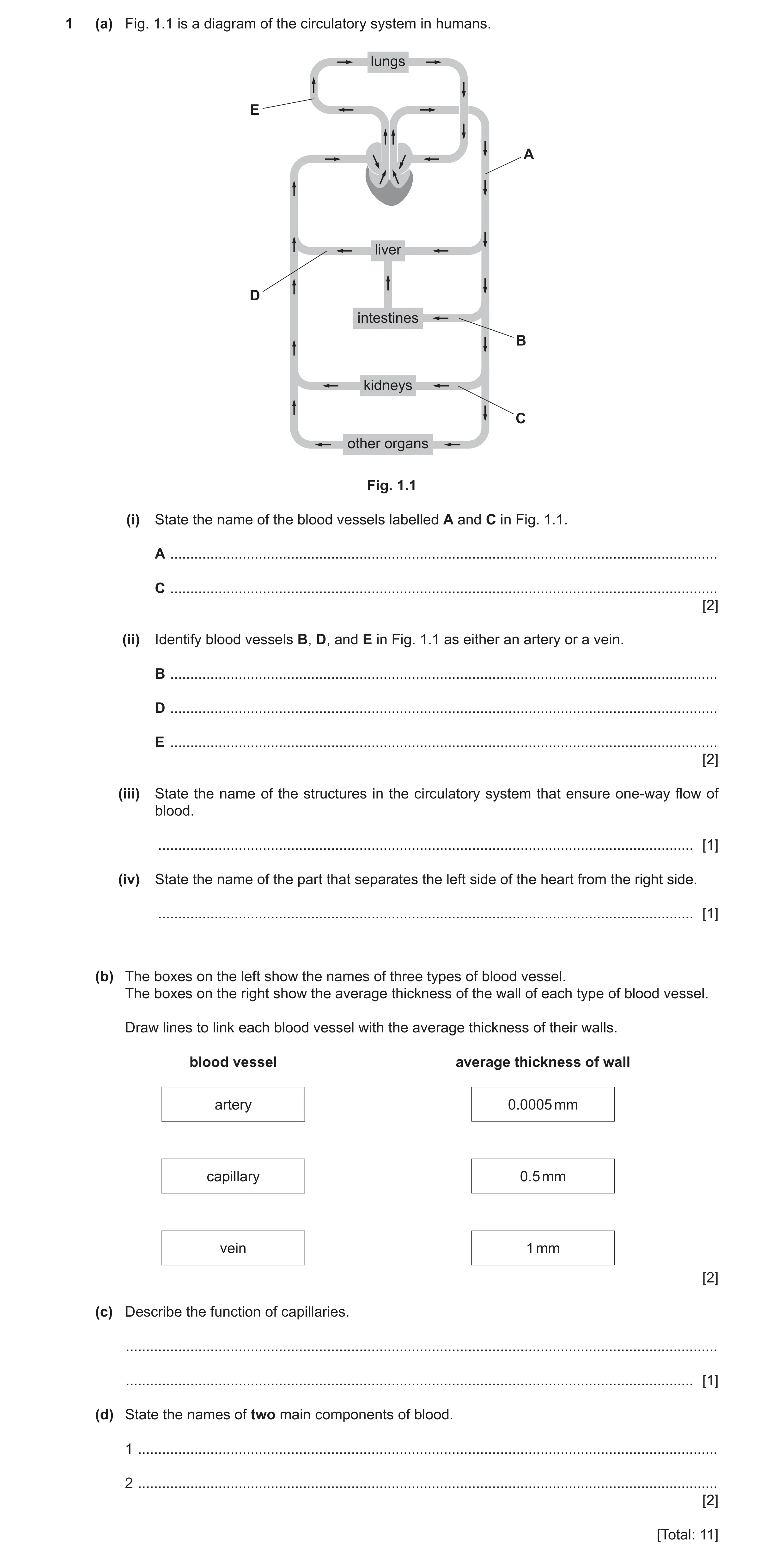
1 (a) Fig. 1.1 is a diagram of the circulatory system in humans. lungs E A liver D intestines B kidneys C other organs Fig. 1.1 (i) State the name of the blood vessels labelled A and C in Fig. 1.1. A … C … [2] (ii) Identify blood vessels B, D, and E in Fig. 1.1 as either an artery or a vein. B … D … E … [2] (iii) State the name of the structures in the circulatory system that ensure one-way flow of blood. … [1] (iv) State the name of the part that separates the left side of the heart from the right side. … [1] (b) The boxes on the left show the names of three types of blood vessel. The boxes on the right show the average thickness of the wall of each type of blood vessel. Draw lines to link each blood vessel with the average thickness of their walls. blood vessel average thickness of wall artery 0.0005 mm capillary 0.5 mm vein 1 mm [2] (c) Describe the function of capillaries. … … [1] (d) State the names of two main components of blood. 1 … 2 … [2] [Total: 11]
11 marks
Mark scheme: Question Answer Marks 1(a)(i) A – aorta ; 2 C – renal artery ; 1(a)(ii) B – artery 2 D – vein E – artery ;; 1(a)(iii) valves ; 1 1(a)(iv) septum ; 1 1(b) 2 artery 0.0005 mm capillary 0.5 mm vein 1 mm ;; 1(c) exchange of (named) material between blood and tissue (cells) / AW ; 1 1(d) any two from: 2 red blood cells ; white blood cells ; plasma ; platelets ;
This question in 0654/32 Feb/March 2023
Q12 · State the word that describes a disease-causing organism 0654/32 Feb/March 2025
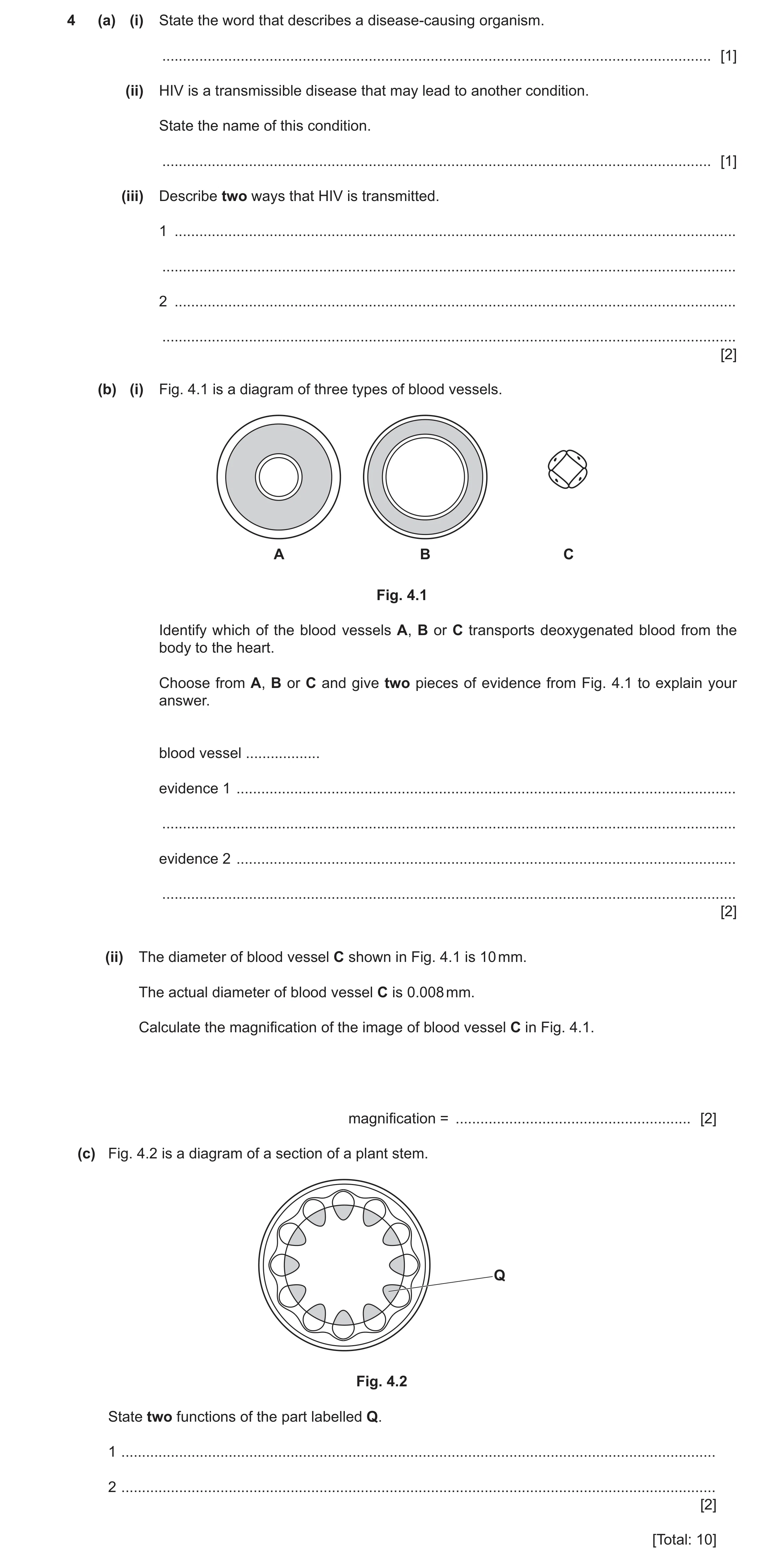
4 (a) (i) State the word that describes a disease-causing organism. … [1] (ii) HIV is a transmissible disease that may lead to another condition. State the name of this condition. … [1] (iii) Describe two ways that HIV is transmitted. 1 … … 2 … … [2] (b) (i) Fig. 4.1 is a diagram of three types of blood vessels. A B C Fig. 4.1 Identify which of the blood vessels A, B or C transports deoxygenated blood from the body to the heart. Choose from A, B or C and give two pieces of evidence from Fig. 4.1 to explain your answer. blood vessel … evidence 1 … … evidence 2 … … [2] (ii) The diameter of blood vessel C shown in Fig. 4.1 is 10 mm. The actual diameter of blood vessel C is 0.008 mm. Calculate the magnification of the image of blood vessel C in Fig. 4.1. magnification = … [2] (c) Fig. 4.2 is a diagram of a section of a plant stem. Q Fig. 4.2 State two functions of the part labelled Q. 1 … 2 … [2] [Total: 10]
10 marks
Mark scheme: 4(a)(i) pathogen ; 1 4(a)(ii) AIDS ; 1 4(a)(iii) any two from: 2 unprotected sexual intercourse sharing needles blood transfusions AVP ; ; 4(b)(i) B (no mark) 2 thin wall ; large lumen ; 4(b)(ii) 10 image size 2 Magnification = or ; 0.008 actual size 1250 ; 4(c) any two from: 2 support transport of water transport of mineral ions ; ;
This question in 0654/32 Feb/March 2025
 1 / 19
 2 / 19
 3 / 19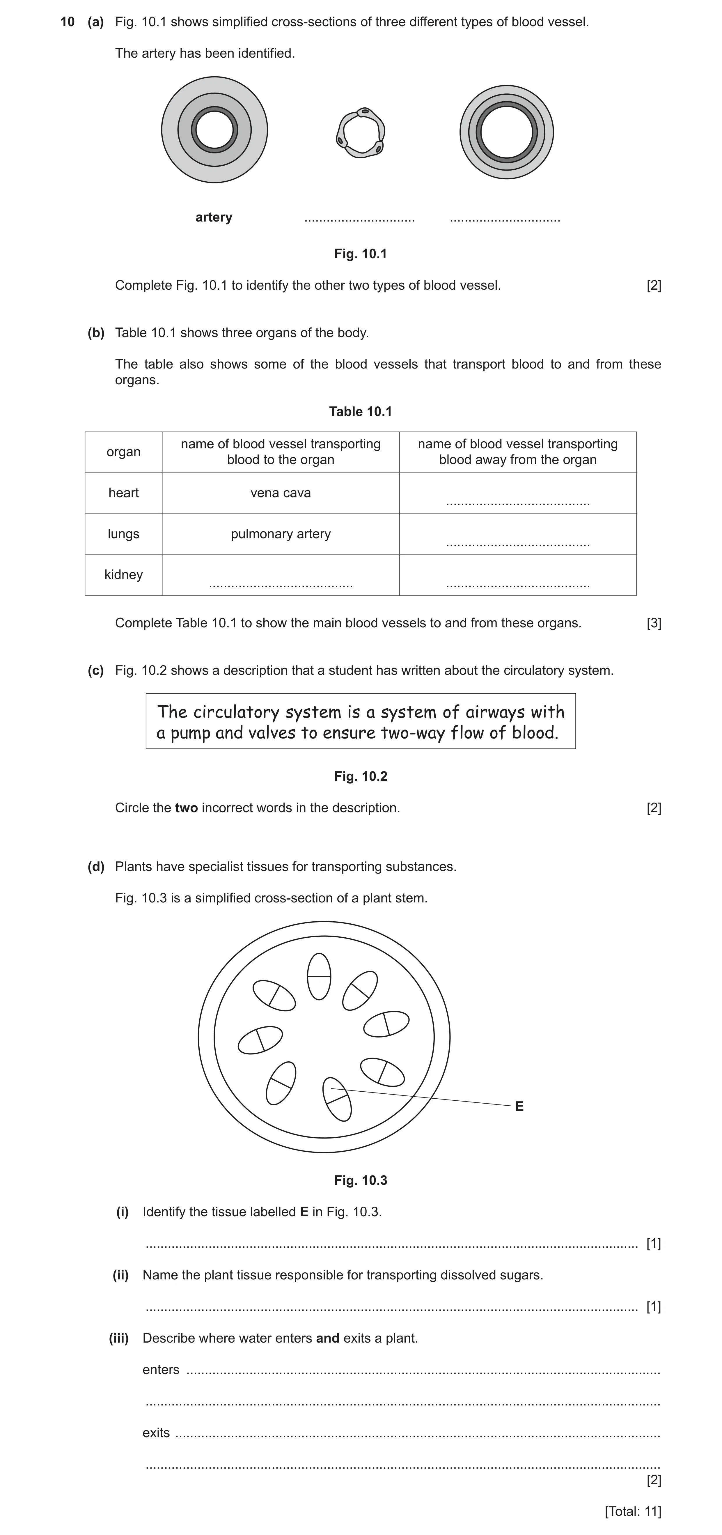
 14 / 19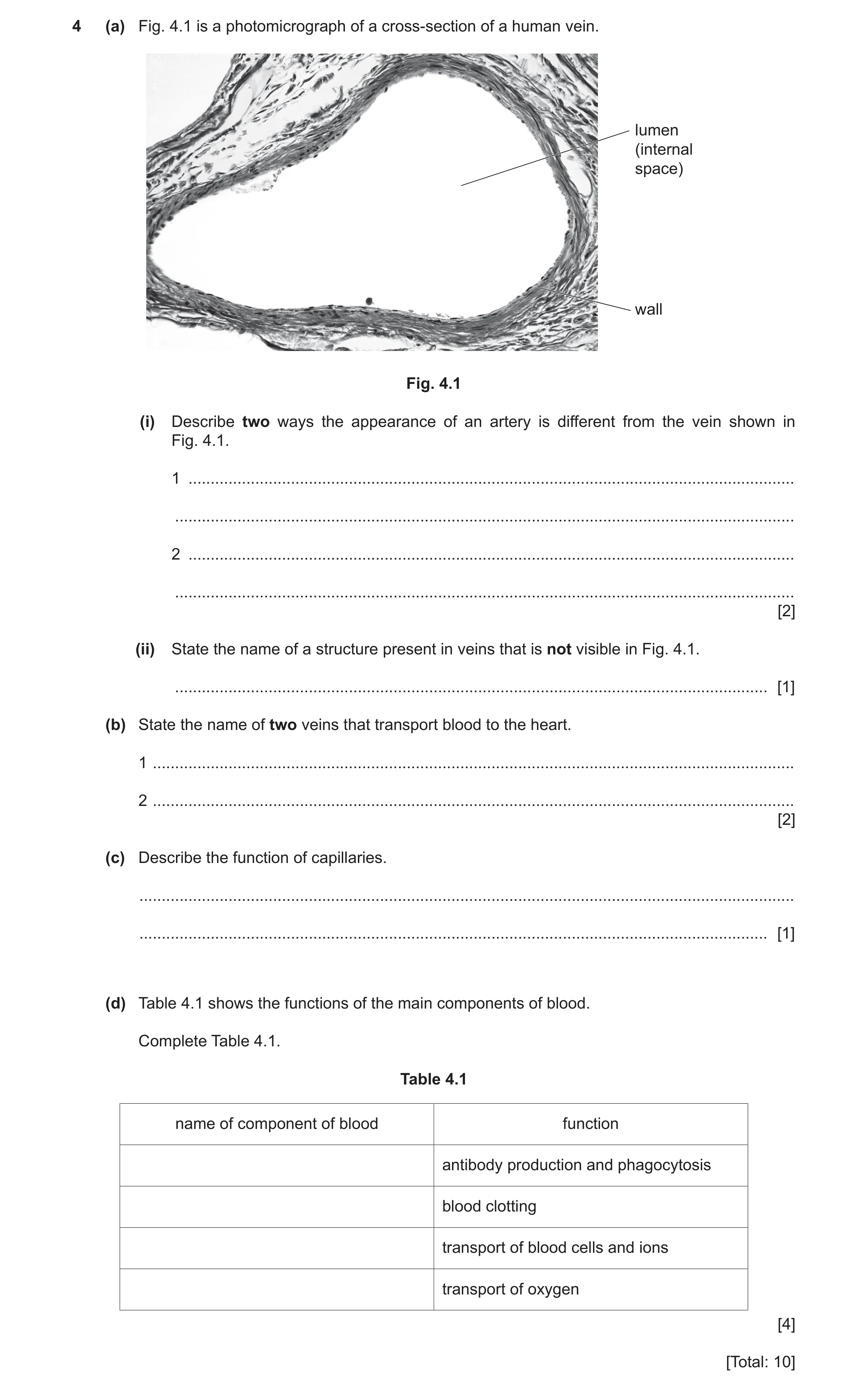
 8
8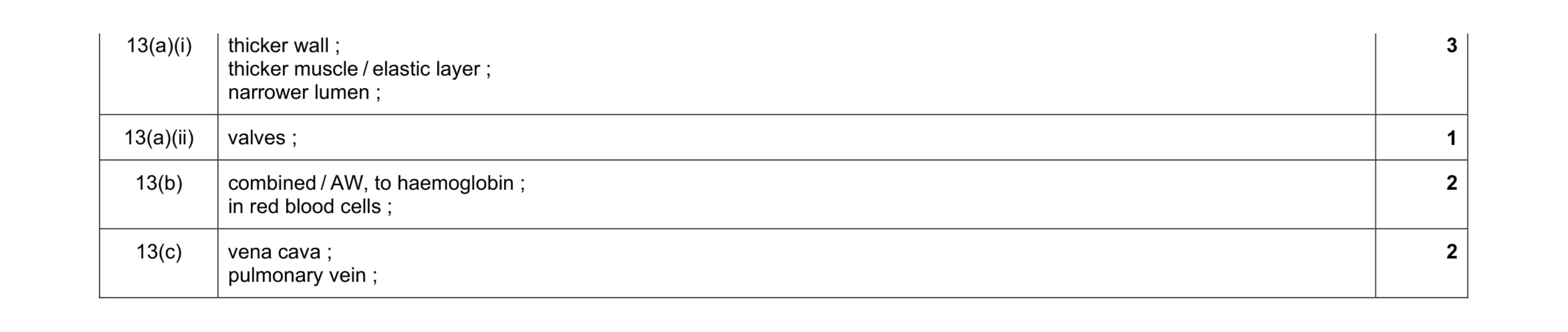 8
8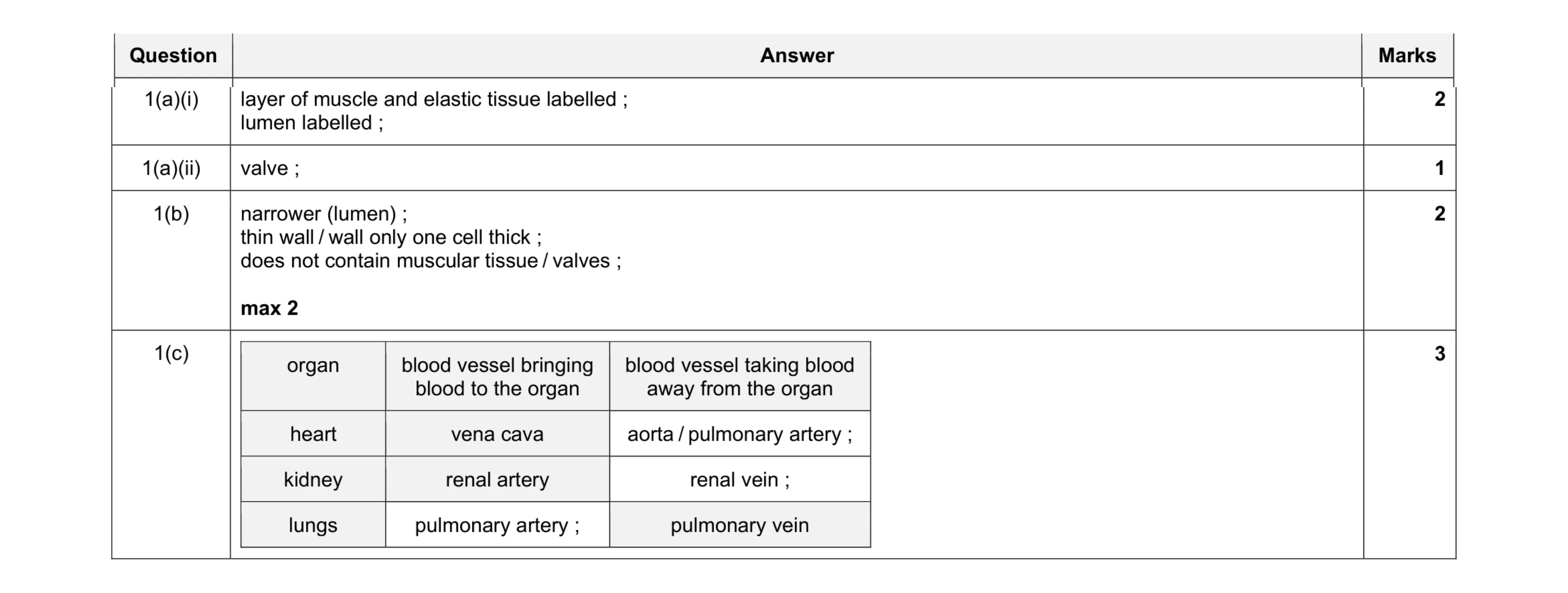 8
8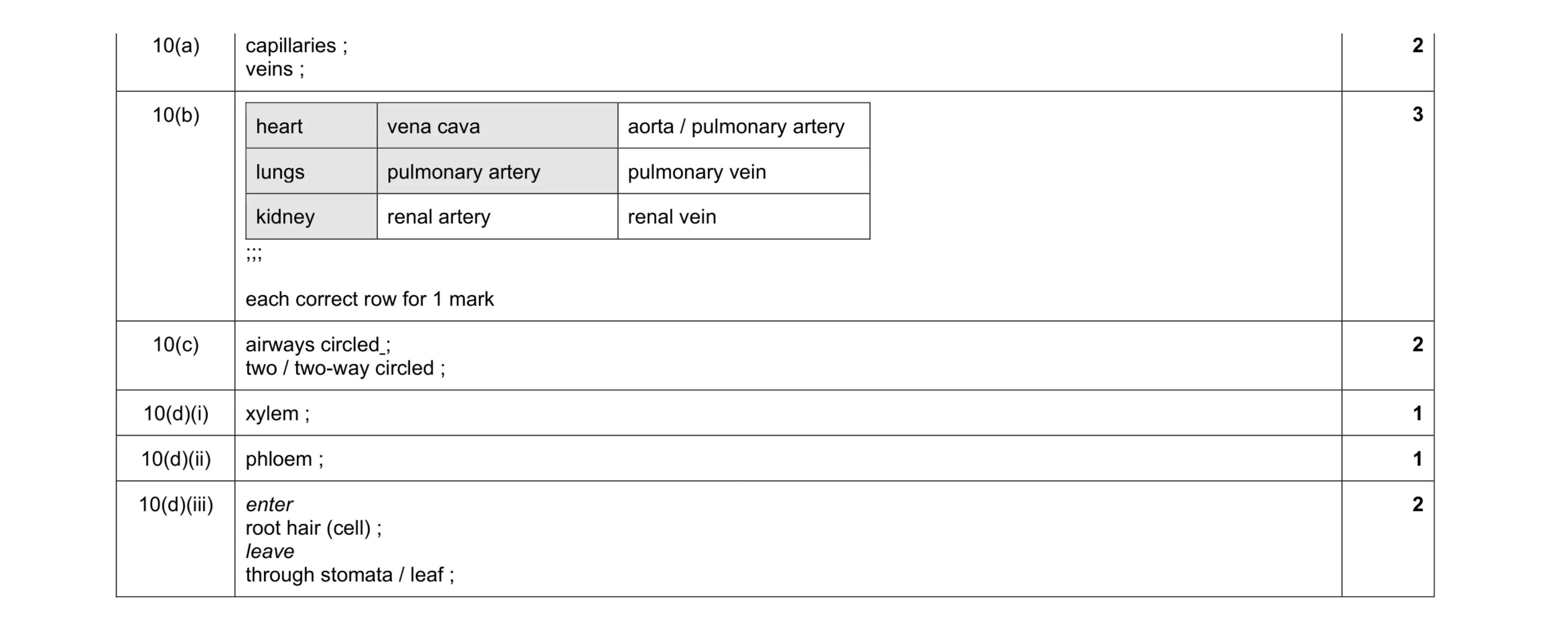 11
11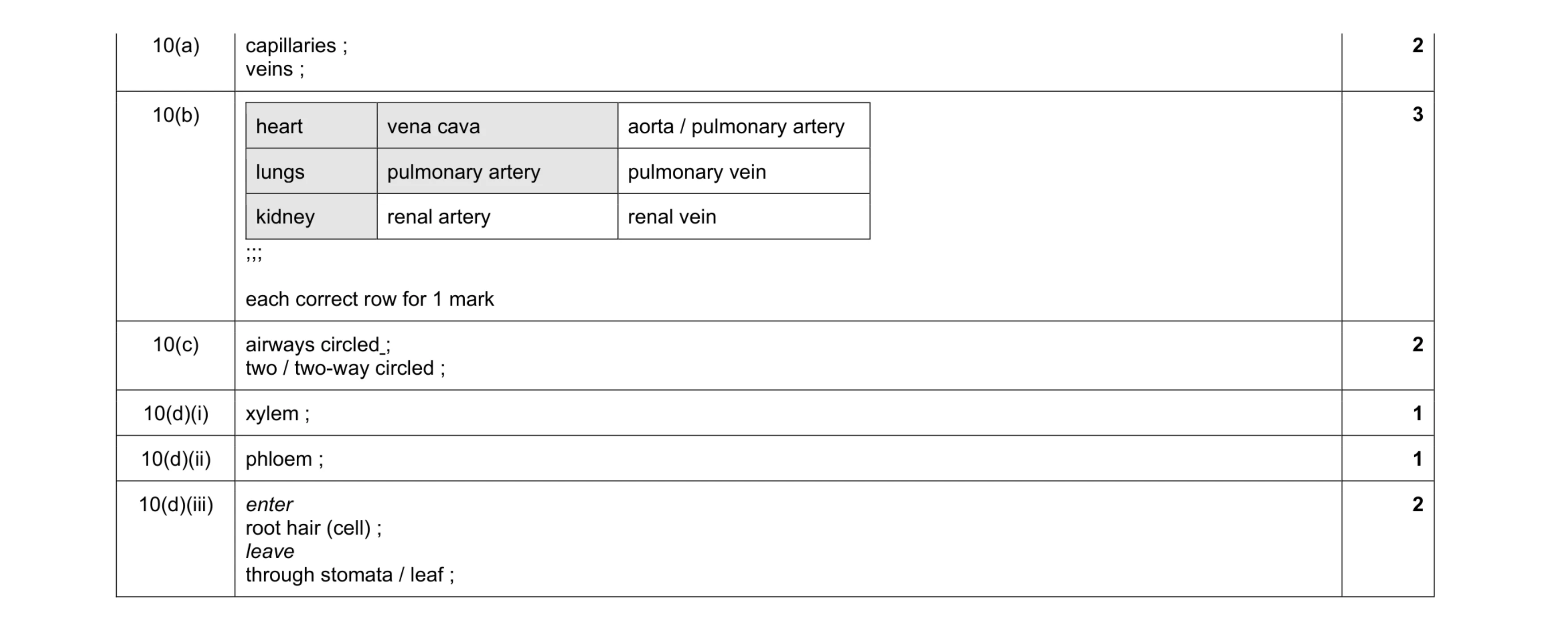 11
11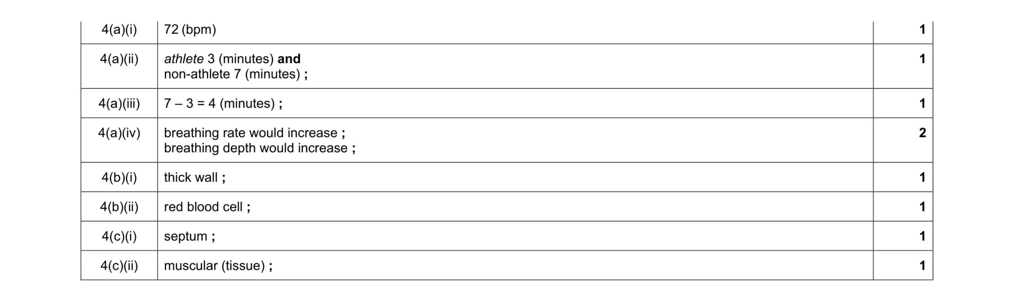 9
9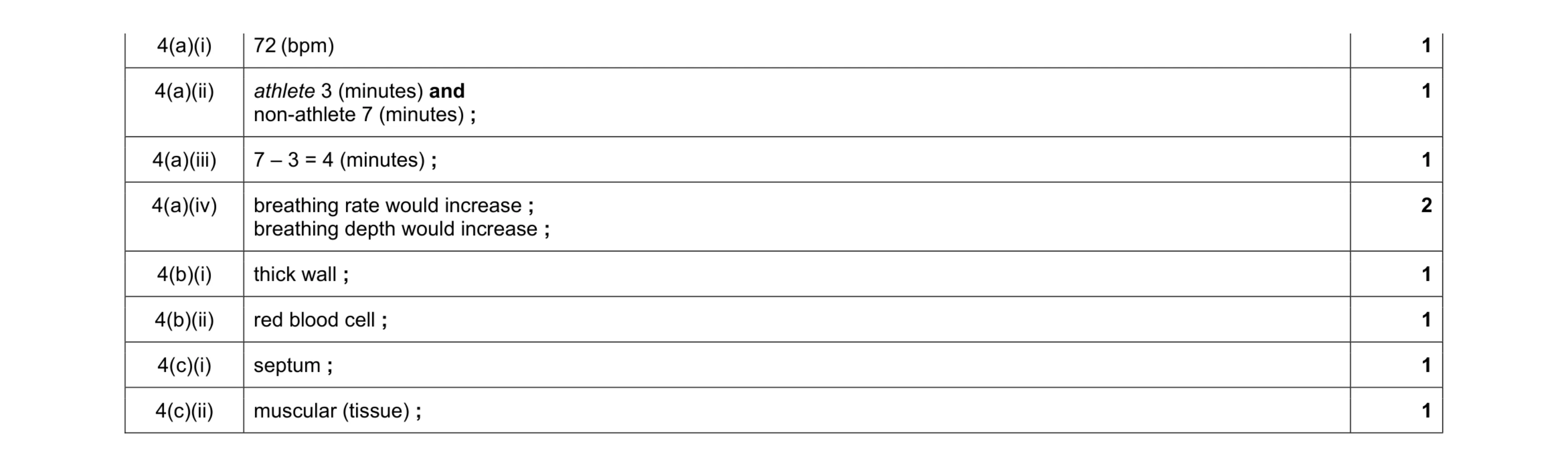 9
9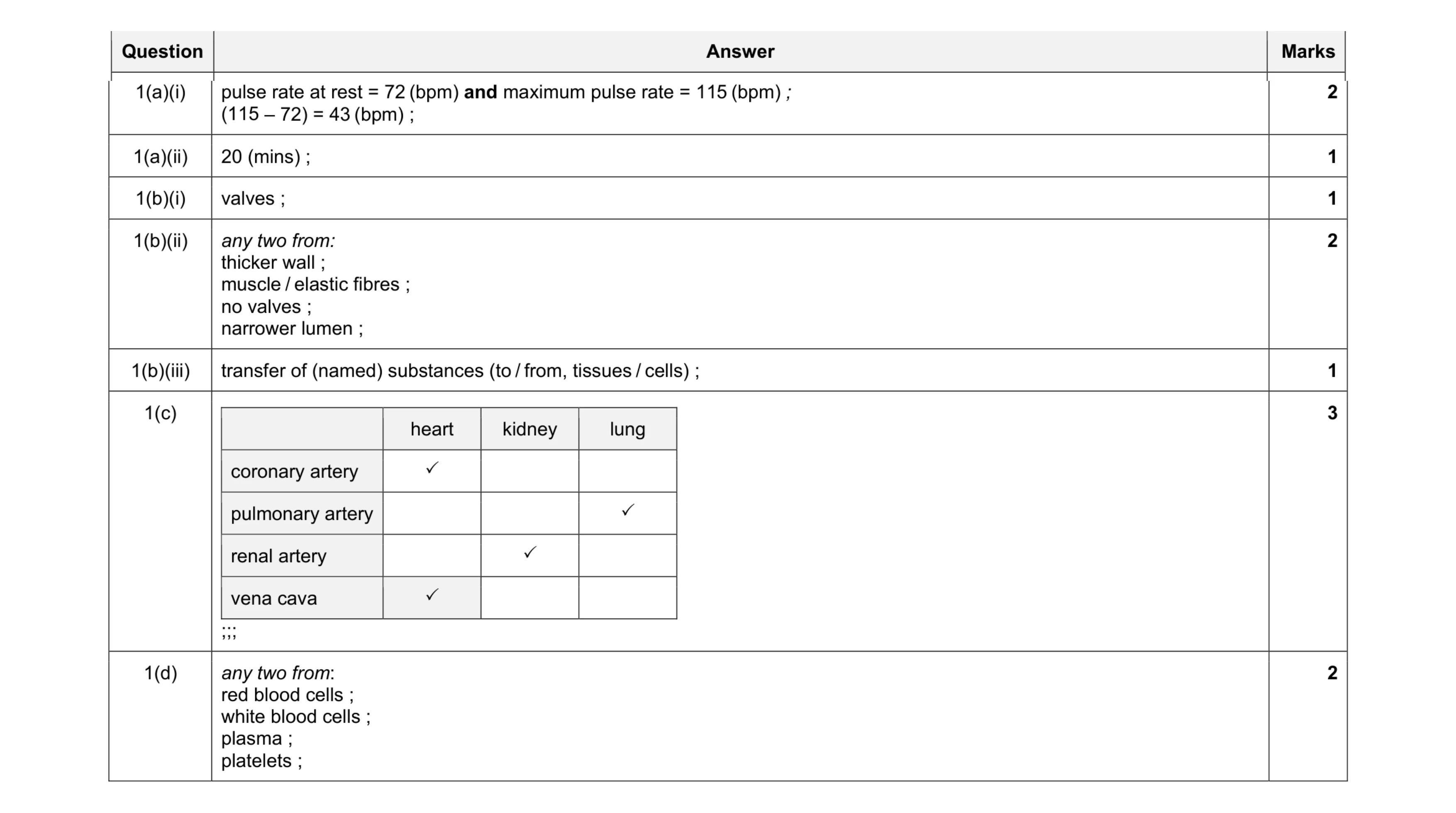 12
12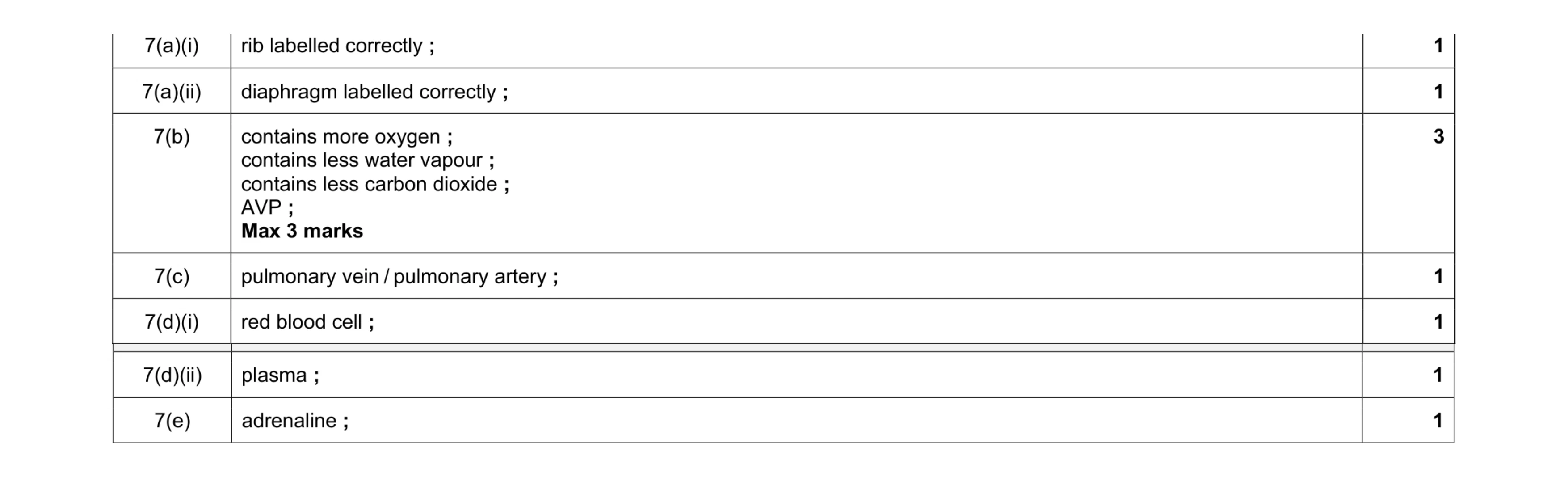 9
9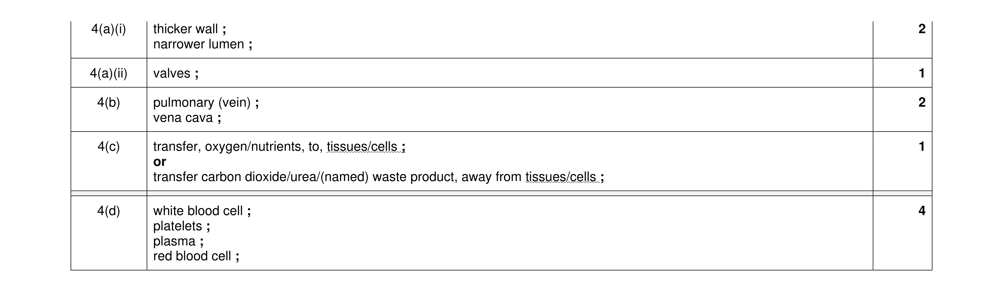 10
10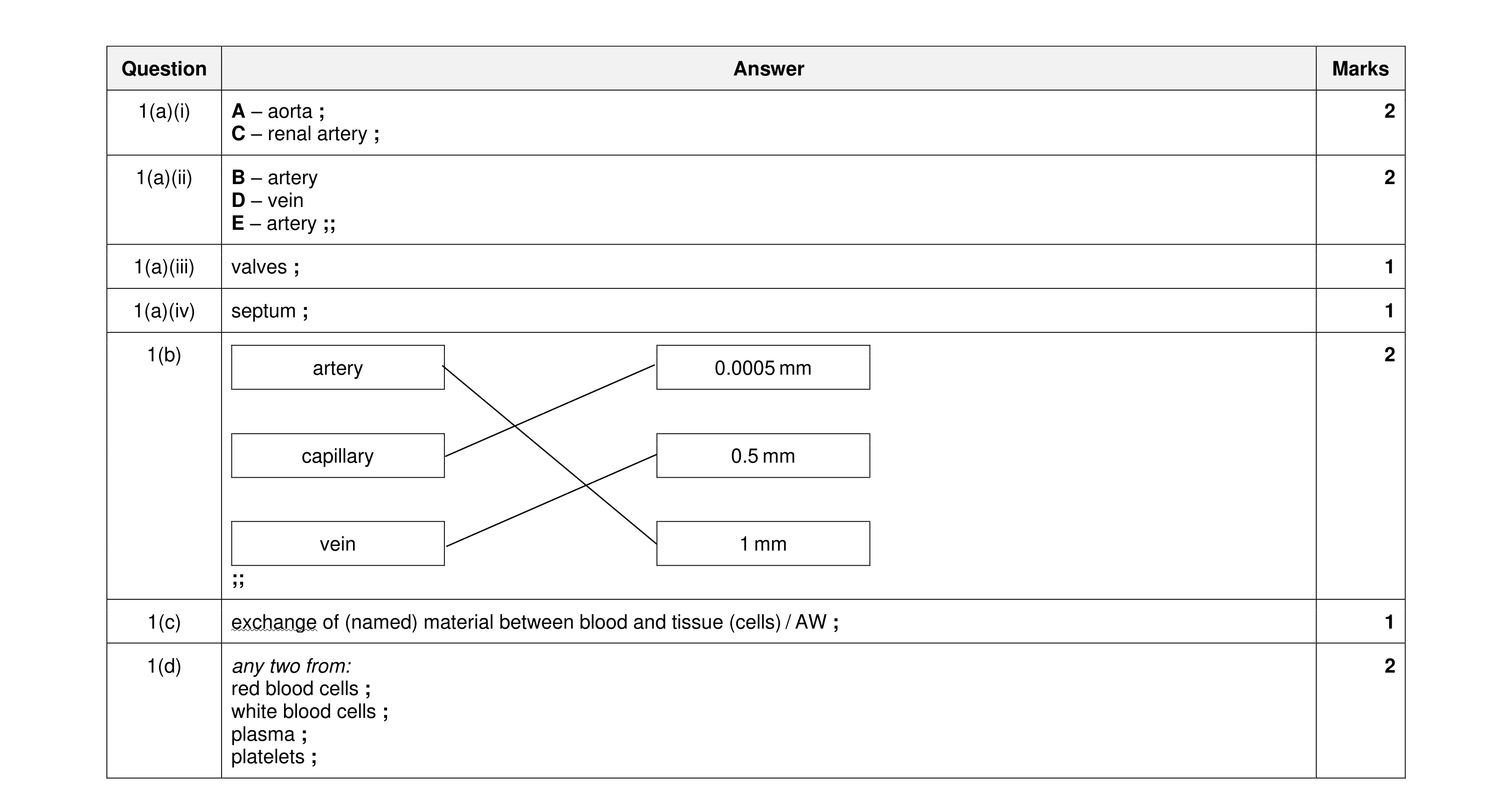 11
11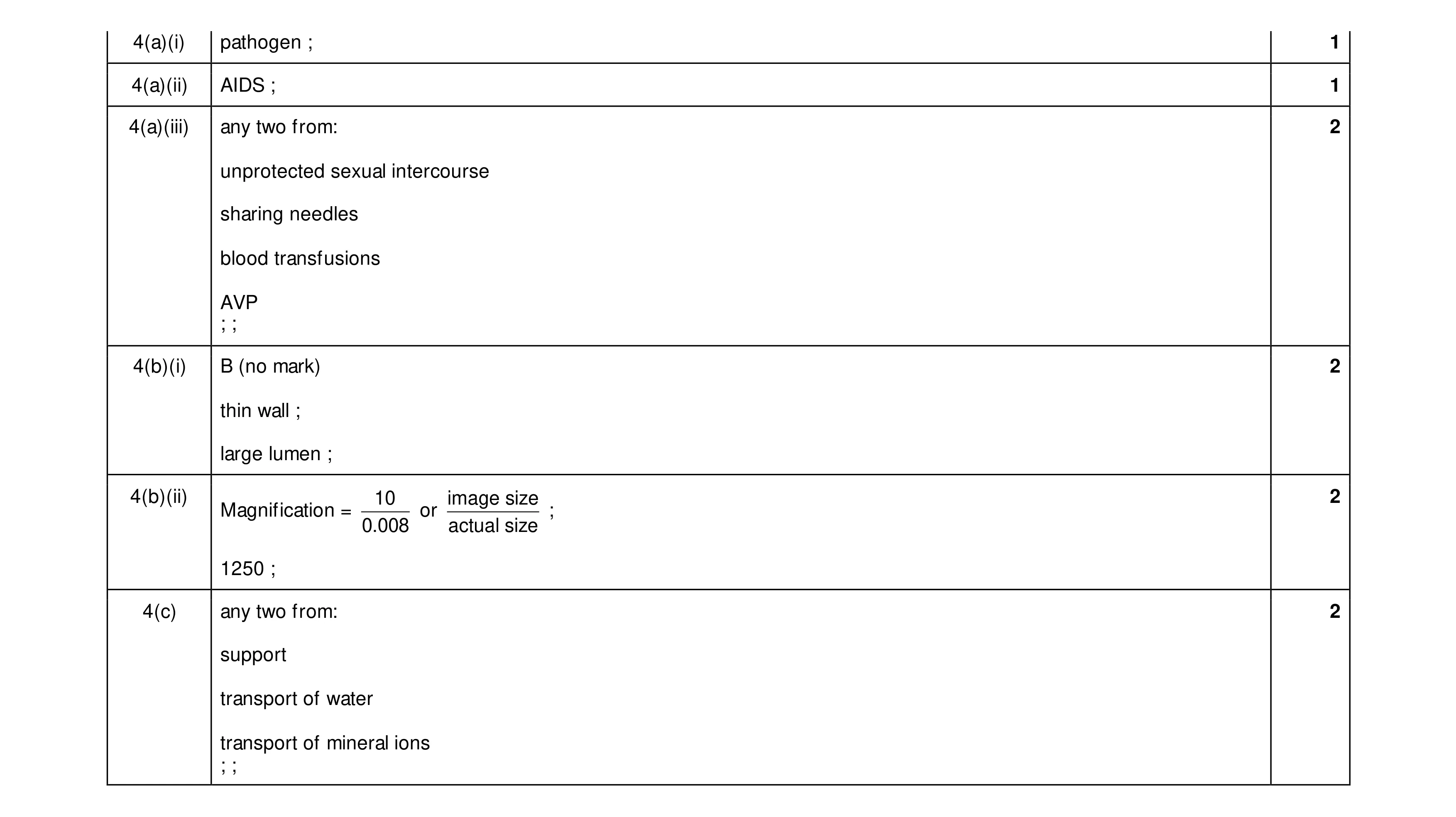 10
10