Cambridge A Level Biology 9700 — 2025 Oct/Nov Paper 2 · Variant 3
9700/23/O/N/25 · 6 questions · 60 marks · 75 min
The question paper and its mark scheme, free to read here and free to download. This is Cambridge’s own paper, exactly as it was sat.
Question paper20 pages
Mark scheme18 pages
Answers below. Sit the paper first if you are practising.
Questions as text
Q1 · Many substances can move through cell surface membranes between the cytoplasm of animal…
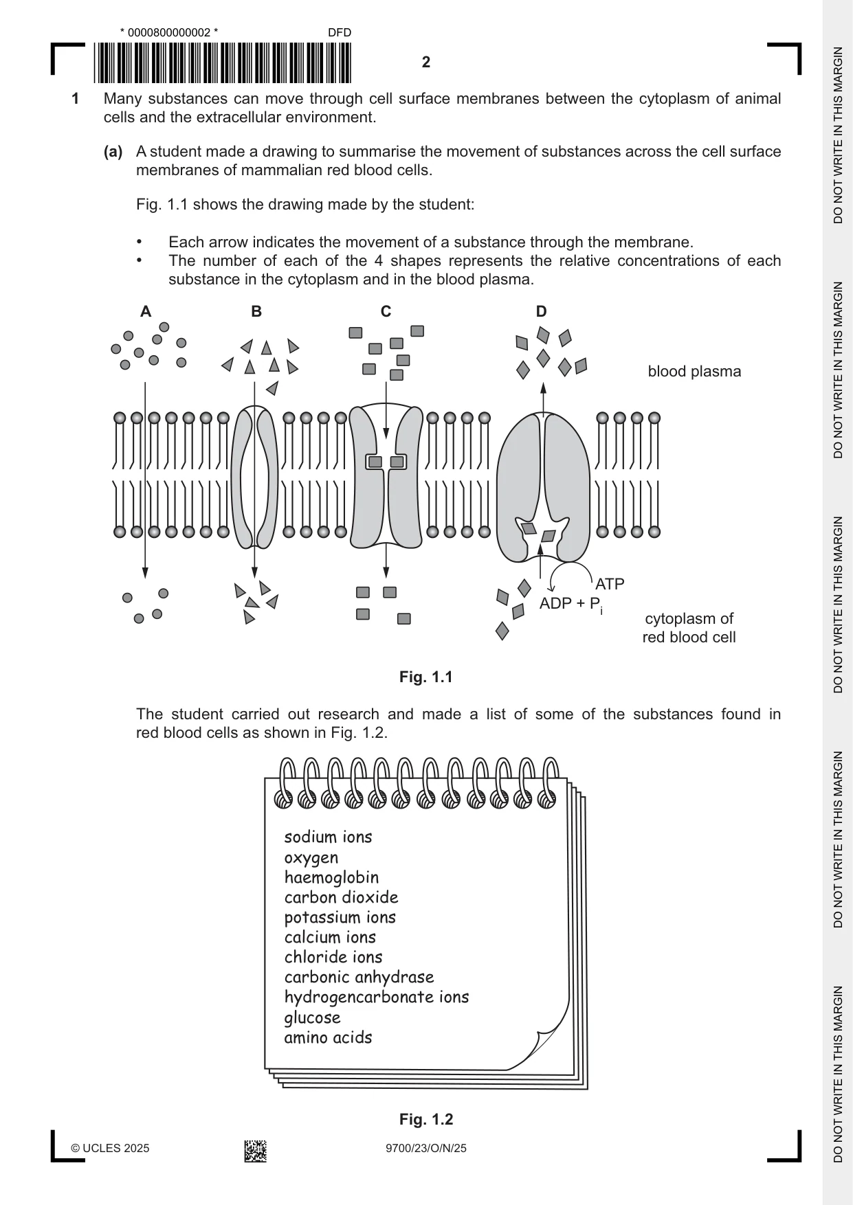
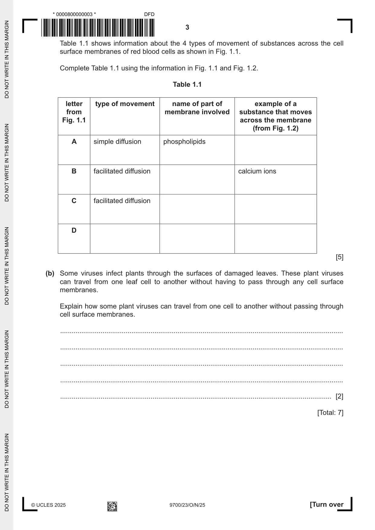
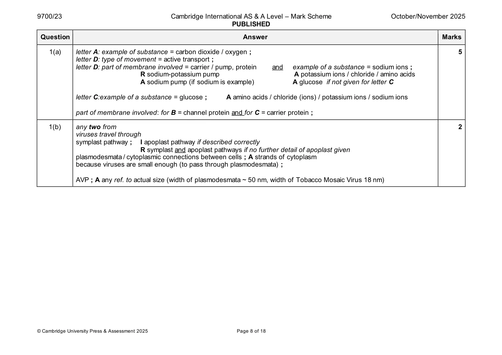
1 Many substances can move through cell surface membranes between the cytoplasm of animal cells and the extracellular environment. (a) A student made a drawing to summarise the movement of substances across the cell surface membranes of mammalian red blood cells. Fig. 1.1 shows the drawing made by the student: • Each arrow indicates the movement of a substance through the membrane. • The number of each of the 4 shapes represents the relative concentrations of each substance in the cytoplasm and in the blood plasma. A B C D blood plasma ATP ADP + Pi cytoplasm of red blood cell Fig. 1.1 The student carried out research and made a list of some of the substances found in red blood cells as shown in Fig. 1.2. sodium ions oxygen haemoglobin carbon dioxide potassium ions calcium ions chloride ions carbonic anhydrase hydrogencarbonate ions glucose amino acids Fig. 1.2 Table 1.1 shows information about the 4 types of movement of substances across the cell surface membranes of red blood cells as shown in Fig. 1.1. Complete Table 1.1 using the information in Fig. 1.1 and Fig. 1.2. Table 1.1 letter type of movement name of part of example of a from membrane involved substance that moves Fig. 1.1 across the membrane (from Fig. 1.2) A simple diffusion phospholipids B facilitated diffusion calcium ions C facilitated diffusion D [5] (b) Some viruses infect plants through the surfaces of damaged leaves. These plant viruses can travel from one leaf cell to another without having to pass through any cell surface membranes. Explain how some plant viruses can travel from one cell to another without passing through cell surface membranes. ................................................................................................................................................... ................................................................................................................................................... ................................................................................................................................................... ................................................................................................................................................... ............................................................................................................................................. [2] [Total: 7]
Mark scheme: Question Answer Marks 1(a) letter A: example of substance = carbon dioxide / oxygen ; 5 letter D: type of movement = active transport ; letter D: part of membrane involved = carrier / pump, protein and example of a substance = sodium ions ; R sodium-potassium pump A potassium ions / chloride / amino acids A sodium pump (if sodium is example) A glucose if not given for letter C letter C:example of a substance = glucose ; A amino acids / chloride (ions) / potassium ions / sodium ions part of membrane involved: for B = channel protein and for C = carrier protein ; 1(b) any two from 2 viruses travel through symplast pathway ; I apoplast pathway if described correctly R symplast and apoplast pathways if no further detail of apoplast given plasmodesmata / cytoplasmic connections between cells ; A strands of cytoplasm because viruses are small enough (to pass through plasmodesmata) ; AVP ; A any ref. to actual size (width of plasmodesmata ~ 50 nm, width of Tobacco Mosaic Virus 18 nm)
Q2 · Collagen is a fibrous protein found in many tissues in animals
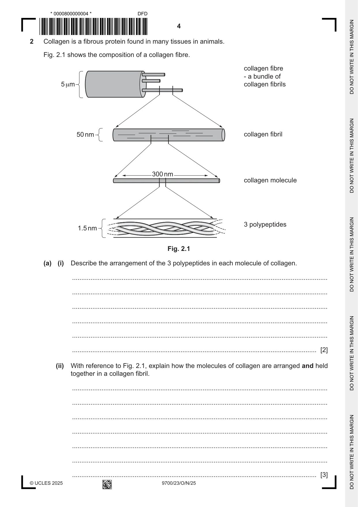
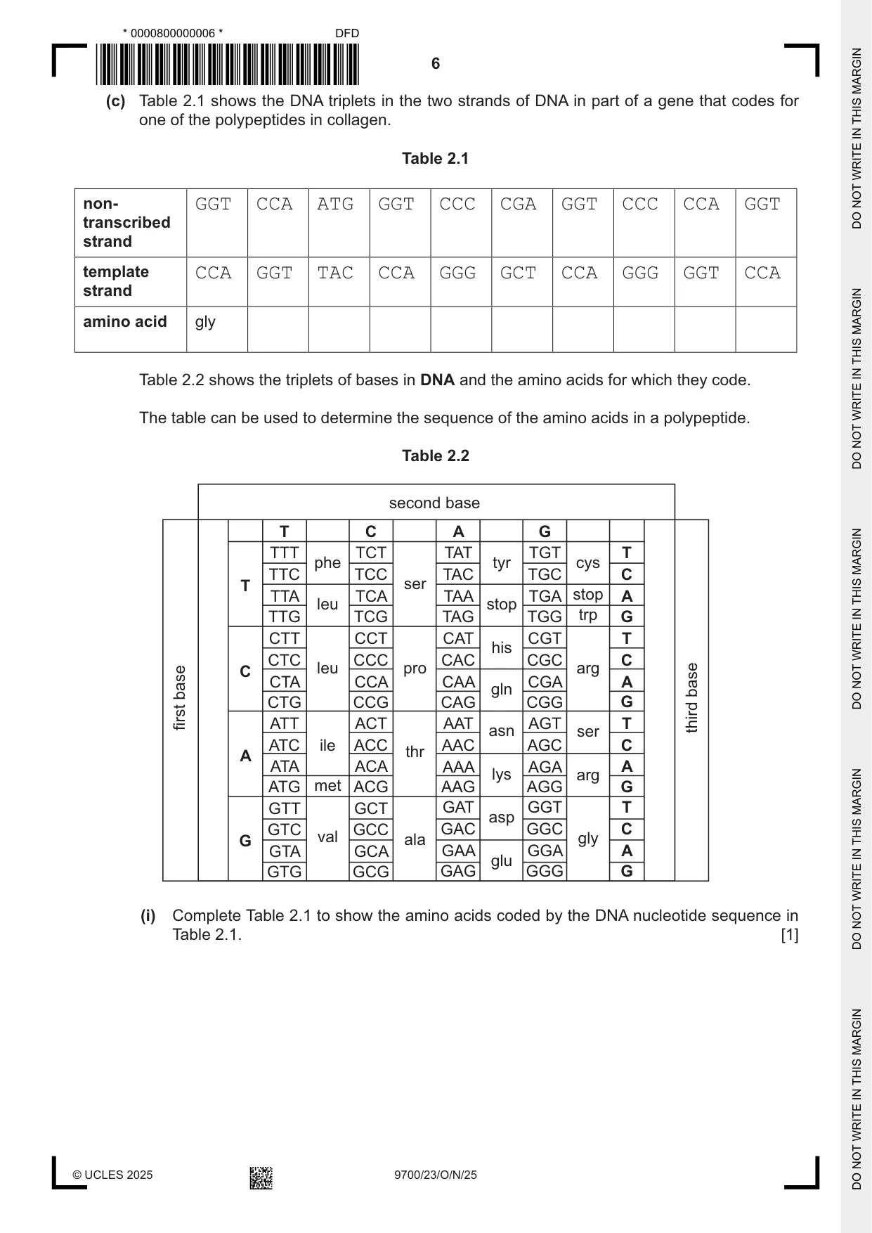
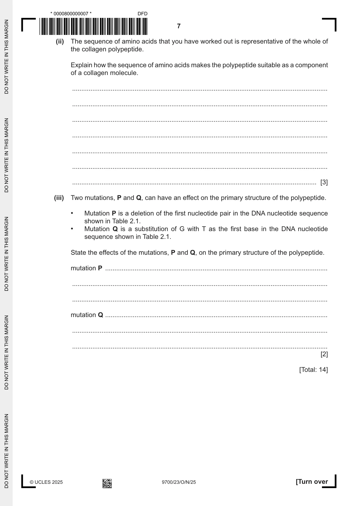
2 Collagen is a fibrous protein found in many tissues in animals. Fig. 2.1 shows the composition of a collagen fibre. collagen fibre - a bundle of 5μm collagen fibrils 50nm collagen fibril 300nm collagen molecule 3 polypeptides 1.5nm Fig. 2.1 (a) (i) Describe the arrangement of the 3 polypeptides in each molecule of collagen. ........................................................................................................................................... ........................................................................................................................................... ........................................................................................................................................... ........................................................................................................................................... ........................................................................................................................................... ..................................................................................................................................... [2] (ii) With reference to Fig. 2.1, explain how the molecules of collagen are arranged and held together in a collagen fibril. ........................................................................................................................................... ........................................................................................................................................... ........................................................................................................................................... ........................................................................................................................................... ........................................................................................................................................... ........................................................................................................................................... ................ ............................................................................................................... [3] (b) Collagen fibres and elastic fibres are found in the structures of the gas exchange system of mammals. (i) Suggest two properties of collagen that contribute to the function of cartilage in the trachea in the gas exchange system. ........................................................................................................................................... ........................................................................................................................................... ........................................................................................................................................... ........................................................................................................................................... ..................................................................................................................................... [2] (ii) State the function of elastic fibres in the alveoli in the lungs. ..................................................................................................................................... [1] (c) Table 2.1 shows the DNA triplets in the two strands of DNA in part of a gene that codes for one of the polypeptides in collagen. Table 2.1 non- GGT CCA ATG GGT CCC CGA GGT CCC CCA GGT transcribed strand template CCA GGT TAC CCA GGG GCT CCA GGG GGT CCA strand amino acid gly Table 2.2 shows the triplets of bases in DNA and the amino acids for which they code. The table can be used to determine the sequence of the amino acids in a polypeptide. Table 2.2 second base T C A G TTT TCT TAT TGT T phe tyr cys TTC TCC TAC TGC C T ser TTA TCA TAA TGA stop A leu stop TTG TCG TAG TGG trp G CTT CCT CAT CGT T his CTC CCC CAC CGC C C leu pro arg CTA CCA CAA CGA A base gln base CTG CCG CAG CGG G first ATT ACT AAT AGT T third asn ser ATC ile ACC AAC AGC C thr A ATA ACA AAA AGA A lys arg ATG met ACG AAG AGG G GTT GCT GAT GGT T asp GTC GCC GAC GGC C G val ala gly GTA GCA GAA GGA A glu GTG GCG GAG GGG G (i) Complete Table 2.1 to show the amino acids coded by the DNA nucleotide sequence in Table 2.1. [1] (ii) The sequence of amino acids that you have worked out is representative of the whole of the collagen polypeptide. Explain how the sequence of amino acids makes the polypeptide suitable as a component of a collagen molecule. ........................................................................................................................................... ........................................................................................................................................... ........................................................................................................................................... ........................................................................................................................................... ........................................................................................................................................... ........................................................................................................................................... ..................................................................................................................................... [3] (iii) Two mutations, P and Q, can have an effect on the primary structure of the polypeptide. • Mutation P is a deletion of the first nucleotide pair in the DNA nucleotide sequence shown in Table 2.1. • Mutation Q is a substitution of G with T as the first base in the DNA nucleotide sequence shown in Table 2.1. State the effects of the mutations, P and Q, on the primary structure of the polypeptide. mutation P ......................................................................................................................... ........................................................................................................................................... ........................................................................................................................................... mutation Q ......................................................................................................................... ........................................................................................................................................... ........................................................................................................................................... [2] [Total: 14]
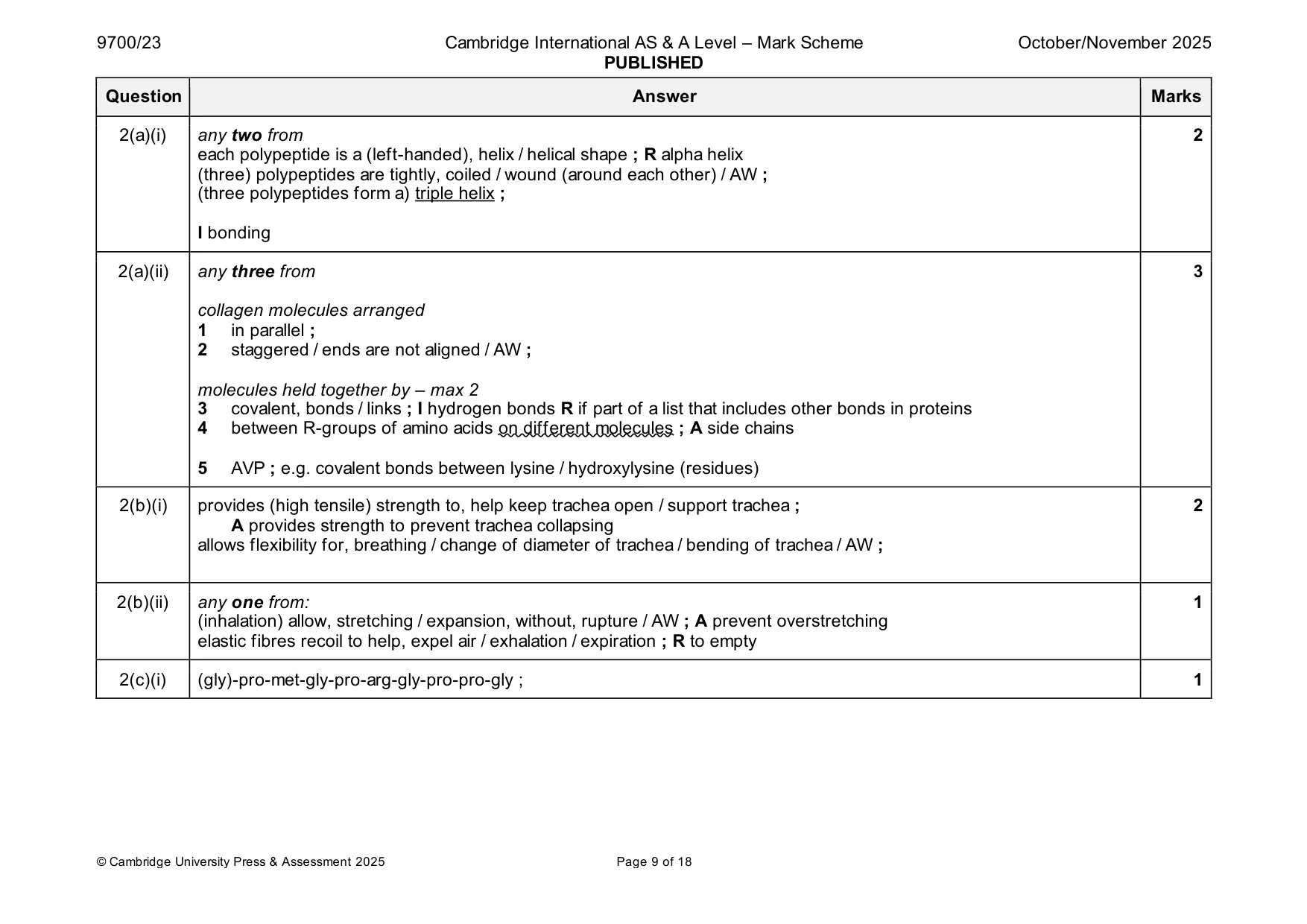
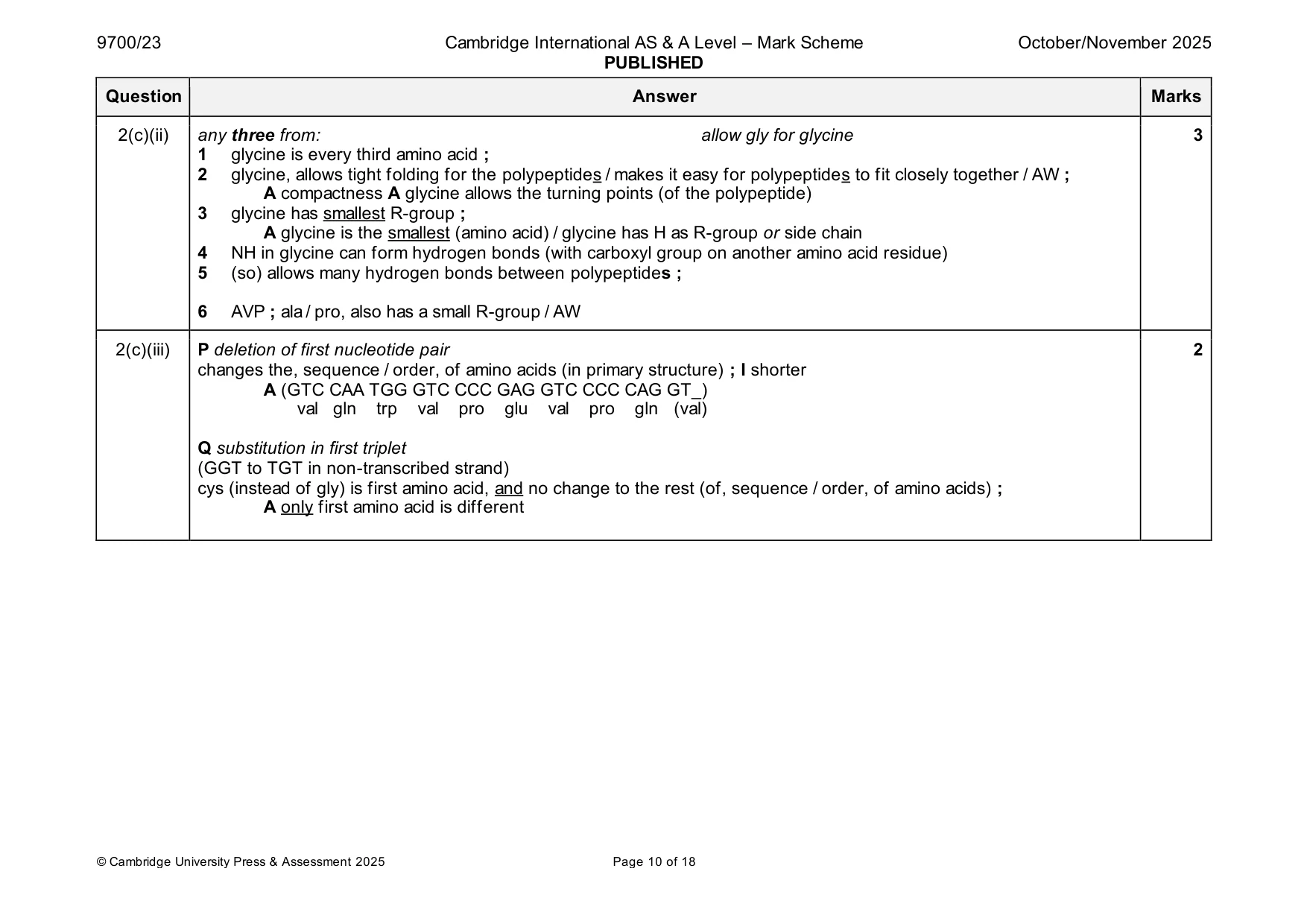
Mark scheme: 2(a)(i) any two from 2 each polypeptide is a (left-handed), helix / helical shape ; R alpha helix (three) polypeptides are tightly, coiled / wound (around each other) / AW ; (three polypeptides form a) triple helix ; I bonding 2(a)(ii) any three from 3 collagen molecules arranged 1 in parallel ; 2 staggered / ends are not aligned / AW ; molecules held together by – max 2 3 covalent, bonds / links ; I hydrogen bonds R if part of a list that includes other bonds in proteins 4 between R-groups of amino acids on different molecules ; A side chains 5 AVP ; e.g. covalent bonds between lysine / hydroxylysine (residues) 2(b)(i) provides (high tensile) strength to, help keep trachea open / support trachea ; 2 A provides strength to prevent trachea collapsing allows flexibility for, breathing / change of diameter of trachea / bending of trachea / AW ; 2(b)(ii) any one from: 1 (inhalation) allow, stretching / expansion, without, rupture / AW ; A prevent overstretching elastic fibres recoil to help, expel air / exhalation / expiration ; R to empty 2(c)(i) (gly)-pro-met-gly-pro-arg-gly-pro-pro-gly ; 1 2(c)(ii) any three from: allow gly for glycine 3 1 glycine is every third amino acid ; 2 glycine, allows tight folding for the polypeptides / makes it easy for polypeptides to fit closely together / AW ; A compactness A glycine allows the turning points (of the polypeptide) 3 glycine has smallest R-group ; A glycine is the smallest (amino acid) / glycine has H as R-group or side chain 4 NH in glycine can form hydrogen bonds (with carboxyl group on another amino acid residue) 5 (so) allows many hydrogen bonds between polypeptides ; 6 AVP ; ala / pro, also has a small R-group / AW 2(c)(iii) P deletion of first nucleotide pair 2 changes the, sequence / order, of amino acids (in primary structure) ; I shorter A (GTC CAA TGG GTC CCC GAG GTC CCC CAG GT_) val gln trp val pro glu val pro gln (val) Q substitution in first triplet (GGT to TGT in non-transcribed strand) cys (instead of gly) is first amino acid, and no change to the rest (of, sequence / order, of amino acids) ; A only first amino acid is different
Q3 · Describe the induced-fit hypothesis of enzyme action
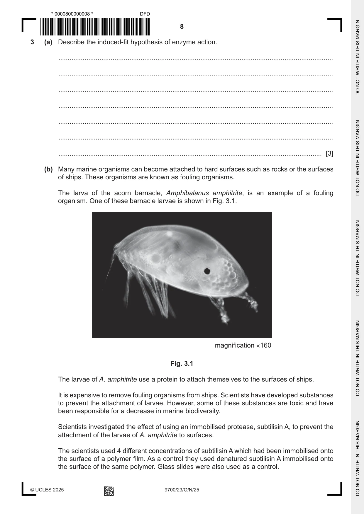
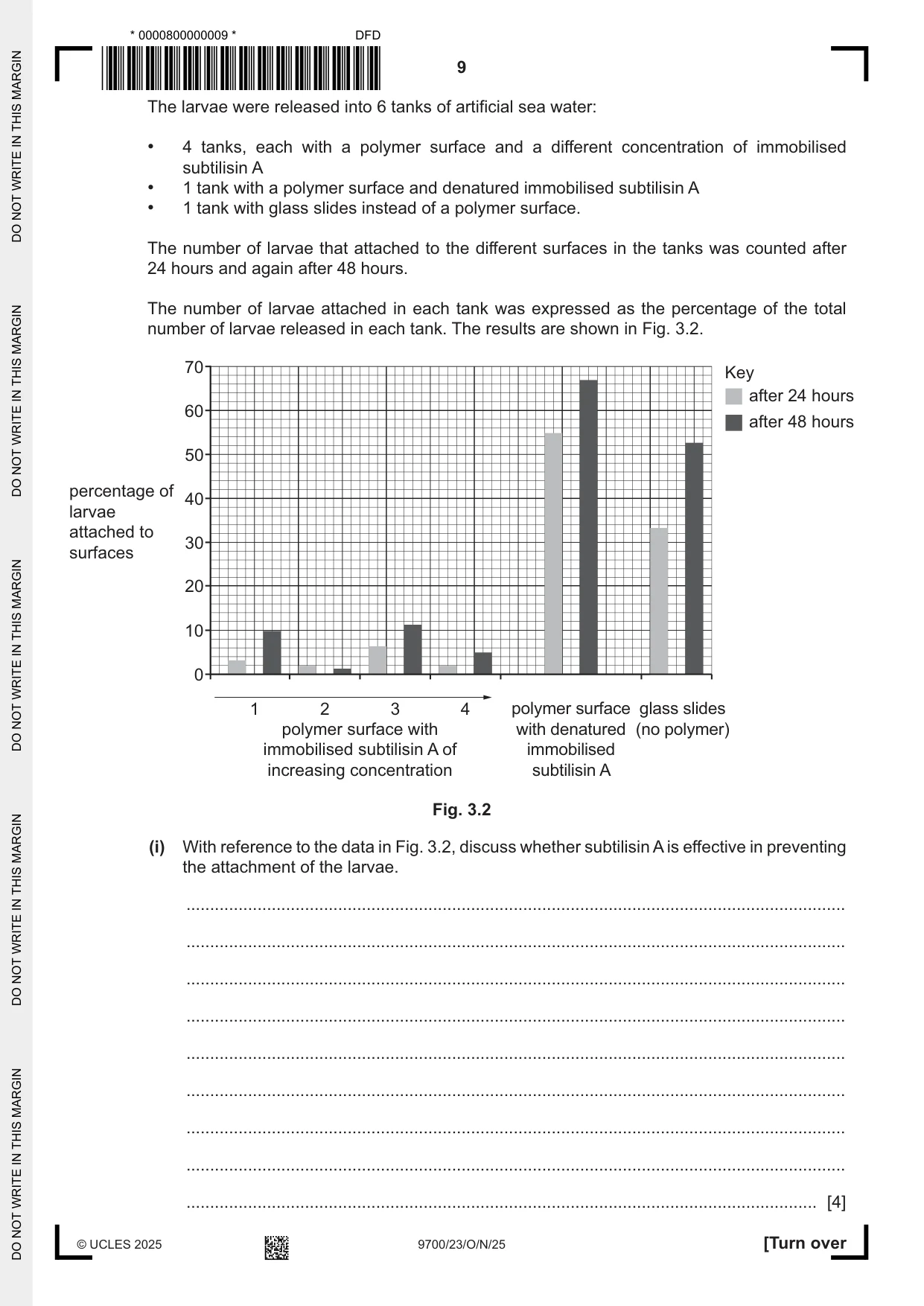
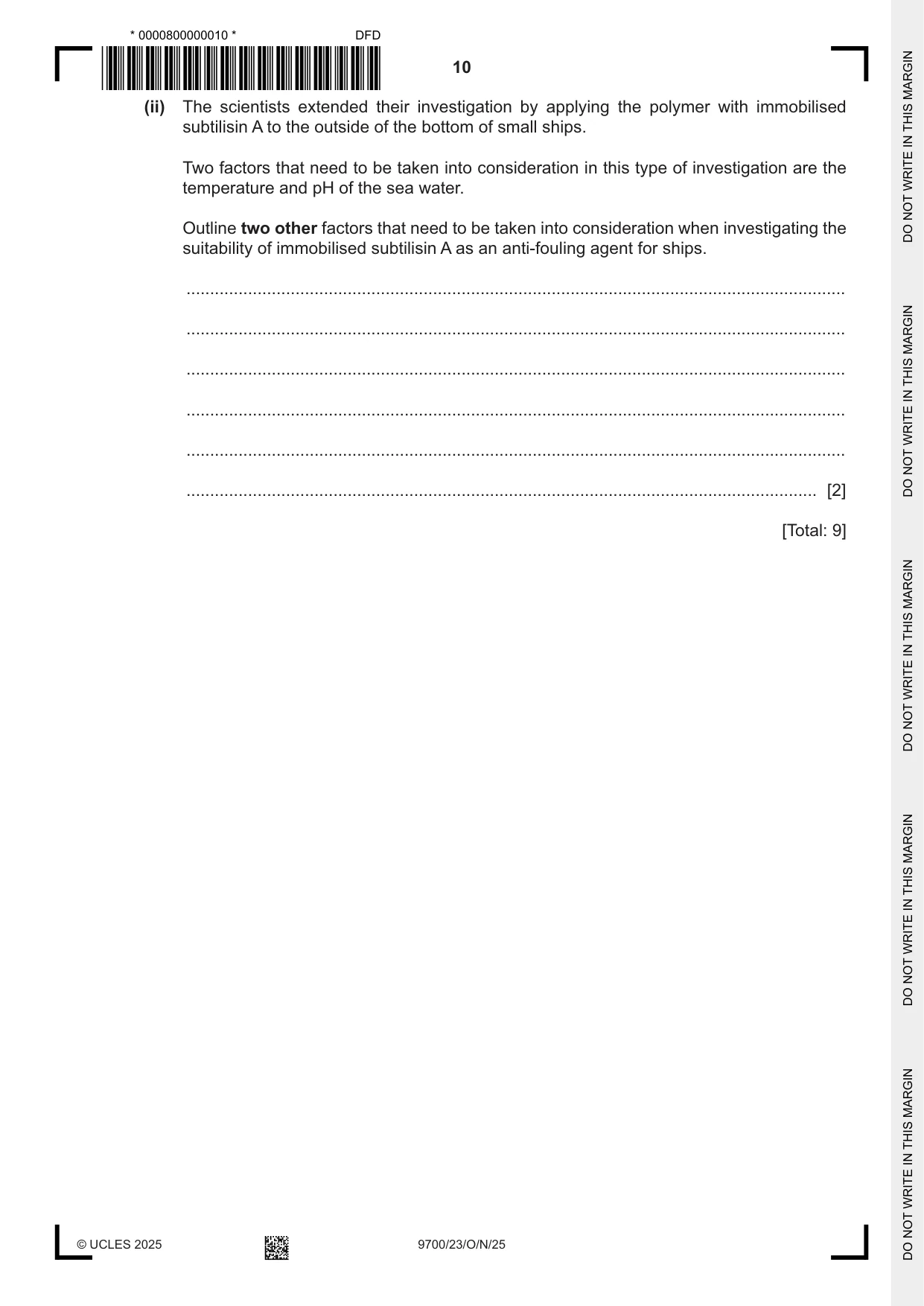
3 (a) Describe the induced-fit hypothesis of enzyme action. ................................................................................................................................................... ................................................................................................................................................... ................................................................................................................................................... ................................................................................................................................................... ................................................................................................................................................... ................................................................................................................................................... ............................................................................................................................................. [3] (b) Many marine organisms can become attached to hard surfaces such as rocks or the surfaces of ships. These organisms are known as fouling organisms. The larva of the acorn barnacle, Amphibalanus amphitrite, is an example of a fouling organism. One of these barnacle larvae is shown in Fig. 3.1. magnification ×160 Fig. 3.1 The larvae of A. amphitrite use a protein to attach themselves to the surfaces of ships. It is expensive to remove fouling organisms from ships. Scientists have developed substances to prevent the attachment of larvae. However, some of these substances are toxic and have been responsible for a decrease in marine biodiversity. Scientists investigated the effect of using an immobilised protease, subtilisin A, to prevent the attachment of the larvae of A. amphitrite to surfaces. The scientists used 4 different concentrations of subtilisin A which had been immobilised onto the surface of a polymer film. As a control they used denatured subtilisin A immobilised onto the surface of the same polymer. Glass slides were also used as a control. The larvae were released into 6 tanks of artificial sea water: • 4 tanks, each with a polymer surface and a different concentration of immobilised subtilisin A • 1 tank with a polymer surface and denatured immobilised subtilisin A • 1 tank with glass slides instead of a polymer surface. The number of larvae that attached to the different surfaces in the tanks was counted after 24 hours and again after 48 hours. The number of larvae attached in each tank was expressed as the percentage of the total number of larvae released in each tank. The results are shown in Fig. 3.2. 70 Key after 24 hours 60 after 48 hours 50 percentage of 40 larvae attached to 30 surfaces 20 10 0 1 2 3 4 polymer surface glass slides polymer surface with with denatured (no polymer) immobilised subtilisin A of immobilised increasing concentration subtilisin A Fig. 3.2 (i) With reference to the data in Fig. 3.2, discuss whether subtilisin A is effective in preventing the attachment of the larvae. ........................................................................................................................................... ........................................................................................................................................... ........................................................................................................................................... ........................................................................................................................................... ........................................................................................................................................... ........................................................................................................................................... ........................................................................................................................................... ........................................................................................................................................... ..................................................................................................................................... [4] (ii) The scientists extended their investigation by applying the polymer with immobilised subtilisin A to the outside of the bottom of small ships. Two factors that need to be taken into consideration in this type of investigation are the temperature and pH of the sea water. Outline two other factors that need to be taken into consideration when investigating the suitability of immobilised subtilisin A as an anti-fouling agent for ships. ........................................................................................................................................... ........................................................................................................................................... ........................................................................................................................................... ........................................................................................................................................... ........................................................................................................................................... ..................................................................................................................................... [2] [Total: 9] Question 4 starts on page 12.
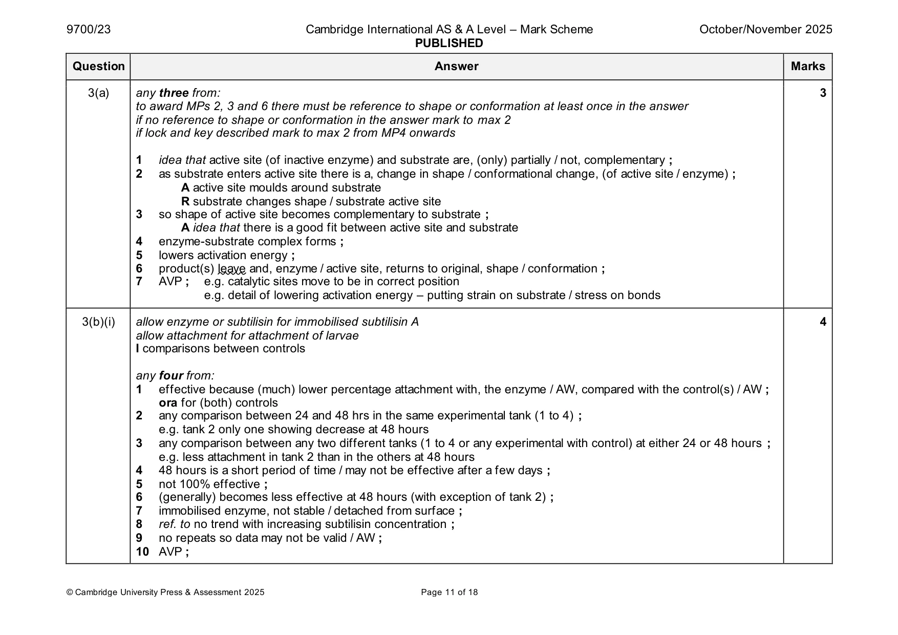
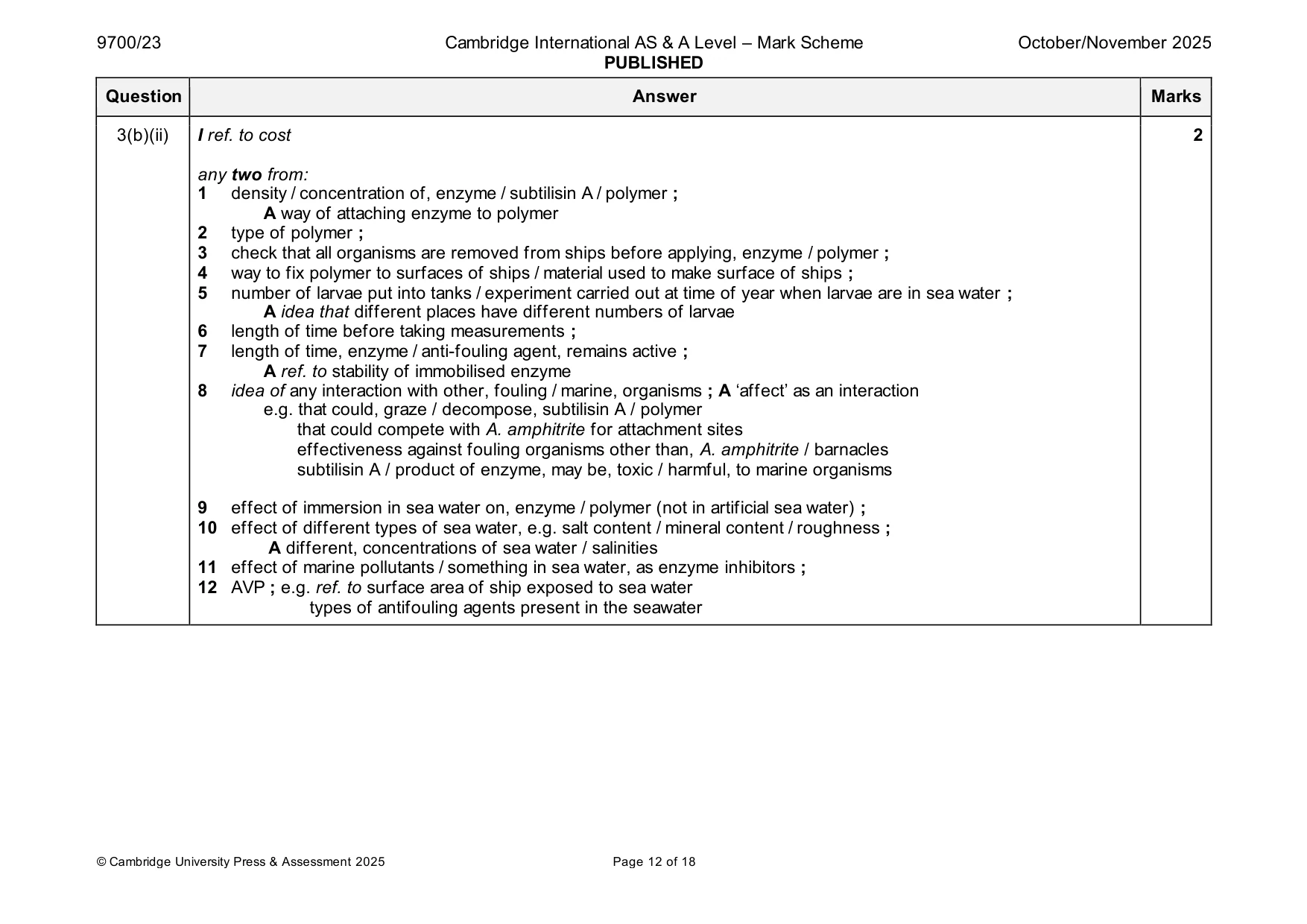
Mark scheme: 3(a) any three from: 3 to award MPs 2, 3 and 6 there must be reference to shape or conformation at least once in the answer if no reference to shape or conformation in the answer mark to max 2 if lock and key described mark to max 2 from MP4 onwards 1 idea that active site (of inactive enzyme) and substrate are, (only) partially / not, complementary ; 2 as substrate enters active site there is a, change in shape / conformational change, (of active site / enzyme) ; A active site moulds around substrate R substrate changes shape / substrate active site 3 so shape of active site becomes complementary to substrate ; A idea that there is a good fit between active site and substrate 4 enzyme-substrate complex forms ; 5 lowers activation energy ; 6 product(s) leave and, enzyme / active site, returns to original, shape / conformation ; 7 AVP ; e.g. catalytic sites move to be in correct position e.g. detail of lowering activation energy – putting strain on substrate / stress on bonds 3(b)(i) allow enzyme or subtilisin for immobilised subtilisin A 4 allow attachment for attachment of larvae I comparisons between controls any four from: 1 effective because (much) lower percentage attachment with, the enzyme / AW, compared with the control(s) / AW ; ora for (both) controls 2 any comparison between 24 and 48 hrs in the same experimental tank (1 to 4) ; e.g. tank 2 only one showing decrease at 48 hours 3 any comparison between any two different tanks (1 to 4 or any experimental with control) at either 24 or 48 hours ; e.g. less attachment in tank 2 than in the others at 48 hours 4 48 hours is a short period of time / may not be effective after a few days ; 5 not 100% effective ; 6 (generally) becomes less effective at 48 hours (with exception of tank 2) ; 7 immobilised enzyme, not stable / detached from surface ; 8 ref. to no trend with increasing subtilisin concentration ; 9 no repeats so data may not be valid / AW ; 10 AVP ; 3(b)(ii) I ref. to cost 2 any two from: 1 density / concentration of, enzyme / subtilisin A / polymer ; A way of attaching enzyme to polymer 2 type of polymer ; 3 check that all organisms are removed from ships before applying, enzyme / polymer ; 4 way to fix polymer to surfaces of ships / material used to make surface of ships ; 5 number of larvae put into tanks / experiment carried out at time of year when larvae are in sea water ; A idea that different places have different numbers of larvae 6 length of time before taking measurements ; 7 length of time, enzyme / anti-fouling agent, remains active ; A ref. to stability of immobilised enzyme 8 idea of any interaction with other, fouling / marine, organisms ; A ‘affect’ as an interaction e.g. that could, graze / decompose, subtilisin A / polymer that could compete with A. amphitrite for attachment sites effectiveness against fouling organisms other than, A. amphitrite / barnacles subtilisin A / product of enzyme, may be, toxic / harmful, to marine organisms 9 effect of immersion in sea water on, enzyme / polymer (not in artificial sea water) ; 10 effect of different types of sea water, e.g. salt content / mineral content / roughness ; A different, concentrations of sea water / salinities 11 effect of marine pollutants / something in sea water, as enzyme inhibitors ; 12 AVP ; e.g. ref. to surface area of ship exposed to sea water types of antifouling agents present in the seawater
Q4 · A phosphorylated nucleotide which is one of the monomers that is used to synthesise DNA…
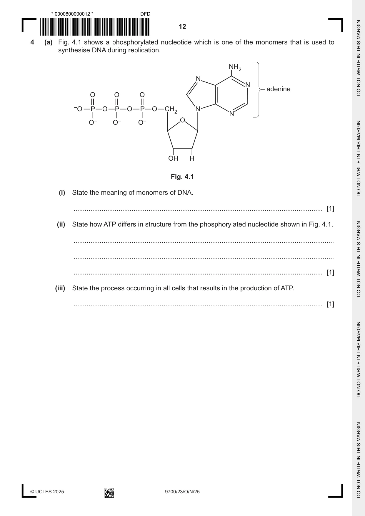
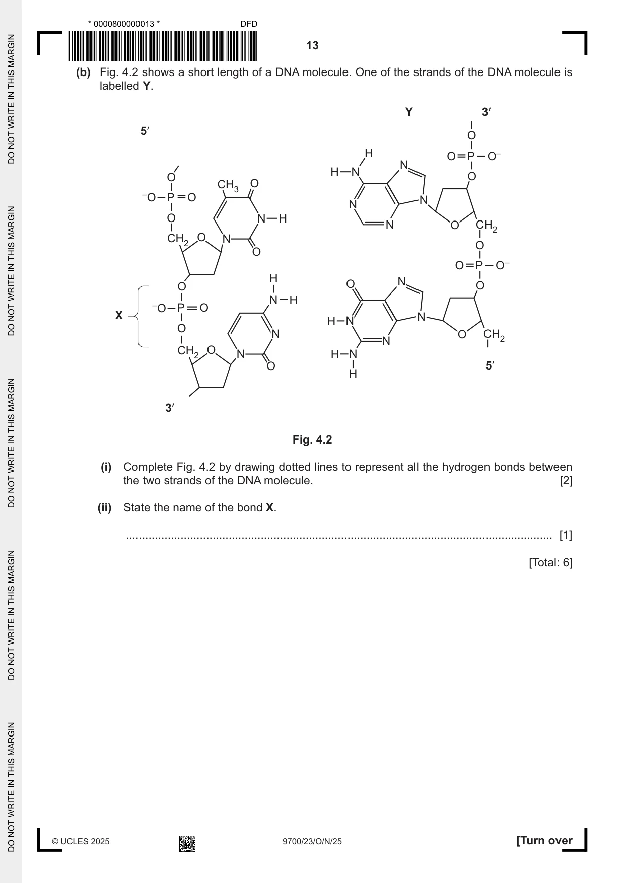
4 (a) Fig. 4.1 shows a phosphorylated nucleotide which is one of the monomers that is used to synthesise DNA during replication. NH2 N N adenine O O O –O P O P O P O CH2 N N O– O– O– O OH H Fig. 4.1 (i) State the meaning of monomers of DNA. ..................................................................................................................................... [1] (ii) State how ATP differs in structure from the phosphorylated nucleotide shown in Fig. 4.1. ........................................................................................................................................... ........................................................................................................................................... ..................................................................................................................................... [1] (iii) State the process occurring in all cells that results in the production of ATP. ..................................................................................................................................... [1] (b) Fig. 4.2 shows a short length of a DNA molecule. One of the strands of the DNA molecule is labelled Y. Y 3ʹ 5ʹ O H O P O– N H N O O CH3 O –O P O N N O N H N O CH2 CH2 O N O O O P O– H O N O O N H –O P O X N H N O N O CH2 N CH2 O N H N O 5ʹ H 3ʹ Fig. 4.2 (i) Complete Fig. 4.2 by drawing dotted lines to represent all the hydrogen bonds between the two strands of the DNA molecule. [2] (ii) State the name of the bond X. ..................................................................................................................................... [1] [Total: 6]
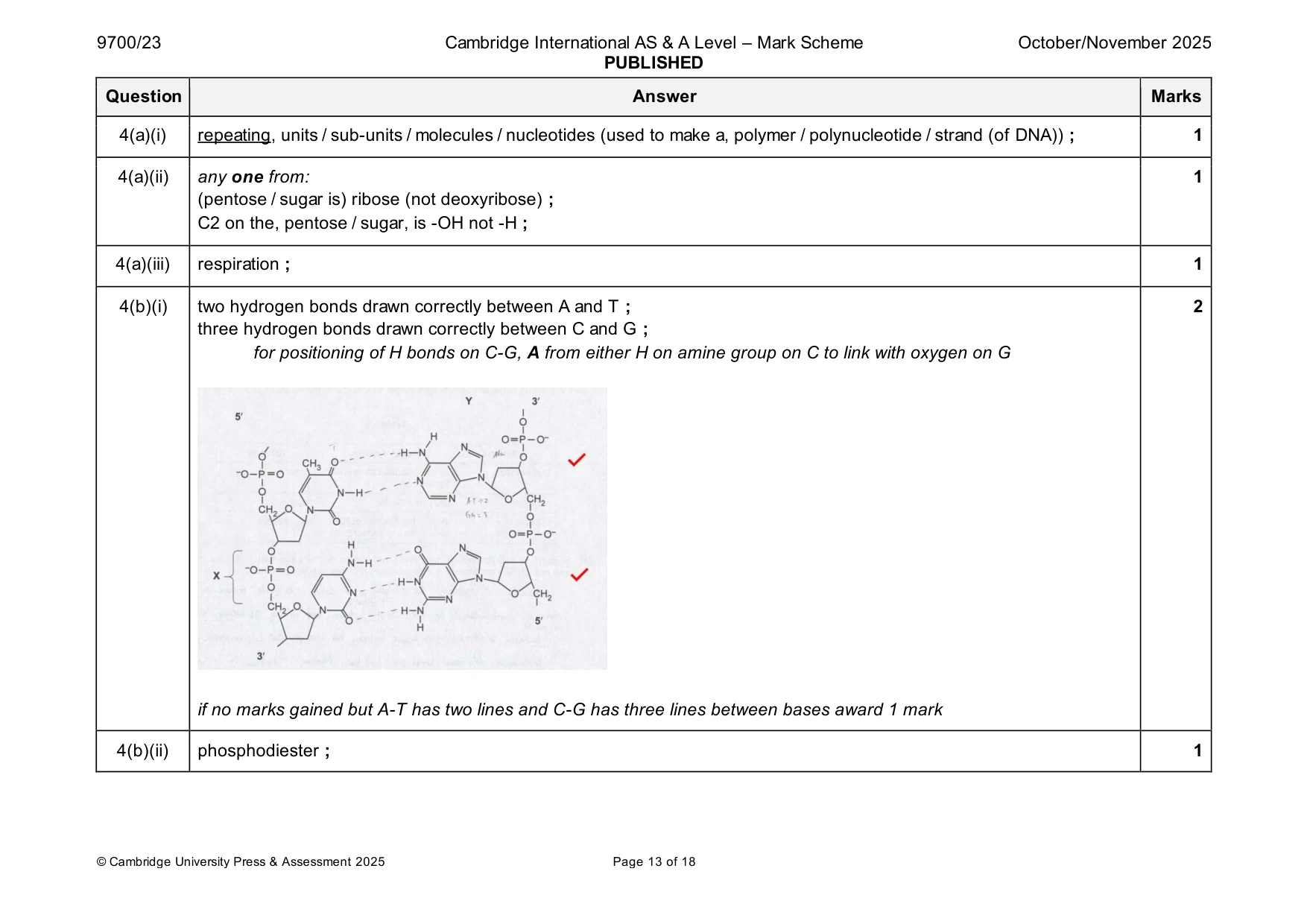
Mark scheme: 4(a)(i) repeating, units / sub-units / molecules / nucleotides (used to make a, polymer / polynucleotide / strand (of DNA)) ; 1 4(a)(ii) any one from: 1 (pentose / sugar is) ribose (not deoxyribose) ; C2 on the, pentose / sugar, is -OH not -H ; 4(a)(iii) respiration ; 1 4(b)(i) two hydrogen bonds drawn correctly between A and T ; 2 three hydrogen bonds drawn correctly between C and G ; for positioning of H bonds on C-G, A from either H on amine group on C to link with oxygen on G if no marks gained but A-T has two lines and C-G has three lines between bases award 1 mark 4(b)(ii) phosphodiester ; 1
More questions on Structure of nucleic acids and replication of DNA
Q5 · Inhibitors are substances that prevent biological processes in a variety of different ways
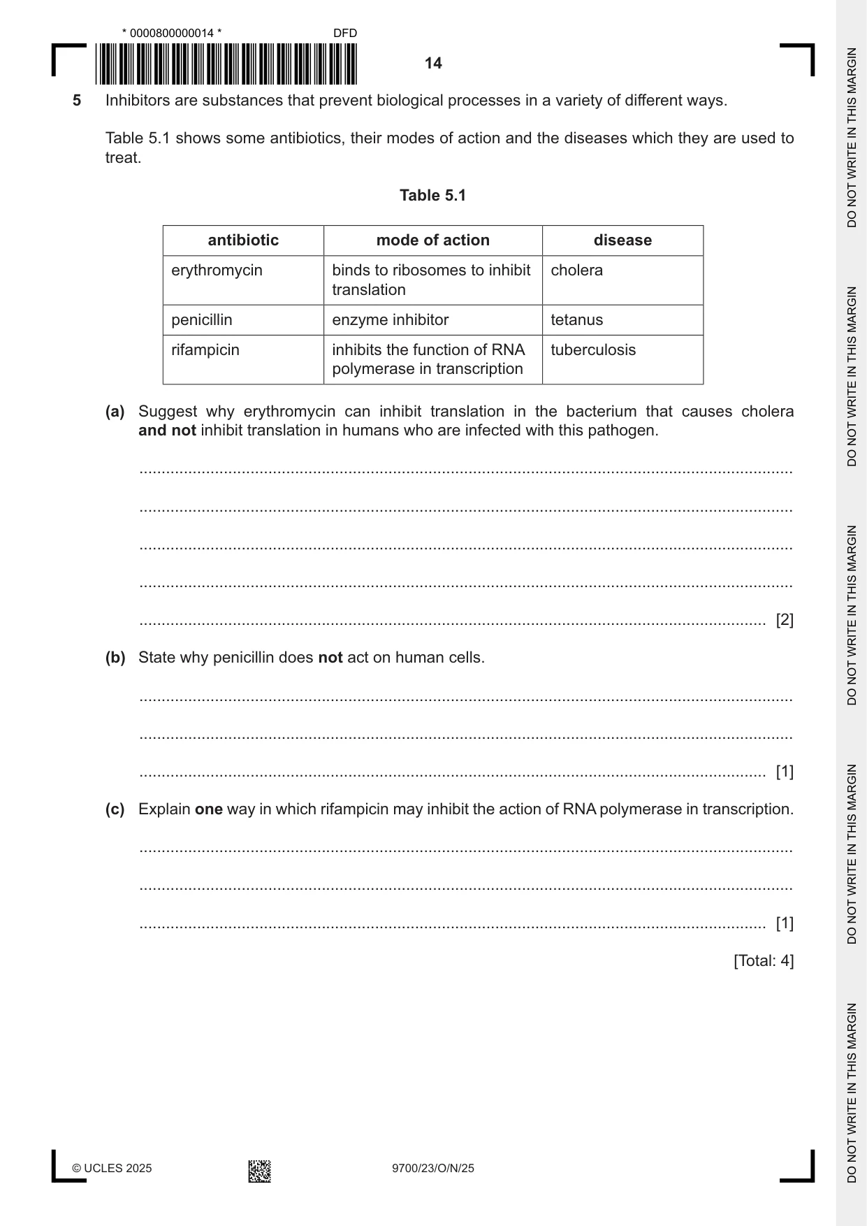
5 Inhibitors are substances that prevent biological processes in a variety of different ways. Table 5.1 shows some antibiotics, their modes of action and the diseases which they are used to treat. Table 5.1 antibiotic mode of action disease erythromycin binds to ribosomes to inhibit cholera translation penicillin enzyme inhibitor tetanus rifampicin inhibits the function of RNA tuberculosis polymerase in transcription (a) Suggest why erythromycin can inhibit translation in the bacterium that causes cholera and not inhibit translation in humans who are infected with this pathogen. ................................................................................................................................................... ................................................................................................................................................... ................................................................................................................................................... ................................................................................................................................................... ............................................................................................................................................. [2] (b) State why penicillin does not act on human cells. ................................................................................................................................................... ................................................................................................................................................... ............................................................................................................................................. [1] (c) Explain one way in which rifampicin may inhibit the action of RNA polymerase in transcription. ................................................................................................................................................... ................................................................................................................................................... ............................................................................................................................................. [1] [Total: 4]
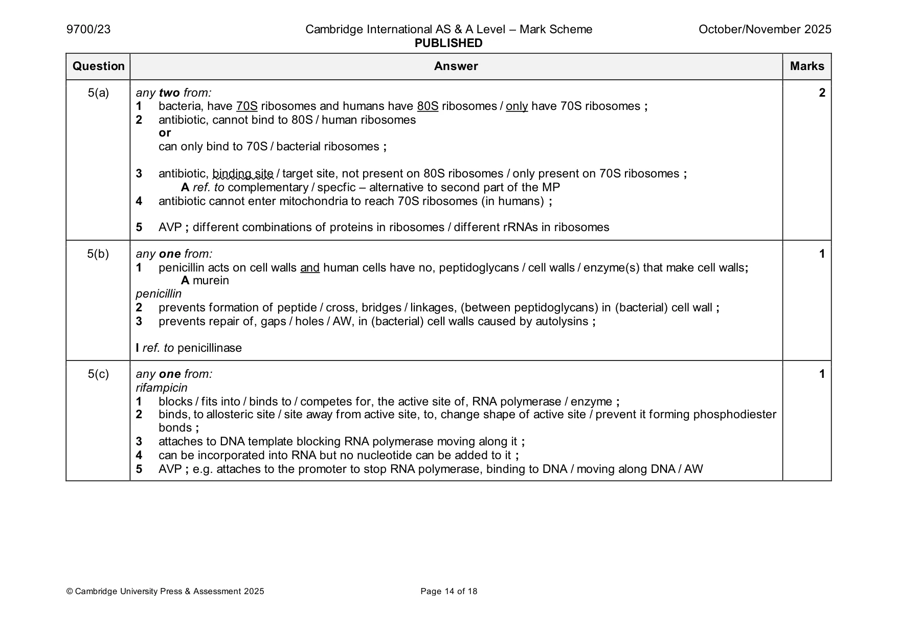
Mark scheme: 5(a) any two from: 2 1 bacteria, have 70S ribosomes and humans have 80S ribosomes / only have 70S ribosomes ; 2 antibiotic, cannot bind to 80S / human ribosomes or can only bind to 70S / bacterial ribosomes ; 3 antibiotic, binding site / target site, not present on 80S ribosomes / only present on 70S ribosomes ; A ref. to complementary / specfic – alternative to second part of the MP 4 antibiotic cannot enter mitochondria to reach 70S ribosomes (in humans) ; 5 AVP ; different combinations of proteins in ribosomes / different rRNAs in ribosomes 5(b) any one from: 1 1 penicillin acts on cell walls and human cells have no, peptidoglycans / cell walls / enzyme(s) that make cell walls; A murein penicillin 2 prevents formation of peptide / cross, bridges / linkages, (between peptidoglycans) in (bacterial) cell wall ; 3 prevents repair of, gaps / holes / AW, in (bacterial) cell walls caused by autolysins ; I ref. to penicillinase 5(c) any one from: 1 rifampicin 1 blocks / fits into / binds to / competes for, the active site of, RNA polymerase / enzyme ; 2 binds, to allosteric site / site away from active site, to, change shape of active site / prevent it forming phosphodiester bonds ; 3 attaches to DNA template blocking RNA polymerase moving along it ; 4 can be incorporated into RNA but no nucleotide can be added to it ; 5 AVP ; e.g. attaches to the promoter to stop RNA polymerase, binding to DNA / moving along DNA / AW
Q6 · A student studied the structure of a mammalian heart
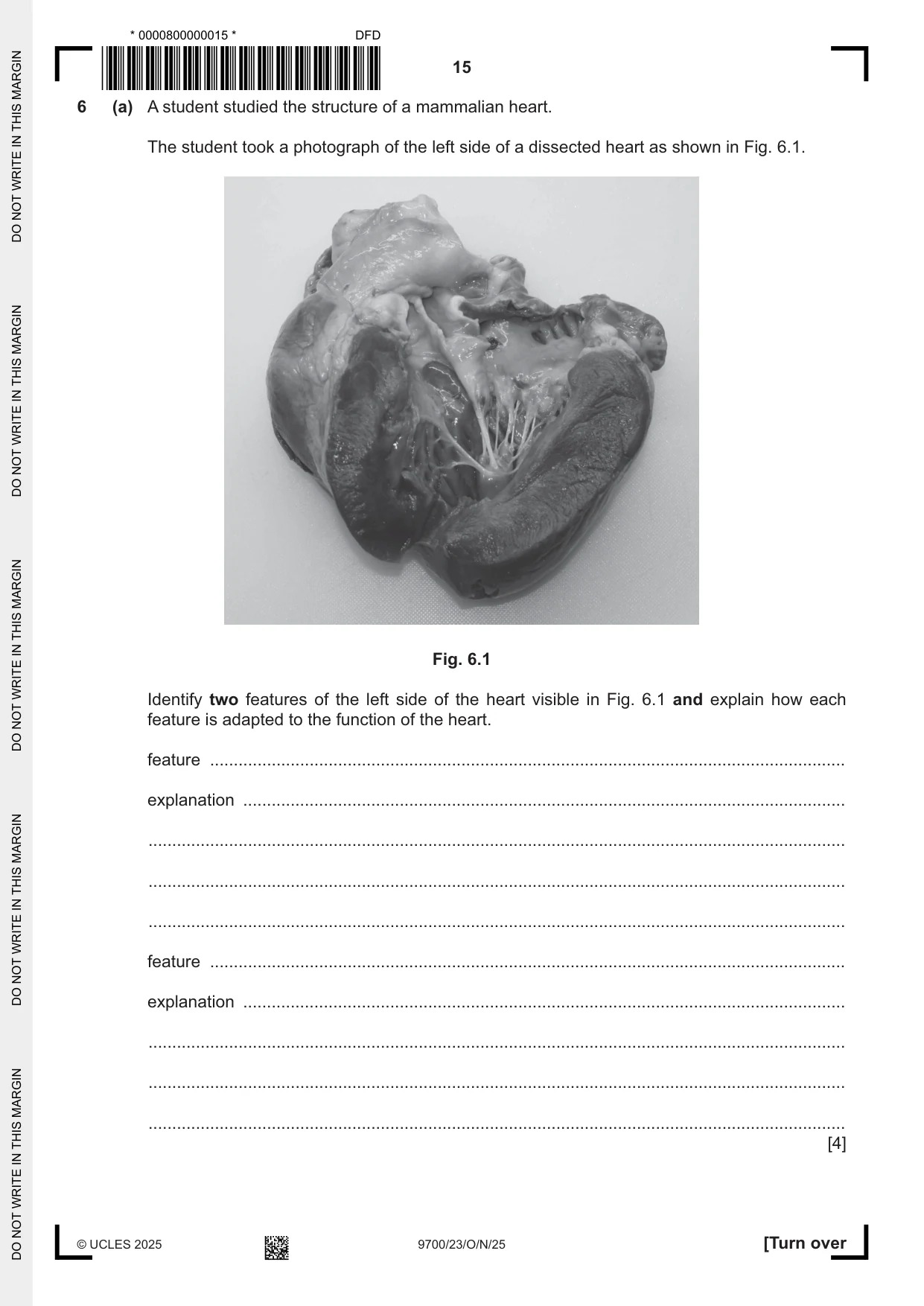
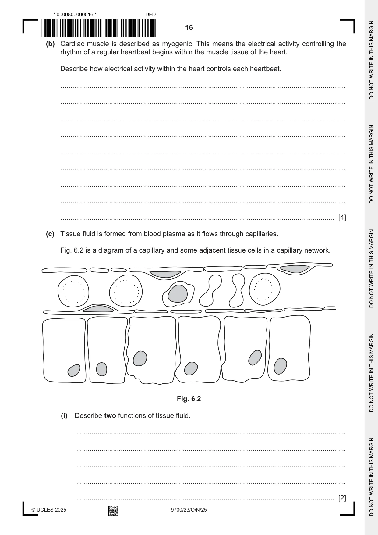
6 (a) A student studied the structure of a mammalian heart. The student took a photograph of the left side of a dissected heart as shown in Fig. 6.1. Fig. 6.1 Identify two features of the left side of the heart visible in Fig. 6.1 and explain how each feature is adapted to the function of the heart. feature ...................................................................................................................................... explanation ............................................................................................................................... ................................................................................................................................................... ................................................................................................................................................... ................................................................................................................................................... feature ...................................................................................................................................... explanation ............................................................................................................................... ................................................................................................................................................... ................................................................................................................................................... ................................................................................................................................................... [4] (b) Cardiac muscle is described as myogenic. This means the electrical activity controlling the rhythm of a regular heartbeat begins within the muscle tissue of the heart. Describe how electrical activity within the heart controls each heartbeat. ................................................................................................................................................... ................................................................................................................................................... ................................................................................................................................................... ................................................................................................................................................... ................................................................................................................................................... ................................................................................................................................................... ................................................................................................................................................... ................................................................................................................................................... ............................................................................................................................................. [4] (c) Tissue fluid is formed from blood plasma as it flows through capillaries. Fig. 6.2 is a diagram of a capillary and some adjacent tissue cells in a capillary network. Fig. 6.2 (i) Describe two functions of tissue fluid. ........................................................................................................................................... ........................................................................................................................................... ........................................................................................................................................... ........................................................................................................................................... ..................................................................................................................................... [2] (ii) With reference to Fig. 6.2, describe how tissue fluid is formed. ........................................................................................................................................... ........................................................................................................................................... ........................................................................................................................................... ........................................................................................................................................... ........................................................................................................................................... ........................................................................................................................................... ..................................................................................................................................... [3] (d) Measles is a common disease caused by a virus. Vaccination to prevent the disease has been very successful. (i) A child receives a vaccine for measles. Explain why only some of the T-lymphocytes in the child respond to the measles vaccine. ........................................................................................................................................... ........................................................................................................................................... ........................................................................................................................................... ........................................................................................................................................... ..................................................................................................................................... [2] (ii) Describe the events that occur in an immune response to a vaccine that result in lymphocytes that provide long-term immunity to measles. ........................................................................................................................................... ........................................................................................................................................... ........................................................................................................................................... ........................................................................................................................................... ........................................................................................................................................... ........................................................................................................................................... ........................................................................................................................................... ........................................................................................................................................... ........................................................................................................................................... ........................................................................................................................................... ..................................................................................................................................... [5]
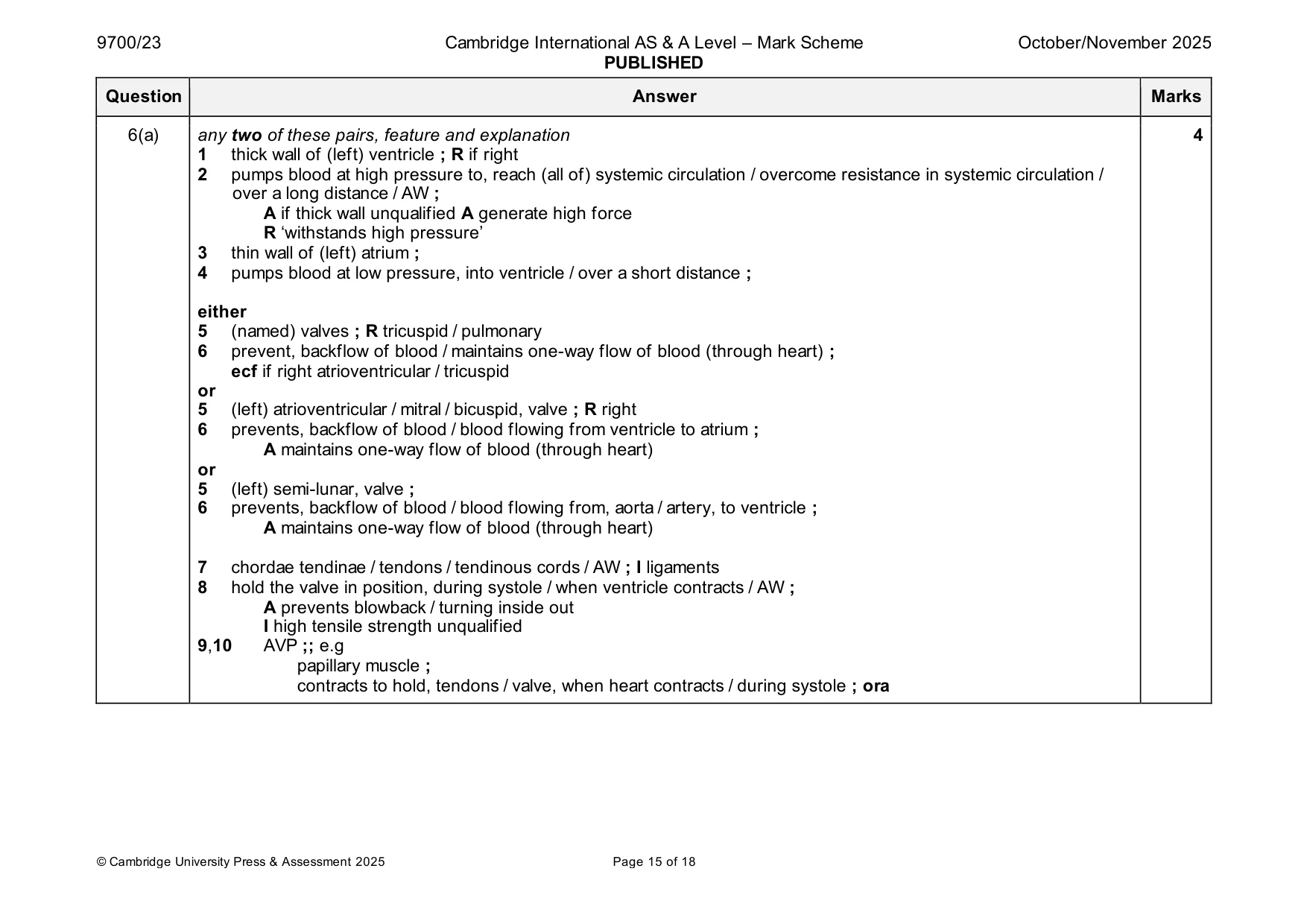
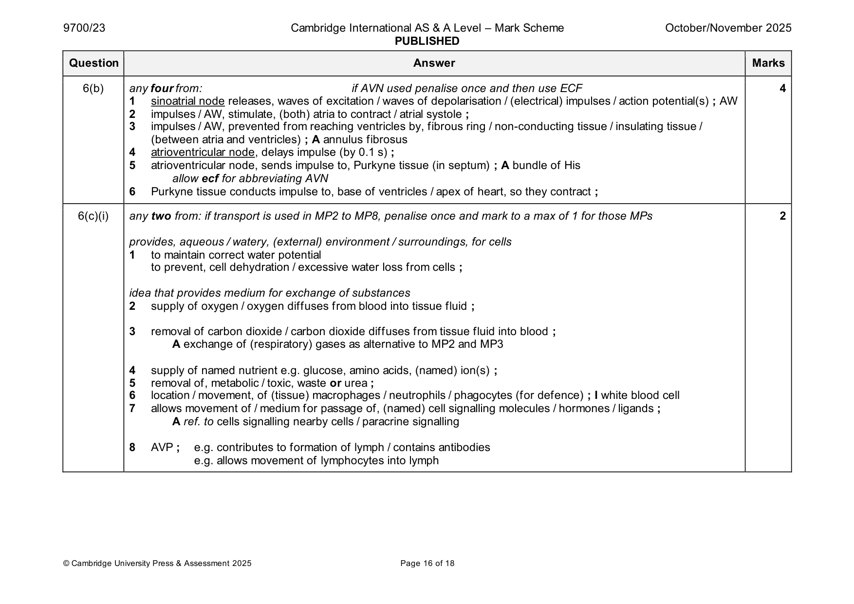
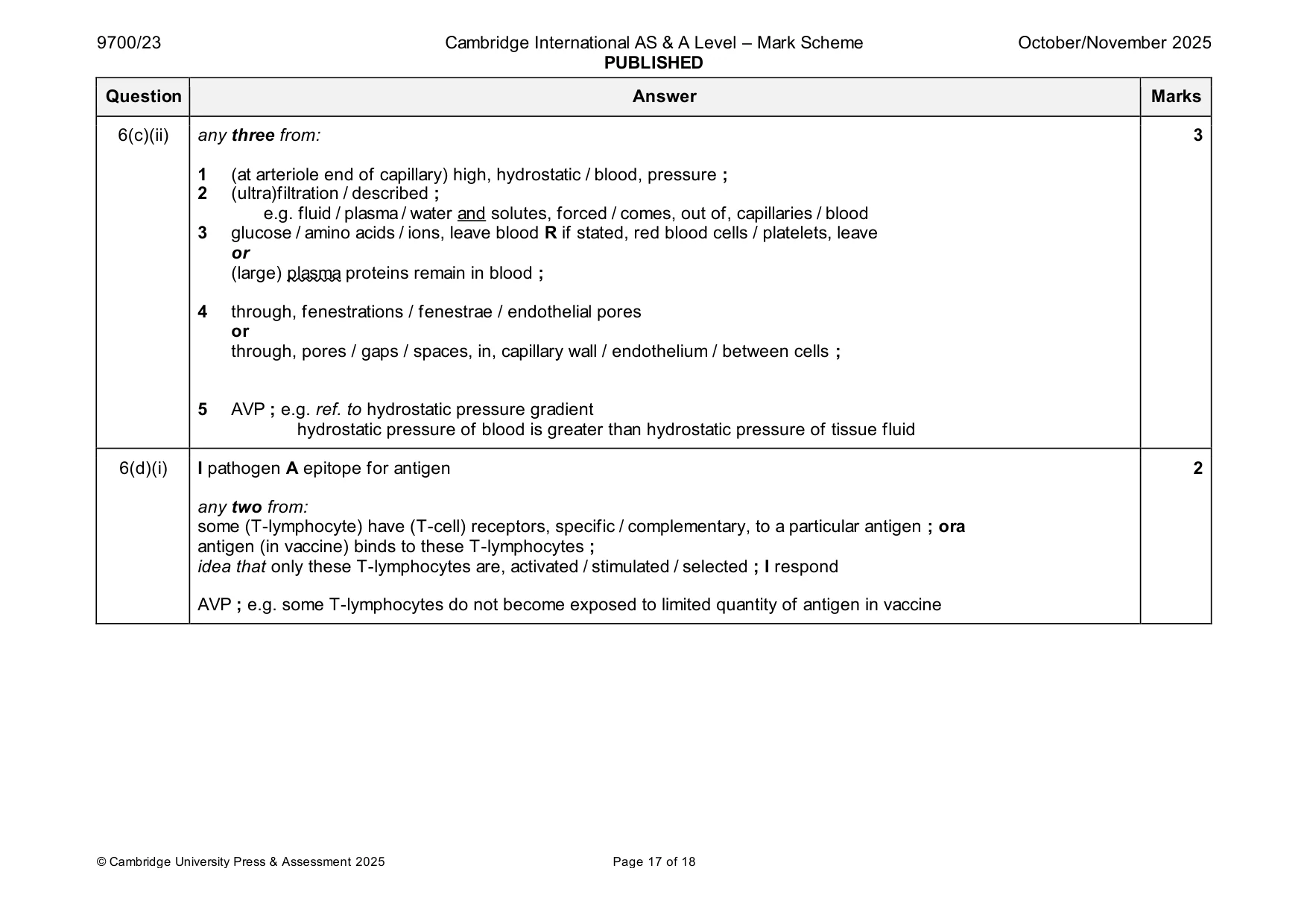
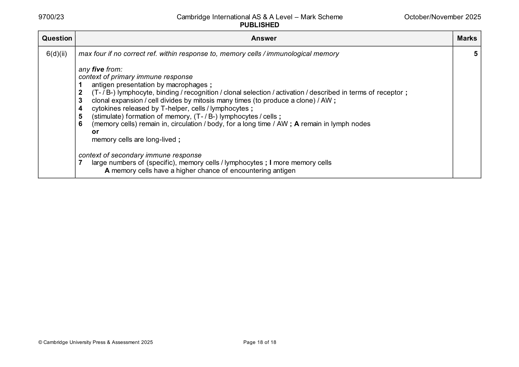
Mark scheme: 6(a) any two of these pairs, feature and explanation 4 1 thick wall of (left) ventricle ; R if right 2 pumps blood at high pressure to, reach (all of) systemic circulation / overcome resistance in systemic circulation / over a long distance / AW ; A if thick wall unqualified A generate high force R ‘withstands high pressure’ 3 thin wall of (left) atrium ; 4 pumps blood at low pressure, into ventricle / over a short distance ; either 5 (named) valves ; R tricuspid / pulmonary 6 prevent, backflow of blood / maintains one-way flow of blood (through heart) ; ecf if right atrioventricular / tricuspid or 5 (left) atrioventricular / mitral / bicuspid, valve ; R right 6 prevents, backflow of blood / blood flowing from ventricle to atrium ; A maintains one-way flow of blood (through heart) or 5 (left) semi-lunar, valve ; 6 prevents, backflow of blood / blood flowing from, aorta / artery, to ventricle ; A maintains one-way flow of blood (through heart) 7 chordae tendinae / tendons / tendinous cords / AW ; I ligaments 8 hold the valve in position, during systole / when ventricle contracts / AW ; A prevents blowback / turning inside out I high tensile strength unqualified 9,10 AVP ;; e.g papillary muscle ; contracts to hold, tendons / valve, when heart contracts / during systole ; ora 6(b) any four from: if AVN used penalise once and then use ECF 4 1 sinoatrial node releases, waves of excitation / waves of depolarisation / (electrical) impulses / action potential(s) ; AW 2 impulses / AW, stimulate, (both) atria to contract / atrial systole ; 3 impulses / AW, prevented from reaching ventricles by, fibrous ring / non-conducting tissue / insulating tissue / (between atria and ventricles) ; A annulus fibrosus 4 atrioventricular node, delays impulse (by 0.1 s) ; 5 atrioventricular node, sends impulse to, Purkyne tissue (in septum) ; A bundle of His allow ecf for abbreviating AVN 6 Purkyne tissue conducts impulse to, base of ventricles / apex of heart, so they contract ; 6(c)(i) any two from: if transport is used in MP2 to MP8, penalise once and mark to a max of 1 for those MPs 2 provides, aqueous / watery, (external) environment / surroundings, for cells 1 to maintain correct water potential to prevent, cell dehydration / excessive water loss from cells ; idea that provides medium for exchange of substances 2 supply of oxygen / oxygen diffuses from blood into tissue fluid ; 3 removal of carbon dioxide / carbon dioxide diffuses from tissue fluid into blood ; A exchange of (respiratory) gases as alternative to MP2 and MP3 4 supply of named nutrient e.g. glucose, amino acids, (named) ion(s) ; 5 removal of, metabolic / toxic, waste or urea ; 6 location / movement, of (tissue) macrophages / neutrophils / phagocytes (for defence) ; I white blood cell 7 allows movement of / medium for passage of, (named) cell signalling molecules / hormones / ligands ; A ref. to cells signalling nearby cells / paracrine signalling 8 AVP ; e.g. contributes to formation of lymph / contains antibodies e.g. allows movement of lymphocytes into lymph 6(c)(ii) any three from: 3 1 (at arteriole end of capillary) high, hydrostatic / blood, pressure ; 2 (ultra)filtration / described ; e.g. fluid / plasma / water and solutes, forced / comes, out of, capillaries / blood 3 glucose / amino acids / ions, leave blood R if stated, red blood cells / platelets, leave or (large) plasma proteins remain in blood ; 4 through, fenestrations / fenestrae / endothelial pores or through, pores / gaps / spaces, in, capillary wall / endothelium / between cells ; 5 AVP ; e.g. ref. to hydrostatic pressure gradient hydrostatic pressure of blood is greater than hydrostatic pressure of tissue fluid 6(d)(i) I pathogen A epitope for antigen 2 any two from: some (T-lymphocyte) have (T-cell) receptors, specific / complementary, to a particular antigen ; ora antigen (in vaccine) binds to these T-lymphocytes ; idea that only these T-lymphocytes are, activated / stimulated / selected ; I respond AVP ; e.g. some T-lymphocytes do not become exposed to limited quantity of antigen in vaccine 6(d)(ii) max four if no correct ref. within response to, memory cells / immunological memory 5 any five from: context of primary immune response 1 antigen presentation by macrophages ; 2 (T- / B-) lymphocyte, binding / recognition / clonal selection / activation / described in terms of receptor ; 3 clonal expansion / cell divides by mitosis many times (to produce a clone) / AW ; 4 cytokines released by T-helper, cells / lymphocytes ; 5 (stimulate) formation of memory, (T- / B-) lymphocytes / cells ; 6 (memory cells) remain in, circulation / body, for a long time / AW ; A remain in lymph nodes or memory cells are long-lived ; context of secondary immune response 7 large numbers of (specific), memory cells / lymphocytes ; I more memory cells A memory cells have a higher chance of encountering antigen
What was in this paper
The subtopics covered by these 6 questions, and how many questions each got. Open one in a new tab to see every Cambridge question on it.
What you needed in this session
Cambridge’s own grade thresholds for 2025 Oct/Nov, Paper 2 · Variant 3. A higher threshold means an easier paper — the bar moves with how the cohort did.