Cambridge A Level Biology 9700 — 2025 May/June Paper 2 · Variant 3
9700/23/M/J/25 · 6 questions · 60 marks · 75 min
The question paper and its mark scheme, free to read here and free to download. This is Cambridge’s own paper, exactly as it was sat.
Question paper16 pages
Mark scheme17 pages
Answers below. Sit the paper first if you are practising.
Questions as text
Q1 · Ranunculus is a group (genus) of dicotyledonous plants that includes more than 1600…
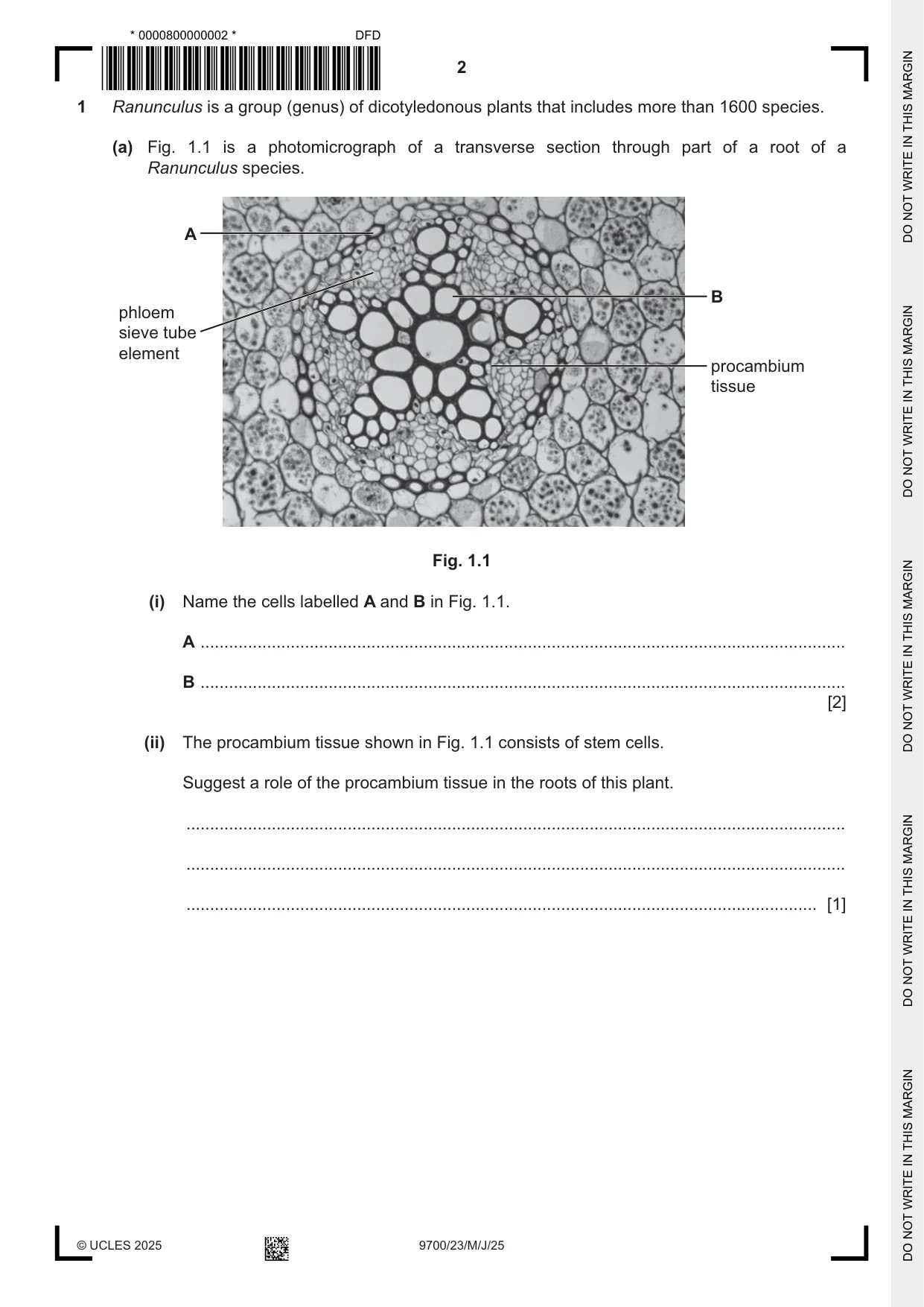
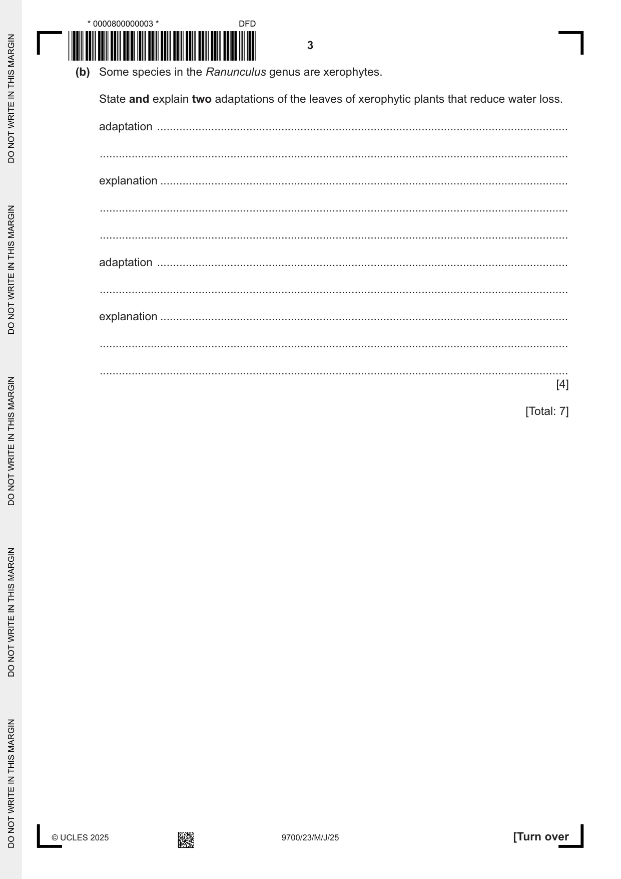
1 Ranunculus is a group (genus) of dicotyledonous plants that includes more than 1600 species. (a) Fig. 1.1 is a photomicrograph of a transverse section through part of a root of a Ranunculus species. A B phloem sieve tube element procambium tissue Fig. 1.1 (i) Name the cells labelled A and B in Fig. 1.1. A ........................................................................................................................................ B ........................................................................................................................................ [2] (ii) The procambium tissue shown in Fig. 1.1 consists of stem cells. Suggest a role of the procambium tissue in the roots of this plant. ........................................................................................................................................... ........................................................................................................................................... ..................................................................................................................................... [1] (b) Some species in the Ranunculus genus are xerophytes. State and explain two adaptations of the leaves of xerophytic plants that reduce water loss. adaptation ................................................................................................................................. ................................................................................................................................................... explanation ................................................................................................................................ ................................................................................................................................................... ................................................................................................................................................... adaptation ................................................................................................................................. ................................................................................................................................................... explanation ................................................................................................................................ ................................................................................................................................................... ................................................................................................................................................... [4] [Total: 7]
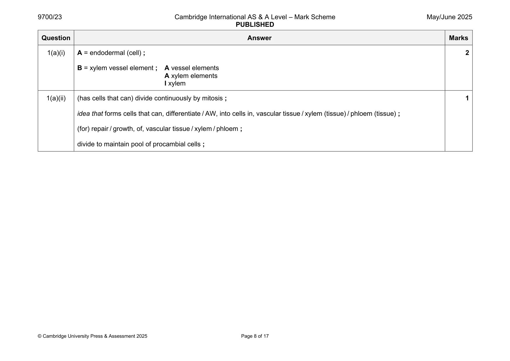
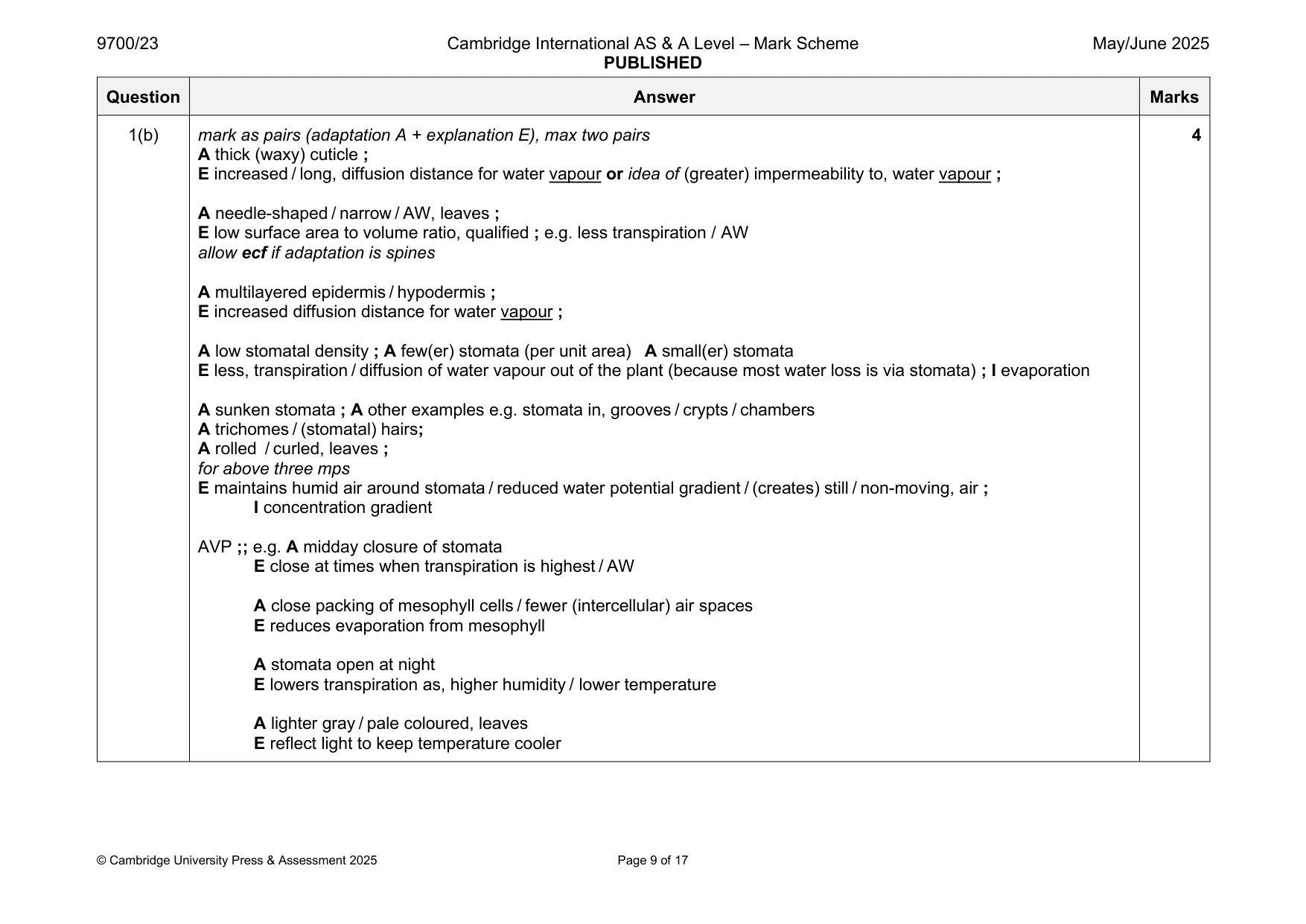
Mark scheme: Question Answer Marks 1(a)(i) A = endodermal (cell) ; 2 B = xylem vessel element ; A vessel elements A xylem elements I xylem 1(a)(ii) (has cells that can) divide continuously by mitosis ; 1 idea that forms cells that can, differentiate / AW, into cells in, vascular tissue / xylem (tissue) / phloem (tissue) ; (for) repair / growth, of, vascular tissue / xylem / phloem ; divide to maintain pool of procambial cells ; 1(b) mark as pairs (adaptation A + explanation E), max two pairs 4 A thick (waxy) cuticle ; E increased / long, diffusion distance for water vapour or idea of (greater) impermeability to, water vapour ; A needle-shaped / narrow / AW, leaves ; E low surface area to volume ratio, qualified ; e.g. less transpiration / AW allow ecf if adaptation is spines A multilayered epidermis / hypodermis ; E increased diffusion distance for water vapour ; A low stomatal density ; A few(er) stomata (per unit area) A small(er) stomata E less, transpiration / diffusion of water vapour out of the plant (because most water loss is via stomata) ; I evaporation A sunken stomata ; A other examples e.g. stomata in, grooves / crypts / chambers A trichomes / (stomatal) hairs; A rolled / curled, leaves ; for above three mps E maintains humid air around stomata / reduced water potential gradient / (creates) still / non-moving, air ; I concentration gradient AVP ;; e.g. A midday closure of stomata E close at times when transpiration is highest / AW A close packing of mesophyll cells / fewer (intercellular) air spaces E reduces evaporation from mesophyll A stomata open at night E lowers transpiration as, higher humidity / lower temperature A lighter gray / pale coloured, leaves E reflect light to keep temperature cooler
Q2 · Bees are insects that produce venom as a means of self‑defence
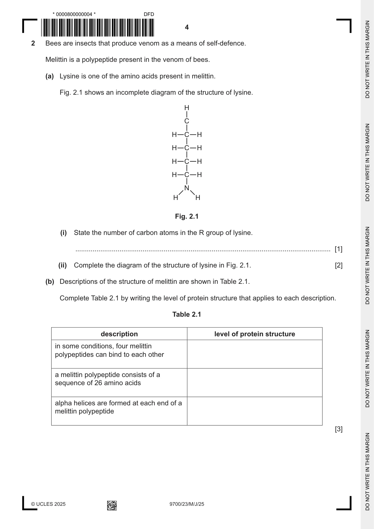
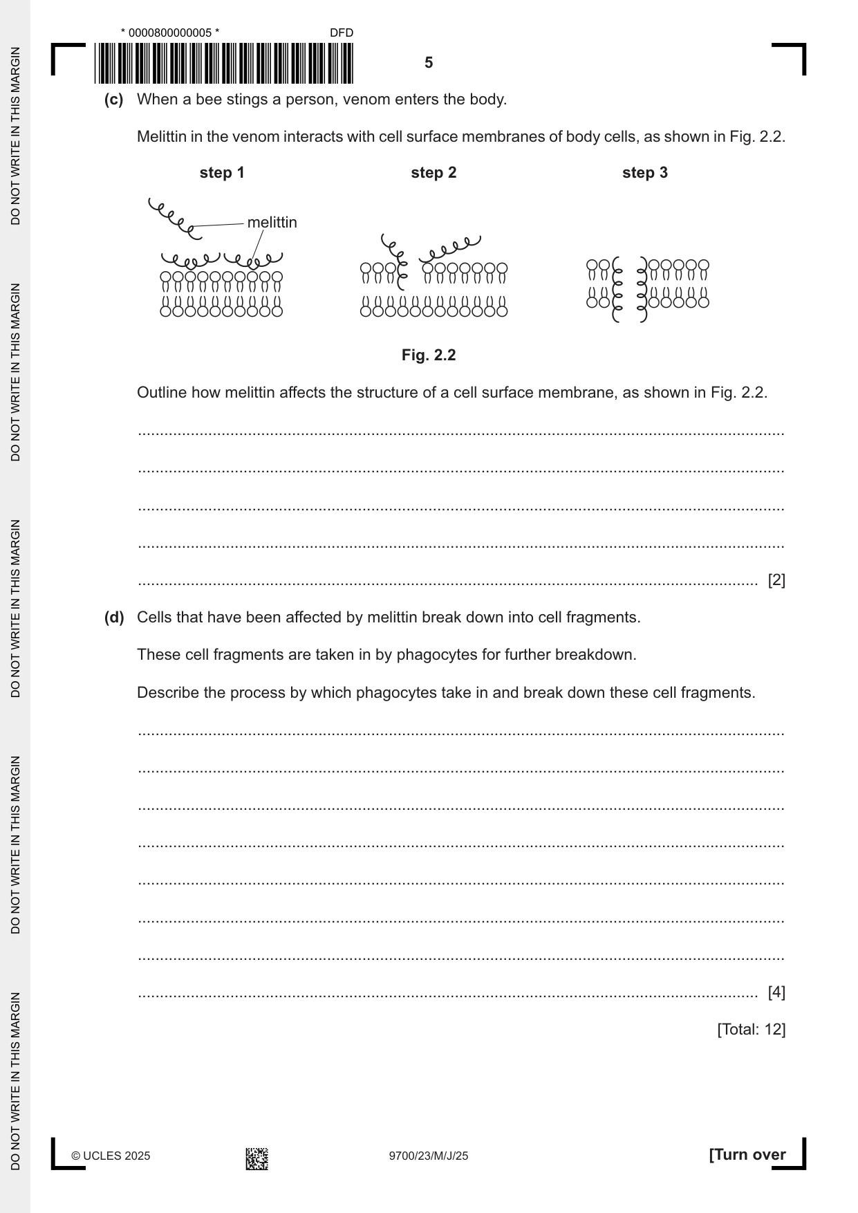
2 Bees are insects that produce venom as a means of self‑defence. Melittin is a polypeptide present in the venom of bees. (a) Lysine is one of the amino acids present in melittin. Fig. 2.1 shows an incomplete diagram of the structure of lysine. H C H C H H C H H C H H C H N H H Fig. 2.1 (i) State the number of carbon atoms in the R group of lysine. ..................................................................................................................................... [1] (ii) Complete the diagram of the structure of lysine in Fig. 2.1. [2] (b) Descriptions of the structure of melittin are shown in Table 2.1. Complete Table 2.1 by writing the level of protein structure that applies to each description. Table 2.1 description level of protein structure in some conditions, four melittin polypeptides can bind to each other a melittin polypeptide consists of a sequence of 26 amino acids alpha helices are formed at each end of a melittin polypeptide [3] (c) When a bee stings a person, venom enters the body. Melittin in the venom interacts with cell surface membranes of body cells, as shown in Fig. 2.2. step 1 step 2 step 3 melittin Fig. 2.2 Outline how melittin affects the structure of a cell surface membrane, as shown in Fig. 2.2. ................................................................................................................................................... ................................................................................................................................................... ................................................................................................................................................... ................................................................................................................................................... ............................................................................................................................................. [2] (d) Cells that have been affected by melittin break down into cell fragments. These cell fragments are taken in by phagocytes for further breakdown. Describe the process by which phagocytes take in and break down these cell fragments. ................................................................................................................................................... ................................................................................................................................................... ................................................................................................................................................... ................................................................................................................................................... ................................................................................................................................................... ................................................................................................................................................... ................................................................................................................................................... ............................................................................................................................................. [4] [Total: 12]
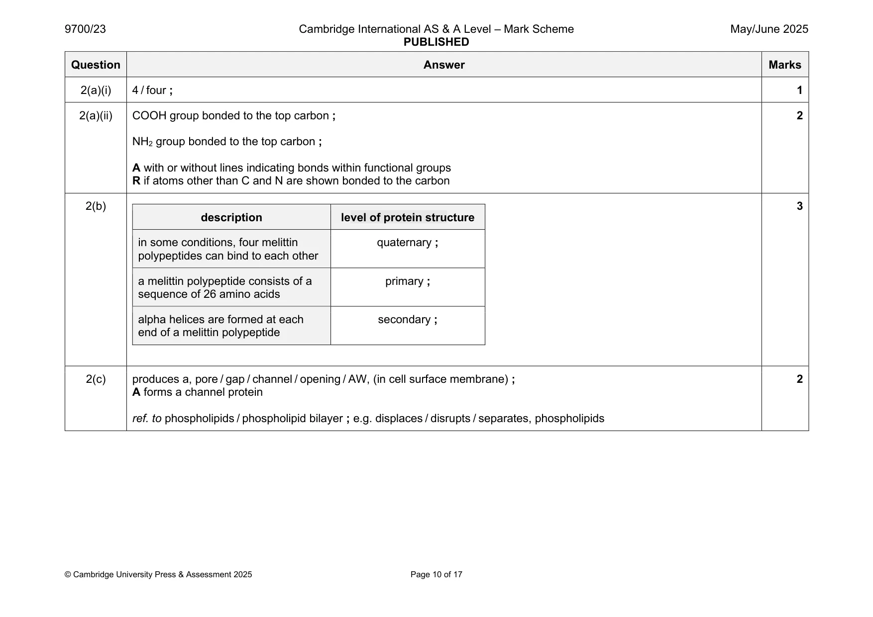
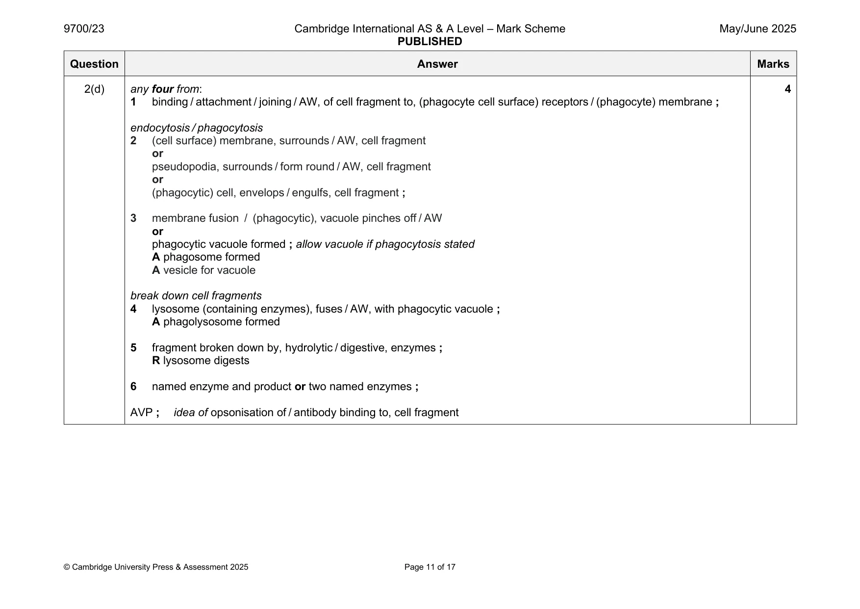
Mark scheme: 2(a)(i) 4 / four ; 1 2(a)(ii) COOH group bonded to the top carbon ; 2 NH2 group bonded to the top carbon ; A with or without lines indicating bonds within functional groups R if atoms other than C and N are shown bonded to the carbon 2(b) 3 description level of protein structure in some conditions, four melittin quaternary ; polypeptides can bind to each other a melittin polypeptide consists of a primary ; sequence of 26 amino acids alpha helices are formed at each secondary ; end of a melittin polypeptide 2(c) produces a, pore / gap / channel / opening / AW, (in cell surface membrane) ; 2 A forms a channel protein ref. to phospholipids / phospholipid bilayer ; e.g. displaces / disrupts / separates, phospholipids 2(d) any four from: 4 1 binding / attachment / joining / AW, of cell fragment to, (phagocyte cell surface) receptors / (phagocyte) membrane ; endocytosis / phagocytosis 2 (cell surface) membrane, surrounds / AW, cell fragment or pseudopodia, surrounds / form round / AW, cell fragment or (phagocytic) cell, envelops / engulfs, cell fragment ; 3 membrane fusion / (phagocytic), vacuole pinches off / AW or phagocytic vacuole formed ; allow vacuole if phagocytosis stated A phagosome formed A vesicle for vacuole break down cell fragments 4 lysosome (containing enzymes), fuses / AW, with phagocytic vacuole ; A phagolysosome formed 5 fragment broken down by, hydrolytic / digestive, enzymes ; R lysosome digests 6 named enzyme and product or two named enzymes ; AVP ; idea of opsonisation of / antibody binding to, cell fragment
Q3 · Staphylococcus epidermidis is a species of bacterium that lives on human skin
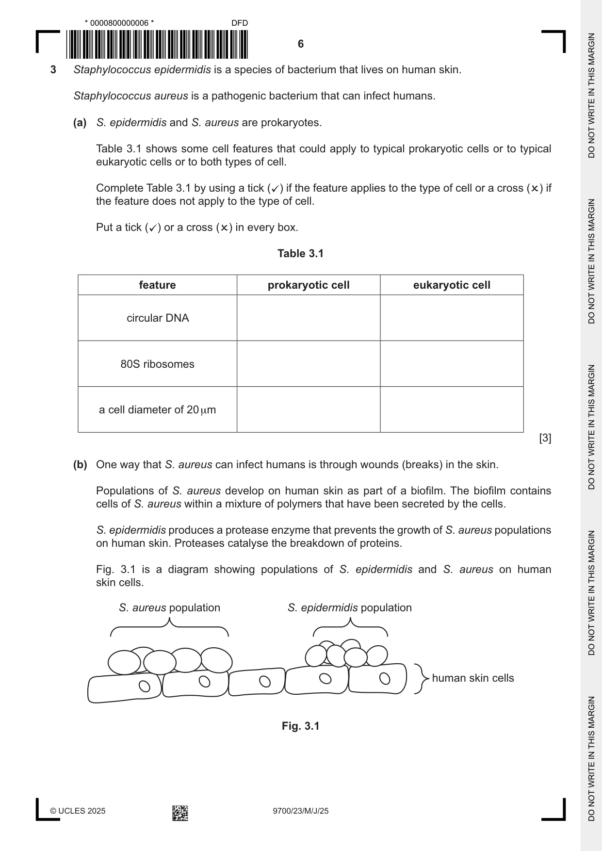
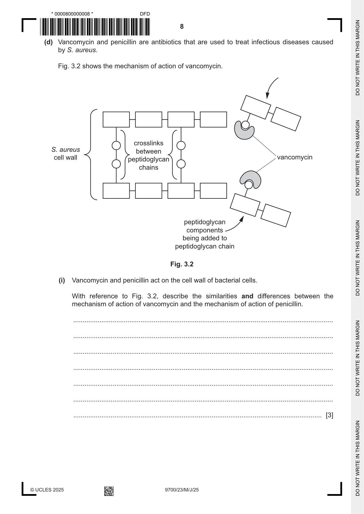
3 Staphylococcus epidermidis is a species of bacterium that lives on human skin. Staphylococcus aureus is a pathogenic bacterium that can infect humans. (a) S. epidermidis and S. aureus are prokaryotes. Table 3.1 shows some cell features that could apply to typical prokaryotic cells or to typical eukaryotic cells or to both types of cell. Complete Table 3.1 by using a tick () if the feature applies to the type of cell or a cross () if the feature does not apply to the type of cell. Put a tick () or a cross () in every box. Table 3.1 feature prokaryotic cell eukaryotic cell circular DNA 80S ribosomes a cell diameter of 20 µm [3] (b) One way that S. aureus can infect humans is through wounds (breaks) in the skin. Populations of S. aureus develop on human skin as part of a biofilm. The biofilm contains cells of S. aureus within a mixture of polymers that have been secreted by the cells. S. epidermidis produces a protease enzyme that prevents the growth of S. aureus populations on human skin. Proteases catalyse the breakdown of proteins. Fig. 3.1 is a diagram showing populations of S. epidermidis and S. aureus on human skin cells. S. aureus population S. epidermidis population human skin cells Fig. 3.1 Suggest and explain how the protease produced by S. epidermidis cells prevents the growth of an S. aureus population on human skin. ................................................................................................................................................... ................................................................................................................................................... ................................................................................................................................................... ................................................................................................................................................... ................................................................................................................................................... ................................................................................................................................................... ............................................................................................................................................. [3] (c) S. aureus can infect many tissues in the human body, including tissues in the gas exchange system. (i) Describe the role of goblet cells in the protection of tissues in the trachea from infection by S. aureus. ........................................................................................................................................... ........................................................................................................................................... ........................................................................................................................................... ..................................................................................................................................... [2] (ii) S. aureus cells can infect tissues by passing in between cells that line the lumen of the trachea. State the name of a cell type, other than goblet cells, that lines the lumen of the trachea. ..................................................................................................................................... [1] (d) Vancomycin and penicillin are antibiotics that are used to treat infectious diseases caused by S. aureus. Fig. 3.2 shows the mechanism of action of vancomycin. crosslinks S. aureus between cell wall peptidoglycan vancomycin chains peptidoglycan components being added to peptidoglycan chain Fig. 3.2 (i) Vancomycin and penicillin act on the cell wall of bacterial cells. With reference to Fig. 3.2, describe the similarities and differences between the mechanism of action of vancomycin and the mechanism of action of penicillin. ........................................................................................................................................... ........................................................................................................................................... ........................................................................................................................................... ........................................................................................................................................... ........................................................................................................................................... ........................................................................................................................................... ..................................................................................................................................... [3] (ii) Some strains of S. aureus are resistant to vancomycin and penicillin. Describe the steps that can be taken to reduce the impact of antibiotic resistance. ........................................................................................................................................... ........................................................................................................................................... ........................................................................................................................................... ........................................................................................................................................... ........................................................................................................................................... ........................................................................................................................................... ........................................................................................................................................... ........................................................................................................................................... ..................................................................................................................................... [3] [Total: 15]
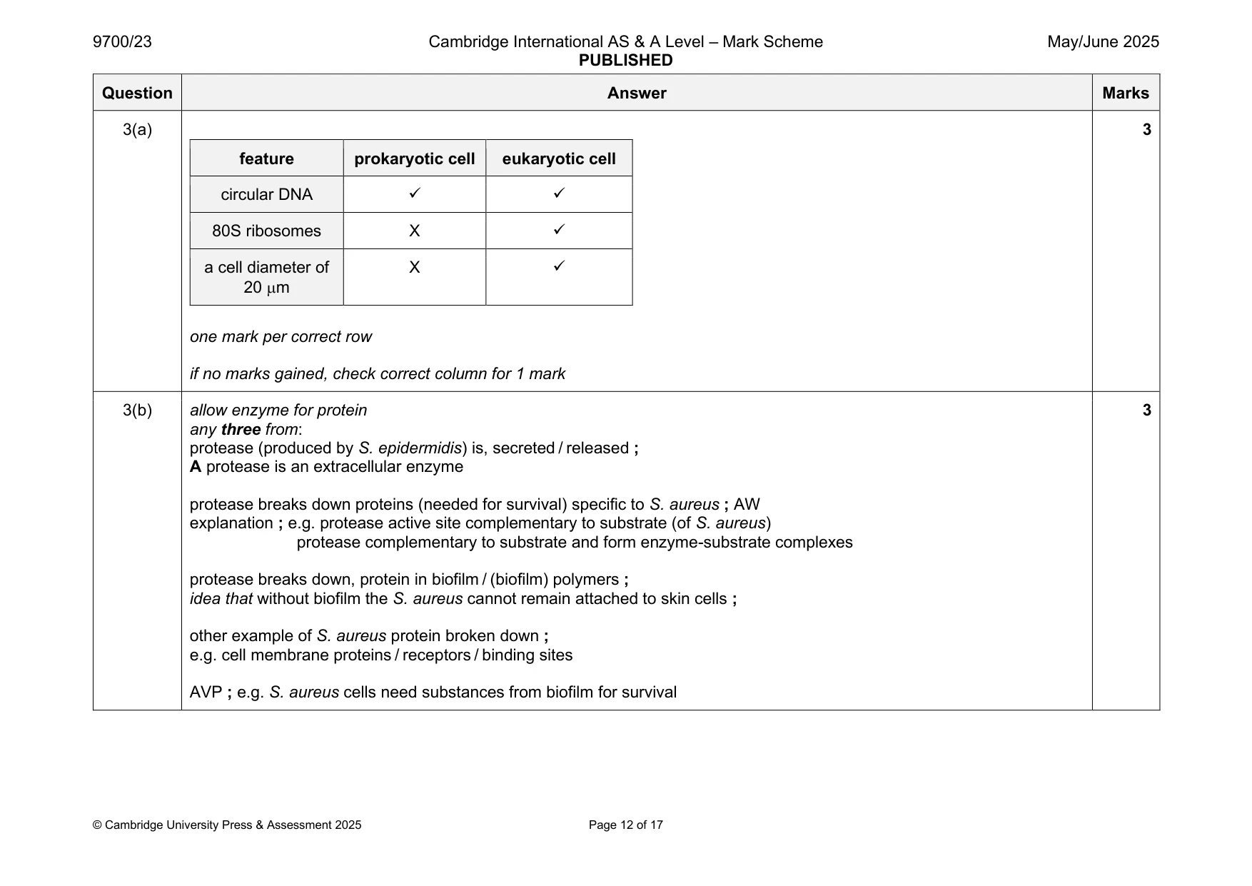
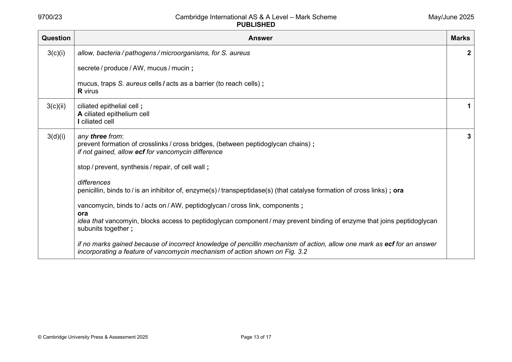
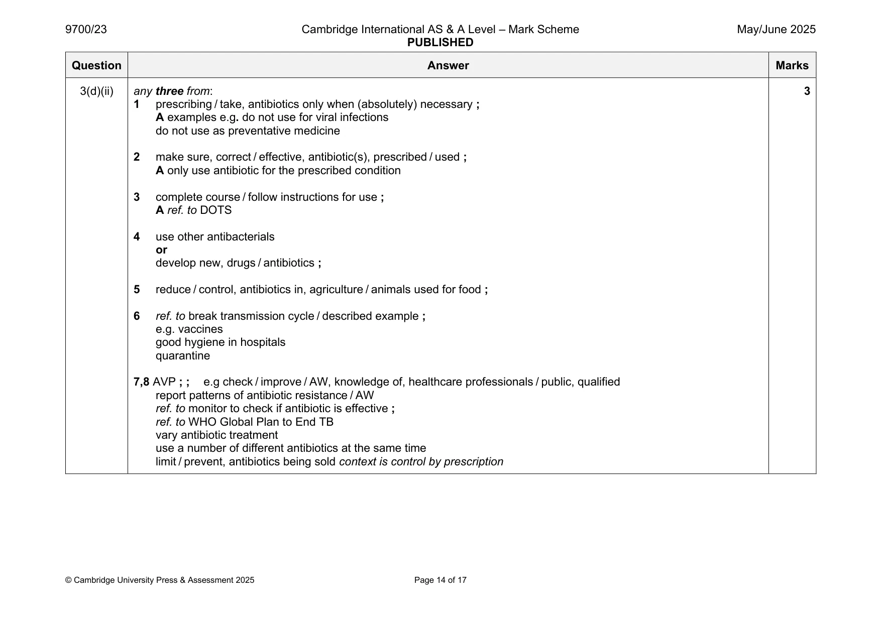
Mark scheme: 3(a) 3 feature prokaryotic cell eukaryotic cell circular DNA ✓ ✓ 80S ribosomes X ✓ a cell diameter of X ✓ 20 m one mark per correct row if no marks gained, check correct column for 1 mark 3(b) allow enzyme for protein 3 any three from: protease (produced by S. epidermidis) is, secreted / released ; A protease is an extracellular enzyme protease breaks down proteins (needed for survival) specific to S. aureus ; AW explanation ; e.g. protease active site complementary to substrate (of S. aureus) protease complementary to substrate and form enzyme-substrate complexes protease breaks down, protein in biofilm / (biofilm) polymers ; idea that without biofilm the S. aureus cannot remain attached to skin cells ; other example of S. aureus protein broken down ; e.g. cell membrane proteins / receptors / binding sites AVP ; e.g. S. aureus cells need substances from biofilm for survival 3(c)(i) allow, bacteria / pathogens / microorganisms, for S. aureus 2 secrete / produce / AW, mucus / mucin ; mucus, traps S. aureus cells / acts as a barrier (to reach cells) ; R virus 3(c)(ii) ciliated epithelial cell ; 1 A ciliated epithelium cell I ciliated cell 3(d)(i) any three from: 3 prevent formation of crosslinks / cross bridges, (between peptidoglycan chains) ; if not gained, allow ecf for vancomycin difference stop / prevent, synthesis / repair, of cell wall ; differences penicillin, binds to / is an inhibitor of, enzyme(s) / transpeptidase(s) (that catalyse formation of cross links) ; ora vancomycin, binds to / acts on / AW, peptidoglycan / cross link, components ; ora idea that vancomyin, blocks access to peptidoglycan component / may prevent binding of enzyme that joins peptidoglycan subunits together ; if no marks gained because of incorrect knowledge of pencillin mechanism of action, allow one mark as ecf for an answer incorporating a feature of vancomycin mechanism of action shown on Fig. 3.2 3(d)(ii) any three from: 3 1 prescribing / take, antibiotics only when (absolutely) necessary ; A examples e.g. do not use for viral infections do not use as preventative medicine 2 make sure, correct / effective, antibiotic(s), prescribed / used ; A only use antibiotic for the prescribed condition 3 complete course / follow instructions for use ; A ref. to DOTS 4 use other antibacterials or develop new, drugs / antibiotics ; 5 reduce / control, antibiotics in, agriculture / animals used for food ; 6 ref. to break transmission cycle / described example ; e.g. vaccines good hygiene in hospitals quarantine 7,8 AVP ; ; e.g check / improve / AW, knowledge of, healthcare professionals / public, qualified report patterns of antibiotic resistance / AW ref. to monitor to check if antibiotic is effective ; ref. to WHO Global Plan to End TB vary antibiotic treatment use a number of different antibiotics at the same time limit / prevent, antibiotics being sold context is control by prescription
Q4 · Lysosomes are membrane‑bound organelles found in mammalian cells
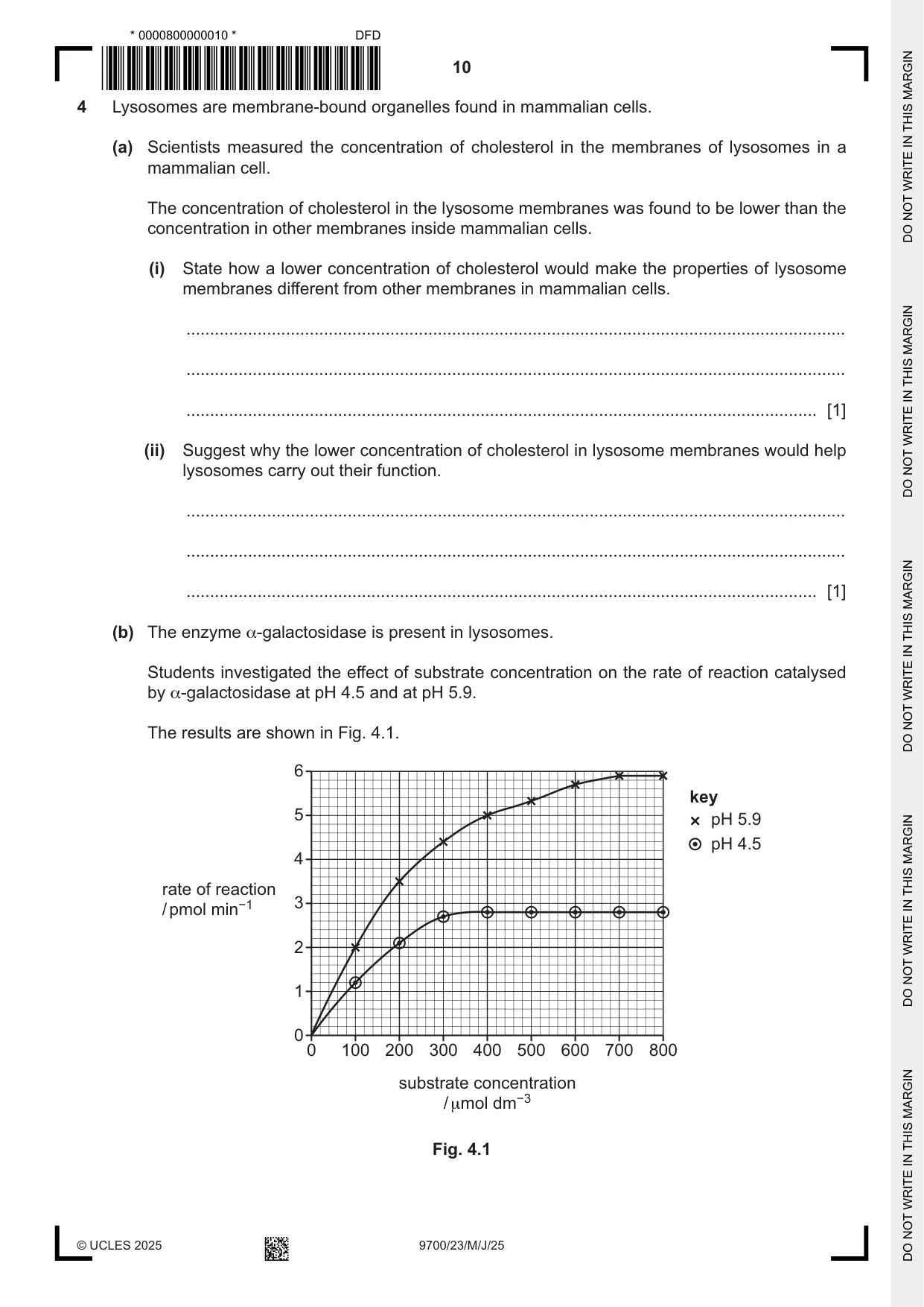
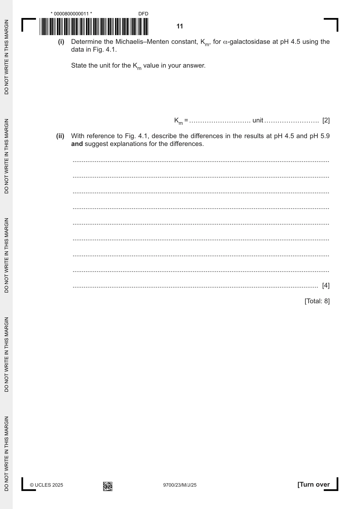
4 Lysosomes are membrane‑bound organelles found in mammalian cells. (a) Scientists measured the concentration of cholesterol in the membranes of lysosomes in a mammalian cell. The concentration of cholesterol in the lysosome membranes was found to be lower than the concentration in other membranes inside mammalian cells. (i) State how a lower concentration of cholesterol would make the properties of lysosome membranes different from other membranes in mammalian cells. ........................................................................................................................................... ........................................................................................................................................... ..................................................................................................................................... [1] (ii) Suggest why the lower concentration of cholesterol in lysosome membranes would help lysosomes carry out their function. ........................................................................................................................................... ........................................................................................................................................... ..................................................................................................................................... [1] (b) The enzyme α‑galactosidase is present in lysosomes. Students investigated the effect of substrate concentration on the rate of reaction catalysed by α‑galactosidase at pH 4.5 and at pH 5.9. The results are shown in Fig. 4.1. 6 key 5 pH 5.9 pH 4.5 4 rate of reaction 3 / pmol min−1 2 1 0 0 100 200 300 400 500 600 700 800 substrate concentration / μmol dm−3 Fig. 4.1 (i) Determine the Michaelis–Menten constant, Km, for α‑galactosidase at pH 4.5 using the data in Fig. 4.1. State the unit for the Km value in your answer. Km = ………………………. unit ……………………. [2] (ii) With reference to Fig. 4.1, describe the differences in the results at pH 4.5 and pH 5.9 and suggest explanations for the differences. ........................................................................................................................................... ........................................................................................................................................... ........................................................................................................................................... ........................................................................................................................................... ........................................................................................................................................... ........................................................................................................................................... ........................................................................................................................................... ........................................................................................................................................... ..................................................................................................................................... [4] [Total: 8]
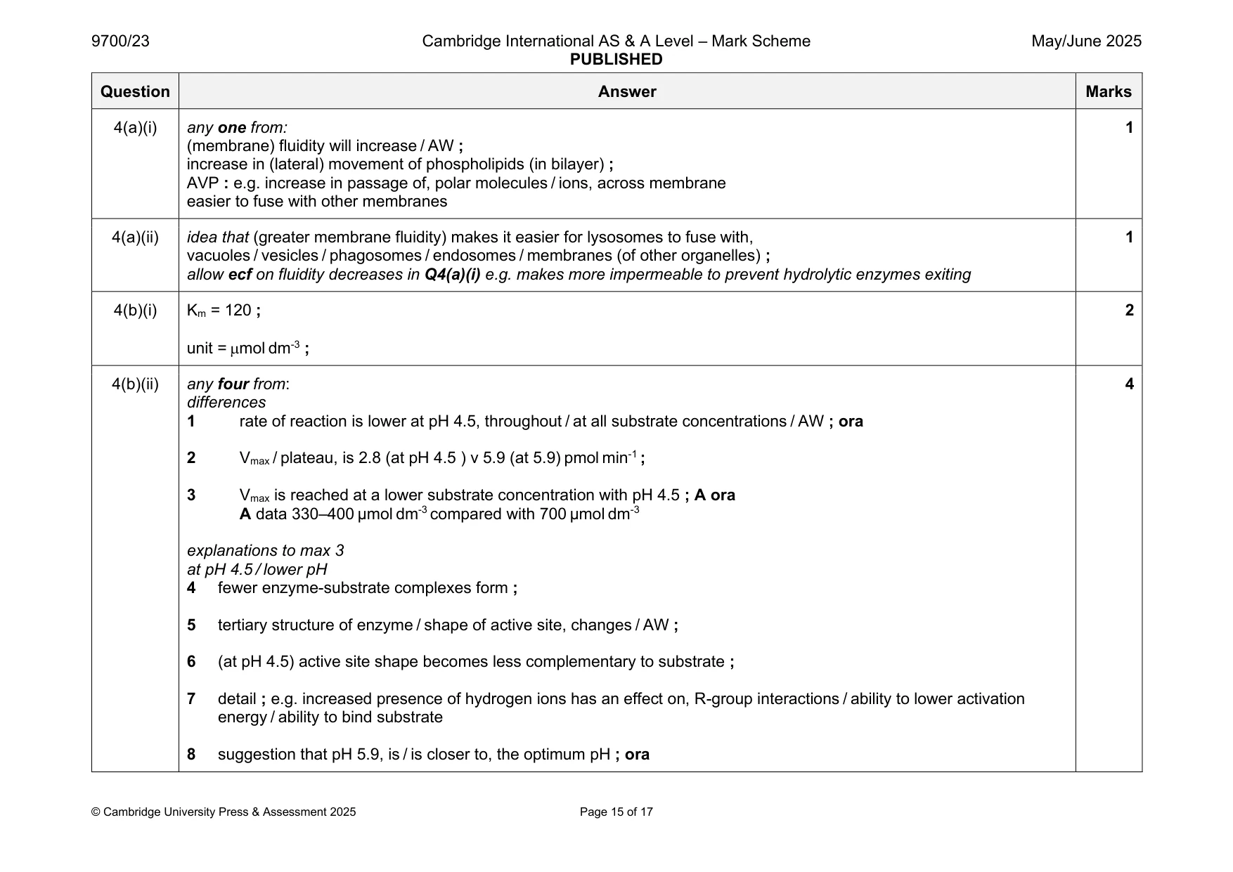
Mark scheme: 4(a)(i) any one from: 1 (membrane) fluidity will increase / AW ; increase in (lateral) movement of phospholipids (in bilayer) ; AVP : e.g. increase in passage of, polar molecules / ions, across membrane easier to fuse with other membranes 4(a)(ii) idea that (greater membrane fluidity) makes it easier for lysosomes to fuse with, 1 vacuoles / vesicles / phagosomes / endosomes / membranes (of other organelles) ; allow ecf on fluidity decreases in Q4(a)(i) e.g. makes more impermeable to prevent hydrolytic enzymes exiting 4(b)(i) Km = 120 ; 2 unit = mol dm-3 ; 4(b)(ii) any four from: 4 differences 1 rate of reaction is lower at pH 4.5, throughout / at all substrate concentrations / AW ; ora 2 Vmax / plateau, is 2.8 (at pH 4.5 ) v 5.9 (at 5.9) pmol min-1 ; 3 Vmax is reached at a lower substrate concentration with pH 4.5 ; A ora A data 330–400 µmol dm-3 compared with 700 µmol dm-3 explanations to max 3 at pH 4.5 / lower pH 4 fewer enzyme-substrate complexes form ; 5 tertiary structure of enzyme / shape of active site, changes / AW ; 6 (at pH 4.5) active site shape becomes less complementary to substrate ; 7 detail ; e.g. increased presence of hydrogen ions has an effect on, R-group interactions / ability to lower activation energy / ability to bind substrate 8 suggestion that pH 5.9, is / is closer to, the optimum pH ; ora
Q5 · Telomeres are lengths of DNA that consist of repetitive nucleotide sequences
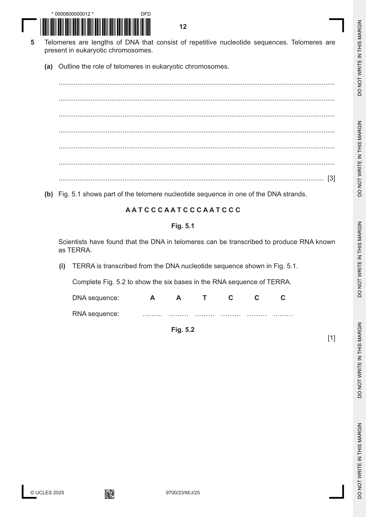
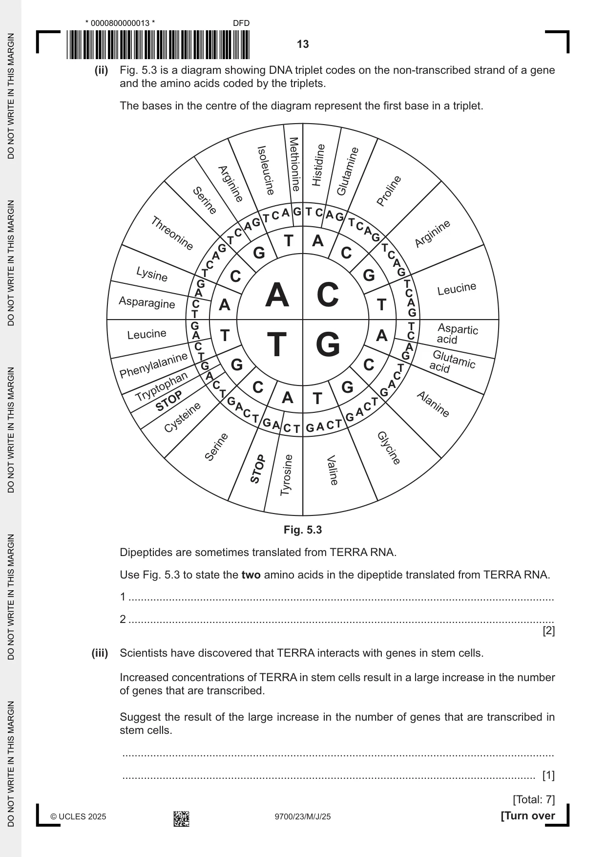
5 Telomeres are lengths of DNA that consist of repetitive nucleotide sequences. Telomeres are present in eukaryotic chromosomes. (a) Outline the role of telomeres in eukaryotic chromosomes. ................................................................................................................................................... ................................................................................................................................................... ................................................................................................................................................... ................................................................................................................................................... ................................................................................................................................................... ................................................................................................................................................... ............................................................................................................................................. [3] (b) Fig. 5.1 shows part of the telomere nucleotide sequence in one of the DNA strands. A A T C C C A A T C C C A A T C C C Fig. 5.1 Scientists have found that the DNA in telomeres can be transcribed to produce RNA known as TERRA. (i) TERRA is transcribed from the DNA nucleotide sequence shown in Fig. 5.1. Complete Fig. 5.2 to show the six bases in the RNA sequence of TERRA. DNA sequence: A A T C C C RNA sequence: Fig. 5.2 [1] (ii) Fig. 5.3 is a diagram showing DNA triplet codes on the non‑transcribed strand of a gene and the amino acids coded by the triplets. The bases in the centre of the diagram represent the first base in a triplet. Histidine Methionine Isoleucine Glutamine Arginine Proline Serine C A G T C A G T G T A C C A T T A G Arginine T Threonine G A G C C C A Lysine T C G G G T A C Leucine A Asparagine C A A C T T G G T Aspartic Leucine A T A C acid C A T T G G Glutamic G G C T acid Phenylalanine A C C C G GA T Tryptophan STOP GA A T CT C A Alanine T G G A C T G A C T Cysteine Serine Glycine STOP Tyrosine Valine Fig. 5.3 Dipeptides are sometimes translated from TERRA RNA. Use Fig. 5.3 to state the two amino acids in the dipeptide translated from TERRA RNA. 1 ......................................................................................................................................... 2 ......................................................................................................................................... [2] (iii) Scientists have discovered that TERRA interacts with genes in stem cells. Increased concentrations of TERRA in stem cells result in a large increase in the number of genes that are transcribed. Suggest the result of the large increase in the number of genes that are transcribed in stem cells. ........................................................................................................................................... ..................................................................................................................................... [1] [Total: 7]
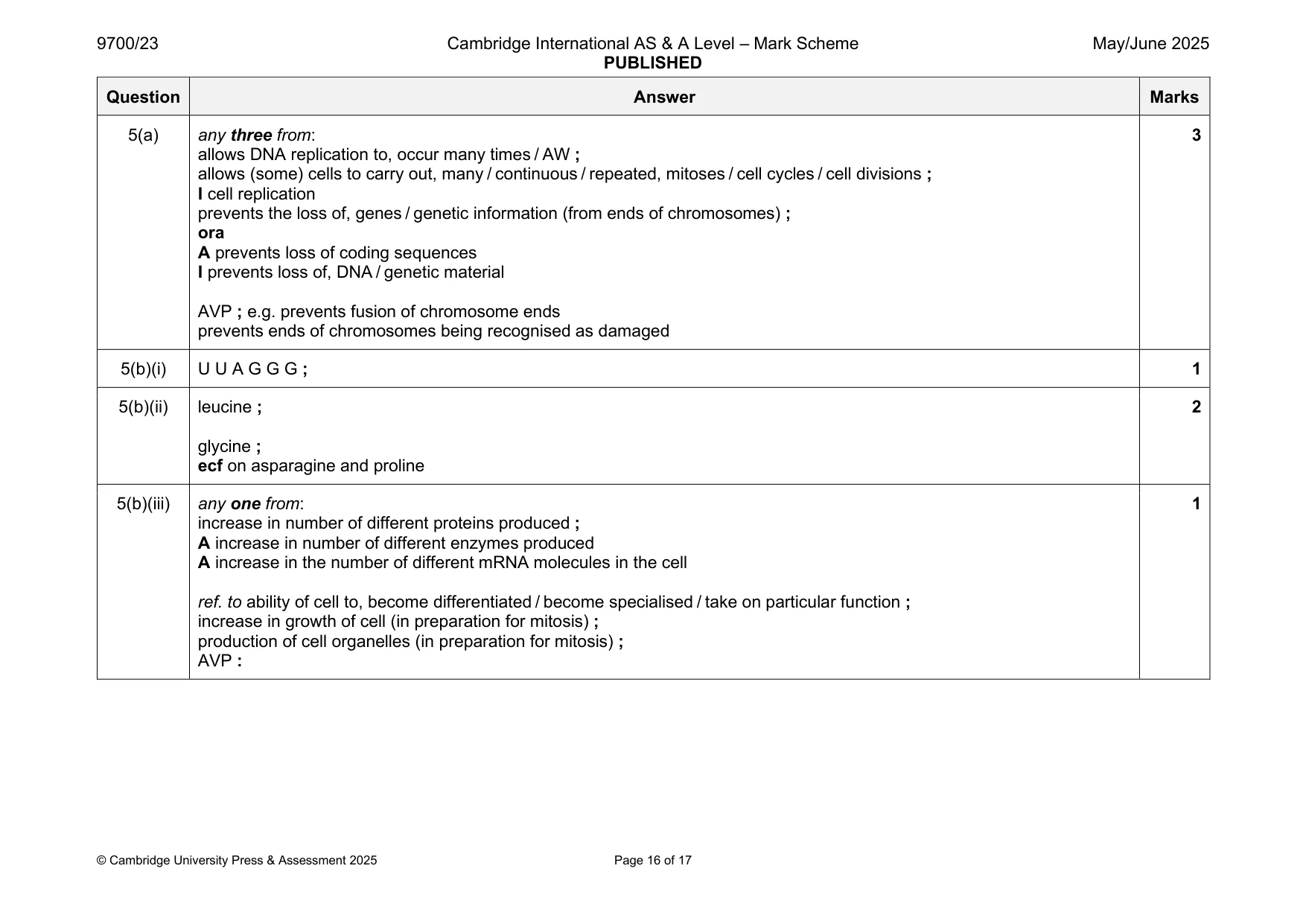
Mark scheme: 5(a) any three from: 3 allows DNA replication to, occur many times / AW ; allows (some) cells to carry out, many / continuous / repeated, mitoses / cell cycles / cell divisions ; I cell replication prevents the loss of, genes / genetic information (from ends of chromosomes) ; ora A prevents loss of coding sequences I prevents loss of, DNA / genetic material AVP ; e.g. prevents fusion of chromosome ends prevents ends of chromosomes being recognised as damaged 5(b)(i) U U A G G G ; 1 5(b)(ii) leucine ; 2 glycine ; ecf on asparagine and proline 5(b)(iii) any one from: 1 increase in number of different proteins produced ; A increase in number of different enzymes produced A increase in the number of different mRNA molecules in the cell ref. to ability of cell to, become differentiated / become specialised / take on particular function ; increase in growth of cell (in preparation for mitosis) ; production of cell organelles (in preparation for mitosis) ; AVP :
Q6 · Monoclonal antibodies can be used in the treatment of disease
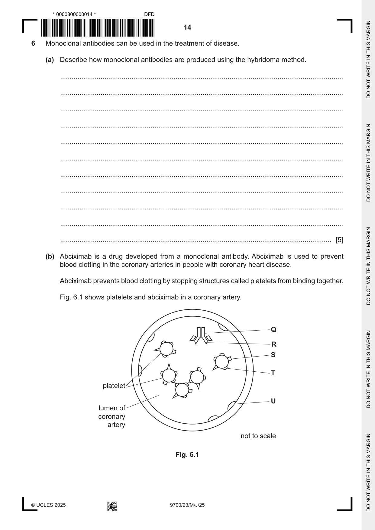
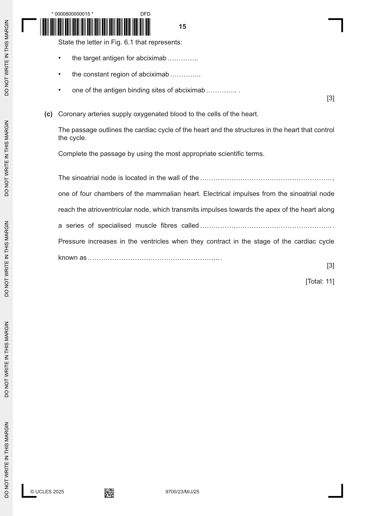
6 Monoclonal antibodies can be used in the treatment of disease. (a) Describe how monoclonal antibodies are produced using the hybridoma method. ................................................................................................................................................... ................................................................................................................................................... ................................................................................................................................................... ................................................................................................................................................... ................................................................................................................................................... ................................................................................................................................................... ................................................................................................................................................... ................................................................................................................................................... ................................................................................................................................................... ................................................................................................................................................... ............................................................................................................................................. [5] (b) Abciximab is a drug developed from a monoclonal antibody. Abciximab is used to prevent blood clotting in the coronary arteries in people with coronary heart disease. Abciximab prevents blood clotting by stopping structures called platelets from binding together. Fig. 6.1 shows platelets and abciximab in a coronary artery. Q R S T platelet U lumen of coronary artery not to scale Fig. 6.1 State the letter in Fig. 6.1 that represents: • the target antigen for abciximab ………….. • the constant region of abciximab ………….. • one of the antigen binding sites of abciximab ………….. . [3] (c) Coronary arteries supply oxygenated blood to the cells of the heart. The passage outlines the cardiac cycle of the heart and the structures in the heart that control the cycle. Complete the passage by using the most appropriate scientific terms. The sinoatrial node is located in the wall of the ………………………………………………….. , one of four chambers of the mammalian heart. Electrical impulses from the sinoatrial node reach the atrioventricular node, which transmits impulses towards the apex of the heart along a series of specialised muscle fibres called ………………………………………………….. . Pressure increases in the ventricles when they contract in the stage of the cardiac cycle known as ………………………………………………….. . [3] [Total: 11]
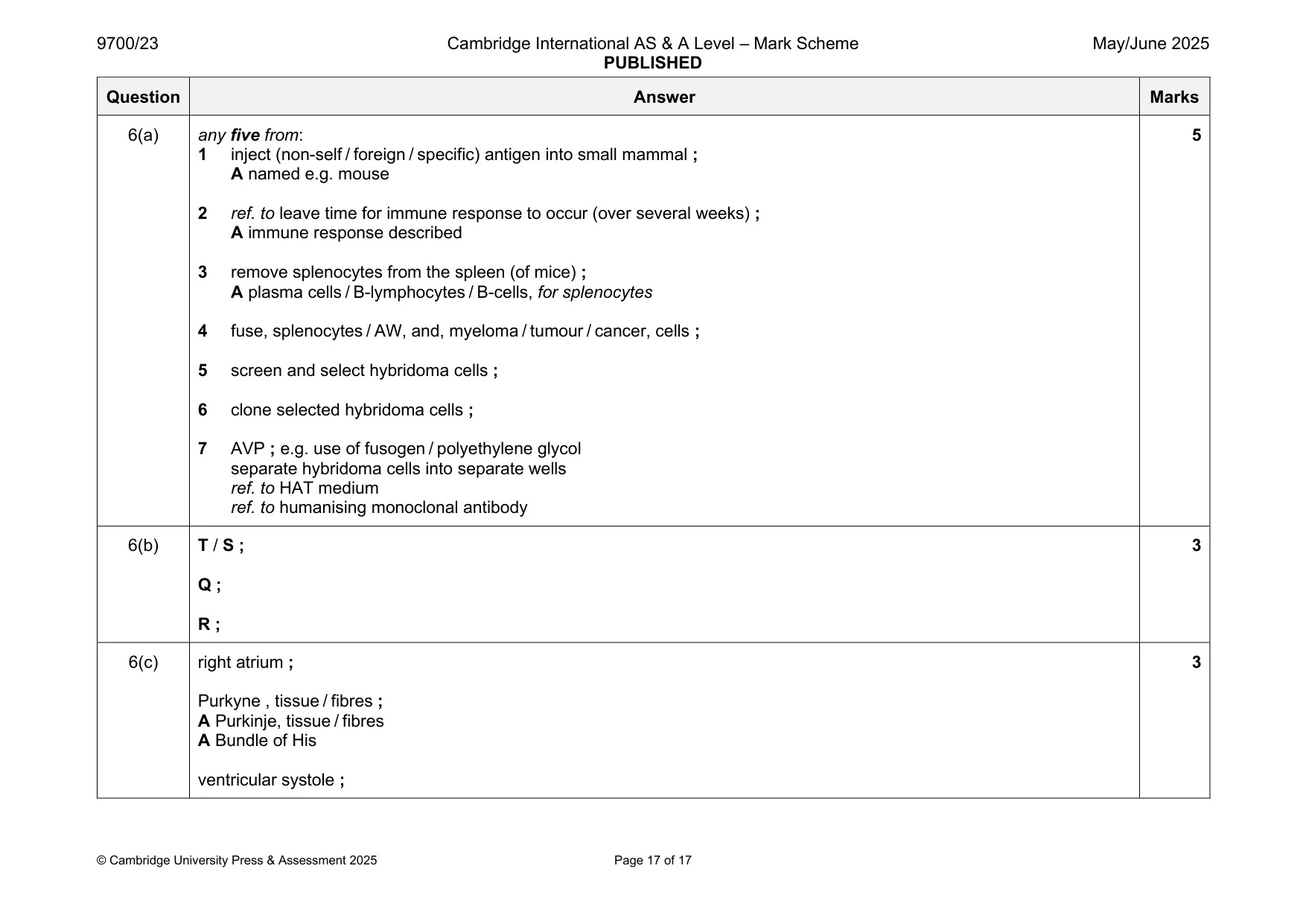
Mark scheme: 6(a) any five from: 5 1 inject (non-self / foreign / specific) antigen into small mammal ; A named e.g. mouse 2 ref. to leave time for immune response to occur (over several weeks) ; A immune response described 3 remove splenocytes from the spleen (of mice) ; A plasma cells / B-lymphocytes / B-cells, for splenocytes 4 fuse, splenocytes / AW, and, myeloma / tumour / cancer, cells ; 5 screen and select hybridoma cells ; 6 clone selected hybridoma cells ; 7 AVP ; e.g. use of fusogen / polyethylene glycol separate hybridoma cells into separate wells ref. to HAT medium ref. to humanising monoclonal antibody 6(b) T / S ; 3 Q ; R ; 6(c) right atrium ; 3 Purkyne , tissue / fibres ; A Purkinje, tissue / fibres A Bundle of His ventricular systole ;
What was in this paper
The subtopics covered by these 6 questions, and how many questions each got. Open one in a new tab to see every Cambridge question on it.
What you needed in this session
Cambridge’s own grade thresholds for 2025 May/June, Paper 2 · Variant 3. A higher threshold means an easier paper — the bar moves with how the cohort did.