Cambridge A Level Biology 9700 — 2025 Oct/Nov Paper 2 · Variant 4
9700/24/O/N/25 · 6 questions · 60 marks · 75 min
The question paper and its mark scheme, free to read here and free to download. This is Cambridge’s own paper, exactly as it was sat.
Question paper16 pages
Mark scheme22 pages
Answers below. Sit the paper first if you are practising.
Questions as text
Q1 · There are similarities and differences between the structure of a typical plant cell and…
1 There are similarities and differences between the structure of a typical plant cell and a typical animal cell. (a) The nucleus of plant cells and animal cells contains chromosomes. In interphase of the cell cycle, individual chromosomes are present but cannot be seen. The chromosome material is known as chromatin. (i) Changes occur in interphase, which result in a difference between the chromatin in the G1 phase compared with the chromatin in the G2 phase. State and explain the difference in chromatin in the G1 phase compared with chromatin in the G2 phase. ........................................................................................................................................... ........................................................................................................................................... ........................................................................................................................................... ........................................................................................................................................... ..................................................................................................................................... [2] (ii) Describe the features of a nucleus, other than containing chromatin. You may use the space below the lines for a diagram. ........................................................................................................................................... ........................................................................................................................................... ........................................................................................................................................... ........................................................................................................................................... ........................................................................................................................................... ........................................................................................................................................... ........................................................................................................................................... [3] (b) Starch, cellulose and pectins are polysaccharides found in plant cells but not in animal cells. Pectins are complex polysaccharides that are found in the cell wall. (i) Describe the structural features of starch that are different from the structural features of cellulose. ........................................................................................................................................... ........................................................................................................................................... ........................................................................................................................................... ........................................................................................................................................... ........................................................................................................................................... ........................................................................................................................................... ........................................................................................................................................... ........................................................................................................................................... ..................................................................................................................................... [4] (ii) Cell wall pectins can vary in different plant cell types and in different stages of cell development. Pectin molecules are released from cells as basic structures and then modified within the cell wall by adding side chains. RG‑I is a pectin molecule with a variable structure. The basic structure is a repeated disaccharide made from two different monosaccharides. RG‑I has three different side chains that can be added in different positions. Monoclonal antibodies (mAbs) are used to investigate the structure, location and role of cell wall pectins. Suggest and explain why scientists need to use a number of different monoclonal antibodies when investigating a pectin such as RG‑I. ........................................................................................................................................... ........................................................................................................................................... ........................................................................................................................................... ........................................................................................................................................... ........................................................................................................................................... ........................................................................................................................................... ..................................................................................................................................... [3] [Total: 12]
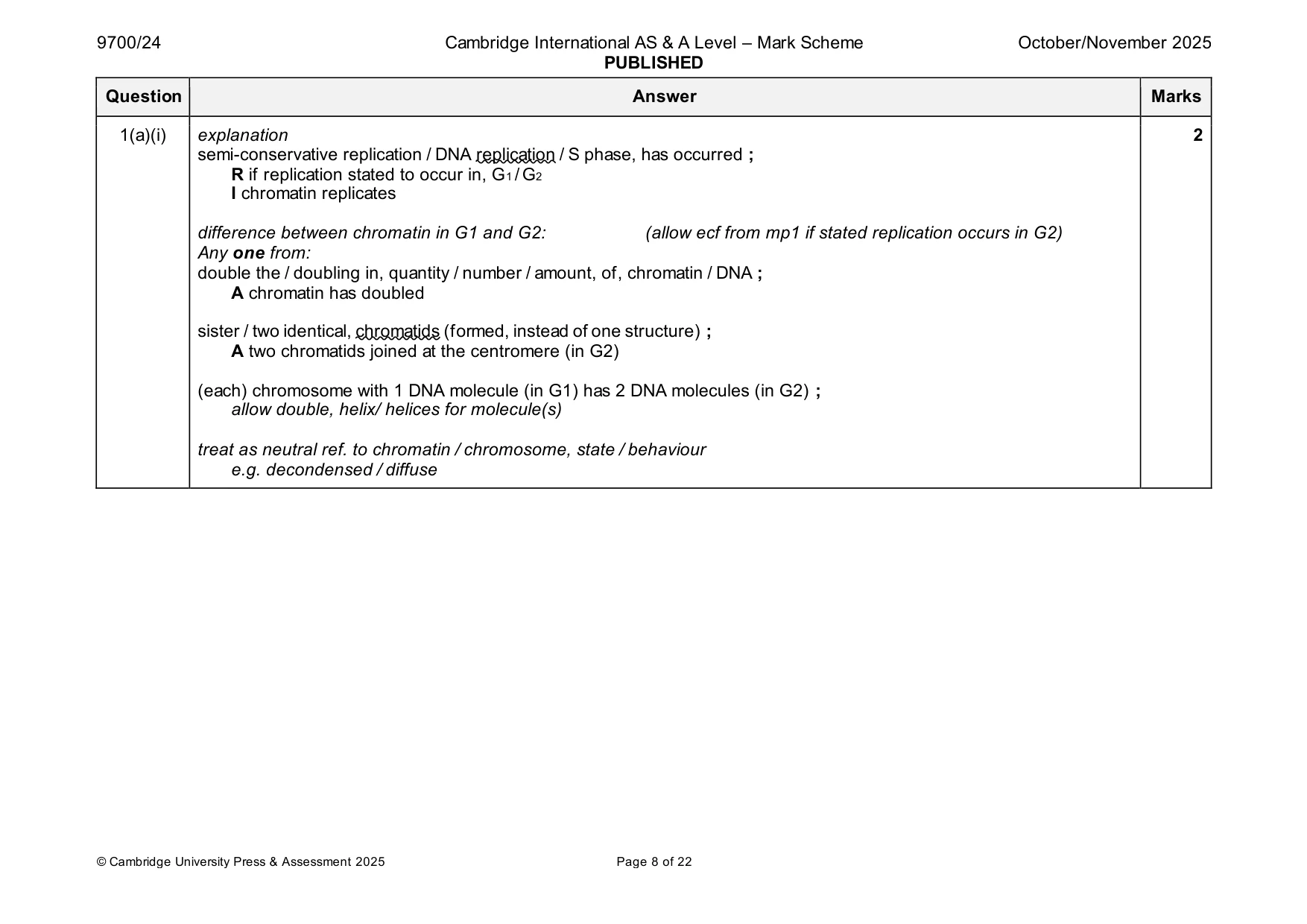
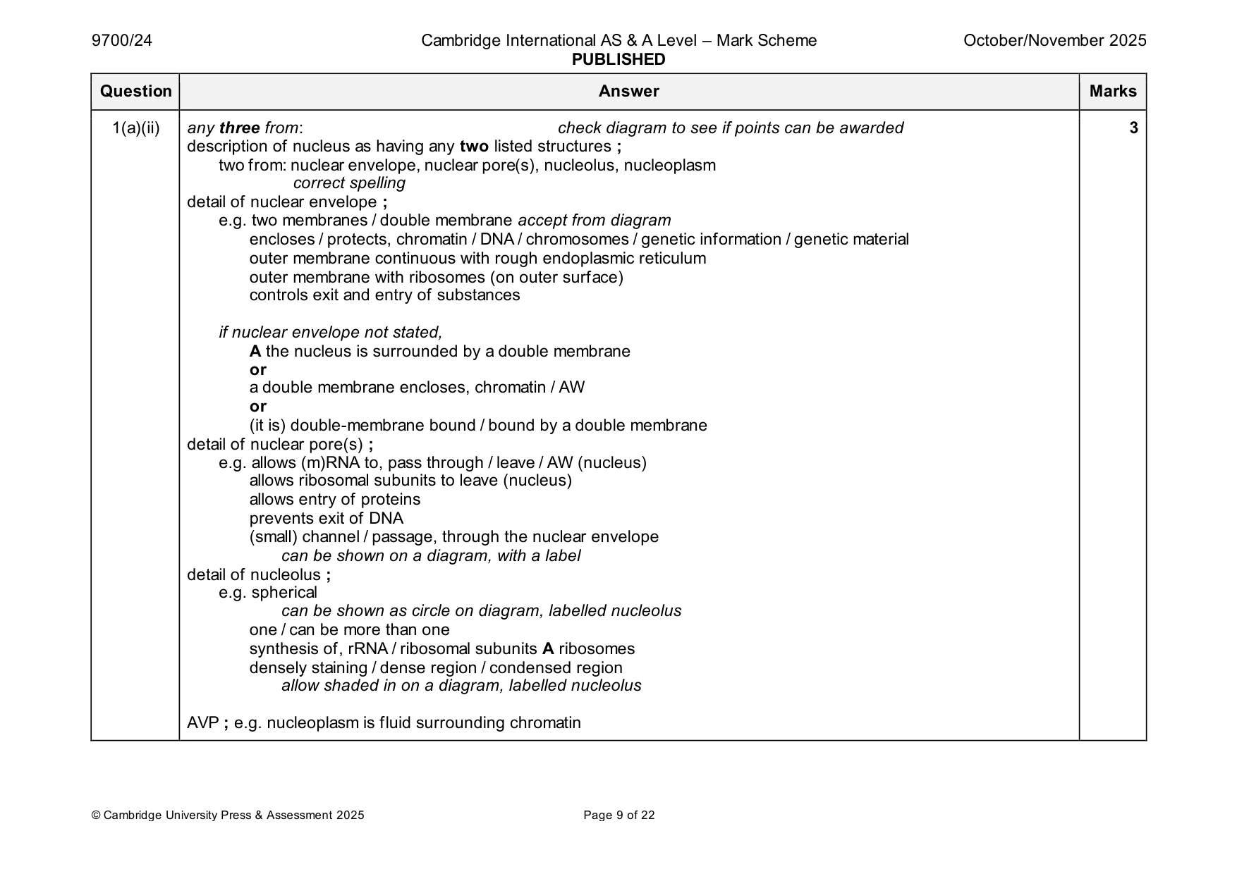
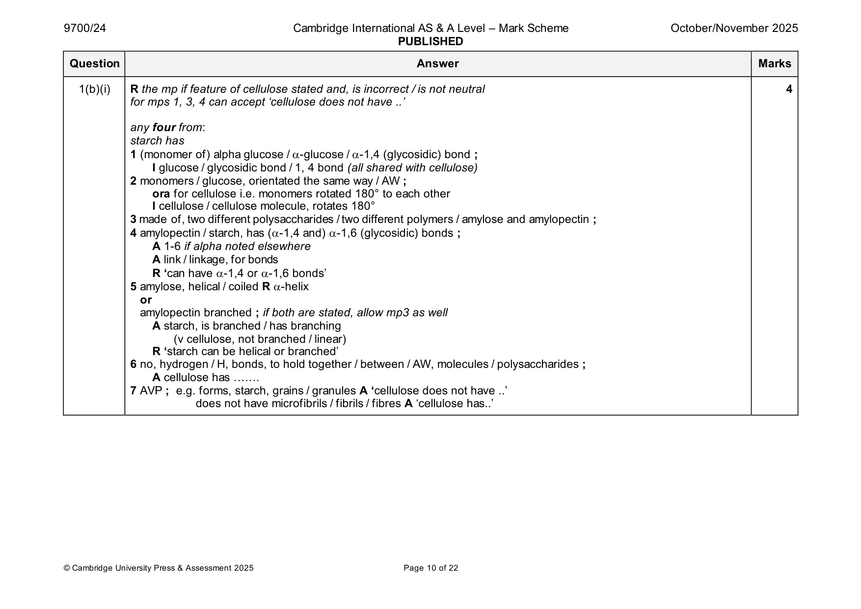
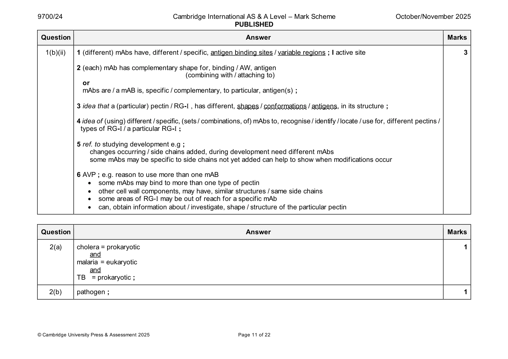
Mark scheme: Question Answer Marks 1(a)(i) explanation 2 semi-conservative replication / DNA replication / S phase, has occurred ; R if replication stated to occur in, G1 / G2 I chromatin replicates difference between chromatin in G1 and G2: (allow ecf from mp1 if stated replication occurs in G2) Any one from: double the / doubling in, quantity / number / amount, of, chromatin / DNA ; A chromatin has doubled sister / two identical, chromatids (formed, instead of one structure) ; A two chromatids joined at the centromere (in G2) (each) chromosome with 1 DNA molecule (in G1) has 2 DNA molecules (in G2) ; allow double, helix/ helices for molecule(s) treat as neutral ref. to chromatin / chromosome, state / behaviour e.g. decondensed / diffuse 1(a)(ii) any three from: check diagram to see if points can be awarded 3 description of nucleus as having any two listed structures ; two from: nuclear envelope, nuclear pore(s), nucleolus, nucleoplasm correct spelling detail of nuclear envelope ; e.g. two membranes / double membrane accept from diagram encloses / protects, chromatin / DNA / chromosomes / genetic information / genetic material outer membrane continuous with rough endoplasmic reticulum outer membrane with ribosomes (on outer surface) controls exit and entry of substances if nuclear envelope not stated, A the nucleus is surrounded by a double membrane or a double membrane encloses, chromatin / AW or (it is) double-membrane bound / bound by a double membrane detail of nuclear pore(s) ; e.g. allows (m)RNA to, pass through / leave / AW (nucleus) allows ribosomal subunits to leave (nucleus) allows entry of proteins prevents exit of DNA (small) channel / passage, through the nuclear envelope can be shown on a diagram, with a label detail of nucleolus ; e.g. spherical can be shown as circle on diagram, labelled nucleolus one / can be more than one synthesis of, rRNA / ribosomal subunits A ribosomes densely staining / dense region / condensed region allow shaded in on a diagram, labelled nucleolus AVP ; e.g. nucleoplasm is fluid surrounding chromatin 1(b)(i) R the mp if feature of cellulose stated and, is incorrect / is not neutral 4 for mps 1, 3, 4 can accept ‘cellulose does not have ..’ any four from: starch has 1 (monomer of) alpha glucose / -glucose / -1,4 (glycosidic) bond ; I glucose / glycosidic bond / 1, 4 bond (all shared with cellulose) 2 monomers / glucose, orientated the same way / AW ; ora for cellulose i.e. monomers rotated 180° to each other I cellulose / cellulose molecule, rotates 180° 3 made of, two different polysaccharides / two different polymers / amylose and amylopectin ; 4 amylopectin / starch, has (-1,4 and) -1,6 (glycosidic) bonds ; A 1-6 if alpha noted elsewhere A link / linkage, for bonds R ‘can have -1,4 or -1,6 bonds’ 5 amylose, helical / coiled R -helix or amylopectin branched ; if both are stated, allow mp3 as well A starch, is branched / has branching (v cellulose, not branched / linear) R ‘starch can be helical or branched’ 6 no, hydrogen / H, bonds, to hold together / between / AW, molecules / polysaccharides ; A cellulose has ……. 7 AVP ; e.g. forms, starch, grains / granules A ‘cellulose does not have ..’ does not have microfibrils / fibrils / fibres A ‘cellulose has..’ 1(b)(ii) 1 (different) mAbs have, different / specific, antigen binding sites / variable regions ; I active site 3 2 (each) mAb has complementary shape for, binding / AW, antigen (combining with / attaching to) or mAbs are / a mAB is, specific / complementary, to particular, antigen(s) ; 3 idea that a (particular) pectin / RG-I , has different, shapes / conformations / antigens, in its structure ; 4 idea of (using) different / specific, (sets / combinations, of) mAbs to, recognise / identify / locate / use for, different pectins / types of RG-I / a particular RG-I ; 5 ref. to studying development e.g ; changes occurring / side chains added, during development need different mAbs some mAbs may be specific to side chains not yet added can help to show when modifications occur 6 AVP ; e.g. reason to use more than one mAB • some mAbs may bind to more than one type of pectin • other cell wall components, may have, similar structures / same side chains • some areas of RG-I may be out of reach for a specific mAb • can, obtain information about / investigate, shape / structure of the particular pectin
Q2 · Cholera, malaria and tuberculosis (TB) are infectious diseases caused by unicellular…
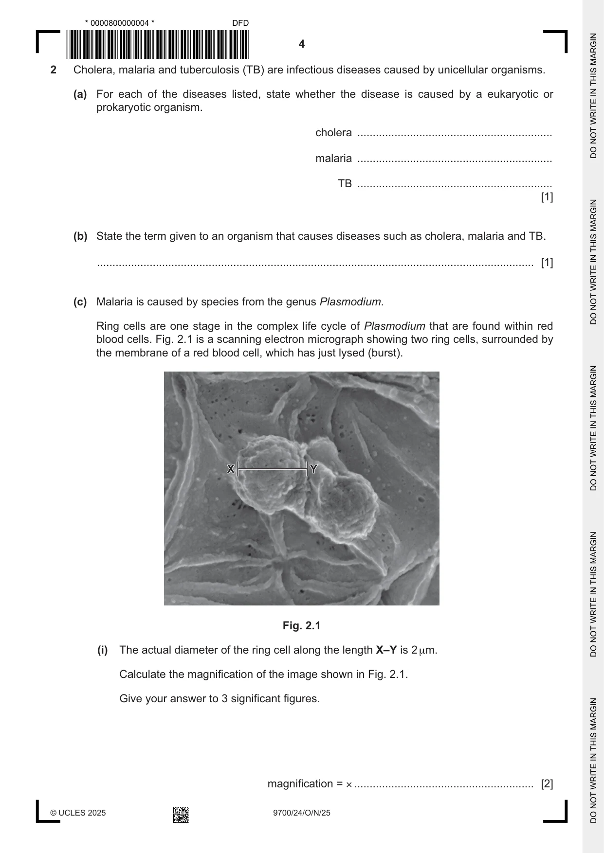
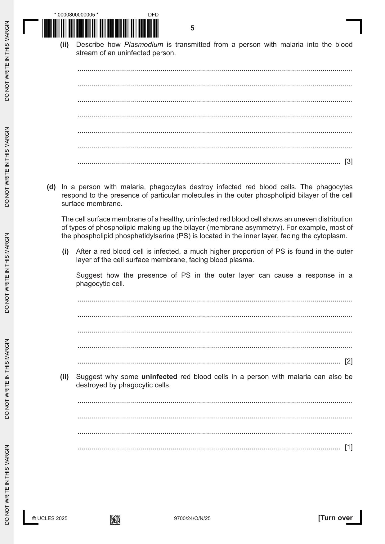
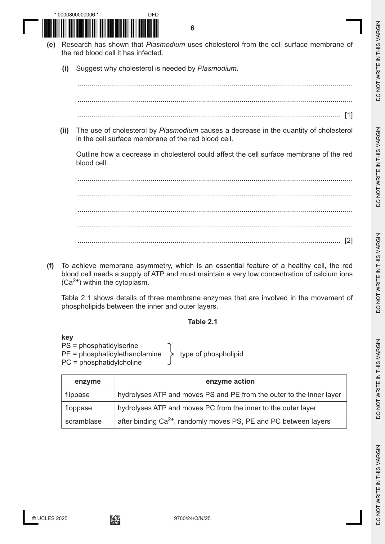
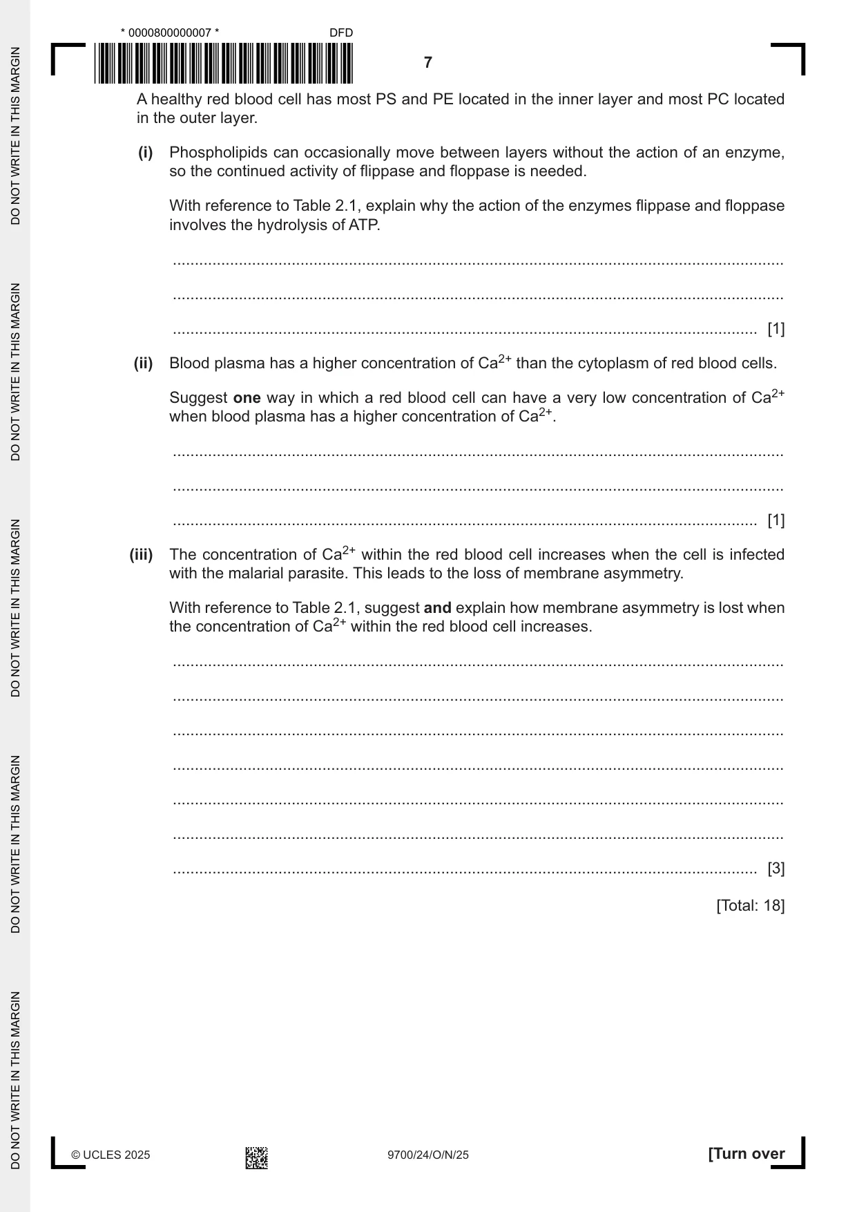
2 Cholera, malaria and tuberculosis (TB) are infectious diseases caused by unicellular organisms. (a) For each of the diseases listed, state whether the disease is caused by a eukaryotic or prokaryotic organism. cholera ............................................................... malaria ............................................................... TB ............................................................... [1] (b) State the term given to an organism that causes diseases such as cholera, malaria and TB. ............................................................................................................................................. [1] (c) Malaria is caused by species from the genus Plasmodium. Ring cells are one stage in the complex life cycle of Plasmodium that are found within red blood cells. Fig. 2.1 is a scanning electron micrograph showing two ring cells, surrounded by the membrane of a red blood cell, which has just lysed (burst). X Y Fig. 2.1 (i) The actual diameter of the ring cell along the length X–Y is 2 µm. Calculate the magnification of the image shown in Fig. 2.1. Give your answer to 3 significant figures. magnification = × .......................................................... [2] (ii) Describe how Plasmodium is transmitted from a person with malaria into the blood stream of an uninfected person. ........................................................................................................................................... ........................................................................................................................................... ........................................................................................................................................... ........................................................................................................................................... ........................................................................................................................................... ........................................................................................................................................... ..................................................................................................................................... [3] (d) In a person with malaria, phagocytes destroy infected red blood cells. The phagocytes respond to the presence of particular molecules in the outer phospholipid bilayer of the cell surface membrane. The cell surface membrane of a healthy, uninfected red blood cell shows an uneven distribution of types of phospholipid making up the bilayer (membrane asymmetry). For example, most of the phospholipid phosphatidylserine (PS) is located in the inner layer, facing the cytoplasm. (i) After a red blood cell is infected, a much higher proportion of PS is found in the outer layer of the cell surface membrane, facing blood plasma. Suggest how the presence of PS in the outer layer can cause a response in a phagocytic cell. ........................................................................................................................................... ........................................................................................................................................... ........................................................................................................................................... ........................................................................................................................................... ..................................................................................................................................... [2] (ii) Suggest why some uninfected red blood cells in a person with malaria can also be destroyed by phagocytic cells. ........................................................................................................................................... ........................................................................................................................................... ........................................................................................................................................... ..................................................................................................................................... [1] (e) Research has shown that Plasmodium uses cholesterol from the cell surface membrane of the red blood cell it has infected. (i) Suggest why cholesterol is needed by Plasmodium. ........................................................................................................................................... ........................................................................................................................................... ..................................................................................................................................... [1] (ii) The use of cholesterol by Plasmodium causes a decrease in the quantity of cholesterol in the cell surface membrane of the red blood cell. Outline how a decrease in cholesterol could affect the cell surface membrane of the red blood cell. ........................................................................................................................................... ........................................................................................................................................... ........................................................................................................................................... ........................................................................................................................................... ..................................................................................................................................... [2] (f) To achieve membrane asymmetry, which is an essential feature of a healthy cell, the red blood cell needs a supply of ATP and must maintain a very low concentration of calcium ions (Ca2+) within the cytoplasm. Table 2.1 shows details of three membrane enzymes that are involved in the movement of phospholipids between the inner and outer layers. Table 2.1 key PS = phosphatidylserine PE = phosphatidylethanolamine type of phospholipid PC = phosphatidylcholine enzyme enzyme action flippase hydrolyses ATP and moves PS and PE from the outer to the inner layer floppase hydrolyses ATP and moves PC from the inner to the outer layer scramblase after binding Ca2+, randomly moves PS, PE and PC between layers A healthy red blood cell has most PS and PE located in the inner layer and most PC located in the outer layer. (i) Phospholipids can occasionally move between layers without the action of an enzyme, so the continued activity of flippase and floppase is needed. With reference to Table 2.1, explain why the action of the enzymes flippase and floppase involves the hydrolysis of ATP. ........................................................................................................................................... ........................................................................................................................................... ..................................................................................................................................... [1] (ii) Blood plasma has a higher concentration of Ca2+ than the cytoplasm of red blood cells. Suggest one way in which a red blood cell can have a very low concentration of Ca2+ when blood plasma has a higher concentration of Ca2+. ........................................................................................................................................... ........................................................................................................................................... ..................................................................................................................................... [1] (iii) The concentration of Ca2+ within the red blood cell increases when the cell is infected with the malarial parasite. This leads to the loss of membrane asymmetry. With reference to Table 2.1, suggest and explain how membrane asymmetry is lost when the concentration of Ca2+ within the red blood cell increases. ........................................................................................................................................... ........................................................................................................................................... ........................................................................................................................................... ........................................................................................................................................... ........................................................................................................................................... ........................................................................................................................................... ..................................................................................................................................... [3] [Total: 18]
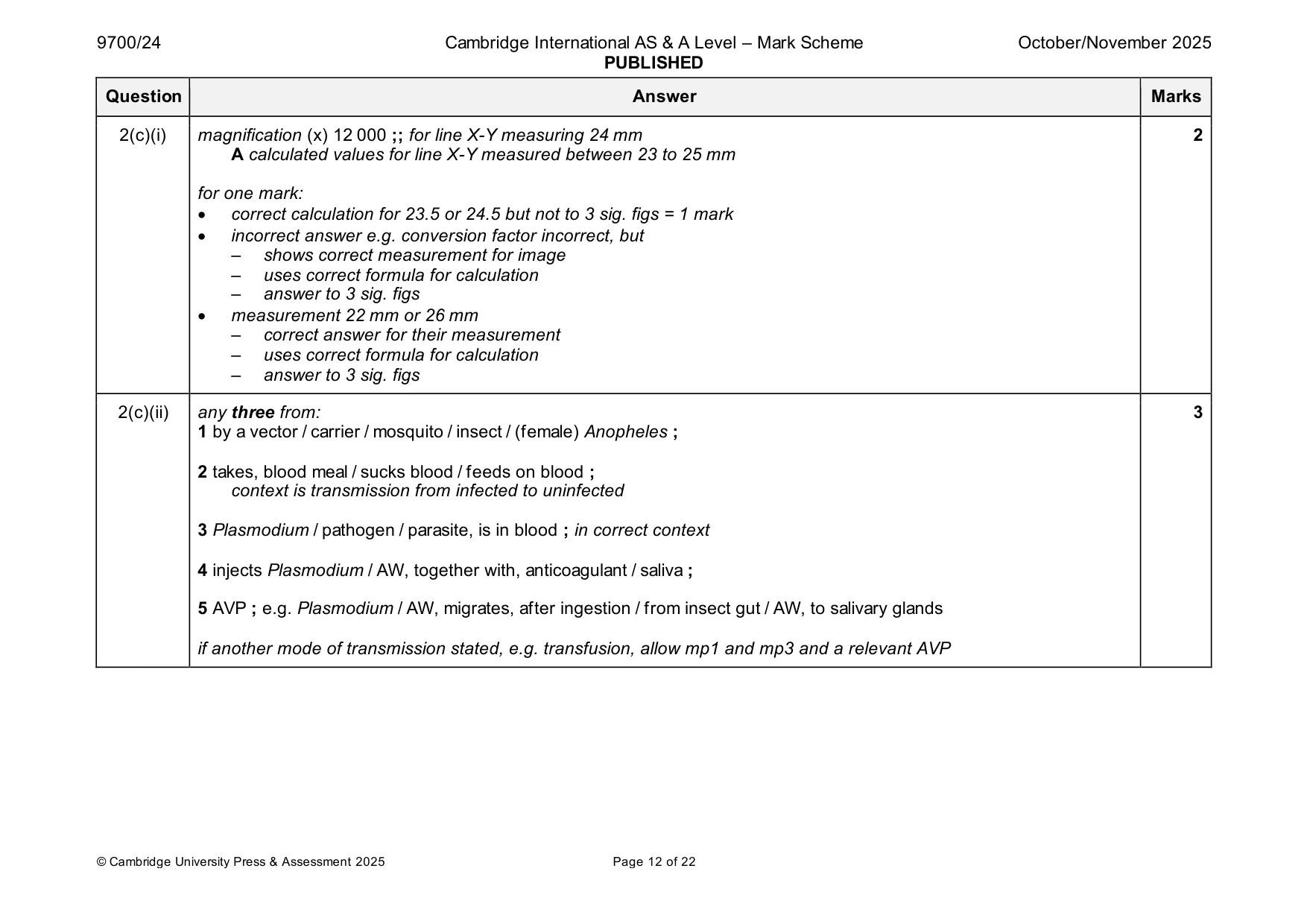
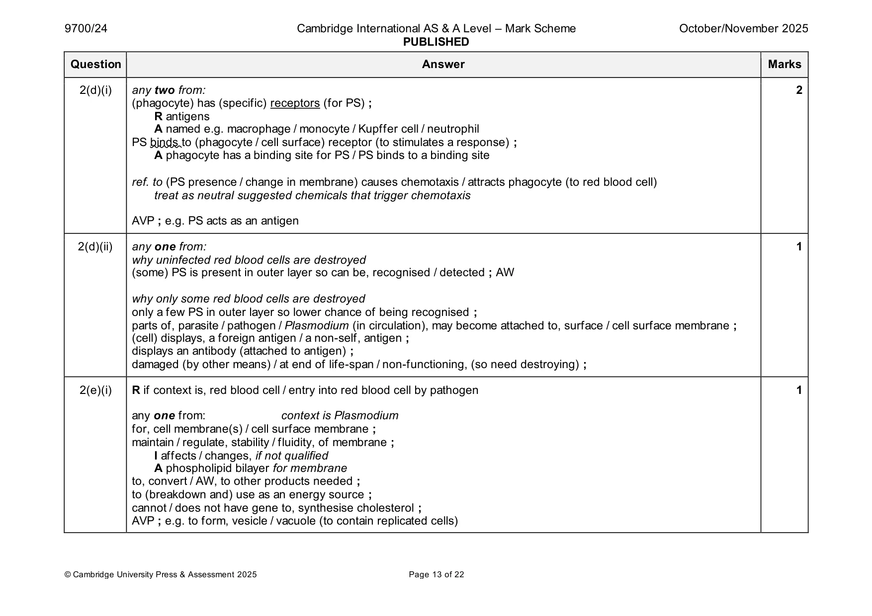
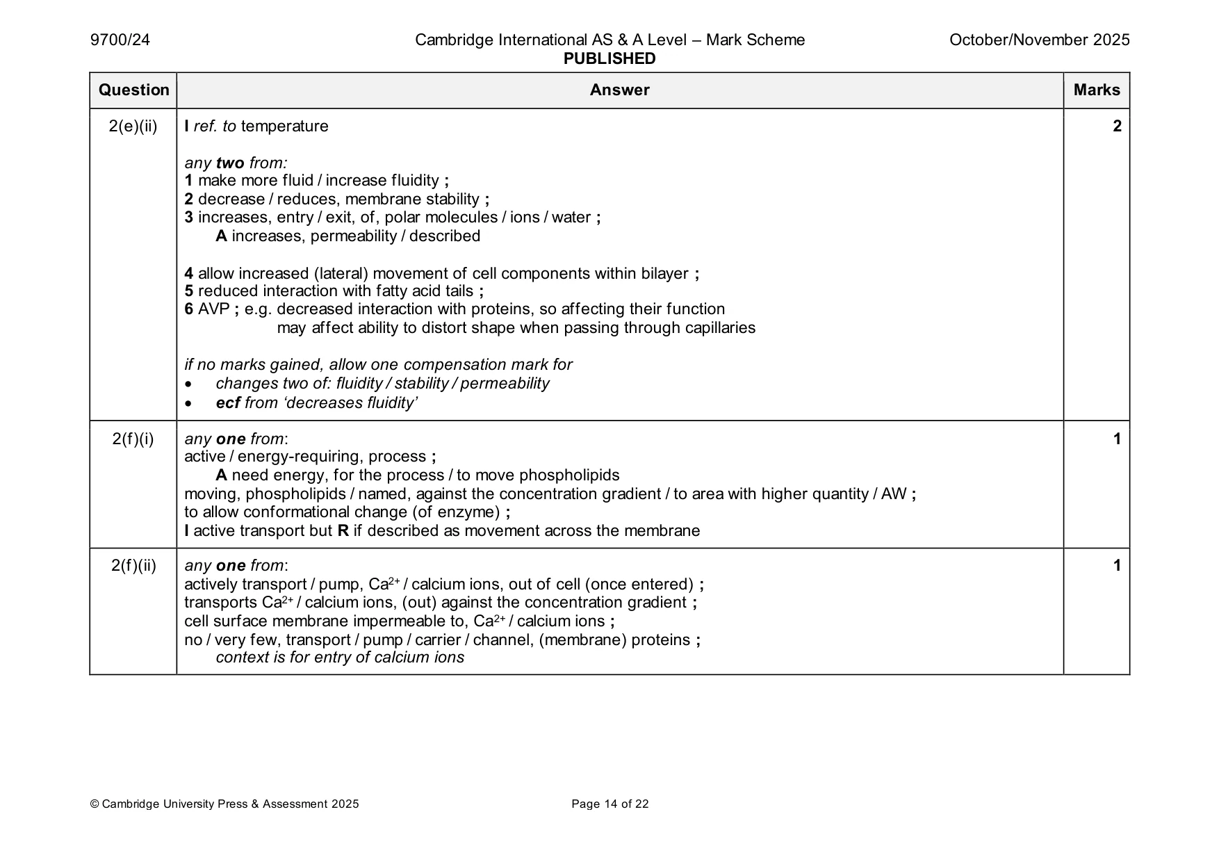
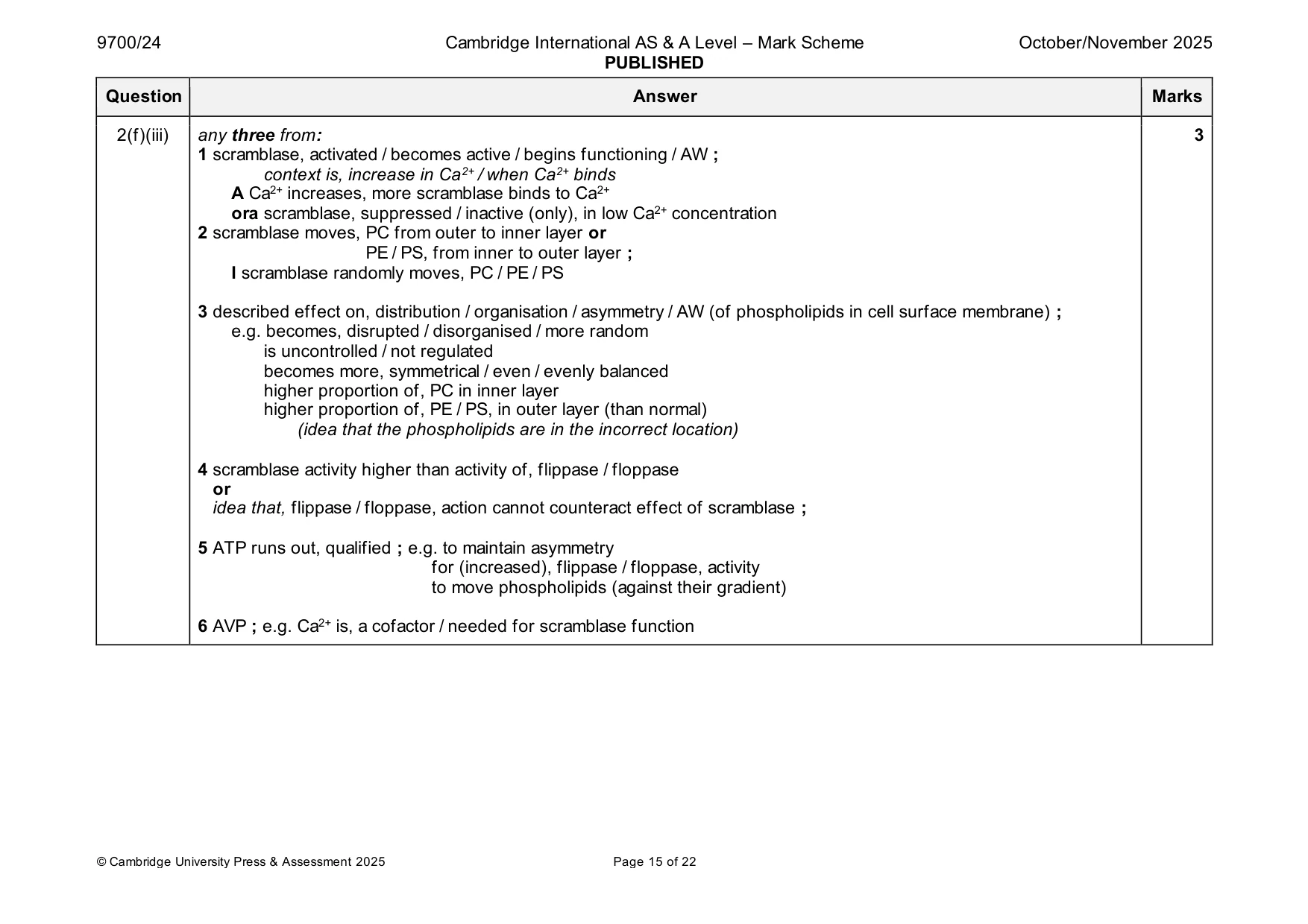
Mark scheme: 2(a) cholera = prokaryotic 1 and malaria = eukaryotic and TB = prokaryotic ; 2(b) pathogen ; 1 2(c)(i) magnification (x) 12 000 ;; for line X-Y measuring 24 mm 2 A calculated values for line X-Y measured between 23 to 25 mm for one mark: • correct calculation for 23.5 or 24.5 but not to 3 sig. figs = 1 mark • incorrect answer e.g. conversion factor incorrect, but – shows correct measurement for image – uses correct formula for calculation – answer to 3 sig. figs • measurement 22 mm or 26 mm – correct answer for their measurement – uses correct formula for calculation – answer to 3 sig. figs 2(c)(ii) any three from: 3 1 by a vector / carrier / mosquito / insect / (female) Anopheles ; 2 takes, blood meal / sucks blood / feeds on blood ; context is transmission from infected to uninfected 3 Plasmodium / pathogen / parasite, is in blood ; in correct context 4 injects Plasmodium / AW, together with, anticoagulant / saliva ; 5 AVP ; e.g. Plasmodium / AW, migrates, after ingestion / from insect gut / AW, to salivary glands if another mode of transmission stated, e.g. transfusion, allow mp1 and mp3 and a relevant AVP 2(d)(i) any two from: 2 (phagocyte) has (specific) receptors (for PS) ; R antigens A named e.g. macrophage / monocyte / Kupffer cell / neutrophil PS binds to (phagocyte / cell surface) receptor (to stimulates a response) ; A phagocyte has a binding site for PS / PS binds to a binding site ref. to (PS presence / change in membrane) causes chemotaxis / attracts phagocyte (to red blood cell) treat as neutral suggested chemicals that trigger chemotaxis AVP ; e.g. PS acts as an antigen 2(d)(ii) any one from: 1 why uninfected red blood cells are destroyed (some) PS is present in outer layer so can be, recognised / detected ; AW why only some red blood cells are destroyed only a few PS in outer layer so lower chance of being recognised ; parts of, parasite / pathogen / Plasmodium (in circulation), may become attached to, surface / cell surface membrane ; (cell) displays, a foreign antigen / a non-self, antigen ; displays an antibody (attached to antigen) ; damaged (by other means) / at end of life-span / non-functioning, (so need destroying) ; 2(e)(i) R if context is, red blood cell / entry into red blood cell by pathogen 1 any one from: context is Plasmodium for, cell membrane(s) / cell surface membrane ; maintain / regulate, stability / fluidity, of membrane ; I affects / changes, if not qualified A phospholipid bilayer for membrane to, convert / AW, to other products needed ; to (breakdown and) use as an energy source ; cannot / does not have gene to, synthesise cholesterol ; AVP ; e.g. to form, vesicle / vacuole (to contain replicated cells) 2(e)(ii) I ref. to temperature 2 any two from: 1 make more fluid / increase fluidity ; 2 decrease / reduces, membrane stability ; 3 increases, entry / exit, of, polar molecules / ions / water ; A increases, permeability / described 4 allow increased (lateral) movement of cell components within bilayer ; 5 reduced interaction with fatty acid tails ; 6 AVP ; e.g. decreased interaction with proteins, so affecting their function may affect ability to distort shape when passing through capillaries if no marks gained, allow one compensation mark for • changes two of: fluidity / stability / permeability • ecf from ‘decreases fluidity’ 2(f)(i) any one from: 1 active / energy-requiring, process ; A need energy, for the process / to move phospholipids moving, phospholipids / named, against the concentration gradient / to area with higher quantity / AW ; to allow conformational change (of enzyme) ; I active transport but R if described as movement across the membrane 2(f)(ii) any one from: 1 actively transport / pump, Ca2+ / calcium ions, out of cell (once entered) ; transports Ca2+ / calcium ions, (out) against the concentration gradient ; cell surface membrane impermeable to, Ca2+ / calcium ions ; no / very few, transport / pump / carrier / channel, (membrane) proteins ; context is for entry of calcium ions 2(f)(iii) any three from: 3 1 scramblase, activated / becomes active / begins functioning / AW ; context is, increase in Ca2+ / when Ca2+ binds A Ca2+ increases, more scramblase binds to Ca2+ ora scramblase, suppressed / inactive (only), in low Ca2+ concentration 2 scramblase moves, PC from outer to inner layer or PE / PS, from inner to outer layer ; I scramblase randomly moves, PC / PE / PS 3 described effect on, distribution / organisation / asymmetry / AW (of phospholipids in cell surface membrane) ; e.g. becomes, disrupted / disorganised / more random is uncontrolled / not regulated becomes more, symmetrical / even / evenly balanced higher proportion of, PC in inner layer higher proportion of, PE / PS, in outer layer (than normal) (idea that the phospholipids are in the incorrect location) 4 scramblase activity higher than activity of, flippase / floppase or idea that, flippase / floppase, action cannot counteract effect of scramblase ; 5 ATP runs out, qualified ; e.g. to maintain asymmetry for (increased), flippase / floppase, activity to move phospholipids (against their gradient) 6 AVP ; e.g. Ca2+ is, a cofactor / needed for scramblase function
Q3 · Small interfering RNA (siRNA) is a short length of double‑stranded RNA (dsRNA)…
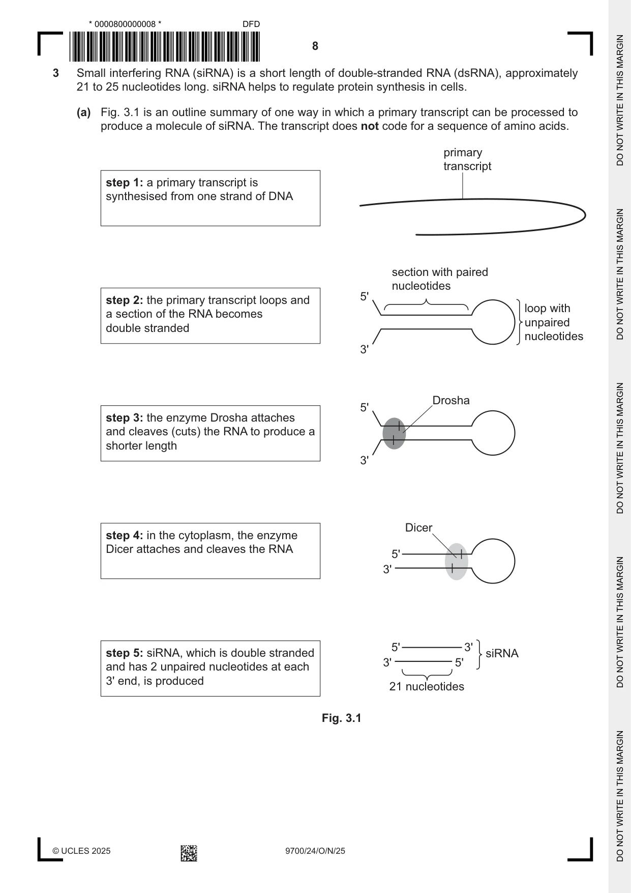
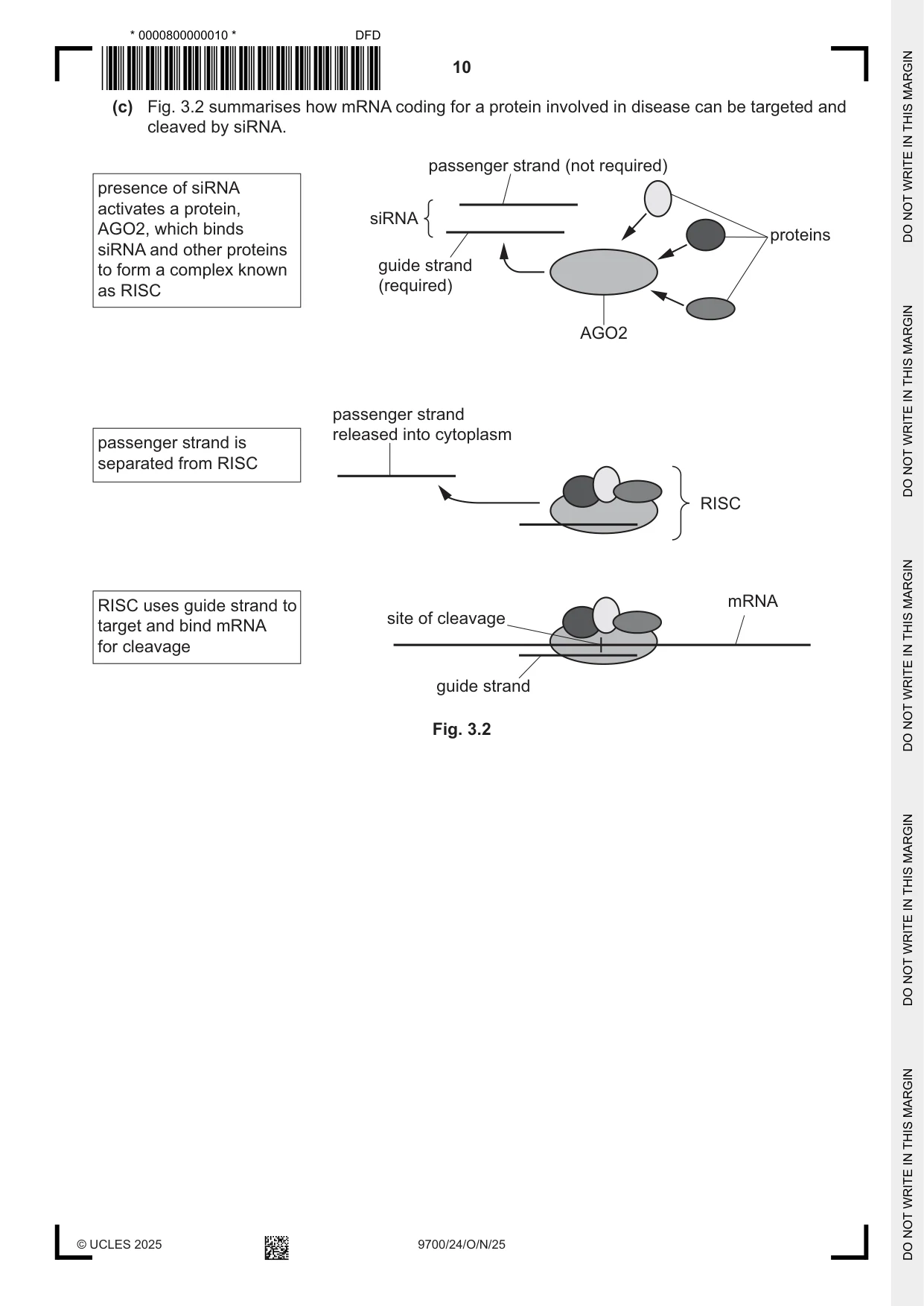
3 Small interfering RNA (siRNA) is a short length of double‑stranded RNA (dsRNA), approximately 21 to 25 nucleotides long. siRNA helps to regulate protein synthesis in cells. (a) Fig. 3.1 is an outline summary of one way in which a primary transcript can be processed to produce a molecule of siRNA. The transcript does not code for a sequence of amino acids. primary transcript step 1: a primary transcript is synthesised from one strand of DNA section with paired nucleotides 5' step 2: the primary transcript loops and loop with a section of the RNA becomes unpaired double stranded nucleotides 3' Drosha 5' step 3: the enzyme Drosha attaches and cleaves (cuts) the RNA to produce a shorter length 3' Dicer step 4: in the cytoplasm, the enzyme Dicer attaches and cleaves the RNA 5' 3' 5' 3' step 5: siRNA, which is double stranded siRNA 3' 5' and has 2 unpaired nucleotides at each 3' end, is produced 21 nucleotides Fig. 3.1 (i) In step 2 in Fig. 3.1, the single‑stranded primary transcript loops and forms a section where dsRNA is present. Explain how it is possible for the double‑stranded section of RNA to be held together in step 2 and maintained this way in steps 3, 4 and 5. ........................................................................................................................................... ........................................................................................................................................... ........................................................................................................................................... ........................................................................................................................................... ..................................................................................................................................... [2] (ii) After step 3, the shorter dsRNA produced by the action of Drosha is transported to the cytoplasm, where it is cleaved further by Dicer to produce ds siRNA. Suggest why two different enzymes, Drosha and Dicer, are needed to cut dsRNA into shorter lengths. ........................................................................................................................................... ........................................................................................................................................... ........................................................................................................................................... ........................................................................................................................................... ..................................................................................................................................... [2] (b) From 2018, siRNA has been used as a therapeutic drug to treat a number of diseases. The presence of molecules of siRNA in the cytoplasm can result in the cleavage of messenger RNA (mRNA) molecules coding for a protein involved in the disease. This prevents the synthesis of the protein. Describe the differences between a molecule of mRNA and a molecule of siRNA, such as the siRNA shown in step 5 in Fig. 3.1. ................................................................................................................................................... ................................................................................................................................................... ................................................................................................................................................... ................................................................................................................................................... ............................................................................................................................................. [2] (c) Fig. 3.2 summarises how mRNA coding for a protein involved in disease can be targeted and cleaved by siRNA. passenger strand (not required) presence of siRNA activates a protein, siRNA AGO2, which binds proteins siRNA and other proteins guide strandto form a complex known (required)as RISC AGO2 passenger strand released into cytoplasmpassenger strand is separated from RISC RISC RISC uses guide strand to mRNA site of cleavagetarget and bind mRNA for cleavage guide strand Fig. 3.2 (i) With reference to Fig. 3.2, state why the passenger strand needs to be separated and released from the RISC. ........................................................................................................................................... ........................................................................................................................................... ..................................................................................................................................... [1] (ii) The aim of siRNA therapy is to prevent or decrease the synthesis of a protein involved in the disease being treated. A target mRNA molecule can be cleaved in a different location by a RISC with a different siRNA. Suggest how cleaving mRNA in different locations will have different effects on protein synthesis and explain how these different effects can result in a lack of functioning protein. ........................................................................................................................................... ........................................................................................................................................... ........................................................................................................................................... ........................................................................................................................................... ........................................................................................................................................... ........................................................................................................................................... ..................................................................................................................................... [3] [Total: 10]
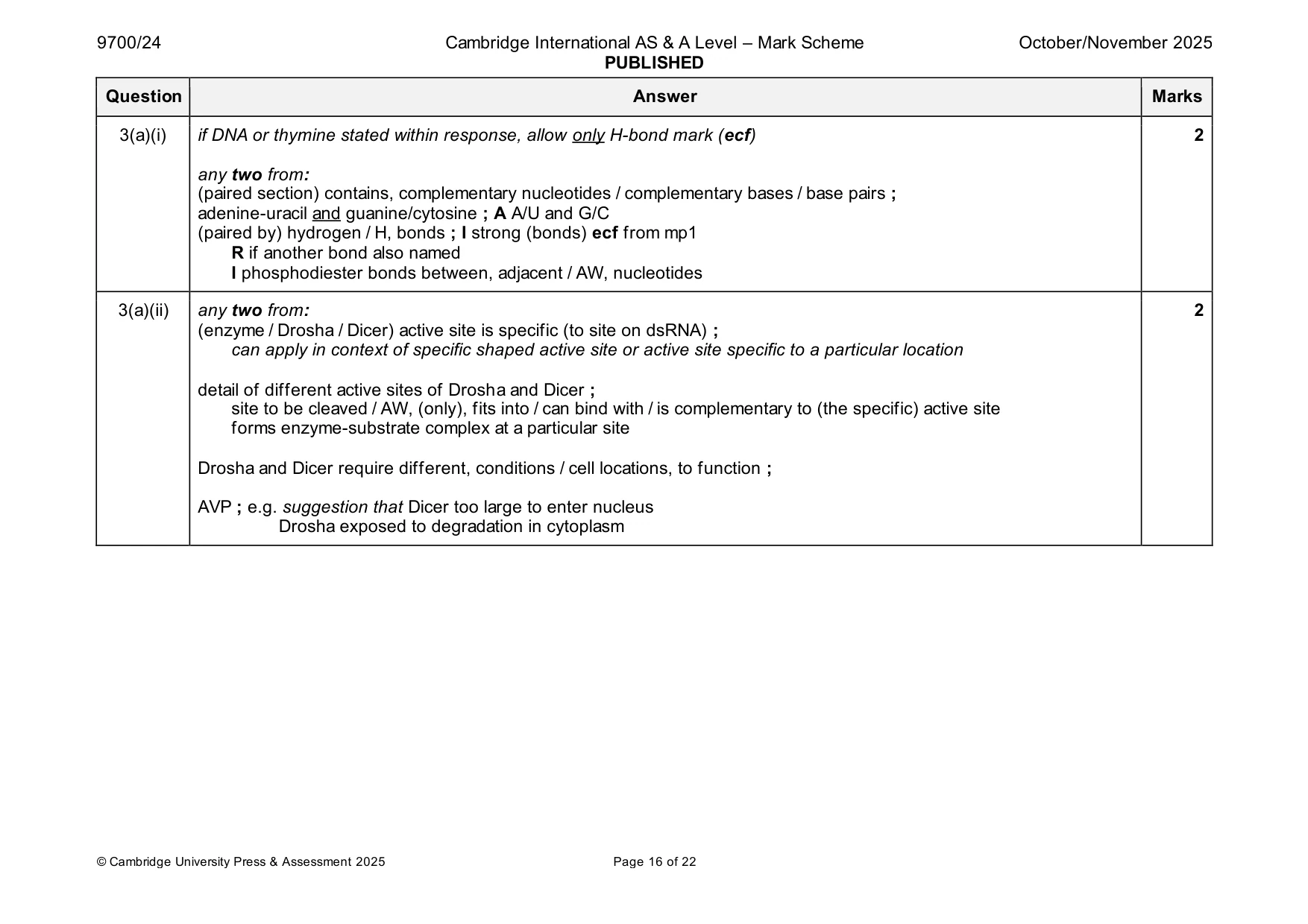
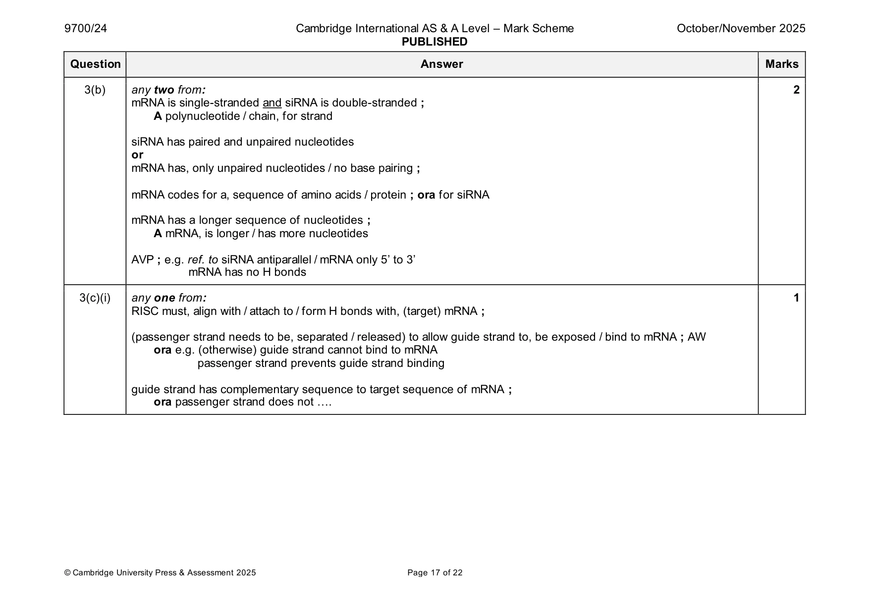
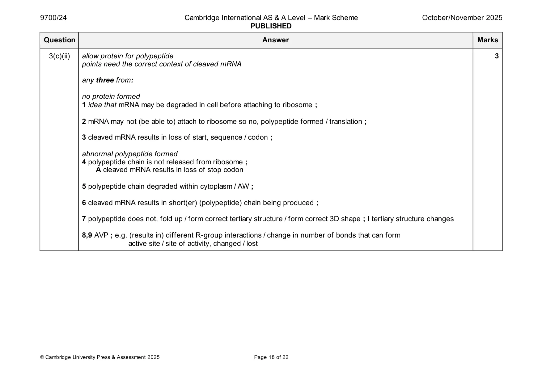
Mark scheme: 3(a)(i) if DNA or thymine stated within response, allow only H-bond mark (ecf) 2 any two from: (paired section) contains, complementary nucleotides / complementary bases / base pairs ; adenine-uracil and guanine/cytosine ; A A/U and G/C (paired by) hydrogen / H, bonds ; I strong (bonds) ecf from mp1 R if another bond also named I phosphodiester bonds between, adjacent / AW, nucleotides 3(a)(ii) any two from: 2 (enzyme / Drosha / Dicer) active site is specific (to site on dsRNA) ; can apply in context of specific shaped active site or active site specific to a particular location detail of different active sites of Drosha and Dicer ; site to be cleaved / AW, (only), fits into / can bind with / is complementary to (the specific) active site forms enzyme-substrate complex at a particular site Drosha and Dicer require different, conditions / cell locations, to function ; AVP ; e.g. suggestion that Dicer too large to enter nucleus Drosha exposed to degradation in cytoplasm 3(b) any two from: 2 mRNA is single-stranded and siRNA is double-stranded ; A polynucleotide / chain, for strand siRNA has paired and unpaired nucleotides or mRNA has, only unpaired nucleotides / no base pairing ; mRNA codes for a, sequence of amino acids / protein ; ora for siRNA mRNA has a longer sequence of nucleotides ; A mRNA, is longer / has more nucleotides AVP ; e.g. ref. to siRNA antiparallel / mRNA only 5’ to 3’ mRNA has no H bonds 3(c)(i) any one from: 1 RISC must, align with / attach to / form H bonds with, (target) mRNA ; (passenger strand needs to be, separated / released) to allow guide strand to, be exposed / bind to mRNA ; AW ora e.g. (otherwise) guide strand cannot bind to mRNA passenger strand prevents guide strand binding guide strand has complementary sequence to target sequence of mRNA ; ora passenger strand does not …. 3(c)(ii) allow protein for polypeptide 3 points need the correct context of cleaved mRNA any three from: no protein formed 1 idea that mRNA may be degraded in cell before attaching to ribosome ; 2 mRNA may not (be able to) attach to ribosome so no, polypeptide formed / translation ; 3 cleaved mRNA results in loss of start, sequence / codon ; abnormal polypeptide formed 4 polypeptide chain is not released from ribosome ; A cleaved mRNA results in loss of stop codon 5 polypeptide chain degraded within cytoplasm / AW ; 6 cleaved mRNA results in short(er) (polypeptide) chain being produced ; 7 polypeptide does not, fold up / form correct tertiary structure / form correct 3D shape ; I tertiary structure changes 8,9 AVP ; e.g. (results in) different R-group interactions / change in number of bonds that can form active site / site of activity, changed / lost
More questions on Structure of nucleic acids and replication of DNA
Q4 · In mammals, the gas exchange system includes a set of branching airways that carry air to…
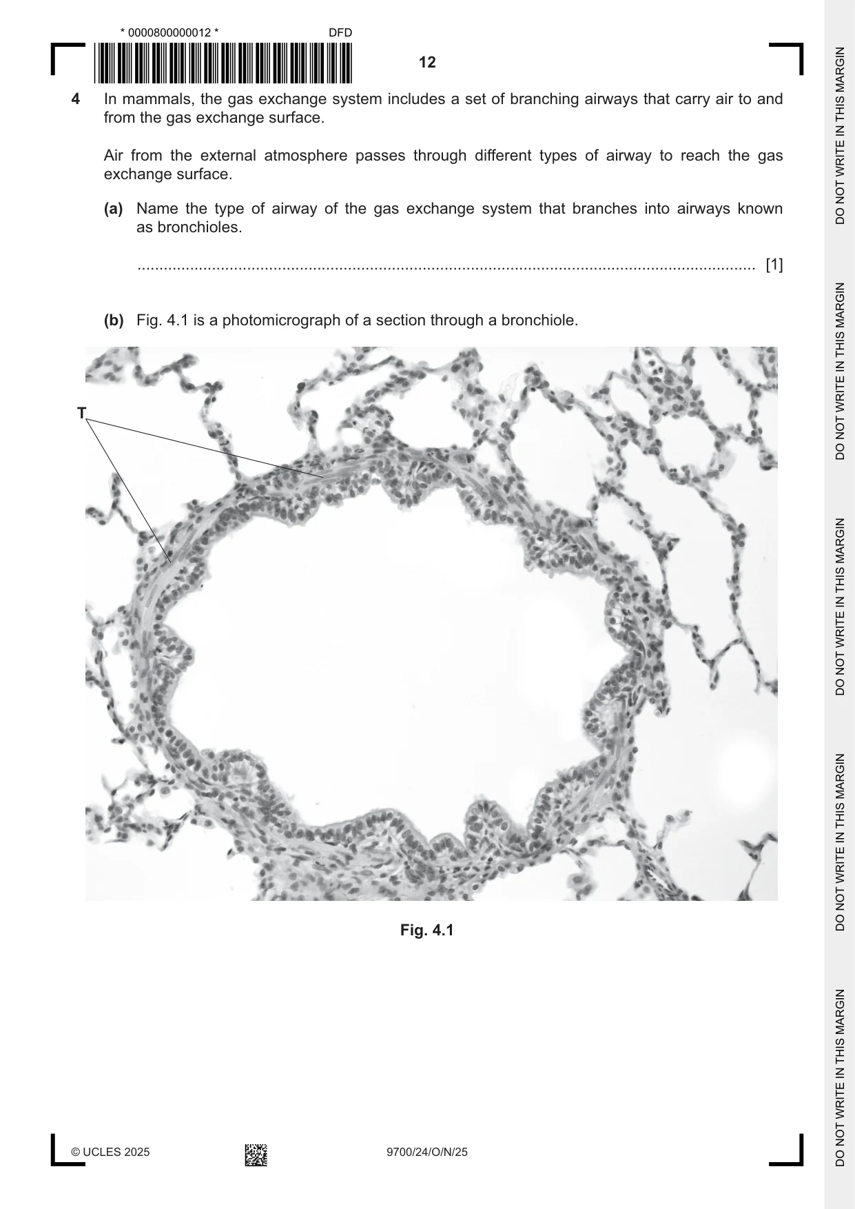
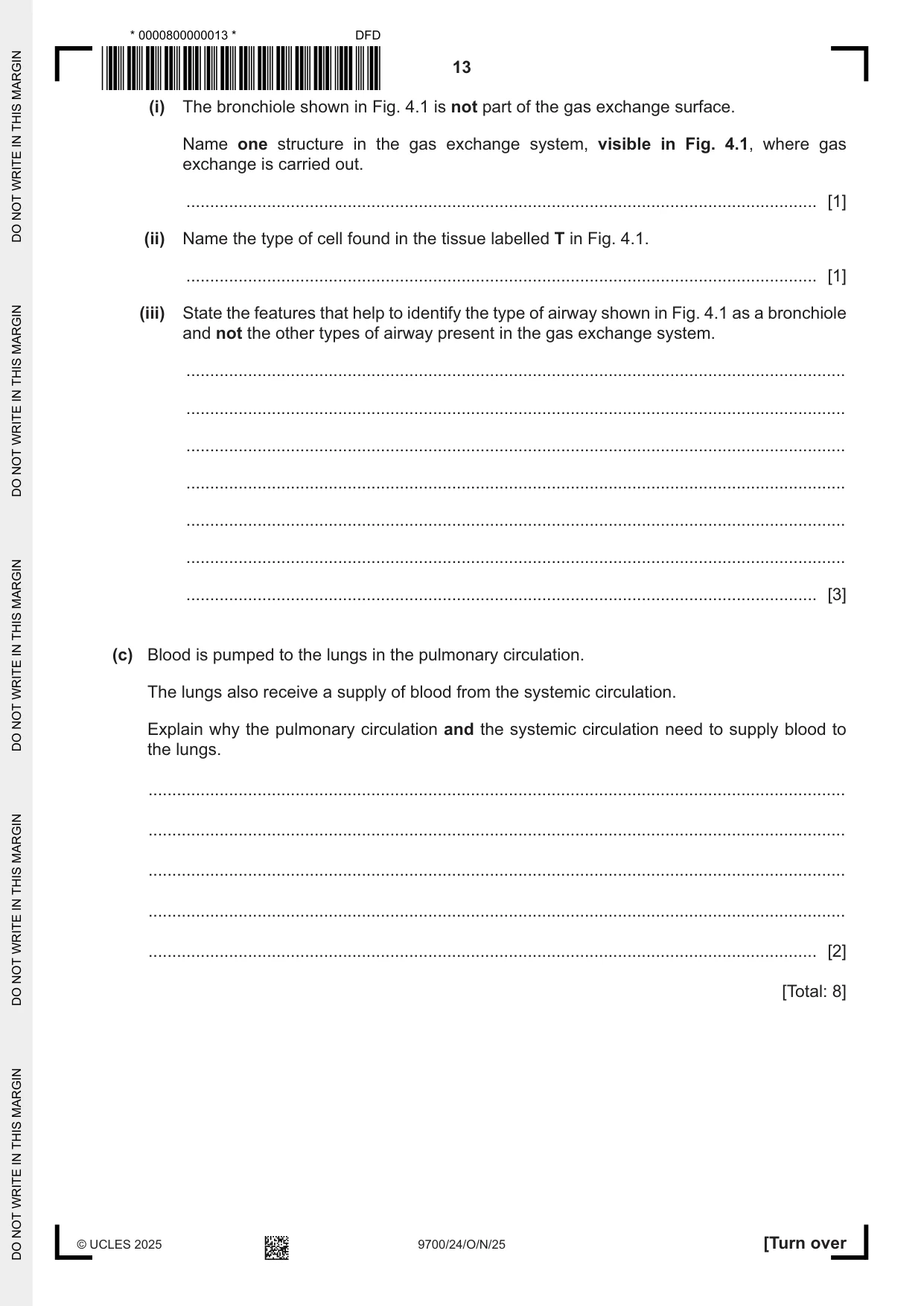
4 In mammals, the gas exchange system includes a set of branching airways that carry air to and from the gas exchange surface. Air from the external atmosphere passes through different types of airway to reach the gas exchange surface. (a) Name the type of airway of the gas exchange system that branches into airways known as bronchioles. ............................................................................................................................................. [1] (b) Fig. 4.1 is a photomicrograph of a section through a bronchiole. T Fig. 4.1 (i) The bronchiole shown in Fig. 4.1 is not part of the gas exchange surface. Name one structure in the gas exchange system, visible in Fig. 4.1, where gas exchange is carried out. ..................................................................................................................................... [1] (ii) Name the type of cell found in the tissue labelled T in Fig. 4.1. ..................................................................................................................................... [1] (iii) State the features that help to identify the type of airway shown in Fig. 4.1 as a bronchiole and not the other types of airway present in the gas exchange system. ........................................................................................................................................... ........................................................................................................................................... ........................................................................................................................................... ........................................................................................................................................... ........................................................................................................................................... ........................................................................................................................................... ..................................................................................................................................... [3] (c) Blood is pumped to the lungs in the pulmonary circulation. The lungs also receive a supply of blood from the systemic circulation. Explain why the pulmonary circulation and the systemic circulation need to supply blood to the lungs. ................................................................................................................................................... ................................................................................................................................................... ................................................................................................................................................... ................................................................................................................................................... ............................................................................................................................................. [2] [Total: 8]
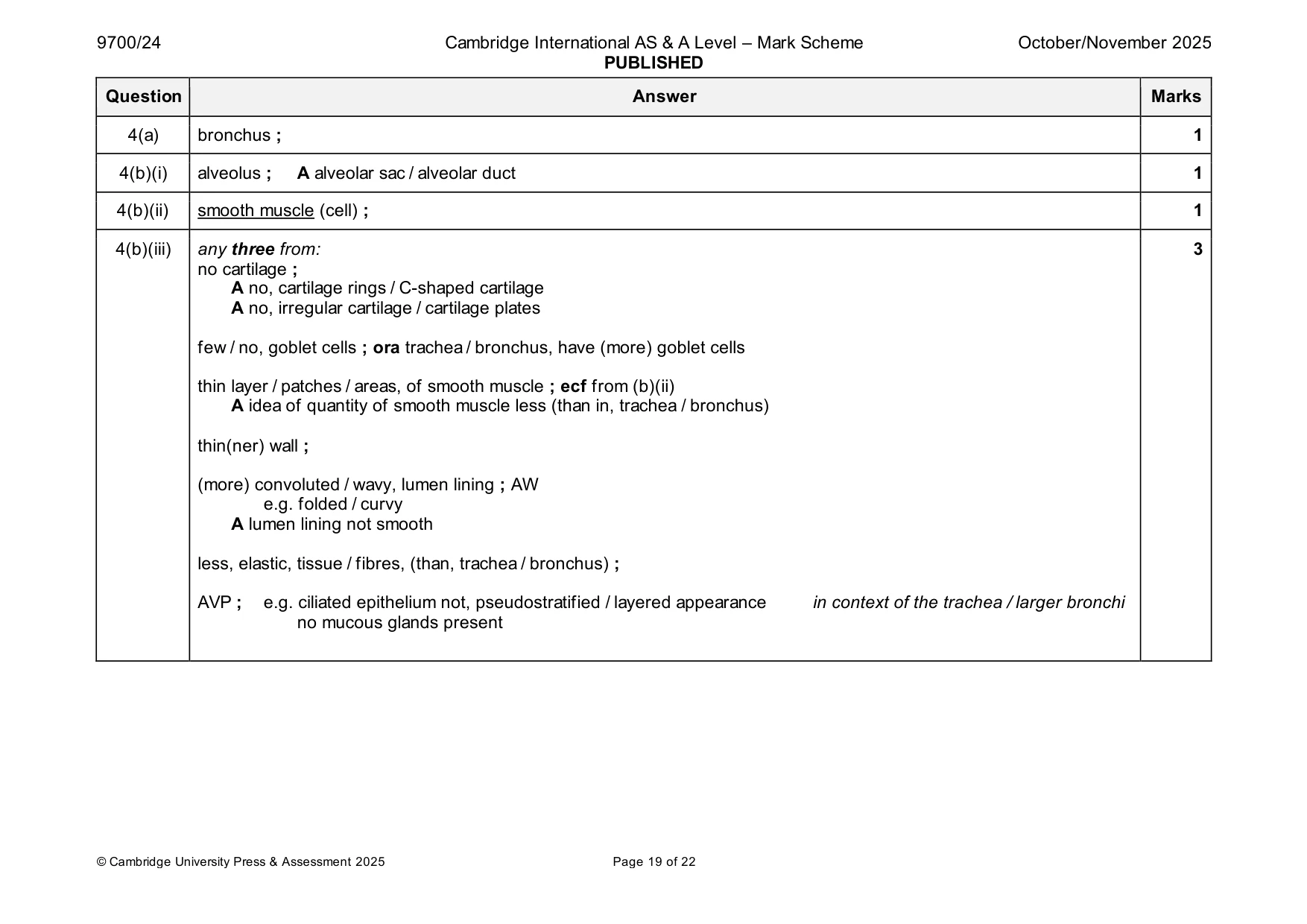
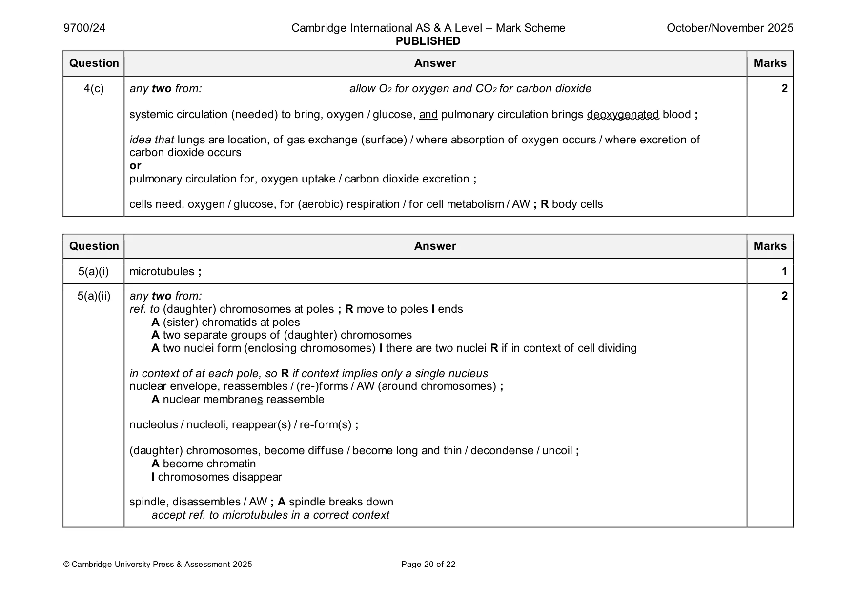
Mark scheme: 4(a) bronchus ; 1 4(b)(i) alveolus ; A alveolar sac / alveolar duct 1 4(b)(ii) smooth muscle (cell) ; 1 4(b)(iii) any three from: 3 no cartilage ; A no, cartilage rings / C-shaped cartilage A no, irregular cartilage / cartilage plates few / no, goblet cells ; ora trachea / bronchus, have (more) goblet cells thin layer / patches / areas, of smooth muscle ; ecf from (b)(ii) A idea of quantity of smooth muscle less (than in, trachea / bronchus) thin(ner) wall ; (more) convoluted / wavy, lumen lining ; AW e.g. folded / curvy A lumen lining not smooth less, elastic, tissue / fibres, (than, trachea / bronchus) ; AVP ; e.g. ciliated epithelium not, pseudostratified / layered appearance in context of the trachea / larger bronchi no mucous glands present 4(c) any two from: allow O2 for oxygen and CO2 for carbon dioxide 2 systemic circulation (needed) to bring, oxygen / glucose, and pulmonary circulation brings deoxygenated blood ; idea that lungs are location, of gas exchange (surface) / where absorption of oxygen occurs / where excretion of carbon dioxide occurs or pulmonary circulation for, oxygen uptake / carbon dioxide excretion ; cells need, oxygen / glucose, for (aerobic) respiration / for cell metabolism / AW ; R body cells
Q5 · Veins transport blood towards the heart
5 Veins transport blood towards the heart. The structure of a vein is adapted to its function. (a) The inner layer of a vein is the tunica intima, composed of a single layer of endothelial cells that form a protective barrier. When the tunica intima of a blood vessel is damaged, endothelial cells can carry out mitosis to allow tissue repair to occur. (i) The spindle that is formed during mitosis is composed of spindle fibres. Name the cell structures that are organised to form spindle fibres during mitosis. ..................................................................................................................................... [1] (ii) Describe the telophase stage of mitosis. ........................................................................................................................................... ........................................................................................................................................... ........................................................................................................................................... ........................................................................................................................................... ........................................................................................................................................... ..................................................................................................................................... [2] (b) Explain how the structure of a vein is related to its function. You do not need to include details of the structure of the tunica intima and its function. ................................................................................................................................................... ................................................................................................................................................... ................................................................................................................................................... ................................................................................................................................................... ................................................................................................................................................... ................................................................................................................................................... ............................................................................................................................................. [3] [Total: 6]
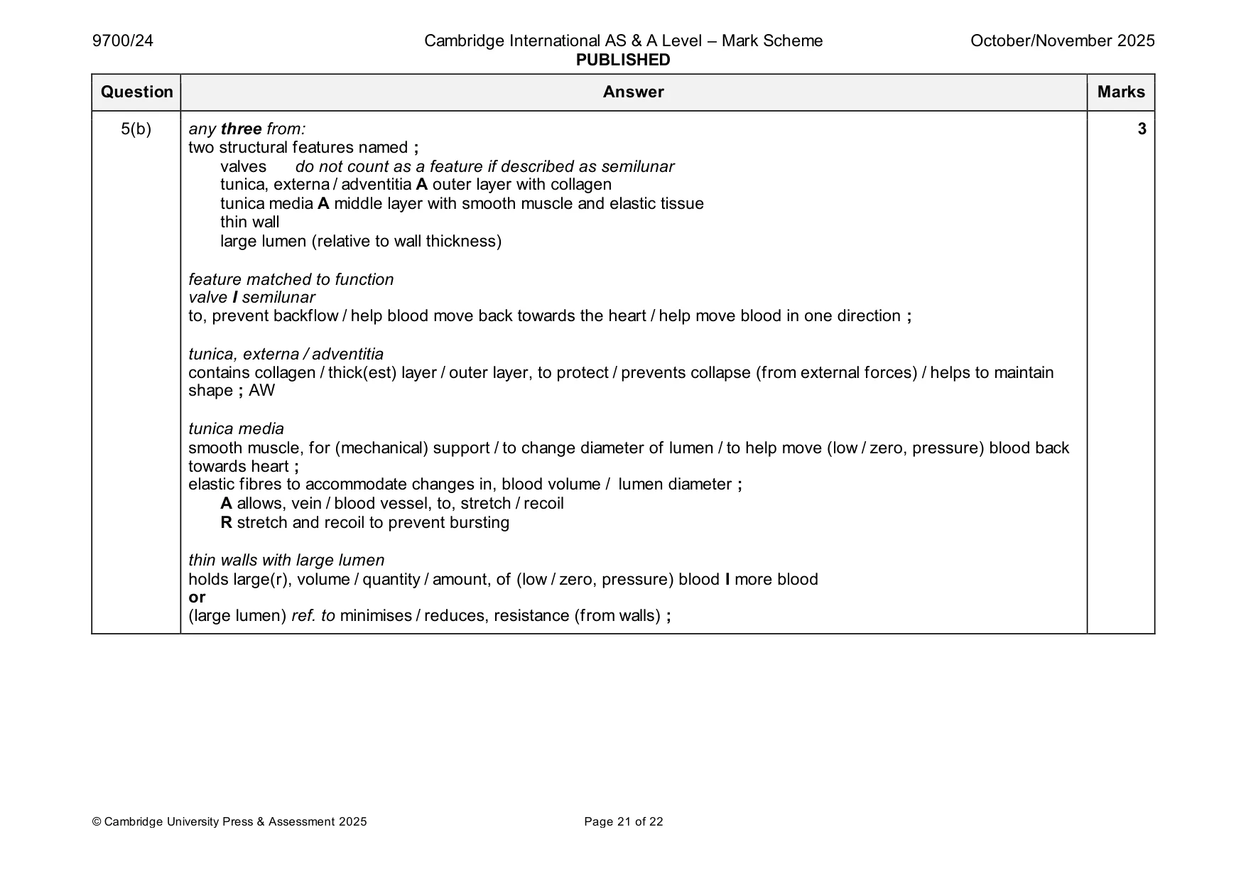
Mark scheme: 5(a)(i) microtubules ; 1 5(a)(ii) any two from: 2 ref. to (daughter) chromosomes at poles ; R move to poles I ends A (sister) chromatids at poles A two separate groups of (daughter) chromosomes A two nuclei form (enclosing chromosomes) I there are two nuclei R if in context of cell dividing in context of at each pole, so R if context implies only a single nucleus nuclear envelope, reassembles / (re-)forms / AW (around chromosomes) ; A nuclear membranes reassemble nucleolus / nucleoli, reappear(s) / re-form(s) ; (daughter) chromosomes, become diffuse / become long and thin / decondense / uncoil ; A become chromatin I chromosomes disappear spindle, disassembles / AW ; A spindle breaks down accept ref. to microtubules in a correct context 5(b) any three from: 3 two structural features named ; valves do not count as a feature if described as semilunar tunica, externa / adventitia A outer layer with collagen tunica media A middle layer with smooth muscle and elastic tissue thin wall large lumen (relative to wall thickness) feature matched to function valve I semilunar to, prevent backflow / help blood move back towards the heart / help move blood in one direction ; tunica, externa / adventitia contains collagen / thick(est) layer / outer layer, to protect / prevents collapse (from external forces) / helps to maintain shape ; AW tunica media smooth muscle, for (mechanical) support / to change diameter of lumen / to help move (low / zero, pressure) blood back towards heart ; elastic fibres to accommodate changes in, blood volume / lumen diameter ; A allows, vein / blood vessel, to, stretch / recoil R stretch and recoil to prevent bursting thin walls with large lumen holds large(r), volume / quantity / amount, of (low / zero, pressure) blood I more blood or (large lumen) ref. to minimises / reduces, resistance (from walls) ;
Q6 · State and explain why the same leaf of a plant can be described as a source or as a sink…
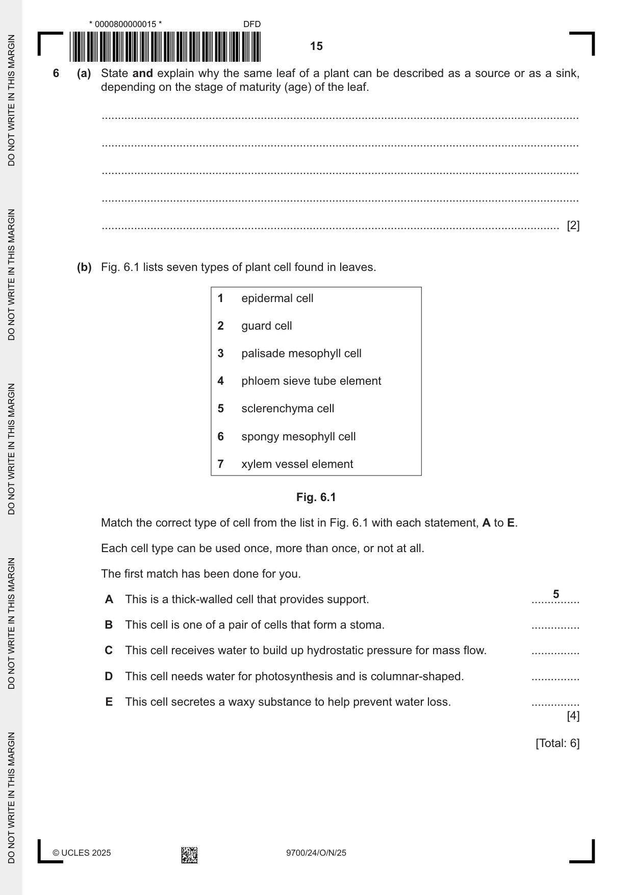
6 (a) State and explain why the same leaf of a plant can be described as a source or as a sink, depending on the stage of maturity (age) of the leaf. ................................................................................................................................................... ................................................................................................................................................... ................................................................................................................................................... ................................................................................................................................................... ............................................................................................................................................. [2] (b) Fig. 6.1 lists seven types of plant cell found in leaves. 1 epidermal cell 2 guard cell 3 palisade mesophyll cell 4 phloem sieve tube element 5 sclerenchyma cell 6 spongy mesophyll cell 7 xylem vessel element Fig. 6.1 Match the correct type of cell from the list in Fig. 6.1 with each statement, A to E. Each cell type can be used once, more than once, or not at all. The first match has been done for you. 5 A This is a thick‑walled cell that provides support. ............... B This cell is one of a pair of cells that form a stoma. ............... C This cell receives water to build up hydrostatic pressure for mass flow. ............... D This cell needs water for photosynthesis and is columnar‑shaped. ............... E This cell secretes a waxy substance to help prevent water loss. ............... [4] [Total: 6]
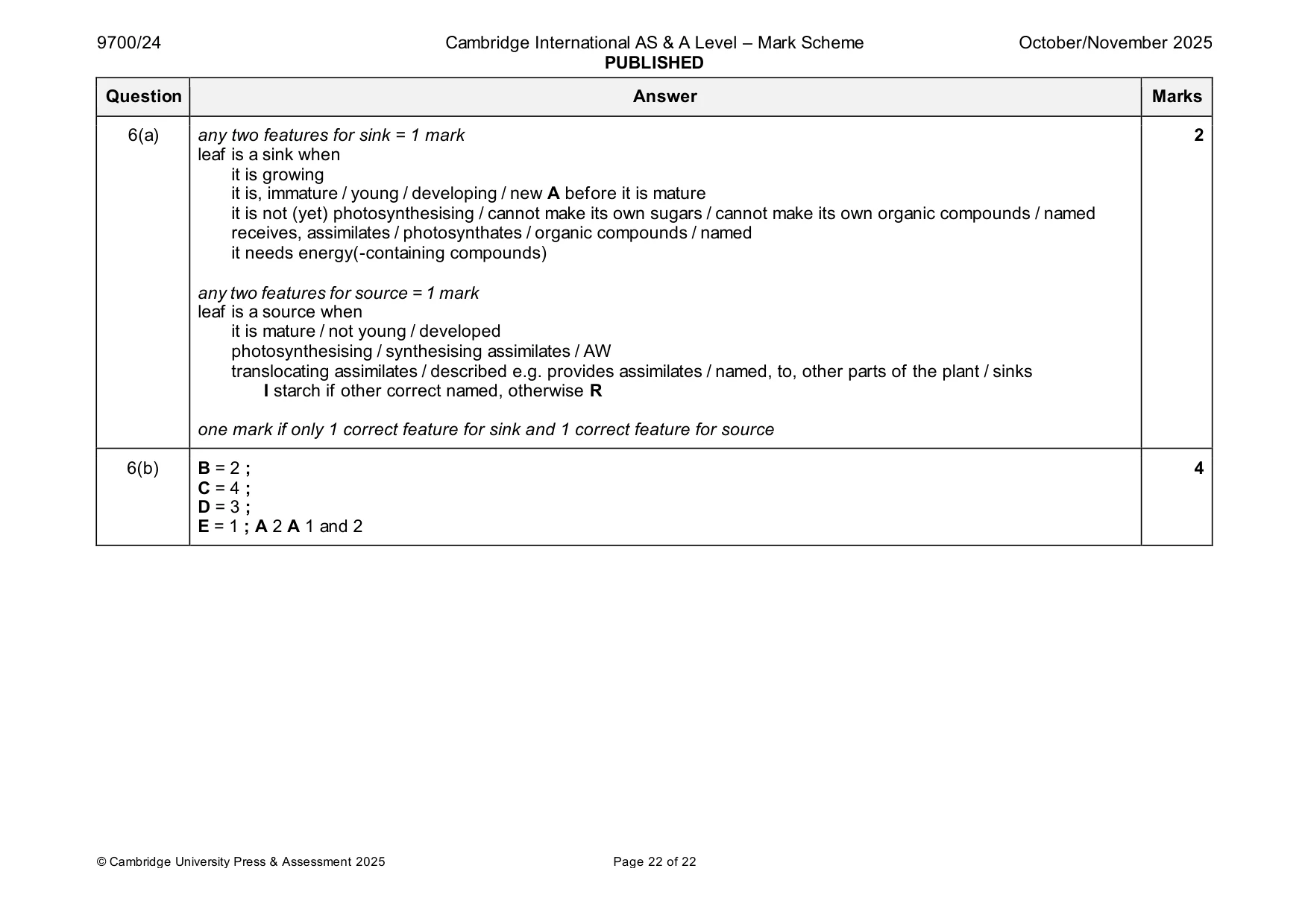
Mark scheme: 6(a) any two features for sink = 1 mark 2 leaf is a sink when it is growing it is, immature / young / developing / new A before it is mature it is not (yet) photosynthesising / cannot make its own sugars / cannot make its own organic compounds / named receives, assimilates / photosynthates / organic compounds / named it needs energy(-containing compounds) any two features for source = 1 mark leaf is a source when it is mature / not young / developed photosynthesising / synthesising assimilates / AW translocating assimilates / described e.g. provides assimilates / named, to, other parts of the plant / sinks I starch if other correct named, otherwise R one mark if only 1 correct feature for sink and 1 correct feature for source 6(b) B = 2 ; 4 C = 4 ; D = 3 ; E = 1 ; A 2 A 1 and 2
More questions on Cells as the basic units of living organisms
What was in this paper
The subtopics covered by these 6 questions, and how many questions each got. Open one in a new tab to see every Cambridge question on it.
What you needed in this session
Cambridge’s own grade thresholds for 2025 Oct/Nov, Paper 2 · Variant 4. A higher threshold means an easier paper — the bar moves with how the cohort did.