Cambridge A Level Biology 9700 — 2024 Oct/Nov Paper 2 · Variant 3
9700/23/O/N/24 · 6 questions · 60 marks · ≈68 min
The question paper and its mark scheme, free to read here and free to download. This is Cambridge’s own paper, exactly as it was sat.
Question paper20 pages
Mark scheme15 pages
Answers below. Sit the paper first if you are practising.
Questions as text
Q1 · The structure of the human gas exchange system
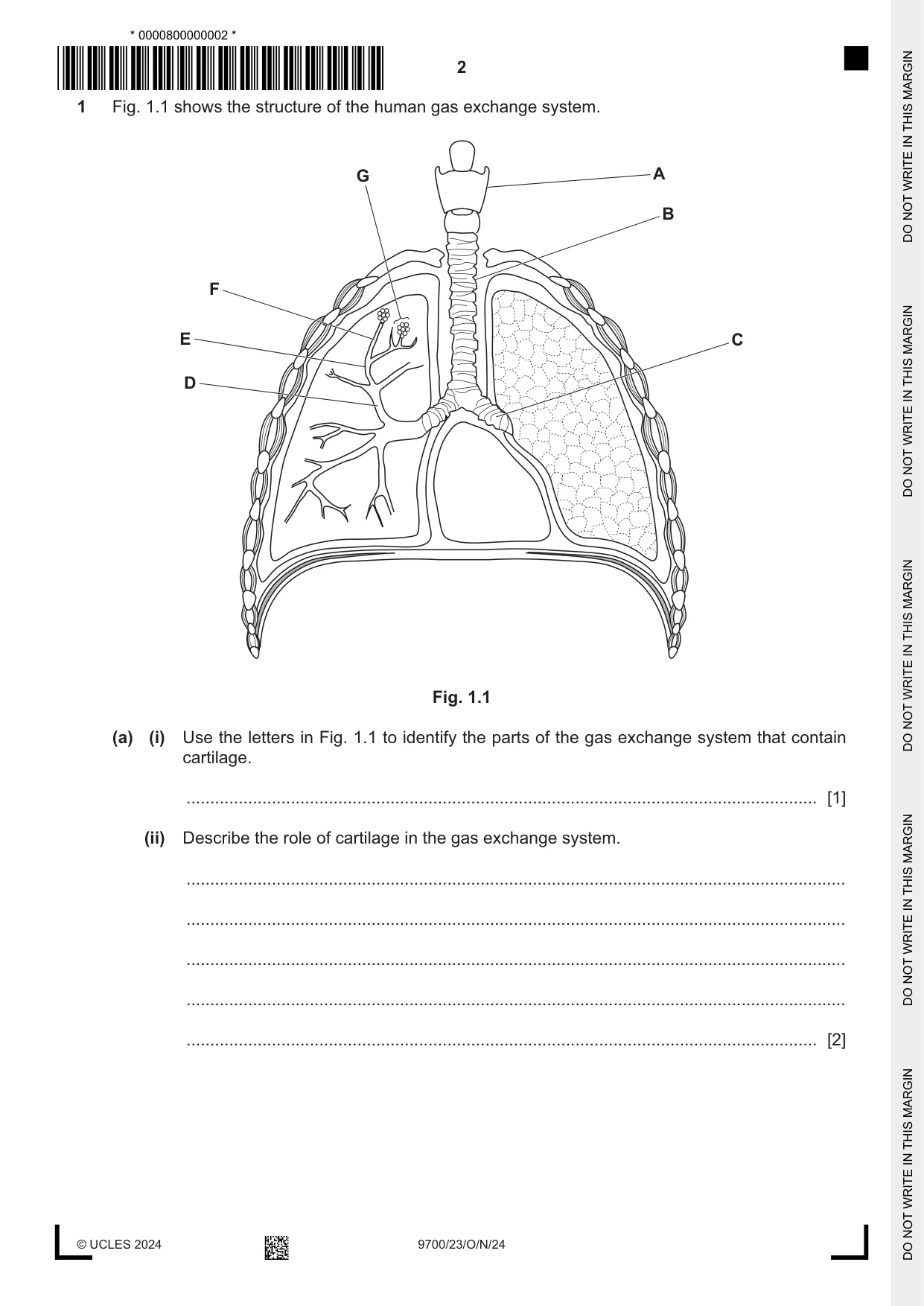
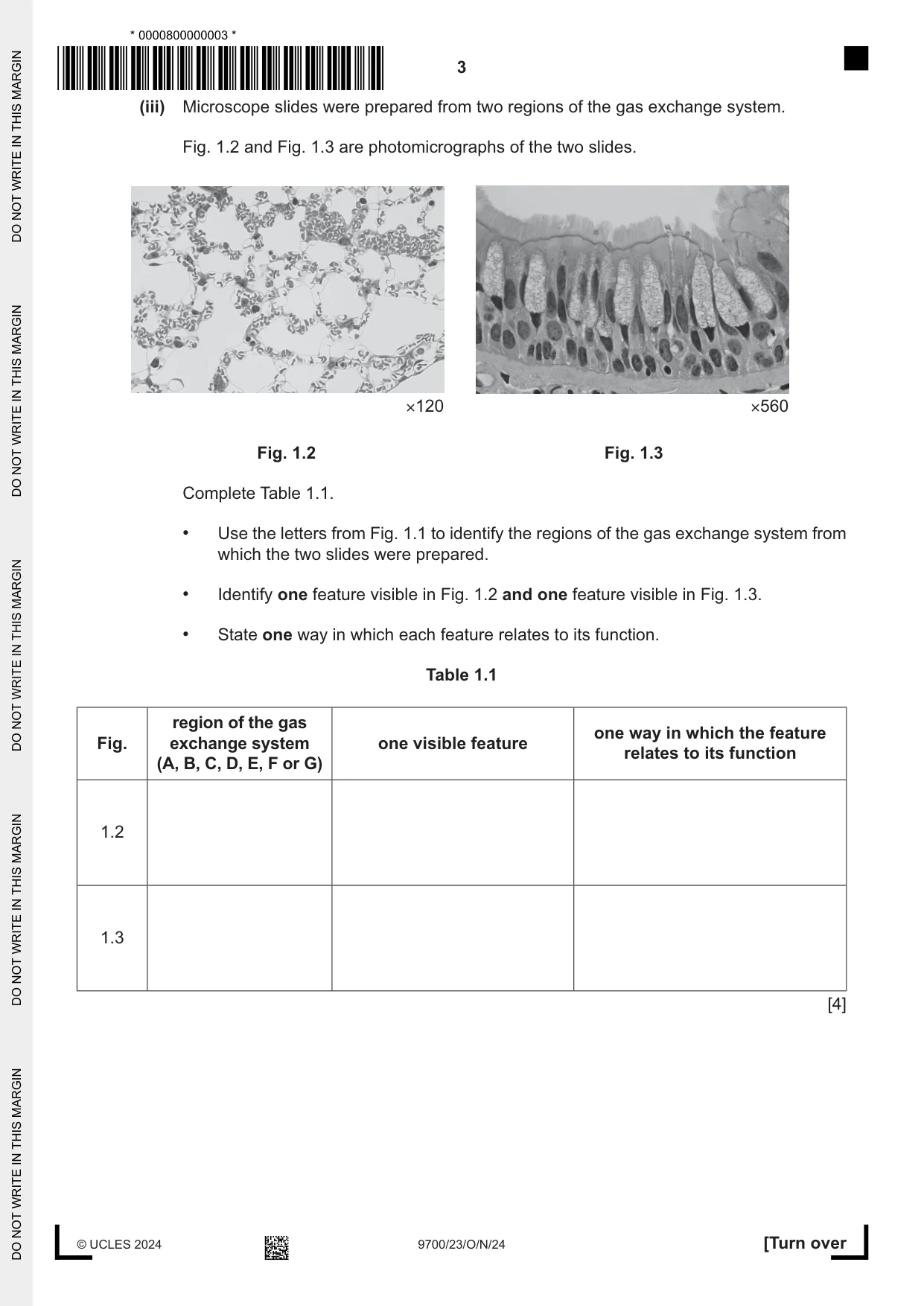
1 Fig. 1.1 shows the structure of the human gas exchange system. G A B F E C D Fig. 1.1 (a) (i) Use the letters in Fig. 1.1 to identify the parts of the gas exchange system that contain cartilage. ..................................................................................................................................... [1] (ii) Describe the role of cartilage in the gas exchange system. ........................................................................................................................................... ........................................................................................................................................... ........................................................................................................................................... ........................................................................................................................................... ..................................................................................................................................... [2] (iii) Microscope slides were prepared from two regions of the gas exchange system. Fig. 1.2 and Fig. 1.3 are photomicrographs of the two slides. ×120 ×560 Fig. 1.2 Fig. 1.3 Complete Table 1.1. • Use the letters from Fig. 1.1 to identify the regions of the gas exchange system from which the two slides were prepared. • Identify one feature visible in Fig. 1.2 and one feature visible in Fig. 1.3. • State one way in which each feature relates to its function. Table 1.1 region of the gas one way in which the feature Fig. exchange system one visible feature relates to its function (A, B, C, D, E, F or G) 1.2 1.3 [4] (b) There is a regular and efficient supply of blood from the heart to the lungs. Describe the sequence of events that occurs in the heart to make sure that there is a regular and efficient supply of blood to the lungs. ................................................................................................................................................... ................................................................................................................................................... ................................................................................................................................................... ................................................................................................................................................... ................................................................................................................................................... ................................................................................................................................................... ................................................................................................................................................... ................................................................................................................................................... ................................................................................................................................................... ................................................................................................................................................... ................................................................................................................................................... ............................................................................................................................................. [5] [Total: 12] Question 2 starts on page 6
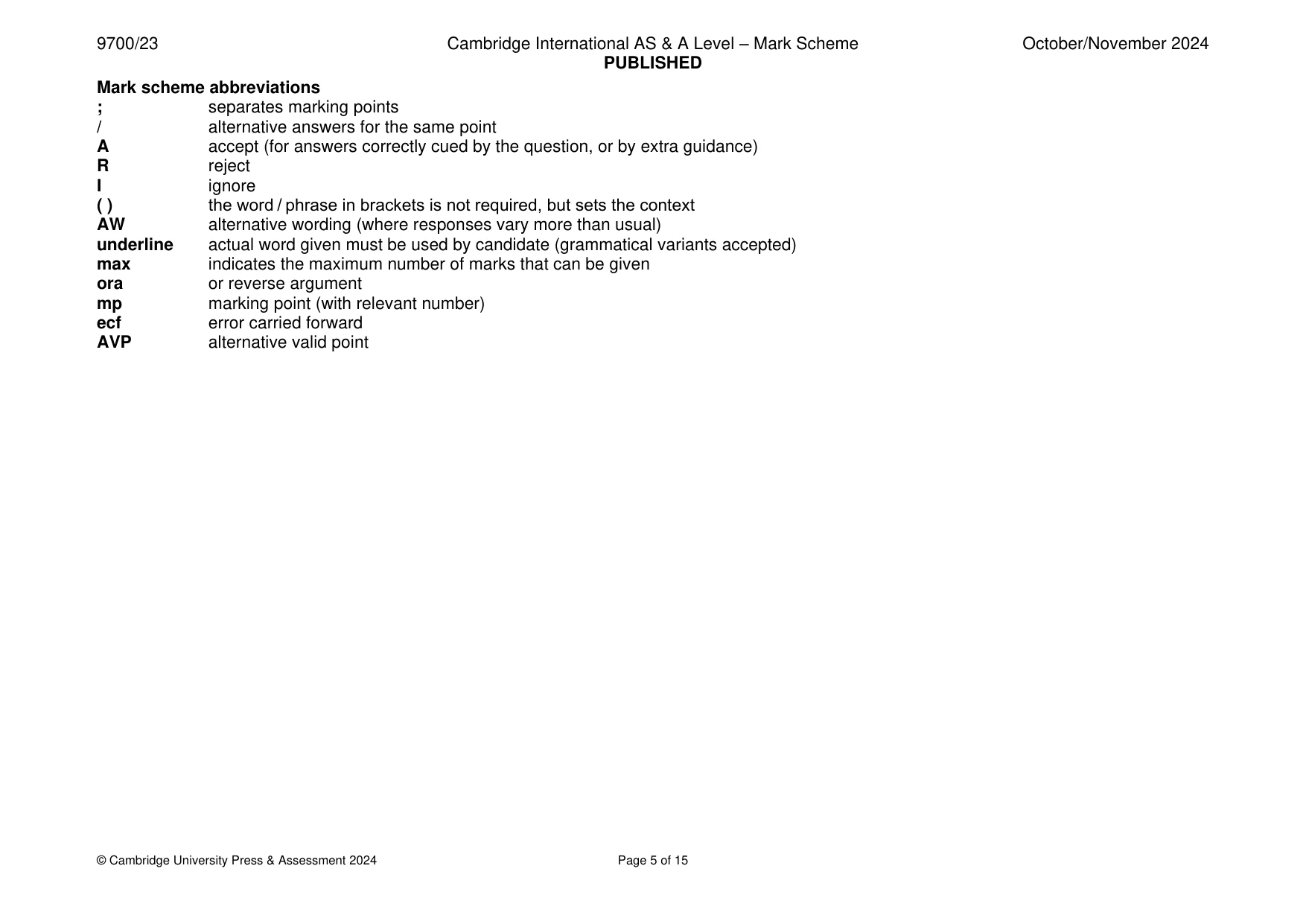
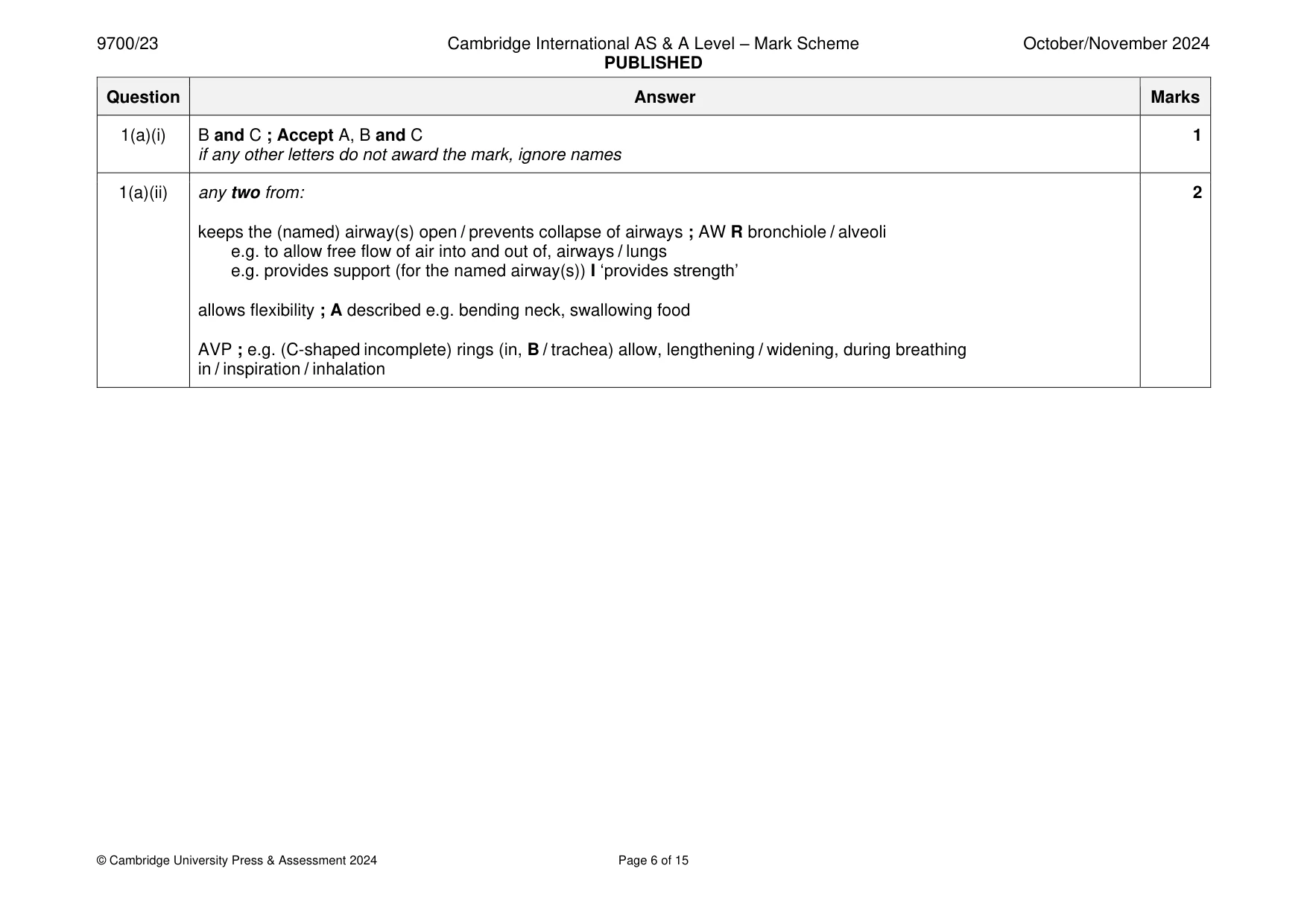
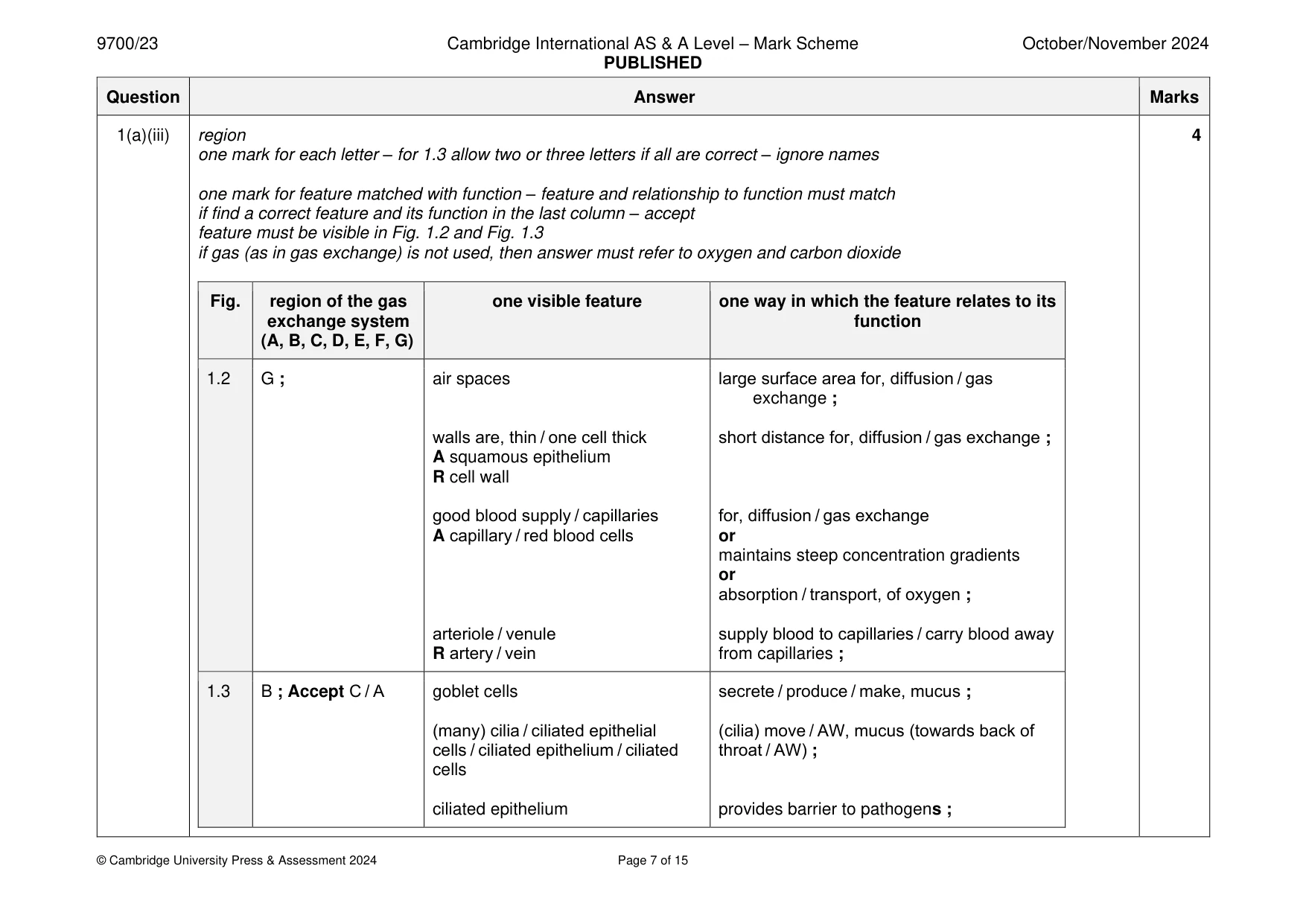
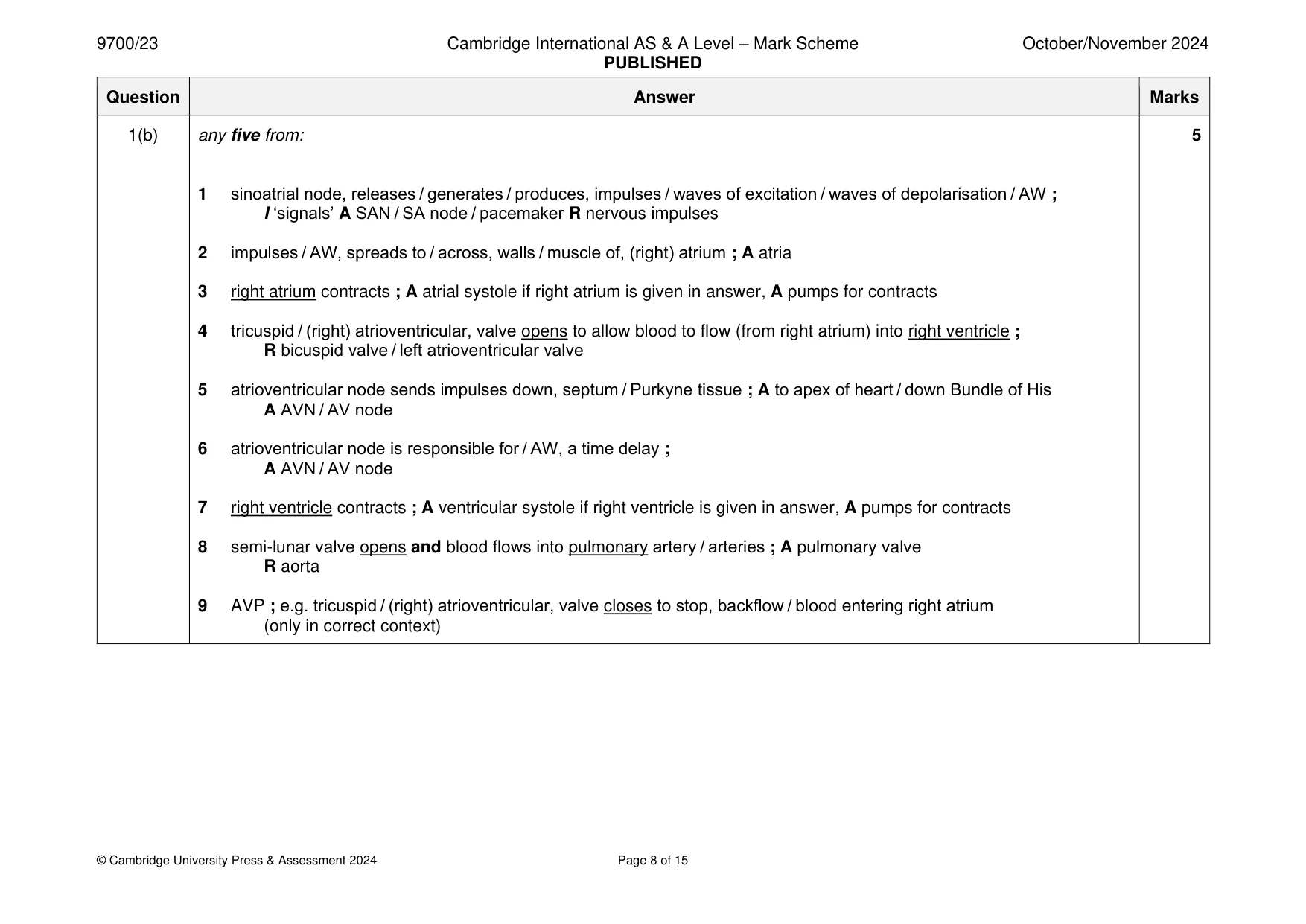
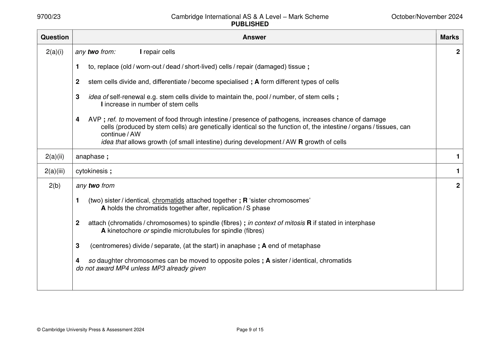
Mark scheme: Question Answer Marks 1(a)(i) B and C ; Accept A, B and C 1 if any other letters do not award the mark, ignore names 1(a)(ii) any two from: 2 keeps the (named) airway(s) open / prevents collapse of airways ; AW R bronchiole / alveoli e.g. to allow free flow of air into and out of, airways / lungs e.g. provides support (for the named airway(s)) I ‘provides strength’ allows flexibility ; A described e.g. bending neck, swallowing food AVP ; e.g. (C-shaped incomplete) rings (in, B / trachea) allow, lengthening / widening, during breathing in / inspiration / inhalation 1(a)(iii) region 4 one mark for each letter – for 1.3 allow two or three letters if all are correct – ignore names one mark for feature matched with function – feature and relationship to function must match if find a correct feature and its function in the last column – accept feature must be visible in Fig. 1.2 and Fig. 1.3 if gas (as in gas exchange) is not used, then answer must refer to oxygen and carbon dioxide Fig. region of the gas one visible feature one way in which the feature relates to its exchange system function (A, B, C, D, E, F, G) 1.2 G ; air spaces large surface area for, diffusion / gas exchange ; walls are, thin / one cell thick short distance for, diffusion / gas exchange ; A squamous epithelium R cell wall good blood supply / capillaries for, diffusion / gas exchange A capillary / red blood cells or maintains steep concentration gradients or absorption / transport, of oxygen ; arteriole / venule supply blood to capillaries / carry blood away R artery / vein from capillaries ; 1.3 B ; Accept C / A goblet cells secrete / produce / make, mucus ; (many) cilia / ciliated epithelial (cilia) move / AW, mucus (towards back of cells / ciliated epithelium / ciliated throat / AW) ; cells ciliated epithelium provides barrier to pathogens ; 1(b) any five from: 5 1 sinoatrial node, releases / generates / produces, impulses / waves of excitation / waves of depolarisation / AW ; I ‘signals’ A SAN / SA node / pacemaker R nervous impulses 2 impulses / AW, spreads to / across, walls / muscle of, (right) atrium ; A atria 3 right atrium contracts ; A atrial systole if right atrium is given in answer, A pumps for contracts 4 tricuspid / (right) atrioventricular, valve opens to allow blood to flow (from right atrium) into right ventricle ; R bicuspid valve / left atrioventricular valve 5 atrioventricular node sends impulses down, septum / Purkyne tissue ; A to apex of heart / down Bundle of His A AVN / AV node 6 atrioventricular node is responsible for / AW, a time delay ; A AVN / AV node 7 right ventricle contracts ; A ventricular systole if right ventricle is given in answer, A pumps for contracts 8 semi-lunar valve opens and blood flows into pulmonary artery / arteries ; A pulmonary valve R aorta 9 AVP ; e.g. tricuspid / (right) atrioventricular, valve closes to stop, backflow / blood entering right atrium (only in correct context)
Q2 · Stem cells are found throughout the human body
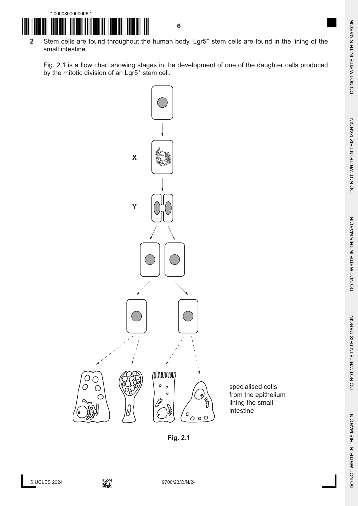
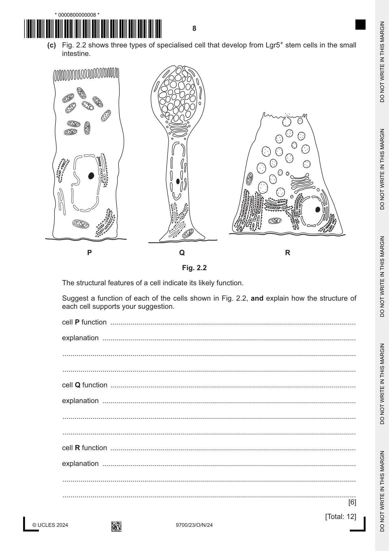
2 Stem cells are found throughout the human body. Lgr5+ stem cells are found in the lining of the small intestine. Fig. 2.1 is a flow chart showing stages in the development of one of the daughter cells produced by the mitotic division of an Lgr5+ stem cell. X Y specialised cells from the epithelium lining the small intestine Fig. 2.1 (a) (i) Explain why stem cells are required in places such as the lining of the small intestine. ........................................................................................................................................... ........................................................................................................................................... ........................................................................................................................................... ........................................................................................................................................... ..................................................................................................................................... [2] (ii) Name the stage of mitosis shown in cell X in Fig. 2.1. ..................................................................................................................................... [1] (iii) State the part of the cell cycle shown at Y in Fig. 2.1. ..................................................................................................................................... [1] (b) Explain the role of centromeres in the cell cycle. ................................................................................................................................................... ................................................................................................................................................... ................................................................................................................................................... ................................................................................................................................................... ............................................................................................................................................. [2] (c) Fig. 2.2 shows three types of specialised cell that develop from Lgr5+ stem cells in the small intestine. P Q R Fig. 2.2 The structural features of a cell indicate its likely function. Suggest a function of each of the cells shown in Fig. 2.2, and explain how the structure of each cell supports your suggestion. cell P function ........................................................................................................................... explanation ............................................................................................................................... ................................................................................................................................................... ................................................................................................................................................... cell Q function ........................................................................................................................... explanation ............................................................................................................................... ................................................................................................................................................... ................................................................................................................................................... cell R function ........................................................................................................................... explanation ............................................................................................................................... ................................................................................................................................................... ................................................................................................................................................... [6] [Total: 12]
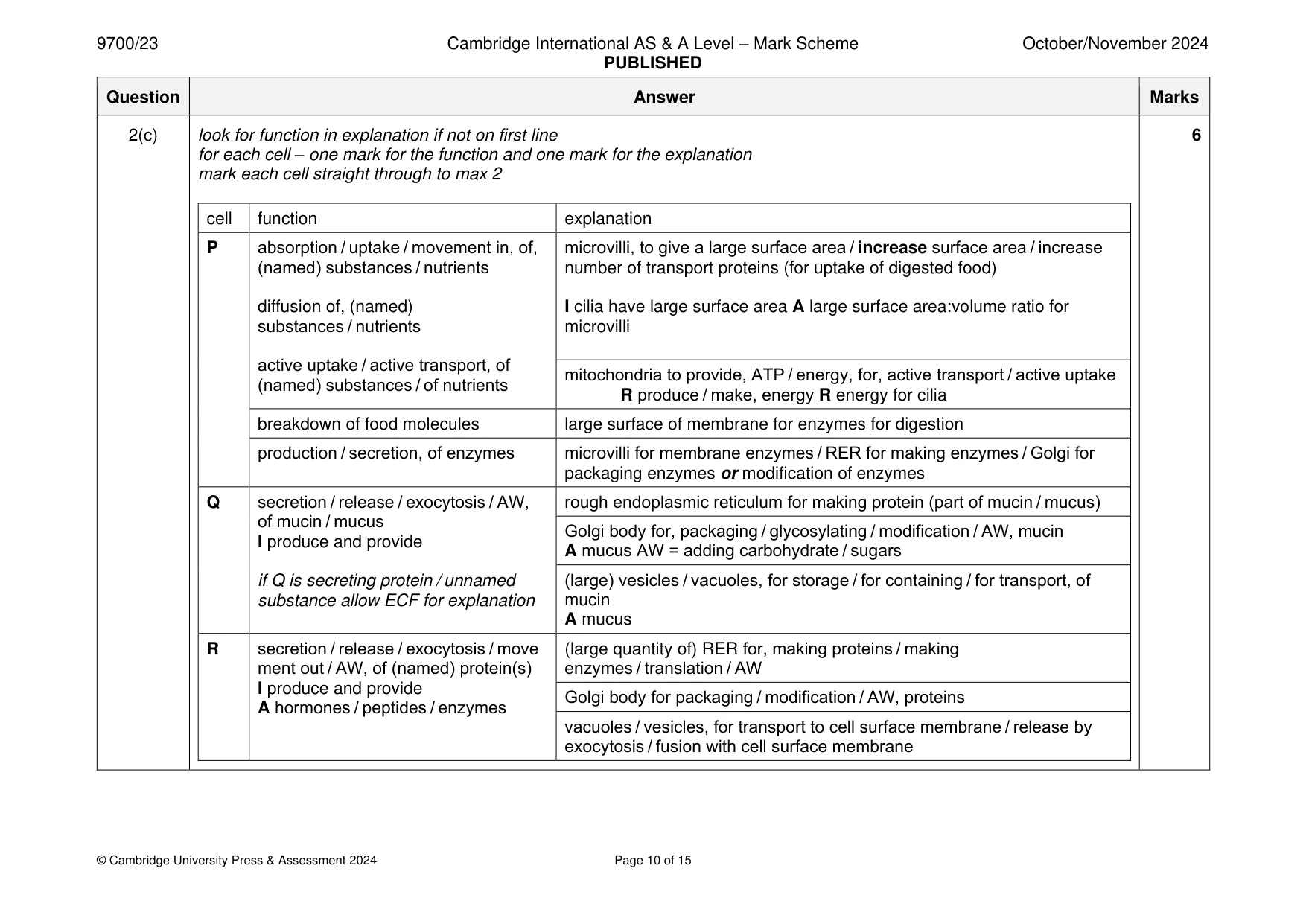
Mark scheme: 2(a)(i) any two from: I repair cells 2 1 to, replace (old / worn-out / dead / short-lived) cells / repair (damaged) tissue ; 2 stem cells divide and, differentiate / become specialised ; A form different types of cells 3 idea of self-renewal e.g. stem cells divide to maintain the, pool / number, of stem cells ; I increase in number of stem cells 4 AVP ; ref. to movement of food through intestine / presence of pathogens, increases chance of damage cells (produced by stem cells) are genetically identical so the function of, the intestine / organs / tissues, can continue / AW idea that allows growth (of small intestine) during development / AW R growth of cells 2(a)(ii) anaphase ; 1 2(a)(iii) cytokinesis ; 1 2(b) any two from 2 1 (two) sister / identical, chromatids attached together ; R ‘sister chromosomes’ A holds the chromatids together after, replication / S phase 2 attach (chromatids / chromosomes) to spindle (fibres) ; in context of mitosis R if stated in interphase A kinetochore or spindle microtubules for spindle (fibres) 3 (centromeres) divide / separate, (at the start) in anaphase ; A end of metaphase 4 so daughter chromosomes can be moved to opposite poles ; A sister / identical, chromatids do not award MP4 unless MP3 already given 2(c) look for function in explanation if not on first line 6 for each cell – one mark for the function and one mark for the explanation mark each cell straight through to max 2 cell function explanation P absorption / uptake / movement in, of, microvilli, to give a large surface area / increase surface area / increase (named) substances / nutrients number of transport proteins (for uptake of digested food) diffusion of, (named) I cilia have large surface area A large surface area:volume ratio for substances / nutrients microvilli active uptake / active transport, of mitochondria to provide, ATP / energy, for, active transport / active uptake (named) substances / of nutrients R produce / make, energy R energy for cilia breakdown of food molecules large surface of membrane for enzymes for digestion production / secretion, of enzymes microvilli for membrane enzymes / RER for making enzymes / Golgi for packaging enzymes or modification of enzymes Q secretion / release / exocytosis / AW, rough endoplasmic reticulum for making protein (part of mucin / mucus) of mucin / mucus Golgi body for, packaging / glycosylating / modification / AW, mucin I produce and provide A mucus AW = adding carbohydrate / sugars if Q is secreting protein / unnamed (large) vesicles / vacuoles, for storage / for containing / for transport, of substance allow ECF for explanation mucin A mucus R secretion / release / exocytosis / move (large quantity of) RER for, making proteins / making ment out / AW, of (named) protein(s) enzymes / translation / AW I produce and provide Golgi body for packaging / modification / AW, proteins A hormones / peptides / enzymes vacuoles / vesicles, for transport to cell surface membrane / release by exocytosis / fusion with cell surface membrane
More questions on Cells as the basic units of living organisms
Q3 · The five bases found in nucleic acids are described as nitrogenous organic compounds
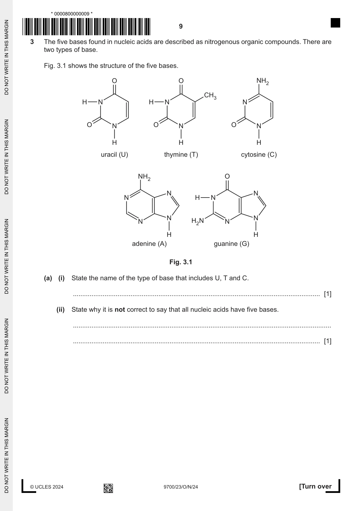
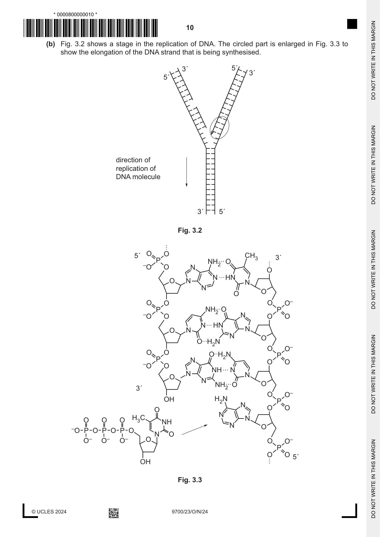
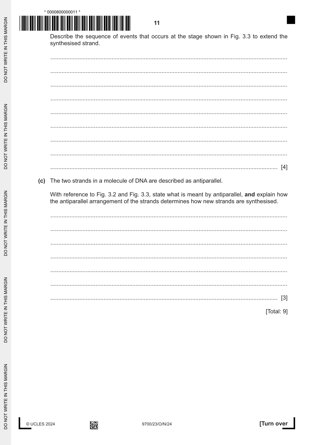
3 The five bases found in nucleic acids are described as nitrogenous organic compounds. There are two types of base. Fig. 3.1 shows the structure of the five bases. O O NH2 CH3 H N H N N O N O N O N H H H uracil (U) thymine (T) cytosine (C) NH2 O N N N H N N N N H2N N H H adenine (A) guanine (G) Fig. 3.1 (a) (i) State the name of the type of base that includes U, T and C. ..................................................................................................................................... [1] (ii) State why it is not correct to say that all nucleic acids have five bases. ........................................................................................................................................... ..................................................................................................................................... [1] (b) Fig. 3.2 shows a stage in the replication of DNA. The circled part is enlarged in Fig. 3.3 to show the elongation of the DNA strand that is being synthesised. 3´ 5´ 3´ 5´ direction of replication of DNA molecule 3´ 5´ Fig. 3.2 5´ O O CH3 3´ P NH2 O –O O N O N HN O N N N O O O O O O– P NH2 O P –O O N O O N HN O N N N O O H2N O O– O O O H2N P P N O O –O O NH N N N O N 3´ NH2 O O O O– OH H2N P N O O O N O O O H3C NH N N O –O P O P O P O N O O– O– O– O O O– P O O 5´ OH Fig. 3.3 Describe the sequence of events that occurs at the stage shown in Fig. 3.3 to extend the synthesised strand. ................................................................................................................................................... ................................................................................................................................................... ................................................................................................................................................... ................................................................................................................................................... ................................................................................................................................................... ................................................................................................................................................... ................................................................................................................................................... ................................................................................................................................................... ............................................................................................................................................. [4] (c) The two strands in a molecule of DNA are described as antiparallel. With reference to Fig. 3.2 and Fig. 3.3, state what is meant by antiparallel, and explain how the antiparallel arrangement of the strands determines how new strands are synthesised. ................................................................................................................................................... ................................................................................................................................................... ................................................................................................................................................... ................................................................................................................................................... ................................................................................................................................................... ................................................................................................................................................... ............................................................................................................................................. [3] [Total: 9]
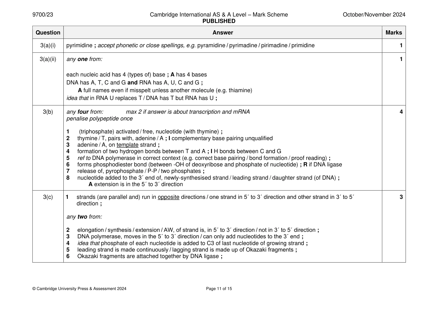
Mark scheme: 3(a)(i) pyrimidine ; accept phonetic or close spellings, e.g. pyramidine / pyrimadine / pirimadine / primidine 1 3(a)(ii) any one from: 1 each nucleic acid has 4 (types of) base ; A has 4 bases DNA has A, T, C and G and RNA has A, U, C and G ; A full names even if misspelt unless another molecule (e.g. thiamine) idea that in RNA U replaces T / DNA has T but RNA has U ; 3(b) any four from: max 2 if answer is about transcription and mRNA 4 penalise polypeptide once 1 (triphosphate) activated / free, nucleotide (with thymine) ; 2 thymine / T, pairs with, adenine / A ; I complementary base pairing unqualified 3 adenine / A, on template strand ; 4 formation of two hydrogen bonds between T and A ; I H bonds between C and G 5 ref to DNA polymerase in correct context (e.g. correct base pairing / bond formation / proof reading) ; 6 forms phosphodiester bond (between -OH of deoxyribose and phosphate of nucleotide) ; R if DNA ligase 7 release of, pyrophosphate / P-P / two phosphates ; 8 nucleotide added to the 3´ end of, newly-synthesised strand / leading strand / daughter strand (of DNA) ; A extension is in the 5´ to 3´ direction 3(c) 1 strands (are parallel and) run in opposite directions / one strand in 5´ to 3´ direction and other strand in 3´ to 5´ 3 direction ; any two from: 2 elongation / synthesis / extension / AW, of strand is, in 5´ to 3´ direction / not in 3´ to 5´ direction ; 3 DNA polymerase, moves in the 5´ to 3´ direction / can only add nucleotides to the 3´ end ; 4 idea that phosphate of each nucleotide is added to C3 of last nucleotide of growing strand ; 5 leading strand is made continuously / lagging strand is made up of Okazaki fragments ; 6 Okazaki fragments are attached together by DNA ligase ;
More questions on Structure of nucleic acids and replication of DNA
Q4 · Biofuels contain alcohols that are produced by the fermentation of sugars derived from…
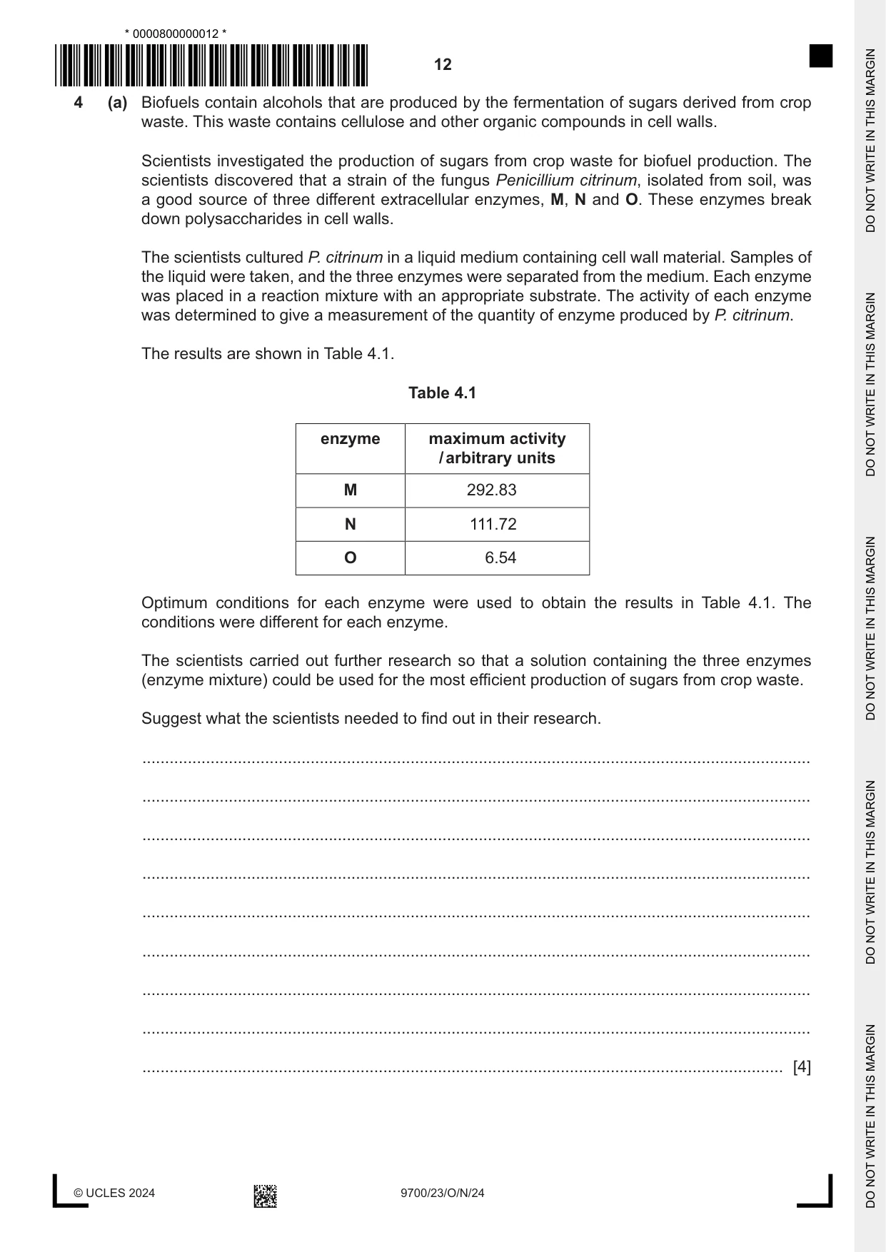
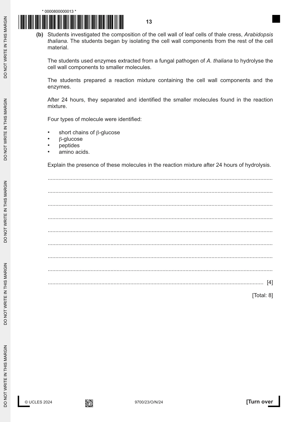
4 (a) Biofuels contain alcohols that are produced by the fermentation of sugars derived from crop waste. This waste contains cellulose and other organic compounds in cell walls. Scientists investigated the production of sugars from crop waste for biofuel production. The scientists discovered that a strain of the fungus Penicillium citrinum, isolated from soil, was a good source of three different extracellular enzymes, M, N and O. These enzymes break down polysaccharides in cell walls. The scientists cultured P. citrinum in a liquid medium containing cell wall material. Samples of the liquid were taken, and the three enzymes were separated from the medium. Each enzyme was placed in a reaction mixture with an appropriate substrate. The activity of each enzyme was determined to give a measurement of the quantity of enzyme produced by P. citrinum. The results are shown in Table 4.1. Table 4.1 enzyme maximum activity / arbitrary units M 292.83 N 111.72 O 6.54 Optimum conditions for each enzyme were used to obtain the results in Table 4.1. The conditions were different for each enzyme. The scientists carried out further research so that a solution containing the three enzymes (enzyme mixture) could be used for the most efficient production of sugars from crop waste. Suggest what the scientists needed to find out in their research. ................................................................................................................................................... ................................................................................................................................................... ................................................................................................................................................... ................................................................................................................................................... ................................................................................................................................................... ................................................................................................................................................... ................................................................................................................................................... ................................................................................................................................................... ............................................................................................................................................. [4] (b) Students investigated the composition of the cell wall of leaf cells of thale cress, Arabidopsis thaliana. The students began by isolating the cell wall components from the rest of the cell material. The students used enzymes extracted from a fungal pathogen of A. thaliana to hydrolyse the cell wall components to smaller molecules. The students prepared a reaction mixture containing the cell wall components and the enzymes. After 24 hours, they separated and identified the smaller molecules found in the reaction mixture. Four types of molecule were identified: • short chains of β-glucose • β-glucose • peptides • amino acids. Explain the presence of these molecules in the reaction mixture after 24 hours of hydrolysis. ................................................................................................................................................... ................................................................................................................................................... ................................................................................................................................................... ................................................................................................................................................... ................................................................................................................................................... ................................................................................................................................................... ................................................................................................................................................... ................................................................................................................................................... ............................................................................................................................................. [4] [Total: 8]
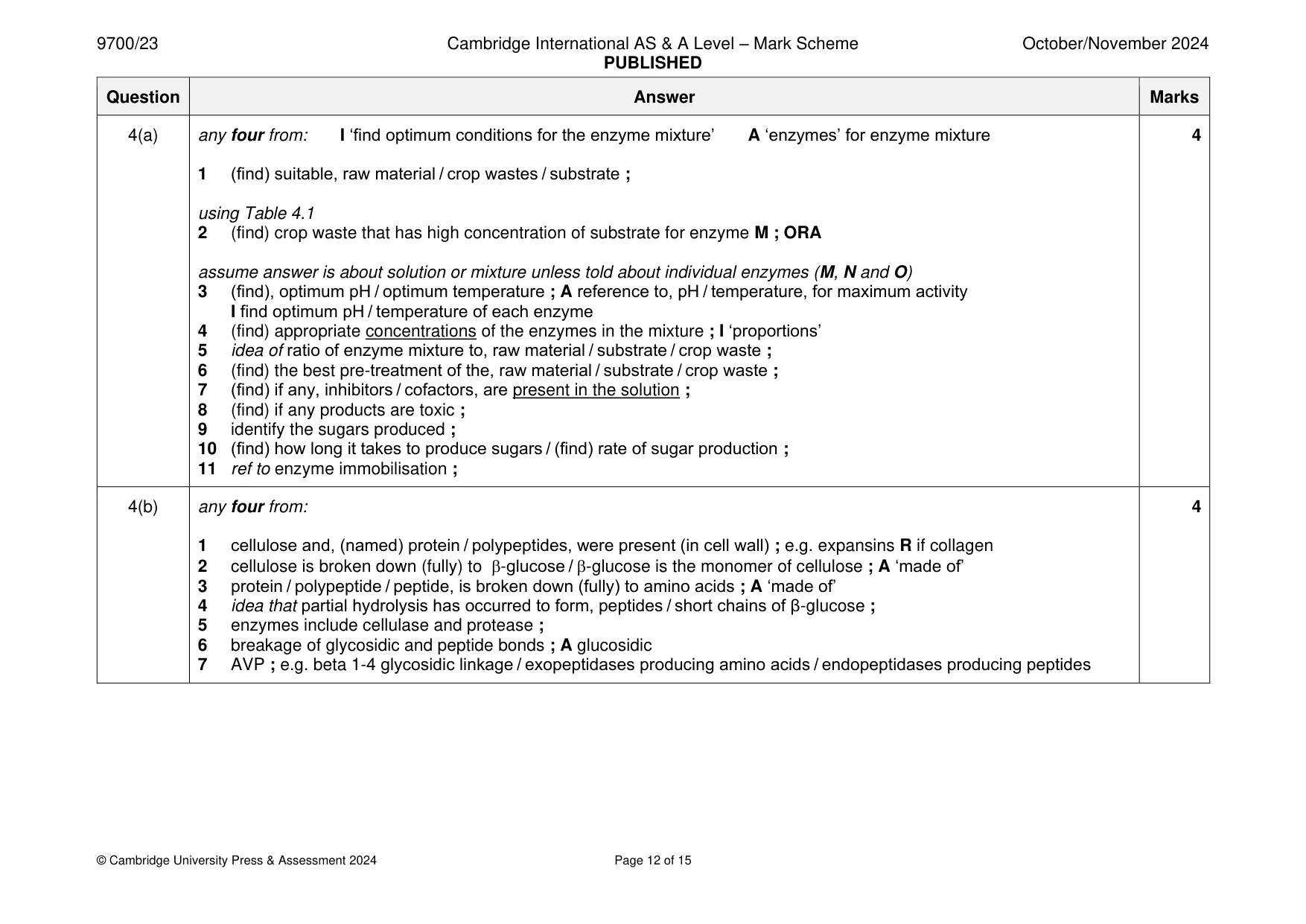
Mark scheme: 4(a) any four from: I ‘find optimum conditions for the enzyme mixture’ A ‘enzymes’ for enzyme mixture 4 1 (find) suitable, raw material / crop wastes / substrate ; using Table 4.1 2 (find) crop waste that has high concentration of substrate for enzyme M ; ORA assume answer is about solution or mixture unless told about individual enzymes (M, N and O) 3 (find), optimum pH / optimum temperature ; A reference to, pH / temperature, for maximum activity I find optimum pH / temperature of each enzyme 4 (find) appropriate concentrations of the enzymes in the mixture ; I ‘proportions’ 5 idea of ratio of enzyme mixture to, raw material / substrate / crop waste ; 6 (find) the best pre-treatment of the, raw material / substrate / crop waste ; 7 (find) if any, inhibitors / cofactors, are present in the solution ; 8 (find) if any products are toxic ; 9 identify the sugars produced ; 10 (find) how long it takes to produce sugars / (find) rate of sugar production ; 11 ref to enzyme immobilisation ; 4(b) any four from: 4 1 cellulose and, (named) protein / polypeptides, were present (in cell wall) ; e.g. expansins R if collagen 2 cellulose is broken down (fully) to -glucose / -glucose is the monomer of cellulose ; A ‘made of’ 3 protein / polypeptide / peptide, is broken down (fully) to amino acids ; A ‘made of’ 4 idea that partial hydrolysis has occurred to form, peptides / short chains of β-glucose ; 5 enzymes include cellulase and protease ; 6 breakage of glycosidic and peptide bonds ; A glucosidic 7 AVP ; e.g. beta 1-4 glycosidic linkage / exopeptidases producing amino acids / endopeptidases producing peptides
Q5 · T-lymphocytes are produced in bone marrow and mature in the thymus gland
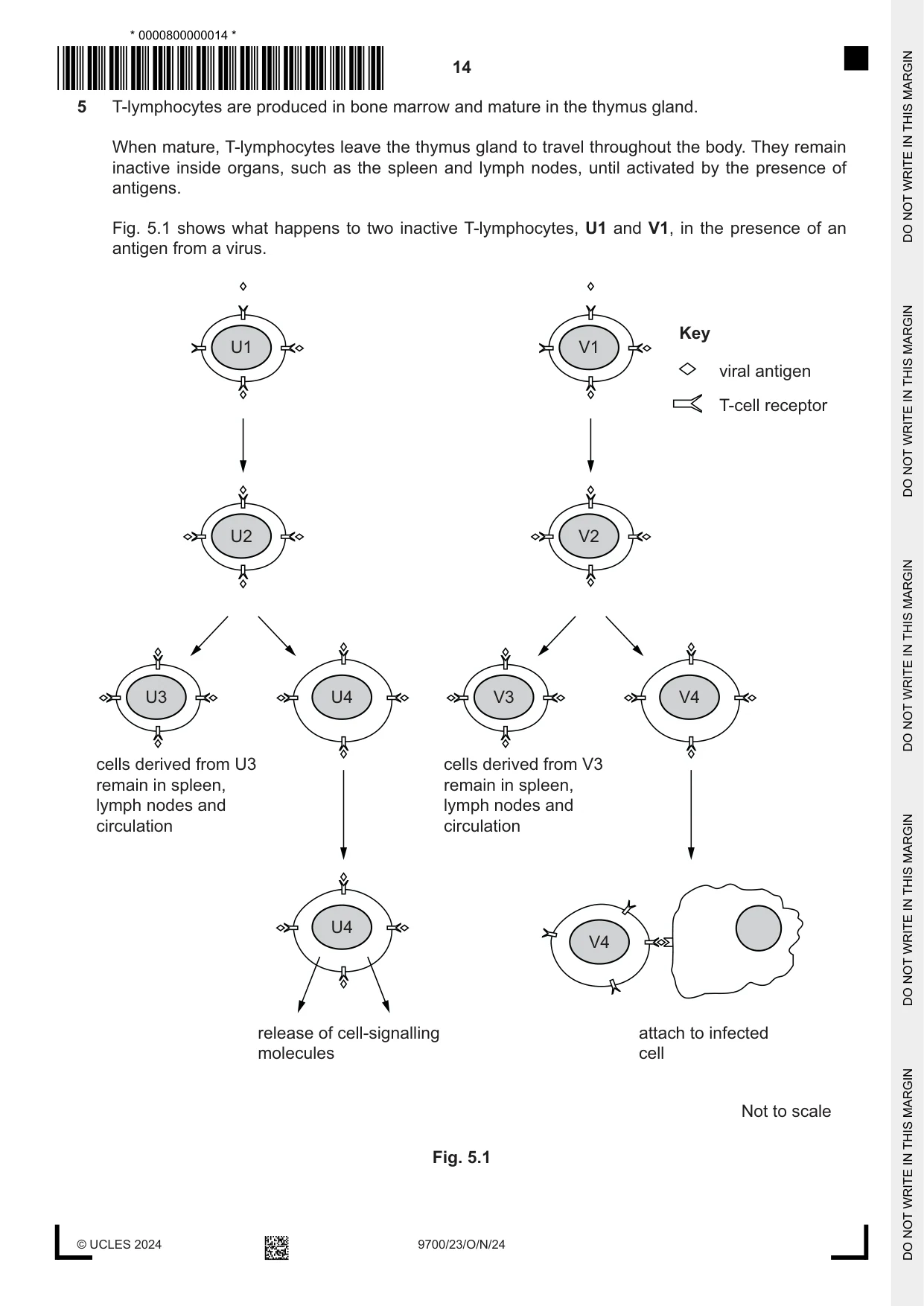
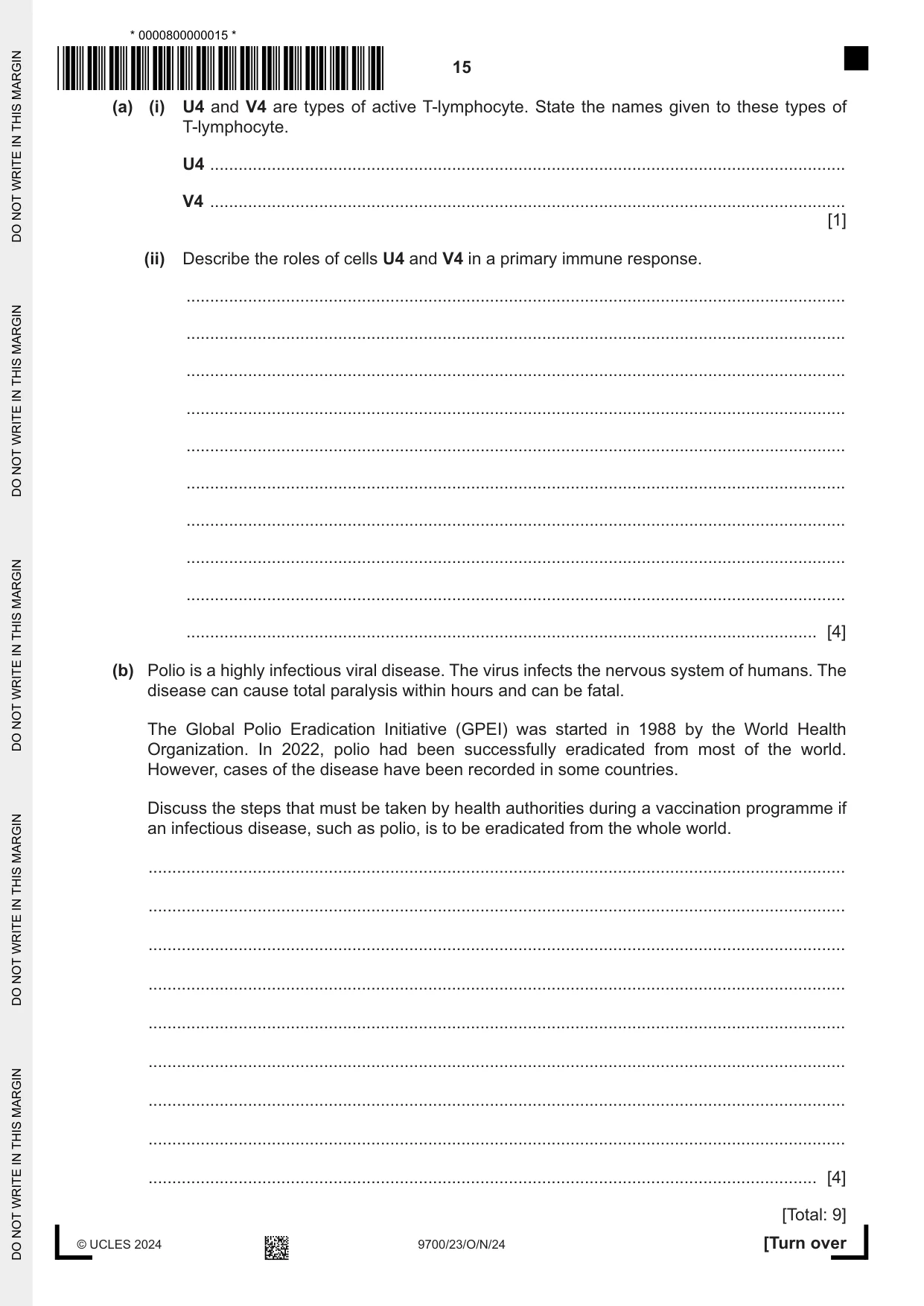
5 T-lymphocytes are produced in bone marrow and mature in the thymus gland. When mature, T-lymphocytes leave the thymus gland to travel throughout the body. They remain inactive inside organs, such as the spleen and lymph nodes, until activated by the presence of antigens. Fig. 5.1 shows what happens to two inactive T-lymphocytes, U1 and V1, in the presence of an antigen from a virus. Key U1 V1 viral antigen T-cell receptor U2 V2 U3 U4 V3 V4 cells derived from U3 cells derived from V3 remain in spleen, remain in spleen, lymph nodes and lymph nodes and circulation circulation U4 V4 release of cell-signalling attach to infected molecules cell Not to scale Fig. 5.1 (a) (i) U4 and V4 are types of active T-lymphocyte. State the names given to these types of T-lymphocyte. U4 ...................................................................................................................................... V4 ...................................................................................................................................... [1] (ii) Describe the roles of cells U4 and V4 in a primary immune response. ........................................................................................................................................... ........................................................................................................................................... ........................................................................................................................................... ........................................................................................................................................... ........................................................................................................................................... ........................................................................................................................................... ........................................................................................................................................... ........................................................................................................................................... ........................................................................................................................................... ..................................................................................................................................... [4] (b) Polio is a highly infectious viral disease. The virus infects the nervous system of humans. The disease can cause total paralysis within hours and can be fatal. The Global Polio Eradication Initiative (GPEI) was started in 1988 by the World Health Organization. In 2022, polio had been successfully eradicated from most of the world. However, cases of the disease have been recorded in some countries. Discuss the steps that must be taken by health authorities during a vaccination programme if an infectious disease, such as polio, is to be eradicated from the whole world. ................................................................................................................................................... ................................................................................................................................................... ................................................................................................................................................... ................................................................................................................................................... ................................................................................................................................................... ................................................................................................................................................... ................................................................................................................................................... ................................................................................................................................................... ............................................................................................................................................. [4] [Total: 9]
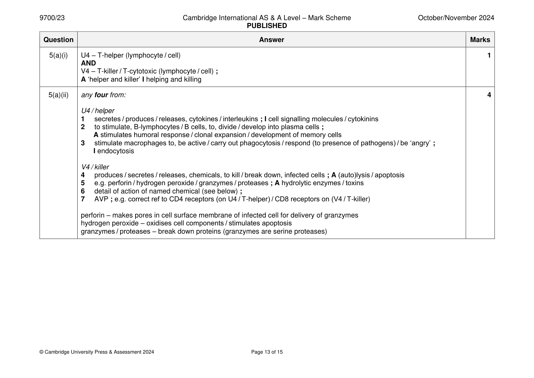
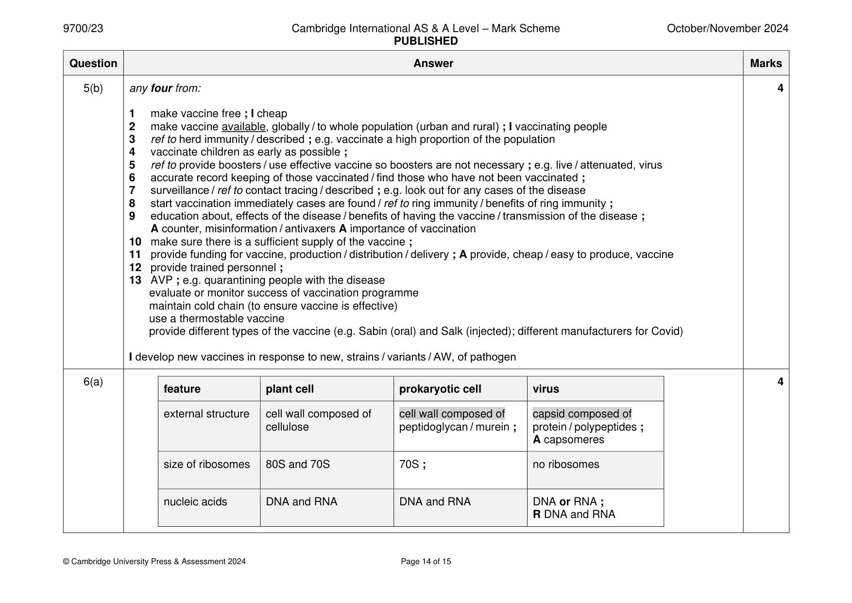
Mark scheme: 5(a)(i) U4 – T-helper (lymphocyte / cell) 1 AND V4 – T-killer / T-cytotoxic (lymphocyte / cell) ; A ‘helper and killer’ I helping and killing 5(a)(ii) any four from: 4 U4 / helper 1 secretes / produces / releases, cytokines / interleukins ; I cell signalling molecules / cytokinins 2 to stimulate, B-lymphocytes / B cells, to, divide / develop into plasma cells ; A stimulates humoral response / clonal expansion / development of memory cells 3 stimulate macrophages to, be active / carry out phagocytosis / respond (to presence of pathogens) / be ‘angry’ ; I endocytosis V4 / killer 4 produces / secretes / releases, chemicals, to kill / break down, infected cells ; A (auto)lysis / apoptosis 5 e.g. perforin / hydrogen peroxide / granzymes / proteases ; A hydrolytic enzymes / toxins 6 detail of action of named chemical (see below) ; 7 AVP ; e.g. correct ref to CD4 receptors (on U4 / T-helper) / CD8 receptors on (V4 / T-killer) perforin – makes pores in cell surface membrane of infected cell for delivery of granzymes hydrogen peroxide – oxidises cell components / stimulates apoptosis granzymes / proteases – break down proteins (granzymes are serine proteases) 5(b) any four from: 4 1 make vaccine free ; I cheap 2 make vaccine available, globally / to whole population (urban and rural) ; I vaccinating people 3 ref to herd immunity / described ; e.g. vaccinate a high proportion of the population 4 vaccinate children as early as possible ; 5 ref to provide boosters / use effective vaccine so boosters are not necessary ; e.g. live / attenuated, virus 6 accurate record keeping of those vaccinated / find those who have not been vaccinated ; 7 surveillance / ref to contact tracing / described ; e.g. look out for any cases of the disease 8 start vaccination immediately cases are found / ref to ring immunity / benefits of ring immunity ; 9 education about, effects of the disease / benefits of having the vaccine / transmission of the disease ; A counter, misinformation / antivaxers A importance of vaccination 10 make sure there is a sufficient supply of the vaccine ; 11 provide funding for vaccine, production / distribution / delivery ; A provide, cheap / easy to produce, vaccine 12 provide trained personnel ; 13 AVP ; e.g. quarantining people with the disease evaluate or monitor success of vaccination programme maintain cold chain (to ensure vaccine is effective) use a thermostable vaccine provide different types of the vaccine (e.g. Sabin (oral) and Salk (injected); different manufacturers for Covid) I develop new vaccines in response to new, strains / variants / AW, of pathogen
Q6 · A student constructed a table to compare the structural features of a plant cell, a…
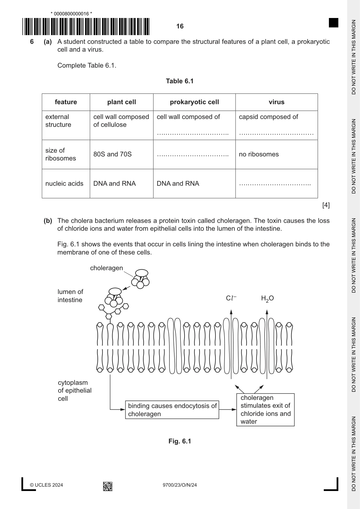
6 (a) A student constructed a table to compare the structural features of a plant cell, a prokaryotic cell and a virus. Complete Table 6.1. Table 6.1 feature plant cell prokaryotic cell virus external cell wall composed cell wall composed of capsid composed of structure of cellulose size of 80S and 70S no ribosomes ribosomes nucleic acids DNA and RNA DNA and RNA [4] (b) The cholera bacterium releases a protein toxin called choleragen. The toxin causes the loss of chloride ions and water from epithelial cells into the lumen of the intestine. Fig. 6.1 shows the events that occur in cells lining the intestine when choleragen binds to the membrane of one of these cells. choleragen lumen of intestine Cl – H2O cytoplasm of epithelial cell choleragen binding causes endocytosis of stimulates exit of choleragen chloride ions and water Fig. 6.1 (i) State one likely source of an outbreak of cholera. ..................................................................................................................................... [1] (ii) With reference to Fig. 6.1, state why choleragen molecules are described as having quaternary structure. ..................................................................................................................................... [1] (iii) State the type of cell membrane component that forms the receptor for choleragen. ..................................................................................................................................... [1] (iv) The process by which chloride ions leave the epithelial cell requires energy. Name the phosphorylated nucleotide that is needed for this process. ..................................................................................................................................... [1] (v) Explain why water also moves from epithelial cells into the lumen of the intestine when choleragen is present. ........................................................................................................................................... ........................................................................................................................................... ........................................................................................................................................... ........................................................................................................................................... ..................................................................................................................................... [2] [Total: 10]
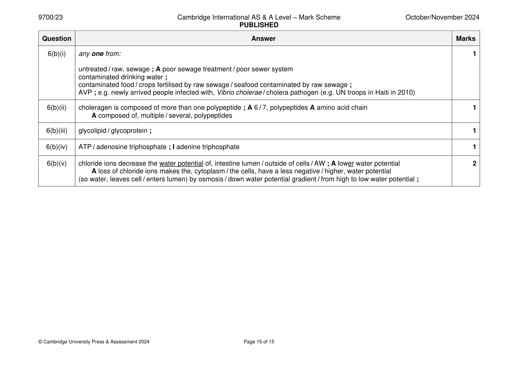
Mark scheme: 6(a) 4 feature plant cell prokaryotic cell virus external structure cell wall composed of cell wall composed of capsid composed of cellulose peptidoglycan / murein ; protein / polypeptides ; A capsomeres size of ribosomes 80S and 70S 70S ; no ribosomes nucleic acids DNA and RNA DNA and RNA DNA or RNA ; R DNA and RNA 6(b)(i) any one from: 1 untreated / raw, sewage ; A poor sewage treatment / poor sewer system contaminated drinking water ; contaminated food / crops fertilised by raw sewage / seafood contaminated by raw sewage ; AVP ; e.g. newly arrived people infected with, Vibrio cholerae / cholera pathogen (e.g. UN troops in Haiti in 2010) 6(b)(ii) choleragen is composed of more than one polypeptide ; A 6 / 7, polypeptides A amino acid chain 1 A composed of, multiple / several, polypeptides 6(b)(iii) glycolipid / glycoprotein ; 1 6(b)(iv) ATP / adenosine triphosphate ; I adenine triphosphate 1 6(b)(v) chloride ions decrease the water potential of, intestine lumen / outside of cells / AW ; A lower water potential 2 A loss of chloride ions makes the, cytoplasm / the cells, have a less negative / higher, water potential (so water, leaves cell / enters lumen) by osmosis / down water potential gradient / from high to low water potential ;
What was in this paper
The subtopics covered by these 6 questions, and how many questions each got. Open one in a new tab to see every Cambridge question on it.
What you needed in this session
Cambridge’s own grade thresholds for 2024 Oct/Nov, Paper 2 · Variant 3. A higher threshold means an easier paper — the bar moves with how the cohort did.