Cambridge A Level Biology 9700 — 2024 May/June Paper 2 · Variant 3
9700/23/M/J/24 · 6 questions · 60 marks · ≈68 min
The question paper and its mark scheme, free to read here and free to download. This is Cambridge’s own paper, exactly as it was sat.
Question paper16 pages
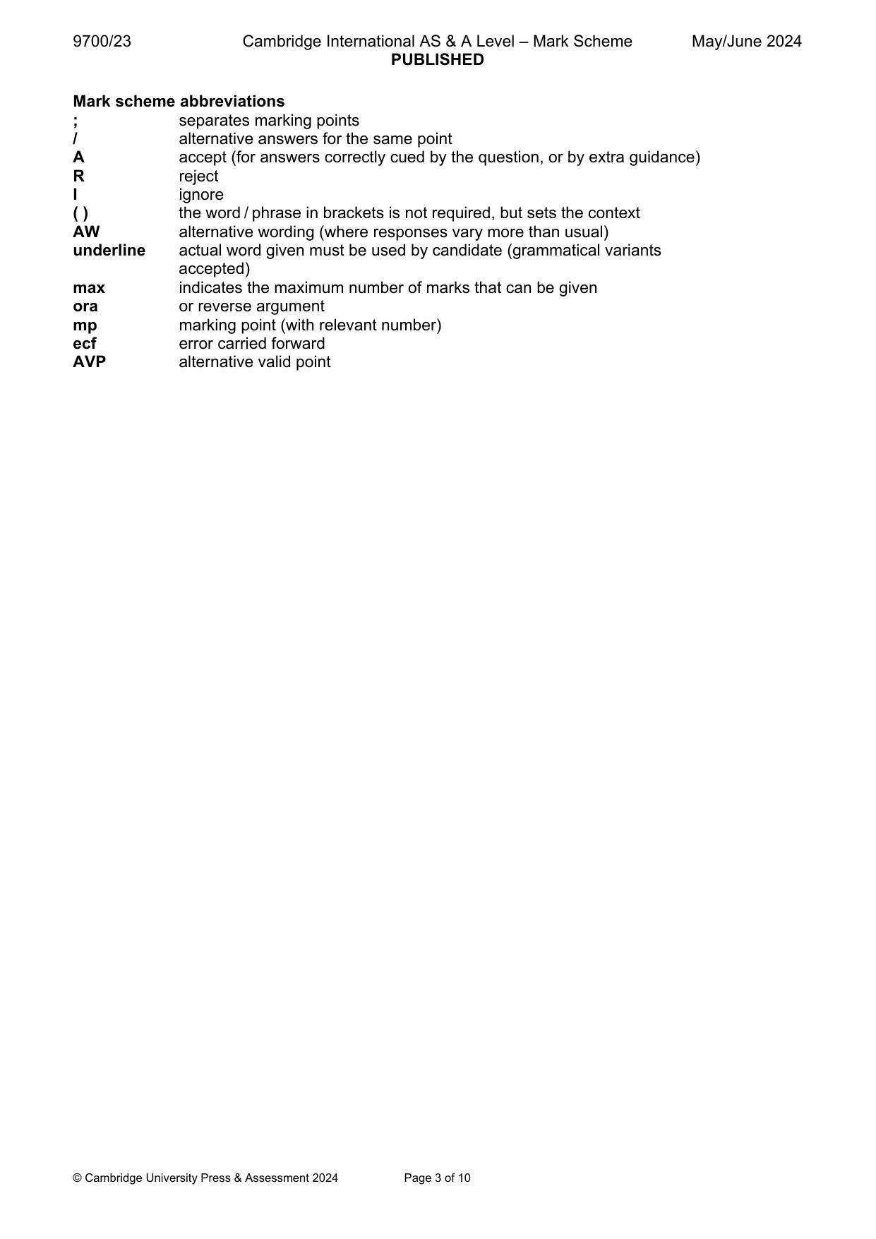
Mark scheme10 pages
Answers below. Sit the paper first if you are practising.
Questions as text
Q1 · The Zika virus is a pathogen that can infect human cells
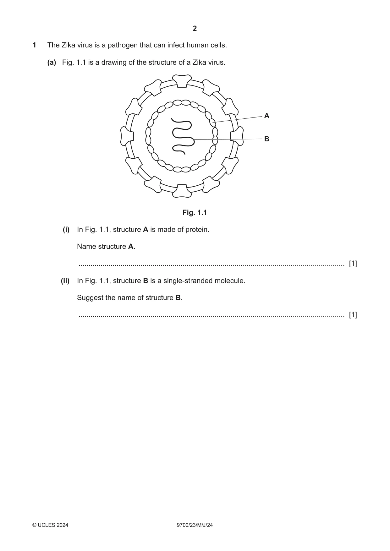
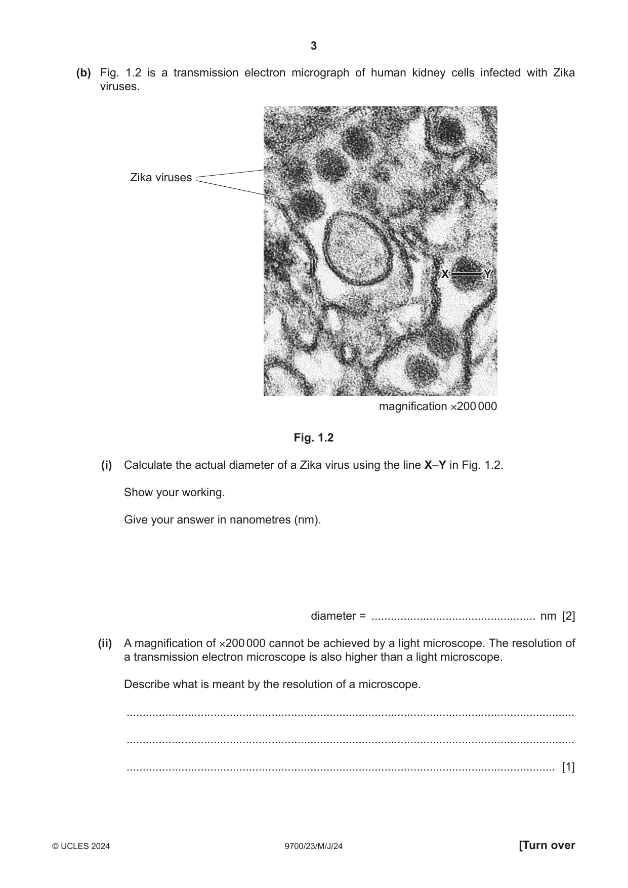
1 The Zika virus is a pathogen that can infect human cells. (a) Fig. 1.1 is a drawing of the structure of a Zika virus. A B Fig. 1.1 (i) In Fig. 1.1, structure A is made of protein. Name structure A. ..................................................................................................................................... [1] (ii) In Fig. 1.1, structure B is a single-stranded molecule. Suggest the name of structure B. ..................................................................................................................................... [1] (b) Fig. 1.2 is a transmission electron micrograph of human kidney cells infected with Zika viruses. Zika viruses X Y magnification ×200 000 Fig. 1.2 (i) Calculate the actual diameter of a Zika virus using the line X–Y in Fig. 1.2. Show your working. Give your answer in nanometres (nm). diameter = ................................................... nm [2] (ii) A magnification of ×200 000 cannot be achieved by a light microscope. The resolution of a transmission electron microscope is also higher than a light microscope. Describe what is meant by the resolution of a microscope. ........................................................................................................................................... ........................................................................................................................................... ..................................................................................................................................... [1] (c) The vector for Zika virus is the mosquito Aedes aegypti. The mosquito feeds on the blood of an infected person and transmits the virus to another person when it feeds again. Describe the similarities and differences between the transmission of Zika virus disease and the transmission of malaria. ................................................................................................................................................... ................................................................................................................................................... ................................................................................................................................................... ................................................................................................................................................... ................................................................................................................................................... ................................................................................................................................................... ............................................................................................................................................. [3] (d) Zika virus vaccines have been developed by scientists. One of the vaccines contains small proteins from the Zika virus. (i) Explain how giving this vaccine to a person can lead to the development of long-term immunity against Zika virus disease. ........................................................................................................................................... ........................................................................................................................................... ........................................................................................................................................... ........................................................................................................................................... ........................................................................................................................................... ........................................................................................................................................... ........................................................................................................................................... ........................................................................................................................................... ..................................................................................................................................... [4] (ii) Explain how a vaccination programme may limit the spread of Zika virus disease through a population. ........................................................................................................................................... ........................................................................................................................................... ........................................................................................................................................... ........................................................................................................................................... ..................................................................................................................................... [2] [Total: 14]
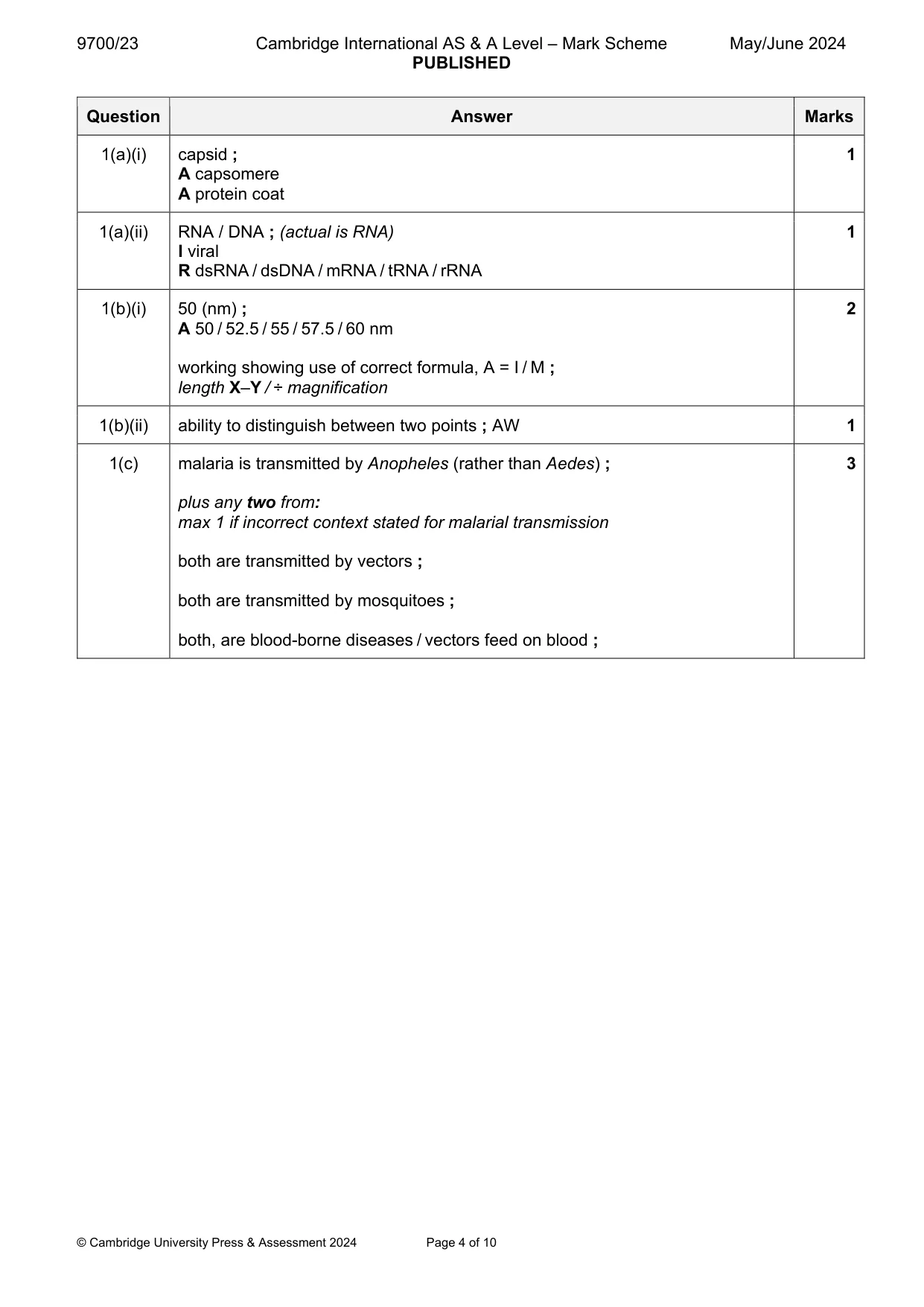
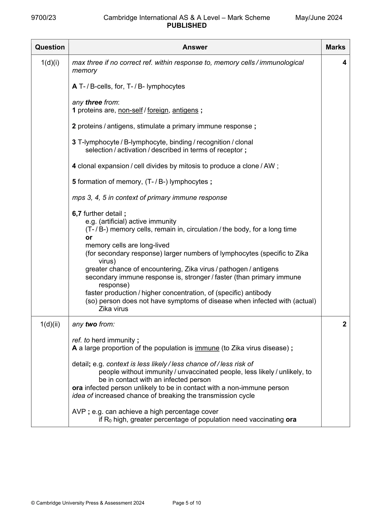
Mark scheme: Question Answer Marks 1(a)(i) capsid ; 1 A capsomere A protein coat 1(a)(ii) RNA / DNA ; (actual is RNA) 1 I viral R dsRNA / dsDNA / mRNA / tRNA / rRNA 1(b)(i) 50 (nm) ; 2 A 50 / 52.5 / 55 / 57.5 / 60 nm working showing use of correct formula, A = I / M ; length X–Y / ÷ magnification 1(b)(ii) ability to distinguish between two points ; AW 1 1(c) malaria is transmitted by Anopheles (rather than Aedes) ; 3 plus any two from: max 1 if incorrect context stated for malarial transmission both are transmitted by vectors ; both are transmitted by mosquitoes ; both, are blood-borne diseases / vectors feed on blood ; 1(d)(i) max three if no correct ref. within response to, memory cells / immunological 4 memory A T- / B-cells, for, T- / B- lymphocytes any three from: 1 proteins are, non-self / foreign, antigens ; 2 proteins / antigens, stimulate a primary immune response ; 3 T-lymphocyte / B-lymphocyte, binding / recognition / clonal selection / activation / described in terms of receptor ; 4 clonal expansion / cell divides by mitosis to produce a clone / AW ; 5 formation of memory, (T- / B-) lymphocytes ; mps 3, 4, 5 in context of primary immune response 6,7 further detail ; e.g. (artificial) active immunity (T- / B-) memory cells, remain in, circulation / the body, for a long time or memory cells are long-lived (for secondary response) larger numbers of lymphocytes (specific to Zika virus) greater chance of encountering, Zika virus / pathogen / antigens secondary immune response is, stronger / faster (than primary immune response) faster production / higher concentration, of (specific) antibody (so) person does not have symptoms of disease when infected with (actual) Zika virus 1(d)(ii) any two from: 2 ref. to herd immunity ; A a large proportion of the population is immune (to Zika virus disease) ; detail; e.g. context is less likely / less chance of / less risk of people without immunity / unvaccinated people, less likely / unlikely, to be in contact with an infected person ora infected person unlikely to be in contact with a non-immune person idea of increased chance of breaking the transmission cycle AVP ; e.g. can achieve a high percentage cover if R0 high, greater percentage of population need vaccinating ora
Q2 · Collagen is a fibrous protein that is found in many tissues in animals
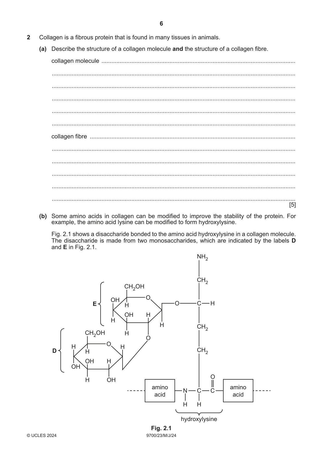
2 Collagen is a fibrous protein that is found in many tissues in animals. (a) Describe the structure of a collagen molecule and the structure of a collagen fibre. collagen molecule ..................................................................................................................... ................................................................................................................................................... ................................................................................................................................................... ................................................................................................................................................... ................................................................................................................................................... ................................................................................................................................................... collagen fibre ............................................................................................................................ ................................................................................................................................................... ................................................................................................................................................... ................................................................................................................................................... ................................................................................................................................................... ................................................................................................................................................... [5] (b) Some amino acids in collagen can be modified to improve the stability of the protein. For example, the amino acid lysine can be modified to form hydroxylysine. Fig. 2.1 shows a disaccharide bonded to the amino acid hydroxylysine in a collagen molecule. The disaccharide is made from two monosaccharides, which are indicated by the labels D and E in Fig. 2.1. NH2 CH2 CH2OH O OH E H O C H OH H H H CH2 CH2OH H O O H H D H CH2 OH H OH O H OH amino amino N C C acid acid H H hydroxylysine Fig. 2.1 (i) Give the precise name of the monosaccharide labelled D in Fig. 2.1. ..................................................................................................................................... [1] (ii) On Fig. 2.1, label the glycosidic bond with the letter G. Write your answer on Fig. 2.1. [1] (iii) On Fig. 2.1, draw a circle around the R group of hydroxylysine and label the circle with the letter R. Write your answer on Fig. 2.1. [1] (c) Osteogenesis imperfecta is a disease that results from a deficiency in collagen. Suggest how a named tissue or structure is affected in a person who has this disease. ................................................................................................................................................... ................................................................................................................................................... ................................................................................................................................................... ............................................................................................................................................. [2] [Total: 10]
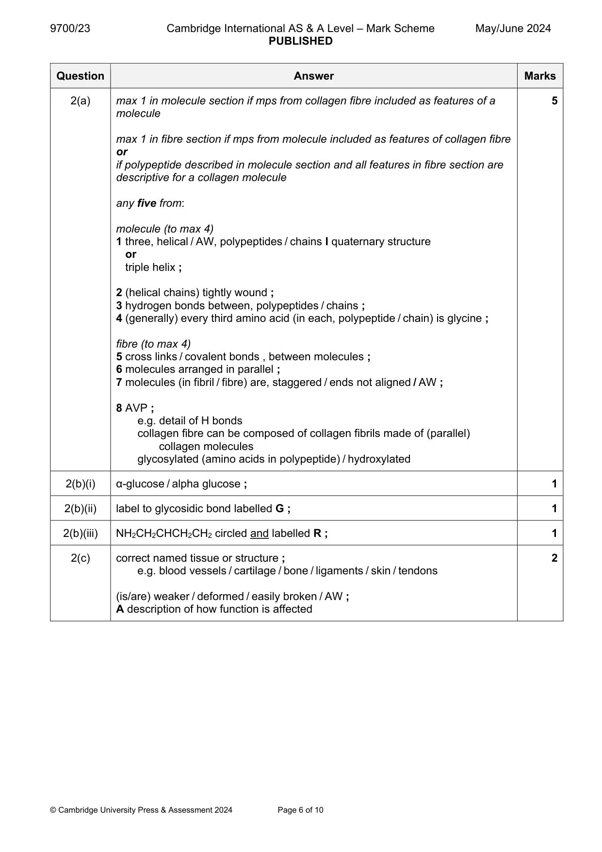
Mark scheme: 2(a) max 1 in molecule section if mps from collagen fibre included as features of a 5 molecule max 1 in fibre section if mps from molecule included as features of collagen fibre or if polypeptide described in molecule section and all features in fibre section are descriptive for a collagen molecule any five from: molecule (to max 4) 1 three, helical / AW, polypeptides / chains I quaternary structure or triple helix ; 2 (helical chains) tightly wound ; 3 hydrogen bonds between, polypeptides / chains ; 4 (generally) every third amino acid (in each, polypeptide / chain) is glycine ; fibre (to max 4) 5 cross links / covalent bonds , between molecules ; 6 molecules arranged in parallel ; 7 molecules (in fibril / fibre) are, staggered / ends not aligned / AW ; 8 AVP ; e.g. detail of H bonds collagen fibre can be composed of collagen fibrils made of (parallel) collagen molecules glycosylated (amino acids in polypeptide) / hydroxylated 2(b)(i) α-glucose / alpha glucose ; 1 2(b)(ii) label to glycosidic bond labelled G ; 1 2(b)(iii) NH2CH2CHCH2CH2 circled and labelled R ; 1 2(c) correct named tissue or structure ; 2 e.g. blood vessels / cartilage / bone / ligaments / skin / tendons (is/are) weaker / deformed / easily broken / AW ; A description of how function is affected
Q3 · Arteries, capillaries and veins are three types of blood vessel
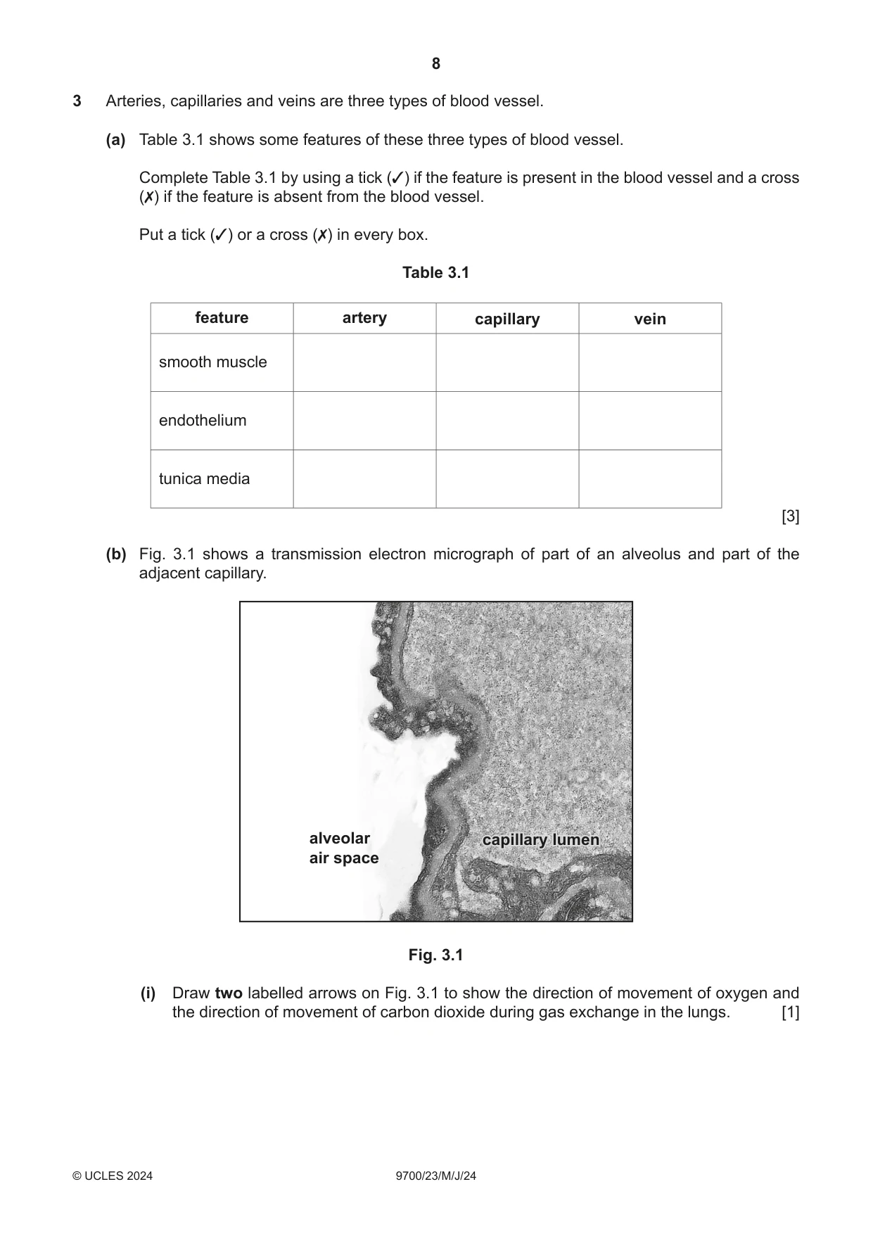
3 Arteries, capillaries and veins are three types of blood vessel. (a) Table 3.1 shows some features of these three types of blood vessel. Complete Table 3.1 by using a tick (3) if the feature is present in the blood vessel and a cross (✗) if the feature is absent from the blood vessel. Put a tick (3) or a cross (✗) in every box. Table 3.1 feature artery capillary vein smooth muscle endothelium tunica media [3] (b) Fig. 3.1 shows a transmission electron micrograph of part of an alveolus and part of the adjacent capillary. alveolar capillarycapillary lumenlumen air space Fig. 3.1 (i) Draw two labelled arrows on Fig. 3.1 to show the direction of movement of oxygen and the direction of movement of carbon dioxide during gas exchange in the lungs. [1] (ii) Suggest and explain how a steep oxygen concentration gradient is maintained in the lungs. ........................................................................................................................................... ........................................................................................................................................... ........................................................................................................................................... ........................................................................................................................................... ........................................................................................................................................... ........................................................................................................................................... ........................................................................................................................................... ........................................................................................................................................... ..................................................................................................................................... [4] (c) The passage outlines the roles of blood vessels associated with the heart. Complete the passage by using the most appropriate scientific terms. The …………………………………. carries blood to the left atrium. After passing from the left atrium to the left ventricle, blood is pumped into the aorta. The aorta is one of two large arteries that carry blood away from the ventricles of the heart. Blood that leaves the heart to enter these arteries must pass through the …………………………………. valves. Oxygenated blood is supplied to the cardiac muscle cells through the …………………………………. arteries. [3] [Total: 11]
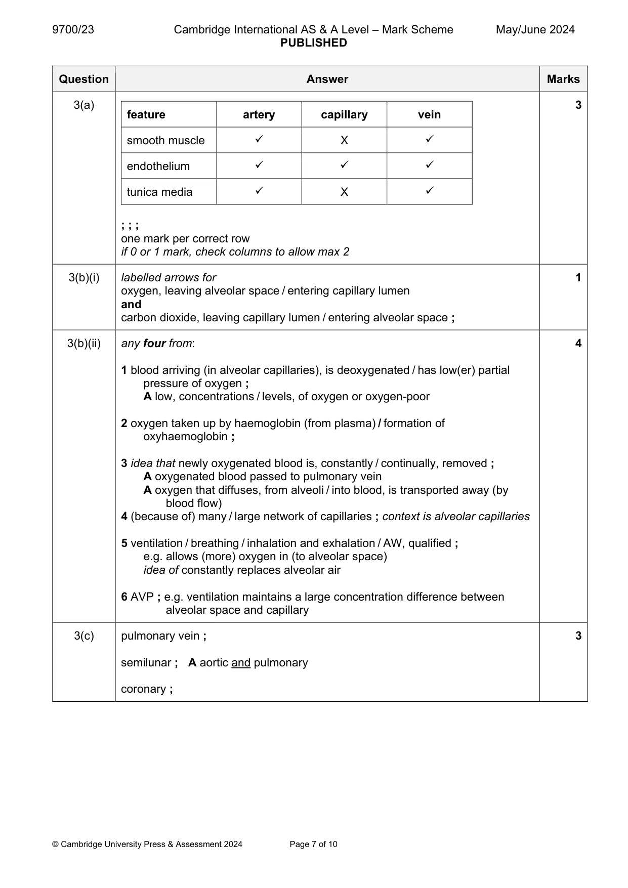
Mark scheme: 3(a) 3 feature artery capillary vein smooth muscle X endothelium tunica media X ; ; ; one mark per correct row if 0 or 1 mark, check columns to allow max 2 3(b)(i) labelled arrows for 1 oxygen, leaving alveolar space / entering capillary lumen and carbon dioxide, leaving capillary lumen / entering alveolar space ; 3(b)(ii) any four from: 4 1 blood arriving (in alveolar capillaries), is deoxygenated / has low(er) partial pressure of oxygen ; A low, concentrations / levels, of oxygen or oxygen-poor 2 oxygen taken up by haemoglobin (from plasma) / formation of oxyhaemoglobin ; 3 idea that newly oxygenated blood is, constantly / continually, removed ; A oxygenated blood passed to pulmonary vein A oxygen that diffuses, from alveoli / into blood, is transported away (by blood flow) 4 (because of) many / large network of capillaries ; context is alveolar capillaries 5 ventilation / breathing / inhalation and exhalation / AW, qualified ; e.g. allows (more) oxygen in (to alveolar space) idea of constantly replaces alveolar air 6 AVP ; e.g. ventilation maintains a large concentration difference between alveolar space and capillary 3(c) pulmonary vein ; 3 semilunar ; A aortic and pulmonary coronary ;
Q4 · Lignin and suberin are polymers that are present in plant tissues
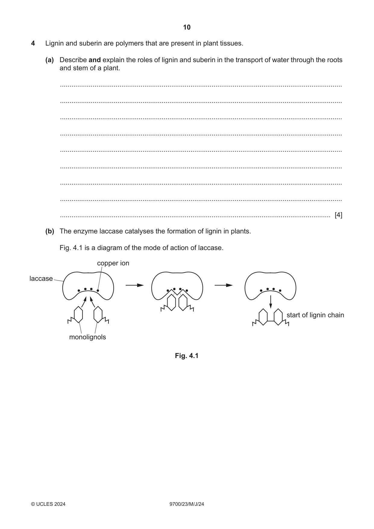
4 Lignin and suberin are polymers that are present in plant tissues. (a) Describe and explain the roles of lignin and suberin in the transport of water through the roots and stem of a plant. ................................................................................................................................................... ................................................................................................................................................... ................................................................................................................................................... ................................................................................................................................................... ................................................................................................................................................... ................................................................................................................................................... ................................................................................................................................................... ................................................................................................................................................... ............................................................................................................................................. [4] (b) The enzyme laccase catalyses the formation of lignin in plants. Fig. 4.1 is a diagram of the mode of action of laccase. copper ion laccase start of lignin chain monolignols Fig. 4.1 Describe and explain the mode of action of laccase when catalysing the formation of lignin. ................................................................................................................................................... ................................................................................................................................................... ................................................................................................................................................... ................................................................................................................................................... ................................................................................................................................................... ................................................................................................................................................... ................................................................................................................................................... ................................................................................................................................................... ................................................................................................................................................... ................................................................................................................................................... ............................................................................................................................................. [5] [Total: 9]
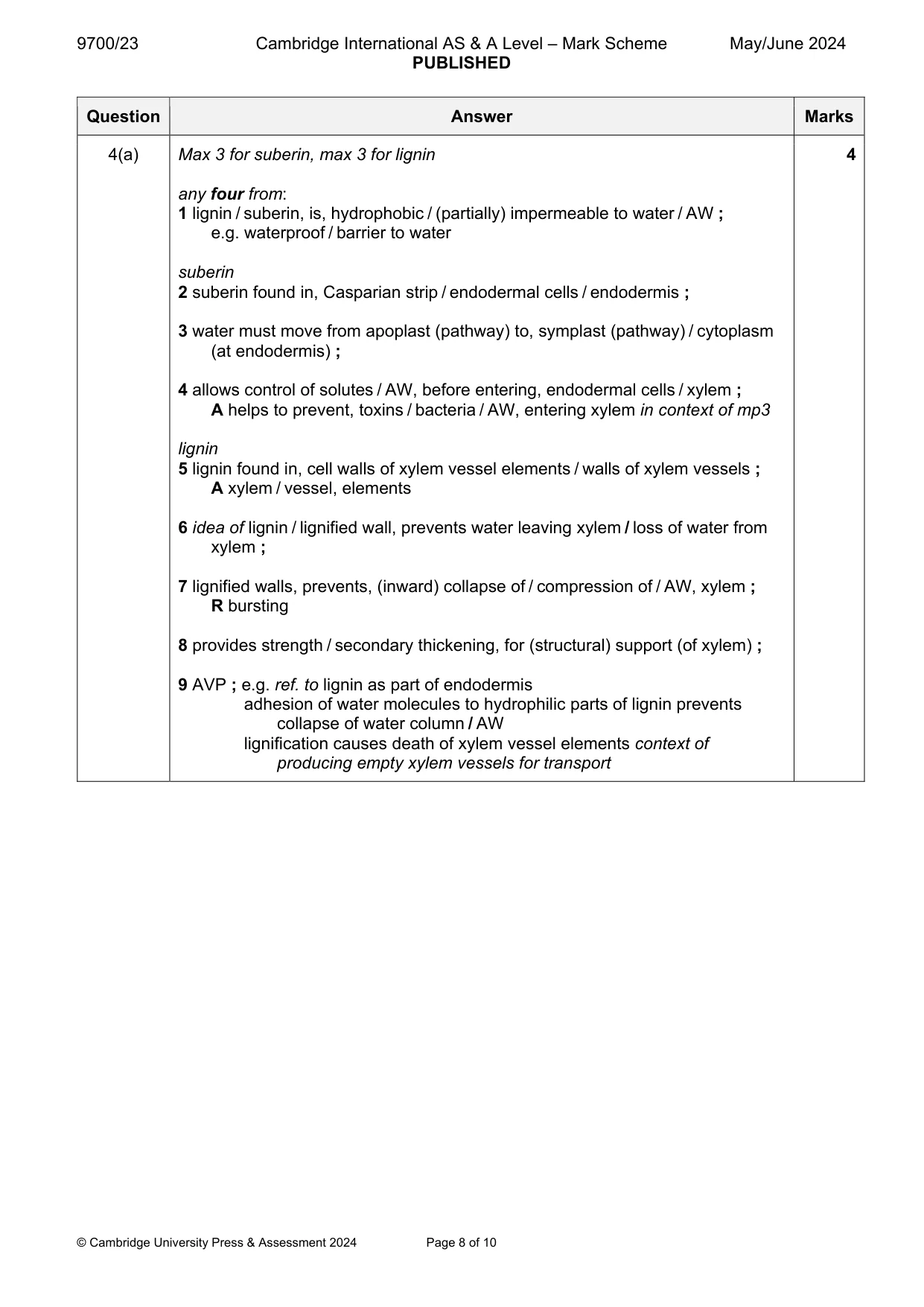
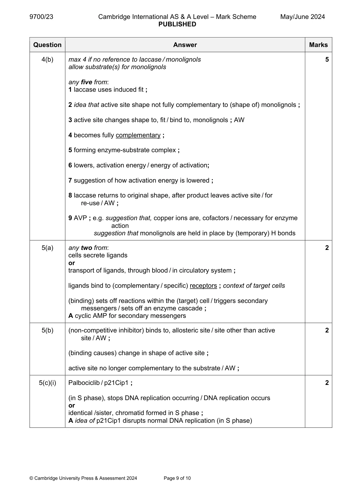
Mark scheme: 4(a) Max 3 for suberin, max 3 for lignin 4 any four from: 1 lignin / suberin, is, hydrophobic / (partially) impermeable to water / AW ; e.g. waterproof / barrier to water suberin 2 suberin found in, Casparian strip / endodermal cells / endodermis ; 3 water must move from apoplast (pathway) to, symplast (pathway) / cytoplasm (at endodermis) ; 4 allows control of solutes / AW, before entering, endodermal cells / xylem ; A helps to prevent, toxins / bacteria / AW, entering xylem in context of mp3 lignin 5 lignin found in, cell walls of xylem vessel elements / walls of xylem vessels ; A xylem / vessel, elements 6 idea of lignin / lignified wall, prevents water leaving xylem / loss of water from xylem ; 7 lignified walls, prevents, (inward) collapse of / compression of / AW, xylem ; R bursting 8 provides strength / secondary thickening, for (structural) support (of xylem) ; 9 AVP ; e.g. ref. to lignin as part of endodermis adhesion of water molecules to hydrophilic parts of lignin prevents collapse of water column / AW lignification causes death of xylem vessel elements context of producing empty xylem vessels for transport 4(b) max 4 if no reference to laccase / monolignols 5 allow substrate(s) for monolignols any five from: 1 laccase uses induced fit ; 2 idea that active site shape not fully complementary to (shape of) monolignols ; 3 active site changes shape to, fit / bind to, monolignols ; AW 4 becomes fully complementary ; 5 forming enzyme-substrate complex ; 6 lowers, activation energy / energy of activation; 7 suggestion of how activation energy is lowered ; 8 laccase returns to original shape, after product leaves active site / for re-use / AW ; 9 AVP ; e.g. suggestion that, copper ions are, cofactors / necessary for enzyme action suggestion that monolignols are held in place by (temporary) H bonds
Q5 · Cyclin-dependent kinases (CDKs) are enzymes that regulate the cell cycle
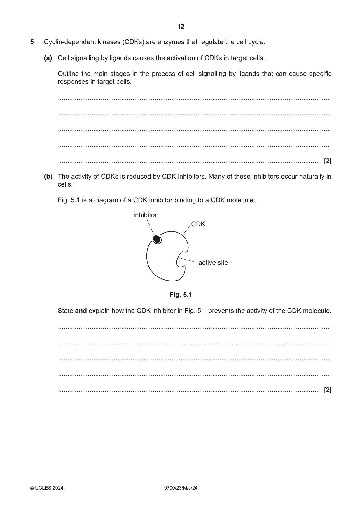
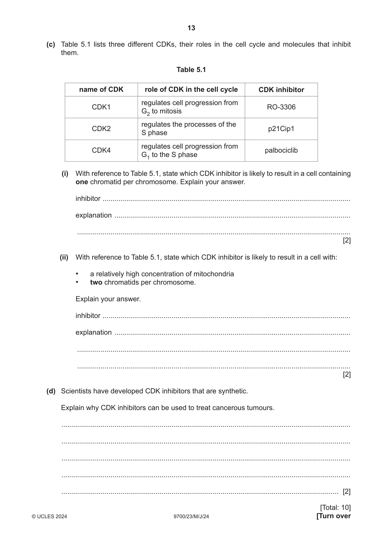
5 Cyclin-dependent kinases (CDKs) are enzymes that regulate the cell cycle. (a) Cell signalling by ligands causes the activation of CDKs in target cells. Outline the main stages in the process of cell signalling by ligands that can cause specific responses in target cells. ................................................................................................................................................... ................................................................................................................................................... ................................................................................................................................................... ................................................................................................................................................... ............................................................................................................................................. [2] (b) The activity of CDKs is reduced by CDK inhibitors. Many of these inhibitors occur naturally in cells. Fig. 5.1 is a diagram of a CDK inhibitor binding to a CDK molecule. inhibitor CDK active site Fig. 5.1 State and explain how the CDK inhibitor in Fig. 5.1 prevents the activity of the CDK molecule. ................................................................................................................................................... ................................................................................................................................................... ................................................................................................................................................... ................................................................................................................................................... ............................................................................................................................................. [2] (c) Table 5.1 lists three different CDKs, their roles in the cell cycle and molecules that inhibit them. Table 5.1 name of CDK role of CDK in the cell cycle CDK inhibitor regulates cell progression from CDK1 RO-3306 G2 to mitosis regulates the processes of the CDK2 p21Cip1 S phase regulates cell progression from CDK4 palbociclib G1 to the S phase (i) With reference to Table 5.1, state which CDK inhibitor is likely to result in a cell containing one chromatid per chromosome. Explain your answer. inhibitor .............................................................................................................................. explanation ........................................................................................................................ ........................................................................................................................................... [2] (ii) With reference to Table 5.1, state which CDK inhibitor is likely to result in a cell with: • a relatively high concentration of mitochondria • two chromatids per chromosome. Explain your answer. inhibitor .............................................................................................................................. explanation ........................................................................................................................ ........................................................................................................................................... ........................................................................................................................................... [2] (d) Scientists have developed CDK inhibitors that are synthetic. Explain why CDK inhibitors can be used to treat cancerous tumours. ................................................................................................................................................... ................................................................................................................................................... ................................................................................................................................................... ................................................................................................................................................... ............................................................................................................................................. [2] [Total: 10]
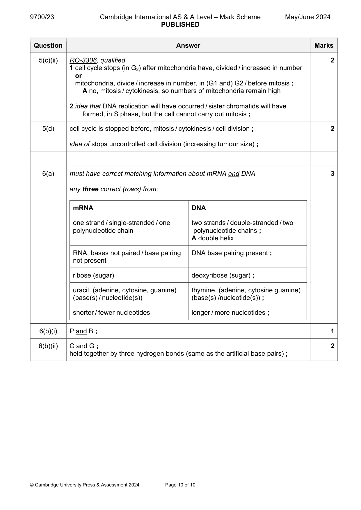
Mark scheme: 5(a) any two from: 2 cells secrete ligands or transport of ligands, through blood / in circulatory system ; ligands bind to (complementary / specific) receptors ; context of target cells (binding) sets off reactions within the (target) cell / triggers secondary messengers / sets off an enzyme cascade ; A cyclic AMP for secondary messengers 5(b) (non-competitive inhibitor) binds to, allosteric site / site other than active 2 site / AW ; (binding causes) change in shape of active site ; active site no longer complementary to the substrate / AW ; 5(c)(i) Palbociclib / p21Cip1 ; 2 (in S phase), stops DNA replication occurring / DNA replication occurs or identical /sister, chromatid formed in S phase ; A idea of p21Cip1 disrupts normal DNA replication (in S phase) 5(c)(ii) RO-3306, qualified 2 1 cell cycle stops (in G2) after mitochondria have, divided / increased in number or mitochondria, divide / increase in number, in (G1 and) G2 / before mitosis ; A no, mitosis / cytokinesis, so numbers of mitochondria remain high 2 idea that DNA replication will have occurred / sister chromatids will have formed, in S phase, but the cell cannot carry out mitosis ; 5(d) cell cycle is stopped before, mitosis / cytokinesis / cell division ; 2 idea of stops uncontrolled cell division (increasing tumour size) ;
More questions on Replication and division of nuclei and cells
Q6 · DNA and RNA are polynucleotides
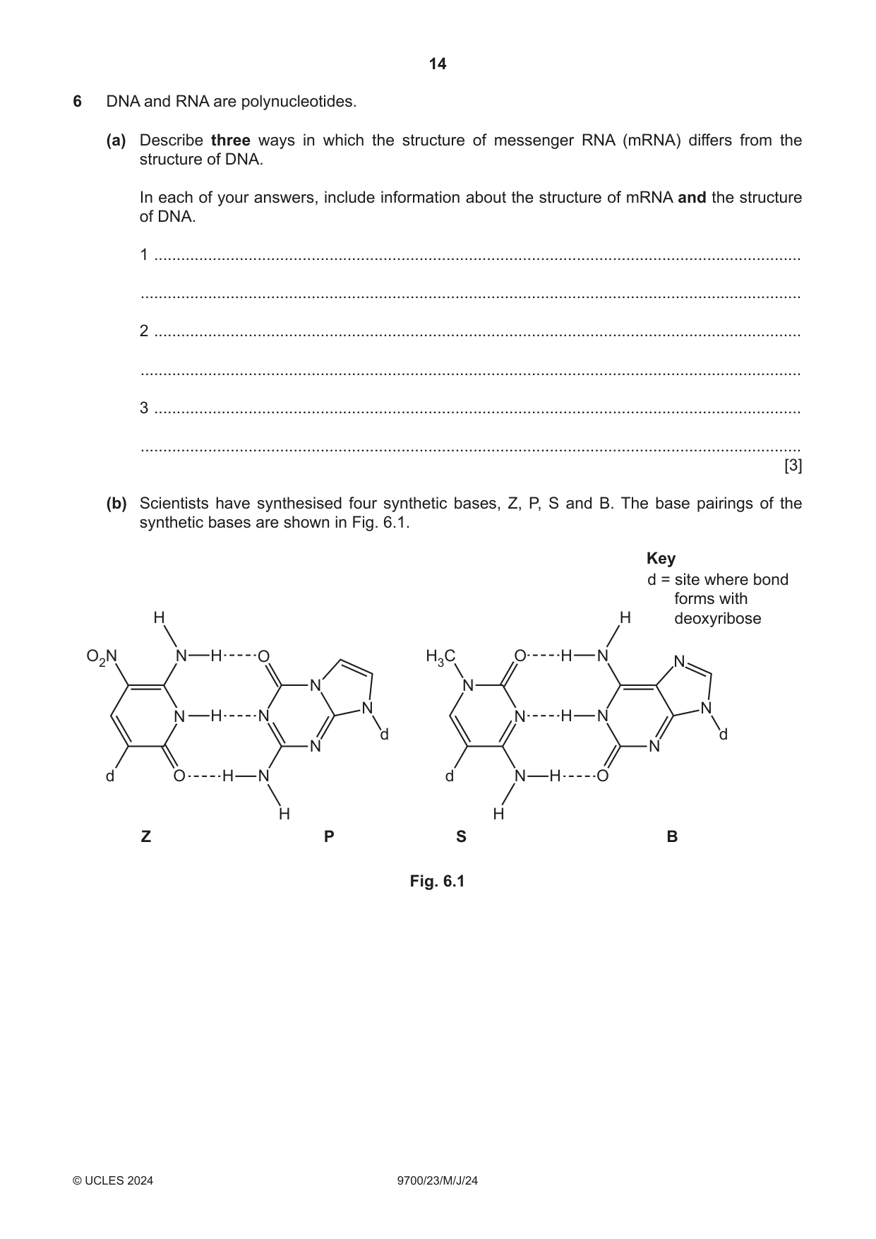
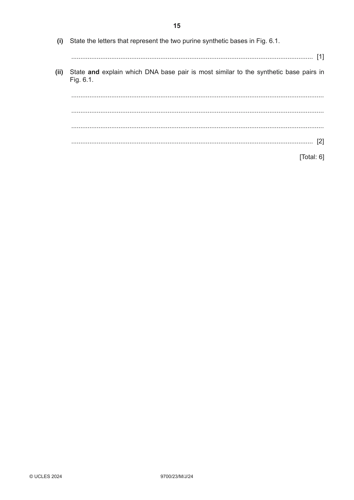
6 DNA and RNA are polynucleotides. (a) Describe three ways in which the structure of messenger RNA (mRNA) differs from the structure of DNA. In each of your answers, include information about the structure of mRNA and the structure of DNA. 1 ................................................................................................................................................ ................................................................................................................................................... 2 ................................................................................................................................................ ................................................................................................................................................... 3 ................................................................................................................................................ ................................................................................................................................................... [3] (b) Scientists have synthesised four synthetic bases, Z, P, S and B. The base pairings of the synthetic bases are shown in Fig. 6.1. Key d = site where bond forms with H H deoxyribose O2N N H O H3C O H N N N N N N N H N N H N d d N N d O H N d N H O H H Z P S B Fig. 6.1 (i) State the letters that represent the two purine synthetic bases in Fig. 6.1. ..................................................................................................................................... [1] (ii) State and explain which DNA base pair is most similar to the synthetic base pairs in Fig. 6.1. ........................................................................................................................................... ........................................................................................................................................... ........................................................................................................................................... ..................................................................................................................................... [2] [Total: 6]
Mark scheme: 6(a) must have correct matching information about mRNA and DNA 3 any three correct (rows) from: mRNA DNA one strand / single-stranded / one two strands / double-stranded / two polynucleotide chain polynucleotide chains ; A double helix RNA, bases not paired / base pairing DNA base pairing present ; not present ribose (sugar) deoxyribose (sugar) ; uracil, (adenine, cytosine, guanine) thymine, (adenine, cytosine guanine) (base(s) / nucleotide(s)) (base(s) /nucleotide(s)) ; shorter / fewer nucleotides longer / more nucleotides ; 6(b)(i) P and B ; 1 6(b)(ii) C and G ; 2 held together by three hydrogen bonds (same as the artificial base pairs) ;
More questions on Structure of nucleic acids and replication of DNA
What was in this paper
The subtopics covered by these 6 questions, and how many questions each got. Open one in a new tab to see every Cambridge question on it.
What you needed in this session
Cambridge’s own grade thresholds for 2024 May/June, Paper 2 · Variant 3. A higher threshold means an easier paper — the bar moves with how the cohort did.