Cambridge A Level Biology 9700 — 2022 May/June Paper 2 · Variant 3
9700/23/M/J/22 · 6 questions · 60 marks · ≈68 min
The question paper and its mark scheme, free to read here and free to download. This is Cambridge’s own paper, exactly as it was sat.
Question paper16 pages
Mark scheme18 pages
Answers below. Sit the paper first if you are practising.
Questions as text
Q1 · The trachea of the gas exchange system branches into two airways, each of which enters a…
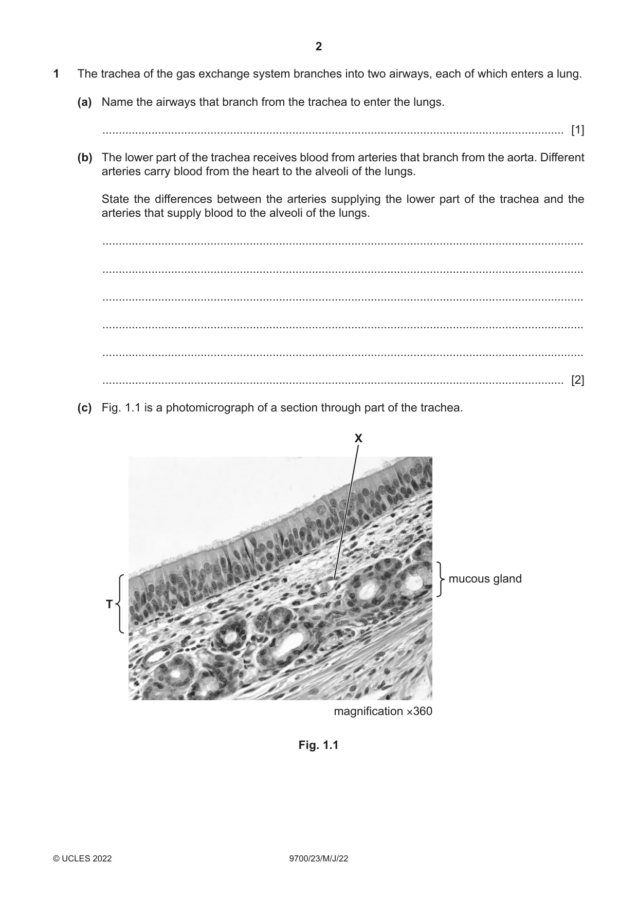
1 The trachea of the gas exchange system branches into two airways, each of which enters a lung. (a) Name the airways that branch from the trachea to enter the lungs. ............................................................................................................................................. [1] (b) The lower part of the trachea receives blood from arteries that branch from the aorta. Different arteries carry blood from the heart to the alveoli of the lungs. State the differences between the arteries supplying the lower part of the trachea and the arteries that supply blood to the alveoli of the lungs. ................................................................................................................................................... ................................................................................................................................................... ................................................................................................................................................... ................................................................................................................................................... ................................................................................................................................................... ............................................................................................................................................. [2] (c) Fig. 1.1 is a photomicrograph of a section through part of the trachea. X mucous gland T magnification ×360 Fig. 1.1 (i) In Fig. 1.1, one of the tissues in the trachea is labelled T. Describe the structural features of tissue T visible in Fig. 1.1. ........................................................................................................................................... ........................................................................................................................................... ........................................................................................................................................... ........................................................................................................................................... ........................................................................................................................................... ........................................................................................................................................... ........................................................................................................................................... ..................................................................................................................................... [3] (ii) Identify structure X in Fig. 1.1 and outline the features that helped your identification. structure X = .......................................................................... ........................................................................................................................................... ........................................................................................................................................... ........................................................................................................................................... ........................................................................................................................................... ........................................................................................................................................... ........................................................................................................................................... ........................................................................................................................................... ..................................................................................................................................... [3] [Total: 9]
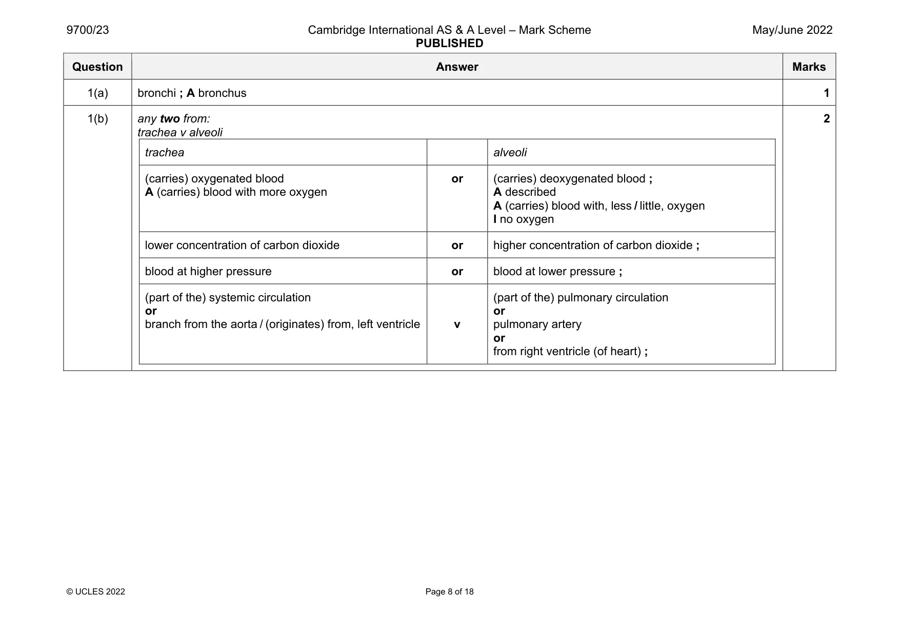
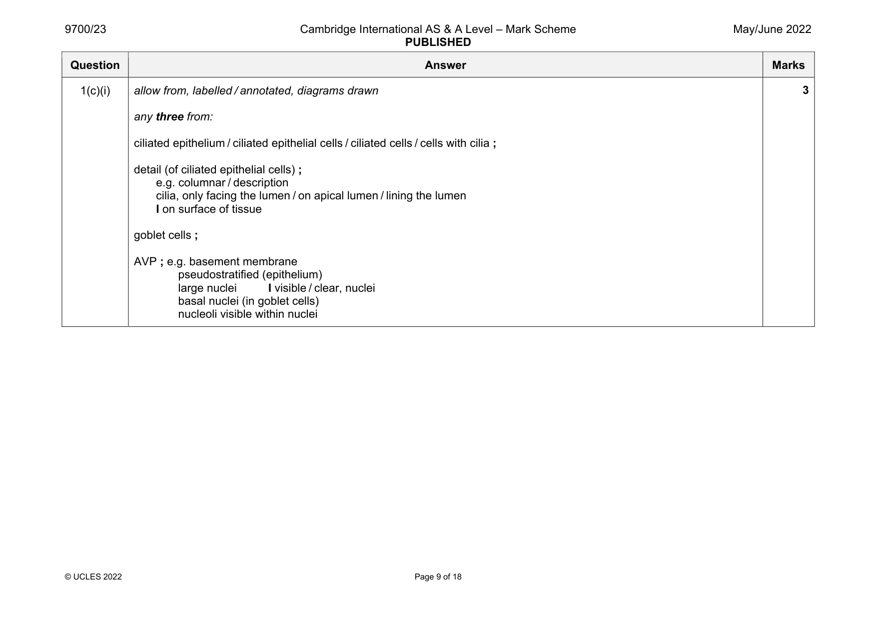
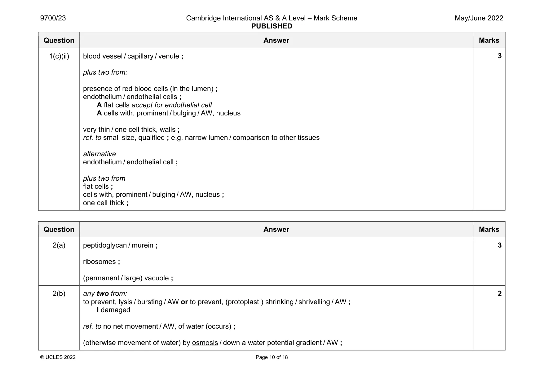
Mark scheme: 1(a) bronchi ; A bronchus 1 1(b) any two from: trachea v alveoli trachea alveoli (carries) oxygenated blood A (carries) blood with more oxygen or (carries) deoxygenated blood ; A described A (carries) blood with, less / little, oxygen I no oxygen lower concentration of carbon dioxide or higher concentration of carbon dioxide ; blood at higher pressure or blood at lower pressure ; (part of the) systemic circulation or branch from the aorta / (originates) from, left ventricle v (part of the) pulmonary circulation or pulmonary artery or from right ventricle (of heart) ; 2 Question Answer Marks 1(c)(i) allow from, labelled / annotated, diagrams drawn any three from: ciliated epithelium / ciliated epithelial cells / ciliated cells / cells with cilia ; detail (of ciliated epithelial cells) ; e.g. columnar / description cilia, only facing the lumen / on apical lumen / lining the lumen I on surface of tissue goblet cells ; AVP ; e.g. basement membrane pseudostratified (epithelium) large nuclei I visible / clear, nuclei basal nuclei (in goblet cells) nucleoli visible within nuclei 3 Question Answer Marks 1(c)(ii) blood vessel / capillary / venule ; plus two from: presence of red blood cells (in the lumen) ; endothelium / endothelial cells ; A flat cells accept for endothelial cell A cells with, prominent / bulging / AW, nucleus very thin / one cell thick, walls ; ref. to small size, qualified ; e.g. narrow lumen / comparison to other tissues alternative endothelium / endothelial cell ; plus two from flat cells ; cells with, prominent / bulging / AW, nucleus ; one cell thick ; 3
Q2 · Bacterial cells are prokaryotic
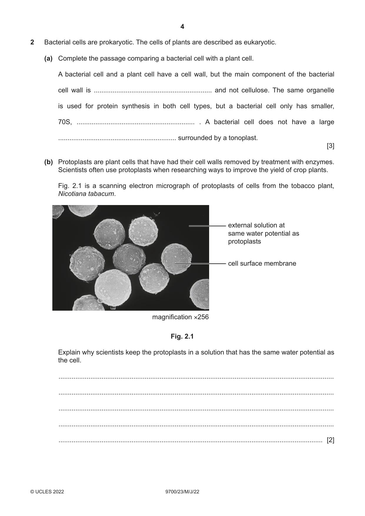
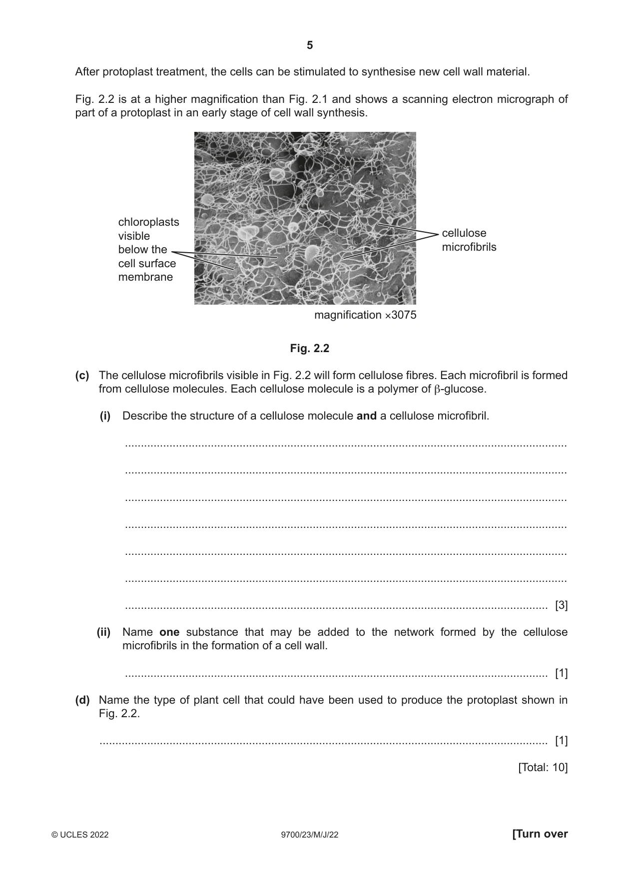
2 Bacterial cells are prokaryotic. The cells of plants are described as eukaryotic. (a) Complete the passage comparing a bacterial cell with a plant cell. A bacterial cell and a plant cell have a cell wall, but the main component of the bacterial cell wall is ............................................................... and not cellulose. The same organelle is used for protein synthesis in both cell types, but a bacterial cell only has smaller, 70S, ............................................................... . A bacterial cell does not have a large ............................................................... surrounded by a tonoplast. [3] (b) Protoplasts are plant cells that have had their cell walls removed by treatment with enzymes. Scientists often use protoplasts when researching ways to improve the yield of crop plants. Fig. 2.1 is a scanning electron micrograph of protoplasts of cells from the tobacco plant, Nicotiana tabacum. external solution at same water potential as protoplasts cell surface membrane magnification ×256 Fig. 2.1 Explain why scientists keep the protoplasts in a solution that has the same water potential as the cell. ................................................................................................................................................... ................................................................................................................................................... ................................................................................................................................................... ................................................................................................................................................... ............................................................................................................................................. [2] After protoplast treatment, the cells can be stimulated to synthesise new cell wall material. Fig. 2.2 is at a higher magnification than Fig. 2.1 and shows a scanning electron micrograph of part of a protoplast in an early stage of cell wall synthesis. chloroplasts visible cellulose below the microfibrils cell surface membrane magnification ×3075 Fig. 2.2 (c) The cellulose microfibrils visible in Fig. 2.2 will form cellulose fibres. Each microfibril is formed from cellulose molecules. Each cellulose molecule is a polymer of β-glucose. (i) Describe the structure of a cellulose molecule and a cellulose microfibril. ........................................................................................................................................... ........................................................................................................................................... ........................................................................................................................................... ........................................................................................................................................... ........................................................................................................................................... ........................................................................................................................................... ..................................................................................................................................... [3] (ii) Name one substance that may be added to the network formed by the cellulose microfibrils in the formation of a cell wall. ..................................................................................................................................... [1] (d) Name the type of plant cell that could have been used to produce the protoplast shown in Fig. 2.2. ............................................................................................................................................. [1] [Total: 10]
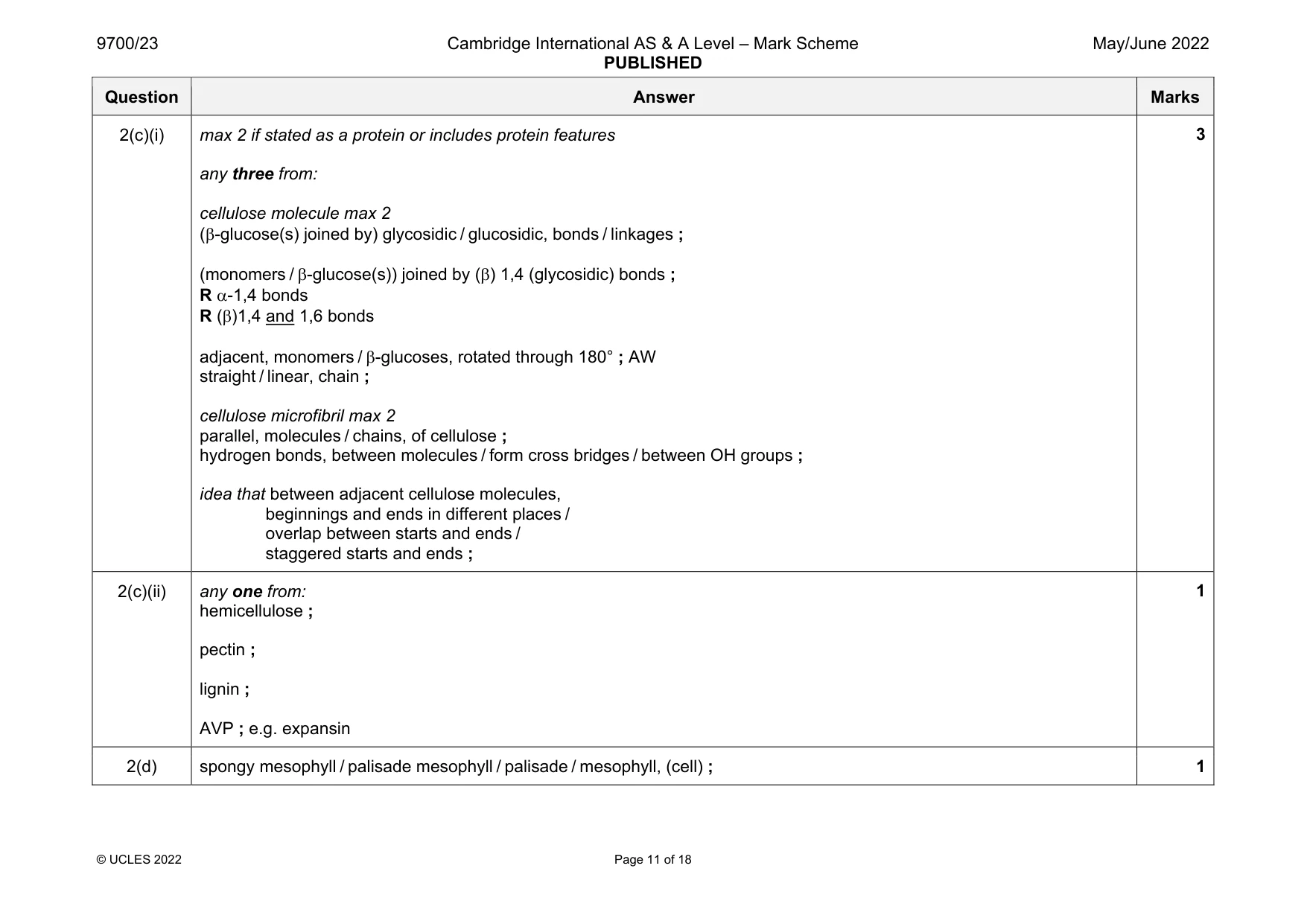
Mark scheme: 2(a) peptidoglycan / murein ; ribosomes ; (permanent / large) vacuole ; 3 2(b) any two from: to prevent, lysis / bursting / AW or to prevent, (protoplast ) shrinking / shrivelling / AW ; I damaged ref. to no net movement / AW, of water (occurs) ; (otherwise movement of water) by osmosis / down a water potential gradient / AW ; 2 Question Answer Marks 2(c)(i) max 2 if stated as a protein or includes protein features any three from: cellulose molecule max 2 (-glucose(s) joined by) glycosidic / glucosidic, bonds / linkages ; (monomers / -glucose(s)) joined by () 1,4 (glycosidic) bonds ; R -1,4 bonds R ()1,4 and 1,6 bonds adjacent, monomers / -glucoses, rotated through 180° ; AW straight / linear, chain ; cellulose microfibril max 2 parallel, molecules / chains, of cellulose ; hydrogen bonds, between molecules / form cross bridges / between OH groups ; idea that between adjacent cellulose molecules, beginnings and ends in different places / overlap between starts and ends / staggered starts and ends ; 3 2(c)(ii) any one from: hemicellulose ; pectin ; lignin ; AVP ; e.g. expansin 1 2(d) spongy mesophyll / palisade mesophyll / palisade / mesophyll, (cell) ; 1
More questions on Cells as the basic units of living organisms
Question 3
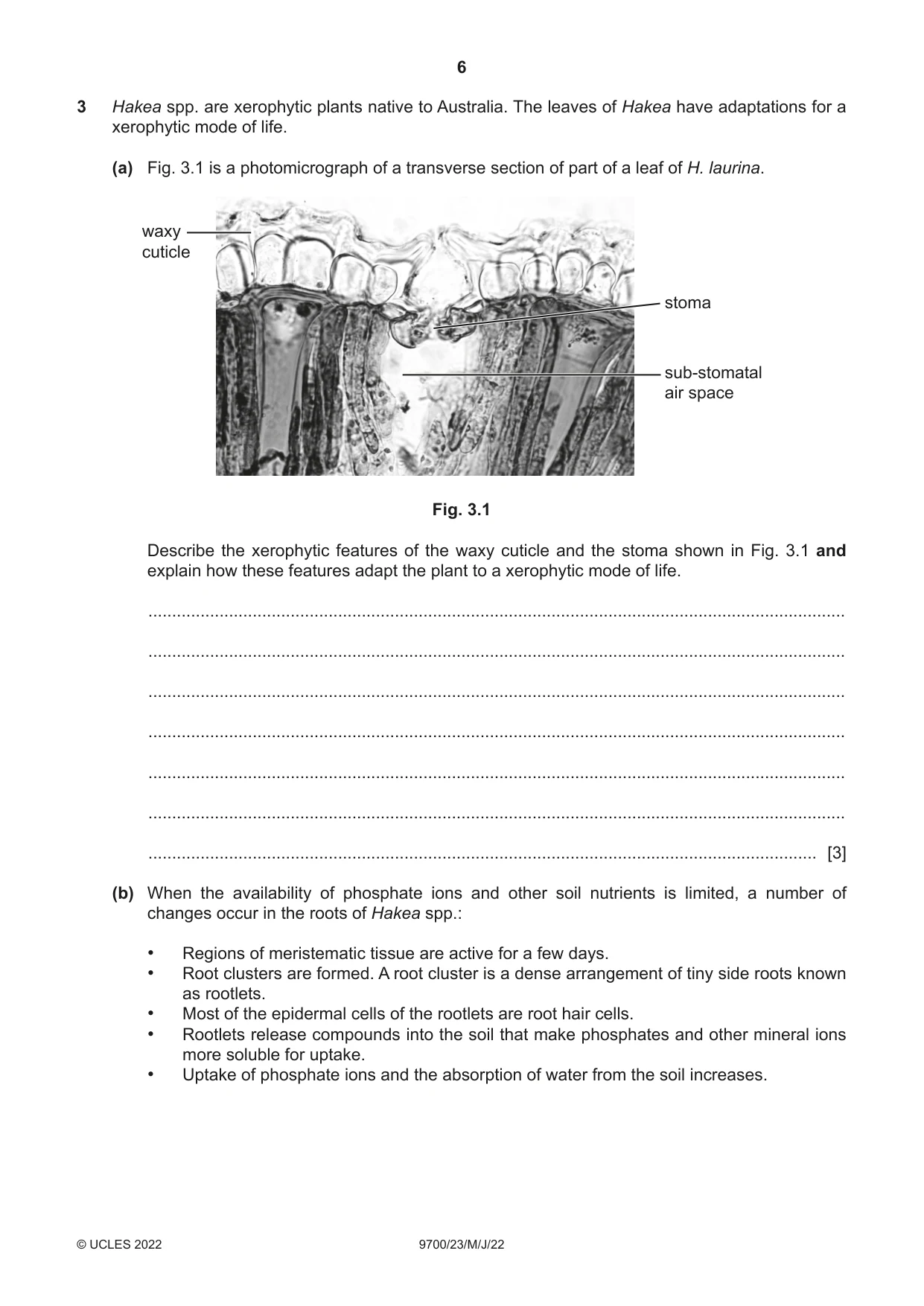
3 Hakea spp. are xerophytic plants native to Australia. The leaves of Hakea have adaptations for a xerophytic mode of life. (a) Fig. 3.1 is a photomicrograph of a transverse section of part of a leaf of H. laurina. waxy cuticle stoma sub-stomatal air space Fig. 3.1 Describe the xerophytic features of the waxy cuticle and the stoma shown in Fig. 3.1 and explain how these features adapt the plant to a xerophytic mode of life. ................................................................................................................................................... ................................................................................................................................................... ................................................................................................................................................... ................................................................................................................................................... ................................................................................................................................................... ................................................................................................................................................... ............................................................................................................................................. [3] (b) When the availability of phosphate ions and other soil nutrients is limited, a number of changes occur in the roots of Hakea spp.: • Regions of meristematic tissue are active for a few days. • Root clusters are formed. A root cluster is a dense arrangement of tiny side roots known as rootlets. • Most of the epidermal cells of the rootlets are root hair cells. • Rootlets release compounds into the soil that make phosphates and other mineral ions more soluble for uptake. • Uptake of phosphate ions and the absorption of water from the soil increases. (i) Meristem cells have a similar role to stem cells in animals. Suggest how meristematic tissue activity leads to the formation of root clusters. ........................................................................................................................................... ........................................................................................................................................... ........................................................................................................................................... ........................................................................................................................................... ........................................................................................................................................... ........................................................................................................................................... ..................................................................................................................................... [3] (ii) Organic anions (negatively charged organic compounds) are released into the soil by rootlets. The concentration of these organic anions can become higher in the soil solution than in the rootlet cells. Suggest and explain how the concentration of organic anions in the soil solution can become higher than in the rootlet cells. ........................................................................................................................................... ........................................................................................................................................... ........................................................................................................................................... ........................................................................................................................................... ........................................................................................................................................... ........................................................................................................................................... ..................................................................................................................................... [3] (iii) Explain how the formation of root clusters can lead to an increase in the uptake of phosphate ions and absorption of water from the soil solution. ........................................................................................................................................... ........................................................................................................................................... ........................................................................................................................................... ........................................................................................................................................... ........................................................................................................................................... ..................................................................................................................................... [2] [Total: 11]
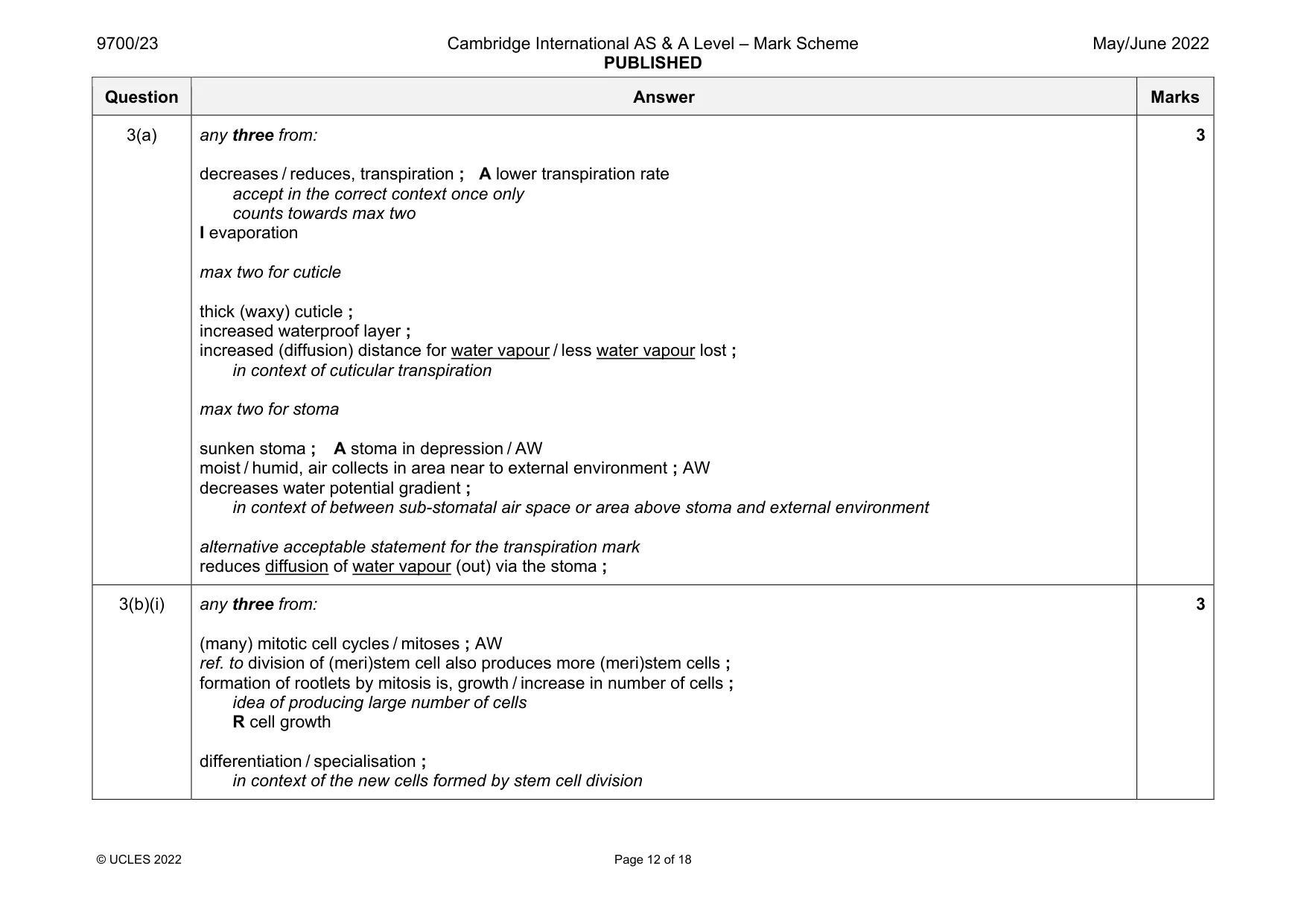
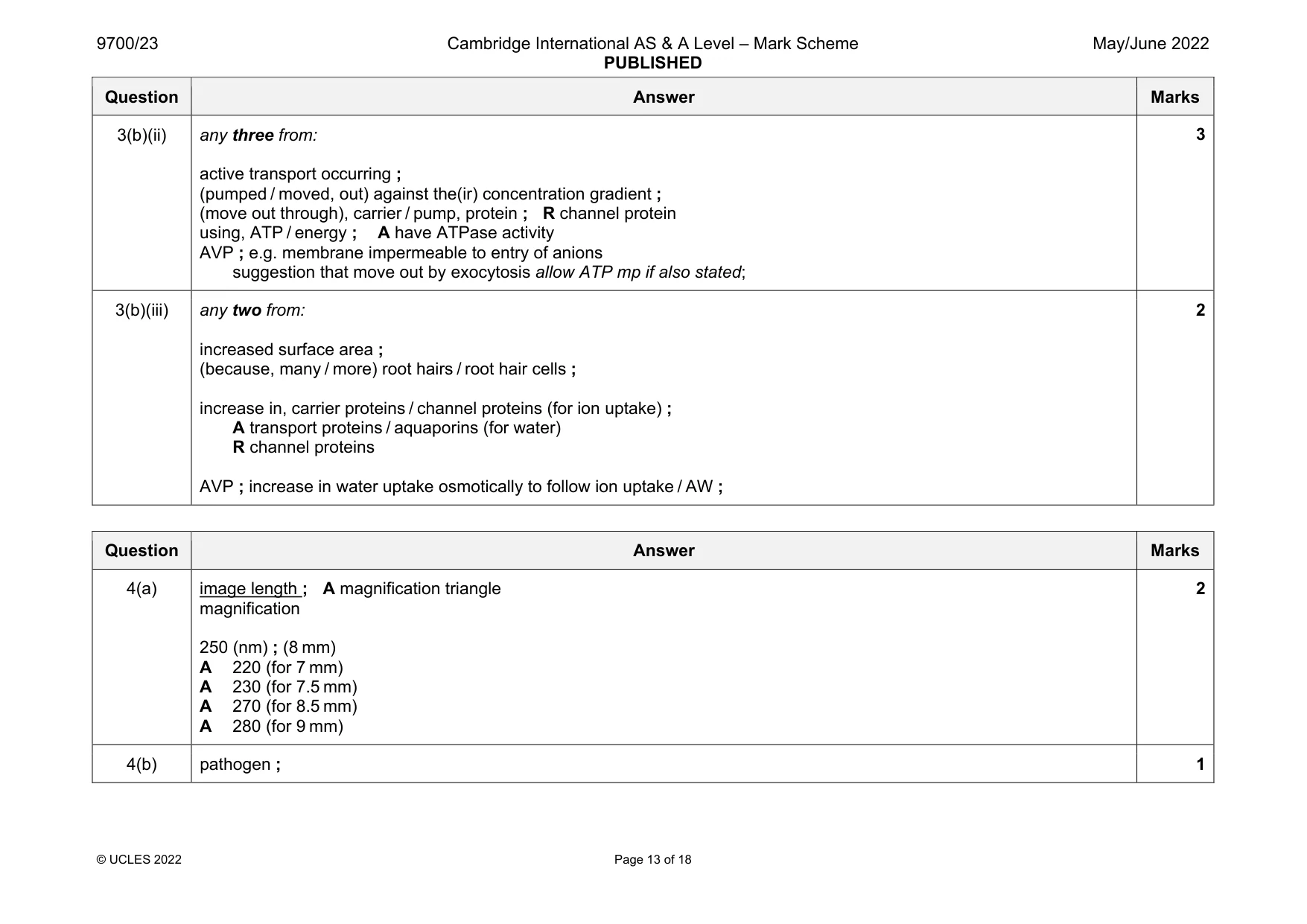
Mark scheme: 3(a) any three from: decreases / reduces, transpiration ; A lower transpiration rate accept in the correct context once only counts towards max two I evaporation max two for cuticle thick (waxy) cuticle ; increased waterproof layer ; increased (diffusion) distance for water vapour / less water vapour lost ; in context of cuticular transpiration max two for stoma sunken stoma ; A stoma in depression / AW moist / humid, air collects in area near to external environment ; AW decreases water potential gradient ; in context of between sub-stomatal air space or area above stoma and external environment alternative acceptable statement for the transpiration mark reduces diffusion of water vapour (out) via the stoma ; 3 3(b)(i) any three from: (many) mitotic cell cycles / mitoses ; AW ref. to division of (meri)stem cell also produces more (meri)stem cells ; formation of rootlets by mitosis is, growth / increase in number of cells ; idea of producing large number of cells R cell growth differentiation / specialisation ; in context of the new cells formed by stem cell division 3 Question Answer Marks 3(b)(ii) any three from: active transport occurring ; (pumped / moved, out) against the(ir) concentration gradient ; (move out through), carrier / pump, protein ; R channel protein using, ATP / energy ; A have ATPase activity AVP ; e.g. membrane impermeable to entry of anions suggestion that move out by exocytosis allow ATP mp if also stated; 3 3(b)(iii) any two from: increased surface area ; (because, many / more) root hairs / root hair cells ; increase in, carrier proteins / channel proteins (for ion uptake) ; A transport proteins / aquaporins (for water) R channel proteins AVP ; increase in water uptake osmotically to follow ion uptake / AW ; 2
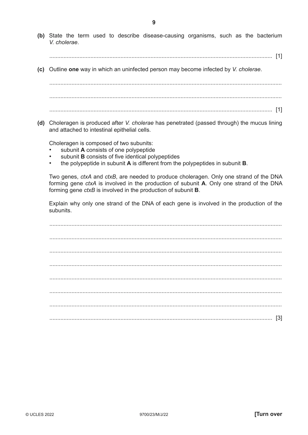
Q4 · There are many different forms of Vibrio cholerae, a bacterium that is found naturally in…
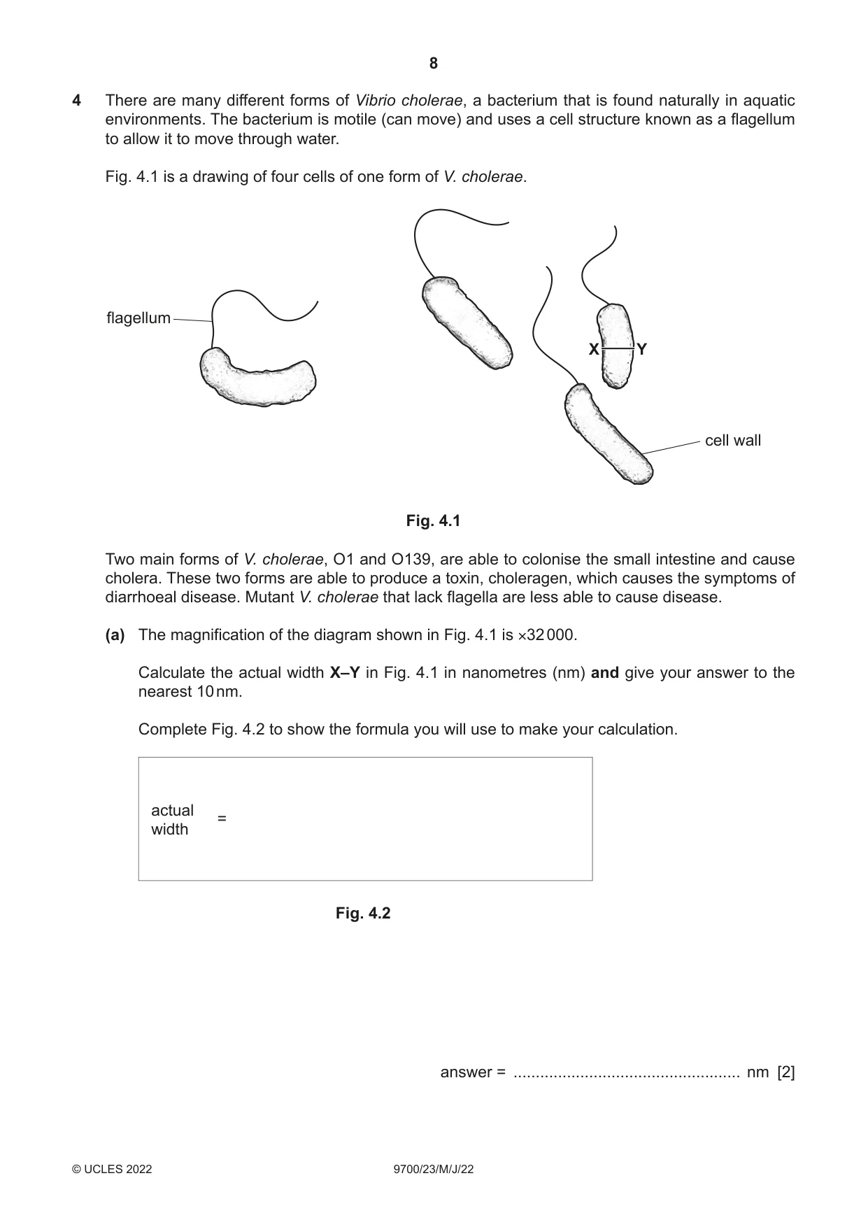
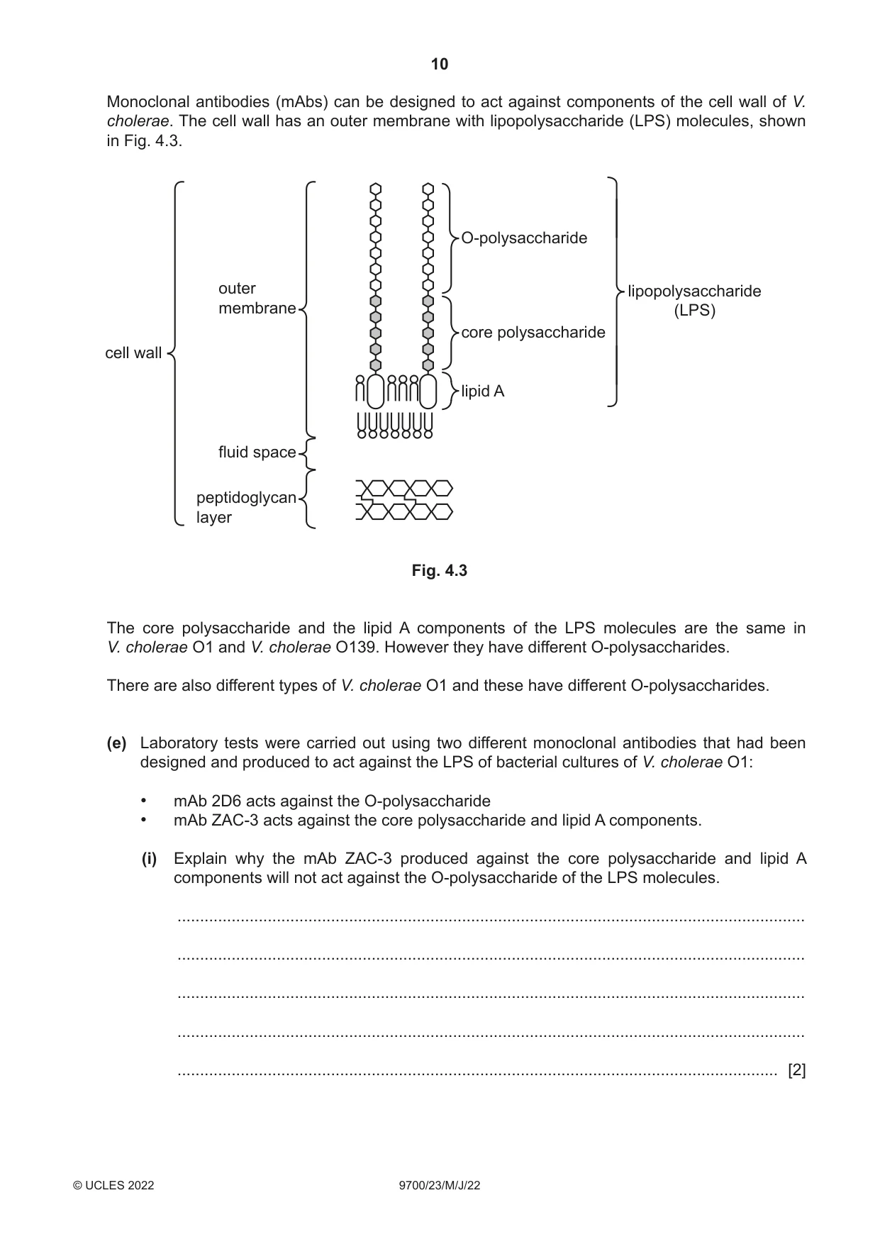
4 There are many different forms of Vibrio cholerae, a bacterium that is found naturally in aquatic environments. The bacterium is motile (can move) and uses a cell structure known as a flagellum to allow it to move through water. Fig. 4.1 is a drawing of four cells of one form of V. cholerae. flagellum X Y cell wall Fig. 4.1 Two main forms of V. cholerae, O1 and O139, are able to colonise the small intestine and cause cholera. These two forms are able to produce a toxin, choleragen, which causes the symptoms of diarrhoeal disease. Mutant V. cholerae that lack flagella are less able to cause disease. (a) The magnification of the diagram shown in Fig. 4.1 is ×32 000. Calculate the actual width X–Y in Fig. 4.1 in nanometres (nm) and give your answer to the nearest 10 nm. Complete Fig. 4.2 to show the formula you will use to make your calculation. actual = width Fig. 4.2 answer = ................................................... nm [2] (b) State the term used to describe disease-causing organisms, such as the bacterium V. cholerae. ............................................................................................................................................. [1] (c) Outline one way in which an uninfected person may become infected by V. cholerae. ................................................................................................................................................... ................................................................................................................................................... ............................................................................................................................................. [1] (d) Choleragen is produced after V. cholerae has penetrated (passed through) the mucus lining and attached to intestinal epithelial cells. Choleragen is composed of two subunits: • subunit A consists of one polypeptide • subunit B consists of five identical polypeptides • the polypeptide in subunit A is different from the polypeptides in subunit B. Two genes, ctxA and ctxB, are needed to produce choleragen. Only one strand of the DNA forming gene ctxA is involved in the production of subunit A. Only one strand of the DNA forming gene ctxB is involved in the production of subunit B. Explain why only one strand of the DNA of each gene is involved in the production of the subunits. ................................................................................................................................................... ................................................................................................................................................... ................................................................................................................................................... ................................................................................................................................................... ................................................................................................................................................... ................................................................................................................................................... ................................................................................................................................................... ............................................................................................................................................. [3] Monoclonal antibodies (mAbs) can be designed to act against components of the cell wall of V. cholerae. The cell wall has an outer membrane with lipopolysaccharide (LPS) molecules, shown in Fig. 4.3. O-polysaccharide outer lipopolysaccharide membrane (LPS) core polysaccharide cell wall lipid A fluid space peptidoglycan layer Fig. 4.3 The core polysaccharide and the lipid A components of the LPS molecules are the same in V. cholerae O1 and V. cholerae O139. However they have different O-polysaccharides. There are also different types of V. cholerae O1 and these have different O-polysaccharides. (e) Laboratory tests were carried out using two different monoclonal antibodies that had been designed and produced to act against the LPS of bacterial cultures of V. cholerae O1: • mAb 2D6 acts against the O-polysaccharide • mAb ZAC-3 acts against the core polysaccharide and lipid A components. (i) Explain why the mAb ZAC-3 produced against the core polysaccharide and lipid A components will not act against the O-polysaccharide of the LPS molecules. ........................................................................................................................................... ........................................................................................................................................... ........................................................................................................................................... ........................................................................................................................................... ..................................................................................................................................... [2] (ii) The results of the tests showed that both mAbs were effective in causing agglutination (clumping) of bacteria and in preventing their motility. This suggests they may be useful for preventing cholera and for treating the disease. Discuss whether mAb 2D6 and mAb ZAC-3 may be useful for preventing cholera and for treating the disease. ........................................................................................................................................... ........................................................................................................................................... ........................................................................................................................................... ........................................................................................................................................... ........................................................................................................................................... ........................................................................................................................................... ........................................................................................................................................... ........................................................................................................................................... ........................................................................................................................................... ..................................................................................................................................... [4] [Total: 13]
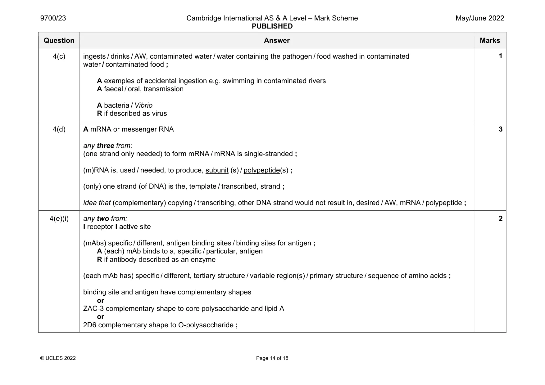
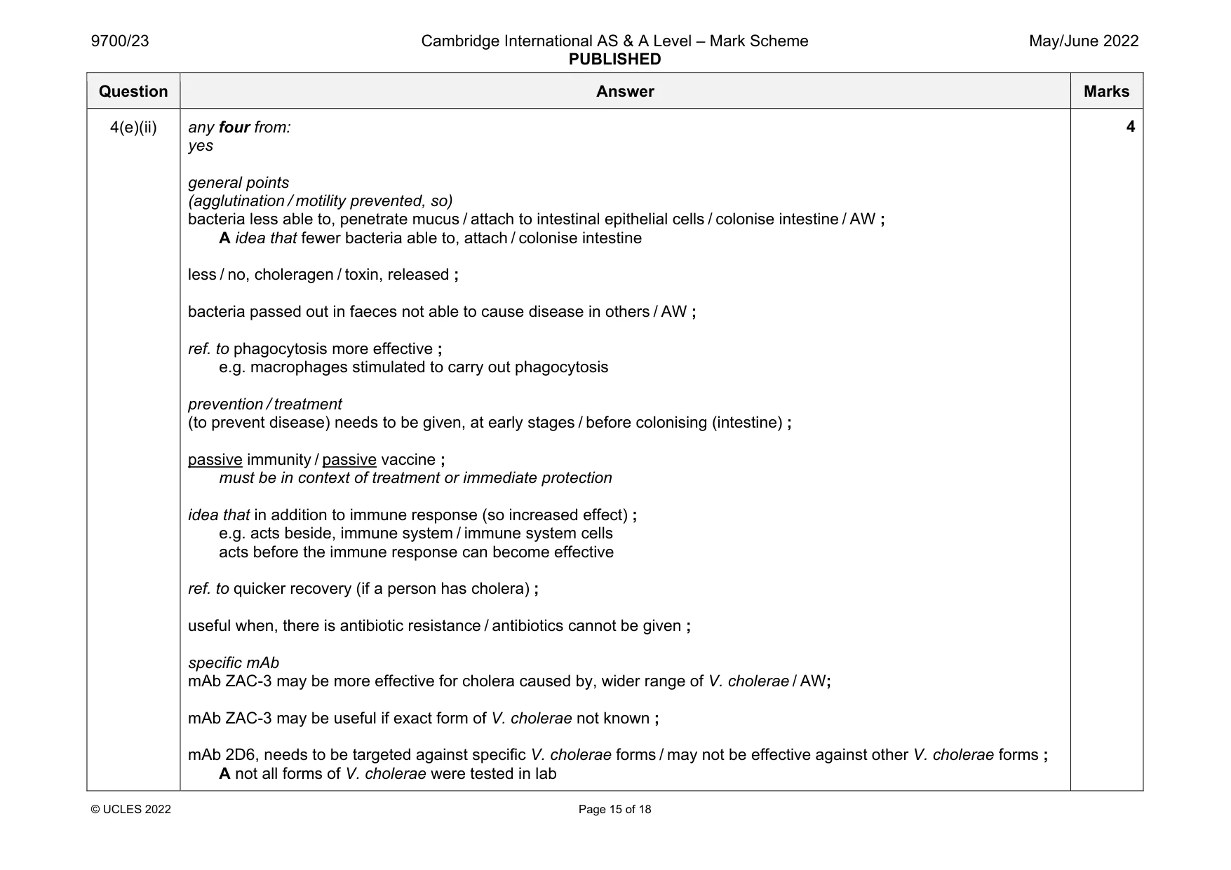
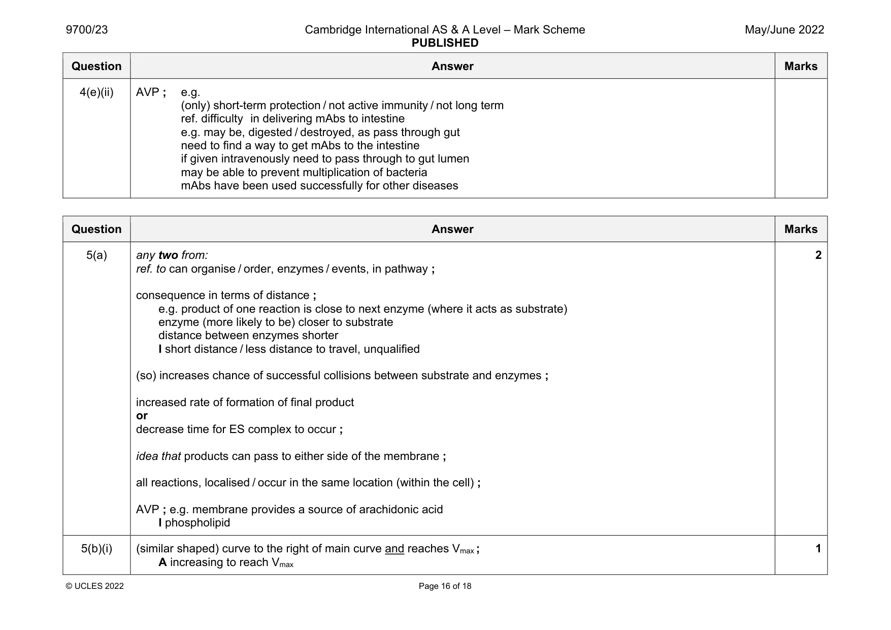
Mark scheme: 4(a) image length ; A magnification triangle magnification 250 (nm) ; (8 mm) A 220 (for 7 mm) A 230 (for 7.5 mm) A 270 (for 8.5 mm) A 280 (for 9 mm) 4(b) pathogen ; 1 Question Answer Marks 4(c) ingests / drinks / AW, contaminated water / water containing the pathogen / food washed in contaminated water / contaminated food ; A examples of accidental ingestion e.g. swimming in contaminated rivers A faecal / oral, transmission A bacteria / Vibrio R if described as virus 1 4(d) A mRNA or messenger RNA any three from: (one strand only needed) to form mRNA / mRNA is single-stranded ; (m)RNA is, used / needed, to produce, subunit (s) / polypeptide(s) ; (only) one strand (of DNA) is the, template / transcribed, strand ; idea that (complementary) copying / transcribing, other DNA strand would not result in, desired / AW, mRNA / polypeptide ; 3 4(e)(i) any two from: I receptor I active site (mAbs) specific / different, antigen binding sites / binding sites for antigen ; A (each) mAb binds to a, specific / particular, antigen R if antibody described as an enzyme (each mAb has) specific / different, tertiary structure / variable region(s) / primary structure / sequence of amino acids ; binding site and antigen have complementary shapes or ZAC-3 complementary shape to core polysaccharide and lipid A or 2D6 complementary shape to O-polysaccharide ; 2 Question Answer Marks 4(e)(ii) any four from: yes general points (agglutination / motility prevented, so) bacteria less able to, penetrate mucus / attach to intestinal epithelial cells / colonise intestine / AW ; A idea that fewer bacteria able to, attach / colonise intestine less / no, choleragen / toxin, released ; bacteria passed out in faeces not able to cause disease in others / AW ; ref. to phagocytosis more effective ; e.g. macrophages stimulated to carry out phagocytosis prevention / treatment (to prevent disease) needs to be given, at early stages / before colonising (intestine) ; passive immunity / passive vaccine ; must be in context of treatment or immediate protection idea that in addition to immune response (so increased effect) ; e.g. acts beside, immune system / immune system cells acts before the immune response can become effective ref. to quicker recovery (if a person has cholera) ; useful when, there is antibiotic resistance / antibiotics cannot be given ; specific mAb mAb ZAC-3 may be more effective for cholera caused by, wider range of V. cholerae / AW; mAb ZAC-3 may be useful if exact form of V. cholerae not known ; mAb 2D6, needs to be targeted against specific V. cholerae forms / may not be effective against other V. cholerae forms ; A not all forms of V. cholerae were tested in lab 4 Question Answer Marks 4(e)(ii) AVP ; e.g. (only) short-term protection / not active immunity / not long term ref. difficulty in delivering mAbs to intestine e.g. may be, digested / destroyed, as pass through gut need to find a way to get mAbs to the intestine if given intravenously need to pass through to gut lumen may be able to prevent multiplication of bacteria mAbs have been used successfully for other diseases
Q5 · Arachidonic acid is a fatty acid that is a common component of phospholipids
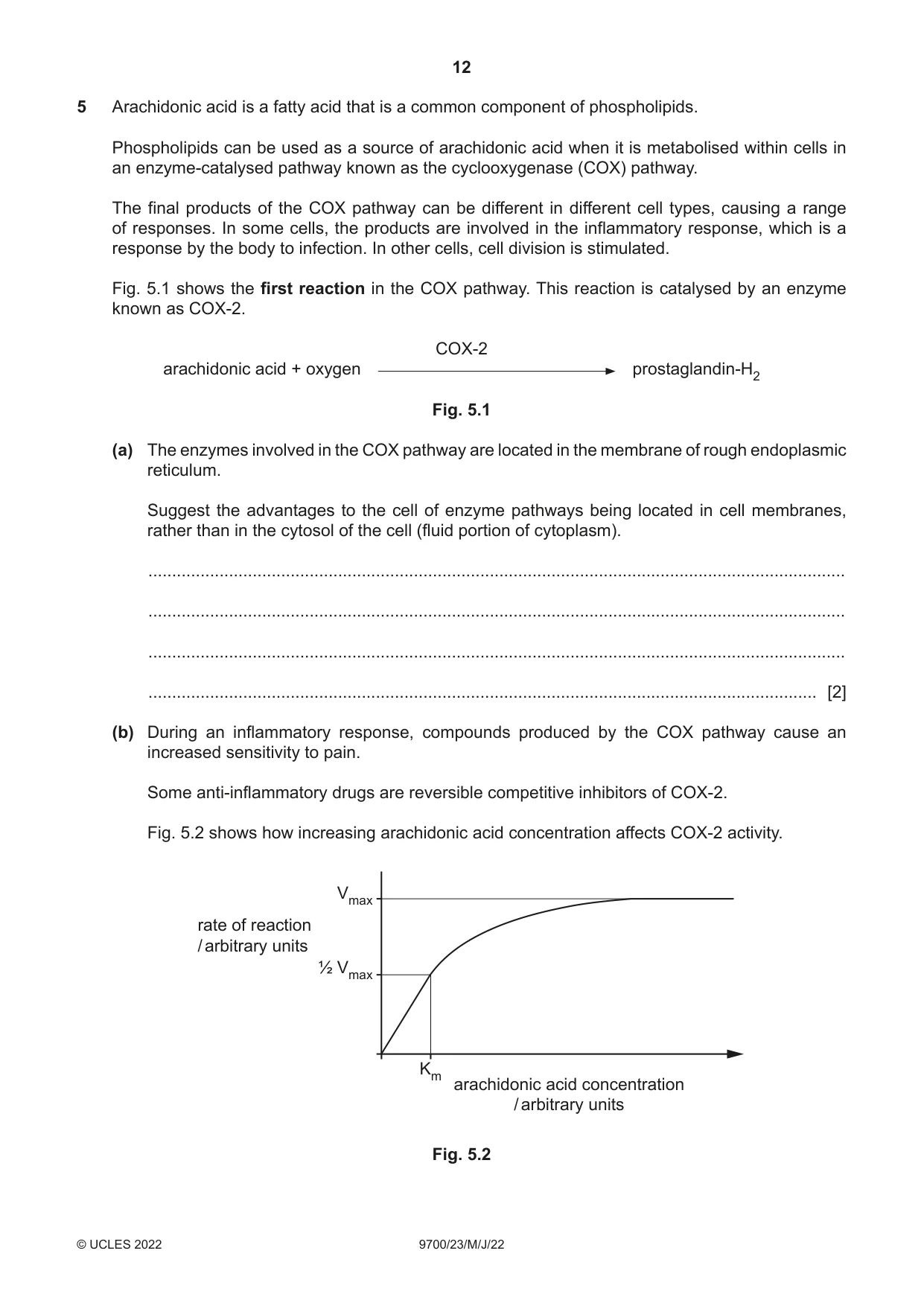
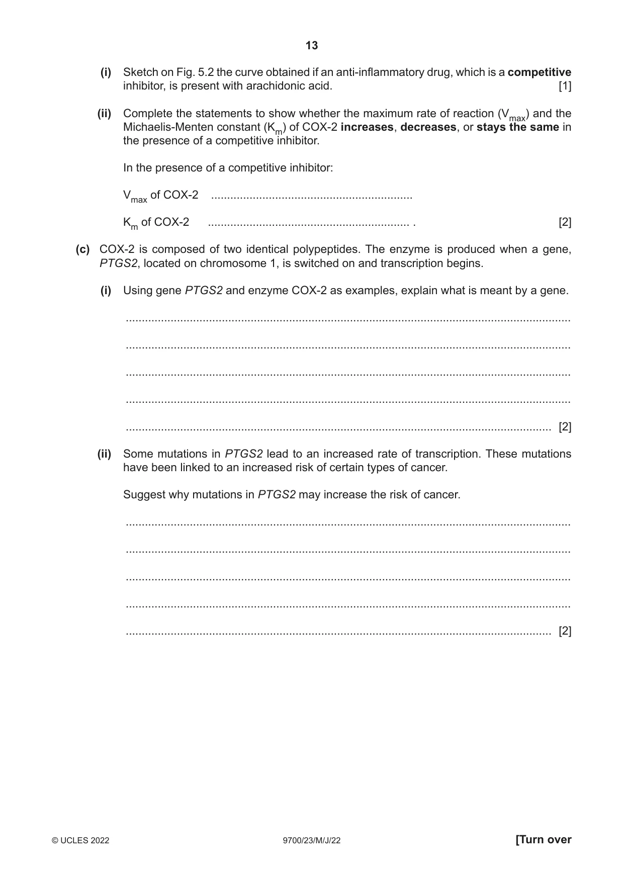
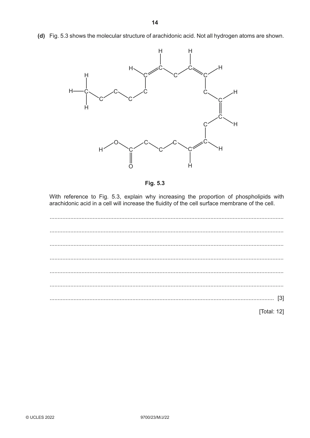
5 Arachidonic acid is a fatty acid that is a common component of phospholipids. Phospholipids can be used as a source of arachidonic acid when it is metabolised within cells in an enzyme-catalysed pathway known as the cyclooxygenase (COX) pathway. The final products of the COX pathway can be different in different cell types, causing a range of responses. In some cells, the products are involved in the inflammatory response, which is a response by the body to infection. In other cells, cell division is stimulated. Fig. 5.1 shows the first reaction in the COX pathway. This reaction is catalysed by an enzyme known as COX-2. COX-2 arachidonic acid + oxygen prostaglandin-H2 Fig. 5.1 (a) The enzymes involved in the COX pathway are located in the membrane of rough endoplasmic reticulum. Suggest the advantages to the cell of enzyme pathways being located in cell membranes, rather than in the cytosol of the cell (fluid portion of cytoplasm). ................................................................................................................................................... ................................................................................................................................................... ................................................................................................................................................... ............................................................................................................................................. [2] (b) During an inflammatory response, compounds produced by the COX pathway cause an increased sensitivity to pain. Some anti-inflammatory drugs are reversible competitive inhibitors of COX-2. Fig. 5.2 shows how increasing arachidonic acid concentration affects COX-2 activity. Vmax rate of reaction / arbitrary units ½ Vmax Km arachidonic acid concentration / arbitrary units Fig. 5.2 (i) Sketch on Fig. 5.2 the curve obtained if an anti-inflammatory drug, which is a competitive inhibitor, is present with arachidonic acid. [1] (ii) Complete the statements to show whether the maximum rate of reaction (Vmax) and the Michaelis-Menten constant (Km) of COX-2 increases, decreases, or stays the same in the presence of a competitive inhibitor. In the presence of a competitive inhibitor: Vmax of COX-2 ............................................................... Km of COX-2 ............................................................... . [2] (c) COX-2 is composed of two identical polypeptides. The enzyme is produced when a gene, PTGS2, located on chromosome 1, is switched on and transcription begins. (i) Using gene PTGS2 and enzyme COX-2 as examples, explain what is meant by a gene. ........................................................................................................................................... ........................................................................................................................................... ........................................................................................................................................... ........................................................................................................................................... ..................................................................................................................................... [2] (ii) Some mutations in PTGS2 lead to an increased rate of transcription. These mutations have been linked to an increased risk of certain types of cancer. Suggest why mutations in PTGS2 may increase the risk of cancer. ........................................................................................................................................... ........................................................................................................................................... ........................................................................................................................................... ........................................................................................................................................... ..................................................................................................................................... [2] (d) Fig. 5.3 shows the molecular structure of arachidonic acid. Not all hydrogen atoms are shown. H H H C C H H C C C H C C C C H C C C H C C H O C C C H C C C H O H Fig. 5.3 With reference to Fig. 5.3, explain why increasing the proportion of phospholipids with arachidonic acid in a cell will increase the fluidity of the cell surface membrane of the cell. ................................................................................................................................................... ................................................................................................................................................... ................................................................................................................................................... ................................................................................................................................................... ................................................................................................................................................... ................................................................................................................................................... ............................................................................................................................................. [3] [Total: 12]
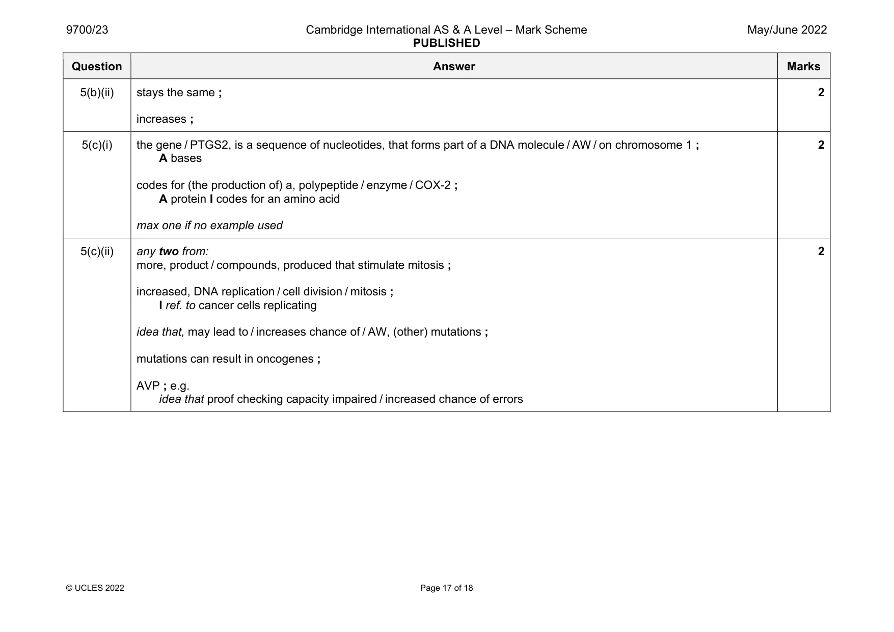
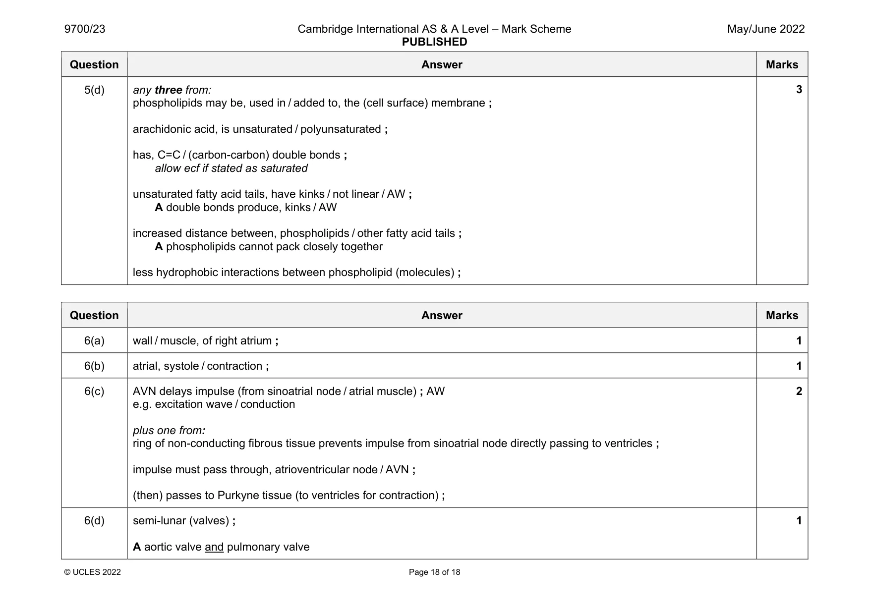
Mark scheme: 5(a) any two from: ref. to can organise / order, enzymes / events, in pathway ; consequence in terms of distance ; e.g. product of one reaction is close to next enzyme (where it acts as substrate) enzyme (more likely to be) closer to substrate distance between enzymes shorter I short distance / less distance to travel, unqualified (so) increases chance of successful collisions between substrate and enzymes ; increased rate of formation of final product or decrease time for ES complex to occur ; idea that products can pass to either side of the membrane ; all reactions, localised / occur in the same location (within the cell) ; AVP ; e.g. membrane provides a source of arachidonic acid I phospholipid 5(b)(i) (similar shaped) curve to the right of main curve and reaches Vmax ; A increasing to reach Vmax 1 Question Answer Marks 5(b)(ii) stays the same ; increases ; 2 5(c)(i) the gene / PTGS2, is a sequence of nucleotides, that forms part of a DNA molecule / AW / on chromosome 1 ; A bases codes for (the production of) a, polypeptide / enzyme / COX-2 ; A protein I codes for an amino acid max one if no example used 2 5(c)(ii) any two from: more, product / compounds, produced that stimulate mitosis ; increased, DNA replication / cell division / mitosis ; I ref. to cancer cells replicating idea that, may lead to / increases chance of / AW, (other) mutations ; mutations can result in oncogenes ; AVP ; e.g. idea that proof checking capacity impaired / increased chance of errors 2 Question Answer Marks 5(d) any three from: phospholipids may be, used in / added to, the (cell surface) membrane ; arachidonic acid, is unsaturated / polyunsaturated ; has, C=C / (carbon-carbon) double bonds ; allow ecf if stated as saturated unsaturated fatty acid tails, have kinks / not linear / AW ; A double bonds produce, kinks / AW increased distance between, phospholipids / other fatty acid tails ; A phospholipids cannot pack closely together less hydrophobic interactions between phospholipid (molecules) ; 3
Q6 · The sinoatrial node, atrioventricular node and the Purkyne tissue have important roles in…
6 The sinoatrial node, atrioventricular node and the Purkyne tissue have important roles in the cardiac cycle. (a) State the precise location in the heart of the sinoatrial node. ............................................................................................................................................. [1] (b) State the part of the cardiac cycle that is directly initiated by the wave of excitation sent out by the sinoatrial node. ............................................................................................................................................. [1] (c) Part of the control of the cardiac cycle involves the contraction of the ventricle walls after the walls of the atria have finished contracting. Outline how this control is achieved. ................................................................................................................................................... ................................................................................................................................................... ................................................................................................................................................... ................................................................................................................................................... ............................................................................................................................................. [2] (d) Name the valves of the heart that open soon after the Purkyne tissue has received an impulse from the atrioventricular node. ............................................................................................................................................. [1] [Total: 5]
Mark scheme: 6(a) wall / muscle, of right atrium ; 1 6(b) atrial, systole / contraction ; 1 6(c) AVN delays impulse (from sinoatrial node / atrial muscle) ; AW e.g. excitation wave / conduction plus one from: ring of non-conducting fibrous tissue prevents impulse from sinoatrial node directly passing to ventricles ; impulse must pass through, atrioventricular node / AVN ; (then) passes to Purkyne tissue (to ventricles for contraction) ; 2 6(d) semi-lunar (valves) ; A aortic valve and pulmonary valve 1
What was in this paper
The subtopics covered by these 6 questions, and how many questions each got. Open one in a new tab to see every Cambridge question on it.
What you needed in this session
Cambridge’s own grade thresholds for 2022 May/June, Paper 2 · Variant 3. A higher threshold means an easier paper — the bar moves with how the cohort did.