Cambridge A Level Biology 9700 — 2023 Oct/Nov Paper 2 · Variant 3
9700/23/O/N/23 · 6 questions · 60 marks · ≈68 min
The question paper and its mark scheme, free to read here and free to download. This is Cambridge’s own paper, exactly as it was sat.
Question paper16 pages

Mark scheme17 pages
Answers below. Sit the paper first if you are practising.
Questions as text
Q1 · The malarial pathogen, Plasmodium falciparum, enters red blood cells after a person…
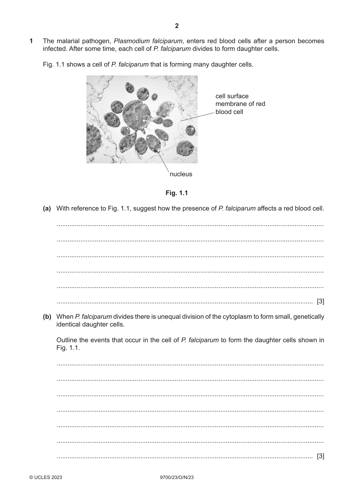
1 The malarial pathogen, Plasmodium falciparum, enters red blood cells after a person becomes infected. After some time, each cell of P. falciparum divides to form daughter cells. Fig. 1.1 shows a cell of P. falciparum that is forming many daughter cells. cell surface membrane of red blood cell nucleus Fig. 1.1 (a) With reference to Fig. 1.1, suggest how the presence of P. falciparum affects a red blood cell. ................................................................................................................................................... ................................................................................................................................................... ................................................................................................................................................... ................................................................................................................................................... ................................................................................................................................................... ............................................................................................................................................. [3] (b) When P. falciparum divides there is unequal division of the cytoplasm to form small, genetically identical daughter cells. Outline the events that occur in the cell of P. falciparum to form the daughter cells shown in Fig. 1.1. ................................................................................................................................................... ................................................................................................................................................... ................................................................................................................................................... ................................................................................................................................................... ................................................................................................................................................... ................................................................................................................................................... ............................................................................................................................................. [3] In 2013, the World Health Organization (WHO) set a target for researchers to create a vaccine for malaria. WHO required the vaccine to show 75% efficacy and be ready for use by 2030. Efficacy is a measure of the effectiveness of a vaccine in reducing the number of new cases of malaria. A trial of the R21/Matrix‑MTM vaccine in Burkina Faso in 2020 achieved a 77% efficacy over a 12‑month period. A control group received a vaccine for rabies. Vaccines stimulate an immune response with the production of antibodies. (c) Explain how antibodies will reduce the spread of the malarial pathogen through the bloodstream. ................................................................................................................................................... ................................................................................................................................................... ................................................................................................................................................... ................................................................................................................................................... ................................................................................................................................................... ................................................................................................................................................... ............................................................................................................................................. [3] (d) Some vaccination programmes have been more successful than others. Discuss the factors that contribute to the success of a vaccination programme. ................................................................................................................................................... ................................................................................................................................................... ................................................................................................................................................... ................................................................................................................................................... ................................................................................................................................................... ................................................................................................................................................... ................................................................................................................................................... ................................................................................................................................................... ................................................................................................................................................... ................................................................................................................................................... ............................................................................................................................................. [5] [Total: 14]
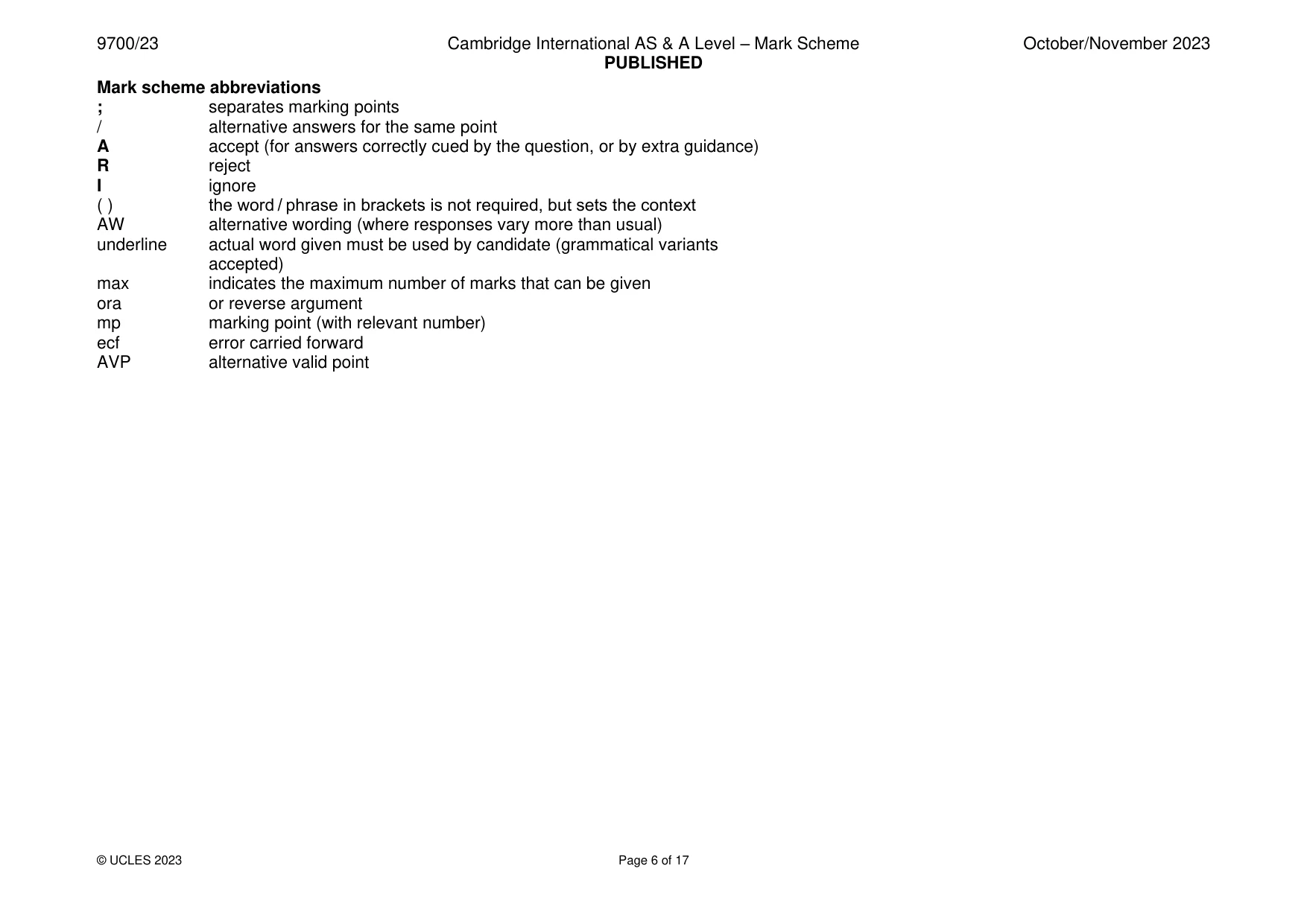
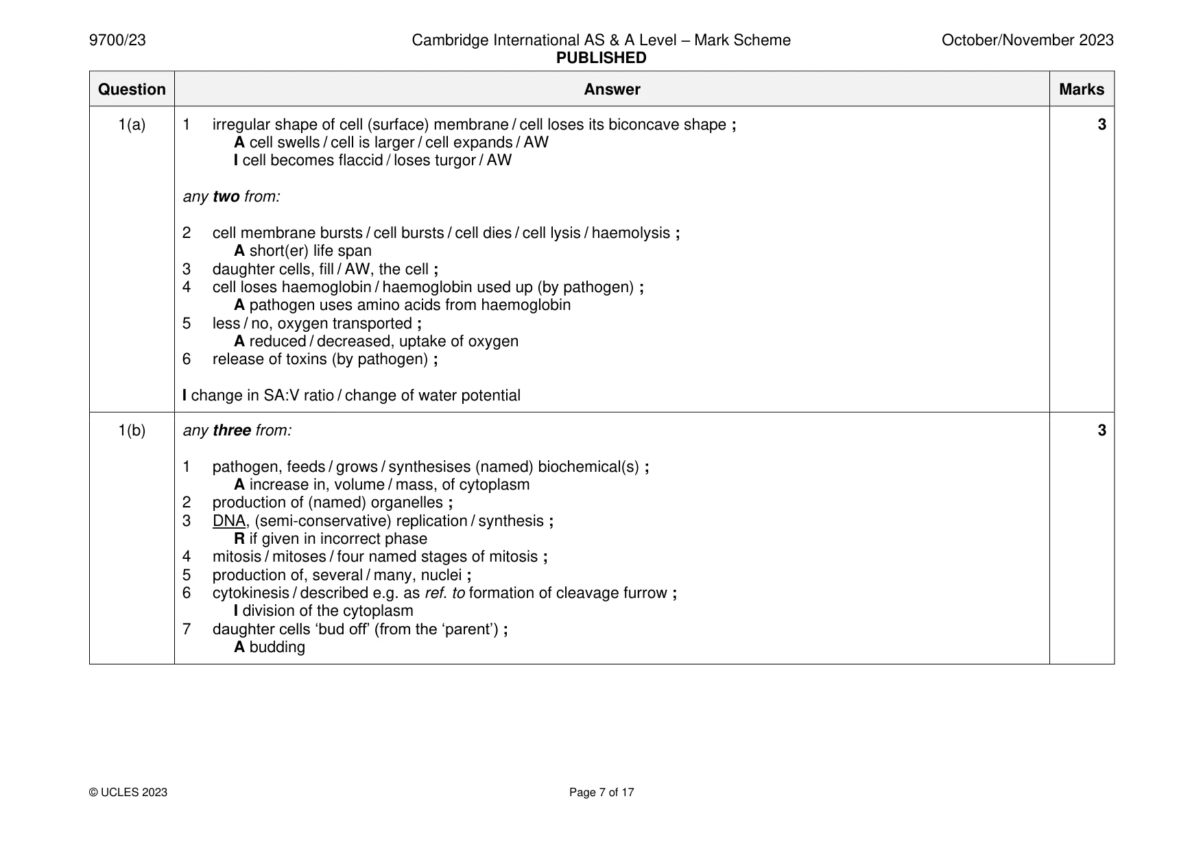
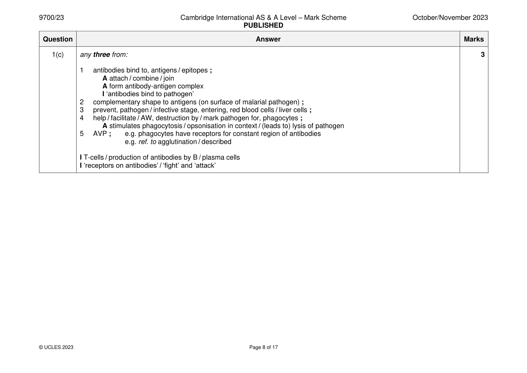
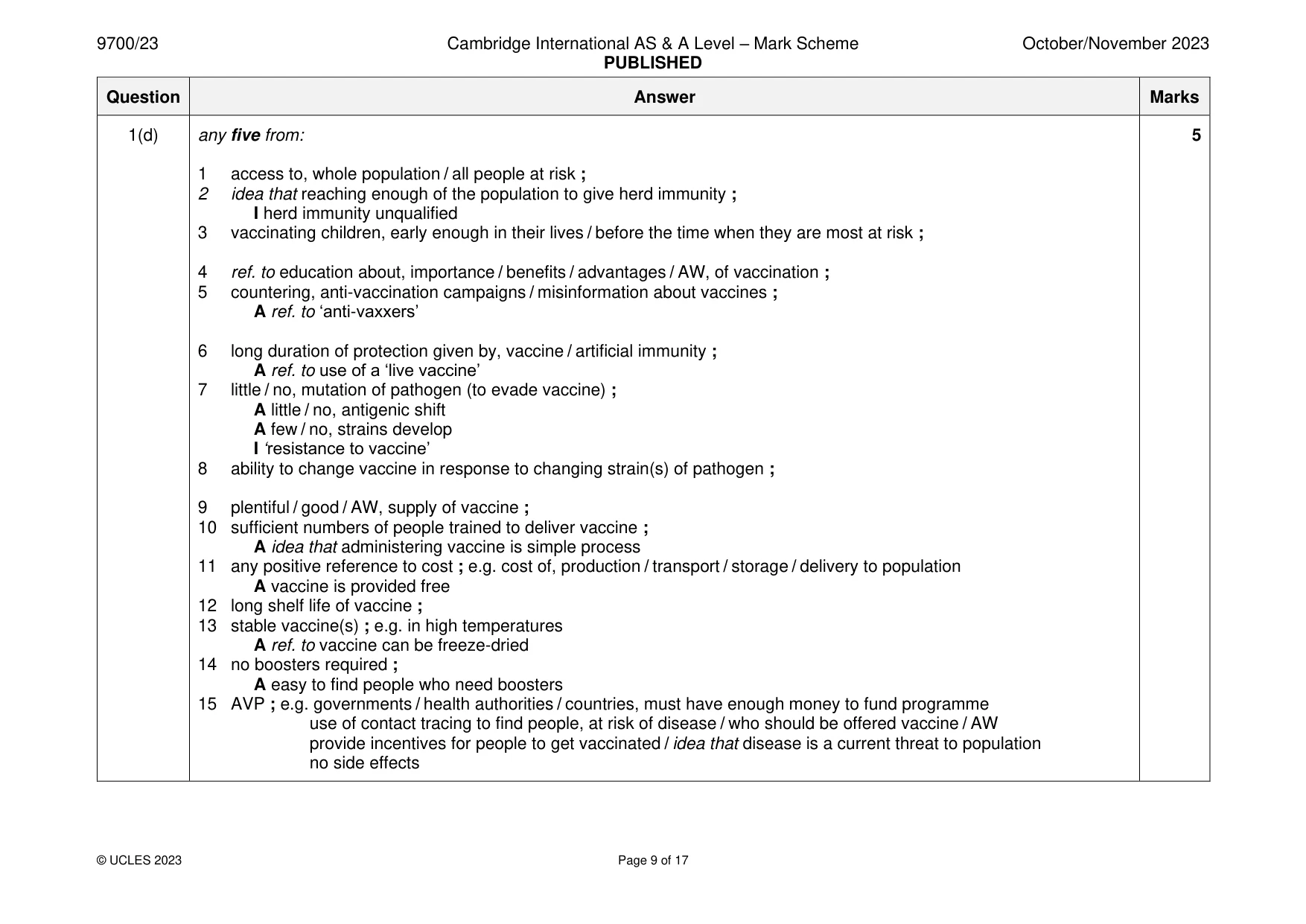
Mark scheme: Question Answer Marks 1(a) 1 irregular shape of cell (surface) membrane / cell loses its biconcave shape ; 3 A cell swells / cell is larger / cell expands / AW I cell becomes flaccid / loses turgor / AW any two from: 2 cell membrane bursts / cell bursts / cell dies / cell lysis / haemolysis ; A short(er) life span 3 daughter cells, fill / AW, the cell ; 4 cell loses haemoglobin / haemoglobin used up (by pathogen) ; A pathogen uses amino acids from haemoglobin 5 less / no, oxygen transported ; A reduced / decreased, uptake of oxygen 6 release of toxins (by pathogen) ; I change in SA:V ratio / change of water potential 1(b) any three from: 3 1 pathogen, feeds / grows / synthesises (named) biochemical(s) ; A increase in, volume / mass, of cytoplasm 2 production of (named) organelles ; 3 DNA, (semi-conservative) replication / synthesis ; R if given in incorrect phase 4 mitosis / mitoses / four named stages of mitosis ; 5 production of, several / many, nuclei ; 6 cytokinesis / described e.g. as ref. to formation of cleavage furrow ; I division of the cytoplasm 7 daughter cells ‘bud off’ (from the ‘parent’) ; A budding 1(c) any three from: 3 1 antibodies bind to, antigens / epitopes ; A attach / combine / join A form antibody-antigen complex I ‘antibodies bind to pathogen’ 2 complementary shape to antigens (on surface of malarial pathogen) ; 3 prevent, pathogen / infective stage, entering, red blood cells / liver cells ; 4 help / facilitate / AW, destruction by / mark pathogen for, phagocytes ; A stimulates phagocytosis / opsonisation in context / (leads to) lysis of pathogen 5 AVP ; e.g. phagocytes have receptors for constant region of antibodies e.g. ref. to agglutination / described I T-cells / production of antibodies by B / plasma cells I ‘receptors on antibodies’ / ‘fight’ and ‘attack’ 1(d) any five from: 5 1 access to, whole population / all people at risk ; 2 idea that reaching enough of the population to give herd immunity ; I herd immunity unqualified 3 vaccinating children, early enough in their lives / before the time when they are most at risk ; 4 ref. to education about, importance / benefits / advantages / AW, of vaccination ; 5 countering, anti-vaccination campaigns / misinformation about vaccines ; A ref. to ‘anti-vaxxers’ 6 long duration of protection given by, vaccine / artificial immunity ; A ref. to use of a ‘live vaccine’ 7 little / no, mutation of pathogen (to evade vaccine) ; A little / no, antigenic shift A few / no, strains develop I ‘resistance to vaccine’ 8 ability to change vaccine in response to changing strain(s) of pathogen ; 9 plentiful / good / AW, supply of vaccine ; 10 sufficient numbers of people trained to deliver vaccine ; A idea that administering vaccine is simple process 11 any positive reference to cost ; e.g. cost of, production / transport / storage / delivery to population A vaccine is provided free 12 long shelf life of vaccine ; 13 stable vaccine(s) ; e.g. in high temperatures A ref. to vaccine can be freeze-dried 14 no boosters required ; A easy to find people who need boosters 15 AVP ; e.g. governments / health authorities / countries, must have enough money to fund programme use of contact tracing to find people, at risk of disease / who should be offered vaccine / AW provide incentives for people to get vaccinated / idea that disease is a current threat to population no side effects
Q2 · Glucose is used in the synthesis of amylose
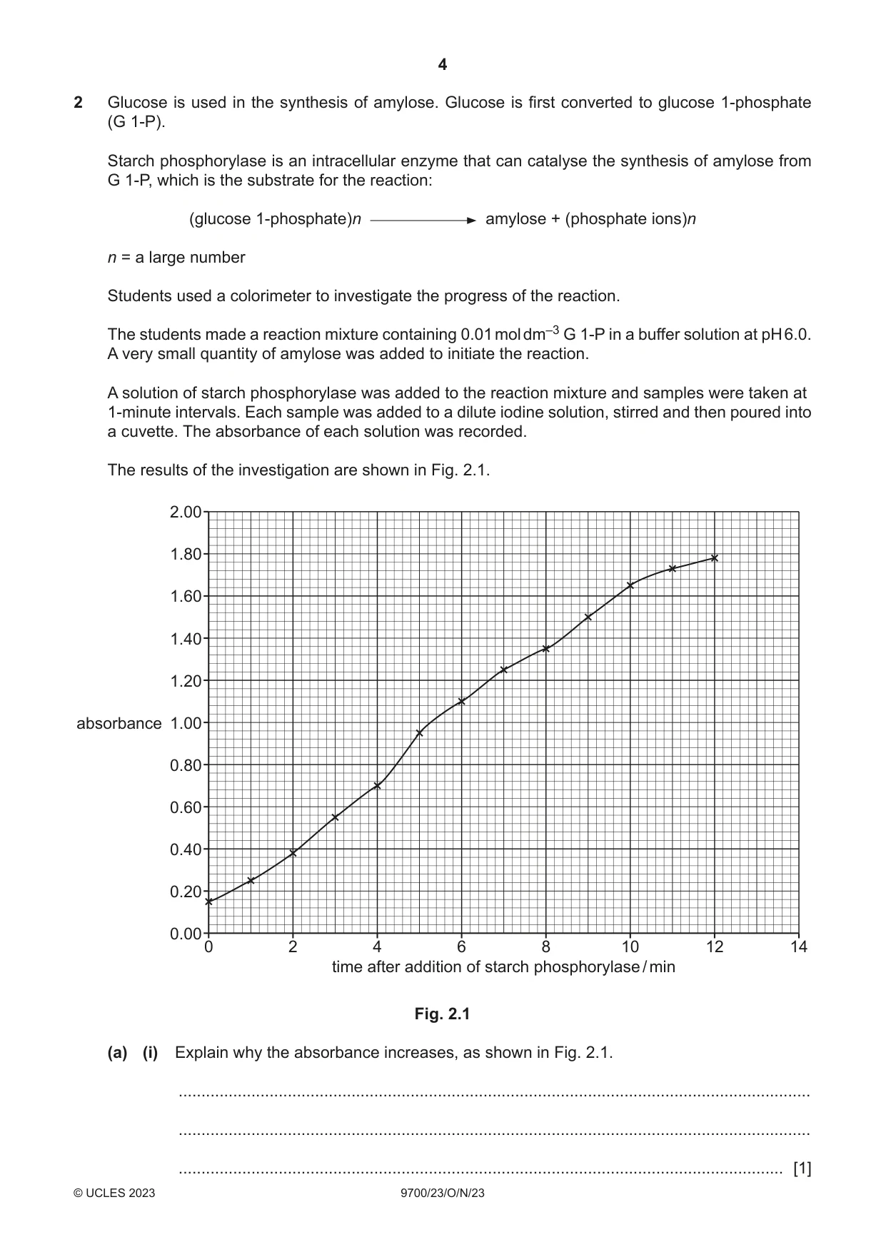
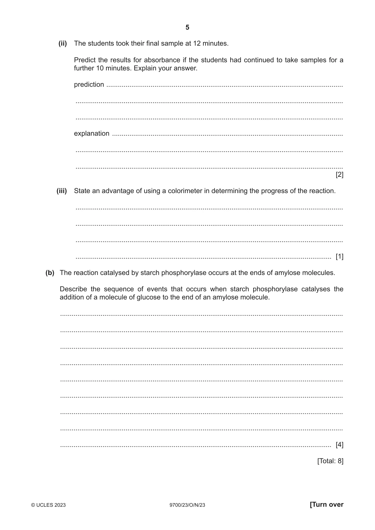
2 Glucose is used in the synthesis of amylose. Glucose is first converted to glucose 1‑phosphate (G 1‑P). Starch phosphorylase is an intracellular enzyme that can catalyse the synthesis of amylose from G 1‑P, which is the substrate for the reaction: (glucose 1‑phosphate)n amylose + (phosphate ions)n n = a large number Students used a colorimeter to investigate the progress of the reaction. The students made a reaction mixture containing 0.01 mol dm–3 G 1‑P in a buffer solution at pH 6.0. A very small quantity of amylose was added to initiate the reaction. A solution of starch phosphorylase was added to the reaction mixture and samples were taken at 1‑minute intervals. Each sample was added to a dilute iodine solution, stirred and then poured into a cuvette. The absorbance of each solution was recorded. The results of the investigation are shown in Fig. 2.1. 2.00 1.80 1.60 1.40 1.20 absorbance 1.00 0.80 0.60 0.40 0.20 0.00 0 2 4 6 8 10 12 14 time after addition of starch phosphorylase / min Fig. 2.1 (a) (i) Explain why the absorbance increases, as shown in Fig. 2.1. ........................................................................................................................................... ........................................................................................................................................... ..................................................................................................................................... [1] (ii) The students took their final sample at 12 minutes. Predict the results for absorbance if the students had continued to take samples for a further 10 minutes. Explain your answer. prediction ........................................................................................................................... ........................................................................................................................................... ........................................................................................................................................... explanation ........................................................................................................................ ........................................................................................................................................... ........................................................................................................................................... [2] (iii) State an advantage of using a colorimeter in determining the progress of the reaction. ........................................................................................................................................... ........................................................................................................................................... ........................................................................................................................................... ..................................................................................................................................... [1] (b) The reaction catalysed by starch phosphorylase occurs at the ends of amylose molecules. Describe the sequence of events that occurs when starch phosphorylase catalyses the addition of a molecule of glucose to the end of an amylose molecule. ................................................................................................................................................... ................................................................................................................................................... ................................................................................................................................................... ................................................................................................................................................... ................................................................................................................................................... ................................................................................................................................................... ................................................................................................................................................... ................................................................................................................................................... ............................................................................................................................................. [4] [Total: 8]
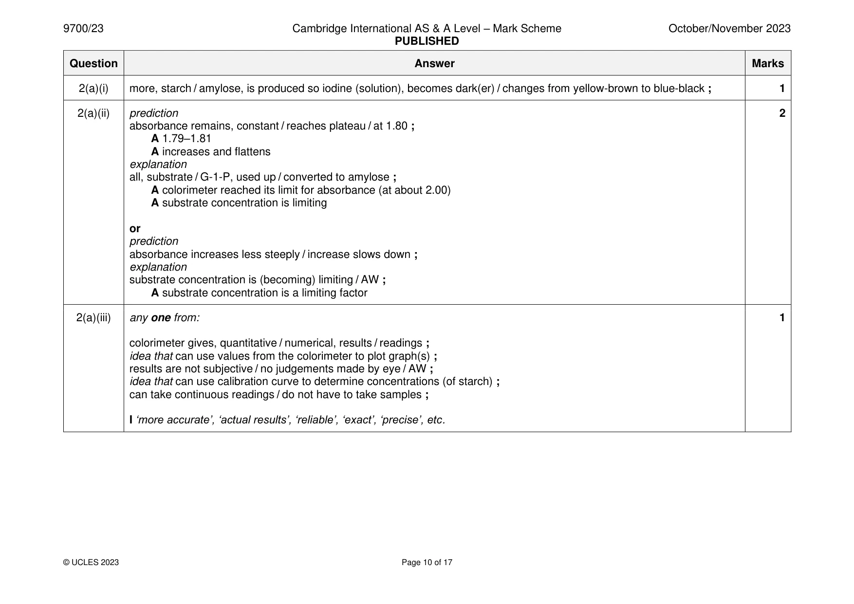
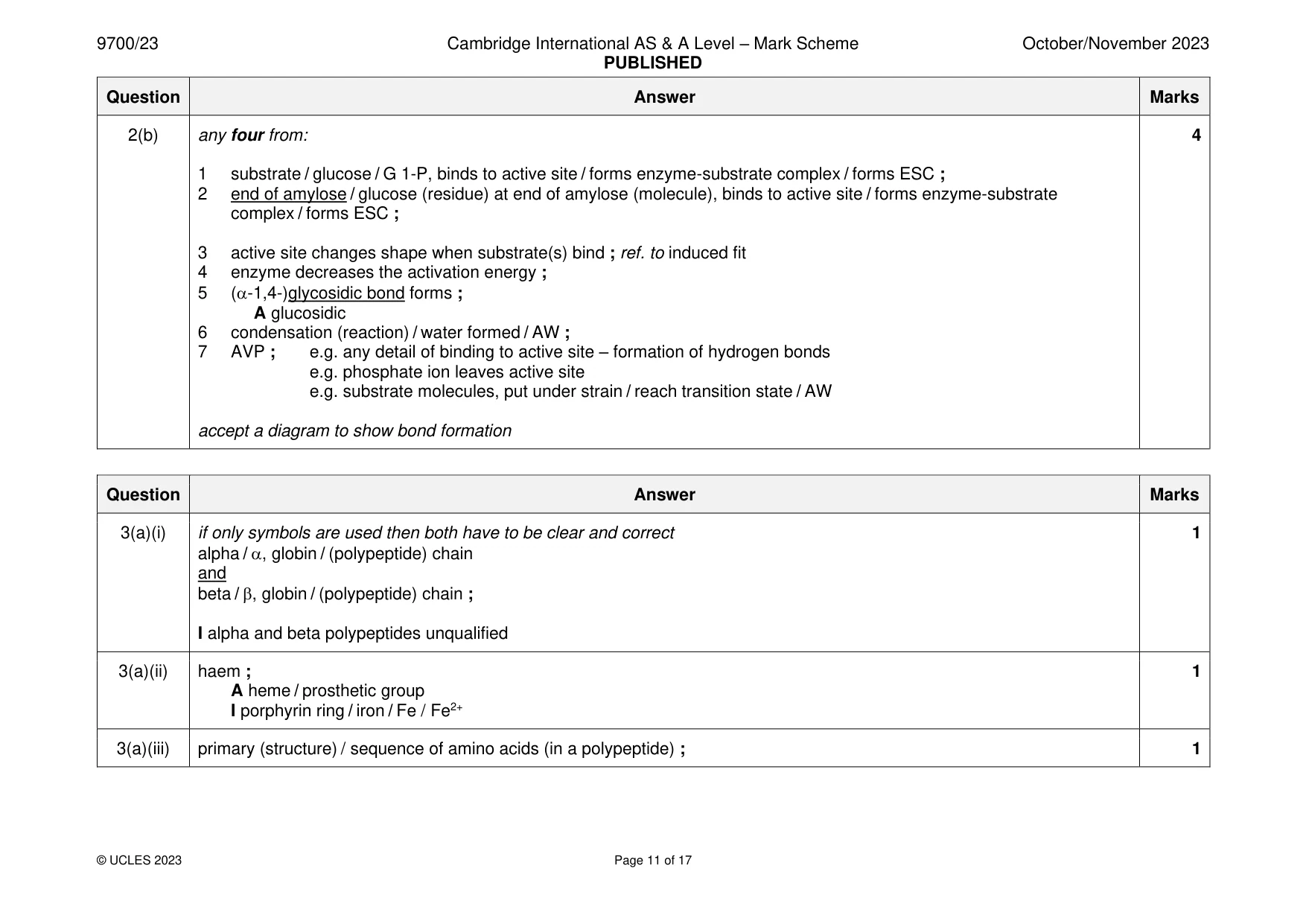
Mark scheme: 2(a)(i) more, starch / amylose, is produced so iodine (solution), becomes dark(er) / changes from yellow-brown to blue-black ; 1 2(a)(ii) prediction 2 absorbance remains, constant / reaches plateau / at 1.80 ; A 1.79–1.81 A increases and flattens explanation all, substrate / G-1-P, used up / converted to amylose ; A colorimeter reached its limit for absorbance (at about 2.00) A substrate concentration is limiting or prediction absorbance increases less steeply / increase slows down ; explanation substrate concentration is (becoming) limiting / AW ; A substrate concentration is a limiting factor 2(a)(iii) any one from: 1 colorimeter gives, quantitative / numerical, results / readings ; idea that can use values from the colorimeter to plot graph(s) ; results are not subjective / no judgements made by eye / AW ; idea that can use calibration curve to determine concentrations (of starch) ; can take continuous readings / do not have to take samples ; I ‘more accurate’, ‘actual results’, ‘reliable’, ‘exact’, ‘precise’, etc. 2(b) any four from: 4 1 substrate / glucose / G 1-P, binds to active site / forms enzyme-substrate complex / forms ESC ; 2 end of amylose / glucose (residue) at end of amylose (molecule), binds to active site / forms enzyme-substrate complex / forms ESC ; 3 active site changes shape when substrate(s) bind ; ref. to induced fit 4 enzyme decreases the activation energy ; 5 (-1,4-)glycosidic bond forms ; A glucosidic 6 condensation (reaction) / water formed / AW ; 7 AVP ; e.g. any detail of binding to active site – formation of hydrogen bonds e.g. phosphate ion leaves active site e.g. substrate molecules, put under strain / reach transition state / AW accept a diagram to show bond formation
Q3 · Haemoglobin is a complex protein molecule made of four separate subunits, as shown in Fig
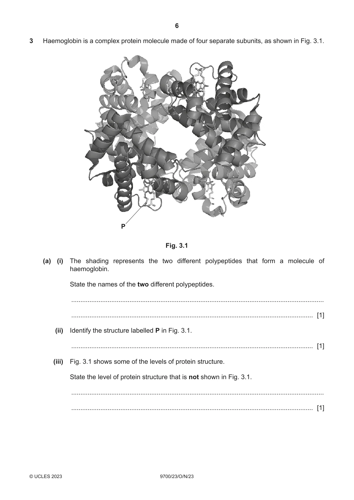
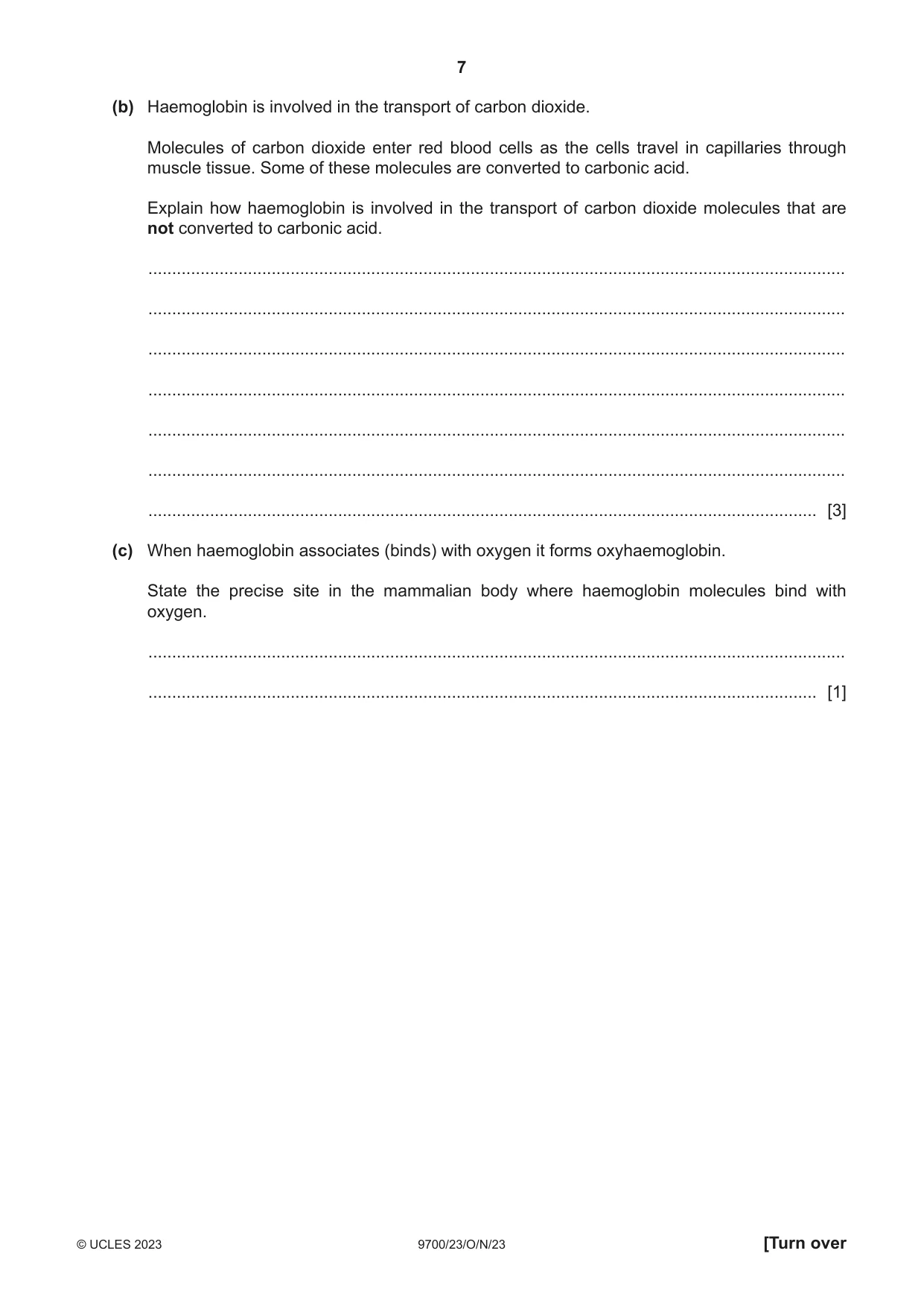
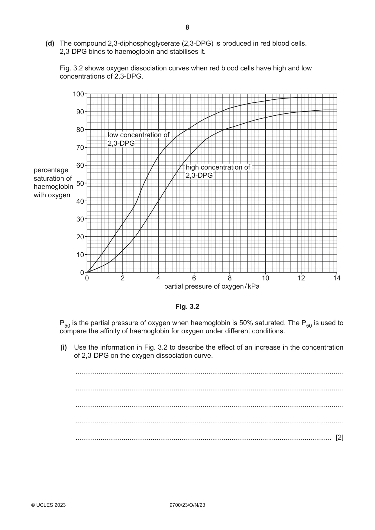
3 Haemoglobin is a complex protein molecule made of four separate subunits, as shown in Fig. 3.1. P Fig. 3.1 (a) (i) The shading represents the two different polypeptides that form a molecule of haemoglobin. State the names of the two different polypeptides. ........................................................................................................................................... ..................................................................................................................................... [1] (ii) Identify the structure labelled P in Fig. 3.1. ..................................................................................................................................... [1] (iii) Fig. 3.1 shows some of the levels of protein structure. State the level of protein structure that is not shown in Fig. 3.1. ........................................................................................................................................... ..................................................................................................................................... [1] (b) Haemoglobin is involved in the transport of carbon dioxide. Molecules of carbon dioxide enter red blood cells as the cells travel in capillaries through muscle tissue. Some of these molecules are converted to carbonic acid. Explain how haemoglobin is involved in the transport of carbon dioxide molecules that are not converted to carbonic acid. ................................................................................................................................................... ................................................................................................................................................... ................................................................................................................................................... ................................................................................................................................................... ................................................................................................................................................... ................................................................................................................................................... ............................................................................................................................................. [3] (c) When haemoglobin associates (binds) with oxygen it forms oxyhaemoglobin. State the precise site in the mammalian body where haemoglobin molecules bind with oxygen. ................................................................................................................................................... ............................................................................................................................................. [1] (d) The compound 2,3‑diphosphoglycerate (2,3‑DPG) is produced in red blood cells. 2,3‑DPG binds to haemoglobin and stabilises it. Fig. 3.2 shows oxygen dissociation curves when red blood cells have high and low concentrations of 2,3‑DPG. 100 90 80 low concentration of 2,3-DPG 70 60 high concentration ofpercentage 2,3-DPGsaturation of 50haemoglobin with oxygen 40 30 20 10 0 0 2 4 6 8 10 12 14 partial pressure of oxygen / kPa Fig. 3.2 P50 is the partial pressure of oxygen when haemoglobin is 50% saturated. The P50 is used to compare the affinity of haemoglobin for oxygen under different conditions. (i) Use the information in Fig. 3.2 to describe the effect of an increase in the concentration of 2,3‑DPG on the oxygen dissociation curve. ........................................................................................................................................... ........................................................................................................................................... ........................................................................................................................................... ........................................................................................................................................... ..................................................................................................................................... [2] (ii) Blood is stored in blood banks for use in hospitals during operations. The concentration of 2,3‑DPG in red blood cells decreases when blood is stored in a blood bank. State and explain the effect that the use of blood taken from a blood bank has on the supply of oxygen to the tissues of a person during an operation. ........................................................................................................................................... ........................................................................................................................................... ........................................................................................................................................... ........................................................................................................................................... ..................................................................................................................................... [2] [Total: 11]
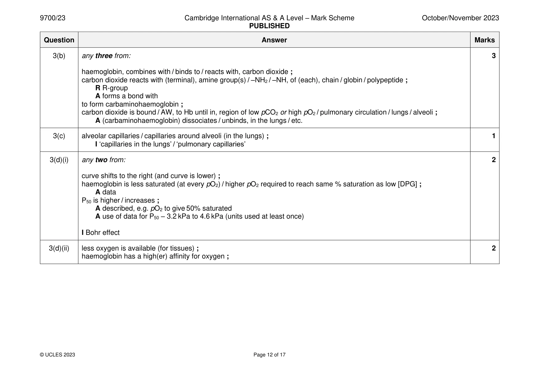
Mark scheme: 3(a)(i) if only symbols are used then both have to be clear and correct 1 alpha / , globin / (polypeptide) chain and beta / , globin / (polypeptide) chain ; I alpha and beta polypeptides unqualified 3(a)(ii) haem ; 1 A heme / prosthetic group I porphyrin ring / iron / Fe / Fe2+ 3(a)(iii) primary (structure) / sequence of amino acids (in a polypeptide) ; 1 3(b) any three from: 3 haemoglobin, combines with / binds to / reacts with, carbon dioxide ; carbon dioxide reacts with (terminal), amine group(s) / –NH2 / –NH, of (each), chain / globin / polypeptide ; R R-group A forms a bond with to form carbaminohaemoglobin ; carbon dioxide is bound / AW, to Hb until in, region of low pCO2 or high pO2 / pulmonary circulation / lungs / alveoli ; A (carbaminohaemoglobin) dissociates / unbinds, in the lungs / etc. 3(c) alveolar capillaries / capillaries around alveoli (in the lungs) ; 1 I ‘capillaries in the lungs’ / ‘pulmonary capillaries’ 3(d)(i) any two from: 2 curve shifts to the right (and curve is lower) ; haemoglobin is less saturated (at every pO2) / higher pO2 required to reach same % saturation as low [DPG] ; A data P50 is higher / increases ; A described, e.g. pO2 to give 50% saturated A use of data for P50 – 3.2 kPa to 4.6 kPa (units used at least once) I Bohr effect 3(d)(ii) less oxygen is available (for tissues) ; 2 haemoglobin has a high(er) affinity for oxygen ;
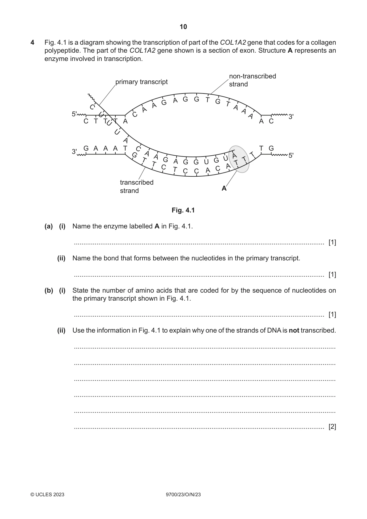
Q4 · A diagram showing the transcription of part of the COL1A2 gene that codes for a collagen…
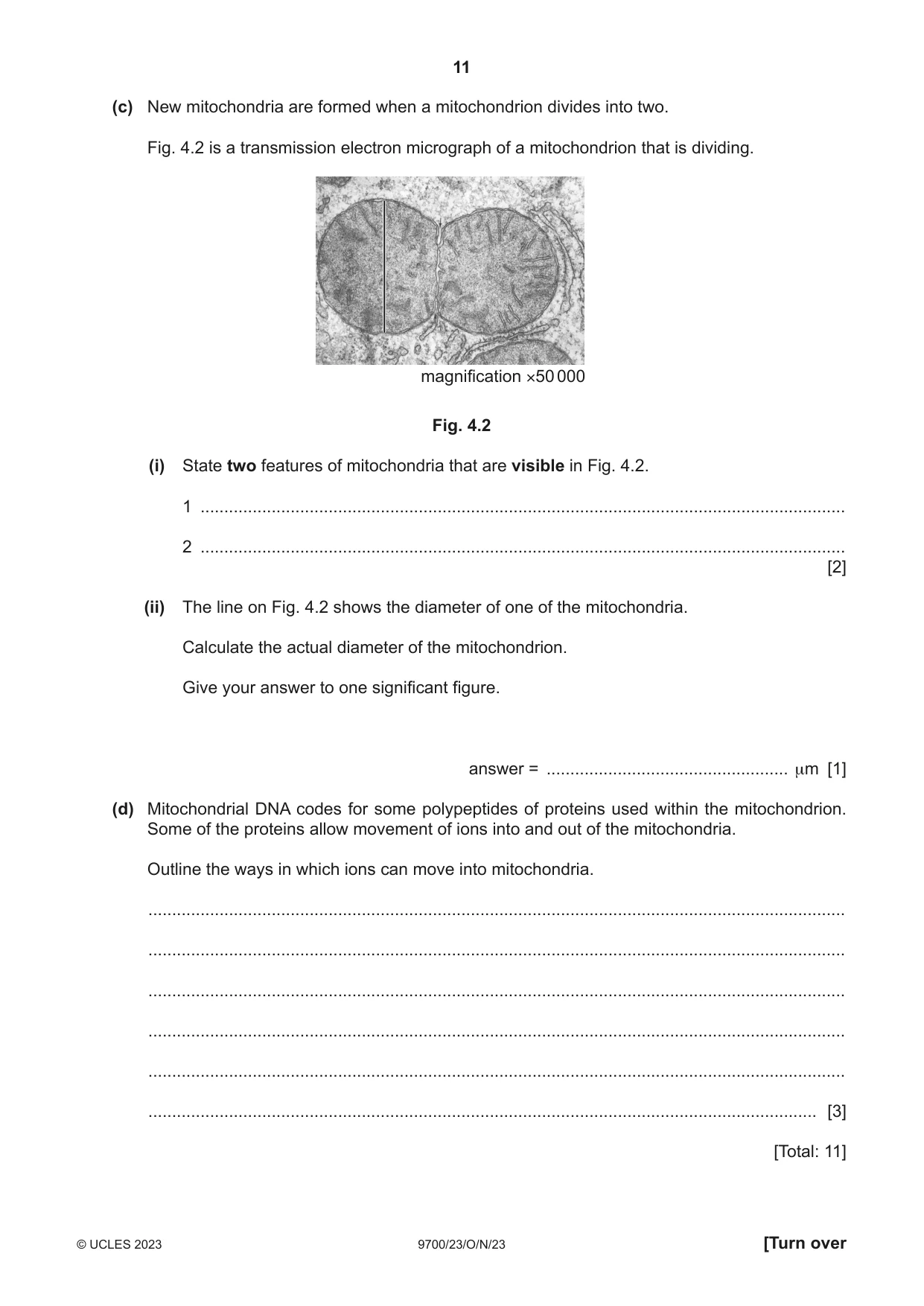
4 Fig. 4.1 is a diagram showing the transcription of part of the COL1A2 gene that codes for a collagen polypeptide. The part of the COL1A2 gene shown is a section of exon. Structure A represents an enzyme involved in transcription. non-transcribed primary transcript strand A G G T G G T C A A A A 5' U C A 3' C T TUT A A C U A G A A A T C T G 3' A T 5' G A A U T G T G A U G G T T A C C T A C C transcribed A strand Fig. 4.1 (a) (i) Name the enzyme labelled A in Fig. 4.1. ..................................................................................................................................... [1] (ii) Name the bond that forms between the nucleotides in the primary transcript. ..................................................................................................................................... [1] (b) (i) State the number of amino acids that are coded for by the sequence of nucleotides on the primary transcript shown in Fig. 4.1. ..................................................................................................................................... [1] (ii) Use the information in Fig. 4.1 to explain why one of the strands of DNA is not transcribed. ........................................................................................................................................... ........................................................................................................................................... ........................................................................................................................................... ........................................................................................................................................... ........................................................................................................................................... ..................................................................................................................................... [2] (c) New mitochondria are formed when a mitochondrion divides into two. Fig. 4.2 is a transmission electron micrograph of a mitochondrion that is dividing. magnification ×50 000 Fig. 4.2 (i) State two features of mitochondria that are visible in Fig. 4.2. 1 ........................................................................................................................................ 2 ........................................................................................................................................ [2] (ii) The line on Fig. 4.2 shows the diameter of one of the mitochondria. Calculate the actual diameter of the mitochondrion. Give your answer to one significant figure. answer = ................................................... μm [1] (d) Mitochondrial DNA codes for some polypeptides of proteins used within the mitochondrion. Some of the proteins allow movement of ions into and out of the mitochondria. Outline the ways in which ions can move into mitochondria. ................................................................................................................................................... ................................................................................................................................................... ................................................................................................................................................... ................................................................................................................................................... ................................................................................................................................................... ............................................................................................................................................. [3] [Total: 11]
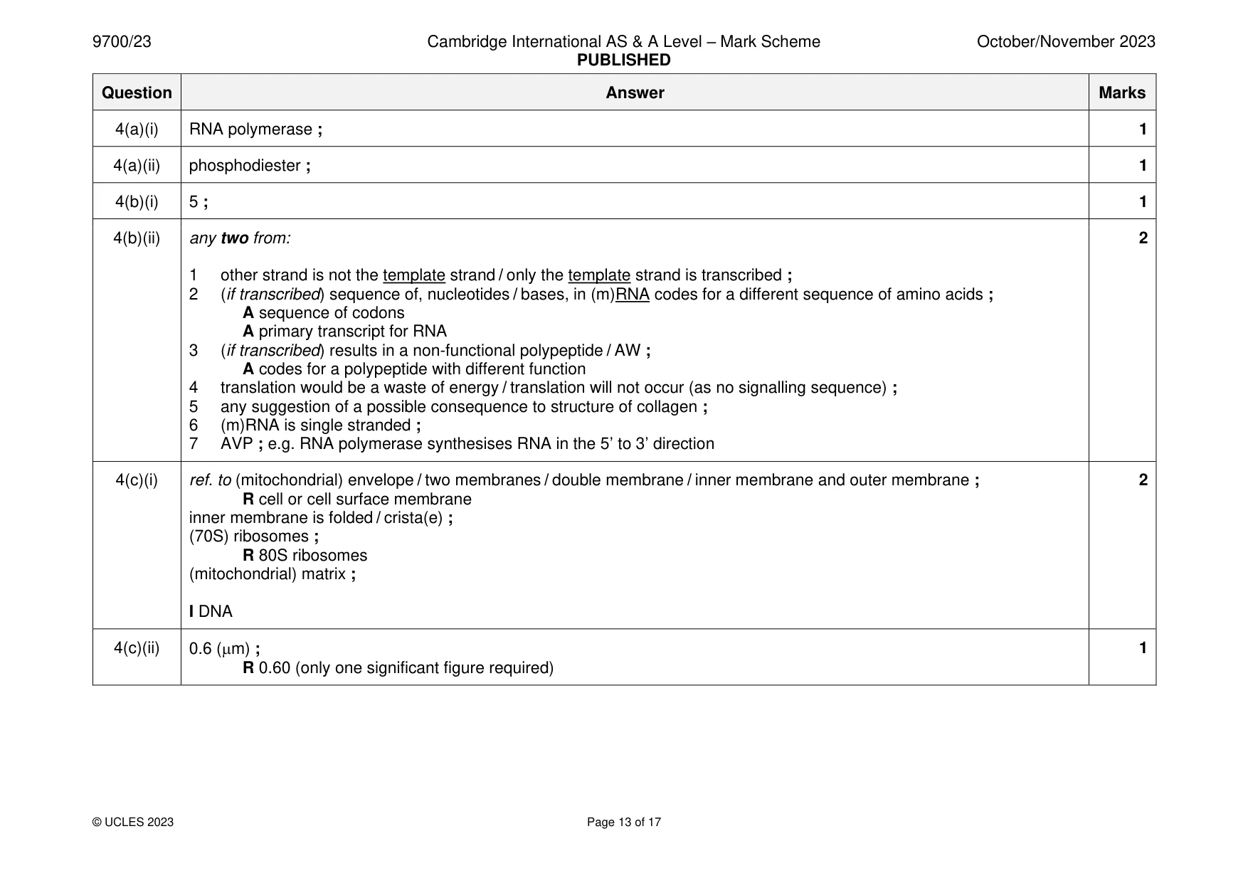
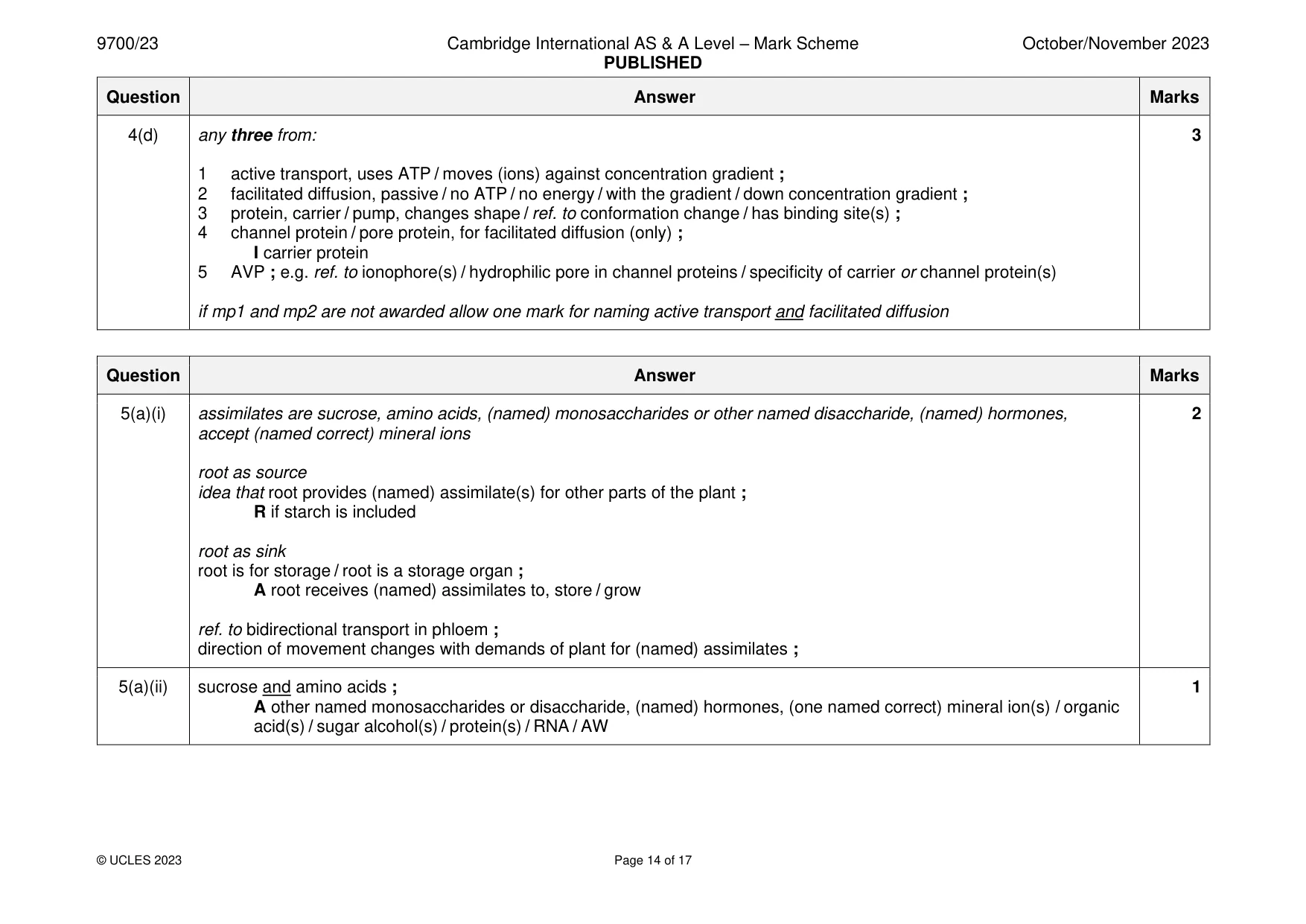
Mark scheme: 4(a)(i) RNA polymerase ; 1 4(a)(ii) phosphodiester ; 1 4(b)(i) 5 ; 1 4(b)(ii) any two from: 2 1 other strand is not the template strand / only the template strand is transcribed ; 2 (if transcribed) sequence of, nucleotides / bases, in (m)RNA codes for a different sequence of amino acids ; A sequence of codons A primary transcript for RNA 3 (if transcribed) results in a non-functional polypeptide / AW ; A codes for a polypeptide with different function 4 translation would be a waste of energy / translation will not occur (as no signalling sequence) ; 5 any suggestion of a possible consequence to structure of collagen ; 6 (m)RNA is single stranded ; 7 AVP ; e.g. RNA polymerase synthesises RNA in the 5’ to 3’ direction 4(c)(i) ref. to (mitochondrial) envelope / two membranes / double membrane / inner membrane and outer membrane ; 2 R cell or cell surface membrane inner membrane is folded / crista(e) ; (70S) ribosomes ; R 80S ribosomes (mitochondrial) matrix ; I DNA 4(c)(ii) 0.6 (m) ; 1 R 0.60 (only one significant figure required) 4(d) any three from: 3 1 active transport, uses ATP / moves (ions) against concentration gradient ; 2 facilitated diffusion, passive / no ATP / no energy / with the gradient / down concentration gradient ; 3 protein, carrier / pump, changes shape / ref. to conformation change / has binding site(s) ; 4 channel protein / pore protein, for facilitated diffusion (only) ; I carrier protein 5 AVP ; e.g. ref. to ionophore(s) / hydrophilic pore in channel proteins / specificity of carrier or channel protein(s) if mp1 and mp2 are not awarded allow one mark for naming active transport and facilitated diffusion
Q5 · In flowering plants, transport of assimilates occurs in phloem tissue between sources and…
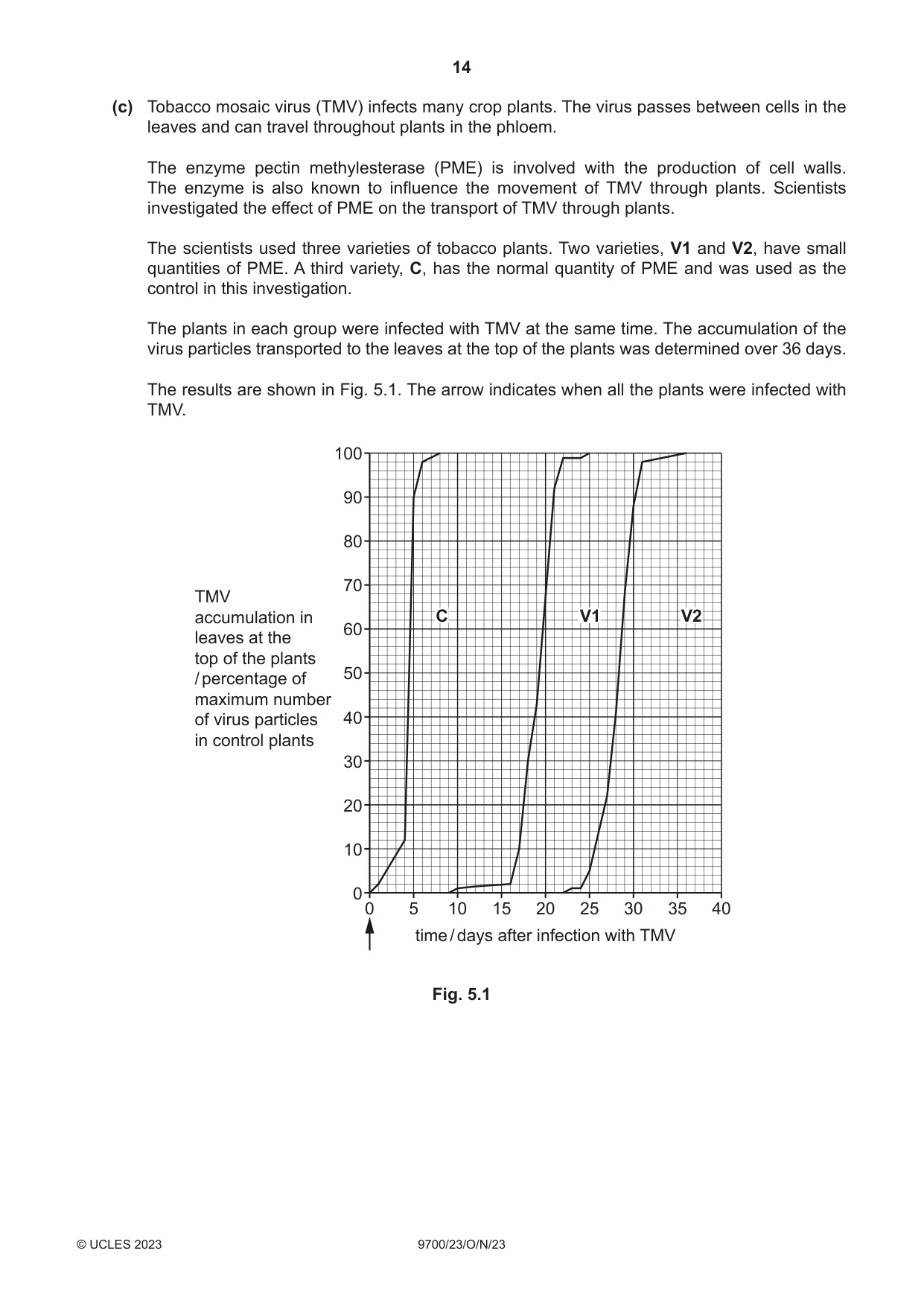
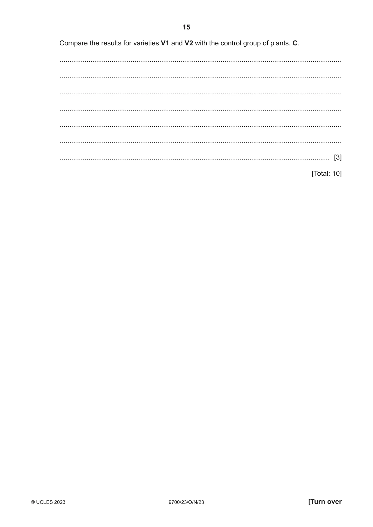
5 In flowering plants, transport of assimilates occurs in phloem tissue between sources and sinks. (a) (i) Explain why a root can be a source and a sink. ........................................................................................................................................... ........................................................................................................................................... ........................................................................................................................................... ........................................................................................................................................... ..................................................................................................................................... [2] (ii) State two examples of assimilates that are transported in the phloem. ..................................................................................................................................... [1] (b) Describe and explain the mechanism that is responsible for the movement of phloem sap in sieve tubes. ................................................................................................................................................... ................................................................................................................................................... ................................................................................................................................................... ................................................................................................................................................... ................................................................................................................................................... ................................................................................................................................................... ................................................................................................................................................... ................................................................................................................................................... ............................................................................................................................................. [4] (c) Tobacco mosaic virus (TMV) infects many crop plants. The virus passes between cells in the leaves and can travel throughout plants in the phloem. The enzyme pectin methylesterase (PME) is involved with the production of cell walls. The enzyme is also known to influence the movement of TMV through plants. Scientists investigated the effect of PME on the transport of TMV through plants. The scientists used three varieties of tobacco plants. Two varieties, V1 and V2, have small quantities of PME. A third variety, C, has the normal quantity of PME and was used as the control in this investigation. The plants in each group were infected with TMV at the same time. The accumulation of the virus particles transported to the leaves at the top of the plants was determined over 36 days. The results are shown in Fig. 5.1. The arrow indicates when all the plants were infected with TMV. 100 90 80 70 TMV accumulation in C V1 V2 60 leaves at the top of the plants 50 / percentage of maximum number of virus particles 40 in control plants 30 20 10 0 0 5 10 15 20 25 30 35 40 time / days after infection with TMV Fig. 5.1 Compare the results for varieties V1 and V2 with the control group of plants, C. ................................................................................................................................................... ................................................................................................................................................... ................................................................................................................................................... ................................................................................................................................................... ................................................................................................................................................... ................................................................................................................................................... ............................................................................................................................................. [3] [Total: 10]
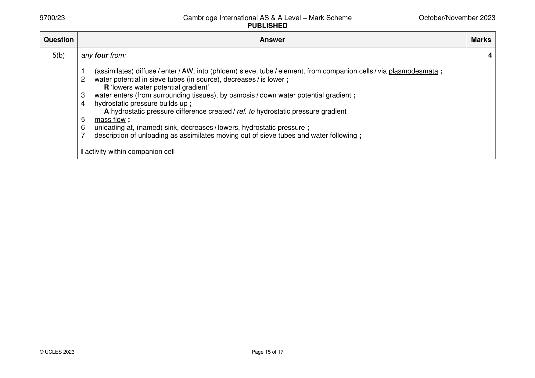
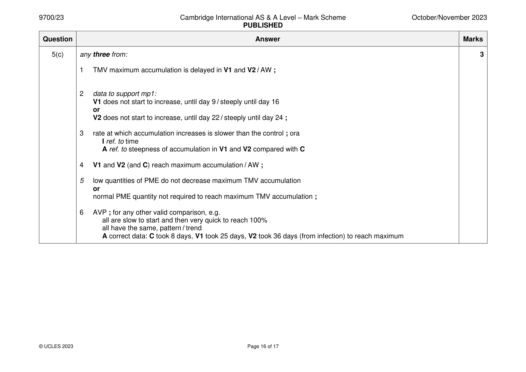
Mark scheme: 5(a)(i) assimilates are sucrose, amino acids, (named) monosaccharides or other named disaccharide, (named) hormones, 2 accept (named correct) mineral ions root as source idea that root provides (named) assimilate(s) for other parts of the plant ; R if starch is included root as sink root is for storage / root is a storage organ ; A root receives (named) assimilates to, store / grow ref. to bidirectional transport in phloem ; direction of movement changes with demands of plant for (named) assimilates ; 5(a)(ii) sucrose and amino acids ; 1 A other named monosaccharides or disaccharide, (named) hormones, (one named correct) mineral ion(s) / organic acid(s) / sugar alcohol(s) / protein(s) / RNA / AW 5(b) any four from: 4 1 (assimilates) diffuse / enter / AW, into (phloem) sieve, tube / element, from companion cells / via plasmodesmata ; 2 water potential in sieve tubes (in source), decreases / is lower ; R ‘lowers water potential gradient’ 3 water enters (from surrounding tissues), by osmosis / down water potential gradient ; 4 hydrostatic pressure builds up ; A hydrostatic pressure difference created / ref. to hydrostatic pressure gradient 5 mass flow ; 6 unloading at, (named) sink, decreases / lowers, hydrostatic pressure ; 7 description of unloading as assimilates moving out of sieve tubes and water following ; I activity within companion cell 5(c) any three from: 3 1 TMV maximum accumulation is delayed in V1 and V2 / AW ; 2 data to support mp1: V1 does not start to increase, until day 9 / steeply until day 16 or V2 does not start to increase, until day 22 / steeply until day 24 ; 3 rate at which accumulation increases is slower than the control ; ora I ref. to time A ref. to steepness of accumulation in V1 and V2 compared with C 4 V1 and V2 (and C) reach maximum accumulation / AW ; 5 low quantities of PME do not decrease maximum TMV accumulation or normal PME quantity not required to reach maximum TMV accumulation ; 6 AVP ; for any other valid comparison, e.g. all are slow to start and then very quick to reach 100% all have the same, pattern / trend A correct data: C took 8 days, V1 took 25 days, V2 took 36 days (from infection) to reach maximum
Q6 · The mammalian heart consists of four chambers
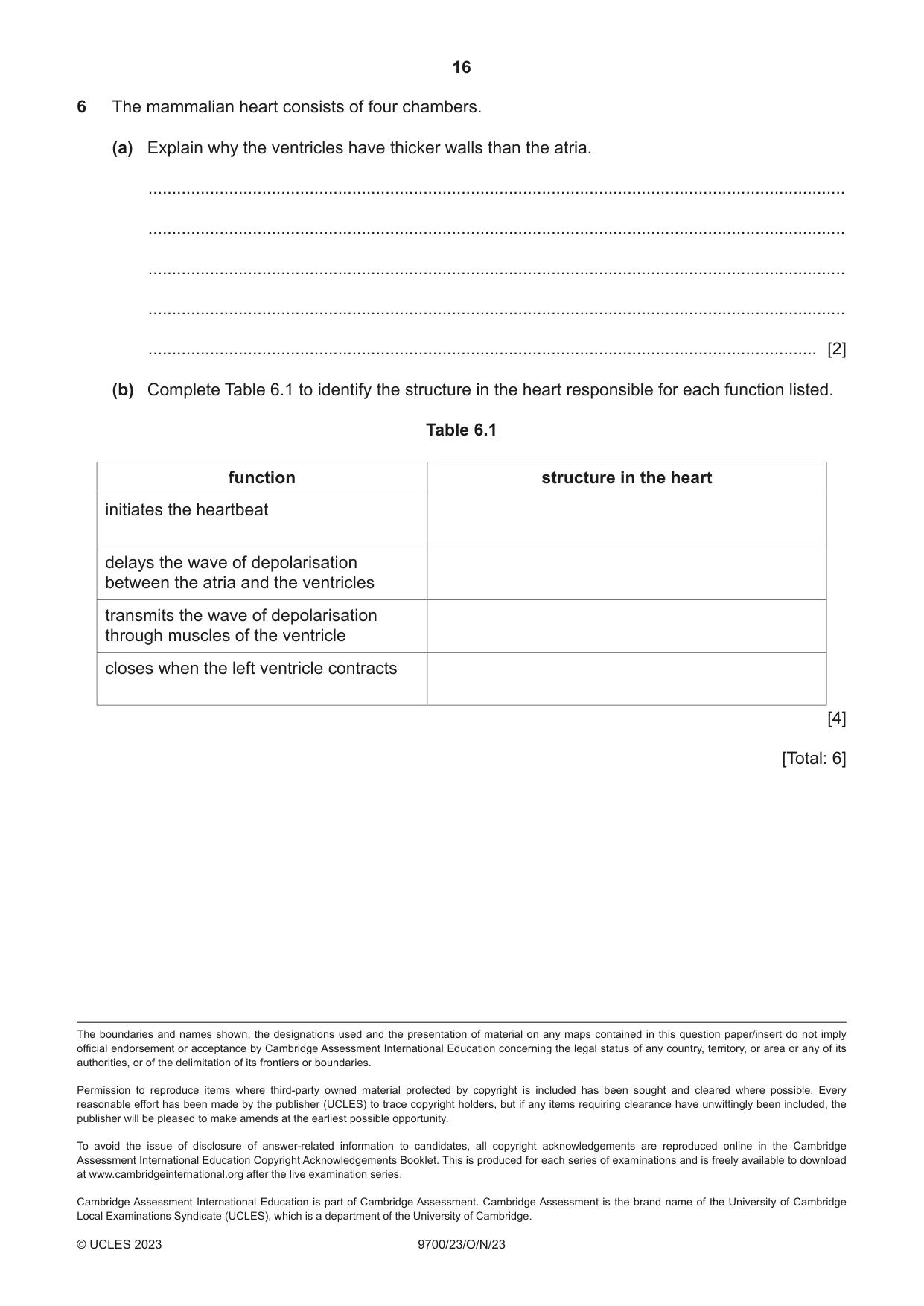
6 The mammalian heart consists of four chambers. (a) Explain why the ventricles have thicker walls than the atria. ................................................................................................................................................... ................................................................................................................................................... ................................................................................................................................................... ................................................................................................................................................... ............................................................................................................................................. [2] (b) Complete Table 6.1 to identify the structure in the heart responsible for each function listed. Table 6.1 function structure in the heart initiates the heartbeat delays the wave of depolarisation between the atria and the ventricles transmits the wave of depolarisation through muscles of the ventricle closes when the left ventricle contracts [4] [Total: 6] The boundaries and names shown, the designations used and the presentation of material on any maps contained in this question paper/insert do not imply official endorsement or acceptance by Cambridge Assessment International Education concerning the legal status of any country, territory, or area or any of its authorities, or of the delimitation of its frontiers or boundaries.
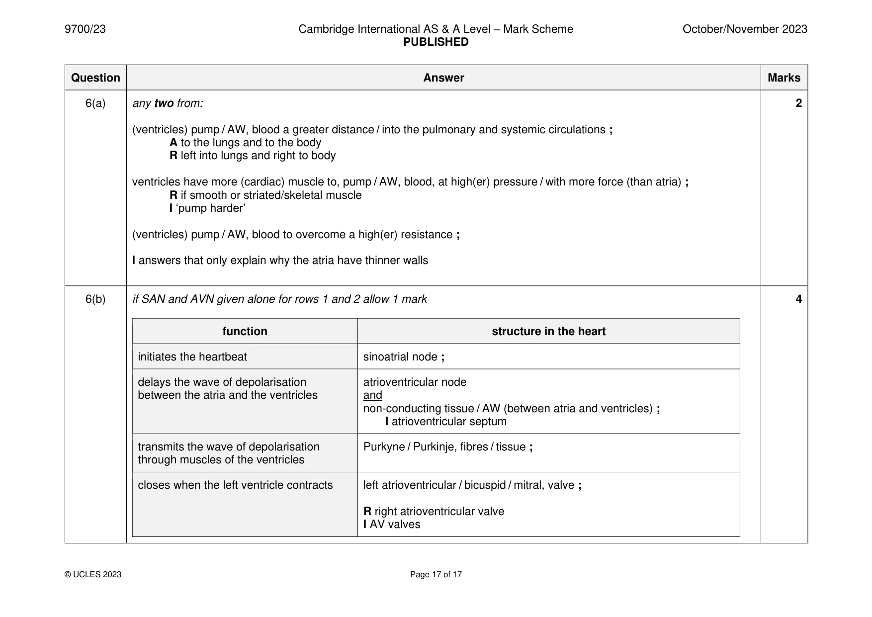
Mark scheme: 6(a) any two from: 2 (ventricles) pump / AW, blood a greater distance / into the pulmonary and systemic circulations ; A to the lungs and to the body R left into lungs and right to body ventricles have more (cardiac) muscle to, pump / AW, blood, at high(er) pressure / with more force (than atria) ; R if smooth or striated/skeletal muscle I ‘pump harder’ (ventricles) pump / AW, blood to overcome a high(er) resistance ; I answers that only explain why the atria have thinner walls 6(b) if SAN and AVN given alone for rows 1 and 2 allow 1 mark 4 function structure in the heart initiates the heartbeat sinoatrial node ; delays the wave of depolarisation atrioventricular node between the atria and the ventricles and non-conducting tissue / AW (between atria and ventricles) ; I atrioventricular septum transmits the wave of depolarisation Purkyne / Purkinje, fibres / tissue ; through muscles of the ventricles closes when the left ventricle contracts left atrioventricular / bicuspid / mitral, valve ; R right atrioventricular valve I AV valves
What was in this paper
The subtopics covered by these 6 questions, and how many questions each got. Open one in a new tab to see every Cambridge question on it.
What you needed in this session
Cambridge’s own grade thresholds for 2023 Oct/Nov, Paper 2 · Variant 3. A higher threshold means an easier paper — the bar moves with how the cohort did.