Cambridge A Level Biology 9700 — 2025 Oct/Nov Paper 2 · Variant 2
9700/22/O/N/25 · 6 questions · 60 marks · 75 min
The question paper and its mark scheme, free to read here and free to download. This is Cambridge’s own paper, exactly as it was sat.
Question paper16 pages
Mark scheme21 pages
Answers below. Sit the paper first if you are practising.
Questions as text
Q1 · The wall of the small intestine is highly folded to form villi
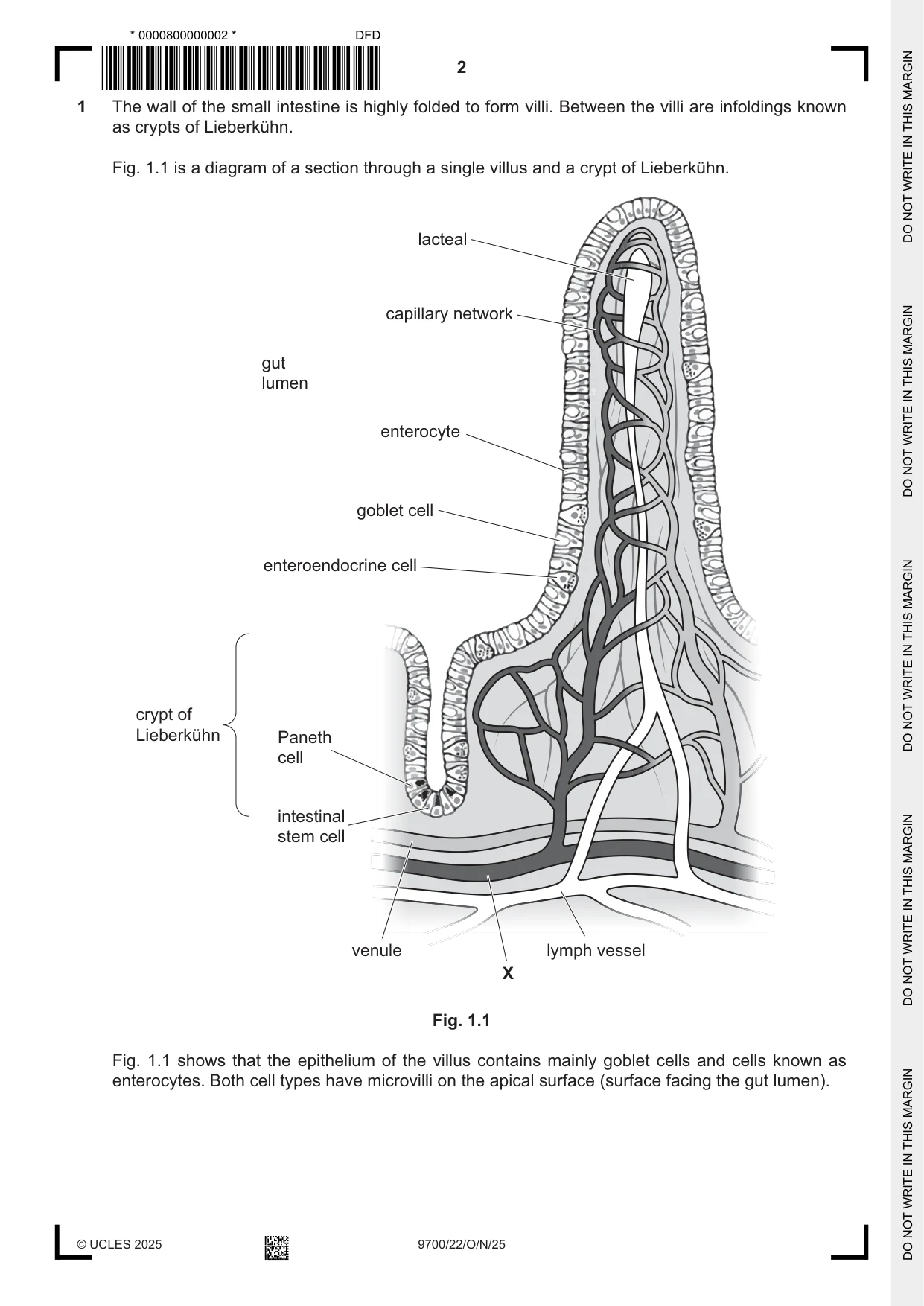
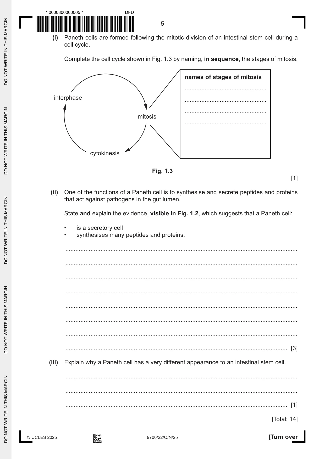
1 The wall of the small intestine is highly folded to form villi. Between the villi are infoldings known as crypts of Lieberkühn. Fig. 1.1 is a diagram of a section through a single villus and a crypt of Lieberkühn. lacteal capillary network gut lumen enterocyte goblet cell enteroendocrine cell crypt of Lieberkühn Paneth cell intestinal stem cell venule lymph vessel X Fig. 1.1 Fig. 1.1 shows that the epithelium of the villus contains mainly goblet cells and cells known as enterocytes. Both cell types have microvilli on the apical surface (surface facing the gut lumen). Goblet cells are involved in the production of mucus. Enterocytes are adapted for the absorption of the soluble products of digestion. These products enter the circulatory system. (a) A student incorrectly stated that an enterocyte has many cilia on its apical surface. Explain the difference between a cilium and a microvillus. ................................................................................................................................................... ................................................................................................................................................... ................................................................................................................................................... ................................................................................................................................................... ................................................................................................................................................... ............................................................................................................................................. [2] (b) Suggest one role of the mucus produced by the goblet cells of the villus. ................................................................................................................................................... ................................................................................................................................................... ............................................................................................................................................. [1] (c) In Fig. 1.1, blood vessel X delivers blood to the capillary network of the villus, where tissue fluid is formed. Some of the fluid passes back into the capillaries and then into the venule. (i) Blood vessel X receives blood from an artery. Name the type of blood vessel represented by X. ..................................................................................................................................... [1] (ii) Describe the formation of tissue fluid in the capillary network of the villus. ........................................................................................................................................... ........................................................................................................................................... ........................................................................................................................................... ........................................................................................................................................... ..................................................................................................................................... [2] (d) In response to the presence of compounds in the gut lumen, enteroendocrine cells synthesise and release peptides (short chains of amino acids) that are cell‑signalling molecules. One of these cell‑signalling molecules is known as GLP‑1. GLP‑1 initiates a number of responses in different body cells. Some of these responses include: • the increase in release of the hormone insulin from cells in the pancreas • the decrease in release of acid from cells in the stomach. Outline, in sequence, the main stages involved in the process of cell signalling by GLP‑1. ................................................................................................................................................... ................................................................................................................................................... ................................................................................................................................................... ................................................................................................................................................... ................................................................................................................................................... ................................................................................................................................................... ............................................................................................................................................. [3] (e) Paneth cells, which are secretory cells, are located between intestinal stem cells at the base of the crypt of Lieberkühn, as shown in Fig. 1.1. A Paneth cell has a very different appearance to an intestinal stem cell. Fig. 1.2 is a transmission electron micrograph of a Paneth cell. Paneth cell nucleus of adjacent intestinal stem cell nucleus Fig. 1.2 (i) Paneth cells are formed following the mitotic division of an intestinal stem cell during a cell cycle. Complete the cell cycle shown in Fig. 1.3 by naming, in sequence, the stages of mitosis. names of stages of mitosis ................................................. interphase ................................................. ................................................. mitosis ................................................. cytokinesis Fig. 1.3 [1] (ii) One of the functions of a Paneth cell is to synthesise and secrete peptides and proteins that act against pathogens in the gut lumen. State and explain the evidence, visible in Fig. 1.2, which suggests that a Paneth cell: • is a secretory cell • synthesises many peptides and proteins. ........................................................................................................................................... ........................................................................................................................................... ........................................................................................................................................... ........................................................................................................................................... ........................................................................................................................................... ........................................................................................................................................... ........................................................................................................................................... ..................................................................................................................................... [3] (iii) Explain why a Paneth cell has a very different appearance to an intestinal stem cell. ........................................................................................................................................... ........................................................................................................................................... ..................................................................................................................................... [1] [Total: 14]
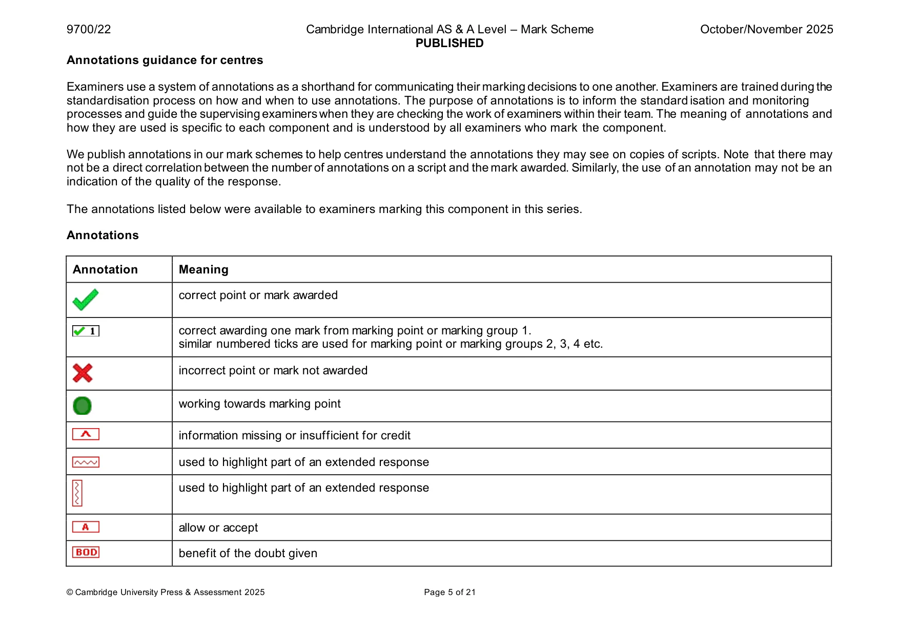
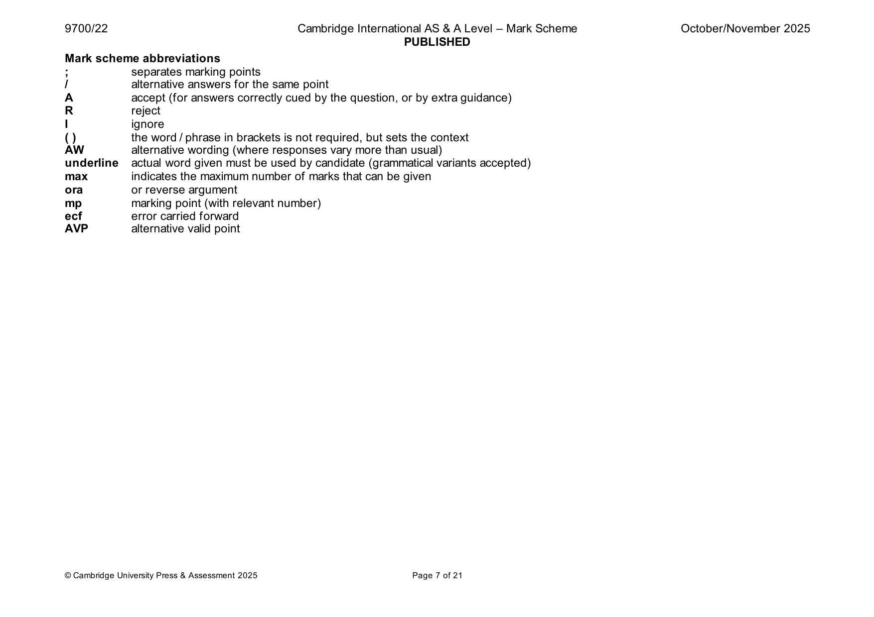
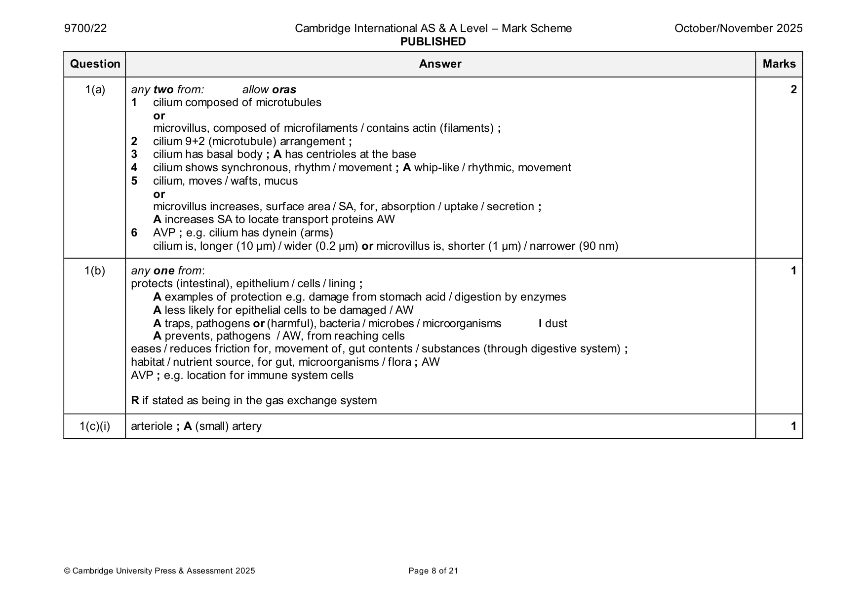
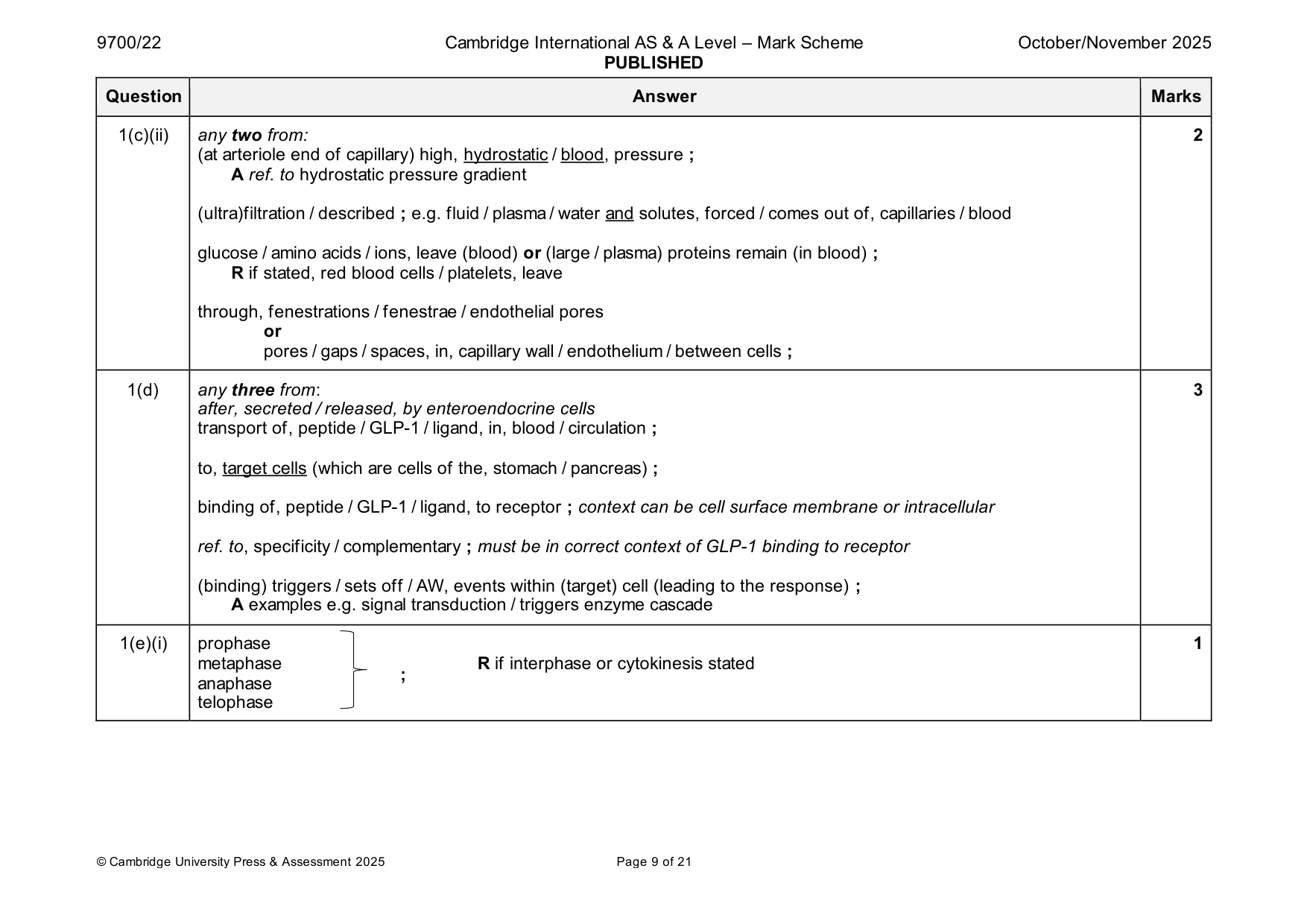
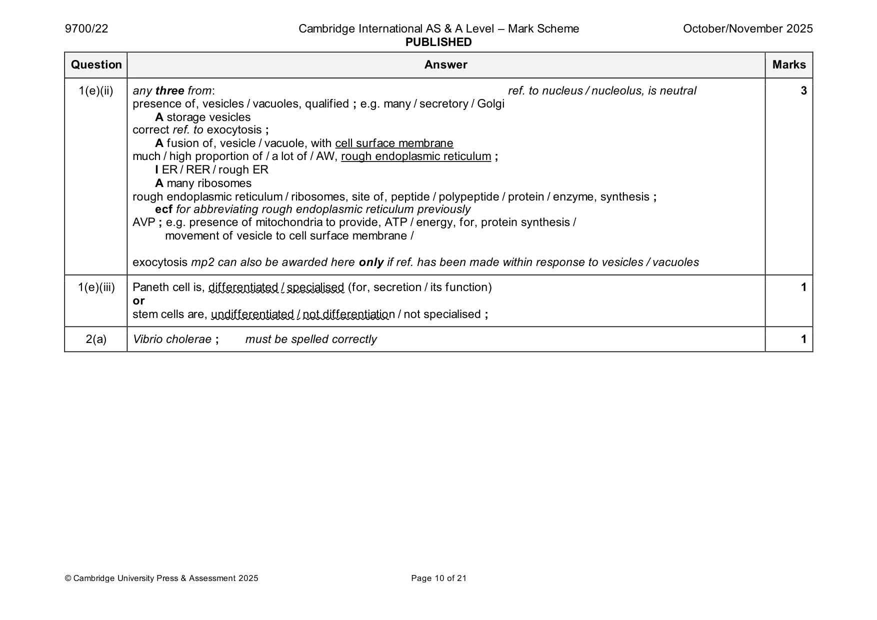
Mark scheme: Question Answer Marks 1(a) any two from: allow oras 2 1 cilium composed of microtubules or microvillus, composed of microfilaments / contains actin (filaments) ; 2 cilium 9+2 (microtubule) arrangement ; 3 cilium has basal body ; A has centrioles at the base 4 cilium shows synchronous, rhythm / movement ; A whip-like / rhythmic, movement 5 cilium, moves / wafts, mucus or microvillus increases, surface area / SA, for, absorption / uptake / secretion ; A increases SA to locate transport proteins AW 6 AVP ; e.g. cilium has dynein (arms) cilium is, longer (10 µm) / wider (0.2 µm) or microvillus is, shorter (1 µm) / narrower (90 nm) 1(b) any one from: 1 protects (intestinal), epithelium / cells / lining ; A examples of protection e.g. damage from stomach acid / digestion by enzymes A less likely for epithelial cells to be damaged / AW A traps, pathogens or (harmful), bacteria / microbes / microorganisms I dust A prevents, pathogens / AW, from reaching cells eases / reduces friction for, movement of, gut contents / substances (through digestive system) ; habitat / nutrient source, for gut, microorganisms / flora ; AW AVP ; e.g. location for immune system cells R if stated as being in the gas exchange system 1(c)(i) arteriole ; A (small) artery 1 1(c)(ii) any two from: 2 (at arteriole end of capillary) high, hydrostatic / blood, pressure ; A ref. to hydrostatic pressure gradient (ultra)filtration / described ; e.g. fluid / plasma / water and solutes, forced / comes out of, capillaries / blood glucose / amino acids / ions, leave (blood) or (large / plasma) proteins remain (in blood) ; R if stated, red blood cells / platelets, leave through, fenestrations / fenestrae / endothelial pores or pores / gaps / spaces, in, capillary wall / endothelium / between cells ; 1(d) any three from: 3 after, secreted / released, by enteroendocrine cells transport of, peptide / GLP-1 / ligand, in, blood / circulation ; to, target cells (which are cells of the, stomach / pancreas) ; binding of, peptide / GLP-1 / ligand, to receptor ; context can be cell surface membrane or intracellular ref. to, specificity / complementary ; must be in correct context of GLP-1 binding to receptor (binding) triggers / sets off / AW, events within (target) cell (leading to the response) ; A examples e.g. signal transduction / triggers enzyme cascade 1(e)(i) prophase 1 metaphase R if interphase or cytokinesis stated ; anaphase telophase 1(e)(ii) any three from: ref. to nucleus / nucleolus, is neutral 3 presence of, vesicles / vacuoles, qualified ; e.g. many / secretory / Golgi A storage vesicles correct ref. to exocytosis ; A fusion of, vesicle / vacuole, with cell surface membrane much / high proportion of / a lot of / AW, rough endoplasmic reticulum ; I ER / RER / rough ER A many ribosomes rough endoplasmic reticulum / ribosomes, site of, peptide / polypeptide / protein / enzyme, synthesis ; ecf for abbreviating rough endoplasmic reticulum previously AVP ; e.g. presence of mitochondria to provide, ATP / energy, for, protein synthesis / movement of vesicle to cell surface membrane / exocytosis mp2 can also be awarded here only if ref. has been made within response to vesicles / vacuoles 1(e)(iii) Paneth cell is, differentiated / specialised (for, secretion / its function) 1 or stem cells are, undifferentiated / not differentiation / not specialised ;
Q2 · Cholera is an infectious intestinal disease caused by a bacterial pathogen
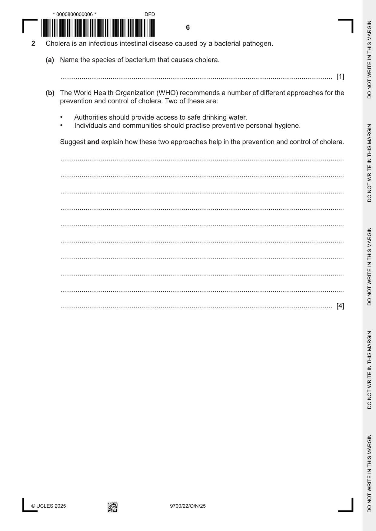
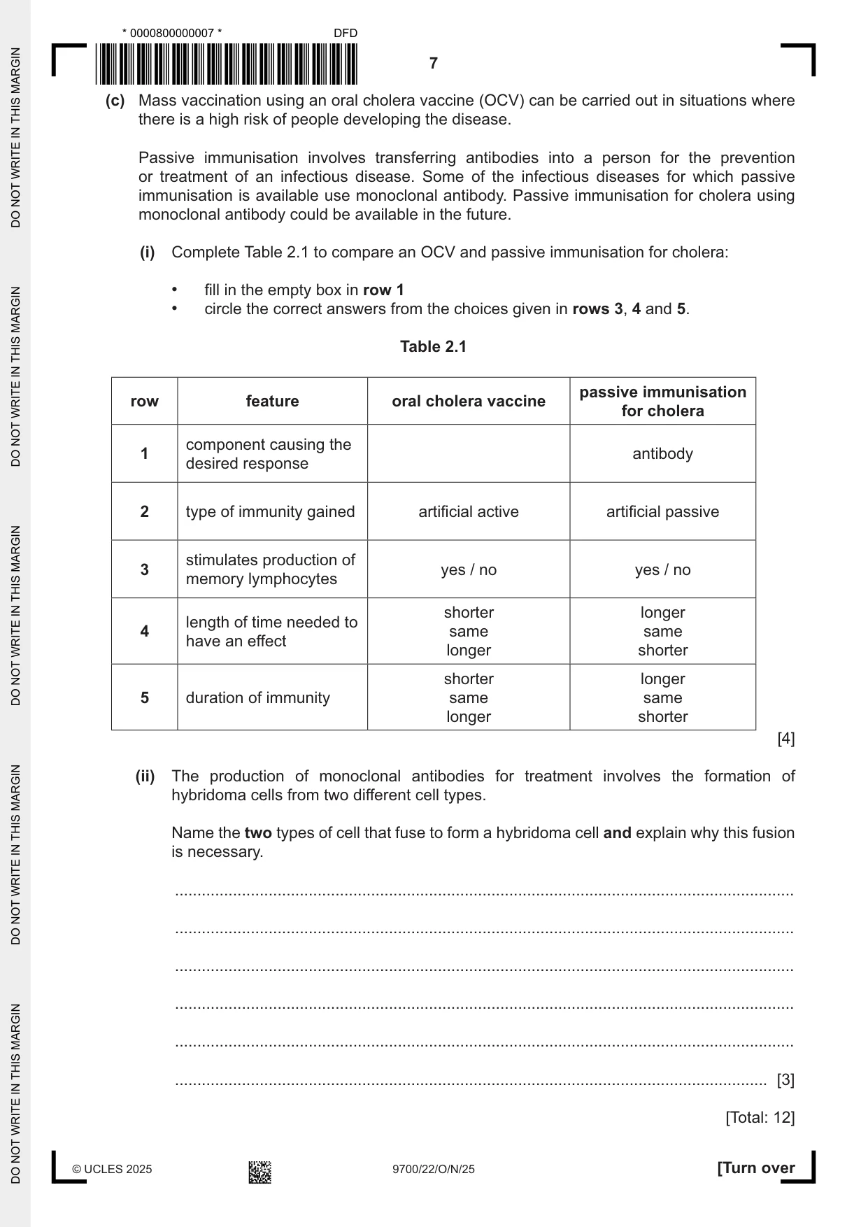
2 Cholera is an infectious intestinal disease caused by a bacterial pathogen. (a) Name the species of bacterium that causes cholera. ............................................................................................................................................. [1] (b) The World Health Organization (WHO) recommends a number of different approaches for the prevention and control of cholera. Two of these are: • Authorities should provide access to safe drinking water. • Individuals and communities should practise preventive personal hygiene. Suggest and explain how these two approaches help in the prevention and control of cholera. ................................................................................................................................................... ................................................................................................................................................... ................................................................................................................................................... ................................................................................................................................................... ................................................................................................................................................... ................................................................................................................................................... ................................................................................................................................................... ................................................................................................................................................... ................................................................................................................................................... ............................................................................................................................................. [4] (c) Mass vaccination using an oral cholera vaccine (OCV) can be carried out in situations where there is a high risk of people developing the disease. Passive immunisation involves transferring antibodies into a person for the prevention or treatment of an infectious disease. Some of the infectious diseases for which passive immunisation is available use monoclonal antibody. Passive immunisation for cholera using monoclonal antibody could be available in the future. (i) Complete Table 2.1 to compare an OCV and passive immunisation for cholera: • fill in the empty box in row 1 • circle the correct answers from the choices given in rows 3, 4 and 5. Table 2.1 passive immunisation row feature oral cholera vaccine for cholera component causing the 1 antibody desired response 2 type of immunity gained artificial active artificial passive stimulates production of 3 yes / no yes / no memory lymphocytes shorter longer length of time needed to 4 same same have an effect longer shorter shorter longer 5 duration of immunity same same longer shorter [4] (ii) The production of monoclonal antibodies for treatment involves the formation of hybridoma cells from two different cell types. Name the two types of cell that fuse to form a hybridoma cell and explain why this fusion is necessary. ........................................................................................................................................... ........................................................................................................................................... ........................................................................................................................................... ........................................................................................................................................... ........................................................................................................................................... ..................................................................................................................................... [3] [Total: 12]
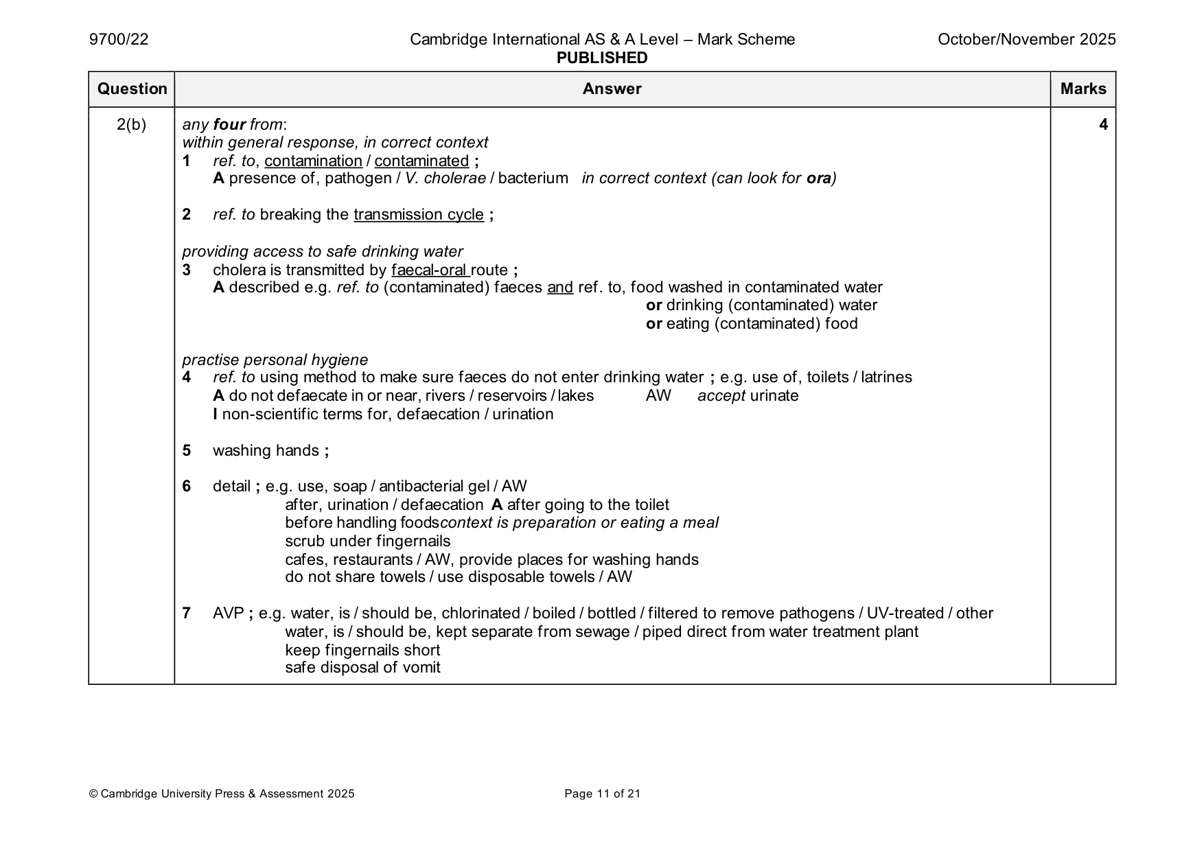
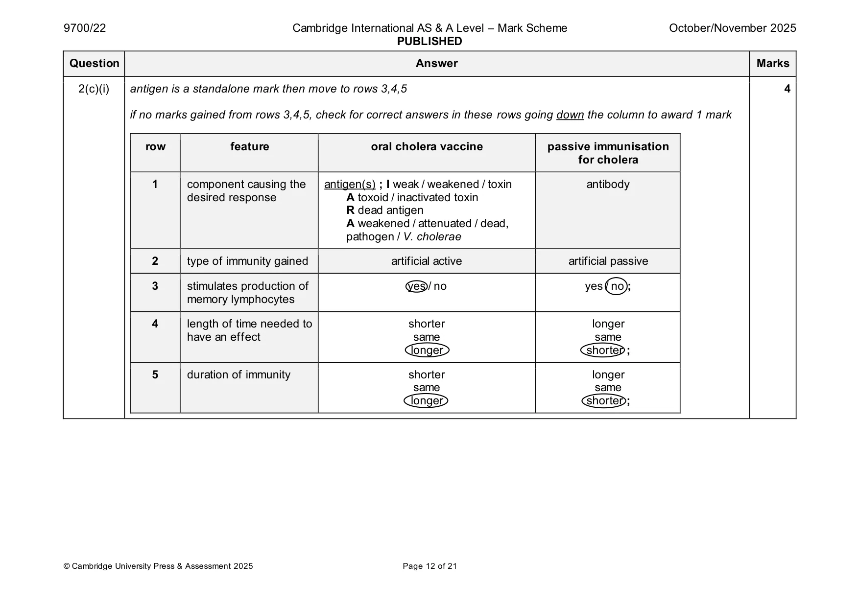
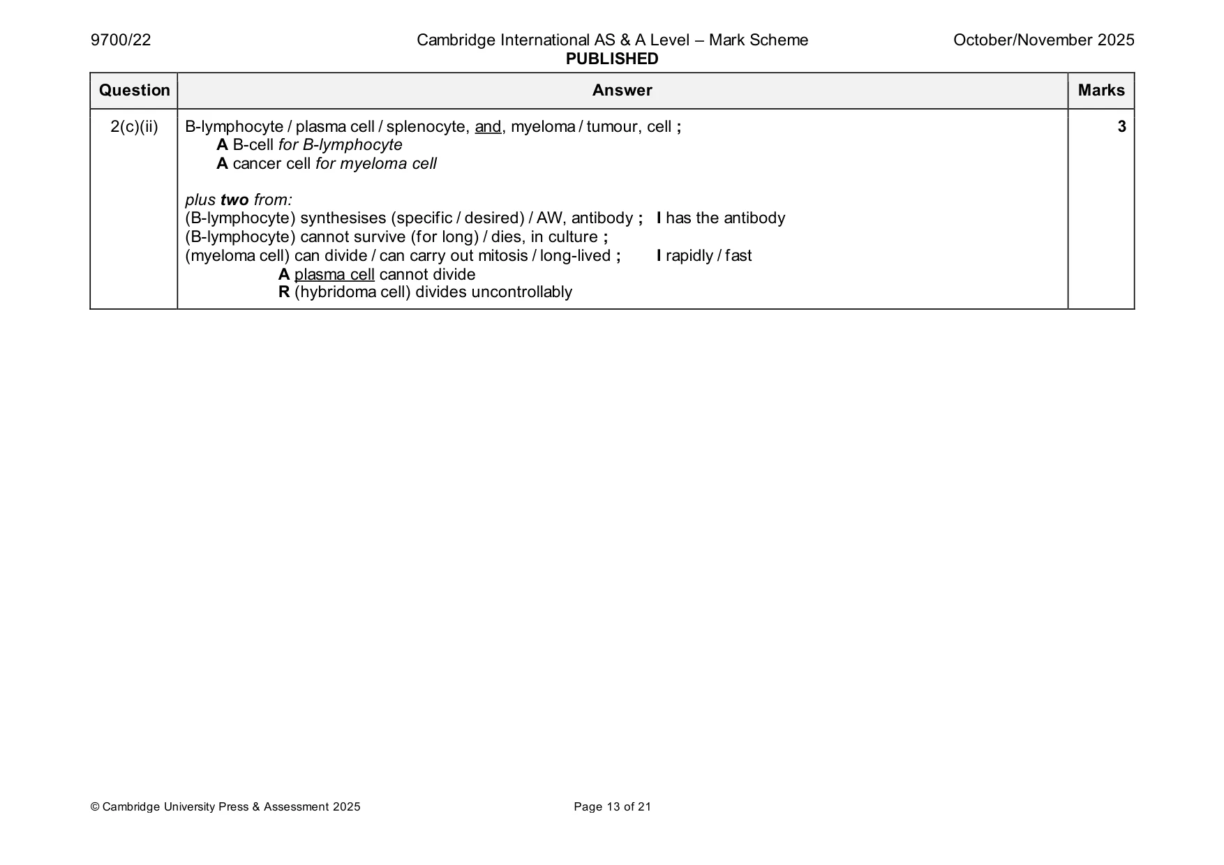
Mark scheme: 2(a) Vibrio cholerae ; must be spelled correctly 1 2(b) any four from: 4 within general response, in correct context 1 ref. to, contamination / contaminated ; A presence of, pathogen / V. cholerae / bacterium in correct context (can look for ora) 2 ref. to breaking the transmission cycle ; providing access to safe drinking water 3 cholera is transmitted by faecal-oral route ; A described e.g. ref. to (contaminated) faeces and ref. to, food washed in contaminated water or drinking (contaminated) water or eating (contaminated) food practise personal hygiene 4 ref. to using method to make sure faeces do not enter drinking water ; e.g. use of, toilets / latrines A do not defaecate in or near, rivers / reservoirs / lakes AW accept urinate I non-scientific terms for, defaecation / urination 5 washing hands ; 6 detail ; e.g. use, soap / antibacterial gel / AW after, urination / defaecation A after going to the toilet before handling foodscontext is preparation or eating a meal scrub under fingernails cafes, restaurants / AW, provide places for washing hands do not share towels / use disposable towels / AW 7 AVP ; e.g. water, is / should be, chlorinated / boiled / bottled / filtered to remove pathogens / UV-treated / other water, is / should be, kept separate from sewage / piped direct from water treatment plant keep fingernails short safe disposal of vomit 2(c)(i) antigen is a standalone mark then move to rows 3,4,5 4 if no marks gained from rows 3,4,5, check for correct answers in these rows going down the column to award 1 mark row feature oral cholera vaccine passive immunisation for cholera 1 component causing the antigen(s) ; I weak / weakened / toxin antibody desired response A toxoid / inactivated toxin R dead antigen A weakened / attenuated / dead, pathogen / V. cholerae 2 type of immunity gained artificial active artificial passive 3 stimulates production of yes / no yes / no ; memory lymphocytes 4 length of time needed to shorter longer have an effect same same longer shorter ; 5 duration of immunity shorter longer same same longer shorter ; 2(c)(ii) B-lymphocyte / plasma cell / splenocyte, and, myeloma / tumour, cell ; 3 A B-cell for B-lymphocyte A cancer cell for myeloma cell plus two from: (B-lymphocyte) synthesises (specific / desired) / AW, antibody ; I has the antibody (B-lymphocyte) cannot survive (for long) / dies, in culture ; (myeloma cell) can divide / can carry out mitosis / long-lived ; I rapidly / fast A plasma cell cannot divide R (hybridoma cell) divides uncontrollably
Q3 · The gene LCT codes for the enzyme lactase
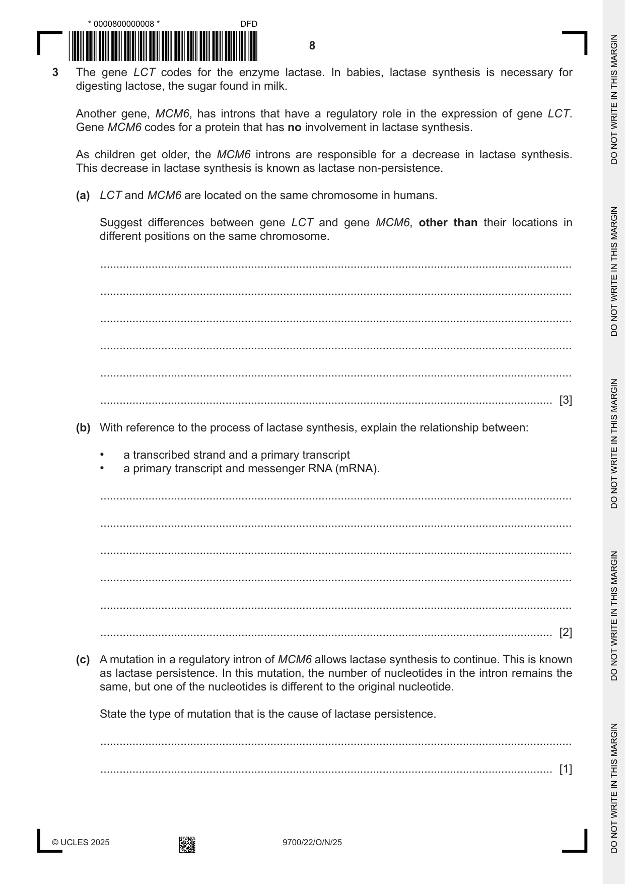
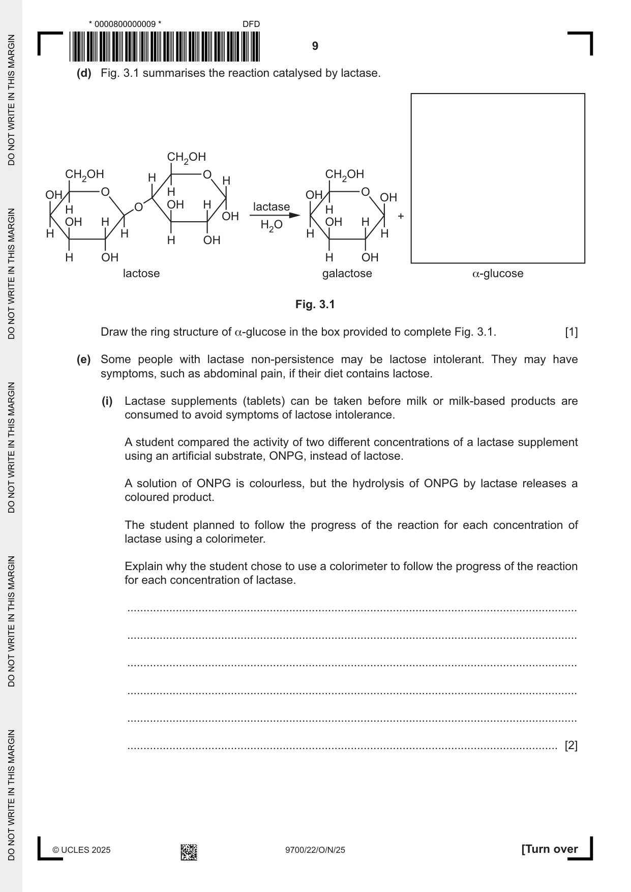
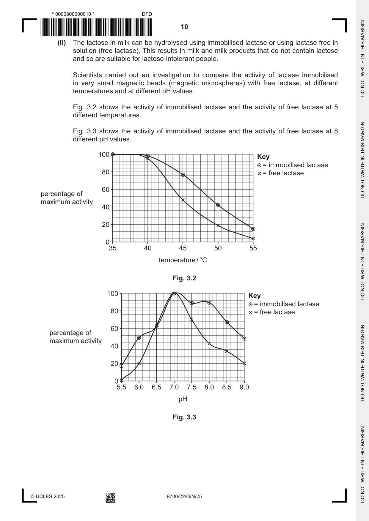
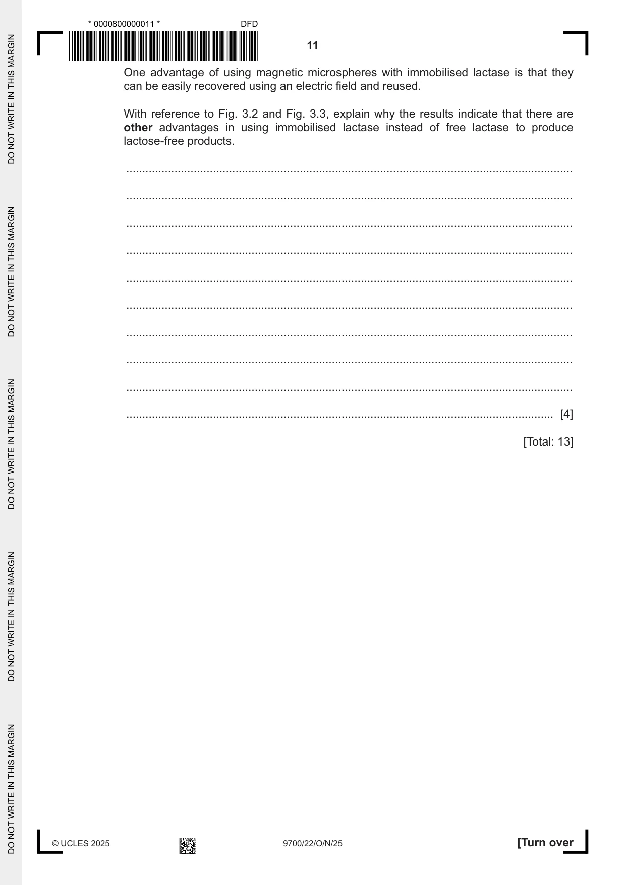
3 The gene LCT codes for the enzyme lactase. In babies, lactase synthesis is necessary for digesting lactose, the sugar found in milk. Another gene, MCM6, has introns that have a regulatory role in the expression of gene LCT. Gene MCM6 codes for a protein that has no involvement in lactase synthesis. As children get older, the MCM6 introns are responsible for a decrease in lactase synthesis. This decrease in lactase synthesis is known as lactase non‑persistence. (a) LCT and MCM6 are located on the same chromosome in humans. Suggest differences between gene LCT and gene MCM6, other than their locations in different positions on the same chromosome. ................................................................................................................................................... ................................................................................................................................................... ................................................................................................................................................... ................................................................................................................................................... ................................................................................................................................................... ............................................................................................................................................. [3] (b) With reference to the process of lactase synthesis, explain the relationship between: • a transcribed strand and a primary transcript • a primary transcript and messenger RNA (mRNA). ................................................................................................................................................... ................................................................................................................................................... ................................................................................................................................................... ................................................................................................................................................... ................................................................................................................................................... ............................................................................................................................................. [2] (c) A mutation in a regulatory intron of MCM6 allows lactase synthesis to continue. This is known as lactase persistence. In this mutation, the number of nucleotides in the intron remains the same, but one of the nucleotides is different to the original nucleotide. State the type of mutation that is the cause of lactase persistence. ................................................................................................................................................... ............................................................................................................................................. [1] (d) Fig. 3.1 summarises the reaction catalysed by lactase. CH2OH CH2OH H O H CH2OH OH O H OH O OH H O OH H lactase H OH + OH H H2O OH HH H H H H OH H OH H OH lactose galactose α-glucose Fig. 3.1 Draw the ring structure of α‑glucose in the box provided to complete Fig. 3.1. [1] (e) Some people with lactase non‑persistence may be lactose intolerant. They may have symptoms, such as abdominal pain, if their diet contains lactose. (i) Lactase supplements (tablets) can be taken before milk or milk‑based products are consumed to avoid symptoms of lactose intolerance. A student compared the activity of two different concentrations of a lactase supplement using an artificial substrate, ONPG, instead of lactose. A solution of ONPG is colourless, but the hydrolysis of ONPG by lactase releases a coloured product. The student planned to follow the progress of the reaction for each concentration of lactase using a colorimeter. Explain why the student chose to use a colorimeter to follow the progress of the reaction for each concentration of lactase. ........................................................................................................................................... ........................................................................................................................................... ........................................................................................................................................... ........................................................................................................................................... ........................................................................................................................................... ..................................................................................................................................... [2] (ii) The lactose in milk can be hydrolysed using immobilised lactase or using lactase free in solution (free lactase). This results in milk and milk products that do not contain lactose and so are suitable for lactose‑intolerant people. Scientists carried out an investigation to compare the activity of lactase immobilised in very small magnetic beads (magnetic microspheres) with free lactase, at different temperatures and at different pH values. Fig. 3.2 shows the activity of immobilised lactase and the activity of free lactase at 5 different temperatures. Fig. 3.3 shows the activity of immobilised lactase and the activity of free lactase at 8 different pH values. 100 Key = immobilised lactase 80 = free lactase 60 percentage of maximum activity 40 20 0 35 40 45 50 55 temperature / °C Fig. 3.2 100 Key = immobilised lactase 80 = free lactase 60 percentage of maximum activity 40 20 0 5.5 6.0 6.5 7.0 7.5 8.0 8.5 9.0 pH Fig. 3.3 One advantage of using magnetic microspheres with immobilised lactase is that they can be easily recovered using an electric field and reused. With reference to Fig. 3.2 and Fig. 3.3, explain why the results indicate that there are other advantages in using immobilised lactase instead of free lactase to produce lactose‑free products. ........................................................................................................................................... ........................................................................................................................................... ........................................................................................................................................... ........................................................................................................................................... ........................................................................................................................................... ........................................................................................................................................... ........................................................................................................................................... ........................................................................................................................................... ........................................................................................................................................... ..................................................................................................................................... [4] [Total: 13]
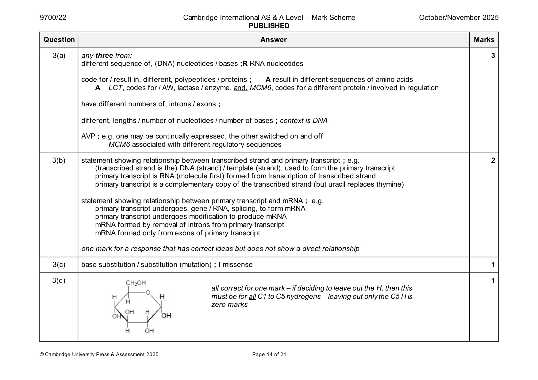
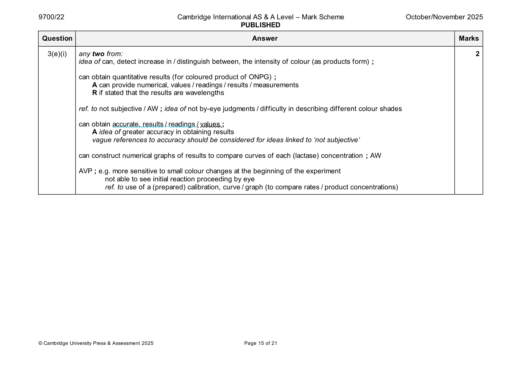
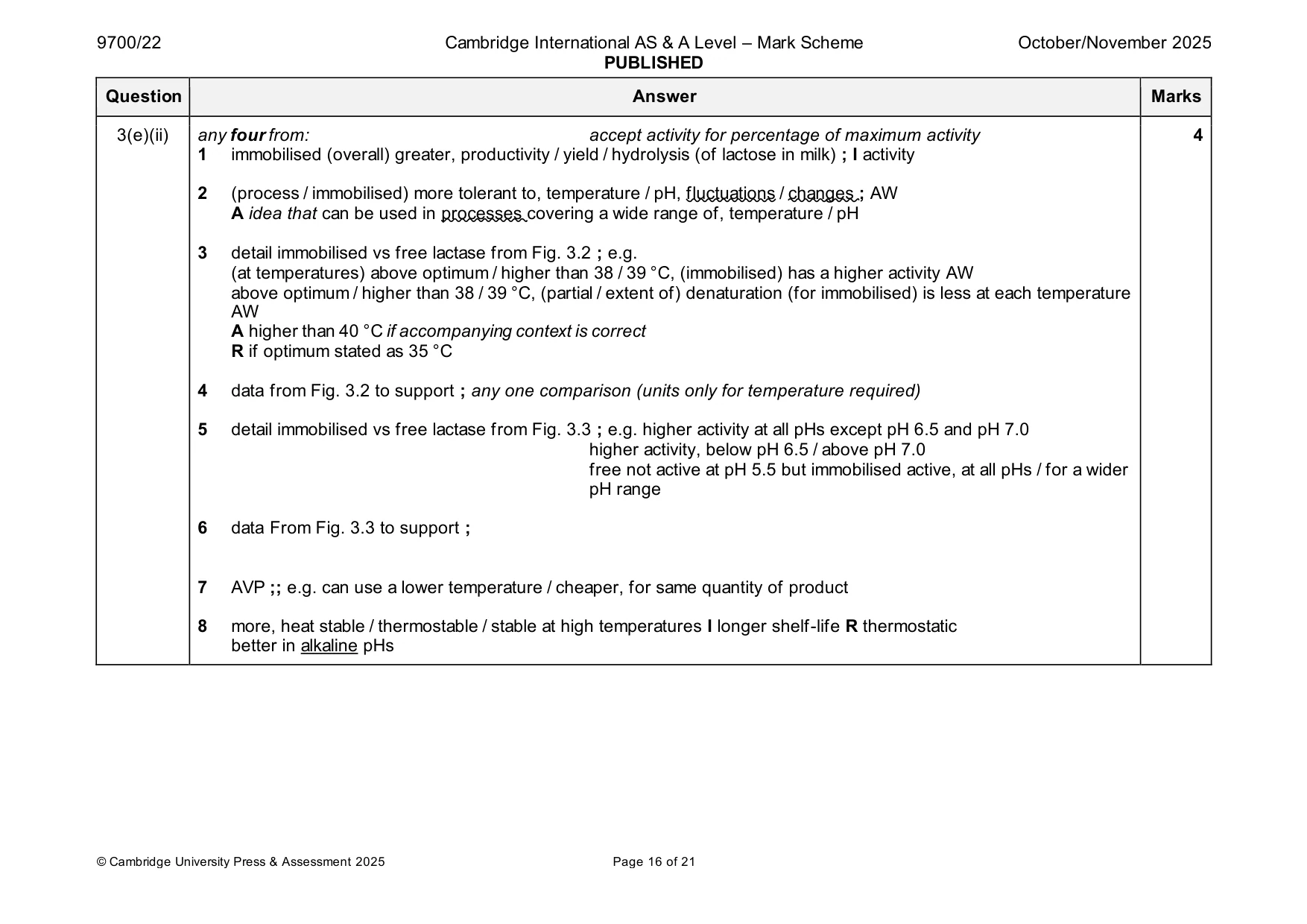
Mark scheme: 3(a) any three from: 3 different sequence of, (DNA) nucleotides / bases ;R RNA nucleotides code for / result in, different, polypeptides / proteins ; A result in different sequences of amino acids A LCT, codes for / AW, lactase / enzyme, and, MCM6, codes for a different protein / involved in regulation have different numbers of, introns / exons ; different, lengths / number of nucleotides / number of bases ; context is DNA AVP ; e.g. one may be continually expressed, the other switched on and off MCM6 associated with different regulatory sequences 3(b) statement showing relationship between transcribed strand and primary transcript ; e.g. 2 (transcribed strand is the) DNA (strand) / template (strand), used to form the primary transcript primary transcript is RNA (molecule first) formed from transcription of transcribed strand primary transcript is a complementary copy of the transcribed strand (but uracil replaces thymine) statement showing relationship between primary transcript and mRNA ; e.g. primary transcript undergoes, gene / RNA, splicing, to form mRNA primary transcript undergoes modification to produce mRNA mRNA formed by removal of introns from primary transcript mRNA formed only from exons of primary transcript one mark for a response that has correct ideas but does not show a direct relationship 3(c) base substitution / substitution (mutation) ; I missense 1 3(d) 1 all correct for one mark – if deciding to leave out the H, then this must be for all C1 to C5 hydrogens – leaving out only the C5 H is zero marks 3(e)(i) any two from: 2 idea of can, detect increase in / distinguish between, the intensity of colour (as products form) ; can obtain quantitative results (for coloured product of ONPG) ; A can provide numerical, values / readings / results / measurements R if stated that the results are wavelengths ref. to not subjective / AW ; idea of not by-eye judgments / difficulty in describing different colour shades can obtain accurate, results / readings / values ; A idea of greater accuracy in obtaining results vague references to accuracy should be considered for ideas linked to ‘not subjective’ can construct numerical graphs of results to compare curves of each (lactase) concentration ; AW AVP ; e.g. more sensitive to small colour changes at the beginning of the experiment not able to see initial reaction proceeding by eye ref. to use of a (prepared) calibration, curve / graph (to compare rates / product concentrations) 3(e)(ii) any four from: accept activity for percentage of maximum activity 4 1 immobilised (overall) greater, productivity / yield / hydrolysis (of lactose in milk) ; I activity 2 (process / immobilised) more tolerant to, temperature / pH, fluctuations / changes ; AW A idea that can be used in processes covering a wide range of, temperature / pH 3 detail immobilised vs free lactase from Fig. 3.2 ; e.g. (at temperatures) above optimum / higher than 38 / 39 °C, (immobilised) has a higher activity AW above optimum / higher than 38 / 39 °C, (partial / extent of) denaturation (for immobilised) is less at each temperature AW A higher than 40 °C if accompanying context is correct R if optimum stated as 35 °C 4 data from Fig. 3.2 to support ; any one comparison (units only for temperature required) 5 detail immobilised vs free lactase from Fig. 3.3 ; e.g. higher activity at all pHs except pH 6.5 and pH 7.0 higher activity, below pH 6.5 / above pH 7.0 free not active at pH 5.5 but immobilised active, at all pHs / for a wider pH range 6 data From Fig. 3.3 to support ; 7 AVP ;; e.g. can use a lower temperature / cheaper, for same quantity of product 8 more, heat stable / thermostable / stable at high temperatures I longer shelf-life R thermostatic better in alkaline pHs
Q4 · Bacteria can be classified according to the type of cell wall they have
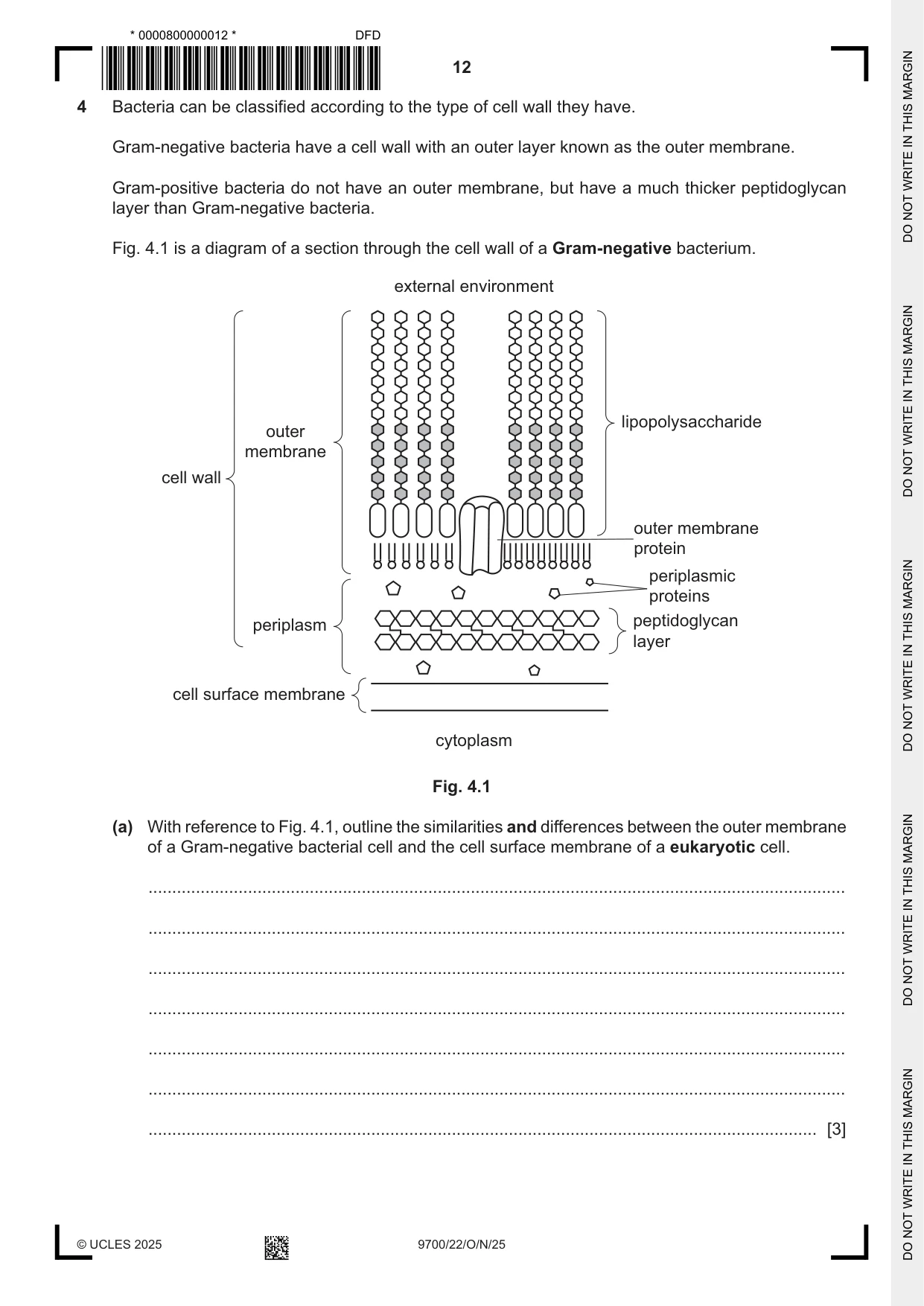
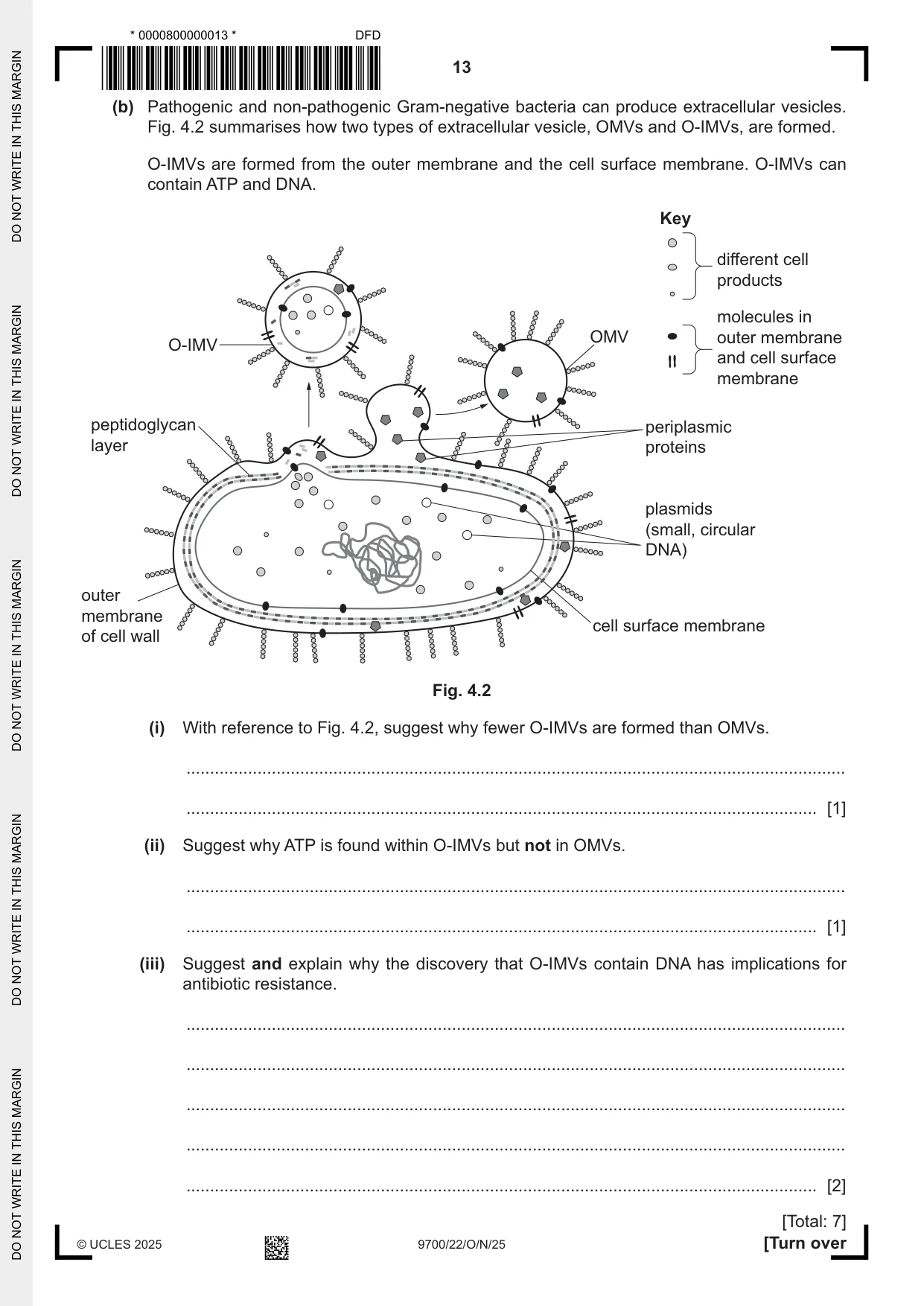
4 Bacteria can be classified according to the type of cell wall they have. Gram‑negative bacteria have a cell wall with an outer layer known as the outer membrane. Gram‑positive bacteria do not have an outer membrane, but have a much thicker peptidoglycan layer than Gram‑negative bacteria. Fig. 4.1 is a diagram of a section through the cell wall of a Gram‑negative bacterium. external environment lipopolysaccharide outer membrane cell wall outer membrane protein periplasmic proteins periplasm peptidoglycan layer cell surface membrane cytoplasm Fig. 4.1 (a) With reference to Fig. 4.1, outline the similarities and differences between the outer membrane of a Gram‑negative bacterial cell and the cell surface membrane of a eukaryotic cell. ................................................................................................................................................... ................................................................................................................................................... ................................................................................................................................................... ................................................................................................................................................... ................................................................................................................................................... ................................................................................................................................................... ............................................................................................................................................. [3] (b) Pathogenic and non‑pathogenic Gram‑negative bacteria can produce extracellular vesicles. Fig. 4.2 summarises how two types of extracellular vesicle, OMVs and O‑IMVs, are formed. O‑IMVs are formed from the outer membrane and the cell surface membrane. O‑IMVs can contain ATP and DNA. Key different cell products molecules in OMV outer membrane O-IMV and cell surface membrane peptidoglycan periplasmic layer proteins plasmids (small, circular DNA) outer membrane cell surface membrane of cell wall Fig. 4.2 (i) With reference to Fig. 4.2, suggest why fewer O‑IMVs are formed than OMVs. ........................................................................................................................................... ..................................................................................................................................... [1] (ii) Suggest why ATP is found within O‑IMVs but not in OMVs. ........................................................................................................................................... ..................................................................................................................................... [1] (iii) Suggest and explain why the discovery that O‑IMVs contain DNA has implications for antibiotic resistance. ........................................................................................................................................... ........................................................................................................................................... ........................................................................................................................................... ........................................................................................................................................... ..................................................................................................................................... [2] [Total: 7]
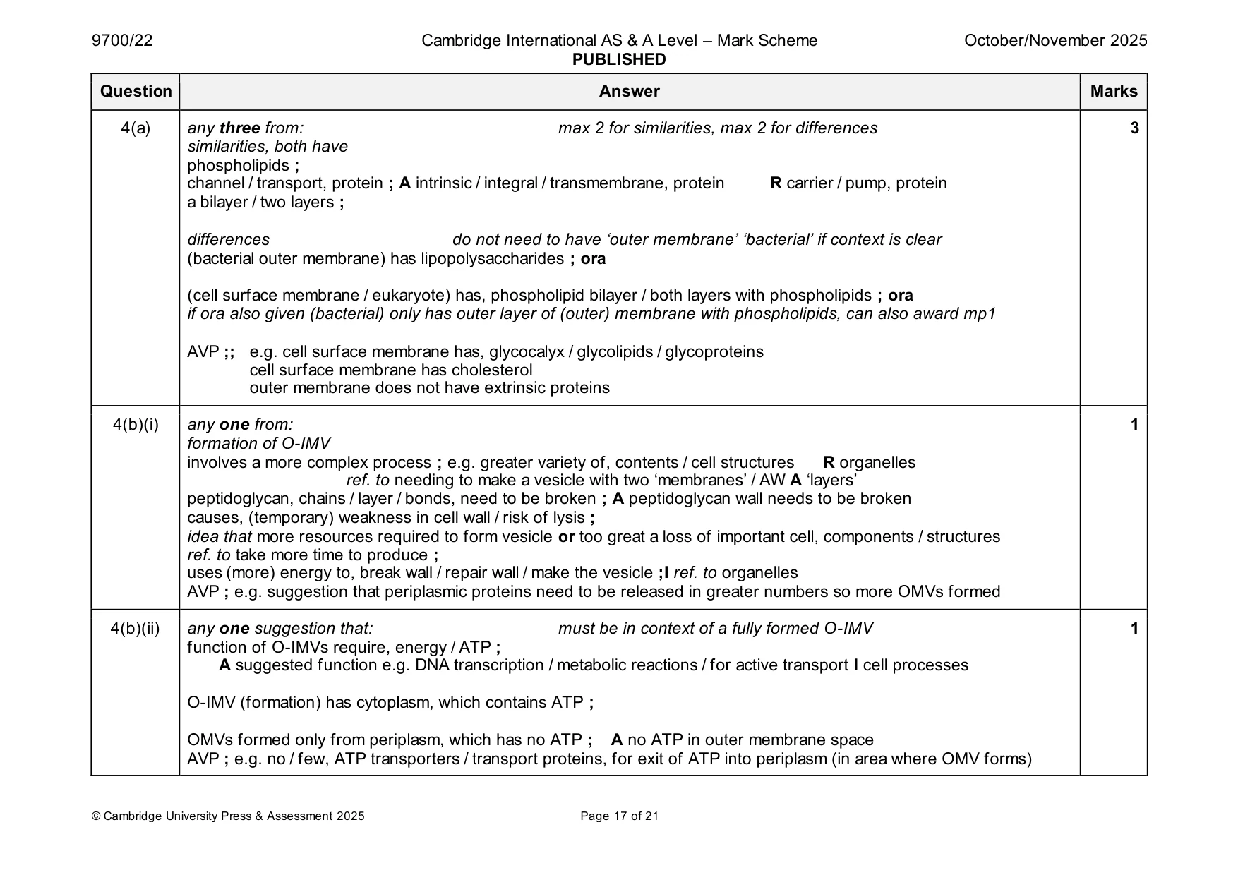
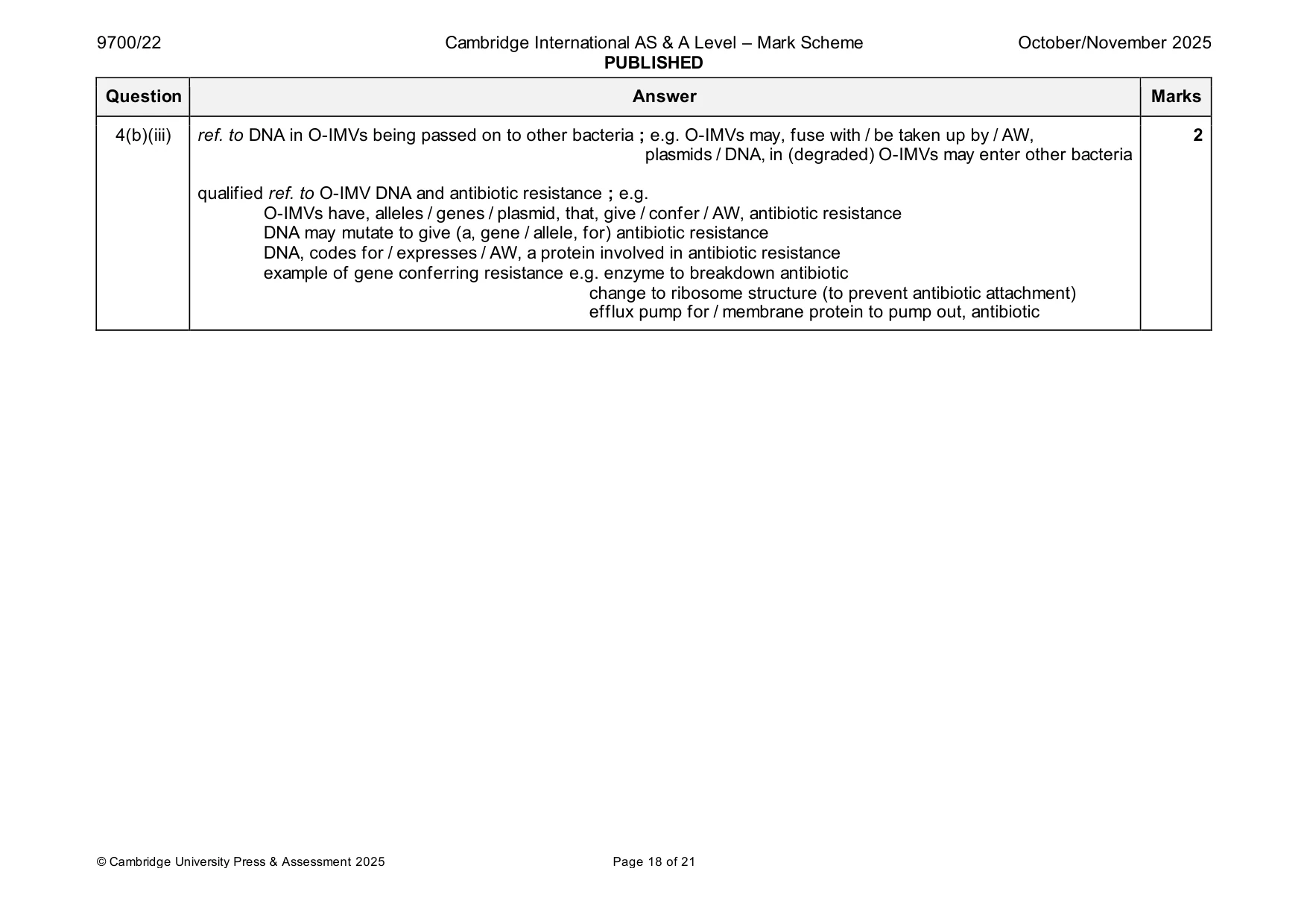
Mark scheme: 4(a) any three from: max 2 for similarities, max 2 for differences 3 similarities, both have phospholipids ; channel / transport, protein ; A intrinsic / integral / transmembrane, protein R carrier / pump, protein a bilayer / two layers ; differences do not need to have ‘outer membrane’ ‘bacterial’ if context is clear (bacterial outer membrane) has lipopolysaccharides ; ora (cell surface membrane / eukaryote) has, phospholipid bilayer / both layers with phospholipids ; ora if ora also given (bacterial) only has outer layer of (outer) membrane with phospholipids, can also award mp1 AVP ;; e.g. cell surface membrane has, glycocalyx / glycolipids / glycoproteins cell surface membrane has cholesterol outer membrane does not have extrinsic proteins 4(b)(i) any one from: 1 formation of O-IMV involves a more complex process ; e.g. greater variety of, contents / cell structures R organelles ref. to needing to make a vesicle with two ‘membranes’ / AW A ‘layers’ peptidoglycan, chains / layer / bonds, need to be broken ; A peptidoglycan wall needs to be broken causes, (temporary) weakness in cell wall / risk of lysis ; idea that more resources required to form vesicle or too great a loss of important cell, components / structures ref. to take more time to produce ; uses (more) energy to, break wall / repair wall / make the vesicle ;I ref. to organelles AVP ; e.g. suggestion that periplasmic proteins need to be released in greater numbers so more OMVs formed 4(b)(ii) any one suggestion that: must be in context of a fully formed O-IMV 1 function of O-IMVs require, energy / ATP ; A suggested function e.g. DNA transcription / metabolic reactions / for active transport I cell processes O-IMV (formation) has cytoplasm, which contains ATP ; OMVs formed only from periplasm, which has no ATP ; A no ATP in outer membrane space AVP ; e.g. no / few, ATP transporters / transport proteins, for exit of ATP into periplasm (in area where OMV forms) 4(b)(iii) ref. to DNA in O-IMVs being passed on to other bacteria ; e.g. O-IMVs may, fuse with / be taken up by / AW, 2 plasmids / DNA, in (degraded) O-IMVs may enter other bacteria qualified ref. to O-IMV DNA and antibiotic resistance ; e.g. O-IMVs have, alleles / genes / plasmid, that, give / confer / AW, antibiotic resistance DNA may mutate to give (a, gene / allele, for) antibiotic resistance DNA, codes for / expresses / AW, a protein involved in antibiotic resistance example of gene conferring resistance e.g. enzyme to breakdown antibiotic change to ribosome structure (to prevent antibiotic attachment) efflux pump for / membrane protein to pump out, antibiotic
Q5 · A plant that is described as a mesophyte has evolved to grow in conditions that do not…
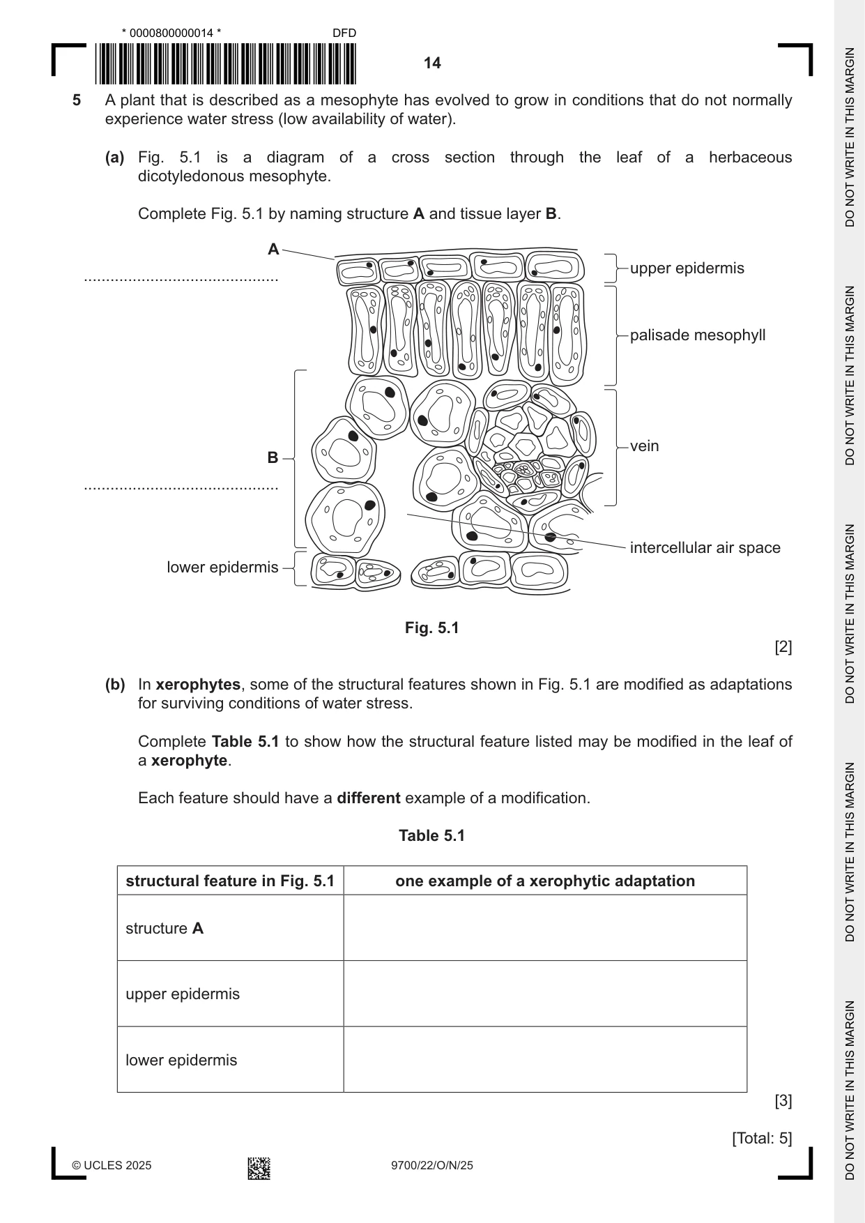
5 A plant that is described as a mesophyte has evolved to grow in conditions that do not normally experience water stress (low availability of water). (a) Fig. 5.1 is a diagram of a cross section through the leaf of a herbaceous dicotyledonous mesophyte. Complete Fig. 5.1 by naming structure A and tissue layer B. A upper epidermis ............................................ palisade mesophyll vein B ............................................ intercellular air space lower epidermis Fig. 5.1 [2] (b) In xerophytes, some of the structural features shown in Fig. 5.1 are modified as adaptations for surviving conditions of water stress. Complete Table 5.1 to show how the structural feature listed may be modified in the leaf of a xerophyte. Each feature should have a different example of a modification. Table 5.1 structural feature in Fig. 5.1 one example of a xerophytic adaptation structure A upper epidermis lower epidermis [3] [Total: 5]
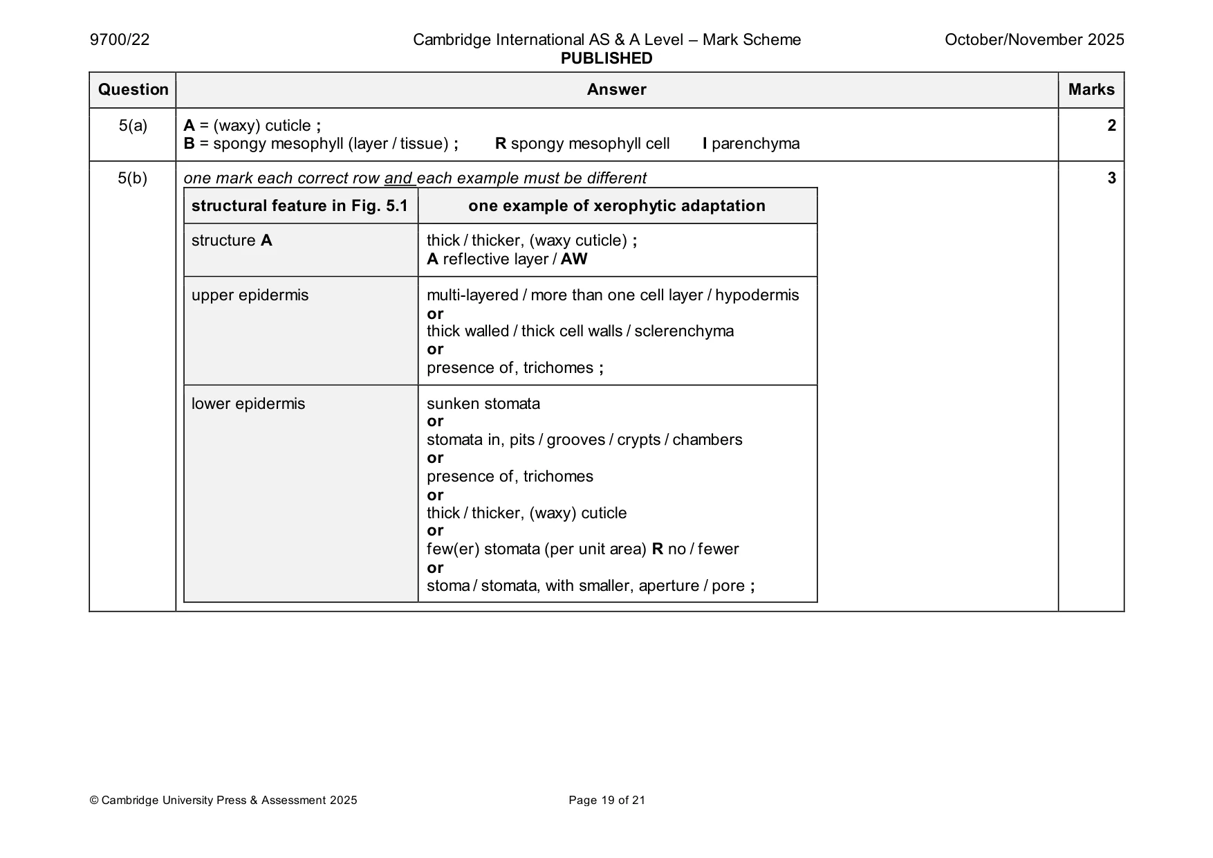
Mark scheme: 5(a) A = (waxy) cuticle ; 2 B = spongy mesophyll (layer / tissue) ; R spongy mesophyll cell I parenchyma 5(b) one mark each correct row and each example must be different 3 structural feature in Fig. 5.1 one example of xerophytic adaptation structure A thick / thicker, (waxy cuticle) ; A reflective layer / AW upper epidermis multi-layered / more than one cell layer / hypodermis or thick walled / thick cell walls / sclerenchyma or presence of, trichomes ; lower epidermis sunken stomata or stomata in, pits / grooves / crypts / chambers or presence of, trichomes or thick / thicker, (waxy) cuticle or few(er) stomata (per unit area) R no / fewer or stoma / stomata, with smaller, aperture / pore ;
Q6 · In the pulmonary circulation of a mammal, deoxygenated blood becomes oxygenated when red…
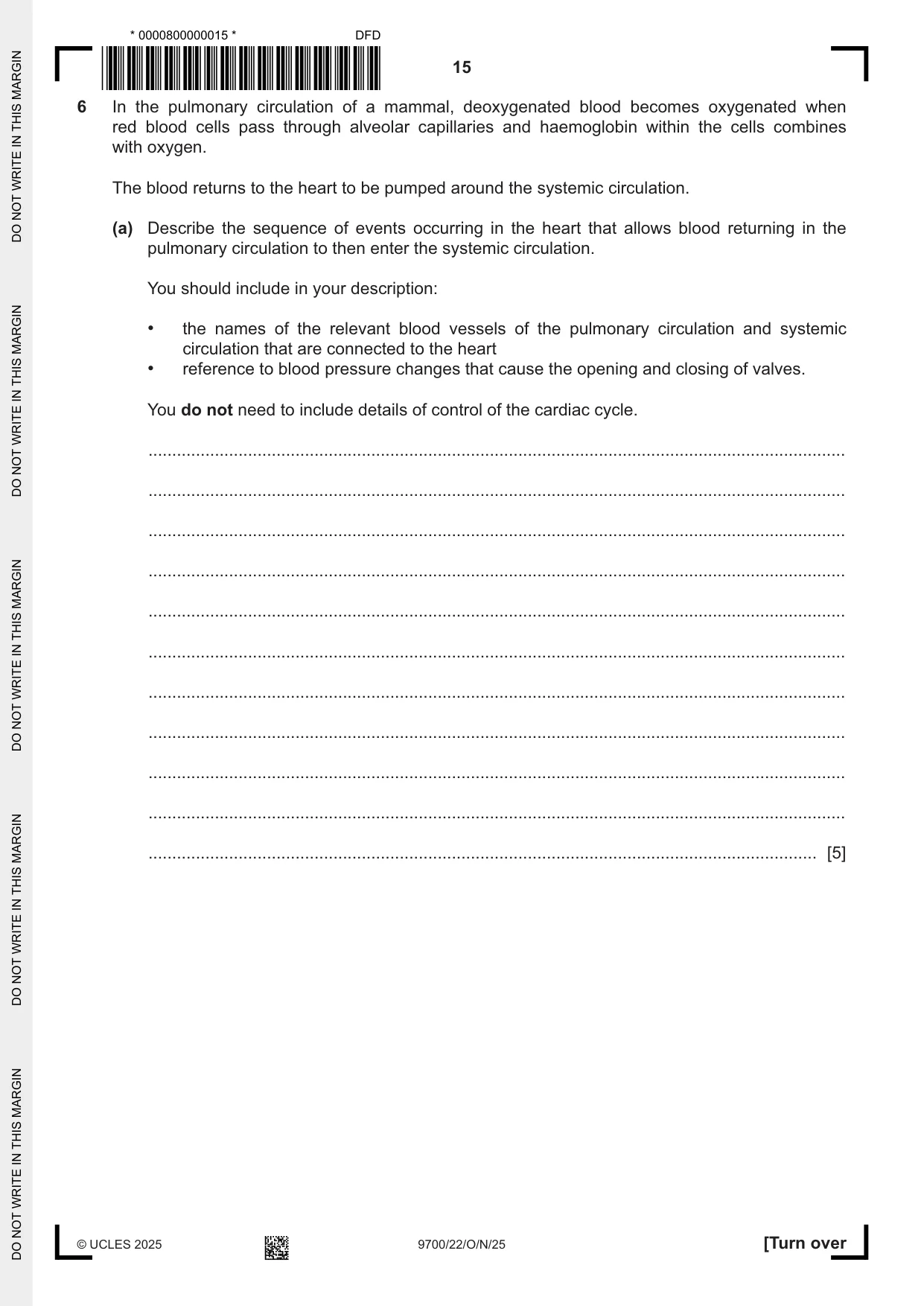
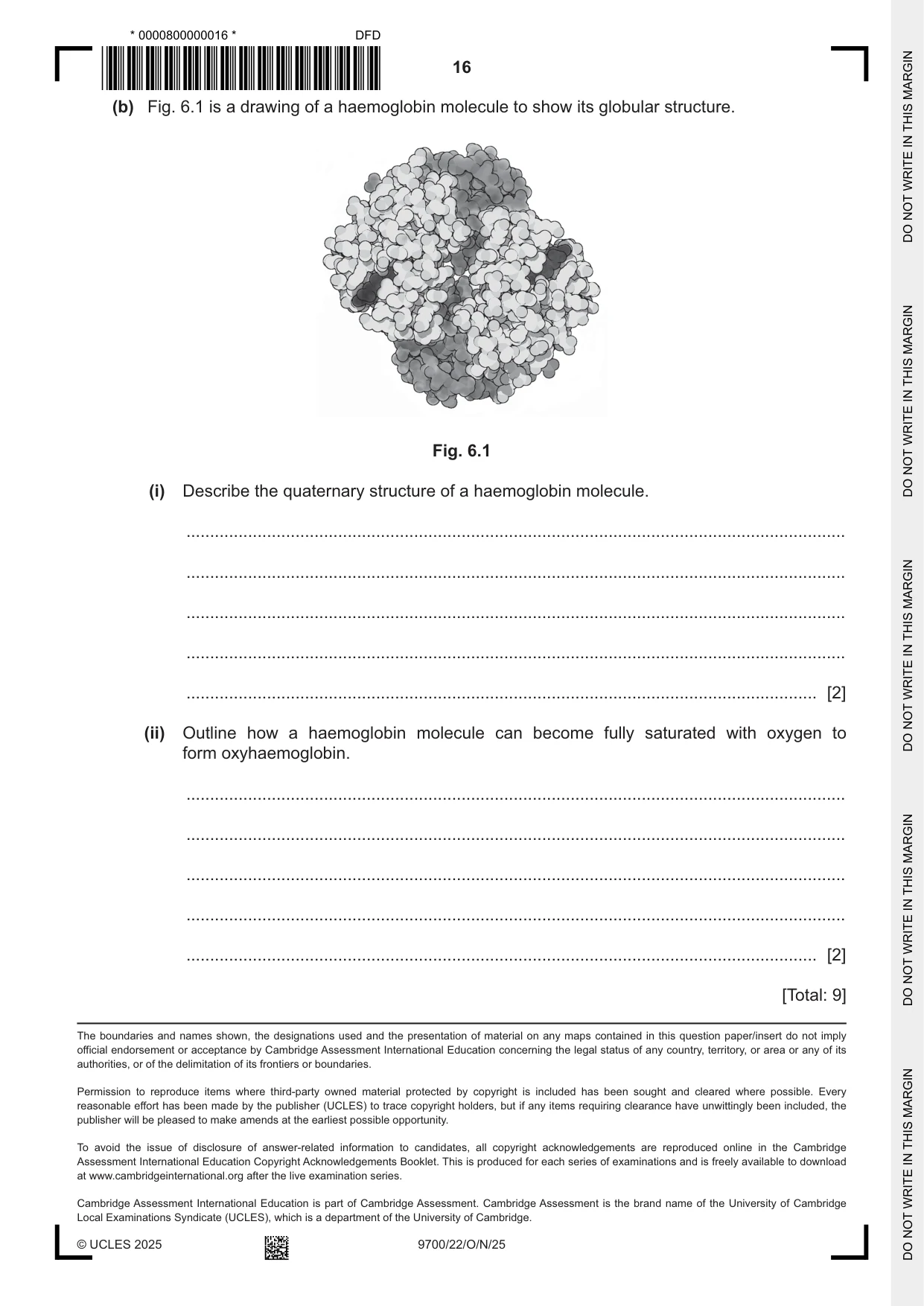
6 In the pulmonary circulation of a mammal, deoxygenated blood becomes oxygenated when red blood cells pass through alveolar capillaries and haemoglobin within the cells combines with oxygen. The blood returns to the heart to be pumped around the systemic circulation. (a) Describe the sequence of events occurring in the heart that allows blood returning in the pulmonary circulation to then enter the systemic circulation. You should include in your description: • the names of the relevant blood vessels of the pulmonary circulation and systemic circulation that are connected to the heart • reference to blood pressure changes that cause the opening and closing of valves. You do not need to include details of control of the cardiac cycle. ................................................................................................................................................... ................................................................................................................................................... ................................................................................................................................................... ................................................................................................................................................... ................................................................................................................................................... ................................................................................................................................................... ................................................................................................................................................... ................................................................................................................................................... ................................................................................................................................................... ................................................................................................................................................... ............................................................................................................................................. [5] (b) Fig. 6.1 is a drawing of a haemoglobin molecule to show its globular structure. Fig. 6.1 (i) Describe the quaternary structure of a haemoglobin molecule. ........................................................................................................................................... ........................................................................................................................................... ........................................................................................................................................... ........................................................................................................................................... ..................................................................................................................................... [2] (ii) Outline how a haemoglobin molecule can become fully saturated with oxygen to form oxyhaemoglobin. ........................................................................................................................................... ........................................................................................................................................... ........................................................................................................................................... ........................................................................................................................................... ..................................................................................................................................... [2] [Total: 9] The boundaries and names shown, the designations used and the presentation of material on any maps contained in this question paper/insert do not imply official endorsement or acceptance by Cambridge Assessment International Education concerning the legal status of any country, territory, or area or any of its authorities, or of the delimitation of its frontiers or boundaries.
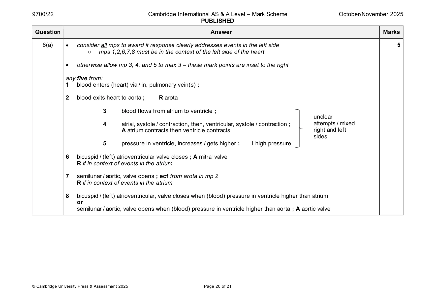
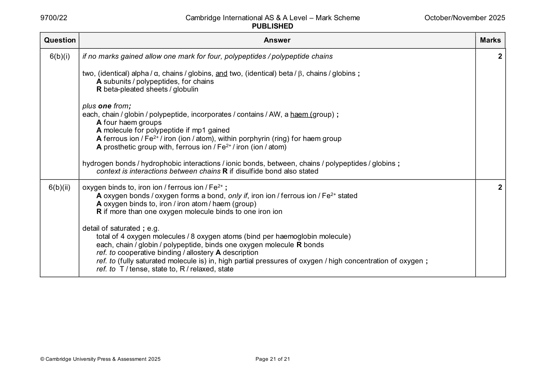
Mark scheme: 6(a) • consider all mps to award if response clearly addresses events in the left side 5 ○ mps 1,2,6,7,8 must be in the context of the left side of the heart • otherwise allow mp 3, 4, and 5 to max 3 – these mark points are inset to the right any five from: 1 blood enters (heart) via / in, pulmonary vein(s) ; 2 blood exits heart to aorta ; R arota 3 blood flows from atrium to ventricle ; unclear 4 atrial, systole / contraction, then, ventricular, systole / contraction ; attempts / mixed A atrium contracts then ventricle contracts right and left sides 5 pressure in ventricle, increases / gets higher ; I high pressure 6 bicuspid / (left) atrioventricular valve closes ; A mitral valve R if in context of events in the atrium 7 semilunar / aortic, valve opens ; ecf from arota in mp 2 R if in context of events in the atrium 8 bicuspid / (left) atrioventricular, valve closes when (blood) pressure in ventricle higher than atrium or semilunar / aortic, valve opens when (blood) pressure in ventricle higher than aorta ; A aortic valve 6(b)(i) if no marks gained allow one mark for four, polypeptides / polypeptide chains 2 two, (identical) alpha / α, chains / globins, and two, (identical) beta / , chains / globins ; A subunits / polypeptides, for chains R beta-pleated sheets / globulin plus one from; each, chain / globin / polypeptide, incorporates / contains / AW, a haem (group) ; A four haem groups A molecule for polypeptide if mp1 gained A ferrous ion / Fe2+ / iron (ion / atom), within porphyrin (ring) for haem group A prosthetic group with, ferrous ion / Fe2+ / iron (ion / atom) hydrogen bonds / hydrophobic interactions / ionic bonds, between, chains / polypeptides / globins ; context is interactions between chains R if disulfide bond also stated 6(b)(ii) oxygen binds to, iron ion / ferrous ion / Fe2+ ; 2 A oxygen bonds / oxygen forms a bond, only if, iron ion / ferrous ion / Fe2+ stated A oxygen binds to, iron / iron atom / haem (group) R if more than one oxygen molecule binds to one iron ion detail of saturated ; e.g. total of 4 oxygen molecules / 8 oxygen atoms (bind per haemoglobin molecule) each, chain / globin / polypeptide, binds one oxygen molecule R bonds ref. to cooperative binding / allostery A description ref. to (fully saturated molecule is) in, high partial pressures of oxygen / high concentration of oxygen ; ref. to T / tense, state to, R / relaxed, state
What was in this paper
The subtopics covered by these 6 questions, and how many questions each got. Open one in a new tab to see every Cambridge question on it.
What you needed in this session
Cambridge’s own grade thresholds for 2025 Oct/Nov, Paper 2 · Variant 2. A higher threshold means an easier paper — the bar moves with how the cohort did.