Cambridge A Level Biology 9700 — 2024 May/June Paper 2 · Variant 2
9700/22/M/J/24 · 6 questions · 60 marks · ≈68 min
The question paper and its mark scheme, free to read here and free to download. This is Cambridge’s own paper, exactly as it was sat.
Question paper16 pages
Mark scheme21 pages
Answers below. Sit the paper first if you are practising.
Questions as text
Q1 · Smooth muscle is a tissue composed of smooth muscle cells
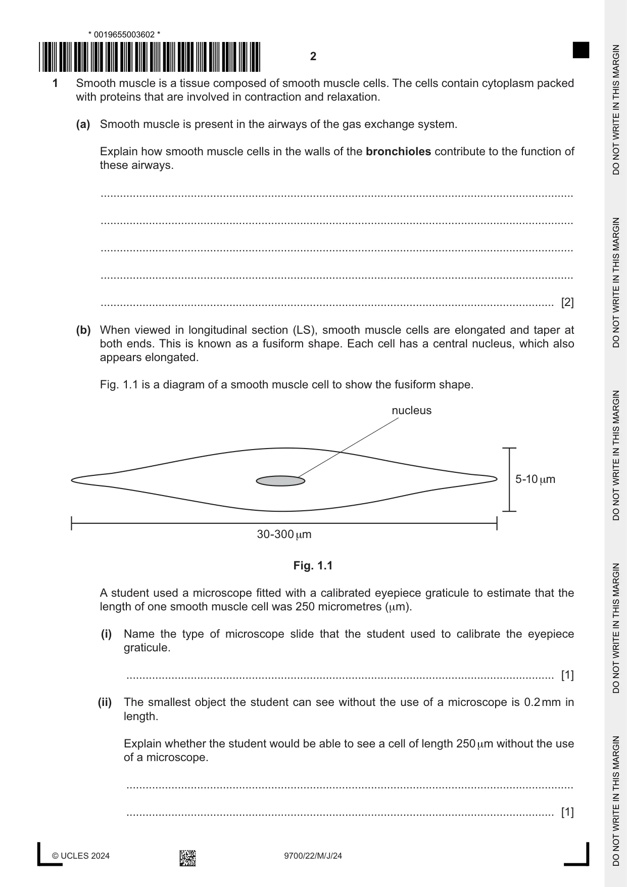
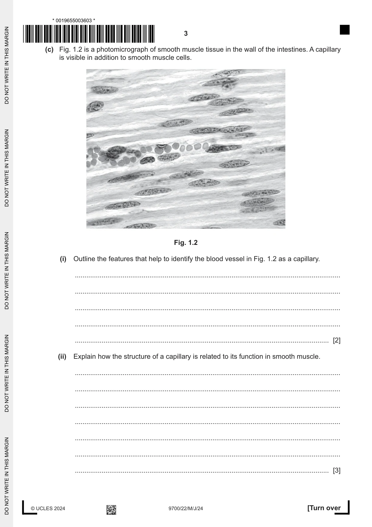
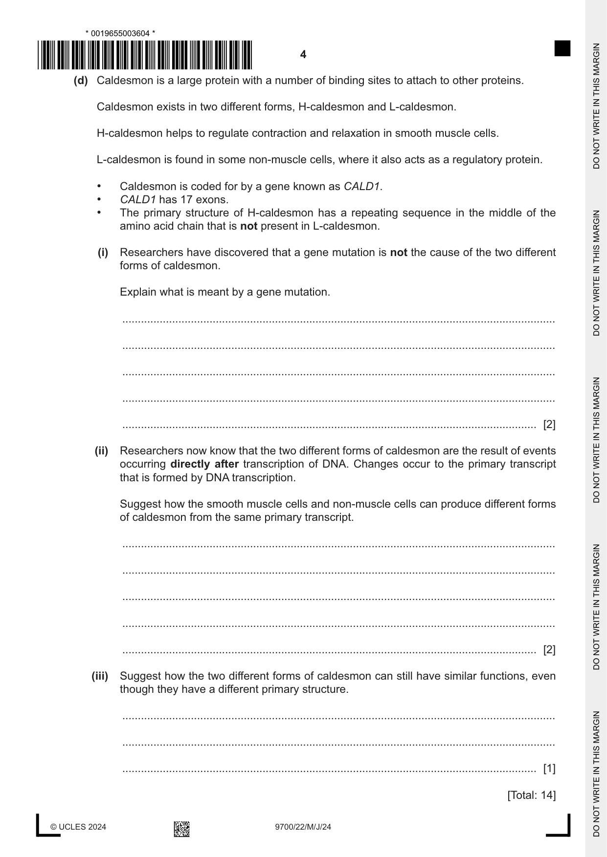
1 Smooth muscle is a tissue composed of smooth muscle cells. The cells contain cytoplasm packed with proteins that are involved in contraction and relaxation. (a) Smooth muscle is present in the airways of the gas exchange system. Explain how smooth muscle cells in the walls of the bronchioles contribute to the function of these airways. ................................................................................................................................................... ................................................................................................................................................... ................................................................................................................................................... ................................................................................................................................................... ............................................................................................................................................. [2] (b) When viewed in longitudinal section (LS), smooth muscle cells are elongated and taper at both ends. This is known as a fusiform shape. Each cell has a central nucleus, which also appears elongated. Fig. 1.1 is a diagram of a smooth muscle cell to show the fusiform shape. nucleus 5-10 μm 30-300 μm Fig. 1.1 A student used a microscope fitted with a calibrated eyepiece graticule to estimate that the length of one smooth muscle cell was 250 micrometres (μm). (i) Name the type of microscope slide that the student used to calibrate the eyepiece graticule. ..................................................................................................................................... [1] (ii) The smallest object the student can see without the use of a microscope is 0.2 mm in length. Explain whether the student would be able to see a cell of length 250 μm without the use of a microscope. ........................................................................................................................................... ..................................................................................................................................... [1] (c) Fig. 1.2 is a photomicrograph of smooth muscle tissue in the wall of the intestines. A capillary is visible in addition to smooth muscle cells. Fig. 1.2 (i) Outline the features that help to identify the blood vessel in Fig. 1.2 as a capillary. ........................................................................................................................................... ........................................................................................................................................... ........................................................................................................................................... ........................................................................................................................................... ..................................................................................................................................... [2] (ii) Explain how the structure of a capillary is related to its function in smooth muscle. ........................................................................................................................................... ........................................................................................................................................... ........................................................................................................................................... ........................................................................................................................................... ........................................................................................................................................... ........................................................................................................................................... ..................................................................................................................................... [3] (d) Caldesmon is a large protein with a number of binding sites to attach to other proteins. Caldesmon exists in two different forms, H-caldesmon and L-caldesmon. H-caldesmon helps to regulate contraction and relaxation in smooth muscle cells. L-caldesmon is found in some non-muscle cells, where it also acts as a regulatory protein. • Caldesmon is coded for by a gene known as CALD1. • CALD1 has 17 exons. • The primary structure of H-caldesmon has a repeating sequence in the middle of the amino acid chain that is not present in L-caldesmon. (i) Researchers have discovered that a gene mutation is not the cause of the two different forms of caldesmon. Explain what is meant by a gene mutation. ........................................................................................................................................... ........................................................................................................................................... ........................................................................................................................................... ........................................................................................................................................... ..................................................................................................................................... [2] (ii) Researchers now know that the two different forms of caldesmon are the result of events occurring directly after transcription of DNA. Changes occur to the primary transcript that is formed by DNA transcription. Suggest how the smooth muscle cells and non-muscle cells can produce different forms of caldesmon from the same primary transcript. ........................................................................................................................................... ........................................................................................................................................... ........................................................................................................................................... ........................................................................................................................................... ..................................................................................................................................... [2] (iii) Suggest how the two different forms of caldesmon can still have similar functions, even though they have a different primary structure. ........................................................................................................................................... ........................................................................................................................................... ..................................................................................................................................... [1] [Total: 14]
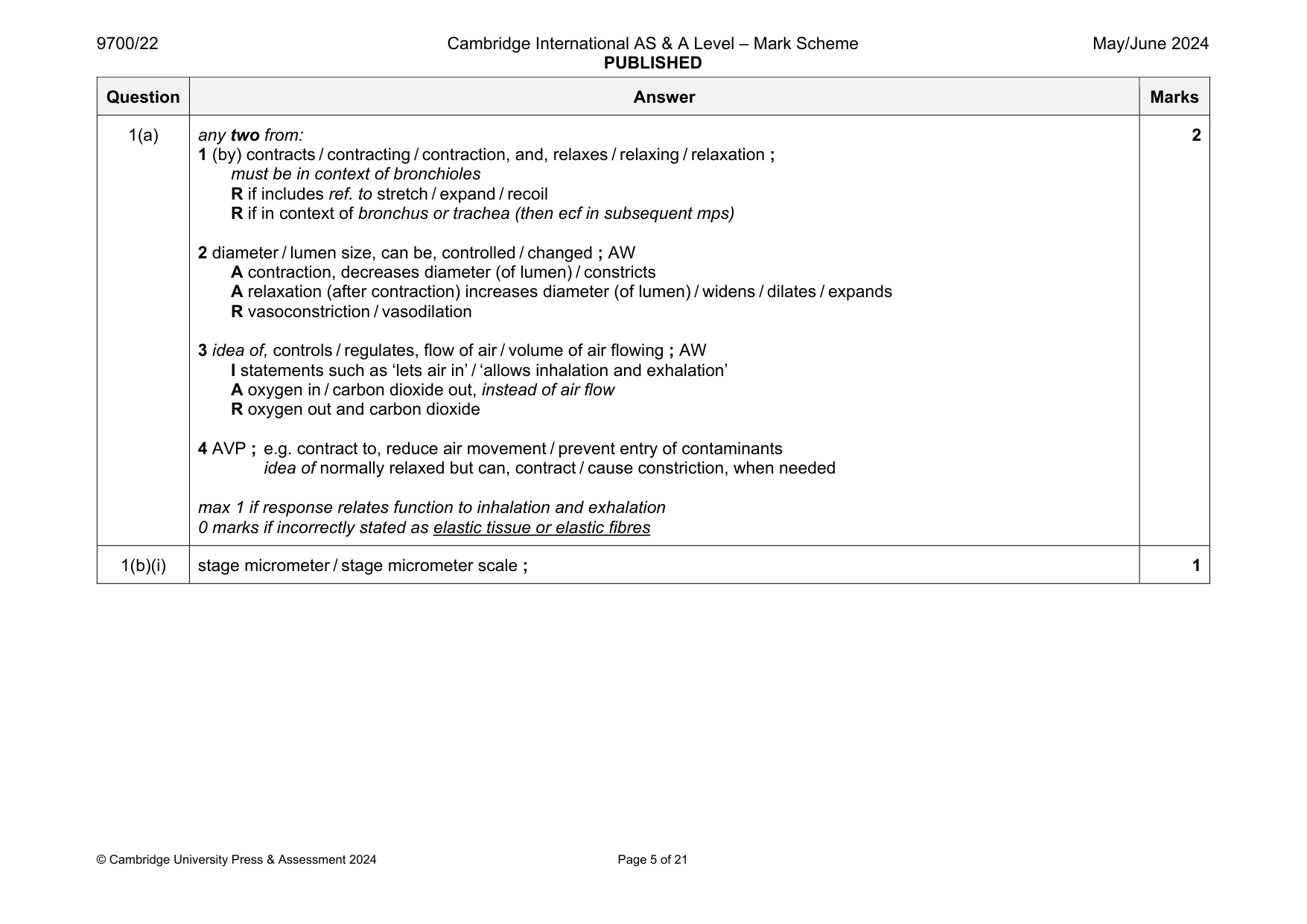
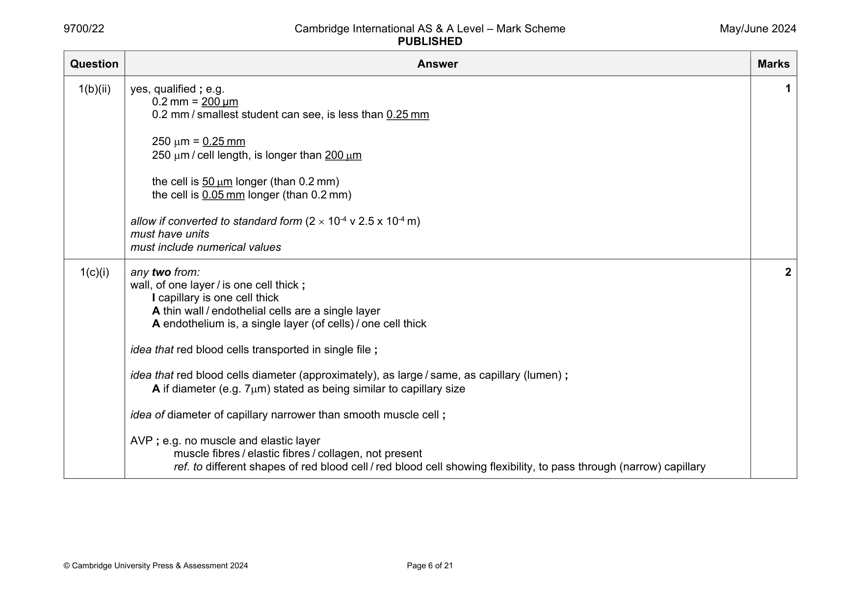
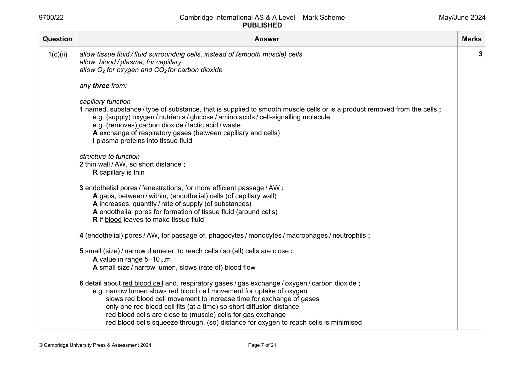
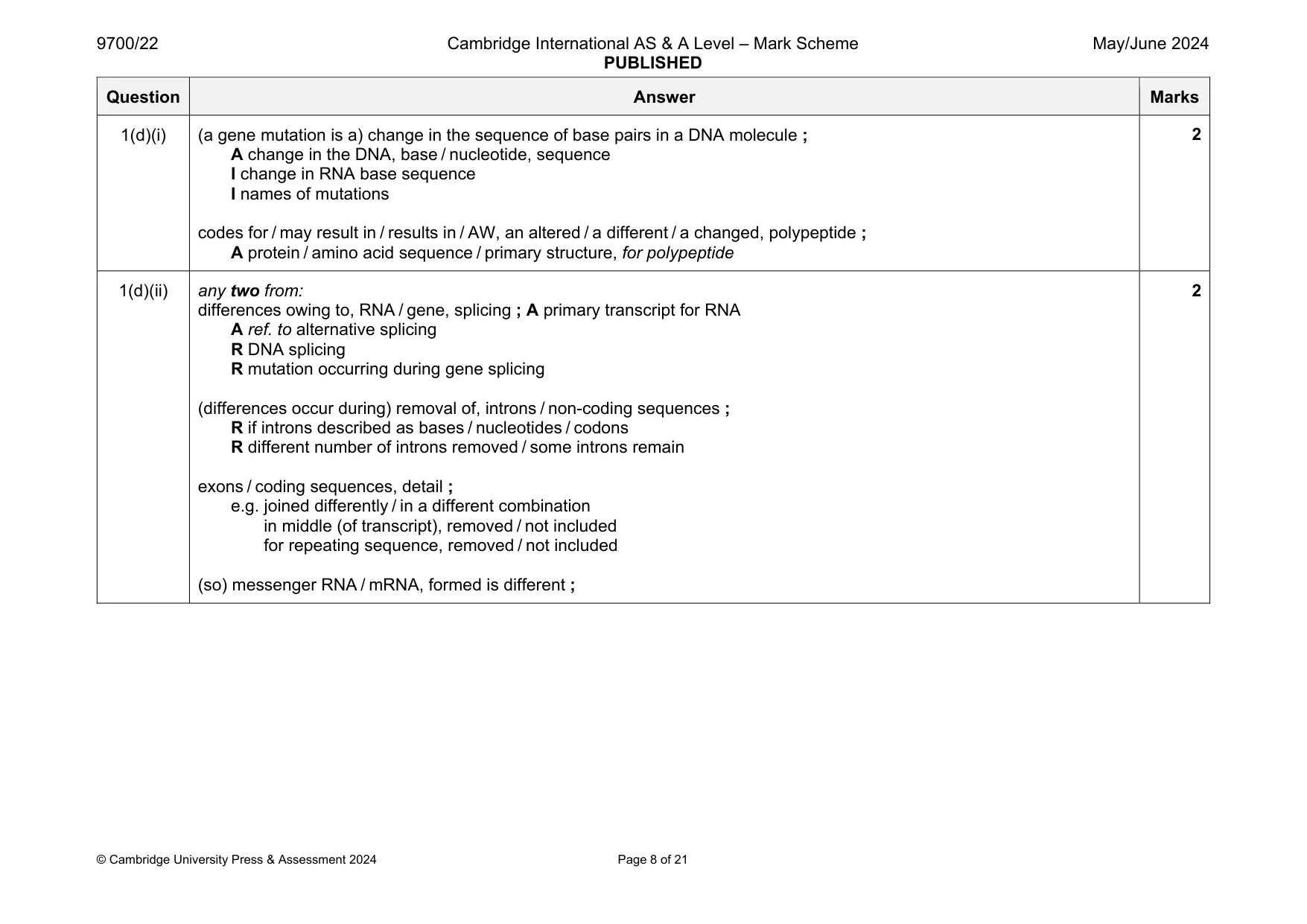
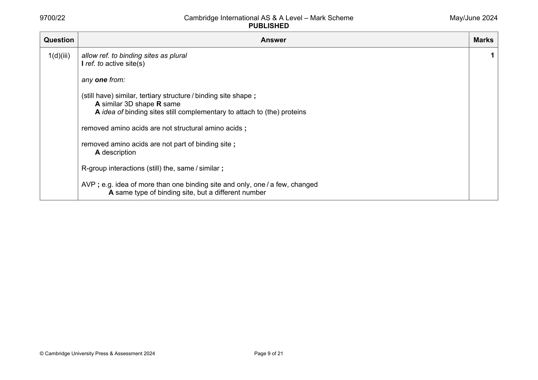
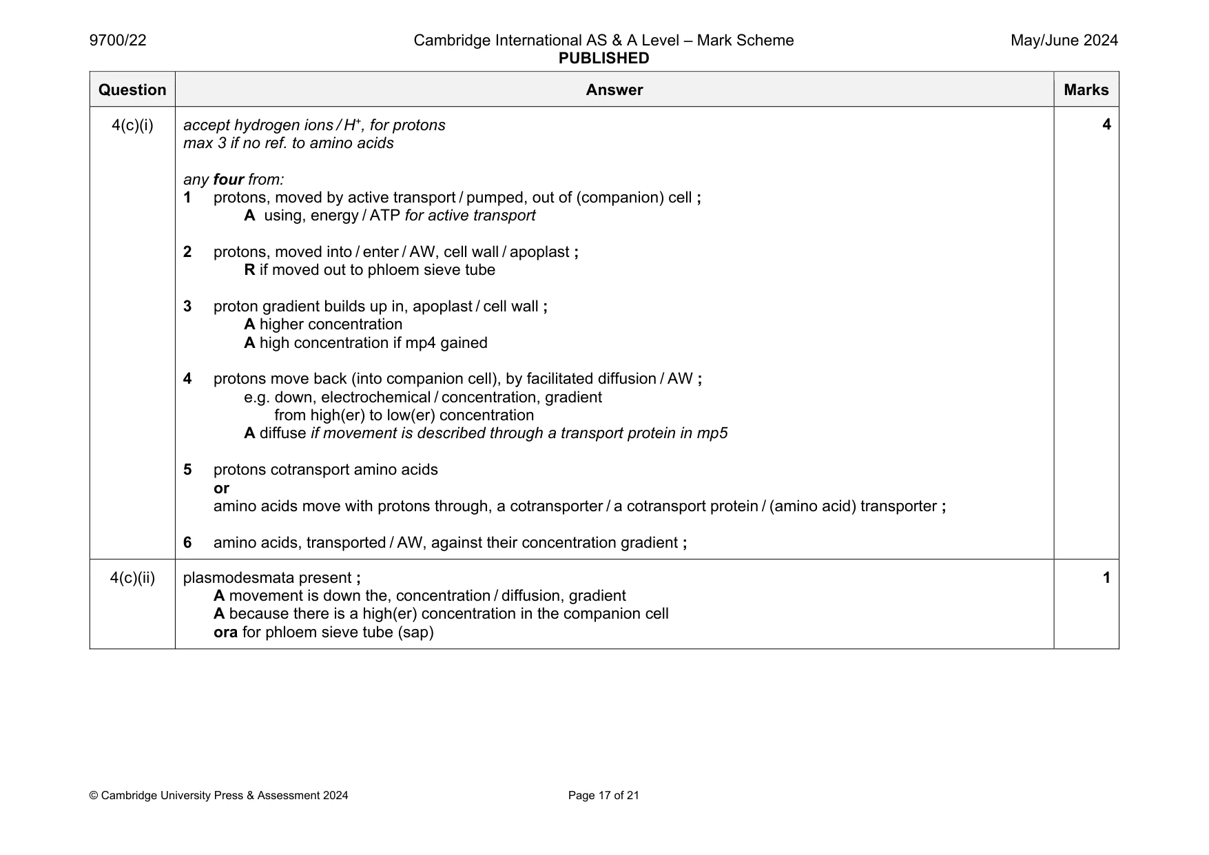
Mark scheme: 1(a) any two from: 1 (by) contracts / contracting / contraction, and, relaxes / relaxing / relaxation ; must be in context of bronchioles R if includes ref. to stretch / expand / recoil R if in context of bronchus or trachea (then ecf in subsequent mps) 2 diameter / lumen size, can be, controlled / changed ; AW A contraction, decreases diameter (of lumen) / constricts A relaxation (after contraction) increases diameter (of lumen) / widens / dilates / expands R vasoconstriction / vasodilation 3 idea of, controls / regulates, flow of air / volume of air flowing ; AW I statements such as ‘lets air in’ / ‘allows inhalation and exhalation’ A oxygen in / carbon dioxide out, instead of air flow R oxygen out and carbon dioxide 4 AVP ; e.g. contract to, reduce air movement / prevent entry of contaminants idea of normally relaxed but can, contract / cause constriction, when needed max 1 if response relates function to inhalation and exhalation 0 marks if incorrectly stated as elastic tissue or elastic fibres 2 1(b)(i) stage micrometer / stage micrometer scale ; 1 Question Answer Marks 1(b)(ii) yes, qualified ; e.g. 0.2 mm = 200 µm 0.2 mm / smallest student can see, is less than 0.25 mm 250 m = 0.25 mm 250 m / cell length, is longer than 200 m the cell is 50 m longer (than 0.2 mm) the cell is 0.05 mm longer (than 0.2 mm) allow if converted to standard form (2 10-4 v 2.5 x 10⁻⁴ m) must have units must include numerical values 1 1(c)(i) any two from: wall, of one layer / is one cell thick ; I capillary is one cell thick A thin wall / endothelial cells are a single layer A endothelium is, a single layer (of cells) / one cell thick idea that red blood cells transported in single file ; idea that red blood cells diameter (approximately), as large / same, as capillary (lumen) ; A if diameter (e.g. 7m) stated as being similar to capillary size idea of diameter of capillary narrower than smooth muscle cell ; AVP ; e.g. no muscle and elastic layer muscle fibres / elastic fibres / collagen, not present ref. to different shapes of red blood cell / red blood cell showing flexibility, to pass through (narrow) capillary 2 Question Answer Marks 1(c)(ii) allow tissue fluid / fluid surrounding cells, instead of (smooth muscle) cells allow, blood / plasma, for capillary allow O2 for oxygen and CO2 for carbon dioxide any three from: capillary function 1 named, substance / type of substance, that is supplied to smooth muscle cells or is a product removed from the cells ; e.g. (supply) oxygen / nutrients / glucose / amino acids / cell-signalling molecule e.g. (removes) carbon dioxide / lactic acid / waste A exchange of respiratory gases (between capillary and cells) I plasma proteins into tissue fluid structure to function 2 thin wall / AW, so short distance ; R capillary is thin 3 endothelial pores / fenestrations, for more efficient passage / AW ; A gaps, between / within, (endothelial) cells (of capillary wall) A increases, quantity / rate of supply (of substances) A endothelial pores for formation of tissue fluid (around cells) R if blood leaves to make tissue fluid 4 (endothelial) pores / AW, for passage of, phagocytes / monocytes / macrophages / neutrophils ; 5 small (size) / narrow diameter, to reach cells / so (all) cells are close ; A value in range 510 m A small size / narrow lumen, slows (rate of) blood flow 6 detail about red blood cell and, respiratory gases / gas exchange / oxygen / carbon dioxide ; e.g. narrow lumen slows red blood cell movement for uptake of oxygen slows red blood cell movement to increase time for exchange of gases only one red blood cell fits (at a time) so short diffusion distance red blood cells are close to (muscle) cells for gas exchange red blood cells squeeze through, (so) distance for oxygen to reach cells is minimised 3 Question Answer Marks 1(d)(i) (a gene mutation is a) change in the sequence of base pairs in a DNA molecule ; A change in the DNA, base / nucleotide, sequence I change in RNA base sequence I names of mutations codes for / may result in / results in / AW, an altered / a different / a changed, polypeptide ; A protein / amino acid sequence / primary structure, for polypeptide 2 1(d)(ii) any two from: differences owing to, RNA / gene, splicing ; A primary transcript for RNA A ref. to alternative splicing R DNA splicing R mutation occurring during gene splicing (differences occur during) removal of, introns / non-coding sequences ; R if introns described as bases / nucleotides / codons R different number of introns removed / some introns remain exons / coding sequences, detail ; e.g. joined differently / in a different combination in middle (of transcript), removed / not included for repeating sequence, removed / not included (so) messenger RNA / mRNA, formed is different ; 2 Question Answer Marks 1(d)(iii) allow ref. to binding sites as plural I ref. to active site(s) any one from: (still have) similar, tertiary structure / binding site shape ; A similar 3D shape R same A idea of binding sites still complementary to attach to (the) proteins removed amino acids are not structural amino acids ; removed amino acids are not part of binding site ; A description R-group interactions (still) the, same / similar ; AVP ; e.g. idea of more than one binding site and only, one / a few, changed A same type of binding site, but a different number 1
Q2 · Alveolar macrophages are cells of the immune system that remain in the alveolar region of…
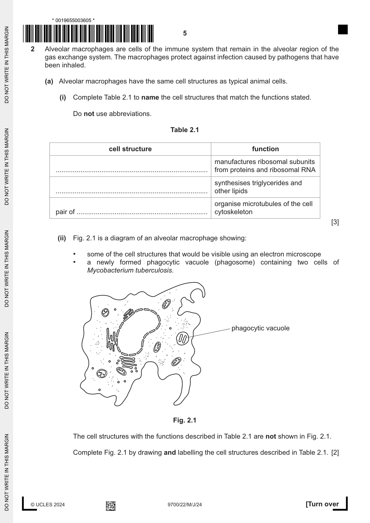
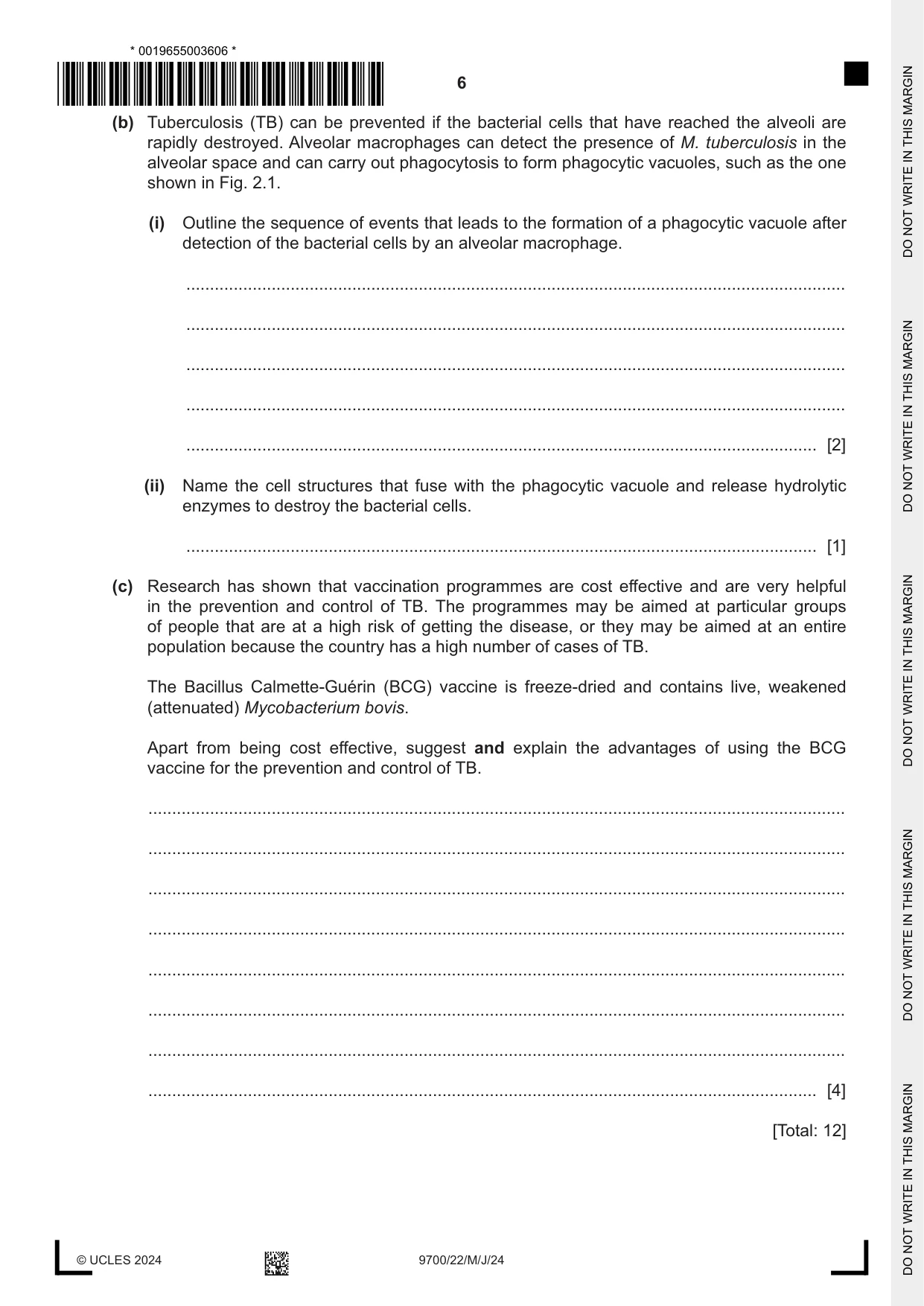
2 Alveolar macrophages are cells of the immune system that remain in the alveolar region of the gas exchange system. The macrophages protect against infection caused by pathogens that have been inhaled. (a) Alveolar macrophages have the same cell structures as typical animal cells. (i) Complete Table 2.1 to name the cell structures that match the functions stated. Do not use abbreviations. Table 2.1 cell structure function manufactures ribosomal subunits ................................................................................ from proteins and ribosomal RNA synthesises triglycerides and ................................................................................ other lipids organise microtubules of the cell pair of ..................................................................... cytoskeleton [3] (ii) Fig. 2.1 is a diagram of an alveolar macrophage showing: • some of the cell structures that would be visible using an electron microscope • a newly formed phagocytic vacuole (phagosome) containing two cells of Mycobacterium tuberculosis. phagocytic vacuole Fig. 2.1 The cell structures with the functions described in Table 2.1 are not shown in Fig. 2.1. Complete Fig. 2.1 by drawing and labelling the cell structures described in Table 2.1. [2] (b) Tuberculosis (TB) can be prevented if the bacterial cells that have reached the alveoli are rapidly destroyed. Alveolar macrophages can detect the presence of M. tuberculosis in the alveolar space and can carry out phagocytosis to form phagocytic vacuoles, such as the one shown in Fig. 2.1. (i) Outline the sequence of events that leads to the formation of a phagocytic vacuole after detection of the bacterial cells by an alveolar macrophage. ........................................................................................................................................... ........................................................................................................................................... ........................................................................................................................................... ........................................................................................................................................... ..................................................................................................................................... [2] (ii) Name the cell structures that fuse with the phagocytic vacuole and release hydrolytic enzymes to destroy the bacterial cells. ..................................................................................................................................... [1] (c) Research has shown that vaccination programmes are cost effective and are very helpful in the prevention and control of TB. The programmes may be aimed at particular groups of people that are at a high risk of getting the disease, or they may be aimed at an entire population because the country has a high number of cases of TB. The Bacillus Calmette-Guérin (BCG) vaccine is freeze-dried and contains live, weakened (attenuated) Mycobacterium bovis. Apart from being cost effective, suggest and explain the advantages of using the BCG vaccine for the prevention and control of TB. ................................................................................................................................................... ................................................................................................................................................... ................................................................................................................................................... ................................................................................................................................................... ................................................................................................................................................... ................................................................................................................................................... ................................................................................................................................................... ............................................................................................................................................. [4] [Total: 12]
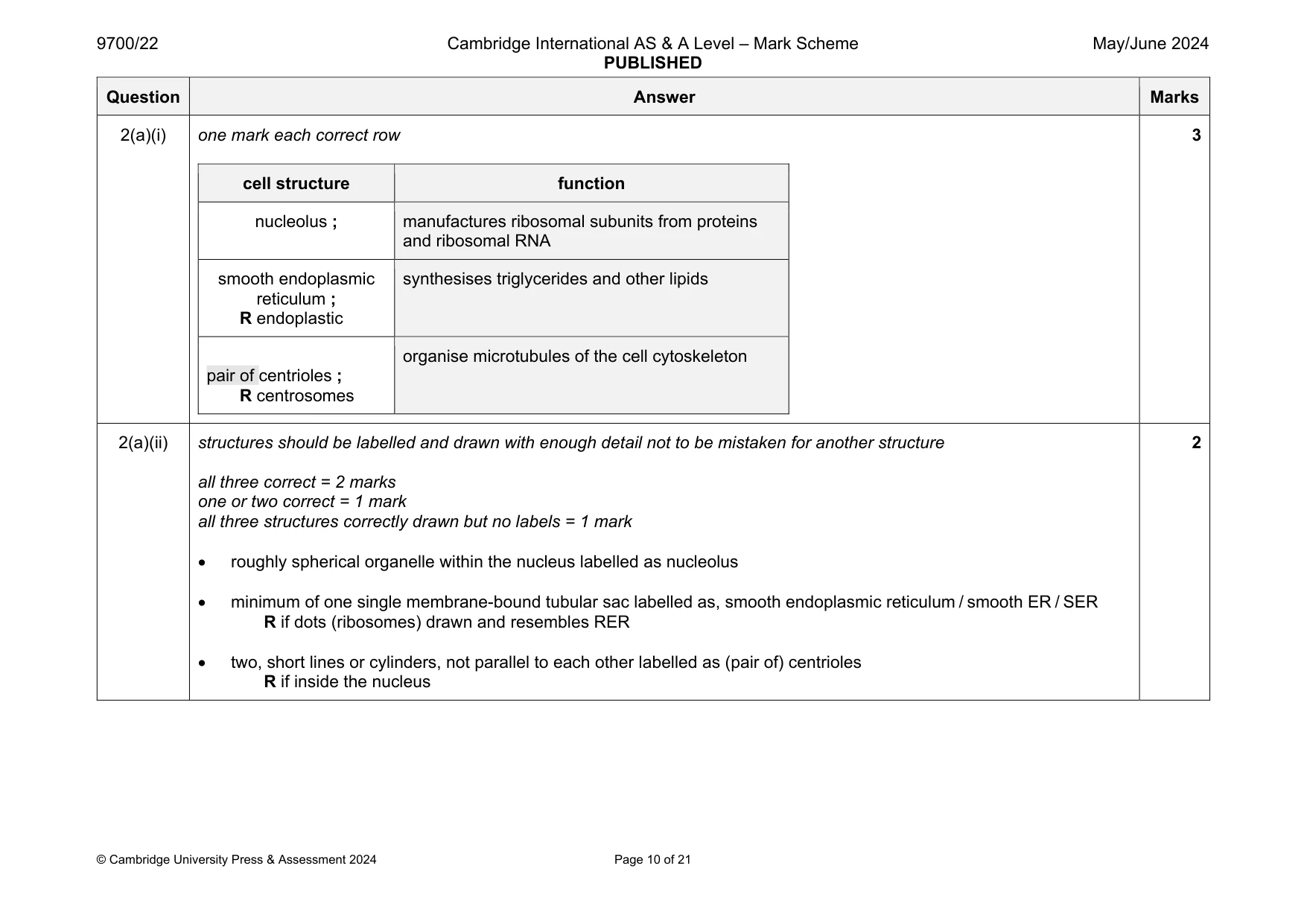
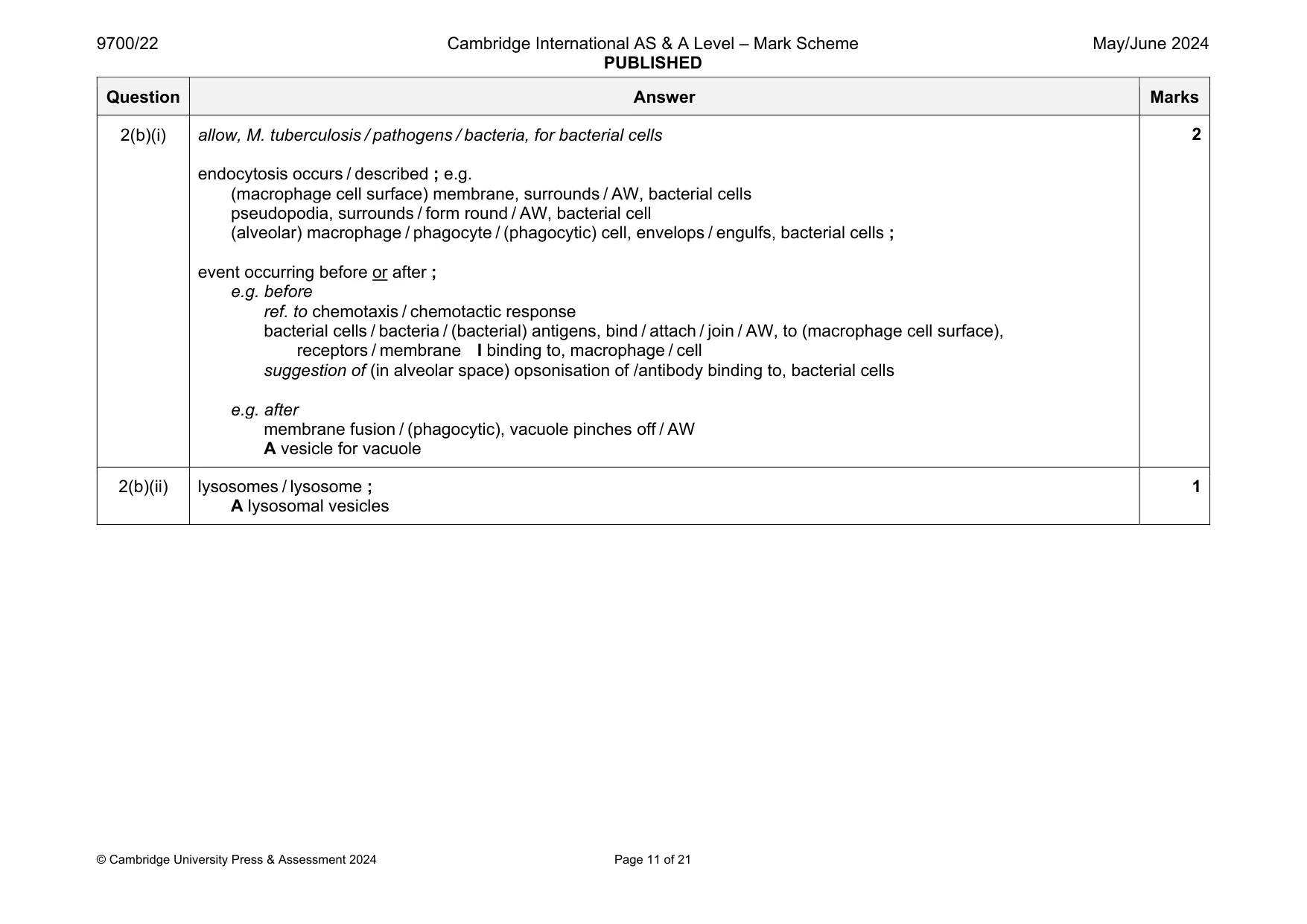
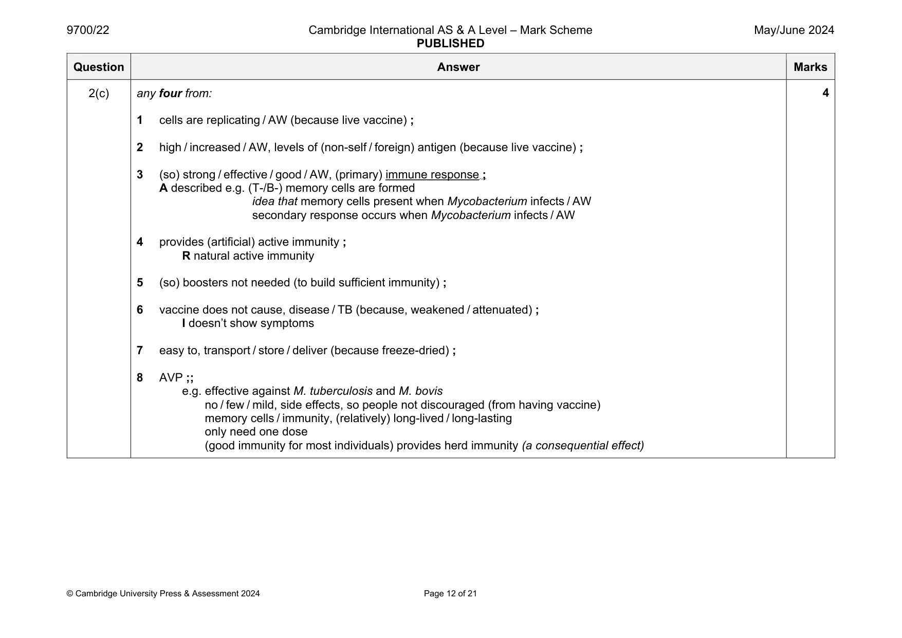
Mark scheme: 2(a)(i) one mark each correct row cell structure function nucleolus ; manufactures ribosomal subunits from proteins and ribosomal RNA smooth endoplasmic reticulum ; R endoplastic synthesises triglycerides and other lipids pair of centrioles ; R centrosomes organise microtubules of the cell cytoskeleton 3 2(a)(ii) structures should be labelled and drawn with enough detail not to be mistaken for another structure all three correct = 2 marks one or two correct = 1 mark all three structures correctly drawn but no labels = 1 mark roughly spherical organelle within the nucleus labelled as nucleolus minimum of one single membrane-bound tubular sac labelled as, smooth endoplasmic reticulum / smooth ER / SER R if dots (ribosomes) drawn and resembles RER two, short lines or cylinders, not parallel to each other labelled as (pair of) centrioles R if inside the nucleus 2 Question Answer Marks 2(b)(i) allow, M. tuberculosis / pathogens / bacteria, for bacterial cells endocytosis occurs / described ; e.g. (macrophage cell surface) membrane, surrounds / AW, bacterial cells pseudopodia, surrounds / form round / AW, bacterial cell (alveolar) macrophage / phagocyte / (phagocytic) cell, envelops / engulfs, bacterial cells ; event occurring before or after ; e.g. before ref. to chemotaxis / chemotactic response bacterial cells / bacteria / (bacterial) antigens, bind / attach / join / AW, to (macrophage cell surface), receptors / membrane I binding to, macrophage / cell suggestion of (in alveolar space) opsonisation of /antibody binding to, bacterial cells e.g. after membrane fusion / (phagocytic), vacuole pinches off / AW A vesicle for vacuole 2 2(b)(ii) lysosomes / lysosome ; A lysosomal vesicles 1 Question Answer Marks 2(c) any four from: 1 cells are replicating / AW (because live vaccine) ; 2 high / increased / AW, levels of (non-self / foreign) antigen (because live vaccine) ; 3 (so) strong / effective / good / AW, (primary) immune response ; A described e.g. (T-/B-) memory cells are formed idea that memory cells present when Mycobacterium infects / AW secondary response occurs when Mycobacterium infects / AW 4 provides (artificial) active immunity ; R natural active immunity 5 (so) boosters not needed (to build sufficient immunity) ; 6 vaccine does not cause, disease / TB (because, weakened / attenuated) ; I doesn’t show symptoms 7 easy to, transport / store / deliver (because freeze-dried) ; 8 AVP ;; e.g. effective against M. tuberculosis and M. bovis no / few / mild, side effects, so people not discouraged (from having vaccine) memory cells / immunity, (relatively) long-lived / long-lasting only need one dose (good immunity for most individuals) provides herd immunity (a consequential effect) 4
More questions on Cells as the basic units of living organisms
Q3 · Lysozyme is an antibacterial enzyme that was discovered in 1921 by Alexander Fleming, the…
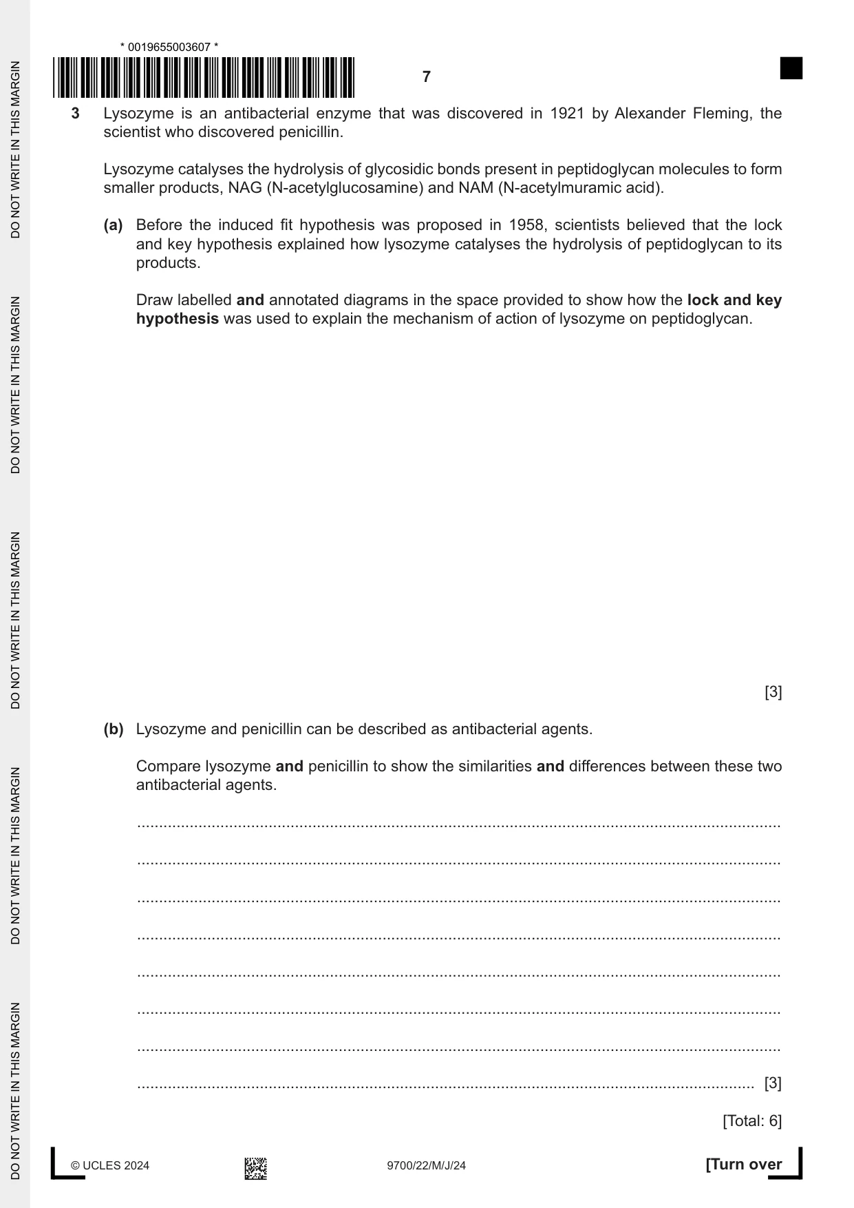
3 Lysozyme is an antibacterial enzyme that was discovered in 1921 by Alexander Fleming, the scientist who discovered penicillin. Lysozyme catalyses the hydrolysis of glycosidic bonds present in peptidoglycan molecules to form smaller products, NAG (N-acetylglucosamine) and NAM (N-acetylmuramic acid). (a) Before the induced fit hypothesis was proposed in 1958, scientists believed that the lock and key hypothesis explained how lysozyme catalyses the hydrolysis of peptidoglycan to its products. Draw labelled and annotated diagrams in the space provided to show how the lock and key hypothesis was used to explain the mechanism of action of lysozyme on peptidoglycan. [3] (b) Lysozyme and penicillin can be described as antibacterial agents. Compare lysozyme and penicillin to show the similarities and differences between these two antibacterial agents. ................................................................................................................................................... ................................................................................................................................................... ................................................................................................................................................... ................................................................................................................................................... ................................................................................................................................................... ................................................................................................................................................... ................................................................................................................................................... ............................................................................................................................................. [3] [Total: 6]
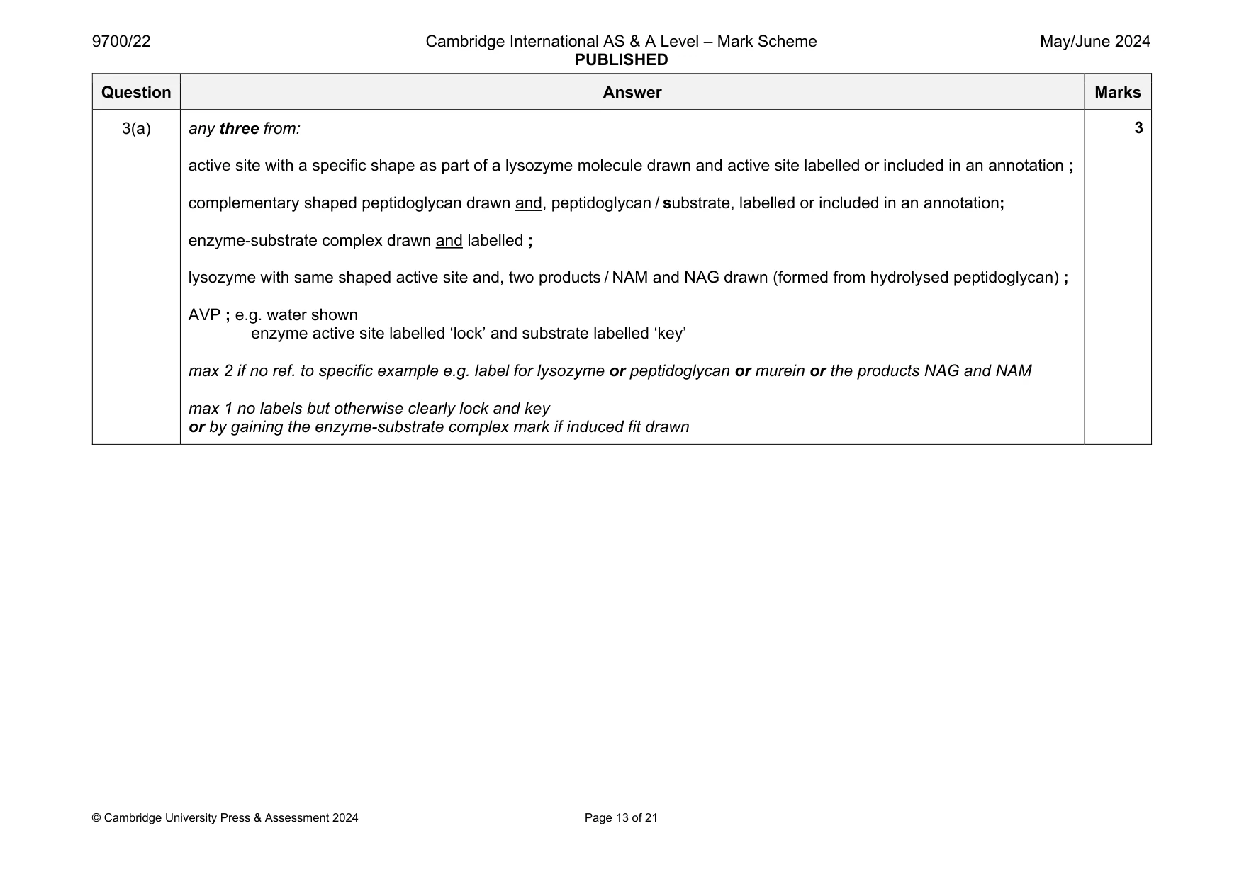
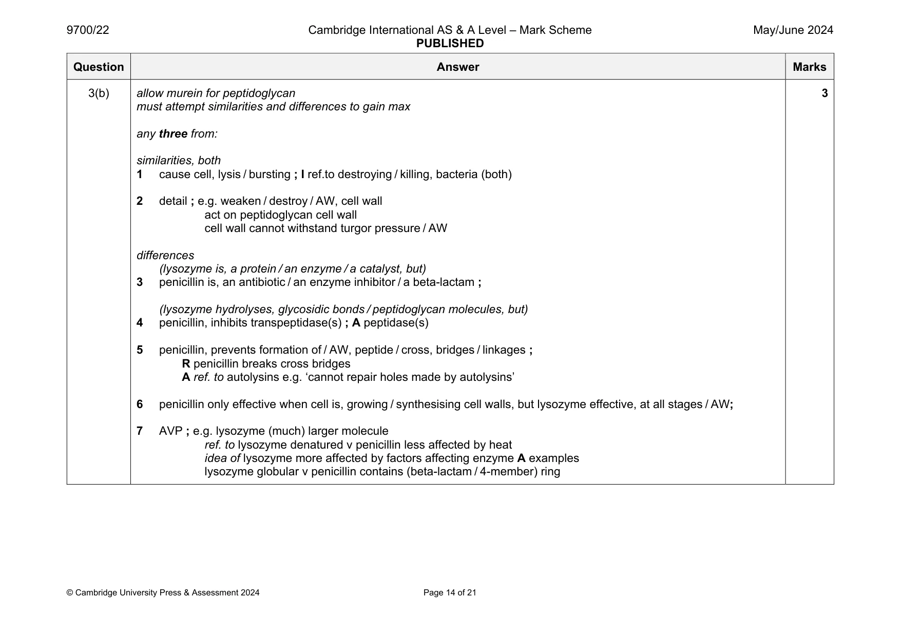
Mark scheme: 3(a) any three from: active site with a specific shape as part of a lysozyme molecule drawn and active site labelled or included in an annotation ; complementary shaped peptidoglycan drawn and, peptidoglycan / substrate, labelled or included in an annotation; enzyme-substrate complex drawn and labelled ; lysozyme with same shaped active site and, two products / NAM and NAG drawn (formed from hydrolysed peptidoglycan) ; AVP ; e.g. water shown enzyme active site labelled ‘lock’ and substrate labelled ‘key’ max 2 if no ref. to specific example e.g. label for lysozyme or peptidoglycan or murein or the products NAG and NAM max 1 no labels but otherwise clearly lock and key or by gaining the enzyme-substrate complex mark if induced fit drawn Question Answer Marks 3(b) allow murein for peptidoglycan must attempt similarities and differences to gain max any three from: similarities, both 1 cause cell, lysis / bursting ; I ref.to destroying / killing, bacteria (both) 2 detail ; e.g. weaken / destroy / AW, cell wall act on peptidoglycan cell wall cell wall cannot withstand turgor pressure / AW differences (lysozyme is, a protein / an enzyme / a catalyst, but) 3 penicillin is, an antibiotic / an enzyme inhibitor / a beta-lactam ; (lysozyme hydrolyses, glycosidic bonds / peptidoglycan molecules, but) 4 penicillin, inhibits transpeptidase(s) ; A peptidase(s) 5 penicillin, prevents formation of / AW, peptide / cross, bridges / linkages ; R penicillin breaks cross bridges A ref. to autolysins e.g. ‘cannot repair holes made by autolysins’ 6 penicillin only effective when cell is, growing / synthesising cell walls, but lysozyme effective, at all stages / AW; 7 AVP ; e.g. lysozyme (much) larger molecule ref. to lysozyme denatured v penicillin less affected by heat idea of lysozyme more affected by factors affecting enzyme A examples lysozyme globular v penicillin contains (beta-lactam / 4-member) ring 3
Q4 · In the mesophyll tissue of leaves, products of photosynthesis can be used to synthesise…
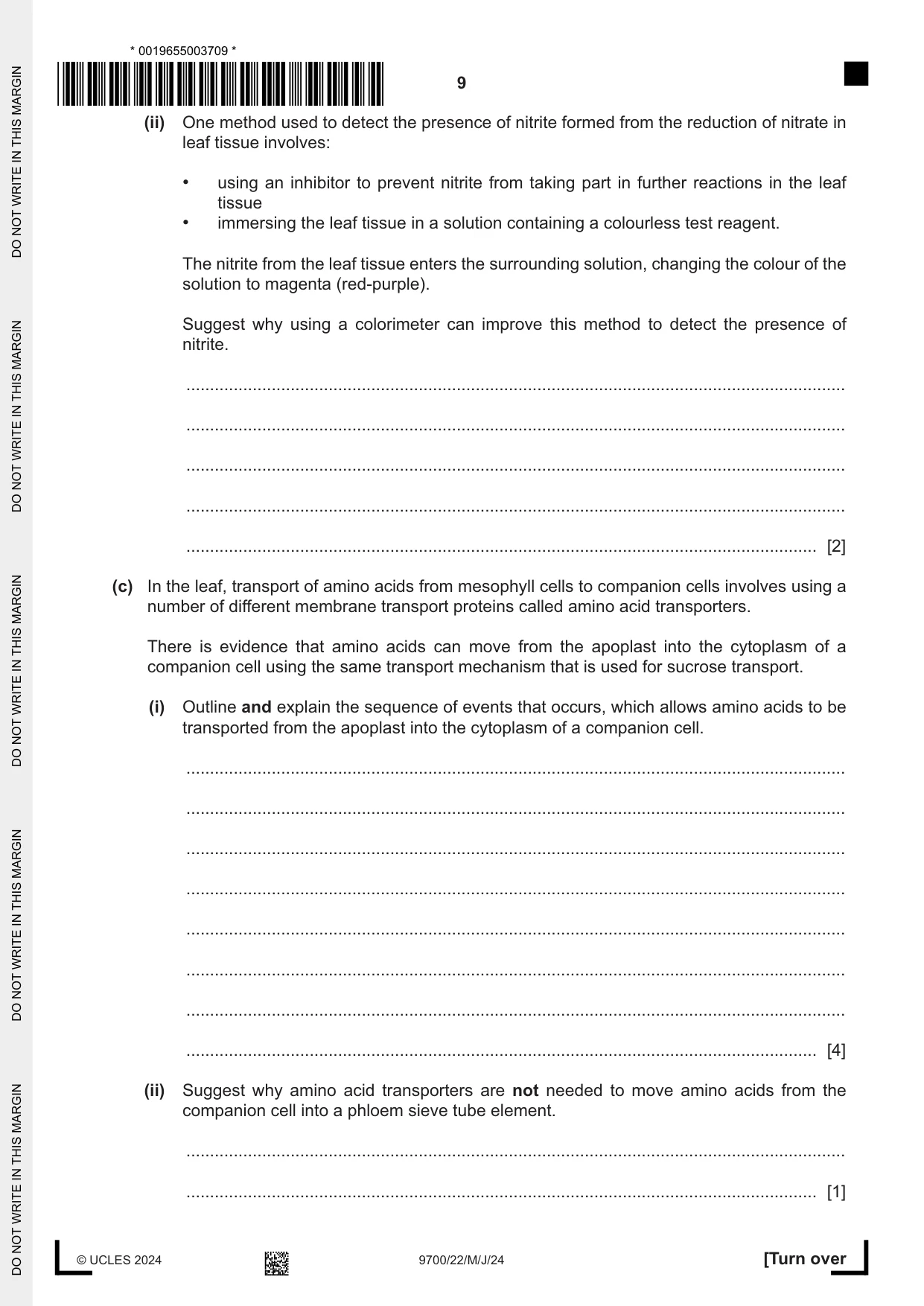
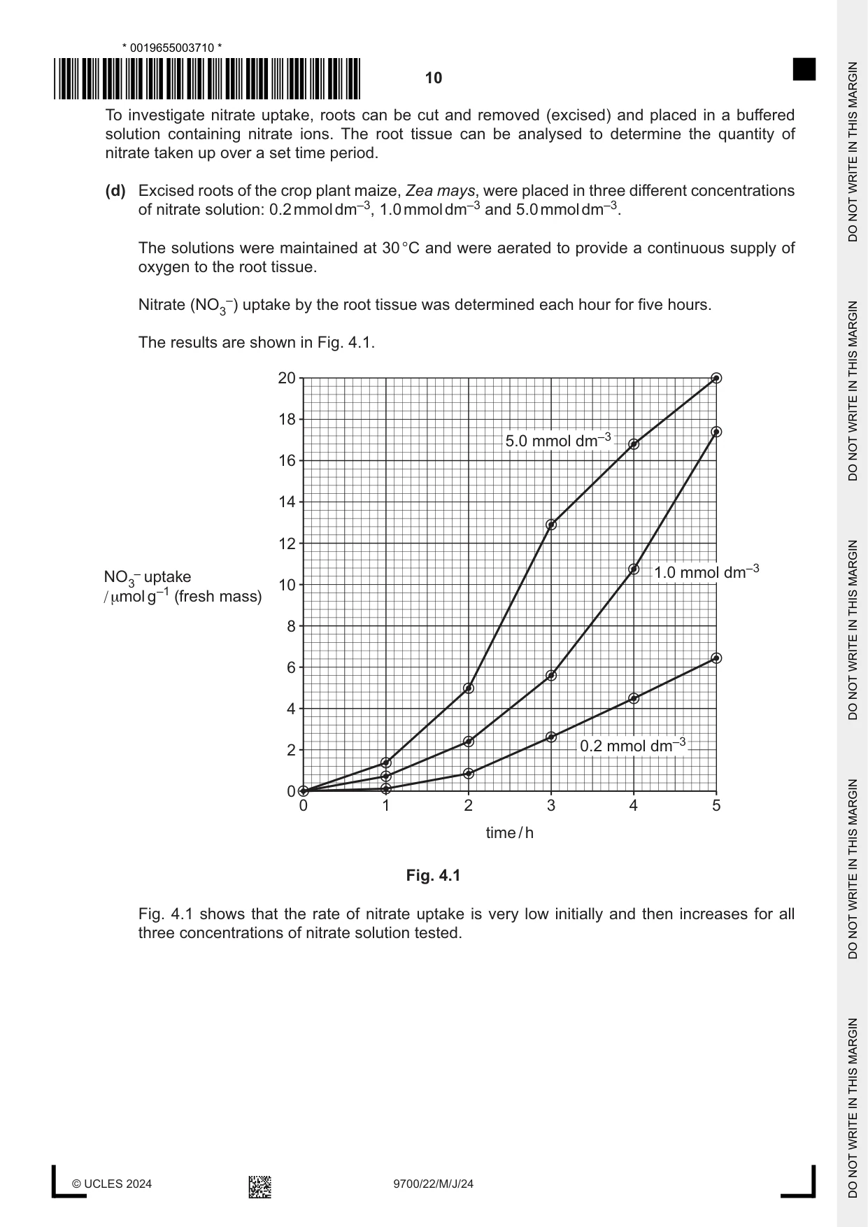
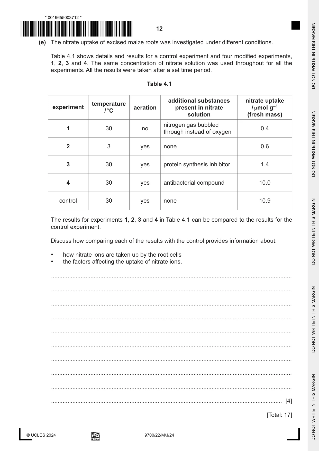
4 In the mesophyll tissue of leaves, products of photosynthesis can be used to synthesise organic compounds, such as the polysaccharide cellulose and some amino acids. A source of nitrogen for amino acid synthesis can be provided by nitrate ions that have been taken up in the roots and transported to the leaves. (a) Describe the structure of a cellulose molecule. ................................................................................................................................................... ................................................................................................................................................... ................................................................................................................................................... ................................................................................................................................................... ................................................................................................................................................... ................................................................................................................................................... ................................................................................................................................................... ............................................................................................................................................. [3] (b) Studies of nitrate uptake and nitrate metabolism help to provide information to scientists who are investigating ways to increase the yield of crop plants. The first step of nitrate metabolism in leaf cells is the reduction of nitrate to nitrite, catalysed by the enzyme nitrate reductase. The activity of the enzyme can be studied by detecting the presence of nitrite formed. (i) Researchers have found that adding nitrate to leaf tissue results in an increase in messenger RNA (mRNA) molecules of the gene NR, which codes for nitrate reductase. State one benefit to leaf cells of an increase in mRNA molecules of gene NR after the addition of nitrate. ........................................................................................................................................... ..................................................................................................................................... [1] (ii) One method used to detect the presence of nitrite formed from the reduction of nitrate in leaf tissue involves: • using an inhibitor to prevent nitrite from taking part in further reactions in the leaf tissue • immersing the leaf tissue in a solution containing a colourless test reagent. The nitrite from the leaf tissue enters the surrounding solution, changing the colour of the solution to magenta (red-purple). Suggest why using a colorimeter can improve this method to detect the presence of nitrite. ........................................................................................................................................... ........................................................................................................................................... ........................................................................................................................................... ........................................................................................................................................... ..................................................................................................................................... [2] (c) In the leaf, transport of amino acids from mesophyll cells to companion cells involves using a number of different membrane transport proteins called amino acid transporters. There is evidence that amino acids can move from the apoplast into the cytoplasm of a companion cell using the same transport mechanism that is used for sucrose transport. (i) Outline and explain the sequence of events that occurs, which allows amino acids to be transported from the apoplast into the cytoplasm of a companion cell. ........................................................................................................................................... ........................................................................................................................................... ........................................................................................................................................... ........................................................................................................................................... ........................................................................................................................................... ........................................................................................................................................... ........................................................................................................................................... ..................................................................................................................................... [4] (ii) Suggest why amino acid transporters are not needed to move amino acids from the companion cell into a phloem sieve tube element. ........................................................................................................................................... ..................................................................................................................................... [1] To investigate nitrate uptake, roots can be cut and removed (excised) and placed in a buffered solution containing nitrate ions. The root tissue can be analysed to determine the quantity of nitrate taken up over a set time period. (d) Excised roots of the crop plant maize, Zea mays, were placed in three different concentrations of nitrate solution: 0.2 mmol dm–3, 1.0 mmol dm–3 and 5.0 mmol dm–3. The solutions were maintained at 30 °C and were aerated to provide a continuous supply of oxygen to the root tissue. Nitrate (NO3–) uptake by the root tissue was determined each hour for five hours. The results are shown in Fig. 4.1. 20 18 5.0 mmol dm–3 16 14 12 – uptake 1.0 mmol dm–3NO3 10 / μmol g–1 (fresh mass) 8 6 4 2 0.2 mmol dm–3 0 0 1 2 3 4 5 time / h Fig. 4.1 Fig. 4.1 shows that the rate of nitrate uptake is very low initially and then increases for all three concentrations of nitrate solution tested. Describe the differences in the rates of nitrate uptake for 5.0 mmol dm–3 nitrate solution compared with 0.2 mmol dm–3 nitrate solution, between 2h and 5h ................................................................................................................................................... ................................................................................................................................................... ................................................................................................................................................... ................................................................................................................................................... ................................................................................................................................................... ............................................................................................................................................. [2] (e) The nitrate uptake of excised maize roots was investigated under different conditions. Table 4.1 shows details and results for a control experiment and four modified experiments, 1, 2, 3 and 4. The same concentration of nitrate solution was used throughout for all the experiments. All the results were taken after a set time period. Table 4.1 additional substances nitrate uptake temperature experiment aeration present in nitrate / μmol g–1 / °C solution (fresh mass) nitrogen gas bubbled 1 30 no 0.4 through instead of oxygen 2 3 yes none 0.6 3 30 yes protein synthesis inhibitor 1.4 4 30 yes antibacterial compound 10.0 control 30 yes none 10.9 The results for experiments 1, 2, 3 and 4 in Table 4.1 can be compared to the results for the control experiment. Discuss how comparing each of the results with the control provides information about: • how nitrate ions are taken up by the root cells • the factors affecting the uptake of nitrate ions. ................................................................................................................................................... ................................................................................................................................................... ................................................................................................................................................... ................................................................................................................................................... ................................................................................................................................................... ................................................................................................................................................... ................................................................................................................................................... ................................................................................................................................................... ................................................................................................................................................... ............................................................................................................................................. [4] [Total: 17]
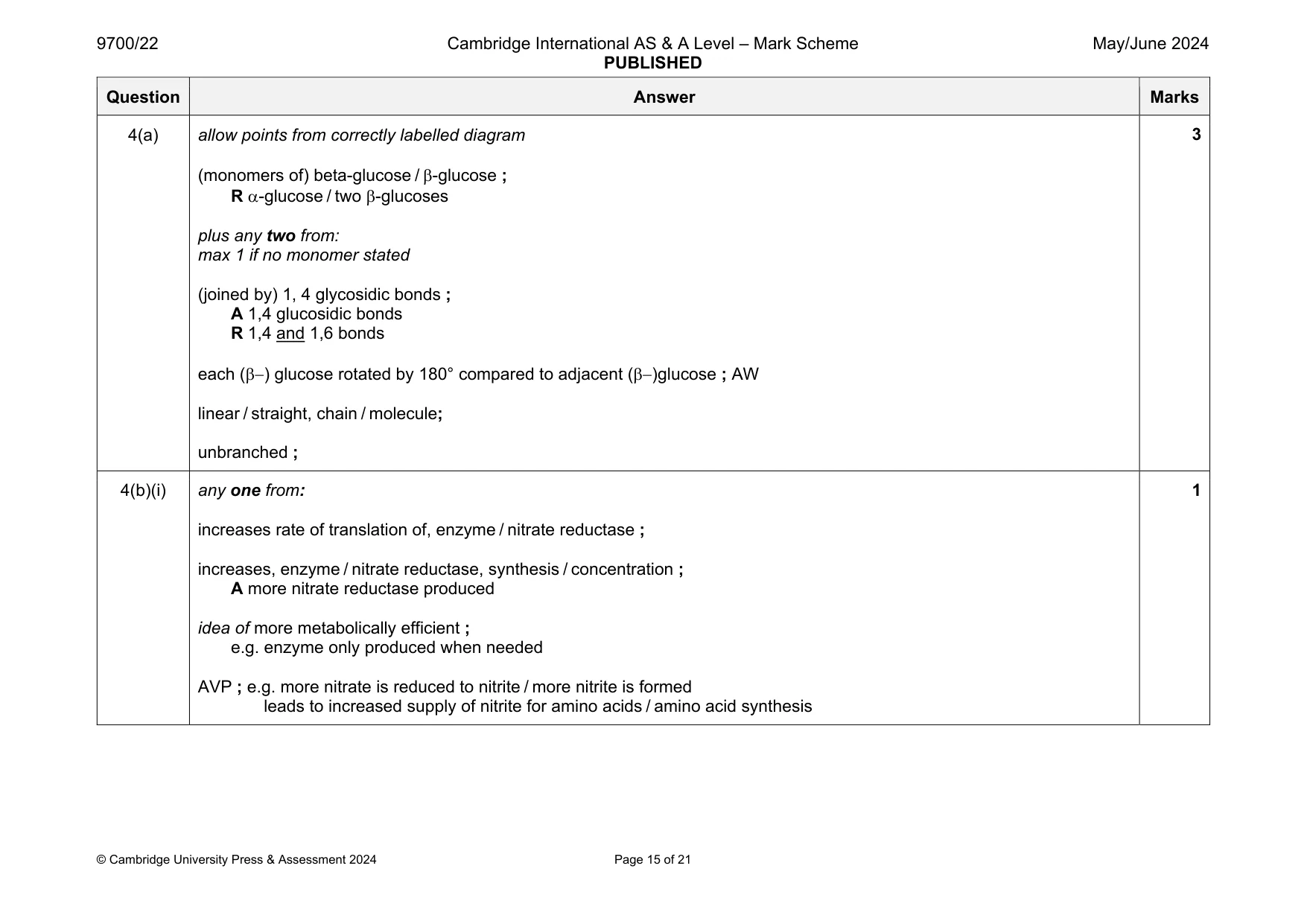
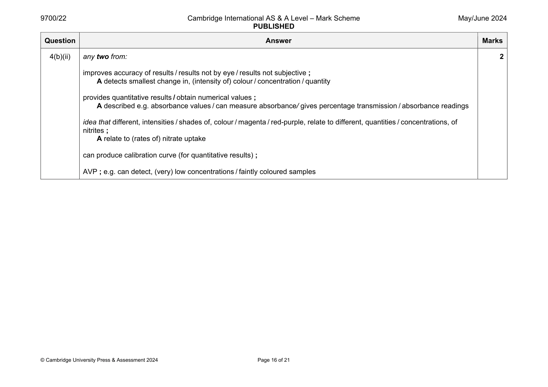
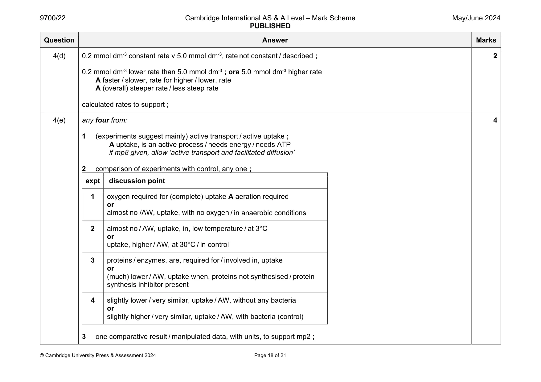
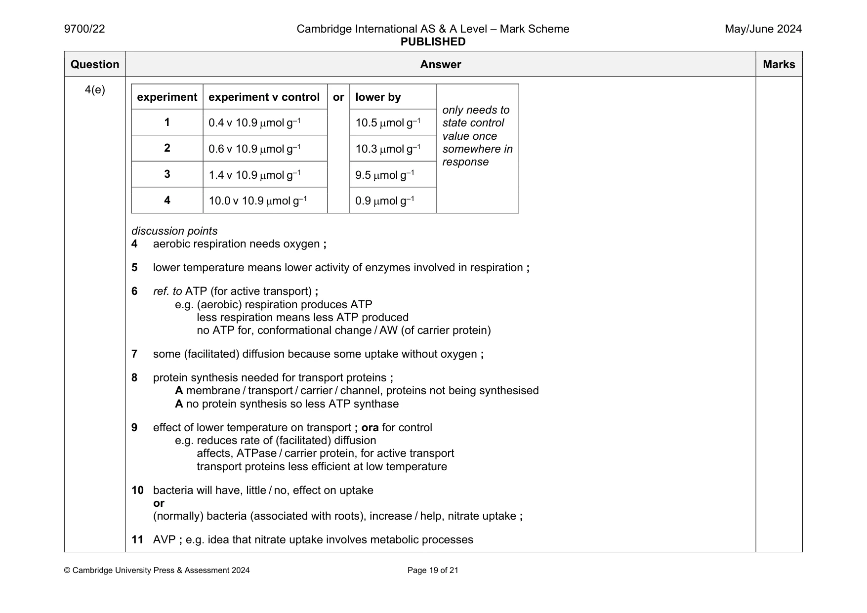
Mark scheme: 4(a) allow points from correctly labelled diagram (monomers of) beta-glucose / -glucose ; R -glucose / two -glucoses plus any two from: max 1 if no monomer stated (joined by) 1, 4 glycosidic bonds ; A 1,4 glucosidic bonds R 1,4 and 1,6 bonds each () glucose rotated by 180° compared to adjacent ()glucose ; AW linear / straight, chain / molecule; unbranched ; 4(b)(i) any one from: increases rate of translation of, enzyme / nitrate reductase ; increases, enzyme / nitrate reductase, synthesis / concentration ; A more nitrate reductase produced idea of more metabolically efficient ; e.g. enzyme only produced when needed AVP ; e.g. more nitrate is reduced to nitrite / more nitrite is formed leads to increased supply of nitrite for amino acids / amino acid synthesis 1 Question Answer Marks 4(b)(ii) any two from: improves accuracy of results / results not by eye / results not subjective ; A detects smallest change in, (intensity of) colour / concentration / quantity provides quantitative results / obtain numerical values ; A described e.g. absorbance values / can measure absorbance/ gives percentage transmission / absorbance readings idea that different, intensities / shades of, colour / magenta / red-purple, relate to different, quantities / concentrations, of nitrites ; A relate to (rates of) nitrate uptake can produce calibration curve (for quantitative results) ; AVP ; e.g. can detect, (very) low concentrations / faintly coloured samples 2 Question Answer Marks 4(c)(i) accept hydrogen ions / H+, for protons max 3 if no ref. to amino acids any four from: 1 protons, moved by active transport / pumped, out of (companion) cell ; A using, energy / ATP for active transport 2 protons, moved into / enter / AW, cell wall / apoplast ; R if moved out to phloem sieve tube 3 proton gradient builds up in, apoplast / cell wall ; A higher concentration A high concentration if mp4 gained 4 protons move back (into companion cell), by facilitated diffusion / AW ; e.g. down, electrochemical / concentration, gradient from high(er) to low(er) concentration A diffuse if movement is described through a transport protein in mp5 5 protons cotransport amino acids or amino acids move with protons through, a cotransporter / a cotransport protein / (amino acid) transporter ; 6 amino acids, transported / AW, against their concentration gradient ; 4 4(c)(ii) plasmodesmata present ; A movement is down the, concentration / diffusion, gradient A because there is a high(er) concentration in the companion cell ora for phloem sieve tube (sap) 1 Question Answer Marks 4(d) 0.2 mmol dm-3 constant rate v 5.0 mmol dm-3, rate not constant / described ; 0.2 mmol dm-3 lower rate than 5.0 mmol dm-3 ; ora 5.0 mmol dm-3 higher rate A faster / slower, rate for higher / lower, rate A (overall) steeper rate / less steep rate calculated rates to support ; 2 4(e) any four from: 1 (experiments suggest mainly) active transport / active uptake ; A uptake, is an active process / needs energy / needs ATP if mp8 given, allow ‘active transport and facilitated diffusion’ 2 comparison of experiments with control, any one ; expt discussion point 1 oxygen required for (complete) uptake A aeration required or almost no /AW, uptake, with no oxygen / in anaerobic conditions 2 almost no / AW, uptake, in, low temperature / at 3°C or uptake, higher / AW, at 30°C / in control 3 proteins / enzymes, are, required for / involved in, uptake or (much) lower / AW, uptake when, proteins not synthesised / protein synthesis inhibitor present 4 slightly lower / very similar, uptake / AW, without any bacteria or slightly higher / very similar, uptake / AW, with bacteria (control) 3 one comparative result / manipulated data, with units, to support mp2 ; 4 Question Answer Marks 4(e) experiment experiment v control or lower by only needs to state control value once somewhere in response 1 0.4 v 10.9 mol g–1 10.5 mol g–1 2 0.6 v 10.9 mol g–1 10.3 mol g–1 3 1.4 v 10.9 mol g–1 9.5 mol g–1 4 10.0 v 10.9 mol g–1 0.9 mol g–1 discussion points 4 aerobic respiration needs oxygen ; 5 lower temperature means lower activity of enzymes involved in respiration ; 6 ref. to ATP (for active transport) ; e.g. (aerobic) respiration produces ATP less respiration means less ATP produced no ATP for, conformational change / AW (of carrier protein) 7 some (facilitated) diffusion because some uptake without oxygen ; 8 protein synthesis needed for transport proteins ; A membrane / transport / carrier / channel, proteins not being synthesised A no protein synthesis so less ATP synthase 9 effect of lower temperature on transport ; ora for control e.g. reduces rate of (facilitated) diffusion affects, ATPase / carrier protein, for active transport transport proteins less efficient at low temperature 10 bacteria will have, little / no, effect on uptake or (normally) bacteria (associated with roots), increase / help, nitrate uptake ; 11 AVP ; e.g. idea that nitrate uptake involves metabolic processes
Q5 · During interphase of the cell cycle, individual chromosomes cannot be seen within the…
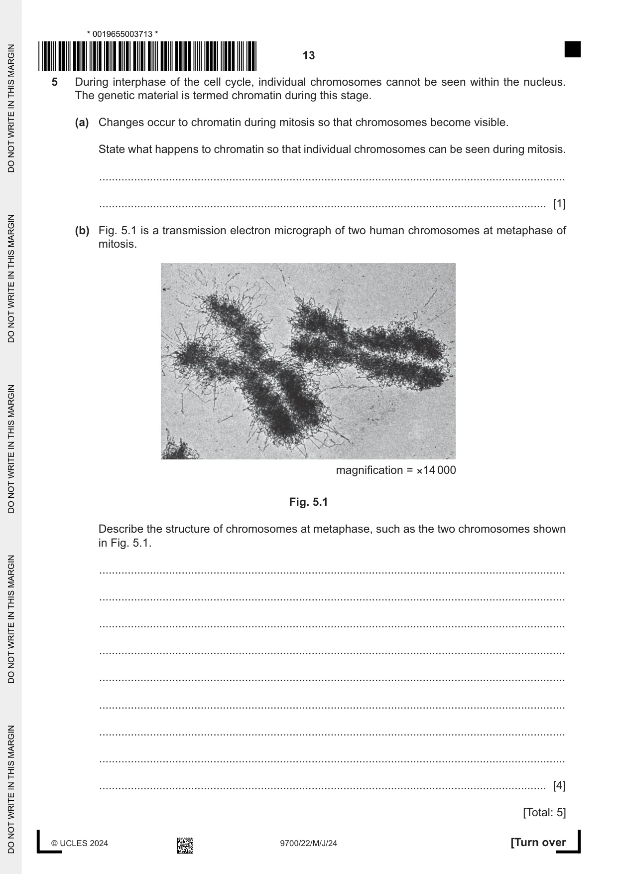
5 During interphase of the cell cycle, individual chromosomes cannot be seen within the nucleus. The genetic material is termed chromatin during this stage. (a) Changes occur to chromatin during mitosis so that chromosomes become visible. State what happens to chromatin so that individual chromosomes can be seen during mitosis. ................................................................................................................................................... ............................................................................................................................................. [1] (b) Fig. 5.1 is a transmission electron micrograph of two human chromosomes at metaphase of mitosis. magnification = ×14 000 Fig. 5.1 Describe the structure of chromosomes at metaphase, such as the two chromosomes shown in Fig. 5.1. ................................................................................................................................................... ................................................................................................................................................... ................................................................................................................................................... ................................................................................................................................................... ................................................................................................................................................... ................................................................................................................................................... ................................................................................................................................................... ................................................................................................................................................... ............................................................................................................................................. [4] [Total: 5]
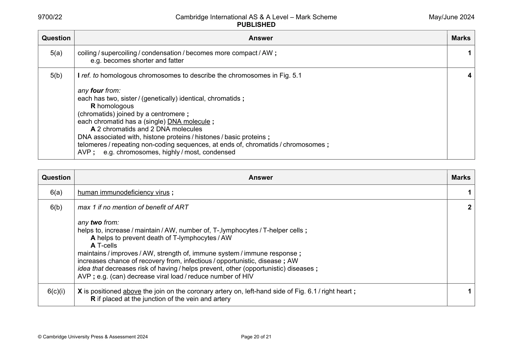
Mark scheme: 5(a) coiling / supercoiling / condensation / becomes more compact / AW ; e.g. becomes shorter and fatter 1 5(b) I ref. to homologous chromosomes to describe the chromosomes in Fig. 5.1 any four from: each has two, sister / (genetically) identical, chromatids ; R homologous (chromatids) joined by a centromere ; each chromatid has a (single) DNA molecule ; A 2 chromatids and 2 DNA molecules DNA associated with, histone proteins / histones / basic proteins ; telomeres / repeating non-coding sequences, at ends of, chromatids / chromosomes ; AVP ; e.g. chromosomes, highly / most, condensed 4
Q6 · Some people who are infected with HIV have HIV / AIDS
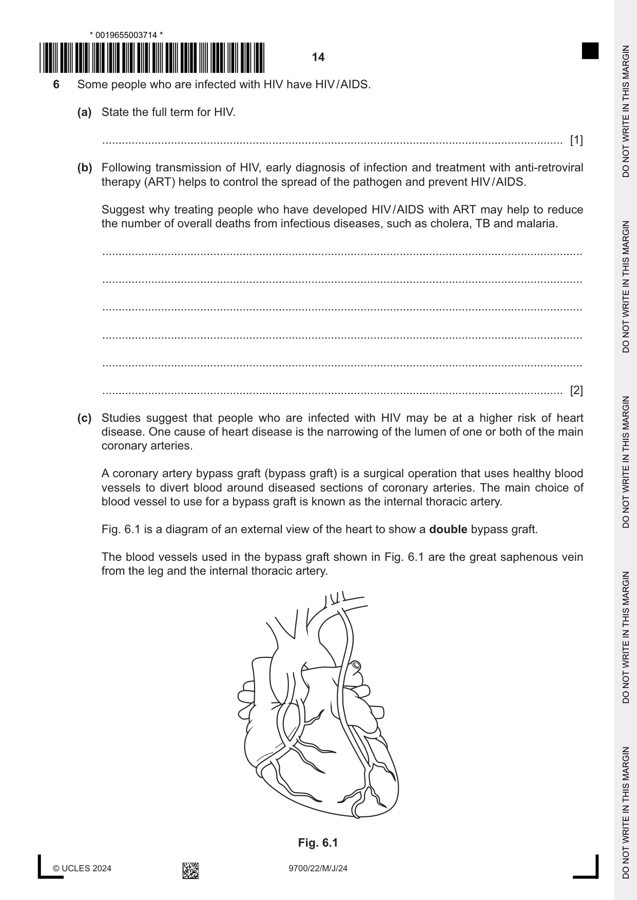
6 Some people who are infected with HIV have HIV / AIDS. (a) State the full term for HIV. ............................................................................................................................................. [1] (b) Following transmission of HIV, early diagnosis of infection and treatment with anti-retroviral therapy (ART) helps to control the spread of the pathogen and prevent HIV / AIDS. Suggest why treating people who have developed HIV / AIDS with ART may help to reduce the number of overall deaths from infectious diseases, such as cholera, TB and malaria. ................................................................................................................................................... ................................................................................................................................................... ................................................................................................................................................... ................................................................................................................................................... ................................................................................................................................................... ............................................................................................................................................. [2] (c) Studies suggest that people who are infected with HIV may be at a higher risk of heart disease. One cause of heart disease is the narrowing of the lumen of one or both of the main coronary arteries. A coronary artery bypass graft (bypass graft) is a surgical operation that uses healthy blood vessels to divert blood around diseased sections of coronary arteries. The main choice of blood vessel to use for a bypass graft is known as the internal thoracic artery. Fig. 6.1 is a diagram of an external view of the heart to show a double bypass graft. The blood vessels used in the bypass graft shown in Fig. 6.1 are the great saphenous vein from the leg and the internal thoracic artery. Fig. 6.1 (i) Draw a cross (X) on Fig. 6.1 to show an area of the right coronary artery that has been bypassed. [1] (ii) After surgery, the wall of the great saphenous vein becomes thicker. Suggest and explain why it is important for the wall of the vein to become thicker after surgery. ........................................................................................................................................... ........................................................................................................................................... ........................................................................................................................................... ........................................................................................................................................... ..................................................................................................................................... [2] [Total: 6]
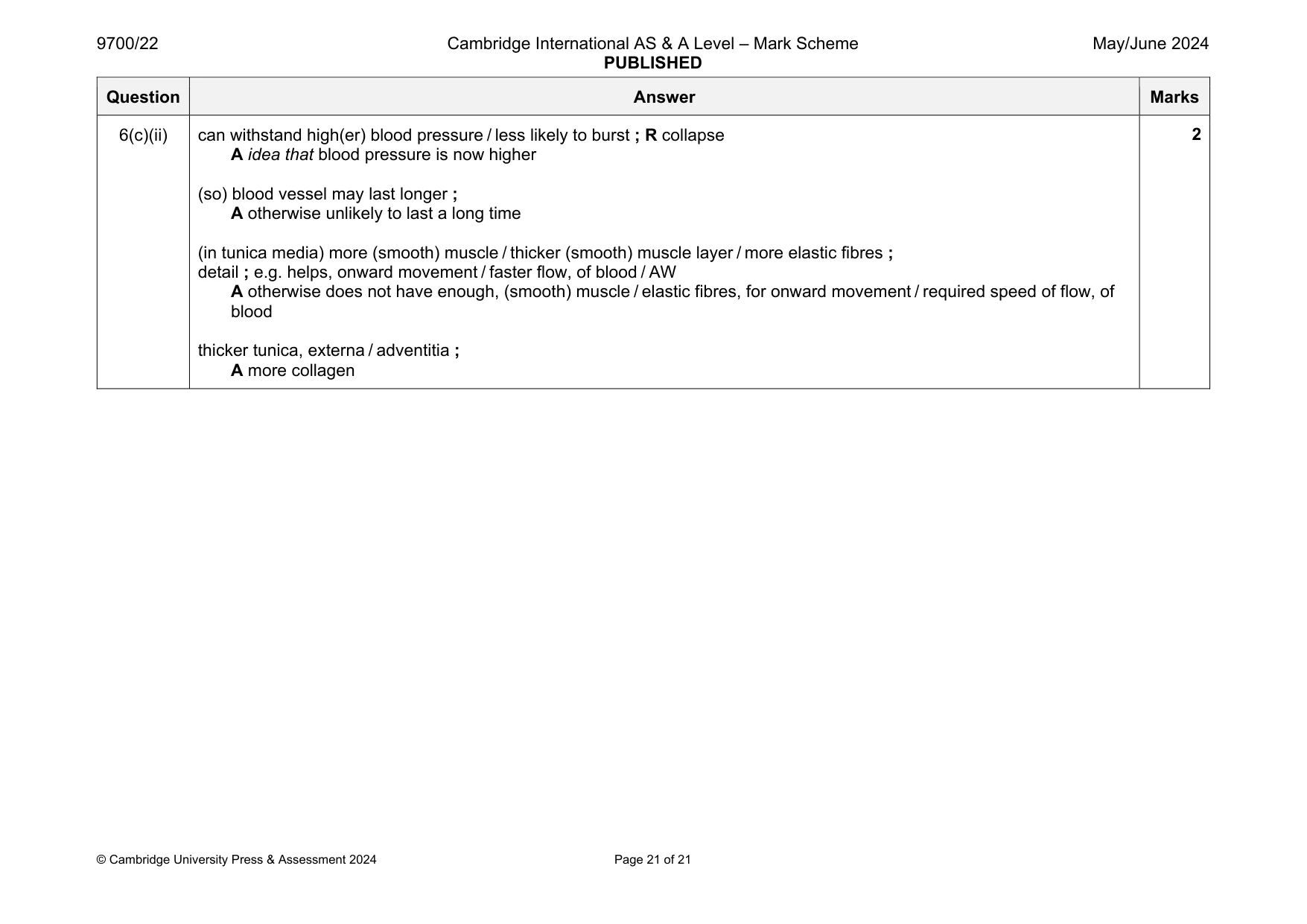
Mark scheme: 6(a) human immunodeficiency virus ; 1 6(b) max 1 if no mention of benefit of ART any two from: helps to, increase / maintain / AW, number of, T-,lymphocytes / T-helper cells ; A helps to prevent death of T-lymphocytes / AW A T-cells maintains / improves / AW, strength of, immune system / immune response ; increases chance of recovery from, infectious / opportunistic, disease ; AW idea that decreases risk of having / helps prevent, other (opportunistic) diseases ; AVP ; e.g. (can) decrease viral load / reduce number of HIV 2 6(c)(i) X is positioned above the join on the coronary artery on, left-hand side of Fig. 6.1 / right heart ; R if placed at the junction of the vein and artery 1 Question Answer Marks 6(c)(ii) can withstand high(er) blood pressure / less likely to burst ; R collapse A idea that blood pressure is now higher (so) blood vessel may last longer ; A otherwise unlikely to last a long time (in tunica media) more (smooth) muscle / thicker (smooth) muscle layer / more elastic fibres ; detail ; e.g. helps, onward movement / faster flow, of blood / AW A otherwise does not have enough, (smooth) muscle / elastic fibres, for onward movement / required speed of flow, of blood thicker tunica, externa / adventitia ; A more collagen 2
What was in this paper
The subtopics covered by these 6 questions, and how many questions each got. Open one in a new tab to see every Cambridge question on it.
What you needed in this session
Cambridge’s own grade thresholds for 2024 May/June, Paper 2 · Variant 2. A higher threshold means an easier paper — the bar moves with how the cohort did.