Cambridge A Level Biology 9700 — 2025 Feb/March Paper 2 · Variant 2
9700/22/F/M/25 · 6 questions · 60 marks · 75 min
The question paper and its mark scheme, free to read here and free to download. This is Cambridge’s own paper, exactly as it was sat.
Question paper24 pages

Mark scheme11 pages
Answers below. Sit the paper first if you are practising.
Questions as text
Q1 · Smilax china is a herbaceous plant
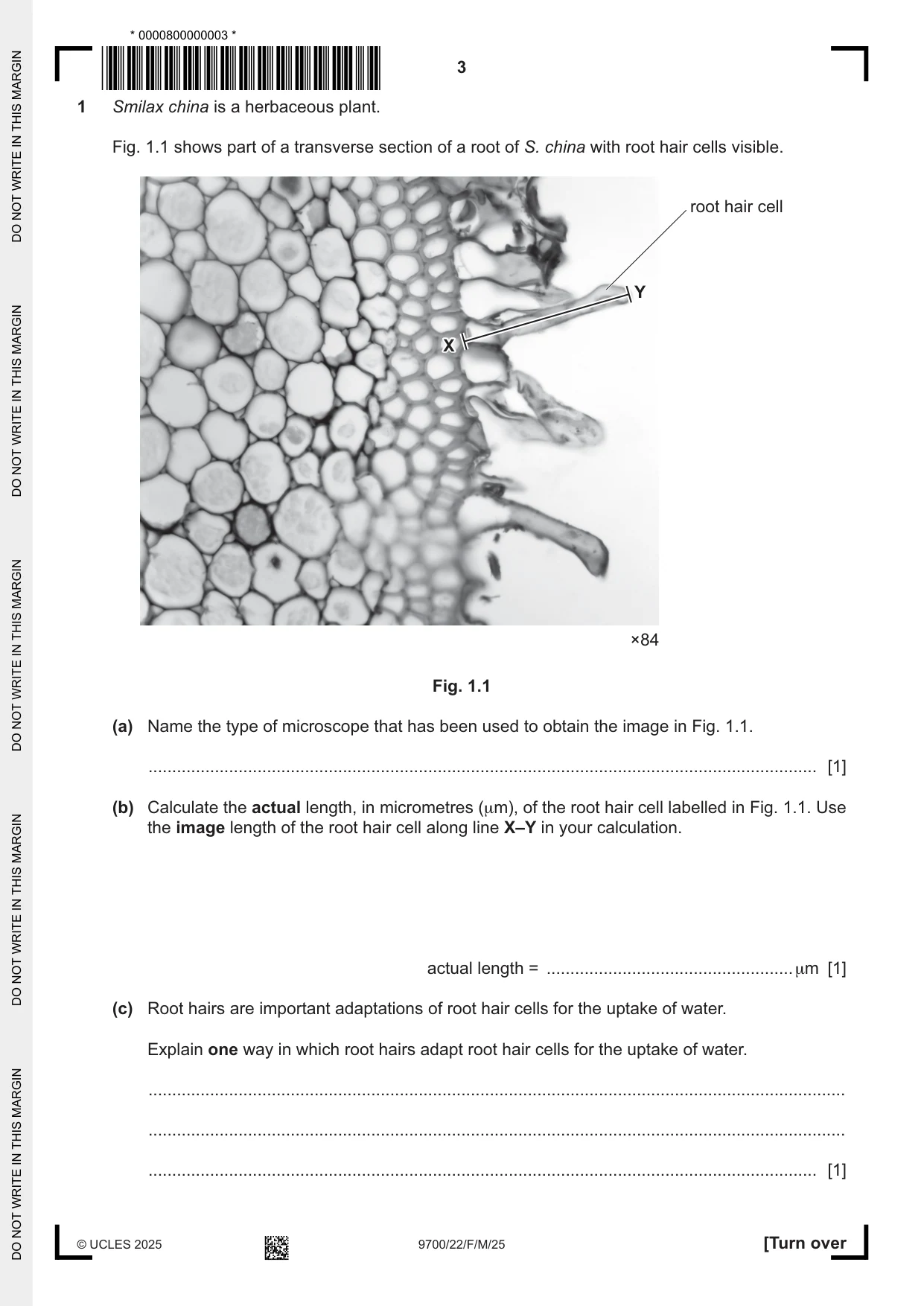
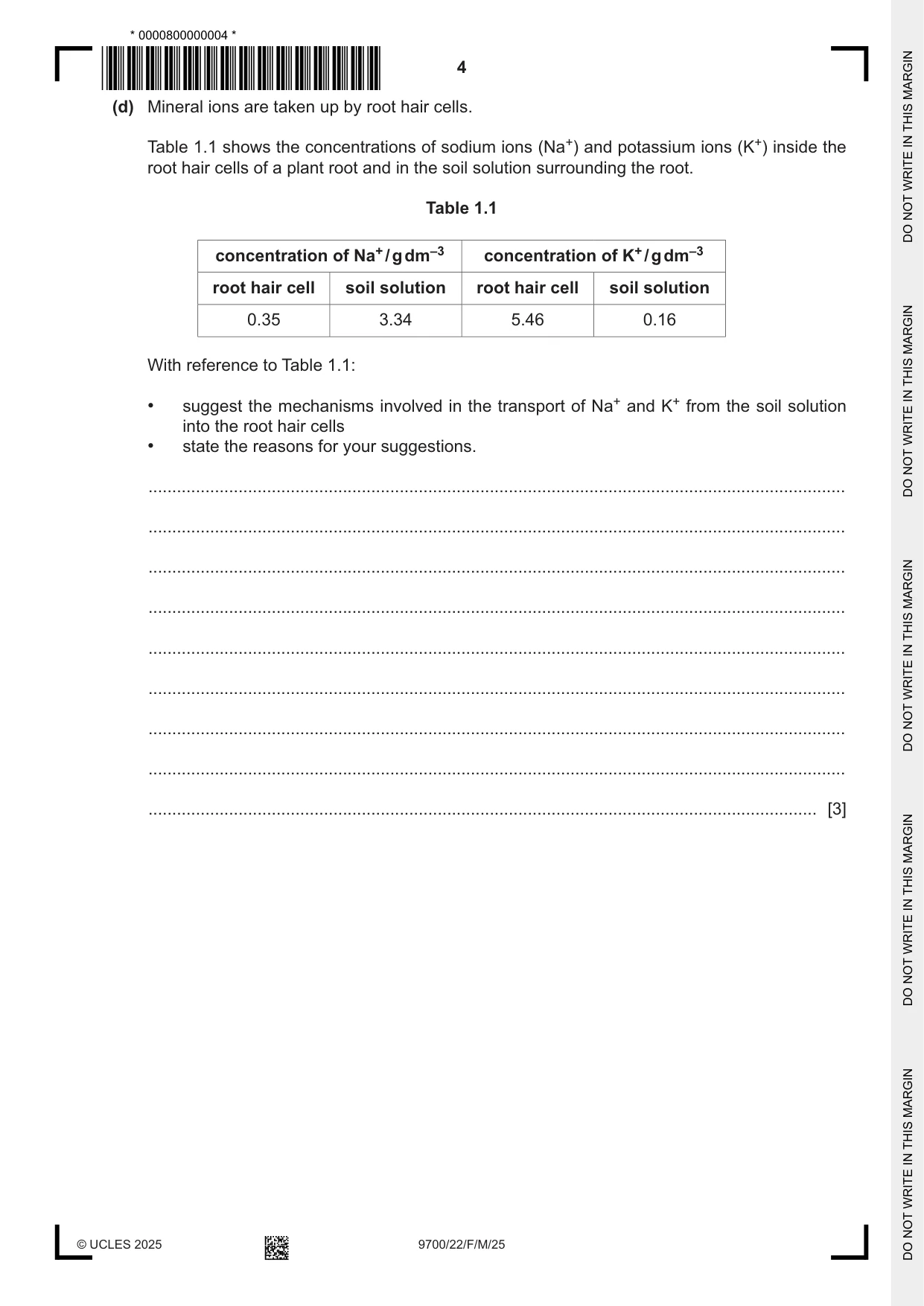
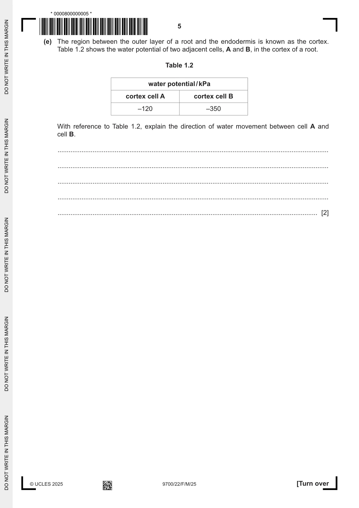
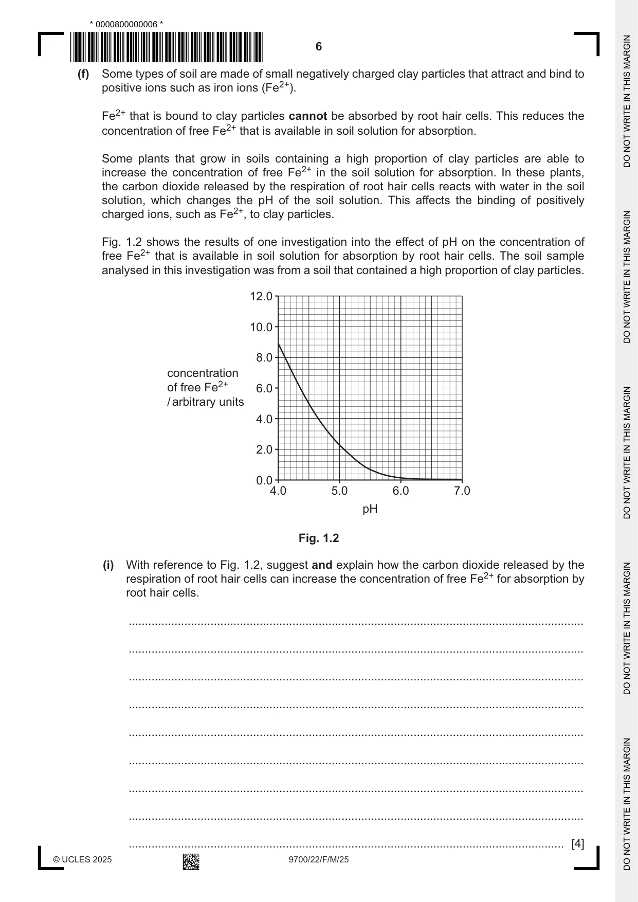
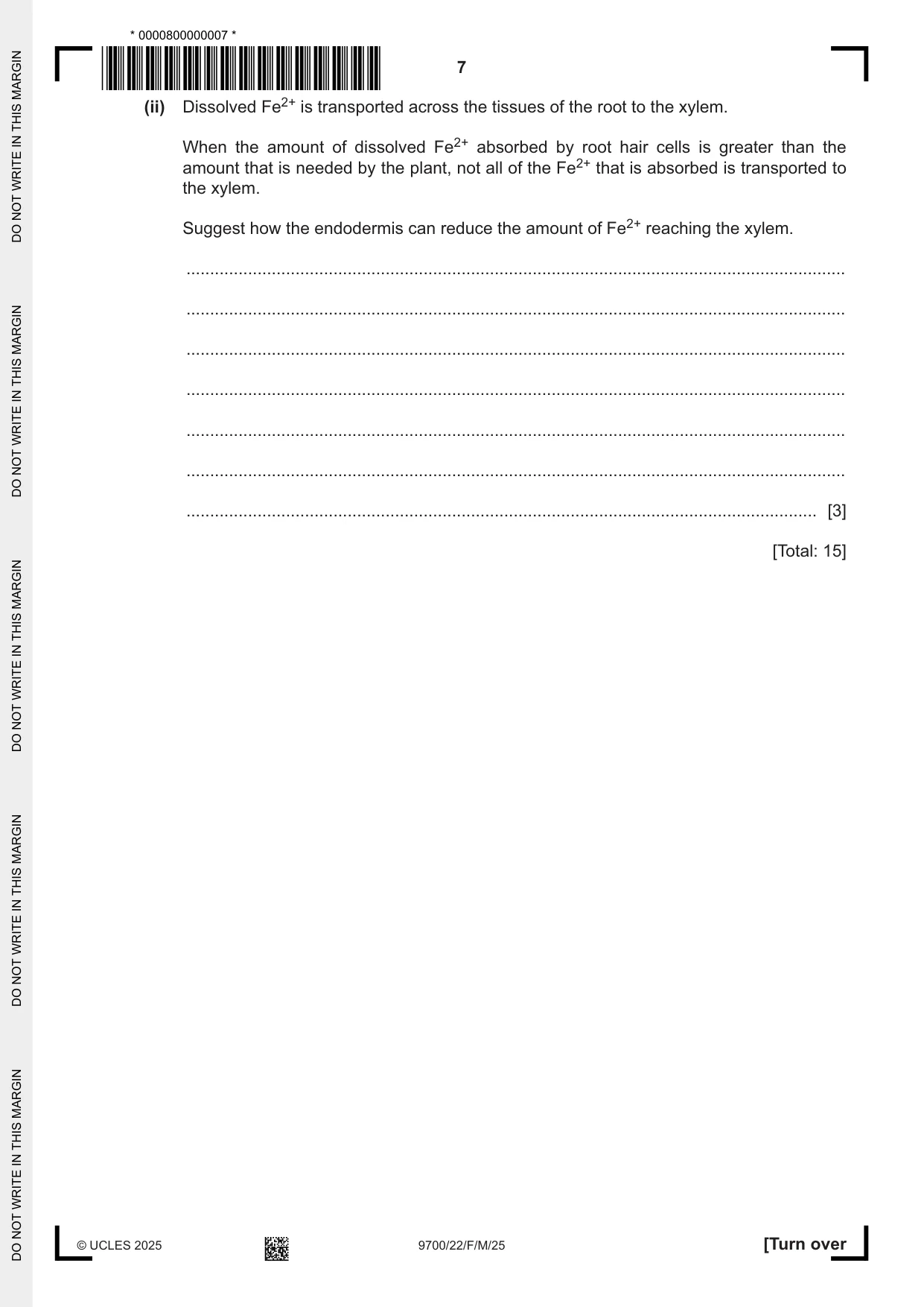
1 Smilax china is a herbaceous plant. Fig. 1.1 shows part of a transverse section of a root of S. china with root hair cells visible. root hair cell Y X ×84 Fig. 1.1 (a) Name the type of microscope that has been used to obtain the image in Fig. 1.1. ............................................................................................................................................. [1] (b) Calculate the actual length, in micrometres (μm), of the root hair cell labelled in Fig. 1.1. Use the image length of the root hair cell along line X–Y in your calculation. actual length = .................................................... μm [1] (c) Root hairs are important adaptations of root hair cells for the uptake of water. Explain one way in which root hairs adapt root hair cells for the uptake of water. ................................................................................................................................................... ................................................................................................................................................... ............................................................................................................................................. [1] (d) Mineral ions are taken up by root hair cells. Table 1.1 shows the concentrations of sodium ions (Na+) and potassium ions (K+) inside the root hair cells of a plant root and in the soil solution surrounding the root. Table 1.1 concentration of Na+ / g dm–3 concentration of K+ / g dm–3 root hair cell soil solution root hair cell soil solution 0.35 3.34 5.46 0.16 With reference to Table 1.1: • suggest the mechanisms involved in the transport of Na+ and K+ from the soil solution into the root hair cells • state the reasons for your suggestions. ................................................................................................................................................... ................................................................................................................................................... ................................................................................................................................................... ................................................................................................................................................... ................................................................................................................................................... ................................................................................................................................................... ................................................................................................................................................... ................................................................................................................................................... ............................................................................................................................................. [3] (e) The region between the outer layer of a root and the endodermis is known as the cortex. Table 1.2 shows the water potential of two adjacent cells, A and B, in the cortex of a root. Table 1.2 water potential / kPa cortex cell A cortex cell B –120 –350 With reference to Table 1.2, explain the direction of water movement between cell A and cell B. ................................................................................................................................................... ................................................................................................................................................... ................................................................................................................................................... ................................................................................................................................................... ............................................................................................................................................. [2] (f) Some types of soil are made of small negatively charged clay particles that attract and bind to positive ions such as iron ions (Fe2+). Fe2+ that is bound to clay particles cannot be absorbed by root hair cells. This reduces the concentration of free Fe2+ that is available in soil solution for absorption. Some plants that grow in soils containing a high proportion of clay particles are able to increase the concentration of free Fe2+ in the soil solution for absorption. In these plants, the carbon dioxide released by the respiration of root hair cells reacts with water in the soil solution, which changes the pH of the soil solution. This affects the binding of positively charged ions, such as Fe2+, to clay particles. Fig. 1.2 shows the results of one investigation into the effect of pH on the concentration of free Fe2+ that is available in soil solution for absorption by root hair cells. The soil sample analysed in this investigation was from a soil that contained a high proportion of clay particles. 12.0 10.0 8.0 concentration of free Fe2+ 6.0 / arbitrary units 4.0 2.0 0.0 4.0 5.0 6.0 7.0 pH Fig. 1.2 (i) With reference to Fig. 1.2, suggest and explain how the carbon dioxide released by the respiration of root hair cells can increase the concentration of free Fe2+ for absorption by root hair cells. ........................................................................................................................................... ........................................................................................................................................... ........................................................................................................................................... ........................................................................................................................................... ........................................................................................................................................... ........................................................................................................................................... ........................................................................................................................................... ........................................................................................................................................... ..................................................................................................................................... [4] (ii) Dissolved Fe2+ is transported across the tissues of the root to the xylem. When the amount of dissolved Fe2+ absorbed by root hair cells is greater than the amount that is needed by the plant, not all of the Fe2+ that is absorbed is transported to the xylem. Suggest how the endodermis can reduce the amount of Fe2+ reaching the xylem. ........................................................................................................................................... ........................................................................................................................................... ........................................................................................................................................... ........................................................................................................................................... ........................................................................................................................................... ........................................................................................................................................... ..................................................................................................................................... [3] [Total: 15]
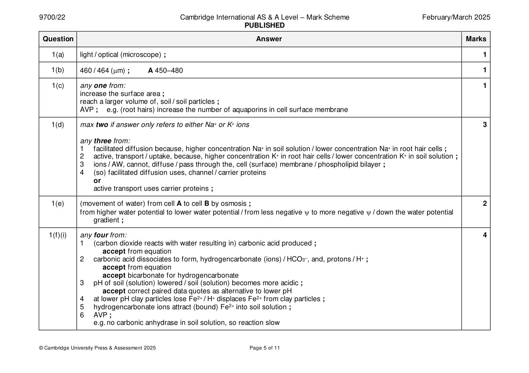
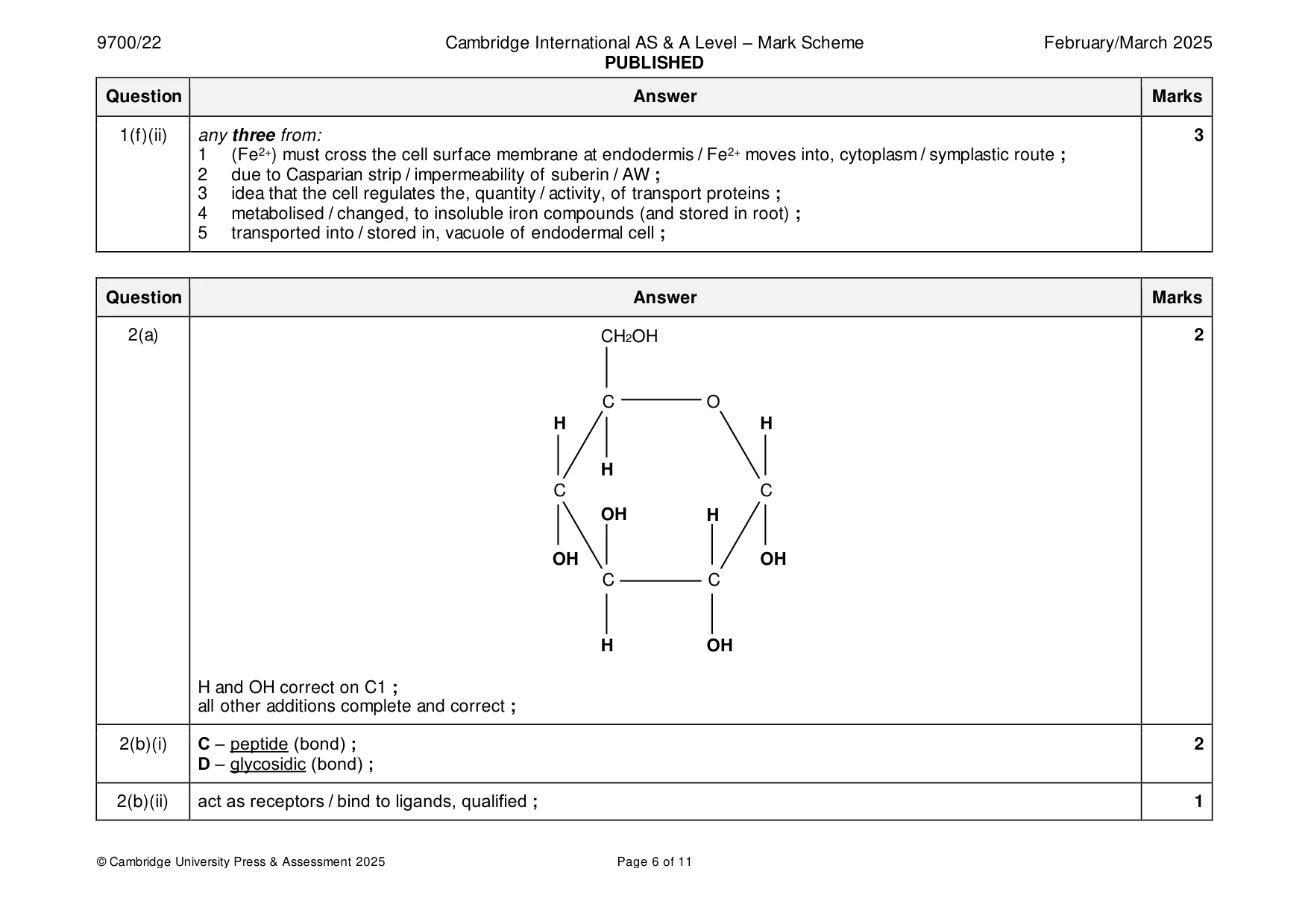
Mark scheme: Question Answer Marks 1(a) light / optical (microscope) ; 1 1(b) 460 / 464 (m) ; A 450–480 1 1(c) any one from: 1 increase the surface area ; reach a larger volume of, soil / soil particles ; AVP ; e.g. (root hairs) increase the number of aquaporins in cell surface membrane 1(d) max two if answer only refers to either Na+ or K+ ions 3 any three from: 1 facilitated diffusion because, higher concentration Na+ in soil solution / lower concentration Na+ in root hair cells ; 2 active, transport / uptake, because, higher concentration K+ in root hair cells / lower concentration K+ in soil solution ; 3 ions / AW, cannot, diffuse / pass through the, cell (surface) membrane / phospholipid bilayer ; 4 (so) facilitated diffusion uses, channel / carrier proteins or active transport uses carrier proteins ; 1(e) (movement of water) from cell A to cell B by osmosis ; 2 from higher water potential to lower water potential / from less negative to more negative / down the water potential gradient ; 1(f)(i) any four from: 4 1 (carbon dioxide reacts with water resulting in) carbonic acid produced ; accept from equation 2 carbonic acid dissociates to form, hydrogencarbonate (ions) / HCO3–, and, protons / H+ ; accept from equation accept bicarbonate for hydrogencarbonate 3 pH of soil (solution) lowered / soil (solution) becomes more acidic ; accept correct paired data quotes as alternative to lower pH 4 at lower pH clay particles lose Fe2+ / H+ displaces Fe2+ from clay particles ; 5 hydrogencarbonate ions attract (bound) Fe2+ into soil solution ; 6 AVP ; e.g. no carbonic anhydrase in soil solution, so reaction slow 1(f)(ii) any three from: 3 1 (Fe2+) must cross the cell surface membrane at endodermis / Fe2+ moves into, cytoplasm / symplastic route ; 2 due to Casparian strip / impermeability of suberin / AW ; 3 idea that the cell regulates the, quantity / activity, of transport proteins ; 4 metabolised / changed, to insoluble iron compounds (and stored in root) ; 5 transported into / stored in, vacuole of endodermal cell ;
Q2 · An incomplete diagram of the structure of an α‑glucose molecule
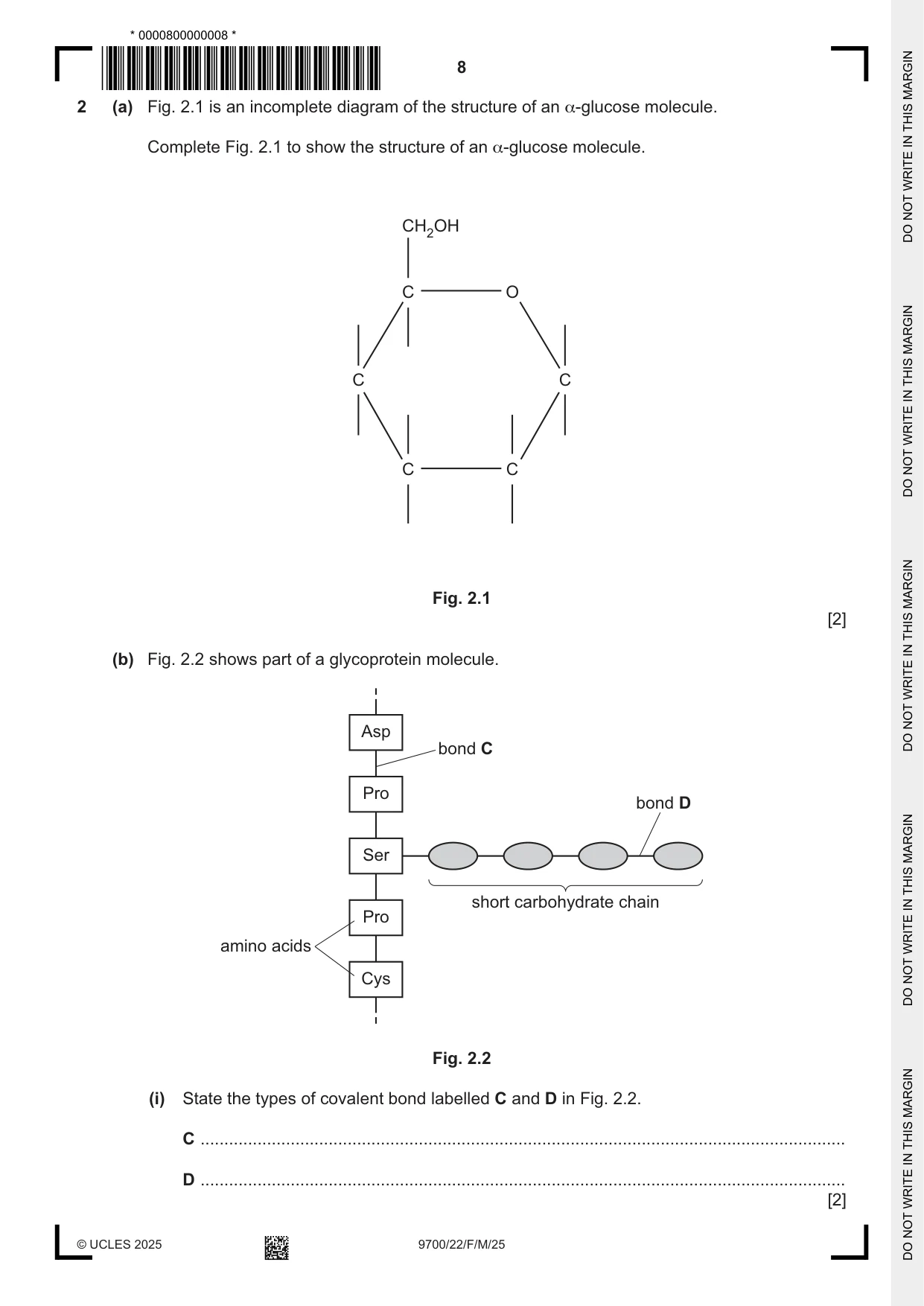
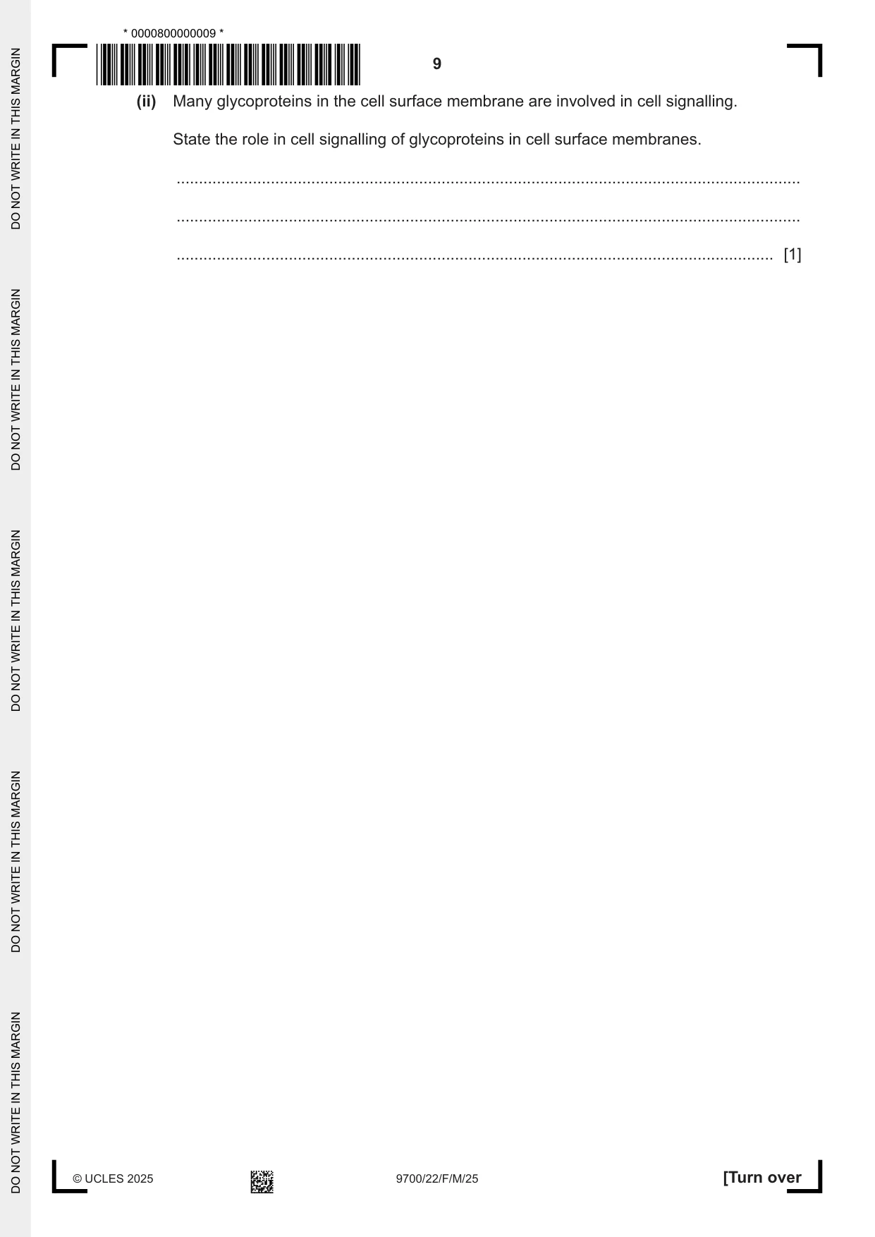
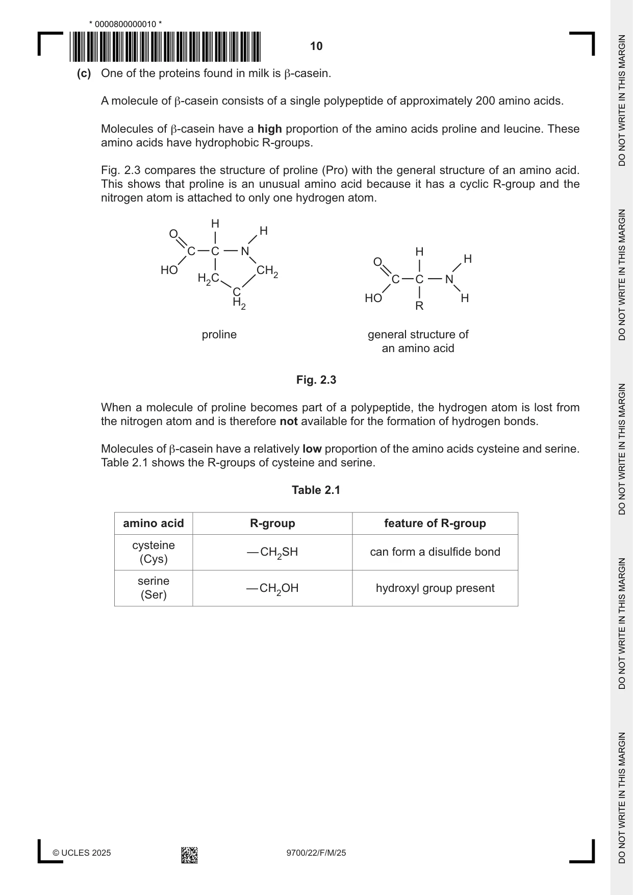
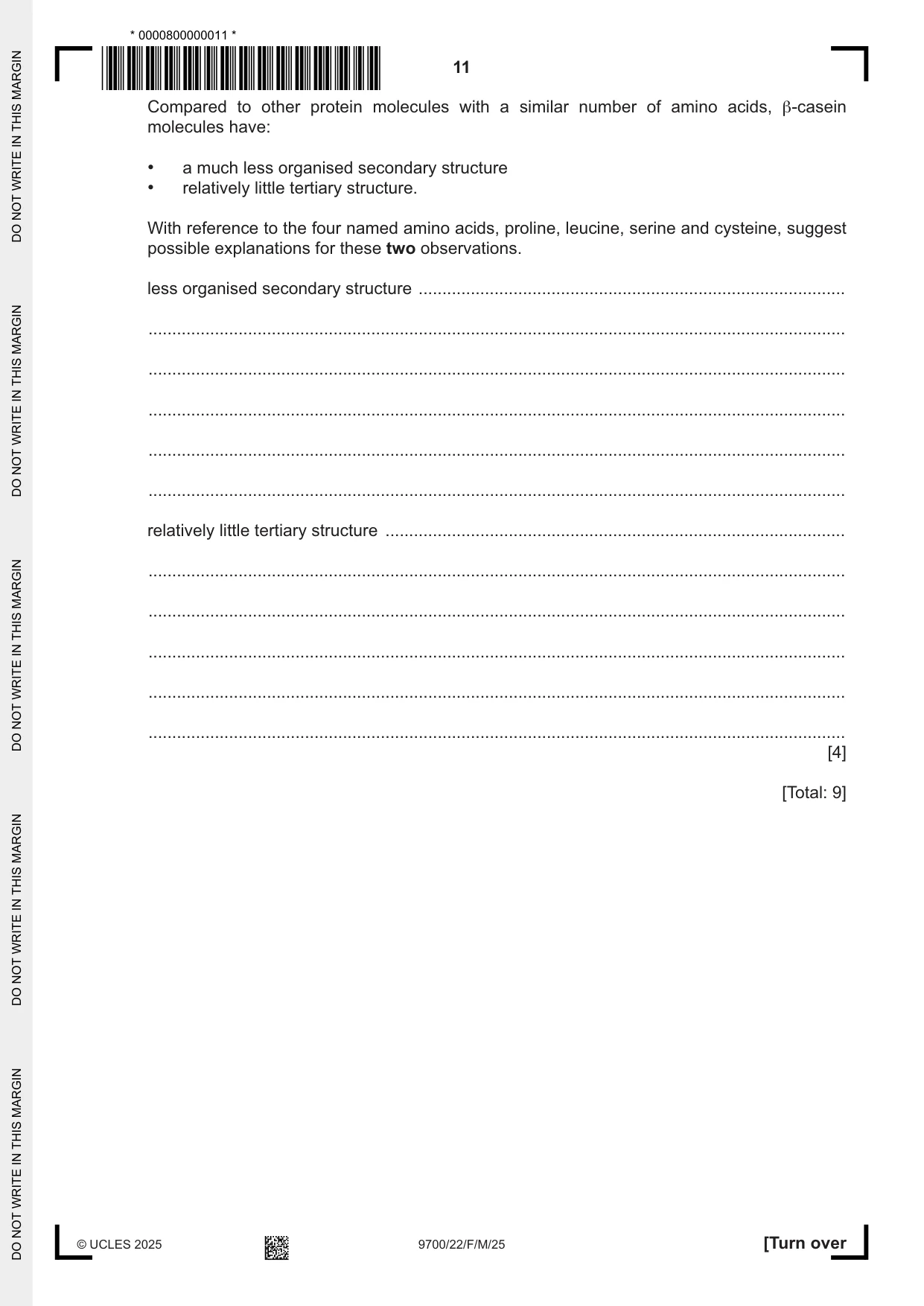
2 (a) Fig. 2.1 is an incomplete diagram of the structure of an α‑glucose molecule. Complete Fig. 2.1 to show the structure of an α‑glucose molecule. CH2OH C O C C C C Fig. 2.1 [2] (b) Fig. 2.2 shows part of a glycoprotein molecule. Asp bond C Pro bond D Ser short carbohydrate chain Pro amino acids Cys Fig. 2.2 (i) State the types of covalent bond labelled C and D in Fig. 2.2. C ........................................................................................................................................ D ........................................................................................................................................ [2] (ii) Many glycoproteins in the cell surface membrane are involved in cell signalling. State the role in cell signalling of glycoproteins in cell surface membranes. ........................................................................................................................................... ........................................................................................................................................... ..................................................................................................................................... [1] (c) One of the proteins found in milk is β‑casein. A molecule of β‑casein consists of a single polypeptide of approximately 200 amino acids. Molecules of β‑casein have a high proportion of the amino acids proline and leucine. These amino acids have hydrophobic R‑groups. Fig. 2.3 compares the structure of proline (Pro) with the general structure of an amino acid. This shows that proline is an unusual amino acid because it has a cyclic R‑group and the nitrogen atom is attached to only one hydrogen atom. H H O C C N H H O HO CH2 H2C C C N C HO H H2 R proline general structure of an amino acid Fig. 2.3 When a molecule of proline becomes part of a polypeptide, the hydrogen atom is lost from the nitrogen atom and is therefore not available for the formation of hydrogen bonds. Molecules of β‑casein have a relatively low proportion of the amino acids cysteine and serine. Table 2.1 shows the R‑groups of cysteine and serine. Table 2.1 amino acid R‑group feature of R‑group cysteine — CH2SH can form a disulfide bond (Cys) serine — CH2OH hydroxyl group present (Ser) Compared to other protein molecules with a similar number of amino acids, β‑casein molecules have: • a much less organised secondary structure • relatively little tertiary structure. With reference to the four named amino acids, proline, leucine, serine and cysteine, suggest possible explanations for these two observations. less organised secondary structure .......................................................................................... ................................................................................................................................................... ................................................................................................................................................... ................................................................................................................................................... ................................................................................................................................................... ................................................................................................................................................... relatively little tertiary structure ................................................................................................. ................................................................................................................................................... ................................................................................................................................................... ................................................................................................................................................... ................................................................................................................................................... ................................................................................................................................................... [4] [Total: 9]
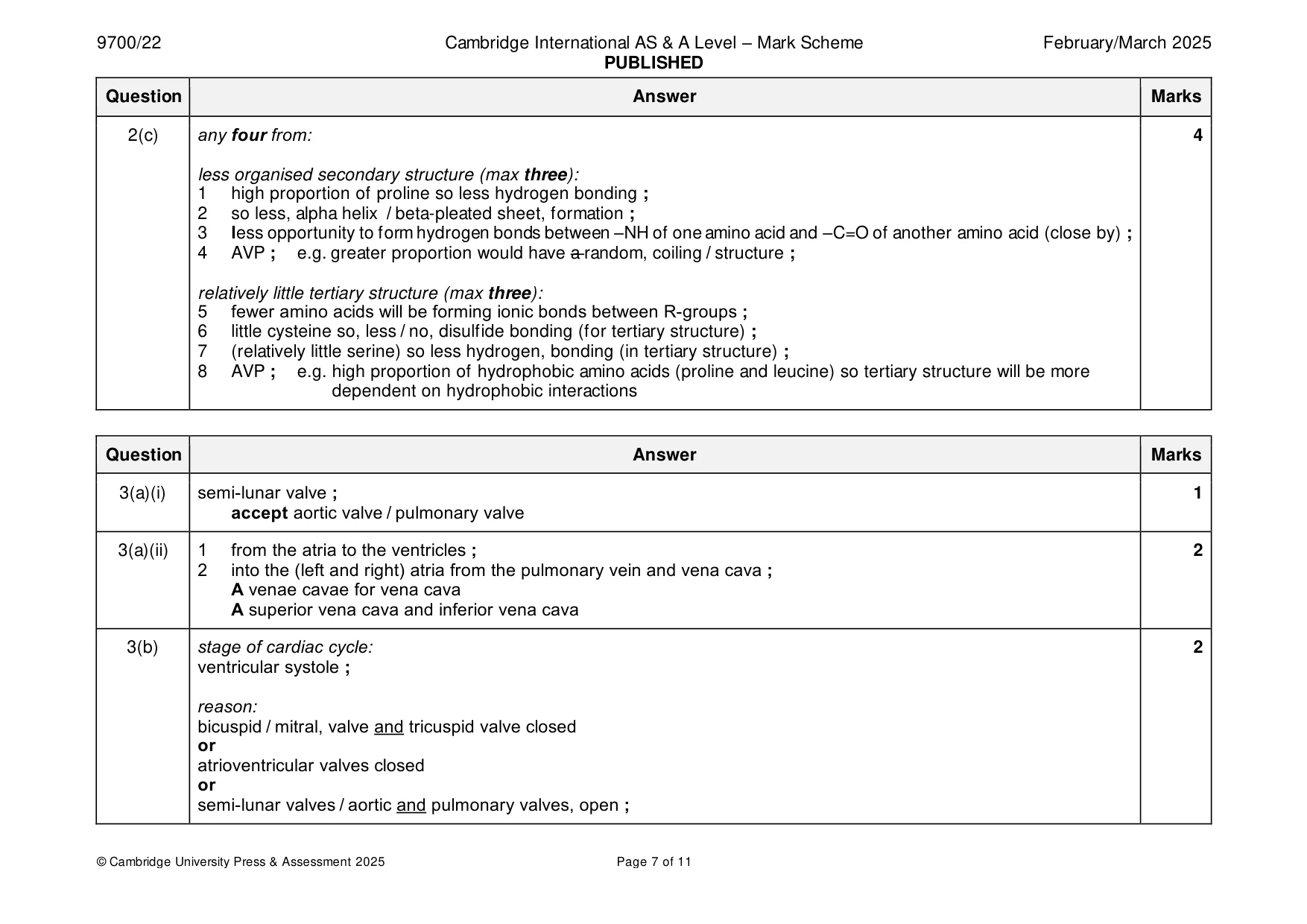
Mark scheme: 2(a) CH2OH 2 C O H H H C C OH H OH OH C C H OH H and OH correct on C1 ; all other additions complete and correct ; 2(b)(i) C – peptide (bond) ; 2 D – glycosidic (bond) ; 2(b)(ii) act as receptors / bind to ligands, qualified ; 1 2(c) any four from: 4 less organised secondary structure (max three): 1 high proportion of proline so less hydrogen bonding ; 2 so less, alpha helix / beta-pleated sheet, formation ; 3 less opportunity to form hydrogen bonds between –NH of one amino acid and –C=O of another amino acid (close by) ; 4 AVP ; e.g. greater proportion would have a random, coiling / structure ; relatively little tertiary structure (max three): 5 fewer amino acids will be forming ionic bonds between R-groups ; 6 little cysteine so, less / no, disulfide bonding (for tertiary structure) ; 7 (relatively little serine) so less hydrogen, bonding (in tertiary structure) ; 8 AVP ; e.g. high proportion of hydrophobic amino acids (proline and leucine) so tertiary structure will be more dependent on hydrophobic interactions
Question 3
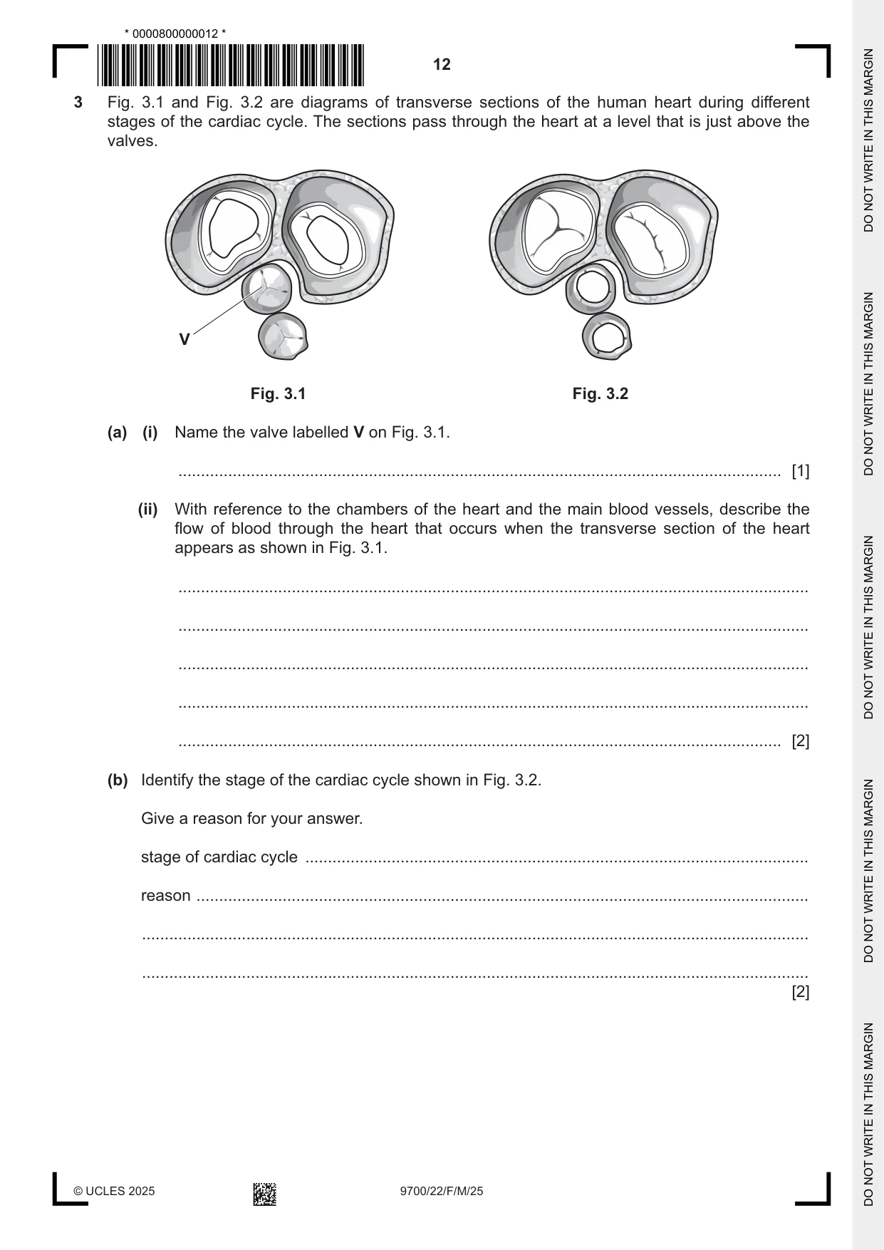
3 Fig. 3.1 and Fig. 3.2 are diagrams of transverse sections of the human heart during different stages of the cardiac cycle. The sections pass through the heart at a level that is just above the valves. V Fig. 3.1 Fig. 3.2 (a) (i) Name the valve labelled V on Fig. 3.1. ..................................................................................................................................... [1] (ii) With reference to the chambers of the heart and the main blood vessels, describe the flow of blood through the heart that occurs when the transverse section of the heart appears as shown in Fig. 3.1. ........................................................................................................................................... ........................................................................................................................................... ........................................................................................................................................... ........................................................................................................................................... ..................................................................................................................................... [2] (b) Identify the stage of the cardiac cycle shown in Fig. 3.2. Give a reason for your answer. stage of cardiac cycle ............................................................................................................... reason ....................................................................................................................................... ................................................................................................................................................... ................................................................................................................................................... [2] (c) The rate and rhythm of the heartbeat are controlled by an area of specialised muscle tissue in the wall of the right atrium, called the sinoatrial node. Describe the sequence of events that control contraction of the ventricles during the cardiac cycle. ................................................................................................................................................... ................................................................................................................................................... ................................................................................................................................................... ................................................................................................................................................... ................................................................................................................................................... ................................................................................................................................................... ...............................................................................................................................................[3] [Total: 8]
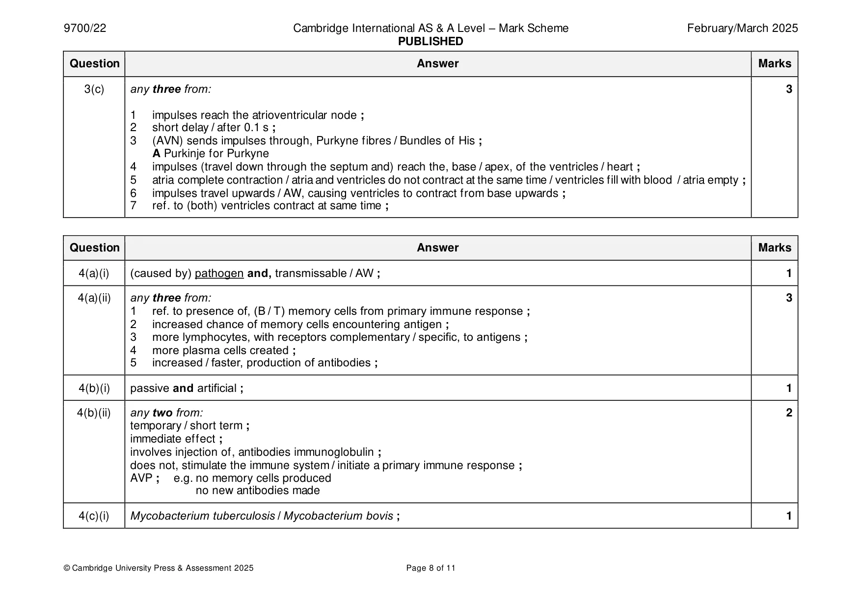
Mark scheme: 3(a)(i) semi-lunar valve ; 1 accept aortic valve / pulmonary valve 3(a)(ii) 1 from the atria to the ventricles ; 2 2 into the (left and right) atria from the pulmonary vein and vena cava ; A venae cavae for vena cava A superior vena cava and inferior vena cava 3(b) stage of cardiac cycle: 2 ventricular systole ; reason: bicuspid / mitral, valve and tricuspid valve closed or atrioventricular valves closed or semi-lunar valves / aortic and pulmonary valves, open ; 3(c) any three from: 3 1 impulses reach the atrioventricular node ; 2 short delay / after 0.1 s ; 3 (AVN) sends impulses through, Purkyne fibres / Bundles of His ; A Purkinje for Purkyne 4 impulses (travel down through the septum and) reach the, base / apex, of the ventricles / heart ; 5 atria complete contraction / atria and ventricles do not contract at the same time / ventricles fill with blood / atria empty ; 6 impulses travel upwards / AW, causing ventricles to contract from base upwards ; 7 ref. to (both) ventricles contract at same time ;
Q4 · Vaccination programmes are widely used to help control the spread of infectious diseases
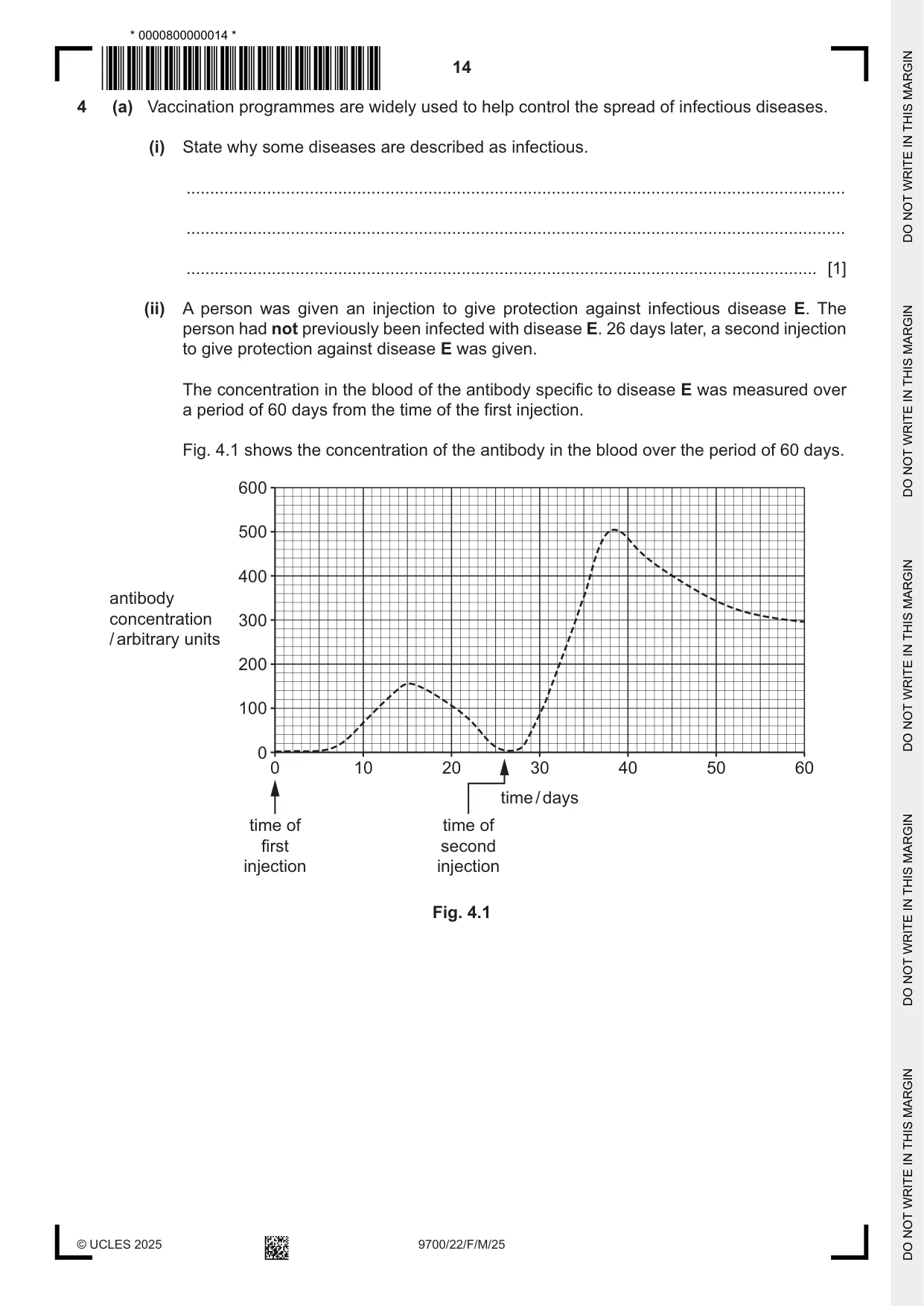
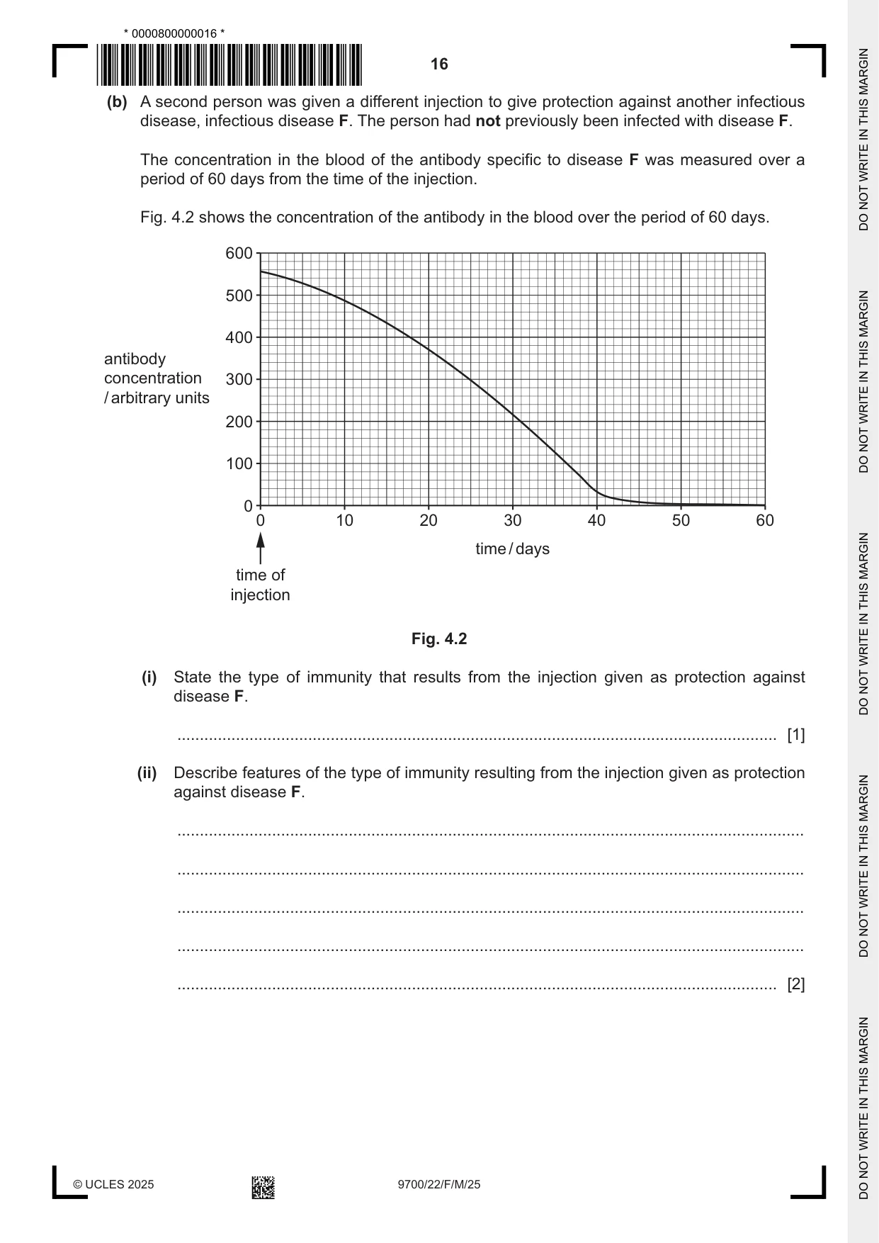
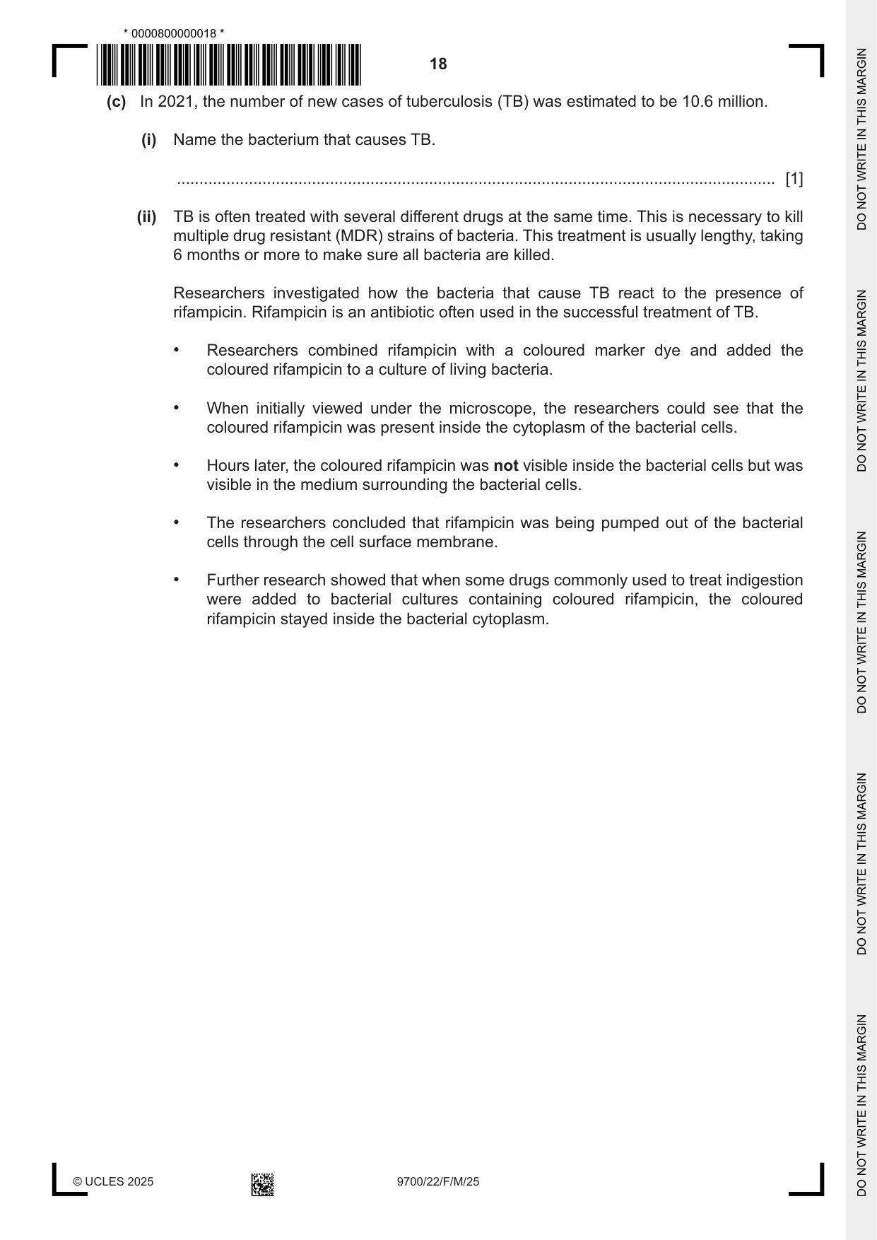
4 (a) Vaccination programmes are widely used to help control the spread of infectious diseases. (i) State why some diseases are described as infectious. ........................................................................................................................................... ........................................................................................................................................... ..................................................................................................................................... [1] (ii) A person was given an injection to give protection against infectious disease E. The person had not previously been infected with disease E. 26 days later, a second injection to give protection against disease E was given. The concentration in the blood of the antibody specific to disease E was measured over a period of 60 days from the time of the first injection. Fig. 4.1 shows the concentration of the antibody in the blood over the period of 60 days. 600 500 400 antibody concentration 300 / arbitrary units 200 100 0 0 10 20 30 40 50 60 time / days time of time of first second injection injection Fig. 4.1 The concentration of the antibody in the blood after the second injection was higher than the concentration after the first injection. Explain why the concentration of the antibody in the blood was higher after the second injection than after the first injection. ........................................................................................................................................... ........................................................................................................................................... ........................................................................................................................................... ........................................................................................................................................... ........................................................................................................................................... ........................................................................................................................................... ..................................................................................................................................... [3] (b) A second person was given a different injection to give protection against another infectious disease, infectious disease F. The person had not previously been infected with disease F. The concentration in the blood of the antibody specific to disease F was measured over a period of 60 days from the time of the injection. Fig. 4.2 shows the concentration of the antibody in the blood over the period of 60 days. 600 500 400 antibody concentration 300 / arbitrary units 200 100 0 0 10 20 30 40 50 60 time / days time of injection Fig. 4.2 (i) State the type of immunity that results from the injection given as protection against disease F. ..................................................................................................................................... [1] (ii) Describe features of the type of immunity resulting from the injection given as protection against disease F. ........................................................................................................................................... ........................................................................................................................................... ........................................................................................................................................... ........................................................................................................................................... ..................................................................................................................................... [2] Question 4 continues on page 18. (c) In 2021, the number of new cases of tuberculosis (TB) was estimated to be 10.6 million. (i) Name the bacterium that causes TB. ..................................................................................................................................... [1] (ii) TB is often treated with several different drugs at the same time. This is necessary to kill multiple drug resistant (MDR) strains of bacteria. This treatment is usually lengthy, taking 6 months or more to make sure all bacteria are killed. Researchers investigated how the bacteria that cause TB react to the presence of rifampicin. Rifampicin is an antibiotic often used in the successful treatment of TB. • Researchers combined rifampicin with a coloured marker dye and added the coloured rifampicin to a culture of living bacteria. • When initially viewed under the microscope, the researchers could see that the coloured rifampicin was present inside the cytoplasm of the bacterial cells. • Hours later, the coloured rifampicin was not visible inside the bacterial cells but was visible in the medium surrounding the bacterial cells. • The researchers concluded that rifampicin was being pumped out of the bacterial cells through the cell surface membrane. • Further research showed that when some drugs commonly used to treat indigestion were added to bacterial cultures containing coloured rifampicin, the coloured rifampicin stayed inside the bacterial cytoplasm. Suggest and explain how knowledge gained from this research could improve TB treatment and reduce the chance of rifampicin‑resistant bacteria developing. ........................................................................................................................................... ........................................................................................................................................... ........................................................................................................................................... ........................................................................................................................................... ........................................................................................................................................... ........................................................................................................................................... ........................................................................................................................................... ........................................................................................................................................... ..................................................................................................................................... [4] [Total: 12]
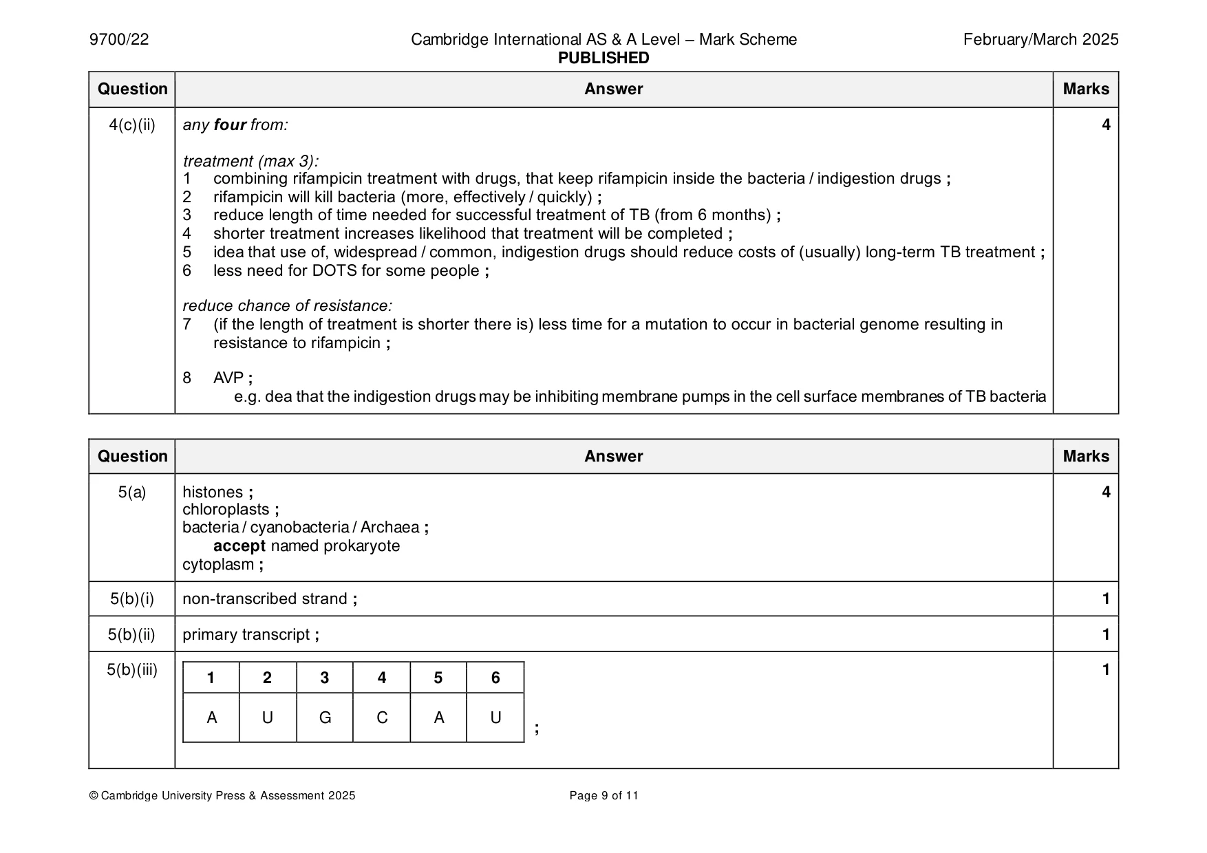
Mark scheme: 4(a)(i) (caused by) pathogen and, transmissable / AW ; 1 4(a)(ii) any three from: 3 1 ref. to presence of, (B / T) memory cells from primary immune response ; 2 increased chance of memory cells encountering antigen ; 3 more lymphocytes, with receptors complementary / specific, to antigens ; 4 more plasma cells created ; 5 increased / faster, production of antibodies ; 4(b)(i) passive and artificial ; 1 4(b)(ii) any two from: 2 temporary / short term ; immediate effect ; involves injection of, antibodies immunoglobulin ; does not, stimulate the immune system / initiate a primary immune response ; AVP ; e.g. no memory cells produced no new antibodies made 4(c)(i) Mycobacterium tuberculosis / Mycobacterium bovis ; 1 4(c)(ii) any four from: 4 treatment (max 3): 1 combining rifampicin treatment with drugs, that keep rifampicin inside the bacteria / indigestion drugs ; 2 rifampicin will kill bacteria (more, effectively / quickly) ; 3 reduce length of time needed for successful treatment of TB (from 6 months) ; 4 shorter treatment increases likelihood that treatment will be completed ; 5 idea that use of, widespread / common, indigestion drugs should reduce costs of (usually) long-term TB treatment ; 6 less need for DOTS for some people ; reduce chance of resistance: 7 (if the length of treatment is shorter there is) less time for a mutation to occur in bacterial genome resulting in resistance to rifampicin ; 8 AVP ; e.g. dea that the indigestion drugs may be inhibiting membrane pumps in the cell surface membranes of TB bacteria
Q5 · Eukaryotic cells and prokaryotic cells contain DNA
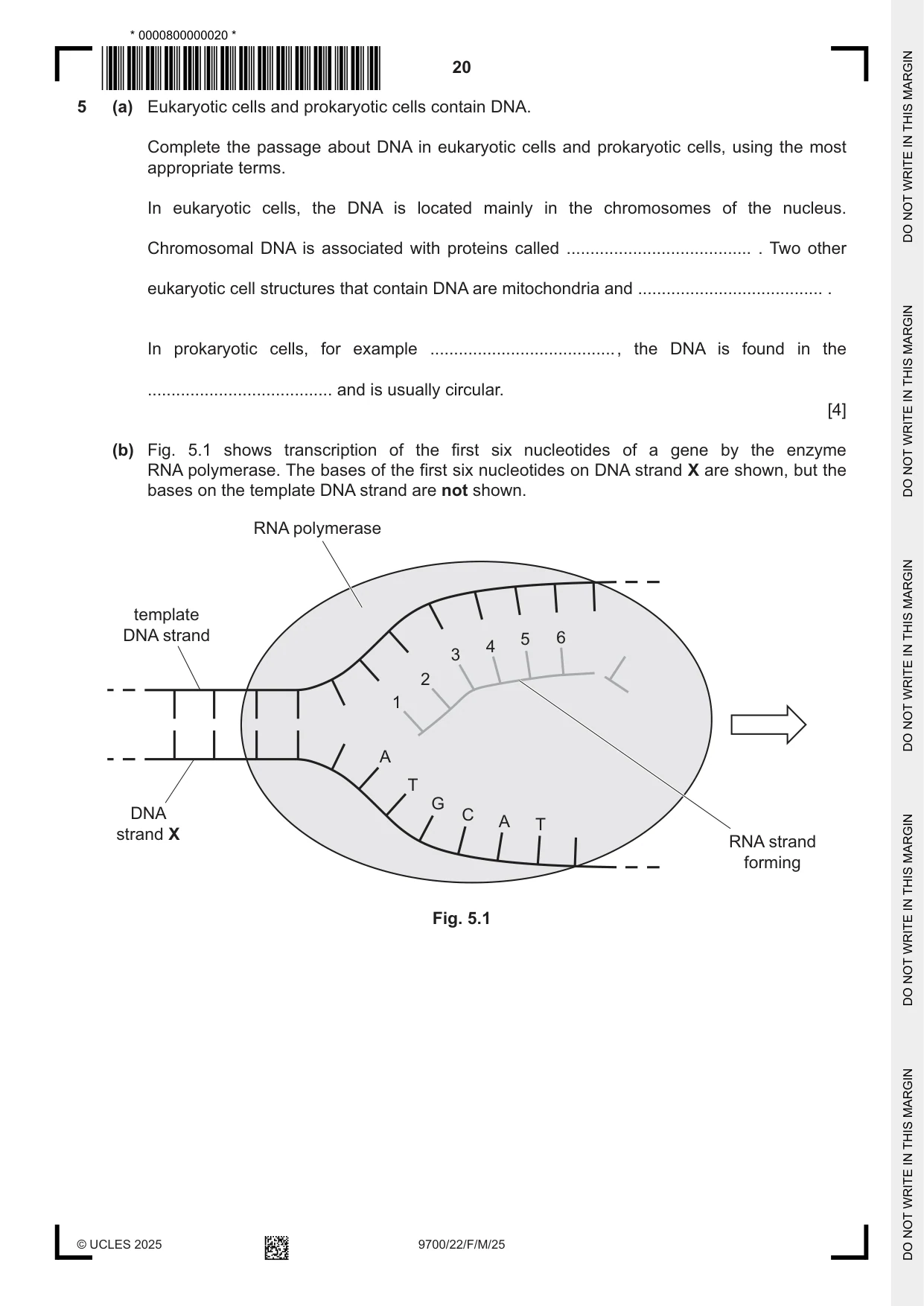
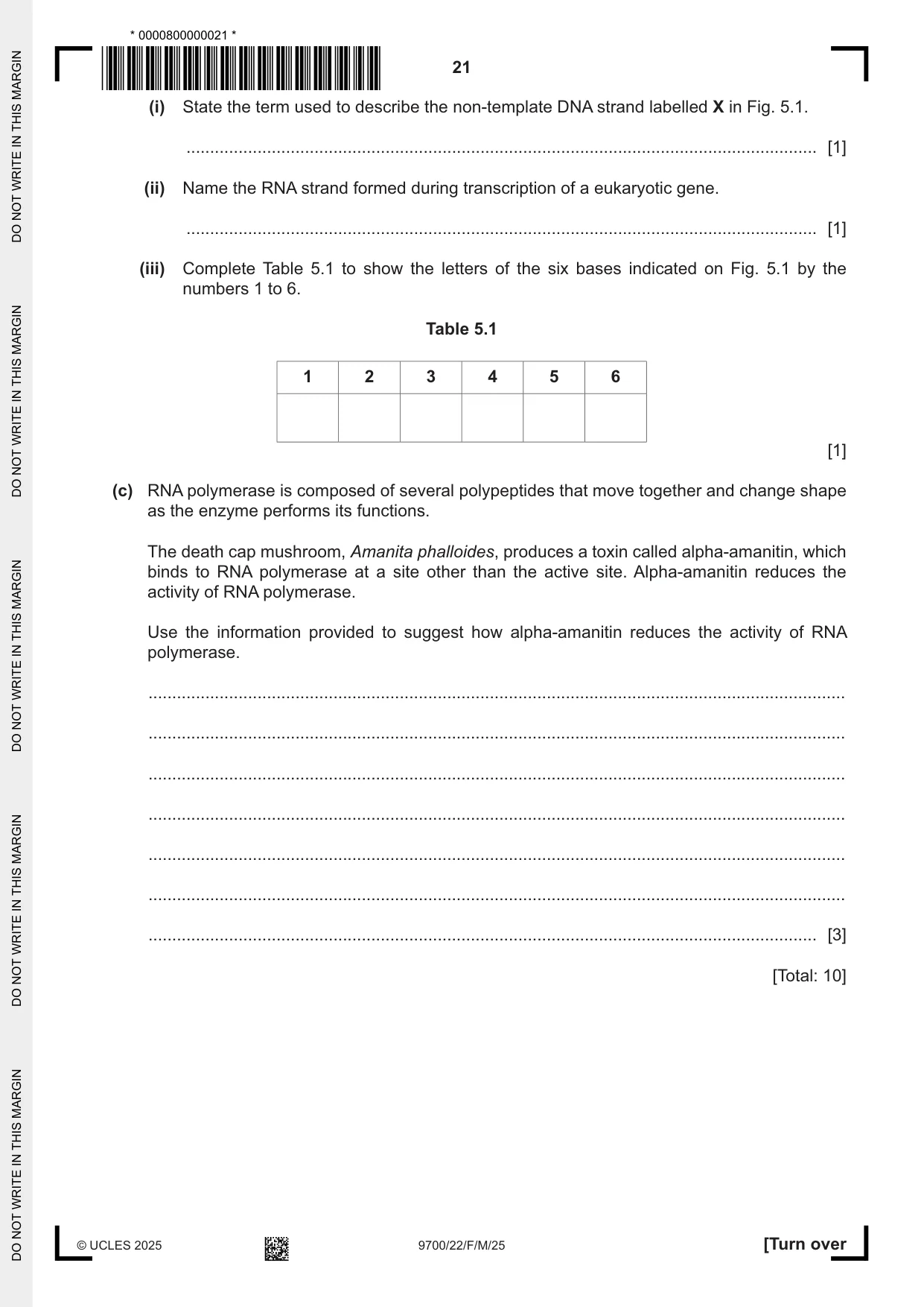
5 (a) Eukaryotic cells and prokaryotic cells contain DNA. Complete the passage about DNA in eukaryotic cells and prokaryotic cells, using the most appropriate terms. In eukaryotic cells, the DNA is located mainly in the chromosomes of the nucleus. Chromosomal DNA is associated with proteins called ....................................... . Two other eukaryotic cell structures that contain DNA are mitochondria and ....................................... . In prokaryotic cells, for example ....................................... , the DNA is found in the ....................................... and is usually circular. [4] (b) Fig. 5.1 shows transcription of the first six nucleotides of a gene by the enzyme RNA polymerase. The bases of the first six nucleotides on DNA strand X are shown, but the bases on the template DNA strand are not shown. RNA polymerase template DNA strand 5 6 4 3 2 1 A T G DNA C A T strand X RNA strand forming Fig. 5.1 (i) State the term used to describe the non‑template DNA strand labelled X in Fig. 5.1. ..................................................................................................................................... [1] (ii) Name the RNA strand formed during transcription of a eukaryotic gene. ..................................................................................................................................... [1] (iii) Complete Table 5.1 to show the letters of the six bases indicated on Fig. 5.1 by the numbers 1 to 6. Table 5.1 1 2 3 4 5 6 [1] (c) RNA polymerase is composed of several polypeptides that move together and change shape as the enzyme performs its functions. The death cap mushroom, Amanita phalloides, produces a toxin called alpha‑amanitin, which binds to RNA polymerase at a site other than the active site. Alpha‑amanitin reduces the activity of RNA polymerase. Use the information provided to suggest how alpha‑amanitin reduces the activity of RNA polymerase. ................................................................................................................................................... ................................................................................................................................................... ................................................................................................................................................... ................................................................................................................................................... ................................................................................................................................................... ................................................................................................................................................... ............................................................................................................................................. [3] [Total: 10]
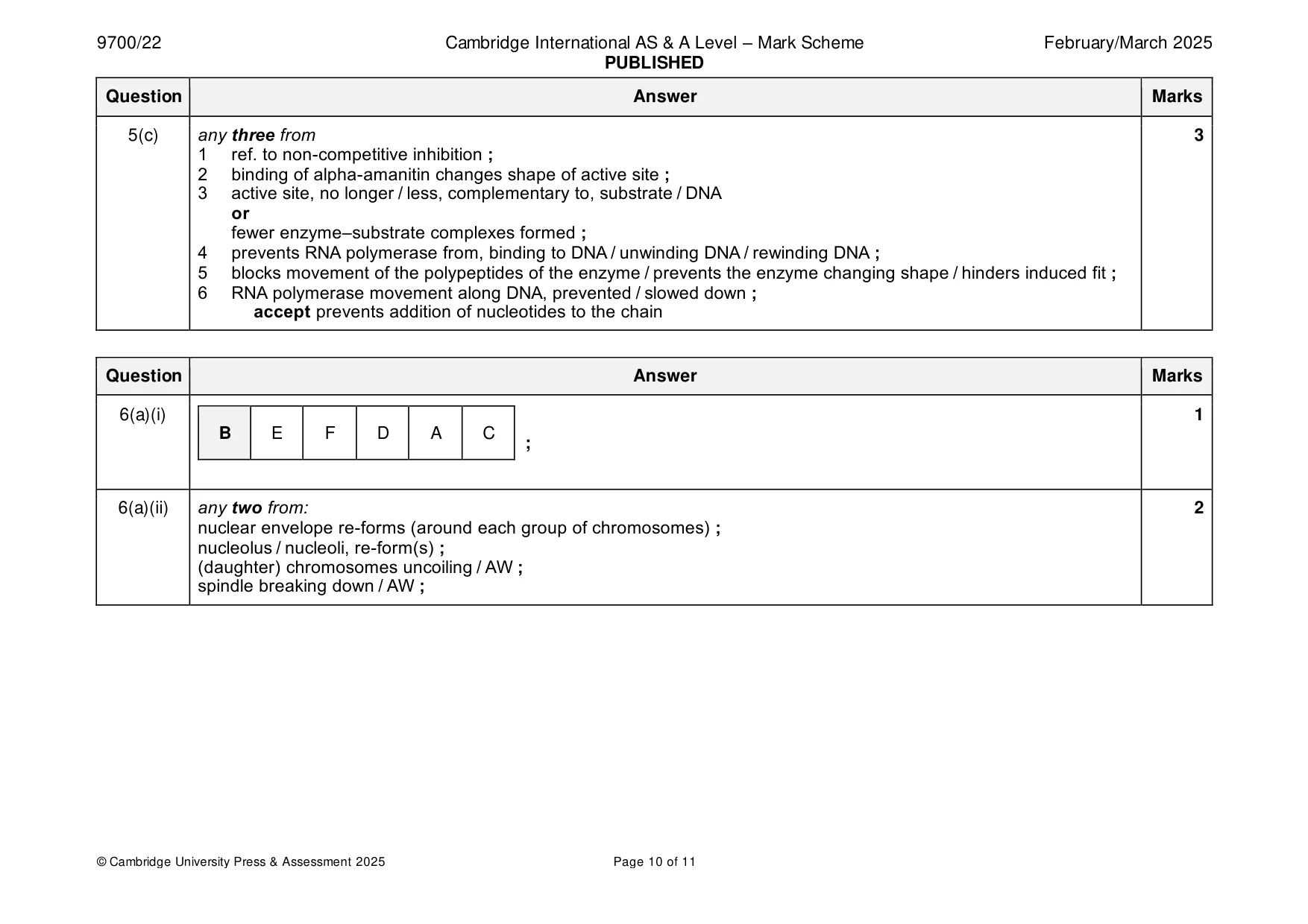
Mark scheme: 5(a) histones ; 4 chloroplasts ; bacteria / cyanobacteria / Archaea ; accept named prokaryote cytoplasm ; 5(b)(i) non-transcribed strand ; 1 5(b)(ii) primary transcript ; 1 5(b)(iii) 1 1 2 3 4 5 6 A U G C A U ; 5(c) any three from 3 1 ref. to non-competitive inhibition ; 2 binding of alpha-amanitin changes shape of active site ; 3 active site, no longer / less, complementary to, substrate / DNA or fewer enzyme–substrate complexes formed ; 4 prevents RNA polymerase from, binding to DNA / unwinding DNA / rewinding DNA ; 5 blocks movement of the polypeptides of the enzyme / prevents the enzyme changing shape / hinders induced fit ; 6 RNA polymerase movement along DNA, prevented / slowed down ; accept prevents addition of nucleotides to the chain
More questions on Cells as the basic units of living organisms
Q6 · Photomicrographs of individual cells from the root tip of an onion, Allium sp., at…
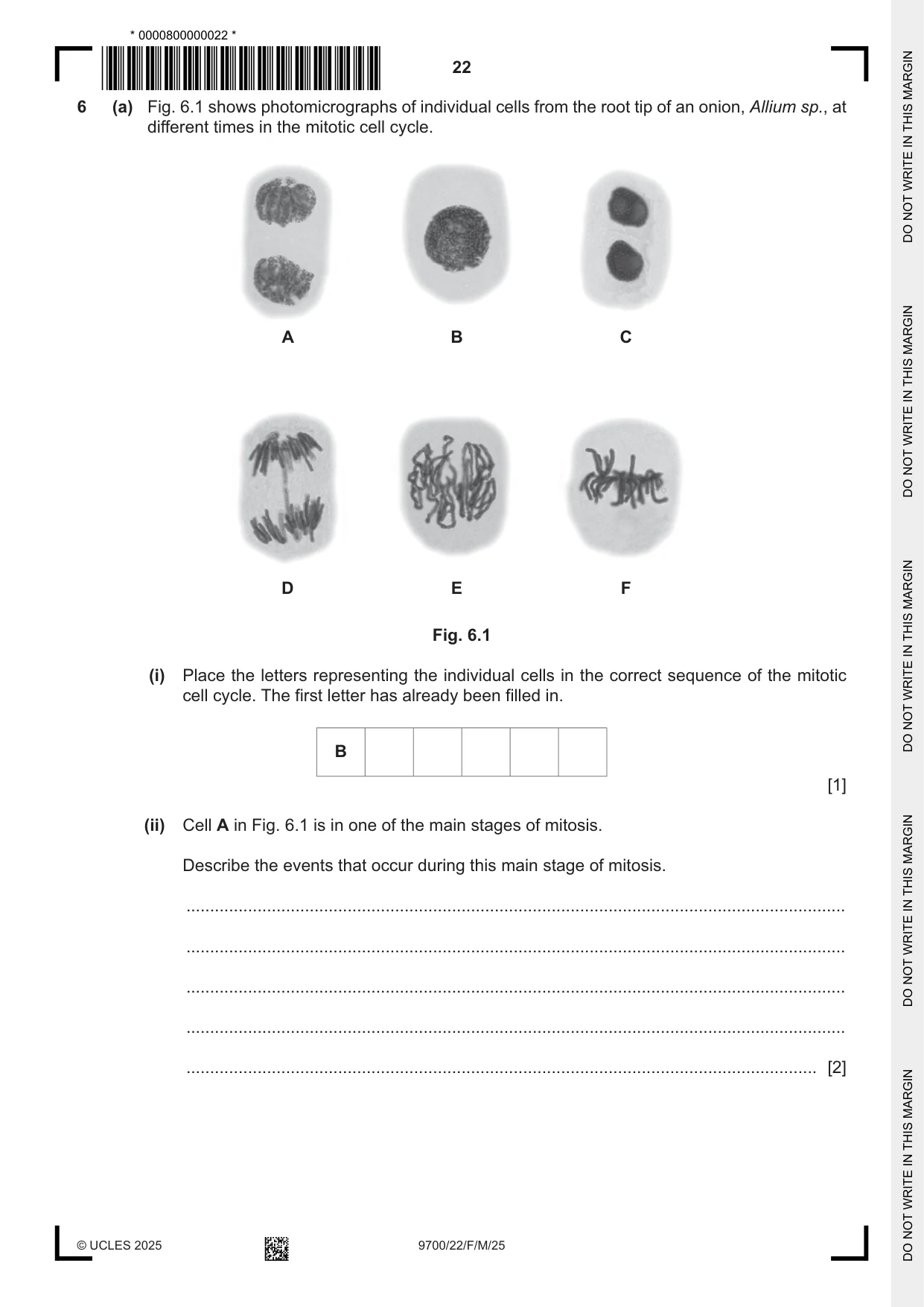
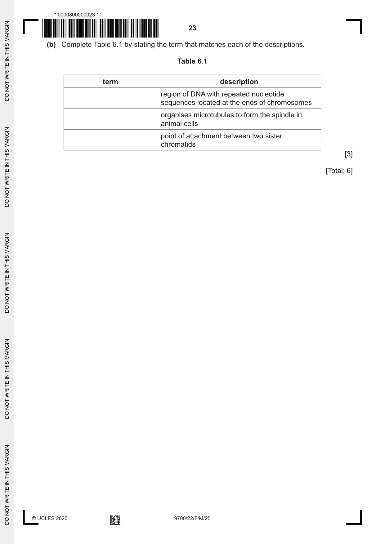
6 (a) Fig. 6.1 shows photomicrographs of individual cells from the root tip of an onion, Allium sp., at different times in the mitotic cell cycle. A B C D E F Fig. 6.1 (i) Place the letters representing the individual cells in the correct sequence of the mitotic cell cycle. The first letter has already been filled in. B [1] (ii) Cell A in Fig. 6.1 is in one of the main stages of mitosis. Describe the events that occur during this main stage of mitosis. ........................................................................................................................................... ........................................................................................................................................... ........................................................................................................................................... ........................................................................................................................................... ..................................................................................................................................... [2] (b) Complete Table 6.1 by stating the term that matches each of the descriptions. Table 6.1 term description region of DNA with repeated nucleotide sequences located at the ends of chromosomes organises microtubules to form the spindle in animal cells point of attachment between two sister chromatids [3] [Total: 6]
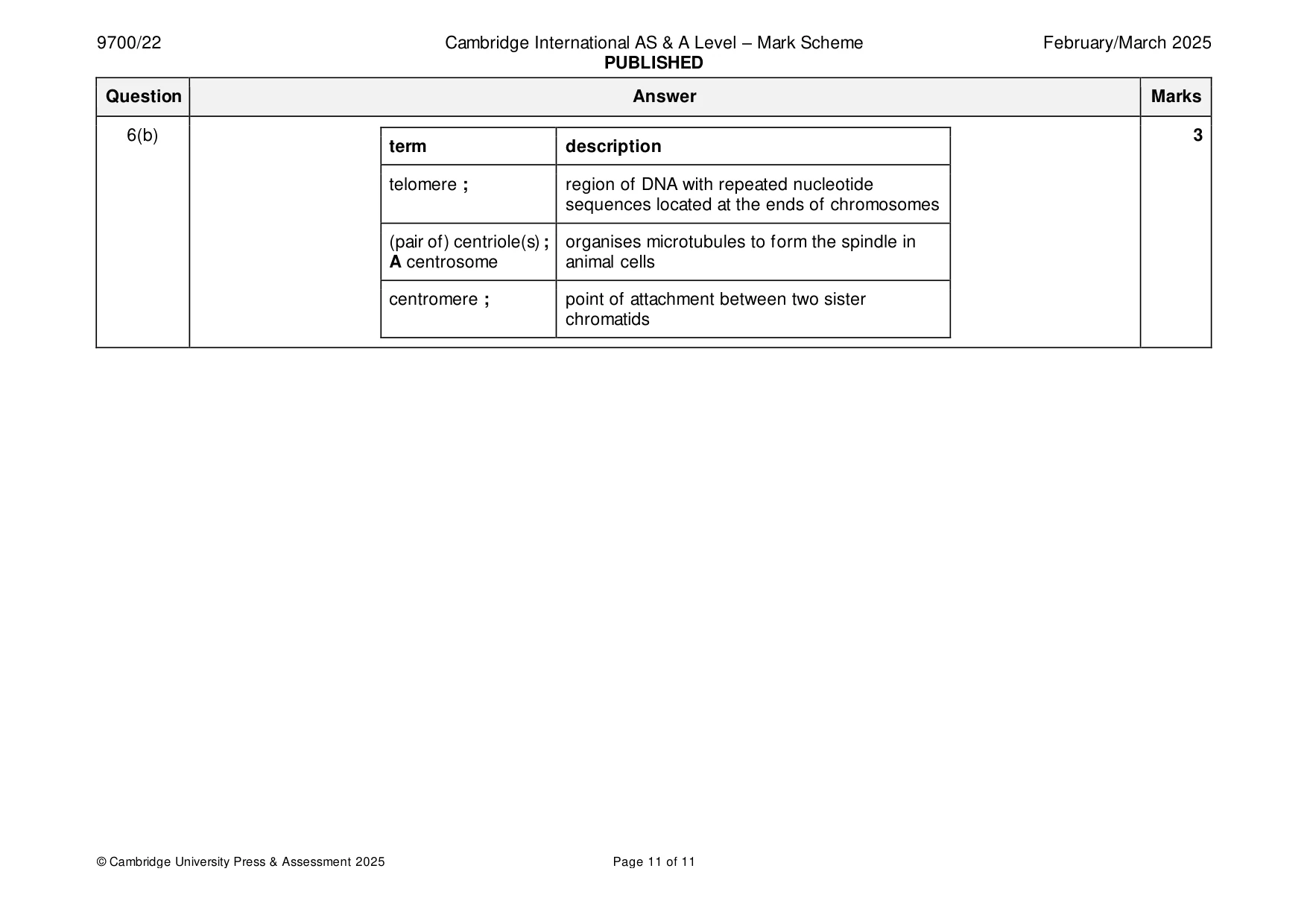
Mark scheme: 6(a)(i) 1 B E F D A C ; 6(a)(ii) any two from: 2 nuclear envelope re-forms (around each group of chromosomes) ; nucleolus / nucleoli, re-form(s) ; (daughter) chromosomes uncoiling / AW ; spindle breaking down / AW ; 6(b) 3 term description telomere ; region of DNA with repeated nucleotide sequences located at the ends of chromosomes (pair of) centriole(s) ; organises microtubules to form the spindle in A centrosome animal cells centromere ; point of attachment between two sister chromatids
What was in this paper
The subtopics covered by these 6 questions, and how many questions each got. Open one in a new tab to see every Cambridge question on it.
What you needed in this session
Cambridge’s own grade thresholds for 2025 Feb/March, Paper 2 · Variant 2. A higher threshold means an easier paper — the bar moves with how the cohort did.