Cambridge A Level Biology 9700 — 2024 Feb/March Paper 2 · Variant 2
9700/22/F/M/24 · 6 questions · 60 marks · ≈68 min
The question paper and its mark scheme, free to read here and free to download. This is Cambridge’s own paper, exactly as it was sat.
Question paper20 pages

Mark scheme13 pages
Answers below. Sit the paper first if you are practising.
Questions as text
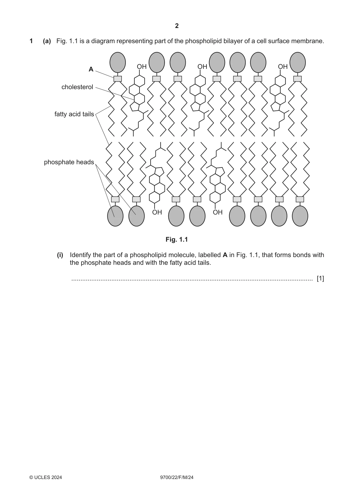
Q1 · A diagram representing part of the phospholipid bilayer of a cell surface membrane
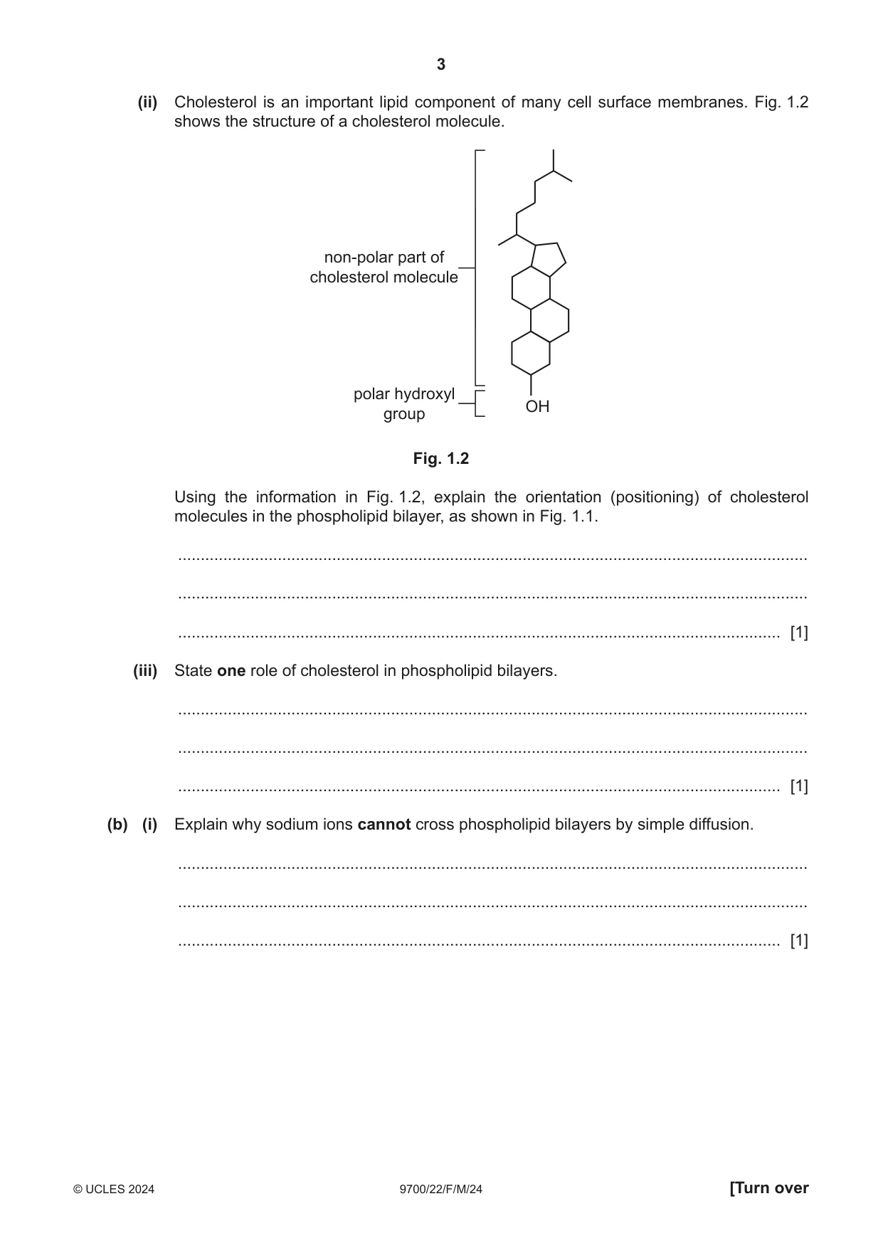
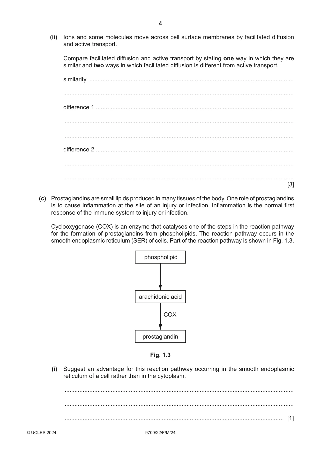
1 (a) Fig. 1.1 is a diagram representing part of the phospholipid bilayer of a cell surface membrane. OH OH OH A cholesterol fatty acid tails phosphate heads OH OH Fig. 1.1 (i) Identify the part of a phospholipid molecule, labelled A in Fig. 1.1, that forms bonds with the phosphate heads and with the fatty acid tails. ..................................................................................................................................... [1] (ii) Cholesterol is an important lipid component of many cell surface membranes. Fig. 1.2 shows the structure of a cholesterol molecule. non-polar part of cholesterol molecule polar hydroxyl OH group Fig. 1.2 Using the information in Fig. 1.2, explain the orientation (positioning) of cholesterol molecules in the phospholipid bilayer, as shown in Fig. 1.1. ........................................................................................................................................... ........................................................................................................................................... ..................................................................................................................................... [1] (iii) State one role of cholesterol in phospholipid bilayers. ........................................................................................................................................... ........................................................................................................................................... ..................................................................................................................................... [1] (b) (i) Explain why sodium ions cannot cross phospholipid bilayers by simple diffusion. ........................................................................................................................................... ........................................................................................................................................... ..................................................................................................................................... [1] (ii) Ions and some molecules move across cell surface membranes by facilitated diffusion and active transport. Compare facilitated diffusion and active transport by stating one way in which they are similar and two ways in which facilitated diffusion is different from active transport. similarity ............................................................................................................................ ........................................................................................................................................... difference 1 ........................................................................................................................ ........................................................................................................................................... ........................................................................................................................................... difference 2 ........................................................................................................................ ........................................................................................................................................... ........................................................................................................................................... [3] (c) Prostaglandins are small lipids produced in many tissues of the body. One role of prostaglandins is to cause inflammation at the site of an injury or infection. Inflammation is the normal first response of the immune system to injury or infection. Cyclooxygenase (COX) is an enzyme that catalyses one of the steps in the reaction pathway for the formation of prostaglandins from phospholipids. The reaction pathway occurs in the smooth endoplasmic reticulum (SER) of cells. Part of the reaction pathway is shown in Fig. 1.3. phospholipid arachidonic acid COX prostaglandin Fig. 1.3 (i) Suggest an advantage for this reaction pathway occurring in the smooth endoplasmic reticulum of a cell rather than in the cytoplasm. ........................................................................................................................................... ........................................................................................................................................... ..................................................................................................................................... [1] (ii) Sometimes inflammation can have side-effects, such as pain. Aspirin is a drug that can be used to reduce these side-effects. Aspirin reduces the catalytic activity of the COX enzyme by modifying the R-group of one of the amino acids. Suggest how modifying the R-group of an amino acid in the COX enzyme can reduce the catalytic activity of the enzyme. ........................................................................................................................................... ........................................................................................................................................... ........................................................................................................................................... ........................................................................................................................................... ........................................................................................................................................... ........................................................................................................................................... ..................................................................................................................................... [3] (iii) Prostaglandins are examples of cell-signalling molecules. Outline the process of cell signalling that leads to a response by the cells involved in inflammation. ........................................................................................................................................... ........................................................................................................................................... ........................................................................................................................................... ........................................................................................................................................... ..................................................................................................................................... [2] [Total: 13]
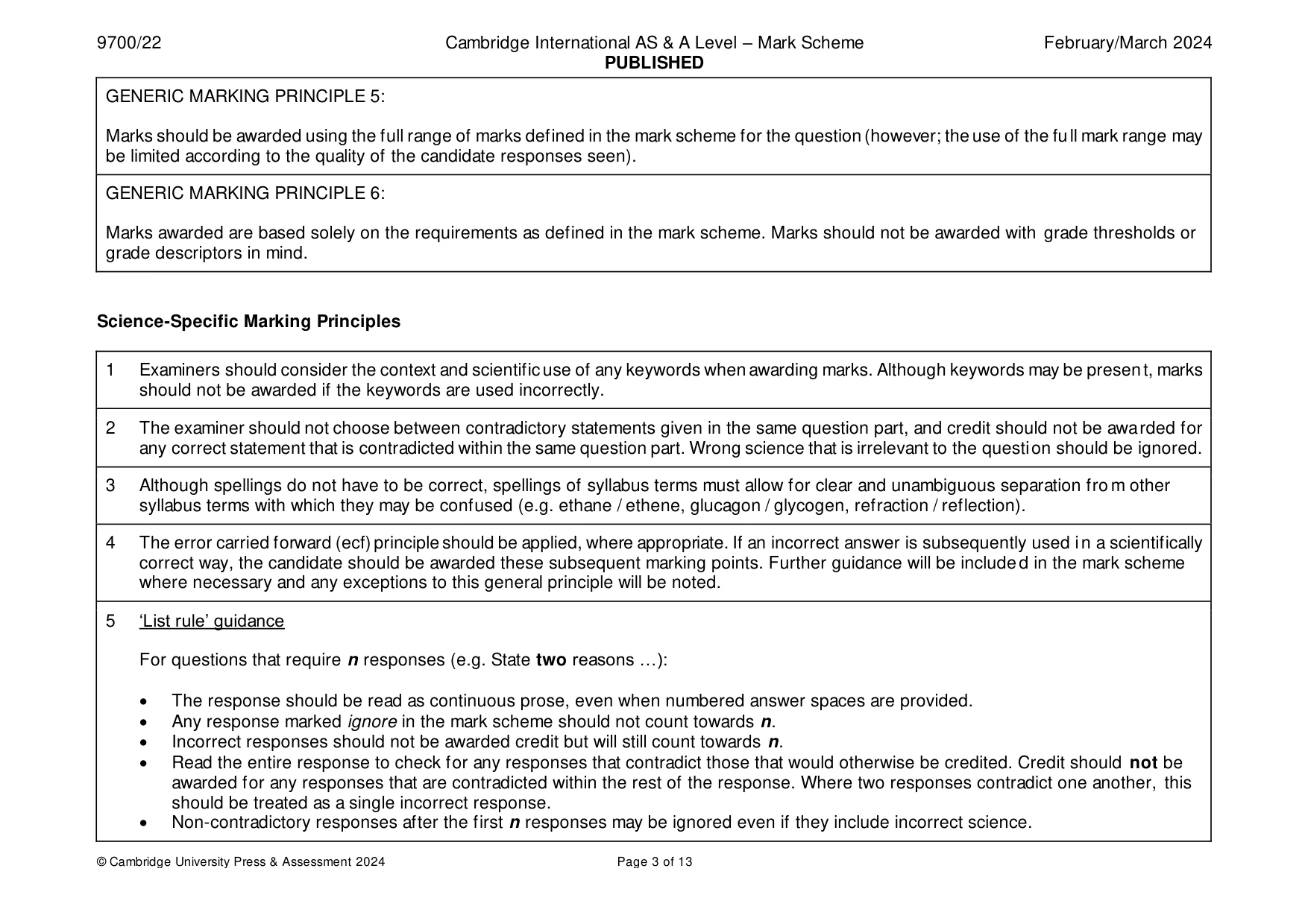
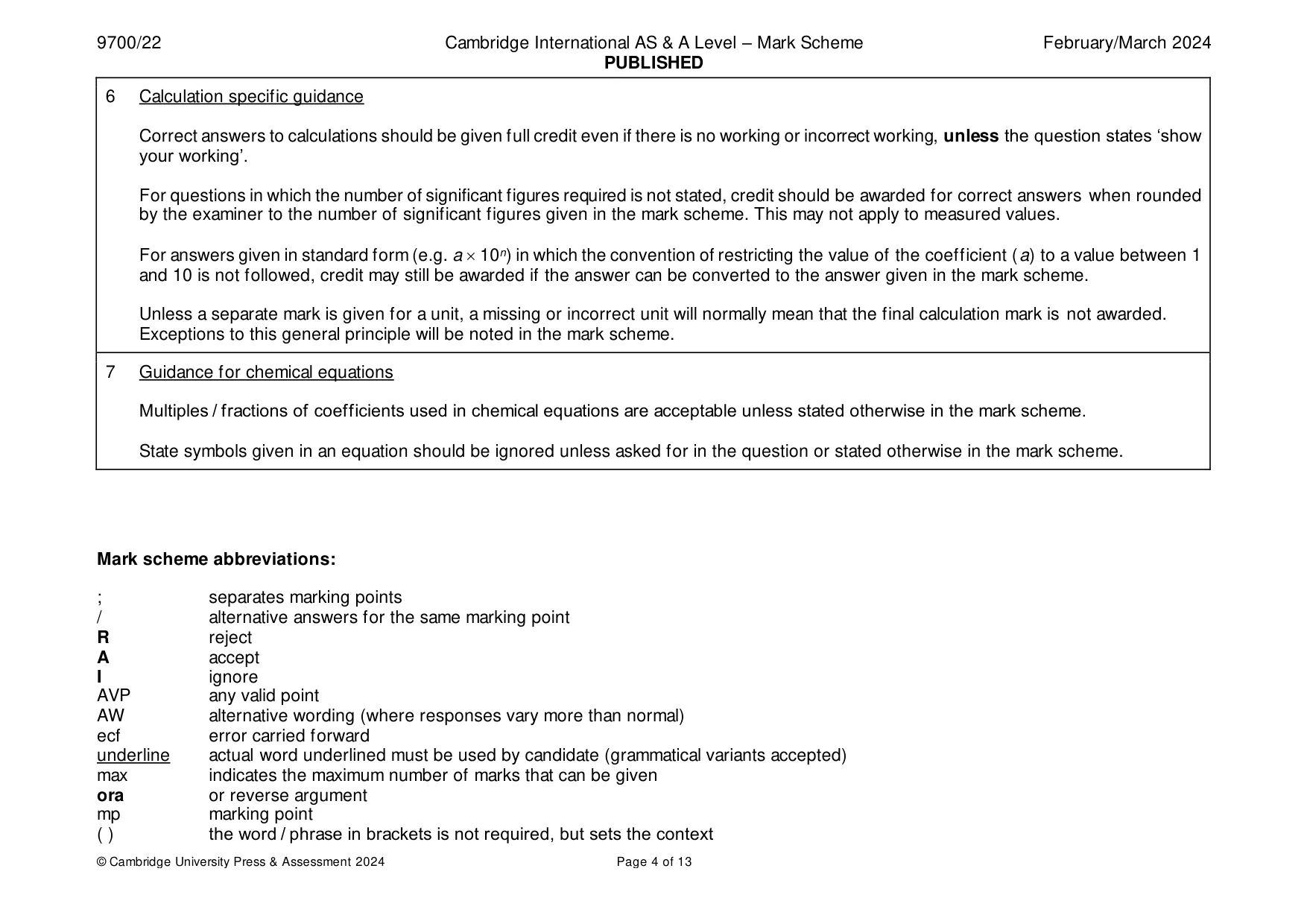
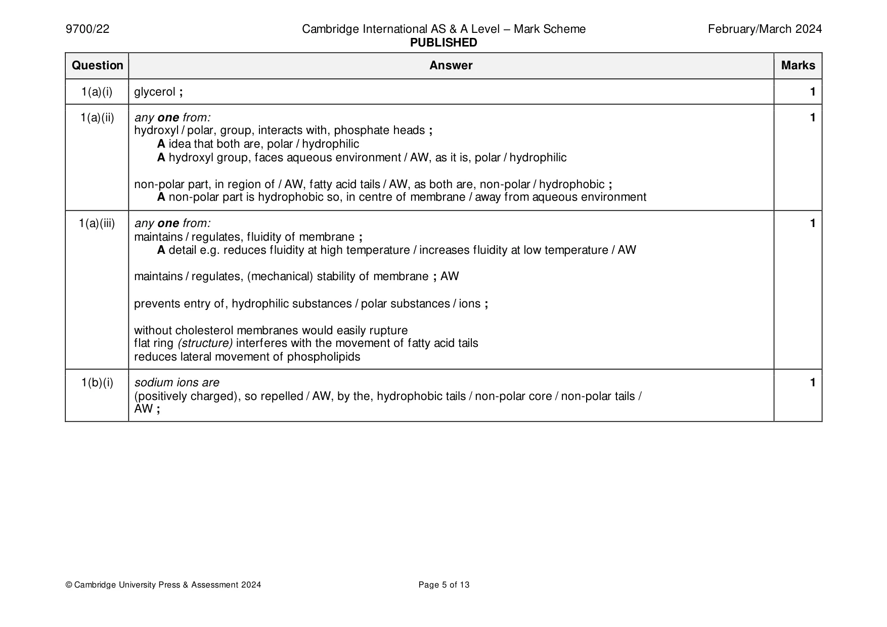
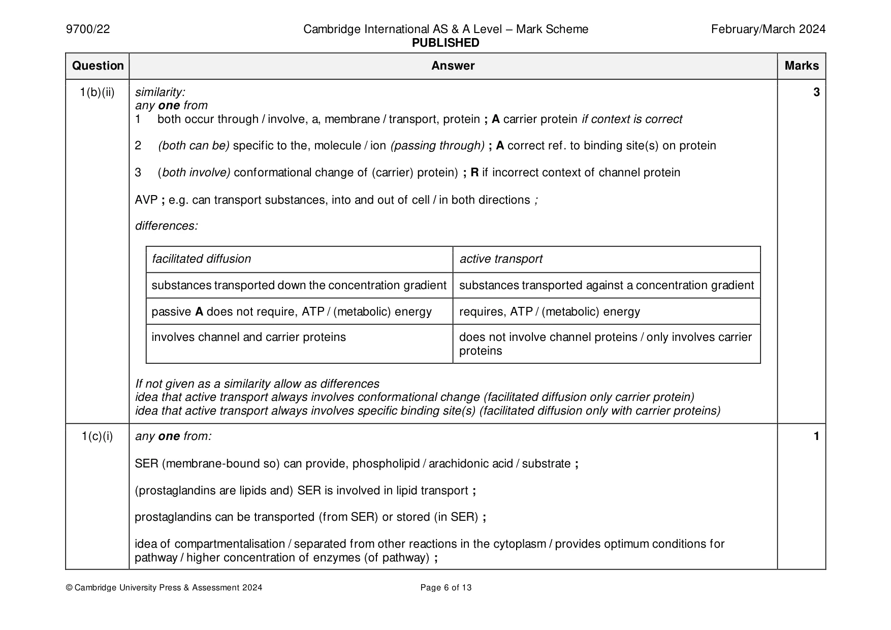
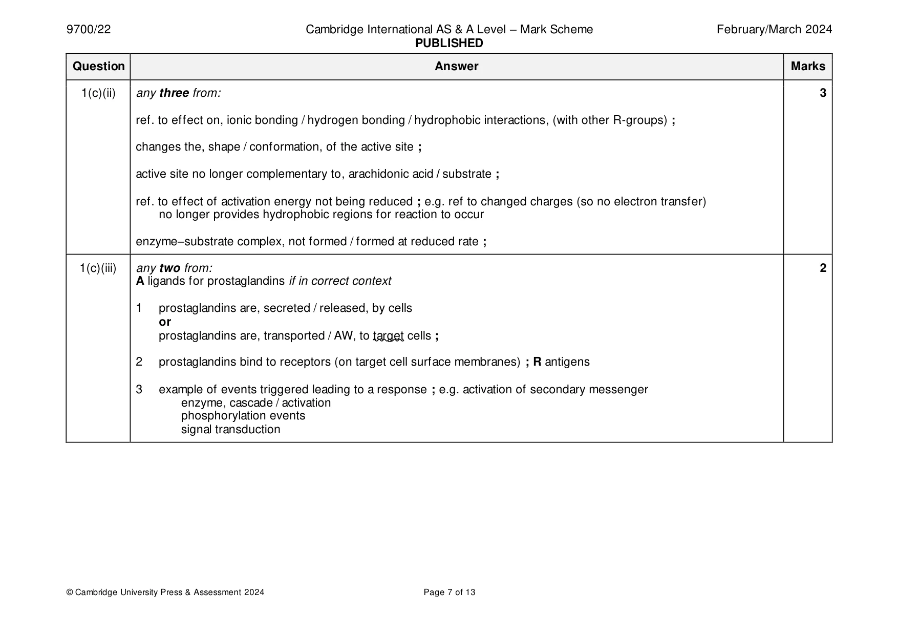
Mark scheme: Question Answer Marks 1(a)(i) glycerol ; 1 1(a)(ii) any one from: 1 hydroxyl / polar, group, interacts with, phosphate heads ; A idea that both are, polar / hydrophilic A hydroxyl group, faces aqueous environment / AW, as it is, polar / hydrophilic non-polar part, in region of / AW, fatty acid tails / AW, as both are, non-polar / hydrophobic ; A non-polar part is hydrophobic so, in centre of membrane / away from aqueous environment 1(a)(iii) any one from: 1 maintains / regulates, fluidity of membrane ; A detail e.g. reduces fluidity at high temperature / increases fluidity at low temperature / AW maintains / regulates, (mechanical) stability of membrane ; AW prevents entry of, hydrophilic substances / polar substances / ions ; without cholesterol membranes would easily rupture flat ring (structure) interferes with the movement of fatty acid tails reduces lateral movement of phospholipids 1(b)(i) sodium ions are 1 (positively charged), so repelled / AW, by the, hydrophobic tails / non-polar core / non-polar tails / AW ; 1(b)(ii) similarity: 3 any one from 1 both occur through / involve, a, membrane / transport, protein ; A carrier protein if context is correct 2 (both can be) specific to the, molecule / ion (passing through) ; A correct ref. to binding site(s) on protein 3 (both involve) conformational change of (carrier) protein) ; R if incorrect context of channel protein AVP ; e.g. can transport substances, into and out of cell / in both directions ; differences: facilitated diffusion active transport substances transported down the concentration gradient substances transported against a concentration gradient passive A does not require, ATP / (metabolic) energy requires, ATP / (metabolic) energy involves channel and carrier proteins does not involve channel proteins / only involves carrier proteins If not given as a similarity allow as differences idea that active transport always involves conformational change (facilitated diffusion only carrier protein) idea that active transport always involves specific binding site(s) (facilitated diffusion only with carrier proteins) 1(c)(i) any one from: 1 SER (membrane-bound so) can provide, phospholipid / arachidonic acid / substrate ; (prostaglandins are lipids and) SER is involved in lipid transport ; prostaglandins can be transported (from SER) or stored (in SER) ; idea of compartmentalisation / separated from other reactions in the cytoplasm / provides optimum conditions for pathway / higher concentration of enzymes (of pathway) ; 1(c)(ii) any three from: 3 ref. to effect on, ionic bonding / hydrogen bonding / hydrophobic interactions, (with other R-groups) ; changes the, shape / conformation, of the active site ; active site no longer complementary to, arachidonic acid / substrate ; ref. to effect of activation energy not being reduced ; e.g. ref to changed charges (so no electron transfer) no longer provides hydrophobic regions for reaction to occur enzyme–substrate complex, not formed / formed at reduced rate ; 1(c)(iii) any two from: 2 A ligands for prostaglandins if in correct context 1 prostaglandins are, secreted / released, by cells or prostaglandins are, transported / AW, to target cells ; 2 prostaglandins bind to receptors (on target cell surface membranes) ; R antigens 3 example of events triggered leading to a response ; e.g. activation of secondary messenger enzyme, cascade / activation phosphorylation events signal transduction
More questions on Cells as the basic units of living organisms
Q2 · Descriptions of three types of white blood cell
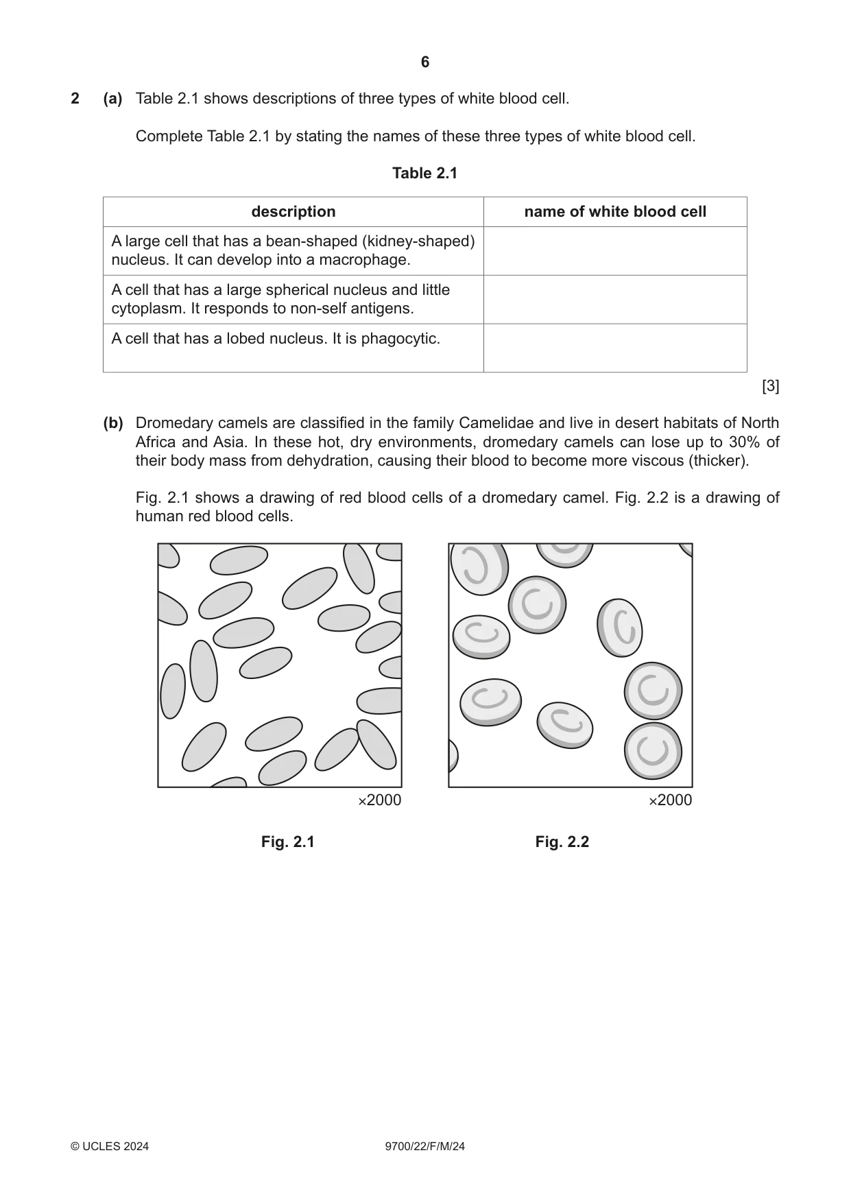
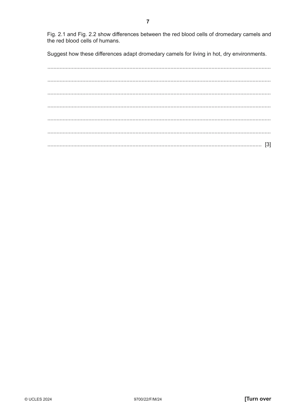
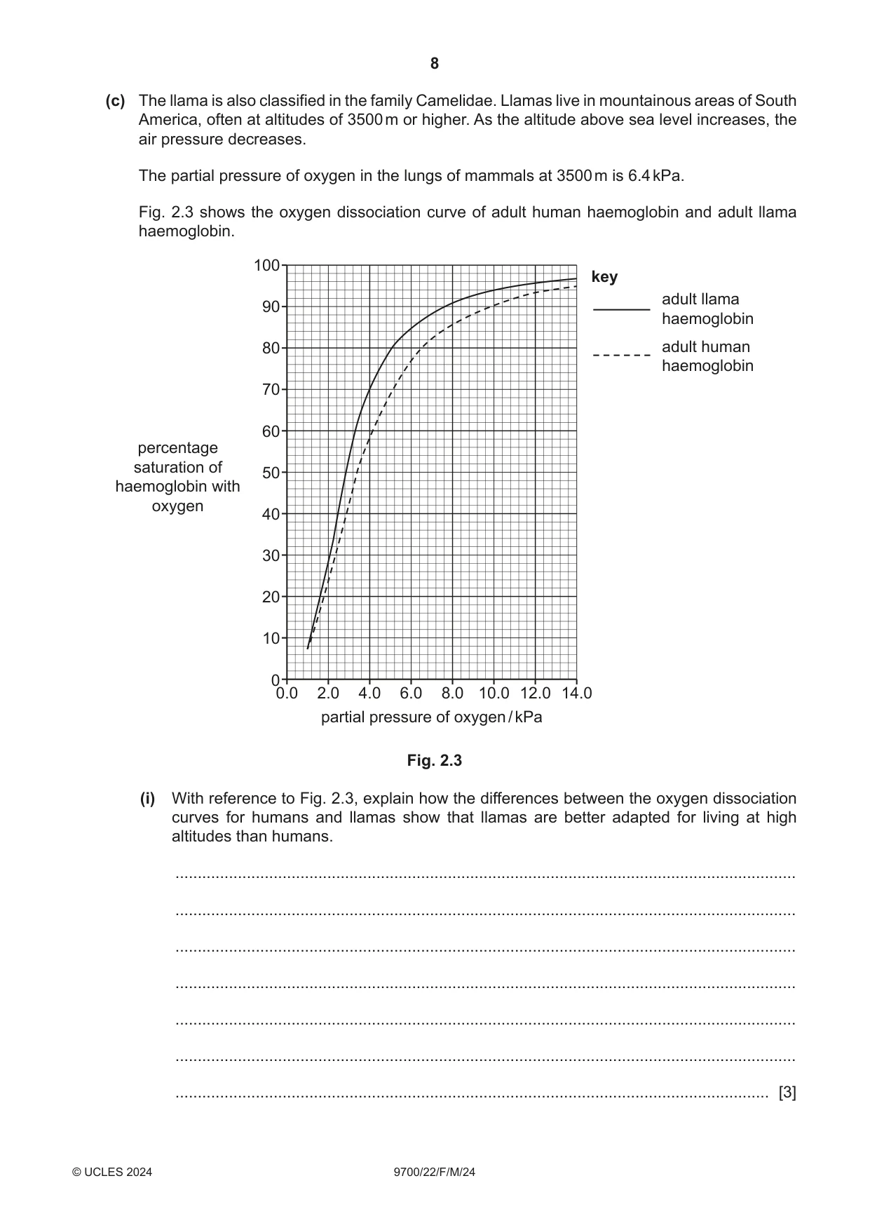
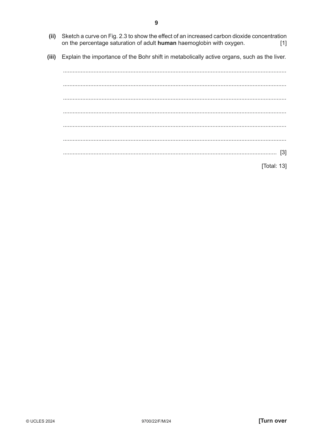
2 (a) Table 2.1 shows descriptions of three types of white blood cell. Complete Table 2.1 by stating the names of these three types of white blood cell. Table 2.1 description name of white blood cell A large cell that has a bean-shaped (kidney-shaped) nucleus. It can develop into a macrophage. A cell that has a large spherical nucleus and little cytoplasm. It responds to non-self antigens. A cell that has a lobed nucleus. It is phagocytic. [3] (b) Dromedary camels are classified in the family Camelidae and live in desert habitats of North Africa and Asia. In these hot, dry environments, dromedary camels can lose up to 30% of their body mass from dehydration, causing their blood to become more viscous (thicker). Fig. 2.1 shows a drawing of red blood cells of a dromedary camel. Fig. 2.2 is a drawing of human red blood cells. ×2000 ×2000 Fig. 2.1 Fig. 2.2 Fig. 2.1 and Fig. 2.2 show differences between the red blood cells of dromedary camels and the red blood cells of humans. Suggest how these differences adapt dromedary camels for living in hot, dry environments. ................................................................................................................................................... ................................................................................................................................................... ................................................................................................................................................... ................................................................................................................................................... ................................................................................................................................................... ................................................................................................................................................... ............................................................................................................................................. [3] (c) The llama is also classified in the family Camelidae. Llamas live in mountainous areas of South America, often at altitudes of 3500 m or higher. As the altitude above sea level increases, the air pressure decreases. The partial pressure of oxygen in the lungs of mammals at 3500 m is 6.4 kPa. Fig. 2.3 shows the oxygen dissociation curve of adult human haemoglobin and adult llama haemoglobin. 100 key adult llama 90 haemoglobin 80 adult human haemoglobin 70 60 percentage saturation of 50 haemoglobin with oxygen 40 30 20 10 0 0.0 2.0 4.0 6.0 8.0 10.0 12.0 14.0 partial pressure of oxygen / kPa Fig. 2.3 (i) With reference to Fig. 2.3, explain how the differences between the oxygen dissociation curves for humans and llamas show that llamas are better adapted for living at high altitudes than humans. ........................................................................................................................................... ........................................................................................................................................... ........................................................................................................................................... ........................................................................................................................................... ........................................................................................................................................... ........................................................................................................................................... ..................................................................................................................................... [3] (ii) Sketch a curve on Fig. 2.3 to show the effect of an increased carbon dioxide concentration on the percentage saturation of adult human haemoglobin with oxygen. [1] (iii) Explain the importance of the Bohr shift in metabolically active organs, such as the liver. ........................................................................................................................................... ........................................................................................................................................... ........................................................................................................................................... ........................................................................................................................................... ........................................................................................................................................... ........................................................................................................................................... ..................................................................................................................................... [3] [Total: 13]
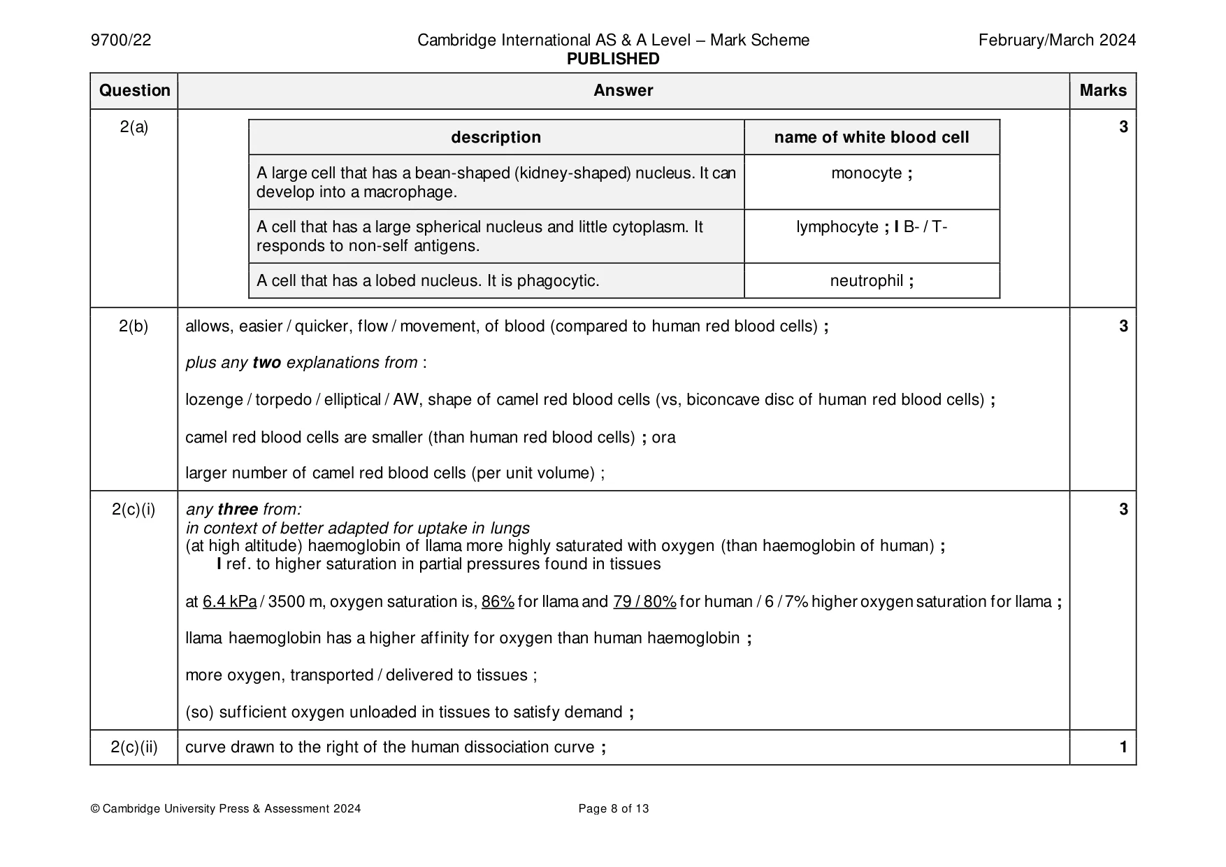
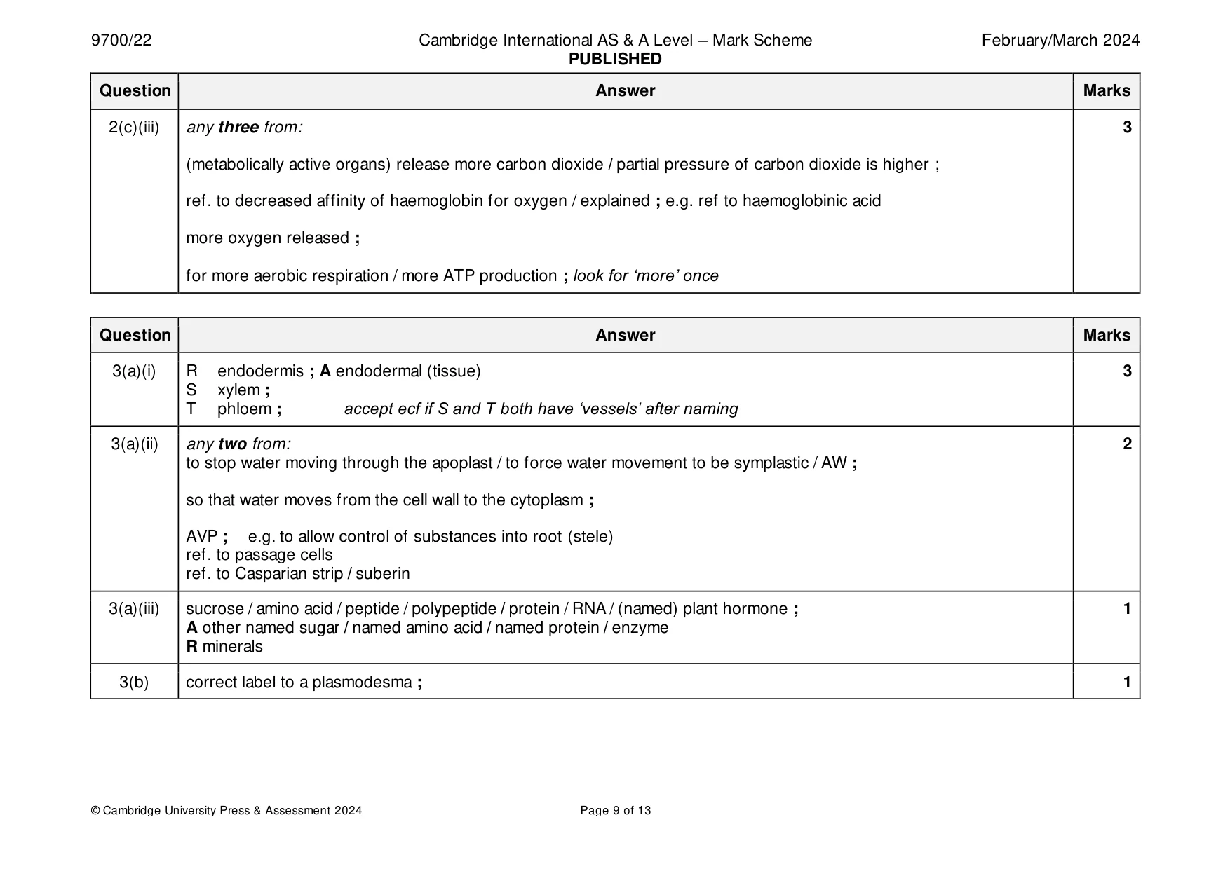
Mark scheme: 2(a) 3 description name of white blood cell A large cell that has a bean-shaped (kidney-shaped) nucleus. It can monocyte ; develop into a macrophage. A cell that has a large spherical nucleus and little cytoplasm. It lymphocyte ; I B- / T- responds to non-self antigens. A cell that has a lobed nucleus. It is phagocytic. neutrophil ; 2(b) allows, easier / quicker, flow / movement, of blood (compared to human red blood cells) ; 3 plus any two explanations from : lozenge / torpedo / elliptical / AW, shape of camel red blood cells (vs, biconcave disc of human red blood cells) ; camel red blood cells are smaller (than human red blood cells) ; ora larger number of camel red blood cells (per unit volume) ; 2(c)(i) any three from: 3 in context of better adapted for uptake in lungs (at high altitude) haemoglobin of llama more highly saturated with oxygen (than haemoglobin of human) ; I ref. to higher saturation in partial pressures found in tissues at 6.4 kPa / 3500 m, oxygen saturation is, 86% for llama and 79 / 80% for human / 6 / 7% higher oxygen saturation for llama ; llama haemoglobin has a higher affinity for oxygen than human haemoglobin ; more oxygen, transported / delivered to tissues ; (so) sufficient oxygen unloaded in tissues to satisfy demand ; 2(c)(ii) curve drawn to the right of the human dissociation curve ; 1 2(c)(iii) any three from: 3 (metabolically active organs) release more carbon dioxide / partial pressure of carbon dioxide is higher ; ref. to decreased affinity of haemoglobin for oxygen / explained ; e.g. ref to haemoglobinic acid more oxygen released ; for more aerobic respiration / more ATP production ; look for ‘more’ once
Q3 · A photomicrograph showing part of a transverse section through the root of an iris, Iris…
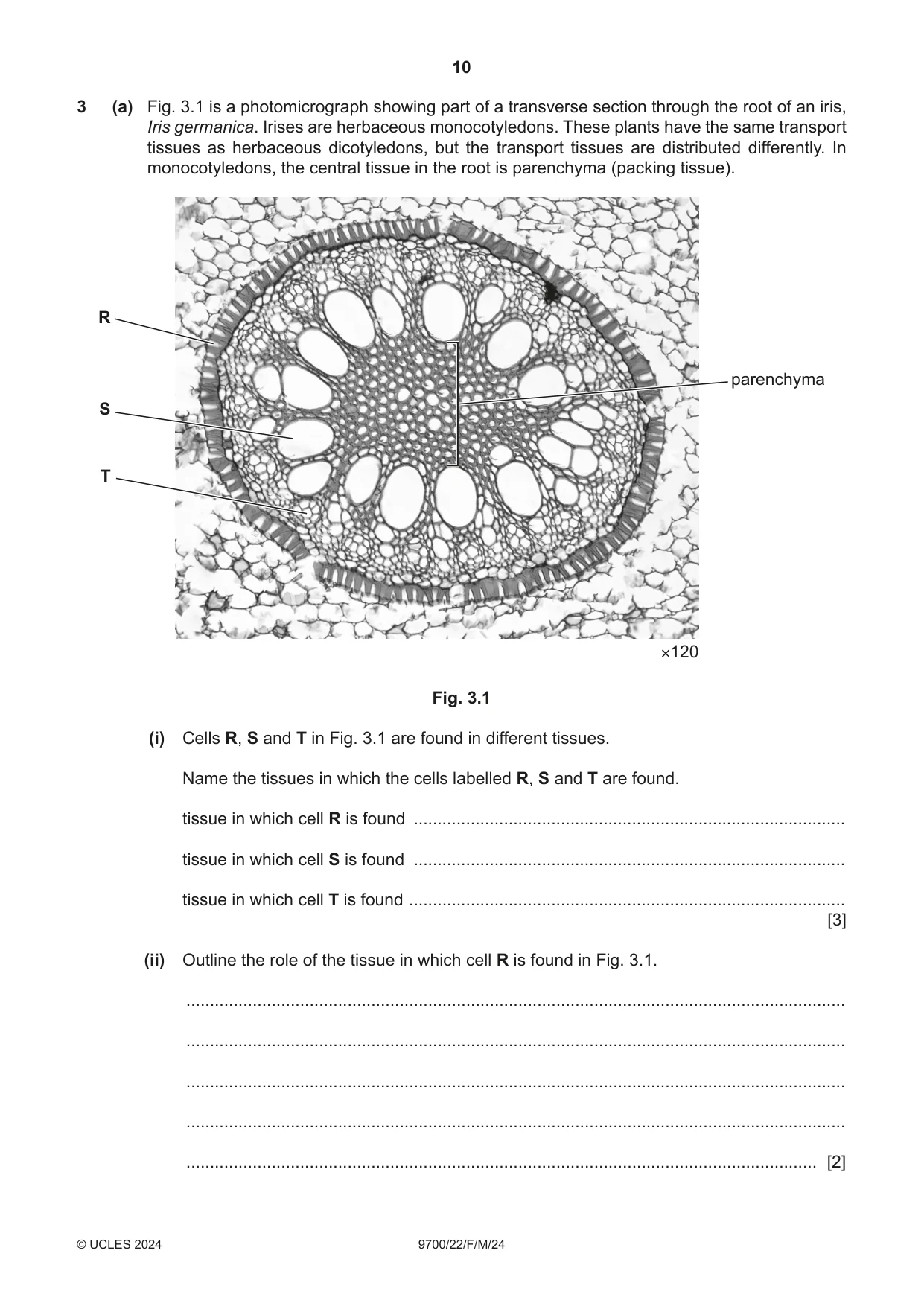
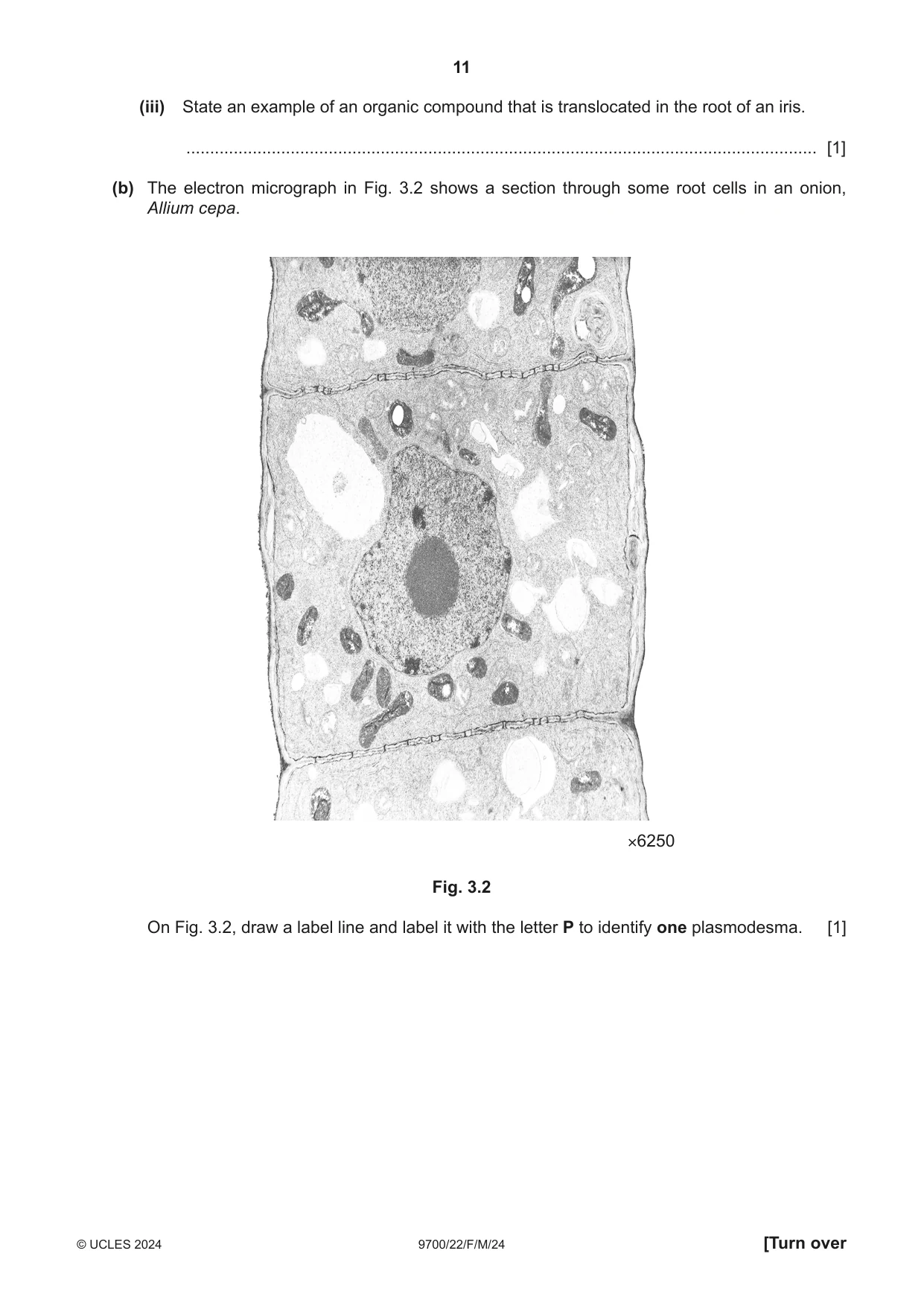
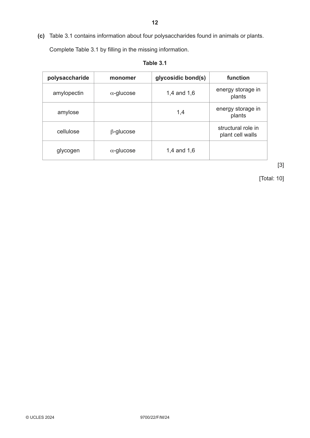
3 (a) Fig. 3.1 is a photomicrograph showing part of a transverse section through the root of an iris, Iris germanica. Irises are herbaceous monocotyledons. These plants have the same transport tissues as herbaceous dicotyledons, but the transport tissues are distributed differently. In monocotyledons, the central tissue in the root is parenchyma (packing tissue). R parenchyma S T ×120 Fig. 3.1 (i) Cells R, S and T in Fig. 3.1 are found in different tissues. Name the tissues in which the cells labelled R, S and T are found. tissue in which cell R is found ........................................................................................... tissue in which cell S is found ........................................................................................... tissue in which cell T is found ............................................................................................ [3] (ii) Outline the role of the tissue in which cell R is found in Fig. 3.1. ........................................................................................................................................... ........................................................................................................................................... ........................................................................................................................................... ........................................................................................................................................... ..................................................................................................................................... [2] (iii) State an example of an organic compound that is translocated in the root of an iris. ..................................................................................................................................... [1] (b) The electron micrograph in Fig. 3.2 shows a section through some root cells in an onion, Allium cepa. ×6250 Fig. 3.2 On Fig. 3.2, draw a label line and label it with the letter P to identify one plasmodesma. [1] (c) Table 3.1 contains information about four polysaccharides found in animals or plants. Complete Table 3.1 by filling in the missing information. Table 3.1 polysaccharide monomer glycosidic bond(s) function energy storage in amylopectin α-glucose 1,4 and 1,6 plants energy storage in amylose 1,4 plants structural role in cellulose β-glucose plant cell walls glycogen α-glucose 1,4 and 1,6 [3] [Total: 10] Question 4 starts on page 14.
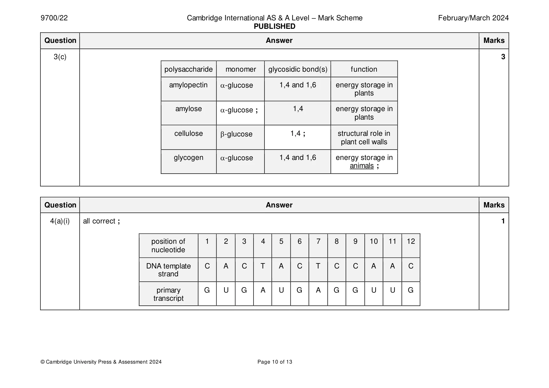
Mark scheme: 3(a)(i) R endodermis ; A endodermal (tissue) 3 S xylem ; T phloem ; accept ecf if S and T both have ‘vessels’ after naming 3(a)(ii) any two from: 2 to stop water moving through the apoplast / to force water movement to be symplastic / AW ; so that water moves from the cell wall to the cytoplasm ; AVP ; e.g. to allow control of substances into root (stele) ref. to passage cells ref. to Casparian strip / suberin 3(a)(iii) sucrose / amino acid / peptide / polypeptide / protein / RNA / (named) plant hormone ; 1 A other named sugar / named amino acid / named protein / enzyme R minerals 3(b) correct label to a plasmodesma ; 1 3(c) 3 polysaccharide monomer glycosidic bond(s) function amylopectin -glucose 1,4 and 1,6 energy storage in plants amylose -glucose ; 1,4 energy storage in plants cellulose -glucose 1,4 ; structural role in plant cell walls glycogen -glucose 1,4 and 1,6 energy storage in animals ;
Q4 · A sequence of 12 nucleotides in the template strand of a short length of a DNA molecule…
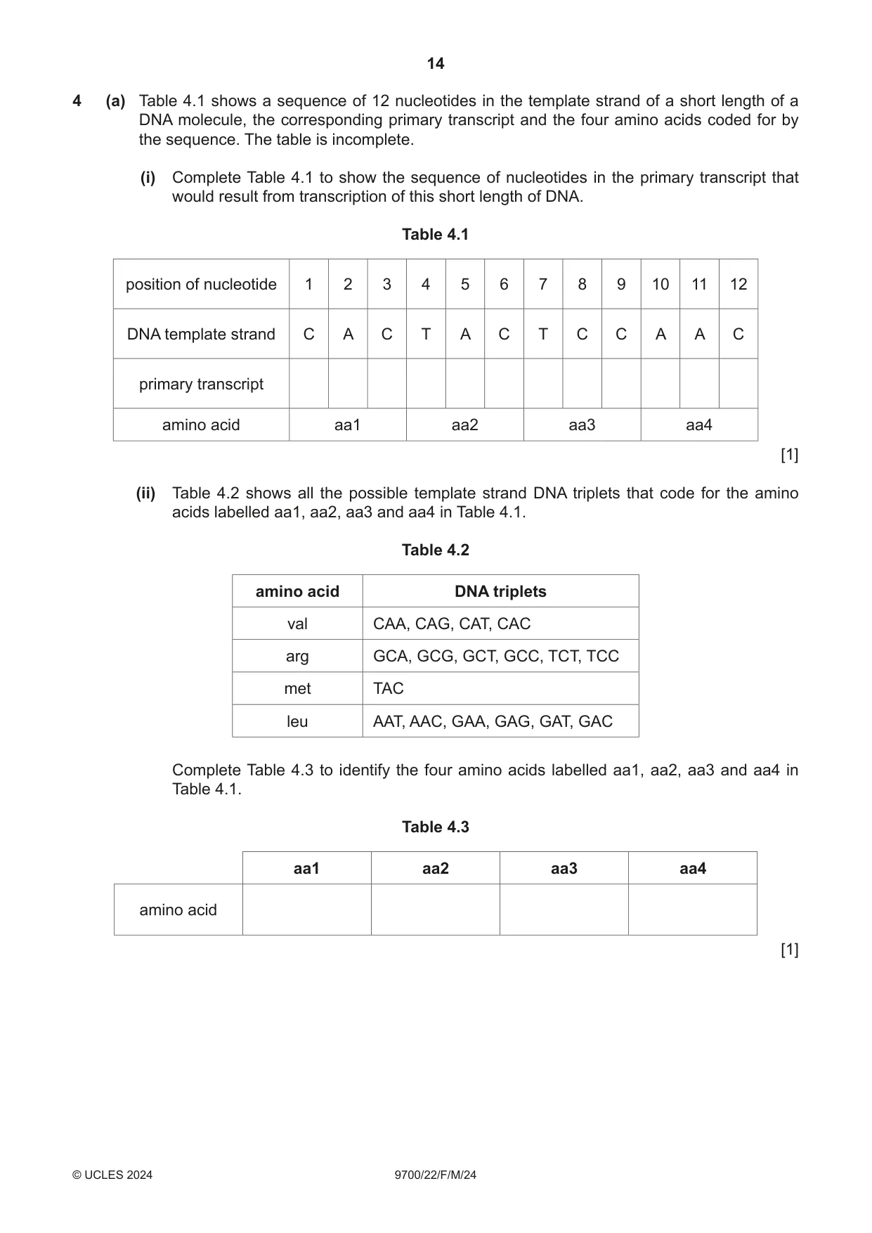
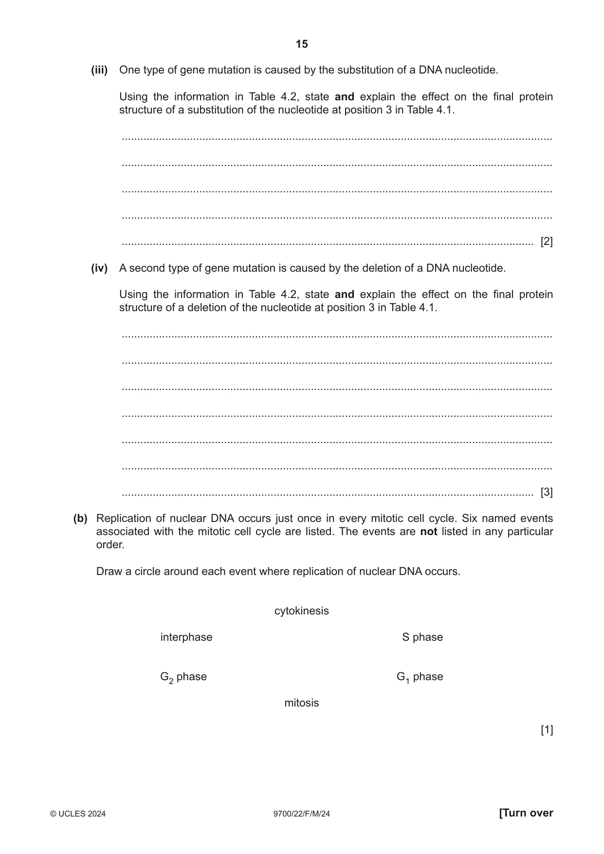
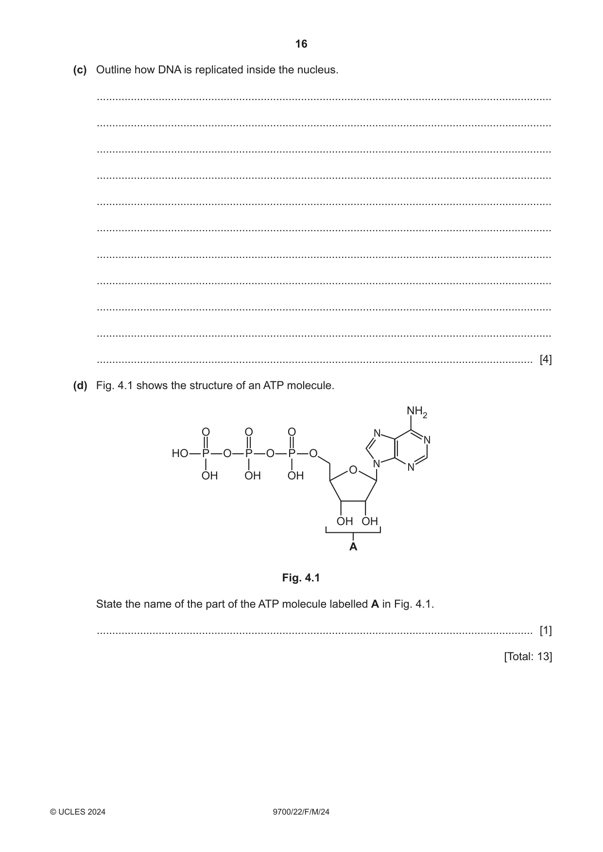
4 (a) Table 4.1 shows a sequence of 12 nucleotides in the template strand of a short length of a DNA molecule, the corresponding primary transcript and the four amino acids coded for by the sequence. The table is incomplete. (i) Complete Table 4.1 to show the sequence of nucleotides in the primary transcript that would result from transcription of this short length of DNA. Table 4.1 position of nucleotide 1 2 3 4 5 6 7 8 9 10 11 12 DNA template strand C A C T A C T C C A A C primary transcript amino acid aa1 aa2 aa3 aa4 [1] (ii) Table 4.2 shows all the possible template strand DNA triplets that code for the amino acids labelled aa1, aa2, aa3 and aa4 in Table 4.1. Table 4.2 amino acid DNA triplets val CAA, CAG, CAT, CAC arg GCA, GCG, GCT, GCC, TCT, TCC met TAC leu AAT, AAC, GAA, GAG, GAT, GAC Complete Table 4.3 to identify the four amino acids labelled aa1, aa2, aa3 and aa4 in Table 4.1. Table 4.3 aa1 aa2 aa3 aa4 amino acid [1] (iii) One type of gene mutation is caused by the substitution of a DNA nucleotide. Using the information in Table 4.2, state and explain the effect on the final protein structure of a substitution of the nucleotide at position 3 in Table 4.1. ........................................................................................................................................... ........................................................................................................................................... ........................................................................................................................................... ........................................................................................................................................... ..................................................................................................................................... [2] (iv) A second type of gene mutation is caused by the deletion of a DNA nucleotide. Using the information in Table 4.2, state and explain the effect on the final protein structure of a deletion of the nucleotide at position 3 in Table 4.1. ........................................................................................................................................... ........................................................................................................................................... ........................................................................................................................................... ........................................................................................................................................... ........................................................................................................................................... ........................................................................................................................................... ..................................................................................................................................... [3] (b) Replication of nuclear DNA occurs just once in every mitotic cell cycle. Six named events associated with the mitotic cell cycle are listed. The events are not listed in any particular order. Draw a circle around each event where replication of nuclear DNA occurs. cytokinesis interphase S phase G2 phase G1 phase mitosis [1] (c) Outline how DNA is replicated inside the nucleus. ................................................................................................................................................... ................................................................................................................................................... ................................................................................................................................................... ................................................................................................................................................... ................................................................................................................................................... ................................................................................................................................................... ................................................................................................................................................... ................................................................................................................................................... ................................................................................................................................................... ................................................................................................................................................... ............................................................................................................................................. [4] (d) Fig. 4.1 shows the structure of an ATP molecule. NH2 O O O N N HO P O P O P O N O N OH OH OH OH OH A Fig. 4.1 State the name of the part of the ATP molecule labelled A in Fig. 4.1. ............................................................................................................................................. [1] [Total: 13]
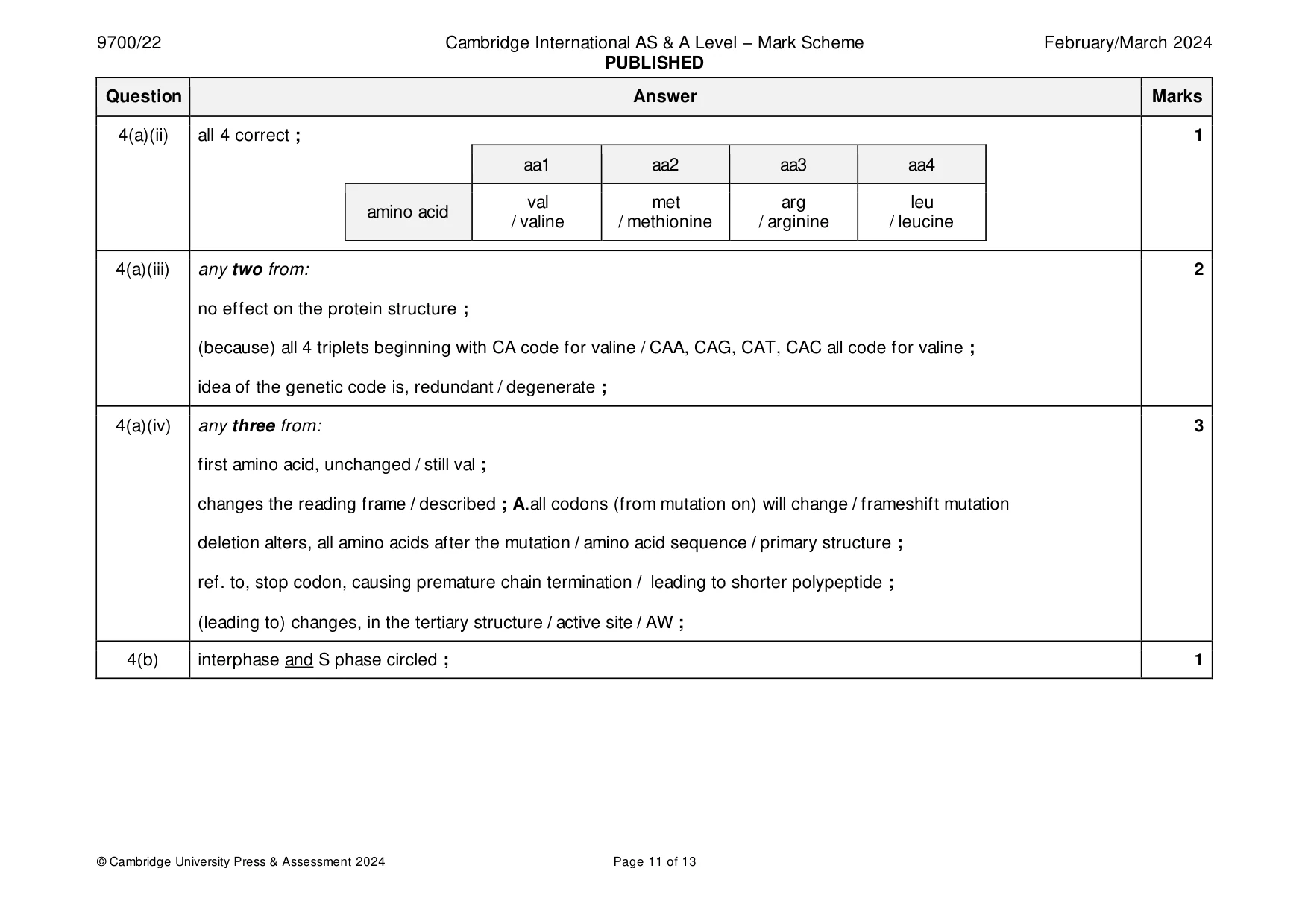
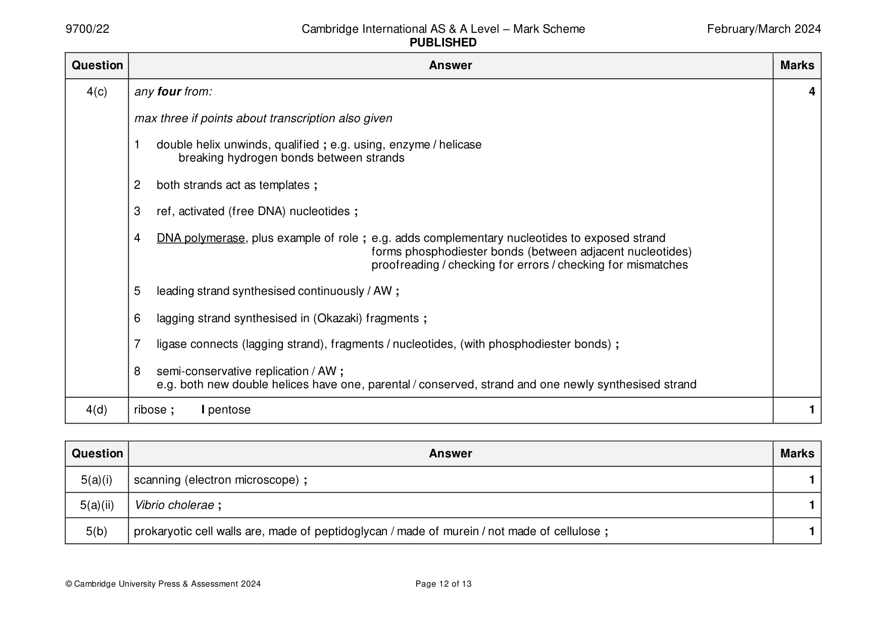
Mark scheme: 4(a)(i) all correct ; 1 position of 1 2 3 4 5 6 7 8 9 10 11 12 nucleotide DNA template C A C T A C T C C A A C strand primary G U G A U G A G G U U G transcript 4(a)(ii) all 4 correct ; 1 aa1 aa2 aa3 aa4 val met arg leu amino acid / valine / methionine / arginine / leucine 4(a)(iii) any two from: 2 no effect on the protein structure ; (because) all 4 triplets beginning with CA code for valine / CAA, CAG, CAT, CAC all code for valine ; idea of the genetic code is, redundant / degenerate ; 4(a)(iv) any three from: 3 first amino acid, unchanged / still val ; changes the reading frame / described ; A.all codons (from mutation on) will change / frameshift mutation deletion alters, all amino acids after the mutation / amino acid sequence / primary structure ; ref. to, stop codon, causing premature chain termination / leading to shorter polypeptide ; (leading to) changes, in the tertiary structure / active site / AW ; 4(b) interphase and S phase circled ; 1 4(c) any four from: 4 max three if points about transcription also given 1 double helix unwinds, qualified ; e.g. using, enzyme / helicase breaking hydrogen bonds between strands 2 both strands act as templates ; 3 ref, activated (free DNA) nucleotides ; 4 DNA polymerase, plus example of role ; e.g. adds complementary nucleotides to exposed strand forms phosphodiester bonds (between adjacent nucleotides) proofreading / checking for errors / checking for mismatches 5 leading strand synthesised continuously / AW ; 6 lagging strand synthesised in (Okazaki) fragments ; 7 ligase connects (lagging strand), fragments / nucleotides, (with phosphodiester bonds) ; 8 semi-conservative replication / AW ; e.g. both new double helices have one, parental / conserved, strand and one newly synthesised strand 4(d) ribose ; I pentose 1
Q5 · The pathogen that causes cholera is a prokaryote
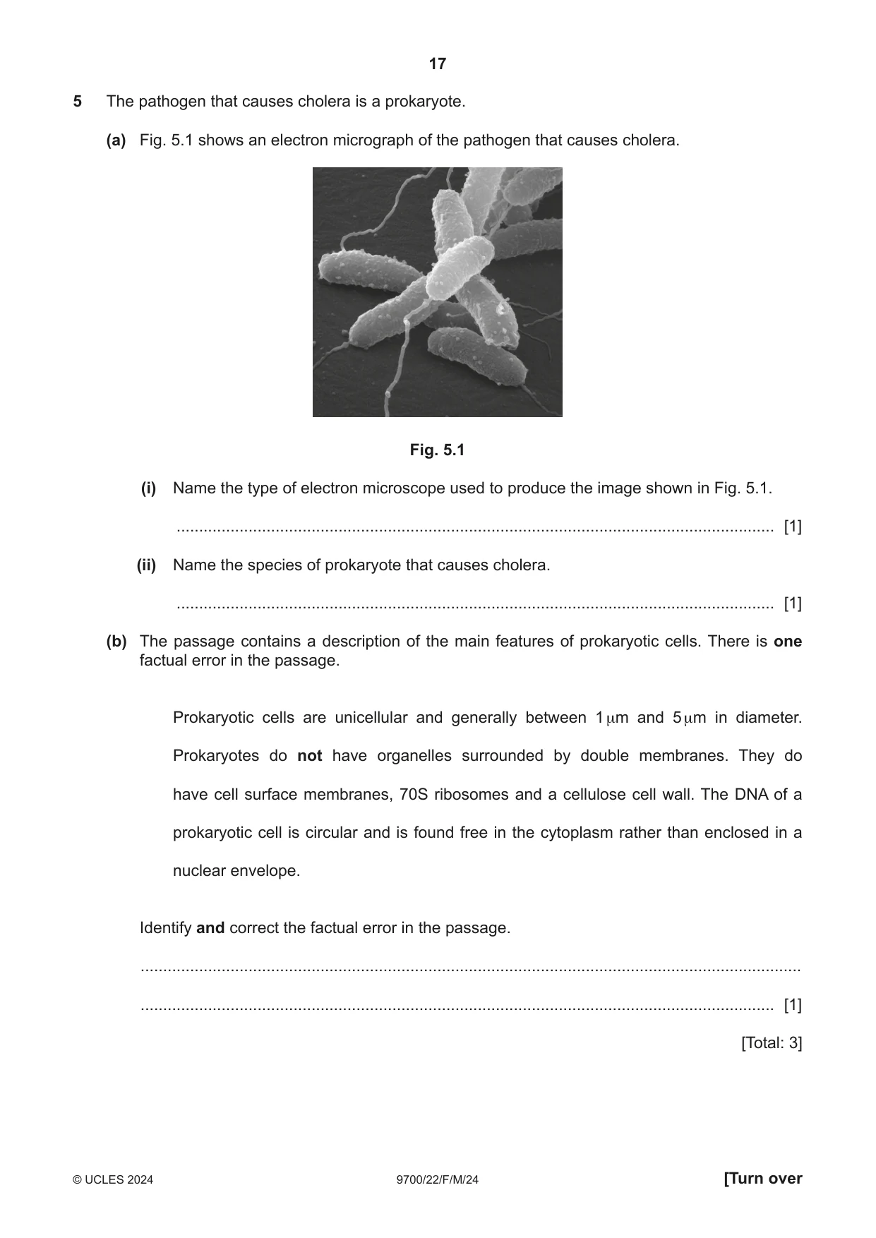
5 The pathogen that causes cholera is a prokaryote. (a) Fig. 5.1 shows an electron micrograph of the pathogen that causes cholera. Fig. 5.1 (i) Name the type of electron microscope used to produce the image shown in Fig. 5.1. ..................................................................................................................................... [1] (ii) Name the species of prokaryote that causes cholera. ..................................................................................................................................... [1] (b) The passage contains a description of the main features of prokaryotic cells. There is one factual error in the passage. Prokaryotic cells are unicellular and generally between 1 μm and 5 μm in diameter. Prokaryotes do not have organelles surrounded by double membranes. They do have cell surface membranes, 70S ribosomes and a cellulose cell wall. The DNA of a prokaryotic cell is circular and is found free in the cytoplasm rather than enclosed in a nuclear envelope. Identify and correct the factual error in the passage. ................................................................................................................................................... ............................................................................................................................................. [1] [Total: 3]
Mark scheme: 5(a)(i) scanning (electron microscope) ; 1 5(a)(ii) Vibrio cholerae ; 1 5(b) prokaryotic cell walls are, made of peptidoglycan / made of murein / not made of cellulose ; 1
Q6 · A simplified diagram representing a section through the human immunodeficiency virus…
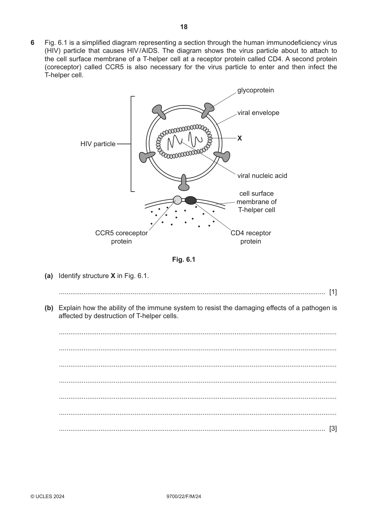
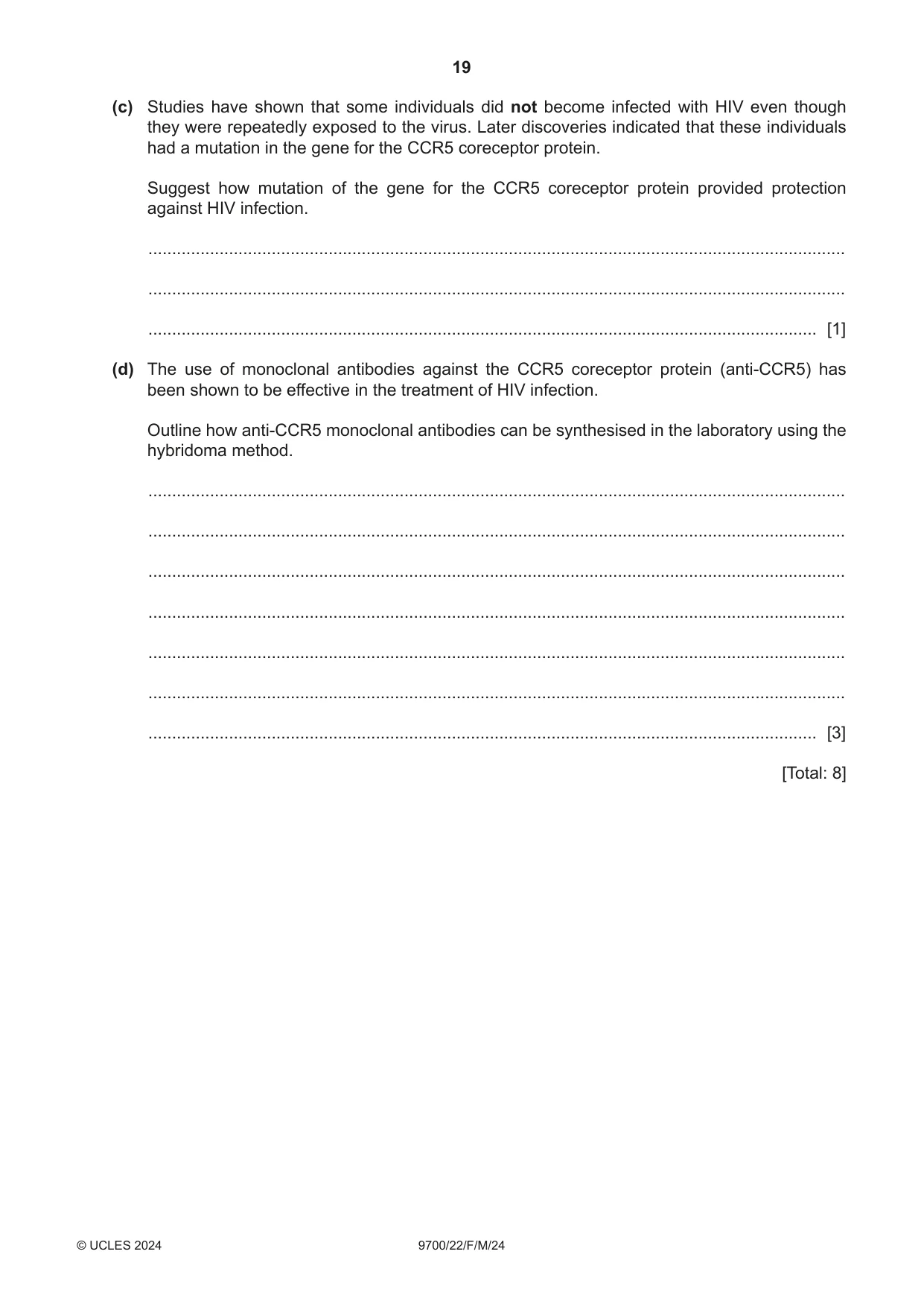
6 Fig. 6.1 is a simplified diagram representing a section through the human immunodeficiency virus (HIV) particle that causes HIV / AIDS. The diagram shows the virus particle about to attach to the cell surface membrane of a T-helper cell at a receptor protein called CD4. A second protein (coreceptor) called CCR5 is also necessary for the virus particle to enter and then infect the T-helper cell. glycoprotein viral envelope X HIV particle viral nucleic acid cell surface membrane of T-helper cell CCR5 coreceptor CD4 receptor protein protein Fig. 6.1 (a) Identify structure X in Fig. 6.1. ............................................................................................................................................. [1] (b) Explain how the ability of the immune system to resist the damaging effects of a pathogen is affected by destruction of T-helper cells. ................................................................................................................................................... ................................................................................................................................................... ................................................................................................................................................... ................................................................................................................................................... ................................................................................................................................................... ................................................................................................................................................... ............................................................................................................................................. [3] (c) Studies have shown that some individuals did not become infected with HIV even though they were repeatedly exposed to the virus. Later discoveries indicated that these individuals had a mutation in the gene for the CCR5 coreceptor protein. Suggest how mutation of the gene for the CCR5 coreceptor protein provided protection against HIV infection. ................................................................................................................................................... ................................................................................................................................................... ............................................................................................................................................. [1] (d) The use of monoclonal antibodies against the CCR5 coreceptor protein (anti-CCR5) has been shown to be effective in the treatment of HIV infection. Outline how anti-CCR5 monoclonal antibodies can be synthesised in the laboratory using the hybridoma method. ................................................................................................................................................... ................................................................................................................................................... ................................................................................................................................................... ................................................................................................................................................... ................................................................................................................................................... ................................................................................................................................................... ............................................................................................................................................. [3] [Total: 8]
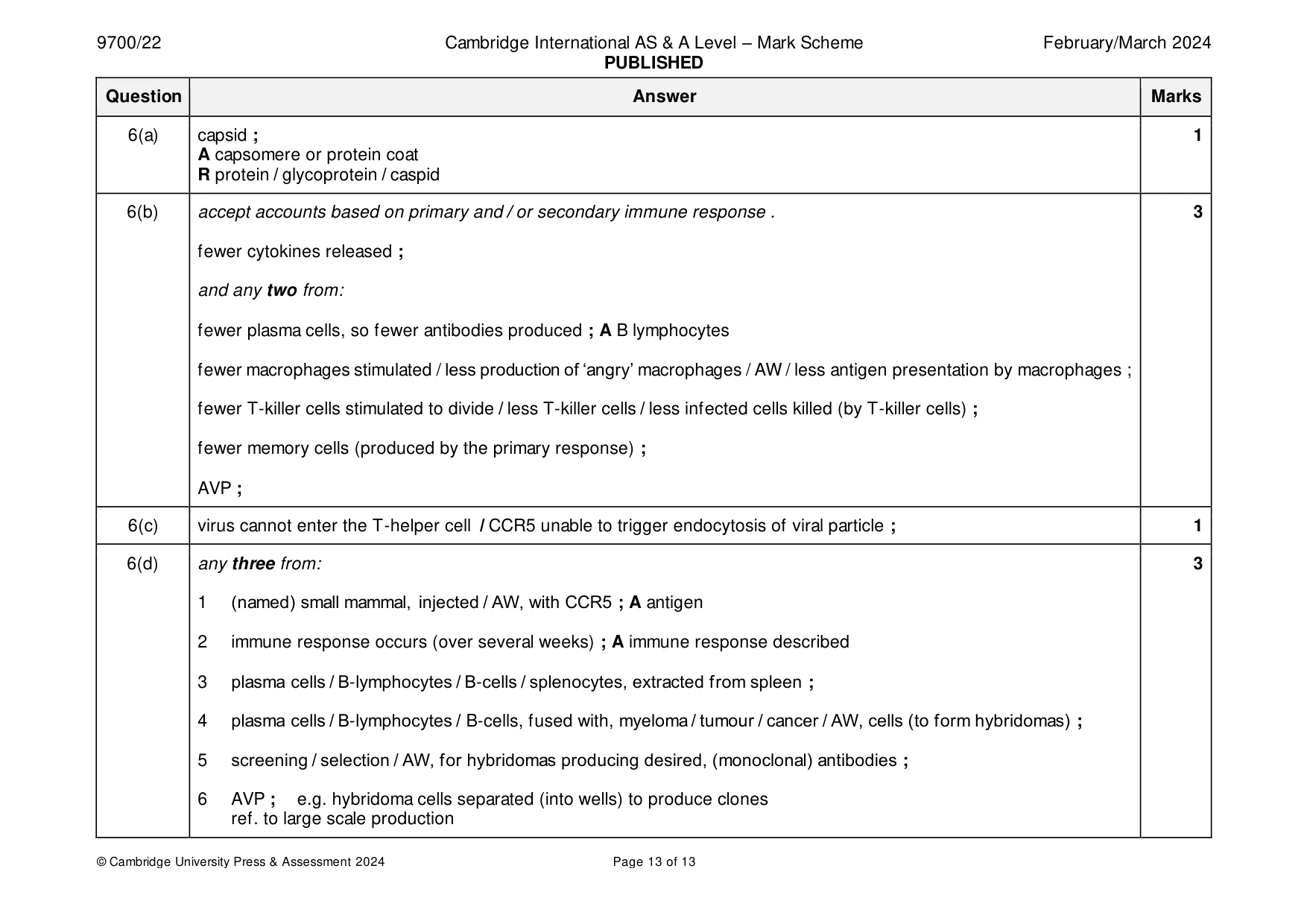
Mark scheme: 6(a) capsid ; 1 A capsomere or protein coat R protein / glycoprotein / caspid 6(b) accept accounts based on primary and / or secondary immune response . 3 fewer cytokines released ; and any two from: fewer plasma cells, so fewer antibodies produced ; A B lymphocytes fewer macrophages stimulated / less production of ‘angry’ macrophages / AW / less antigen presentation by macrophages ; fewer T-killer cells stimulated to divide / less T-killer cells / less infected cells killed (by T-killer cells) ; fewer memory cells (produced by the primary response) ; AVP ; 6(c) virus cannot enter the T-helper cell / CCR5 unable to trigger endocytosis of viral particle ; 1 6(d) any three from: 3 1 (named) small mammal, injected / AW, with CCR5 ; A antigen 2 immune response occurs (over several weeks) ; A immune response described 3 plasma cells / B-lymphocytes / B-cells / splenocytes, extracted from spleen ; 4 plasma cells / B-lymphocytes / B-cells, fused with, myeloma / tumour / cancer / AW, cells (to form hybridomas) ; 5 screening / selection / AW, for hybridomas producing desired, (monoclonal) antibodies ; 6 AVP ; e.g. hybridoma cells separated (into wells) to produce clones ref. to large scale production
What was in this paper
The subtopics covered by these 6 questions, and how many questions each got. Open one in a new tab to see every Cambridge question on it.
What you needed in this session
Cambridge’s own grade thresholds for 2024 Feb/March, Paper 2 · Variant 2. A higher threshold means an easier paper — the bar moves with how the cohort did.