Cambridge A Level Biology 9700 — 2025 May/June Paper 2 · Variant 4
9700/24/M/J/25 · 6 questions · 60 marks · 75 min
The question paper and its mark scheme, free to read here and free to download. This is Cambridge’s own paper, exactly as it was sat.
Question paper16 pages
Mark scheme20 pages
Answers below. Sit the paper first if you are practising.
Questions as text
Q1 · In the roots of plants, specialised tissues are formed from a region in the root tip…
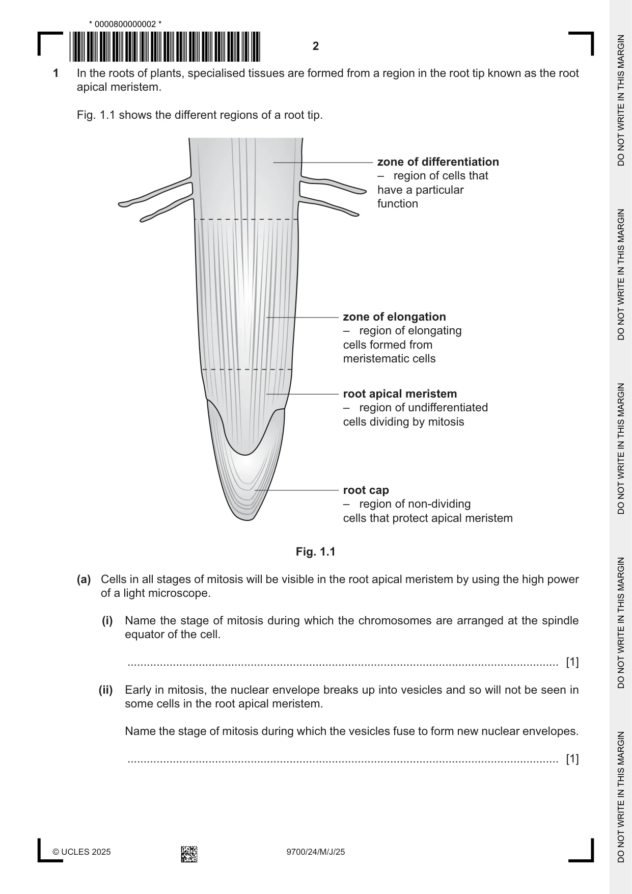
1 In the roots of plants, specialised tissues are formed from a region in the root tip known as the root apical meristem. Fig. 1.1 shows the different regions of a root tip. zone of differentiation – region of cells that have a particular function zone of elongation – region of elongating cells formed from meristematic cells root apical meristem – region of undifferentiated cells dividing by mitosis root cap – region of non-dividing cells that protect apical meristem Fig. 1.1 (a) Cells in all stages of mitosis will be visible in the root apical meristem by using the high power of a light microscope. (i) Name the stage of mitosis during which the chromosomes are arranged at the spindle equator of the cell. ..................................................................................................................................... [1] (ii) Early in mitosis, the nuclear envelope breaks up into vesicles and so will not be seen in some cells in the root apical meristem. Name the stage of mitosis during which the vesicles fuse to form new nuclear envelopes. ..................................................................................................................................... [1] (b) In the zone of elongation, shown in Fig. 1.1, the newly formed cells expand and elongate. Two main events that occur in these cells are: • the entry of water into the cells • the formation of the large vacuole. (i) Outline the process by which water enters the cells. ........................................................................................................................................... ........................................................................................................................................... ........................................................................................................................................... ........................................................................................................................................... ........................................................................................................................................... ........................................................................................................................................... ..................................................................................................................................... [3] (ii) Outline the structure of a fully mature plant vacuole. ........................................................................................................................................... ........................................................................................................................................... ........................................................................................................................................... ..................................................................................................................................... [2] (c) In the zone of differentiation, shown in Fig. 1.1, two types of vascular (transport) tissue form: xylem and phloem. Draw a diagram showing the distribution of the vascular tissue as seen in a transverse section of a root in the zone of differentiation. [2] (d) Starch can be stored in the roots of plants. This can be converted to sucrose, which is loaded into phloem sieve tubes for transport within the phloem sap to growing areas in the stem. After loading of sucrose into phloem sieve tubes, the transport of phloem sap occurs by mass flow. Explain how the phloem sap is moved within the sieve tubes by mass flow from the root storage areas to the growing areas of the stem. ................................................................................................................................................... ................................................................................................................................................... ................................................................................................................................................... ................................................................................................................................................... ................................................................................................................................................... ................................................................................................................................................... ................................................................................................................................................... ................................................................................................................................................... ............................................................................................................................................. [4] [Total: 13]
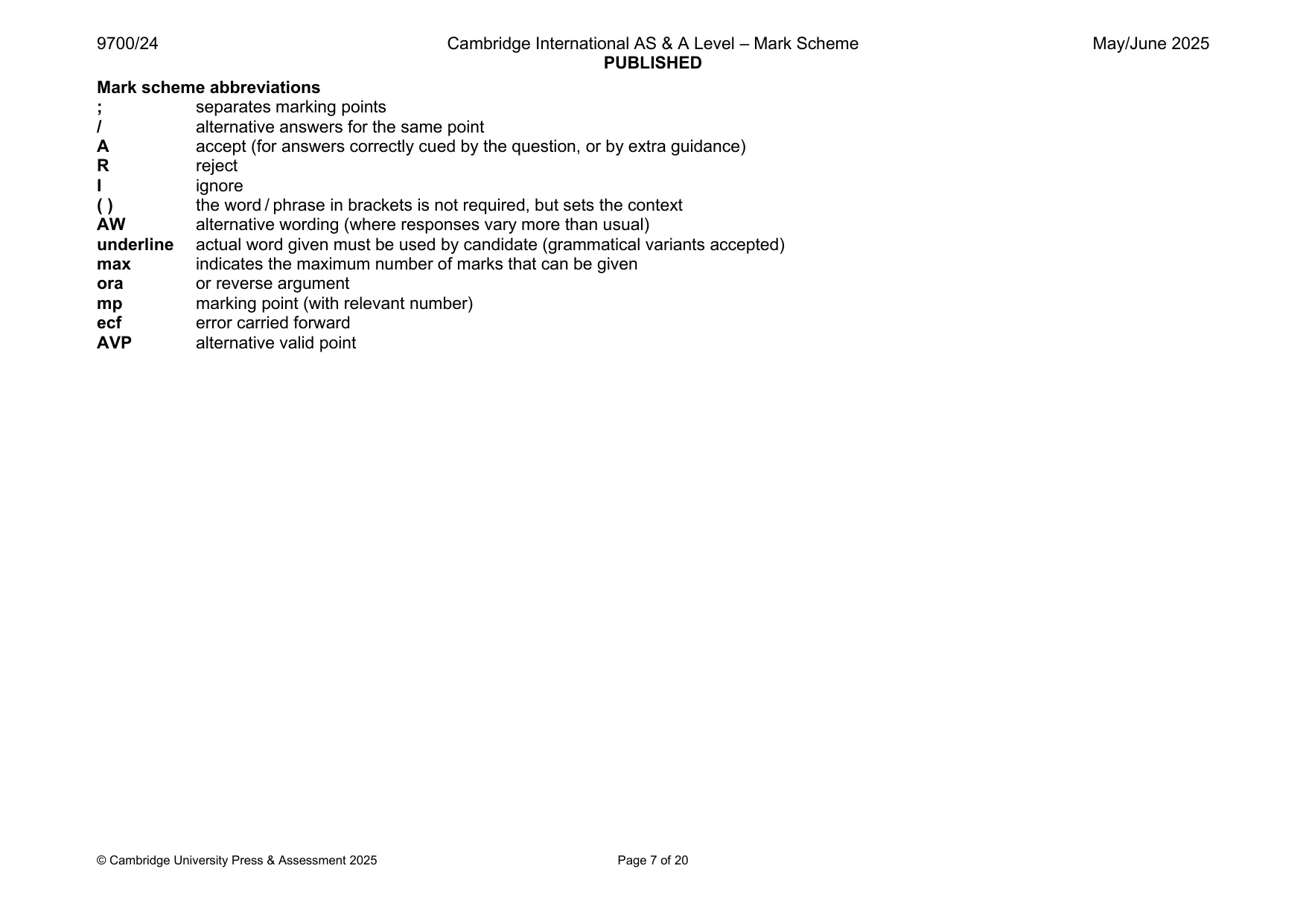
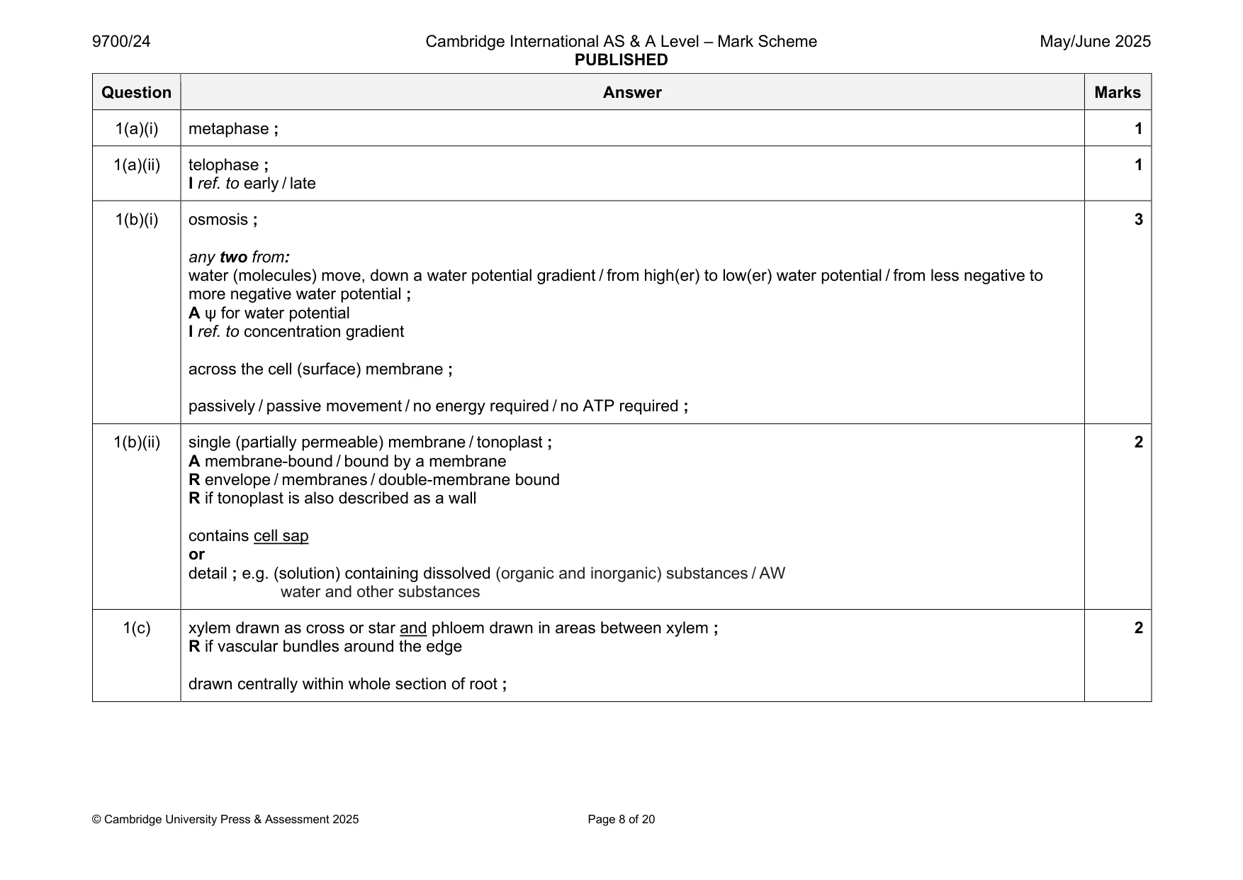
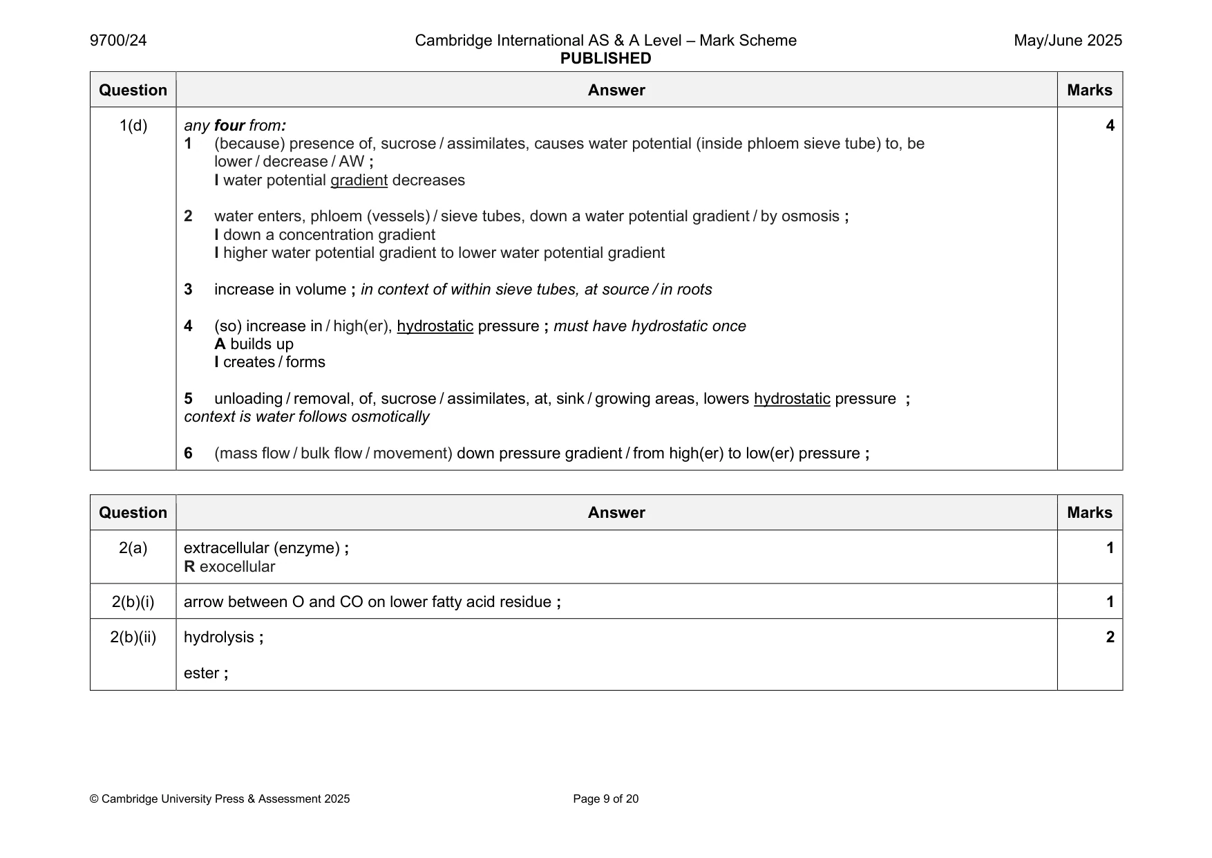
Mark scheme: Question Answer Marks 1(a)(i) metaphase ; 1 1(a)(ii) telophase ; 1 I ref. to early / late 1(b)(i) osmosis ; 3 any two from: water (molecules) move, down a water potential gradient / from high(er) to low(er) water potential / from less negative to more negative water potential ; A ψ for water potential I ref. to concentration gradient across the cell (surface) membrane ; passively / passive movement / no energy required / no ATP required ; 1(b)(ii) single (partially permeable) membrane / tonoplast ; 2 A membrane-bound / bound by a membrane R envelope / membranes / double-membrane bound R if tonoplast is also described as a wall contains cell sap or detail ; e.g. (solution) containing dissolved (organic and inorganic) substances / AW water and other substances 1(c) xylem drawn as cross or star and phloem drawn in areas between xylem ; 2 R if vascular bundles around the edge drawn centrally within whole section of root ; 1(d) any four from: 4 1 (because) presence of, sucrose / assimilates, causes water potential (inside phloem sieve tube) to, be lower / decrease / AW ; I water potential gradient decreases 2 water enters, phloem (vessels) / sieve tubes, down a water potential gradient / by osmosis ; I down a concentration gradient I higher water potential gradient to lower water potential gradient 3 increase in volume ; in context of within sieve tubes, at source / in roots 4 (so) increase in / high(er), hydrostatic pressure ; must have hydrostatic once A builds up I creates / forms 5 unloading / removal, of, sucrose / assimilates, at, sink / growing areas, lowers hydrostatic pressure ; context is water follows osmotically 6 (mass flow / bulk flow / movement) down pressure gradient / from high(er) to low(er) pressure ;
Q2 · The human pancreas synthesises and secretes a number of different digestive enzymes
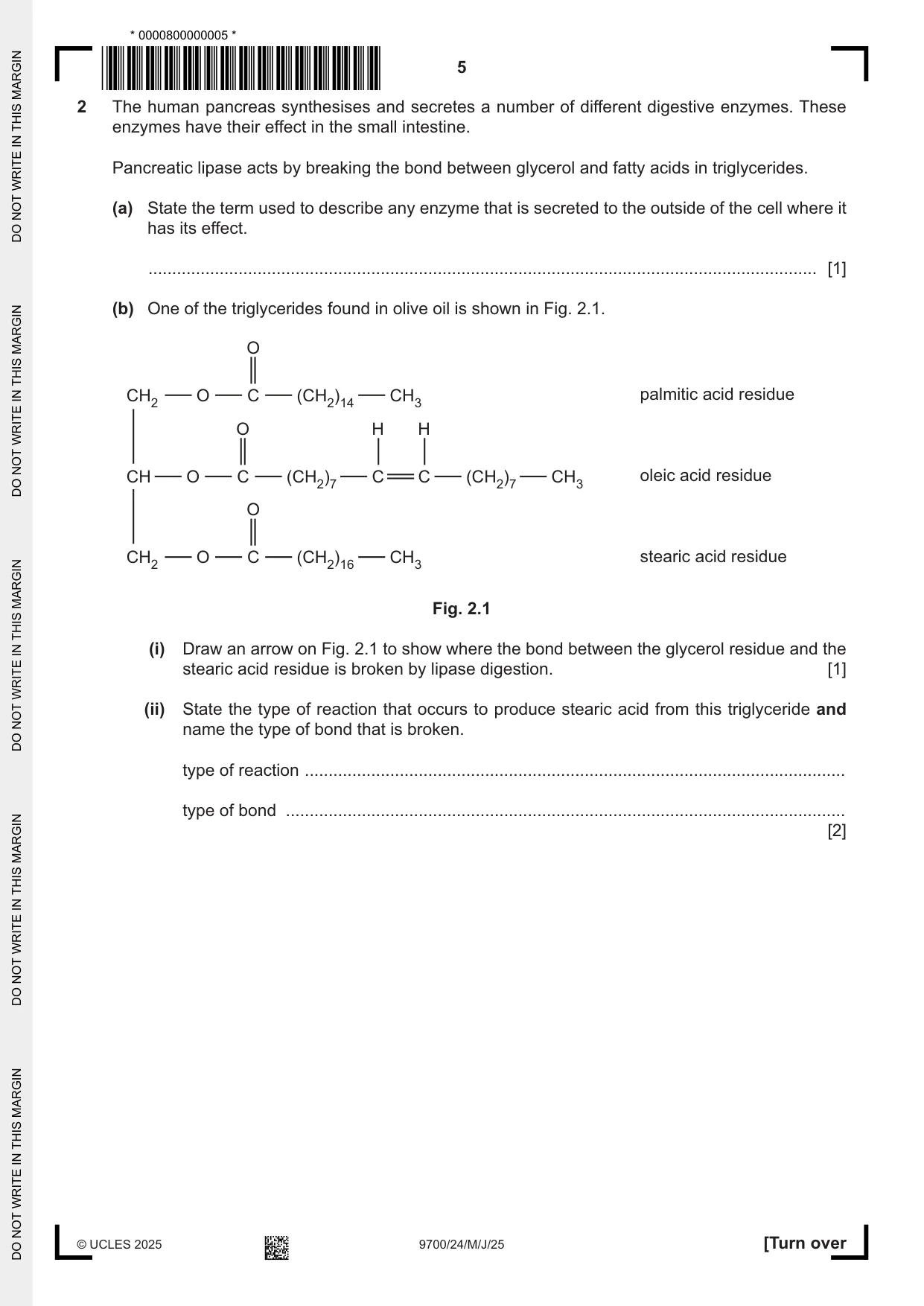
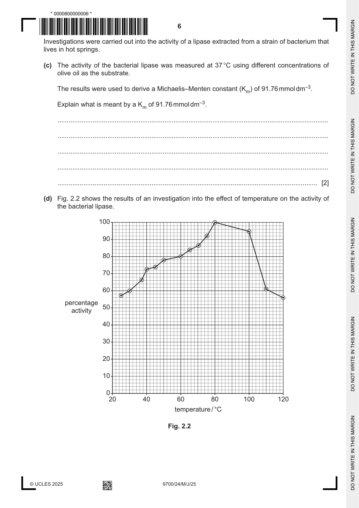
2 The human pancreas synthesises and secretes a number of different digestive enzymes. These enzymes have their effect in the small intestine. Pancreatic lipase acts by breaking the bond between glycerol and fatty acids in triglycerides. (a) State the term used to describe any enzyme that is secreted to the outside of the cell where it has its effect. ............................................................................................................................................. [1] (b) One of the triglycerides found in olive oil is shown in Fig. 2.1. O CH2 O C (CH2)14 CH3 palmitic acid residue O H H CH O C (CH2)7 C C (CH2)7 CH3 oleic acid residue O CH2 O C (CH2)16 CH3 stearic acid residue Fig. 2.1 (i) Draw an arrow on Fig. 2.1 to show where the bond between the glycerol residue and the stearic acid residue is broken by lipase digestion. [1] (ii) State the type of reaction that occurs to produce stearic acid from this triglyceride and name the type of bond that is broken. type of reaction .................................................................................................................. type of bond ...................................................................................................................... [2] Investigations were carried out into the activity of a lipase extracted from a strain of bacterium that lives in hot springs. (c) The activity of the bacterial lipase was measured at 37 °C using different concentrations of olive oil as the substrate. The results were used to derive a Michaelis–Menten constant (Km) of 91.76 mmol dm–3. Explain what is meant by a Km of 91.76 mmol dm–3. ................................................................................................................................................... ................................................................................................................................................... ................................................................................................................................................... ................................................................................................................................................... ............................................................................................................................................. [2] (d) Fig. 2.2 shows the results of an investigation into the effect of temperature on the activity of the bacterial lipase. 100 90 80 70 60 percentage 50 activity 40 30 20 10 0 20 40 60 80 100 120 temperature / °C Fig. 2.2 (i) Describe and explain the results shown in Fig. 2.2. ........................................................................................................................................... ........................................................................................................................................... ........................................................................................................................................... ........................................................................................................................................... ........................................................................................................................................... ........................................................................................................................................... ........................................................................................................................................... ........................................................................................................................................... ........................................................................................................................................... ........................................................................................................................................... ........................................................................................................................................... ........................................................................................................................................... ..................................................................................................................................... [4] (ii) An investigation using human pancreatic lipase at the same range of temperature will produce different results from those shown in Fig. 2.2. Predict how the trend will be different from that shown in Fig. 2.2. ........................................................................................................................................... ........................................................................................................................................... ........................................................................................................................................... ........................................................................................................................................... ..................................................................................................................................... [2] [Total: 12]
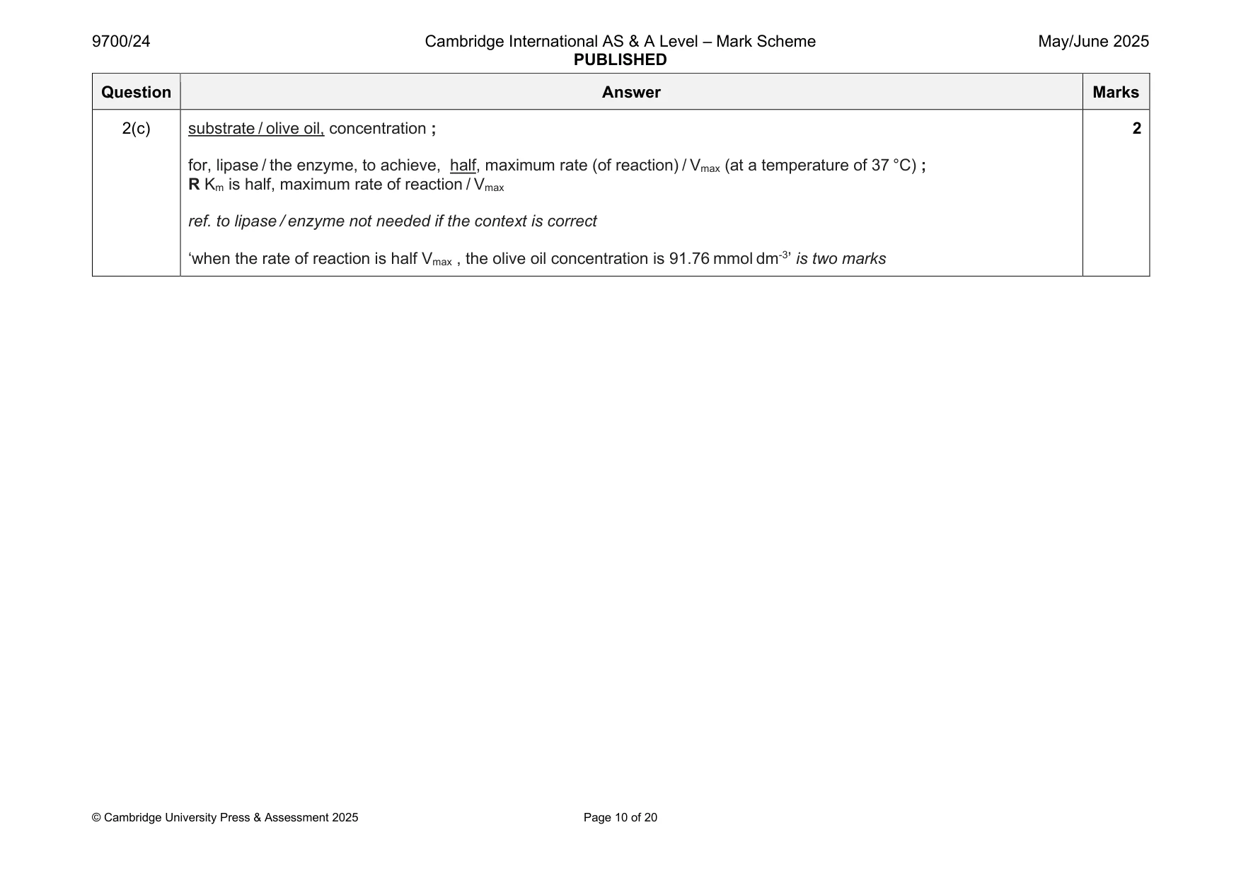
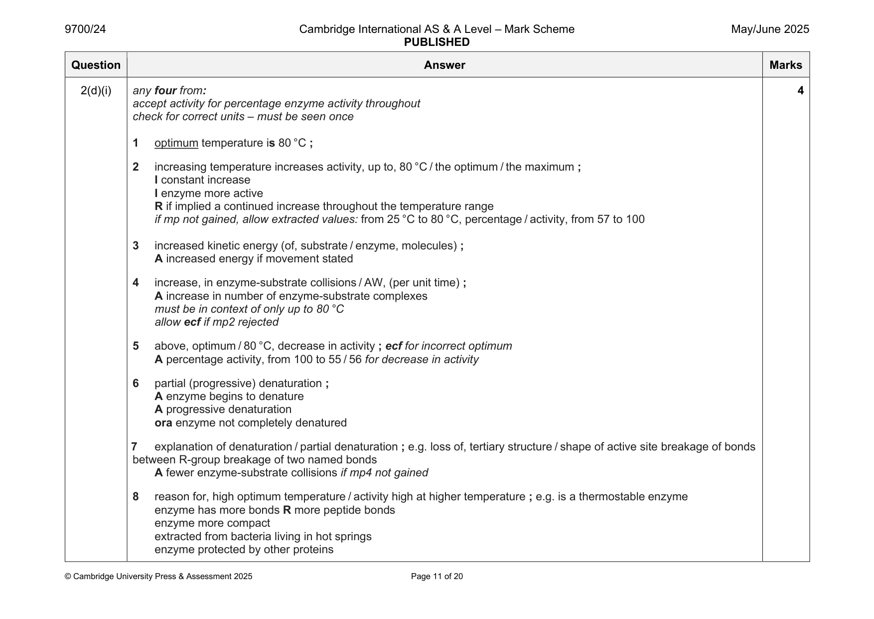
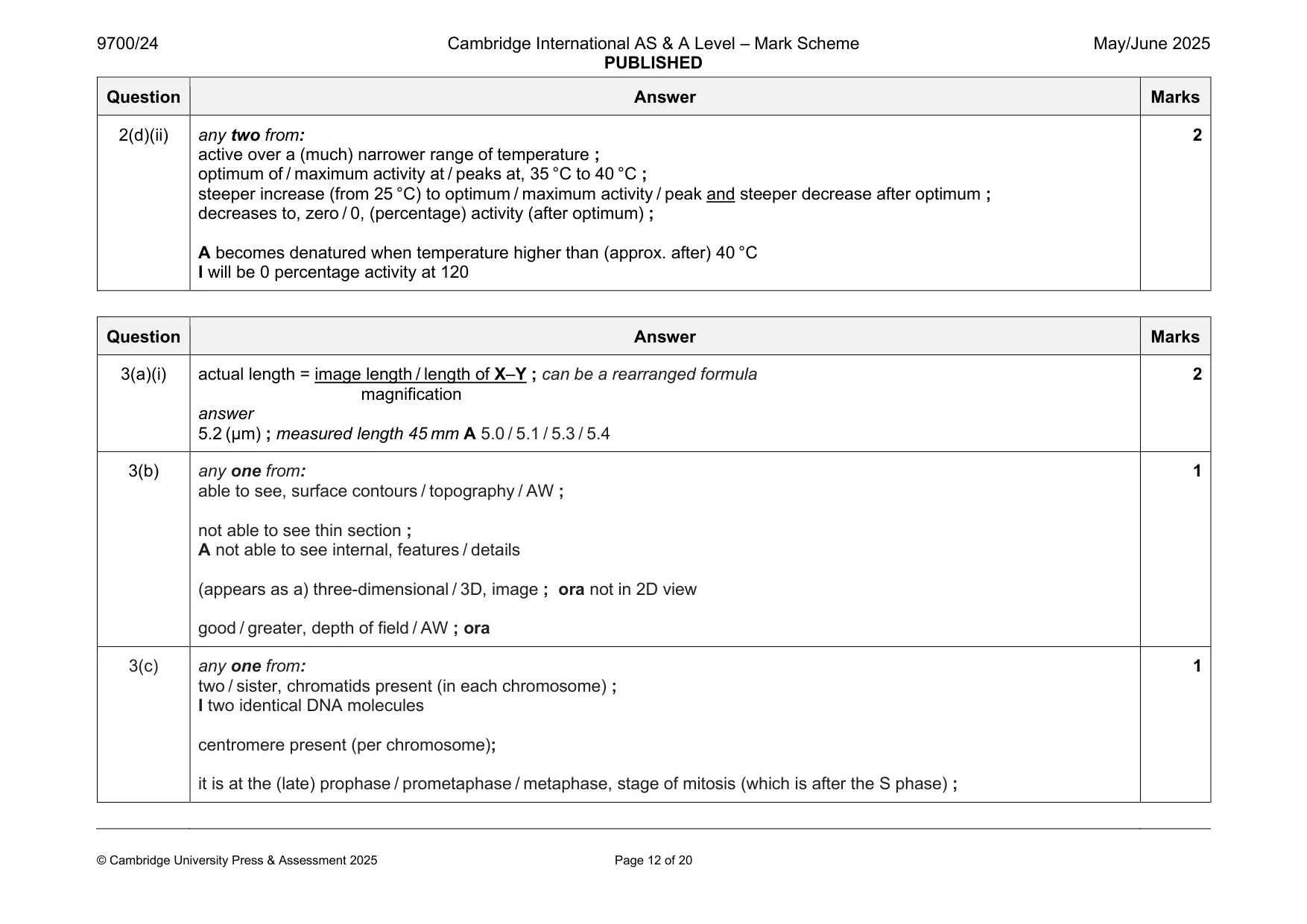
Mark scheme: 2(a) extracellular (enzyme) ; 1 R exocellular 2(b)(i) arrow between O and CO on lower fatty acid residue ; 1 2(b)(ii) hydrolysis ; 2 ester ; 2(c) substrate / olive oil, concentration ; 2 for, lipase / the enzyme, to achieve, half, maximum rate (of reaction) / Vmax (at a temperature of 37 °C) ; R Km is half, maximum rate of reaction / Vmax ref. to lipase / enzyme not needed if the context is correct ‘when the rate of reaction is half Vmax , the olive oil concentration is 91.76 mmol dm-3’ is two marks 2(d)(i) any four from: 4 accept activity for percentage enzyme activity throughout check for correct units – must be seen once 1 optimum temperature is 80 °C ; 2 increasing temperature increases activity, up to, 80 °C / the optimum / the maximum ; I constant increase I enzyme more active R if implied a continued increase throughout the temperature range if mp not gained, allow extracted values: from 25 °C to 80 °C, percentage / activity, from 57 to 100 3 increased kinetic energy (of, substrate / enzyme, molecules) ; A increased energy if movement stated 4 increase, in enzyme-substrate collisions / AW, (per unit time) ; A increase in number of enzyme-substrate complexes must be in context of only up to 80 °C allow ecf if mp2 rejected 5 above, optimum / 80 °C, decrease in activity ; ecf for incorrect optimum A percentage activity, from 100 to 55 / 56 for decrease in activity 6 partial (progressive) denaturation ; A enzyme begins to denature A progressive denaturation ora enzyme not completely denatured 7 explanation of denaturation / partial denaturation ; e.g. loss of, tertiary structure / shape of active site breakage of bonds between R-group breakage of two named bonds A fewer enzyme-substrate collisions if mp4 not gained 8 reason for, high optimum temperature / activity high at higher temperature ; e.g. is a thermostable enzyme enzyme has more bonds R more peptide bonds enzyme more compact extracted from bacteria living in hot springs enzyme protected by other proteins 2(d)(ii) any two from: 2 active over a (much) narrower range of temperature ; optimum of / maximum activity at / peaks at, 35 °C to 40 °C ; steeper increase (from 25 °C) to optimum / maximum activity / peak and steeper decrease after optimum ; decreases to, zero / 0, (percentage) activity (after optimum) ; A becomes denatured when temperature higher than (approx. after) 40 °C I will be 0 percentage activity at 120
Q3 · A scanning electron micrograph of a pair of human chromosomes in a stage of the mitotic…
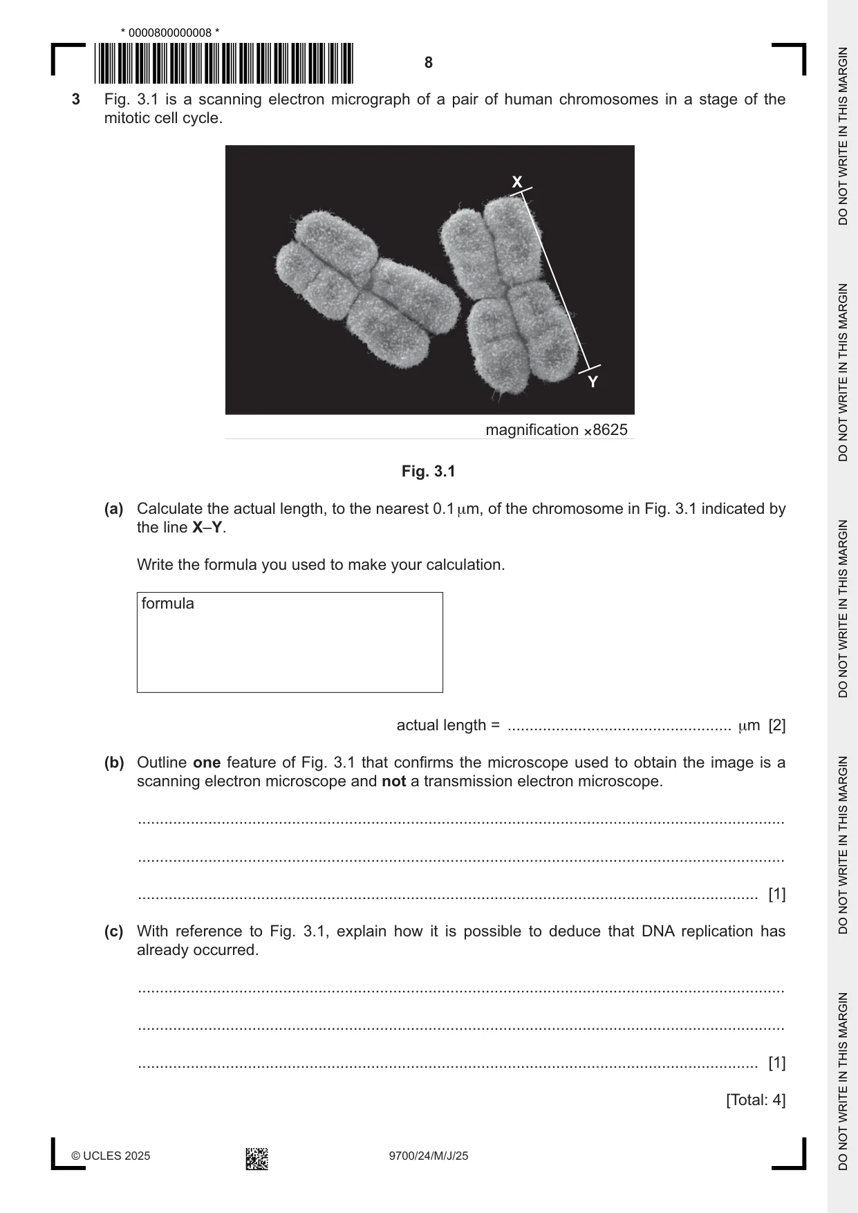
3 Fig. 3.1 is a scanning electron micrograph of a pair of human chromosomes in a stage of the mitotic cell cycle. X Y magnification ×8625 Fig. 3.1 (a) Calculate the actual length, to the nearest 0.1 μm, of the chromosome in Fig. 3.1 indicated by the line X–Y. Write the formula you used to make your calculation. formula actual length = ................................................... μm [2] (b) Outline one feature of Fig. 3.1 that confirms the microscope used to obtain the image is a scanning electron microscope and not a transmission electron microscope. ................................................................................................................................................... ................................................................................................................................................... ............................................................................................................................................. [1] (c) With reference to Fig. 3.1, explain how it is possible to deduce that DNA replication has already occurred. ................................................................................................................................................... ................................................................................................................................................... ............................................................................................................................................. [1] [Total: 4]
Mark scheme: 3(a)(i) actual length = image length / length of X–Y ; can be a rearranged formula 2 magnification answer 5.2 (µm) ; measured length 45 mm A 5.0 / 5.1 / 5.3 / 5.4 3(b) any one from: 1 able to see, surface contours / topography / AW ; not able to see thin section ; A not able to see internal, features / details (appears as a) three-dimensional / 3D, image ; ora not in 2D view good / greater, depth of field / AW ; ora 3(c) any one from: 1 two / sister, chromatids present (in each chromosome) ; I two identical DNA molecules centromere present (per chromosome); it is at the (late) prophase / prometaphase / metaphase, stage of mitosis (which is after the S phase) ;
Q4 · A number of species in the genus Plasmodium are known to cause malaria in humans
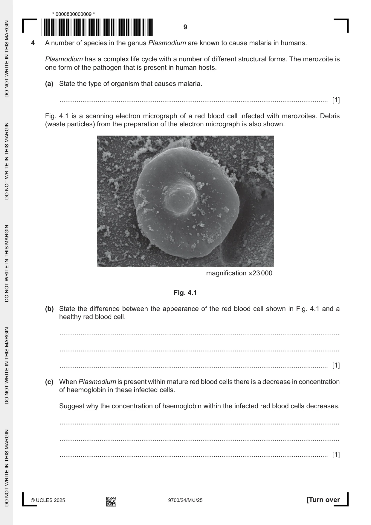
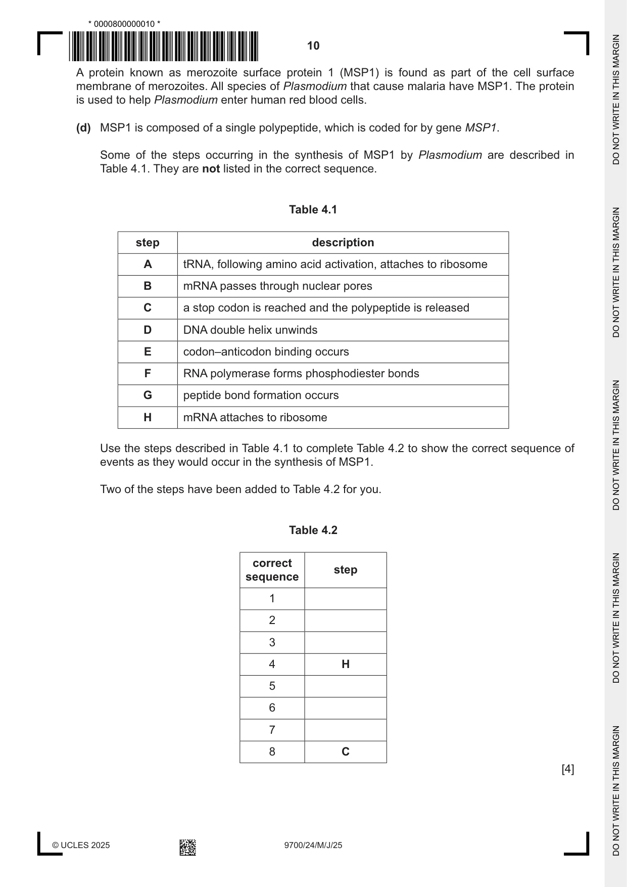
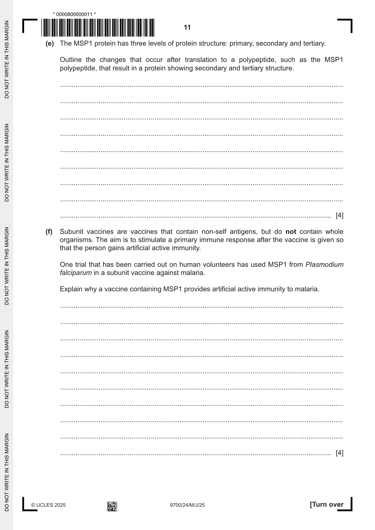
4 A number of species in the genus Plasmodium are known to cause malaria in humans. Plasmodium has a complex life cycle with a number of different structural forms. The merozoite is one form of the pathogen that is present in human hosts. (a) State the type of organism that causes malaria. ............................................................................................................................................. [1] Fig. 4.1 is a scanning electron micrograph of a red blood cell infected with merozoites. Debris (waste particles) from the preparation of the electron micrograph is also shown. magnification ×23 000 Fig. 4.1 (b) State the difference between the appearance of the red blood cell shown in Fig. 4.1 and a healthy red blood cell. ................................................................................................................................................... ................................................................................................................................................... ............................................................................................................................................. [1] (c) When Plasmodium is present within mature red blood cells there is a decrease in concentration of haemoglobin in these infected cells. Suggest why the concentration of haemoglobin within the infected red blood cells decreases. ................................................................................................................................................... ................................................................................................................................................... ............................................................................................................................................. [1] A protein known as merozoite surface protein 1 (MSP1) is found as part of the cell surface membrane of merozoites. All species of Plasmodium that cause malaria have MSP1. The protein is used to help Plasmodium enter human red blood cells. (d) MSP1 is composed of a single polypeptide, which is coded for by gene MSP1. Some of the steps occurring in the synthesis of MSP1 by Plasmodium are described in Table 4.1. They are not listed in the correct sequence. Table 4.1 step description A tRNA, following amino acid activation, attaches to ribosome B mRNA passes through nuclear pores C a stop codon is reached and the polypeptide is released D DNA double helix unwinds E codon–anticodon binding occurs F RNA polymerase forms phosphodiester bonds G peptide bond formation occurs H mRNA attaches to ribosome Use the steps described in Table 4.1 to complete Table 4.2 to show the correct sequence of events as they would occur in the synthesis of MSP1. Two of the steps have been added to Table 4.2 for you. Table 4.2 correct step sequence 1 2 3 4 H 5 6 7 8 C [4] (e) The MSP1 protein has three levels of protein structure: primary, secondary and tertiary. Outline the changes that occur after translation to a polypeptide, such as the MSP1 polypeptide, that result in a protein showing secondary and tertiary structure. ................................................................................................................................................... ................................................................................................................................................... ................................................................................................................................................... ................................................................................................................................................... ................................................................................................................................................... ................................................................................................................................................... ................................................................................................................................................... ................................................................................................................................................... ............................................................................................................................................. [4] (f) Subunit vaccines are vaccines that contain non-self antigens, but do not contain whole organisms. The aim is to stimulate a primary immune response after the vaccine is given so that the person gains artificial active immunity. One trial that has been carried out on human volunteers has used MSP1 from Plasmodium falciparum in a subunit vaccine against malaria. Explain why a vaccine containing MSP1 provides artificial active immunity to malaria. ................................................................................................................................................... ................................................................................................................................................... ................................................................................................................................................... ................................................................................................................................................... ................................................................................................................................................... ................................................................................................................................................... ................................................................................................................................................... ................................................................................................................................................... ................................................................................................................................................... ............................................................................................................................................. [4] (g) The female Anopheles mosquito is the vector of Plasmodium. Discuss the ways in which the vector is controlled to help prevent the transmission of malaria. ................................................................................................................................................... ................................................................................................................................................... ................................................................................................................................................... ................................................................................................................................................... ................................................................................................................................................... ................................................................................................................................................... ................................................................................................................................................... ............................................................................................................................................. [3] [Total: 18]
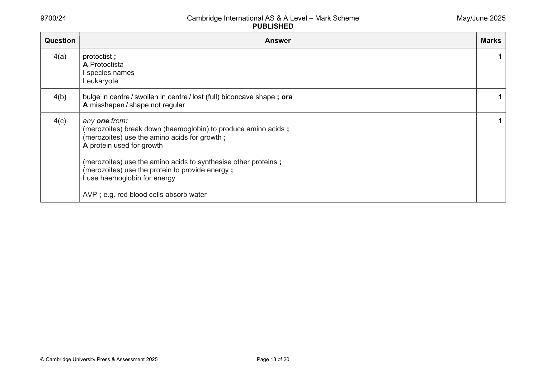
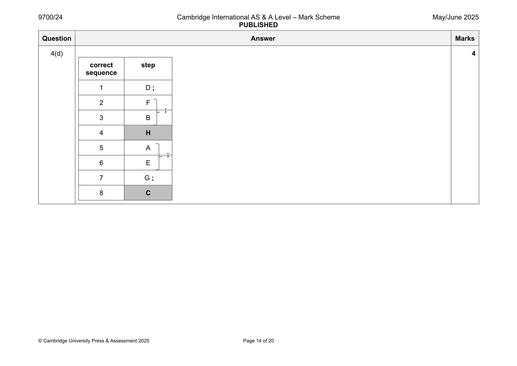
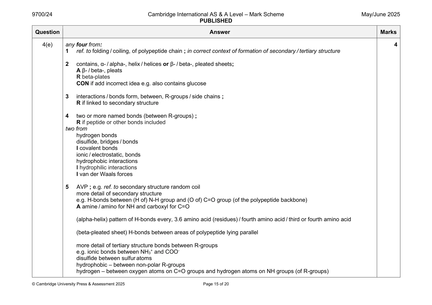
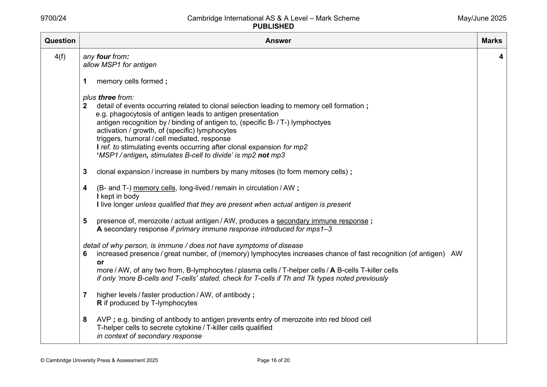
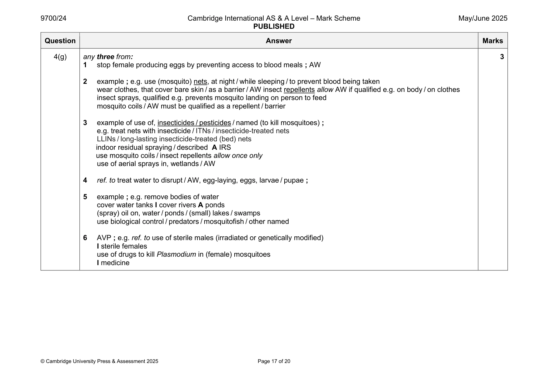
Mark scheme: 4(a) protoctist ; 1 A Protoctista I species names I eukaryote 4(b) bulge in centre / swollen in centre / lost (full) biconcave shape ; ora 1 A misshapen / shape not regular 4(c) any one from: 1 (merozoites) break down (haemoglobin) to produce amino acids ; (merozoites) use the amino acids for growth ; A protein used for growth (merozoites) use the amino acids to synthesise other proteins ; (merozoites) use the protein to provide energy ; I use haemoglobin for energy AVP ; e.g. red blood cells absorb water 4(d) 4 correct step sequence 1 D ; 2 F ; 3 B 4 H 5 A ; 6 E 7 G ; 8 C 4(e) any four from: 4 1 ref. to folding / coiling, of polypeptide chain ; in correct context of formation of secondary / tertiary structure 2 contains, α- / alpha-, helix / helices or β- / beta-, pleated sheets; A β- / beta-, pleats R beta-plates CON if add incorrect idea e.g. also contains glucose 3 interactions / bonds form, between, R-groups / side chains ; R if linked to secondary structure 4 two or more named bonds (between R-groups) ; R if peptide or other bonds included two from hydrogen bonds disulfide, bridges / bonds I covalent bonds ionic / electrostatic, bonds hydrophobic interactions I hydrophilic interactions I van der Waals forces 5 AVP ; e.g. ref. to secondary structure random coil more detail of secondary structure e.g. H-bonds between (H of) N-H group and (O of) C=O group (of the polypeptide backbone) A amine / amino for NH and carboxyl for C=O (alpha-helix) pattern of H-bonds every, 3.6 amino acid (residues) / fourth amino acid / third or fourth amino acid (beta-pleated sheet) H-bonds between areas of polypeptide lying parallel more detail of tertiary structure bonds between R-groups e.g. ionic bonds between NH3+ and COO- disulfide between sulfur atoms hydrophobic – between non-polar R-groups hydrogen – between oxygen atoms on C=O groups and hydrogen atoms on NH groups (of R-groups) 4(f) any four from: 4 allow MSP1 for antigen 1 memory cells formed ; plus three from: 2 detail of events occurring related to clonal selection leading to memory cell formation ; e.g. phagocytosis of antigen leads to antigen presentation antigen recognition by / binding of antigen to, (specific B- / T-) lymphoctyes activation / growth, of (specific) lymphocytes triggers, humoral / cell mediated, response I ref. to stimulating events occurring after clonal expansion for mp2 ‘MSP1 / antigen, stimulates B-cell to divide’ is mp2 not mp3 3 clonal expansion / increase in numbers by many mitoses (to form memory cells) ; 4 (B- and T-) memory cells, long-lived / remain in circulation / AW ; I kept in body I live longer unless qualified that they are present when actual antigen is present 5 presence of, merozoite / actual antigen / AW, produces a secondary immune response ; A secondary response if primary immune response introduced for mps1–3 detail of why person, is immune / does not have symptoms of disease 6 increased presence / great number, of (memory) lymphocytes increases chance of fast recognition (of antigen) AW or more / AW, of any two from, B-lymphocytes / plasma cells / T-helper cells / A B-cells T-killer cells if only ‘more B-cells and T-cells’ stated, check for T-cells if Th and Tk types noted previously 7 higher levels / faster production / AW, of antibody ; R if produced by T-lymphocytes 8 AVP ; e.g. binding of antibody to antigen prevents entry of merozoite into red blood cell T-helper cells to secrete cytokine / T-killer cells qualified in context of secondary response 4(g) any three from: 3 1 stop female producing eggs by preventing access to blood meals ; AW 2 example ; e.g. use (mosquito) nets, at night / while sleeping / to prevent blood being taken wear clothes, that cover bare skin / as a barrier / AW insect repellents allow AW if qualified e.g. on body / on clothes insect sprays, qualified e.g. prevents mosquito landing on person to feed mosquito coils / AW must be qualified as a repellent / barrier 3 example of use of, insecticides / pesticides / named (to kill mosquitoes) ; e.g. treat nets with insecticide / ITNs / insecticide-treated nets LLINs / long-lasting insecticide-treated (bed) nets indoor residual spraying / described A IRS use mosquito coils / insect repellents allow once only use of aerial sprays in, wetlands / AW 4 ref. to treat water to disrupt / AW, egg-laying, eggs, larvae / pupae ; 5 example ; e.g. remove bodies of water cover water tanks I cover rivers A ponds (spray) oil on, water / ponds / (small) lakes / swamps use biological control / predators / mosquitofish / other named 6 AVP ; e.g. ref. to use of sterile males (irradiated or genetically modified) I sterile females use of drugs to kill Plasmodium in (female) mosquitoes I medicine
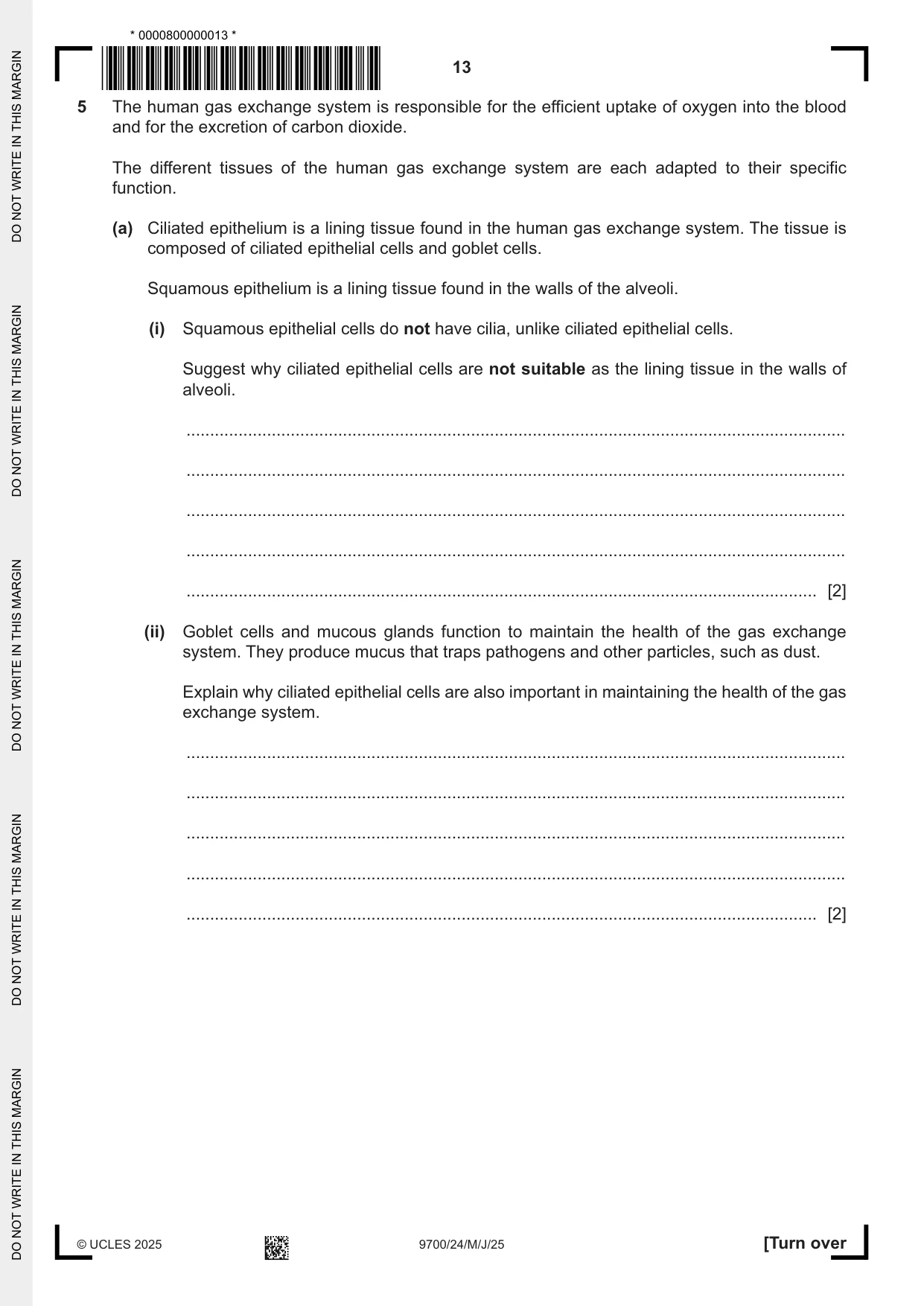
Q5 · The human gas exchange system is responsible for the efficient uptake of oxygen into the…
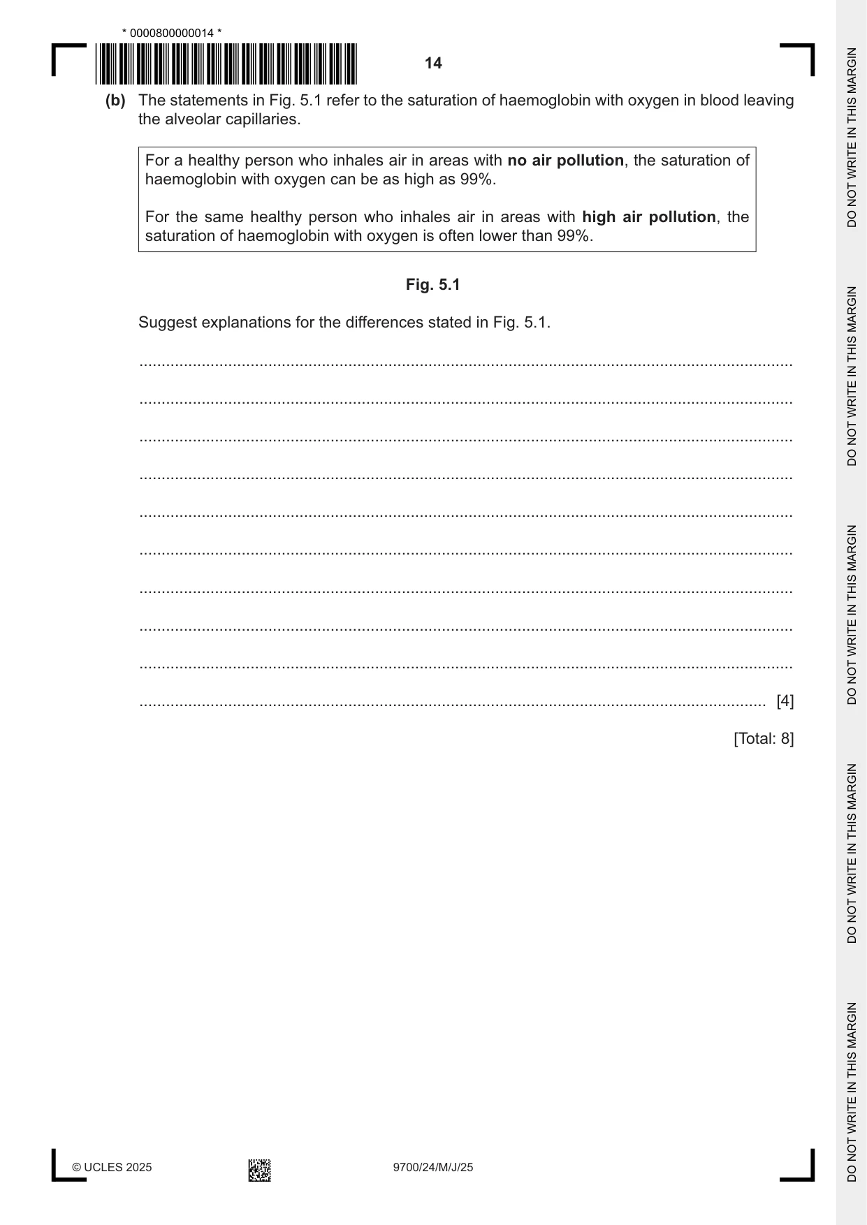
5 The human gas exchange system is responsible for the efficient uptake of oxygen into the blood and for the excretion of carbon dioxide. The different tissues of the human gas exchange system are each adapted to their specific function. (a) Ciliated epithelium is a lining tissue found in the human gas exchange system. The tissue is composed of ciliated epithelial cells and goblet cells. Squamous epithelium is a lining tissue found in the walls of the alveoli. (i) Squamous epithelial cells do not have cilia, unlike ciliated epithelial cells. Suggest why ciliated epithelial cells are not suitable as the lining tissue in the walls of alveoli. ........................................................................................................................................... ........................................................................................................................................... ........................................................................................................................................... ........................................................................................................................................... ..................................................................................................................................... [2] (ii) Goblet cells and mucous glands function to maintain the health of the gas exchange system. They produce mucus that traps pathogens and other particles, such as dust. Explain why ciliated epithelial cells are also important in maintaining the health of the gas exchange system. ........................................................................................................................................... ........................................................................................................................................... ........................................................................................................................................... ........................................................................................................................................... ..................................................................................................................................... [2] (b) The statements in Fig. 5.1 refer to the saturation of haemoglobin with oxygen in blood leaving the alveolar capillaries. For a healthy person who inhales air in areas with no air pollution, the saturation of haemoglobin with oxygen can be as high as 99%. For the same healthy person who inhales air in areas with high air pollution, the saturation of haemoglobin with oxygen is often lower than 99%. Fig. 5.1 Suggest explanations for the differences stated in Fig. 5.1. ................................................................................................................................................... ................................................................................................................................................... ................................................................................................................................................... ................................................................................................................................................... ................................................................................................................................................... ................................................................................................................................................... ................................................................................................................................................... ................................................................................................................................................... ................................................................................................................................................... ............................................................................................................................................. [4] [Total: 8]
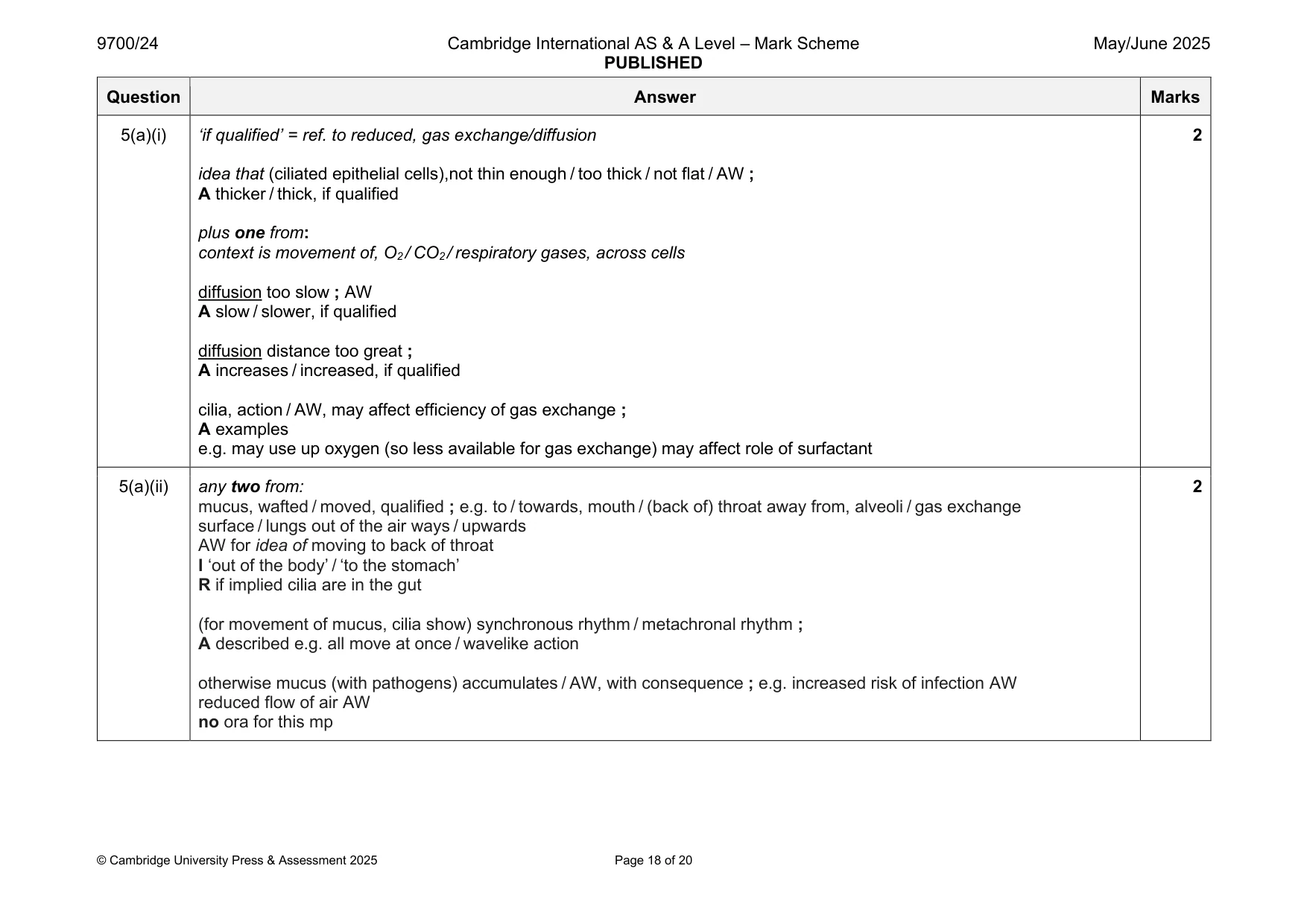
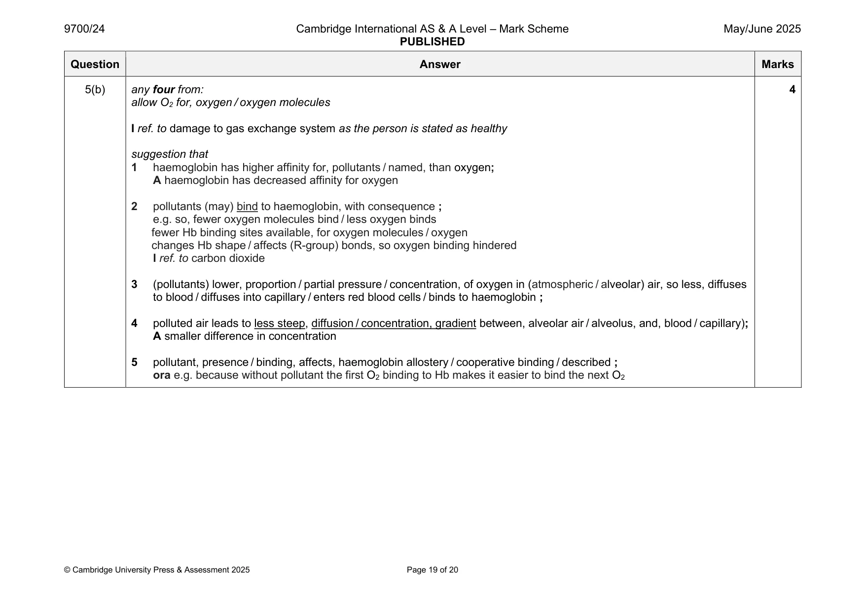
Mark scheme: 5(a)(i) ‘if qualified’ = ref. to reduced, gas exchange/diffusion 2 idea that (ciliated epithelial cells),not thin enough / too thick / not flat / AW ; A thicker / thick, if qualified plus one from: context is movement of, O2 / CO2 / respiratory gases, across cells diffusion too slow ; AW A slow / slower, if qualified diffusion distance too great ; A increases / increased, if qualified cilia, action / AW, may affect efficiency of gas exchange ; A examples e.g. may use up oxygen (so less available for gas exchange) may affect role of surfactant 5(a)(ii) any two from: 2 mucus, wafted / moved, qualified ; e.g. to / towards, mouth / (back of) throat away from, alveoli / gas exchange surface / lungs out of the air ways / upwards AW for idea of moving to back of throat I ‘out of the body’ / ‘to the stomach’ R if implied cilia are in the gut (for movement of mucus, cilia show) synchronous rhythm / metachronal rhythm ; A described e.g. all move at once / wavelike action otherwise mucus (with pathogens) accumulates / AW, with consequence ; e.g. increased risk of infection AW reduced flow of air AW no ora for this mp 5(b) any four from: 4 allow O2 for, oxygen / oxygen molecules I ref. to damage to gas exchange system as the person is stated as healthy suggestion that 1 haemoglobin has higher affinity for, pollutants / named, than oxygen; A haemoglobin has decreased affinity for oxygen 2 pollutants (may) bind to haemoglobin, with consequence ; e.g. so, fewer oxygen molecules bind / less oxygen binds fewer Hb binding sites available, for oxygen molecules / oxygen changes Hb shape / affects (R-group) bonds, so oxygen binding hindered I ref. to carbon dioxide 3 (pollutants) lower, proportion / partial pressure / concentration, of oxygen in (atmospheric / alveolar) air, so less, diffuses to blood / diffuses into capillary / enters red blood cells / binds to haemoglobin ; 4 polluted air leads to less steep, diffusion / concentration, gradient between, alveolar air / alveolus, and, blood / capillary); A smaller difference in concentration 5 pollutant, presence / binding, affects, haemoglobin allostery / cooperative binding / described ; ora e.g. because without pollutant the first O2 binding to Hb makes it easier to bind the next O2
Q6 · The sinoatrial node (SAN), the atrioventricular node (AVN) and the Purkyne fibres have a…
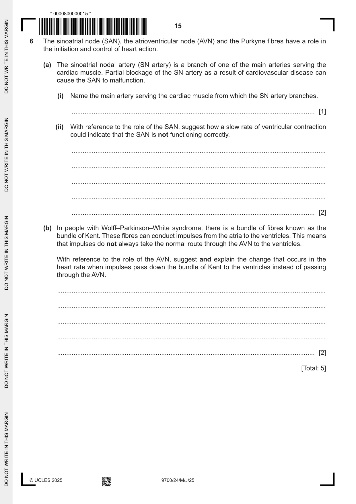
6 The sinoatrial node (SAN), the atrioventricular node (AVN) and the Purkyne fibres have a role in the initiation and control of heart action. (a) The sinoatrial nodal artery (SN artery) is a branch of one of the main arteries serving the cardiac muscle. Partial blockage of the SN artery as a result of cardiovascular disease can cause the SAN to malfunction. (i) Name the main artery serving the cardiac muscle from which the SN artery branches. ..................................................................................................................................... [1] (ii) With reference to the role of the SAN, suggest how a slow rate of ventricular contraction could indicate that the SAN is not functioning correctly. ........................................................................................................................................... ........................................................................................................................................... ........................................................................................................................................... ........................................................................................................................................... ..................................................................................................................................... [2] (b) In people with Wolff–Parkinson–White syndrome, there is a bundle of fibres known as the bundle of Kent. These fibres can conduct impulses from the atria to the ventricles. This means that impulses do not always take the normal route through the AVN to the ventricles. With reference to the role of the AVN, suggest and explain the change that occurs in the heart rate when impulses pass down the bundle of Kent to the ventricles instead of passing through the AVN. ................................................................................................................................................... ................................................................................................................................................... ................................................................................................................................................... ................................................................................................................................................... ............................................................................................................................................. [2] [Total: 5]
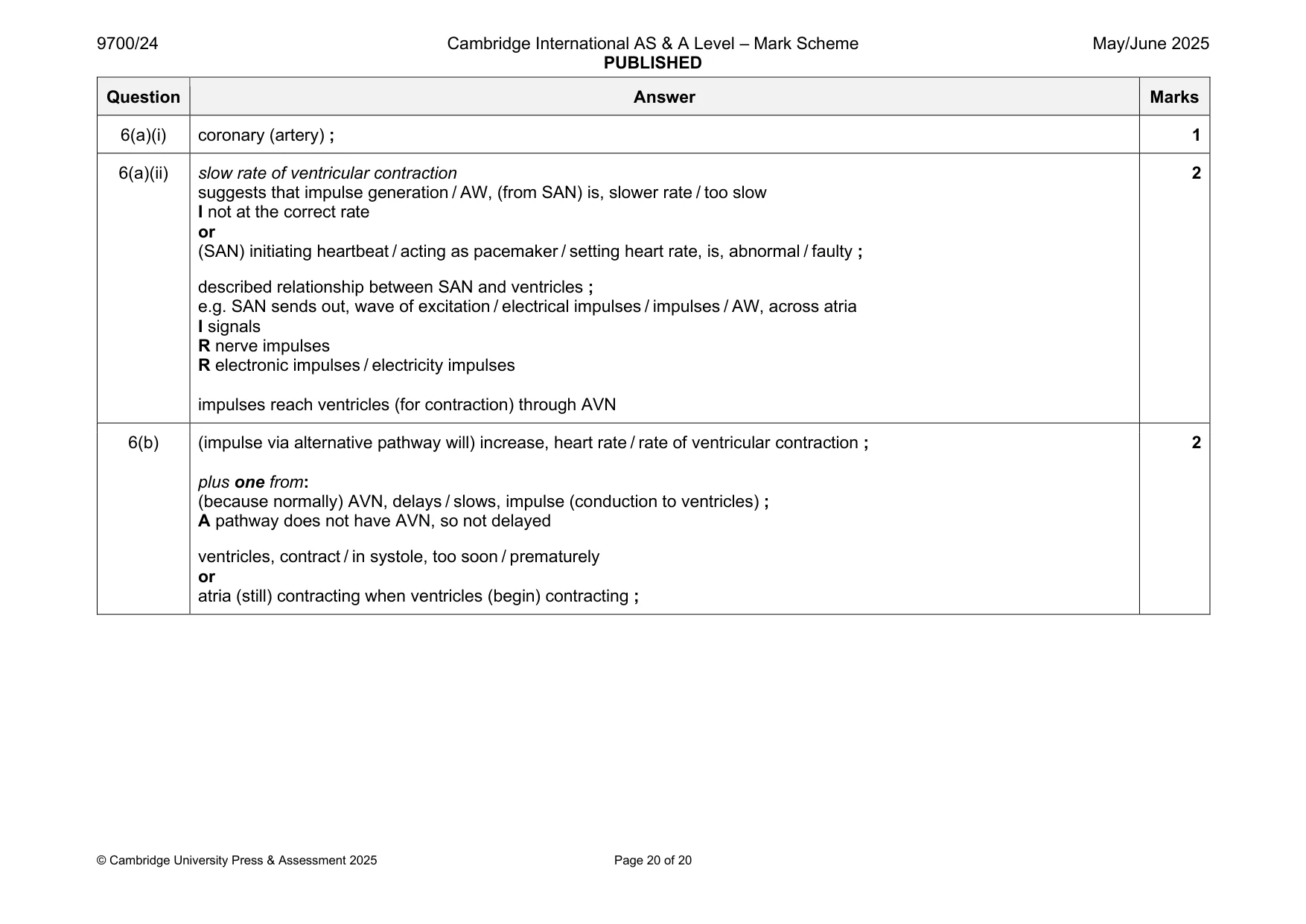
Mark scheme: 6(a)(i) coronary (artery) ; 1 6(a)(ii) slow rate of ventricular contraction 2 suggests that impulse generation / AW, (from SAN) is, slower rate / too slow I not at the correct rate or (SAN) initiating heartbeat / acting as pacemaker / setting heart rate, is, abnormal / faulty ; described relationship between SAN and ventricles ; e.g. SAN sends out, wave of excitation / electrical impulses / impulses / AW, across atria I signals R nerve impulses R electronic impulses / electricity impulses impulses reach ventricles (for contraction) through AVN 6(b) (impulse via alternative pathway will) increase, heart rate / rate of ventricular contraction ; 2 plus one from: (because normally) AVN, delays / slows, impulse (conduction to ventricles) ; A pathway does not have AVN, so not delayed ventricles, contract / in systole, too soon / prematurely or atria (still) contracting when ventricles (begin) contracting ;
What was in this paper
The subtopics covered by these 6 questions, and how many questions each got. Open one in a new tab to see every Cambridge question on it.
What you needed in this session
Cambridge’s own grade thresholds for 2025 May/June, Paper 2 · Variant 4. A higher threshold means an easier paper — the bar moves with how the cohort did.