Cambridge A Level Biology 9700 — 2024 Oct/Nov Paper 2 · Variant 1
9700/21/O/N/24 · 6 questions · 60 marks · ≈68 min
The question paper and its mark scheme, free to read here and free to download. This is Cambridge’s own paper, exactly as it was sat.
Question paper24 pages



Mark scheme16 pages
Answers below. Sit the paper first if you are practising.
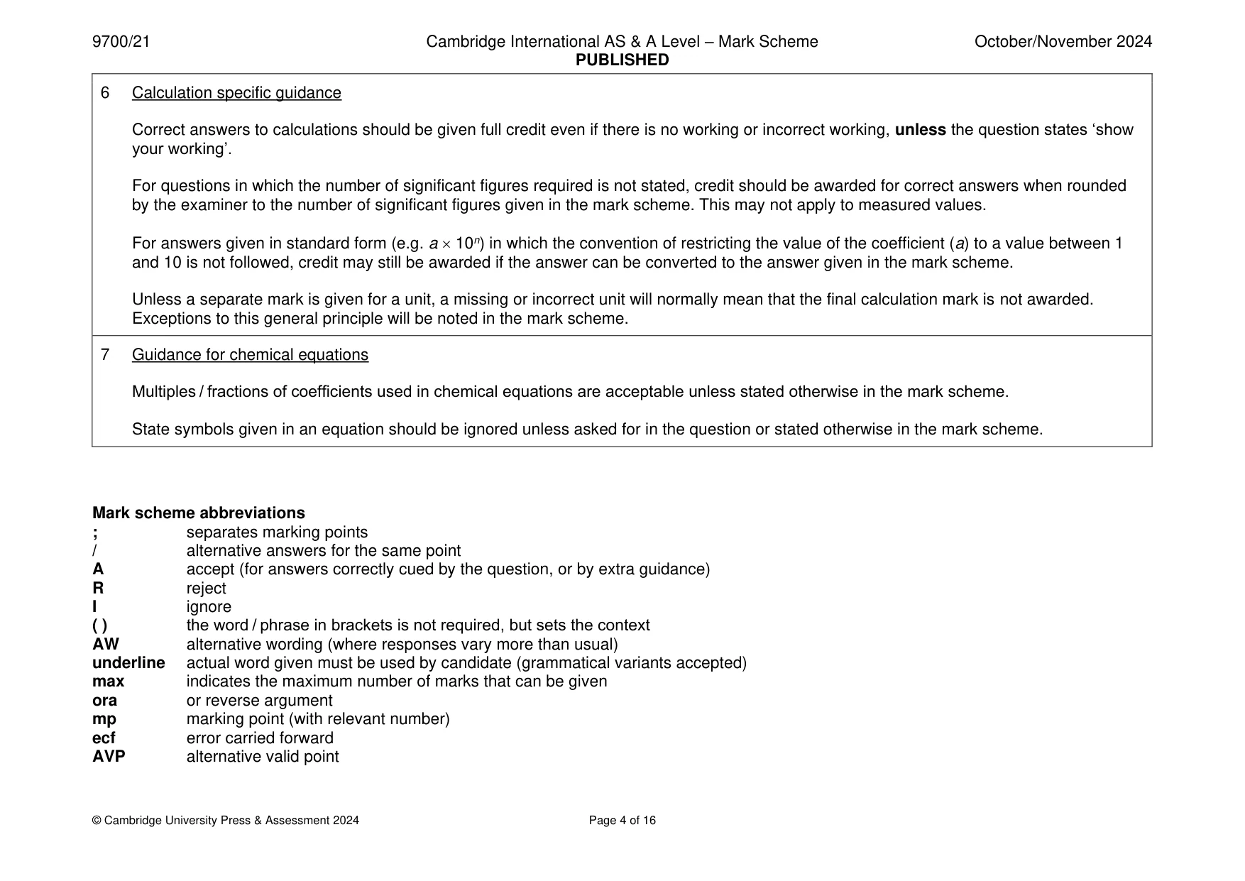
Questions as text
Q1 · Animal cells, plant cells and prokaryotic cells have similarities and differences in…
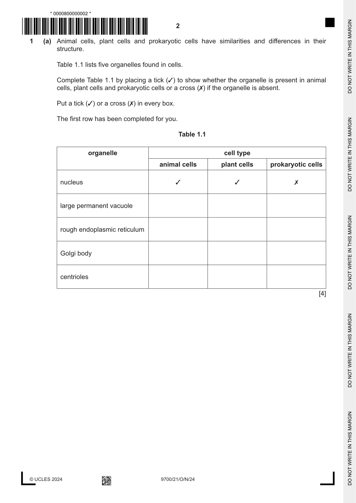
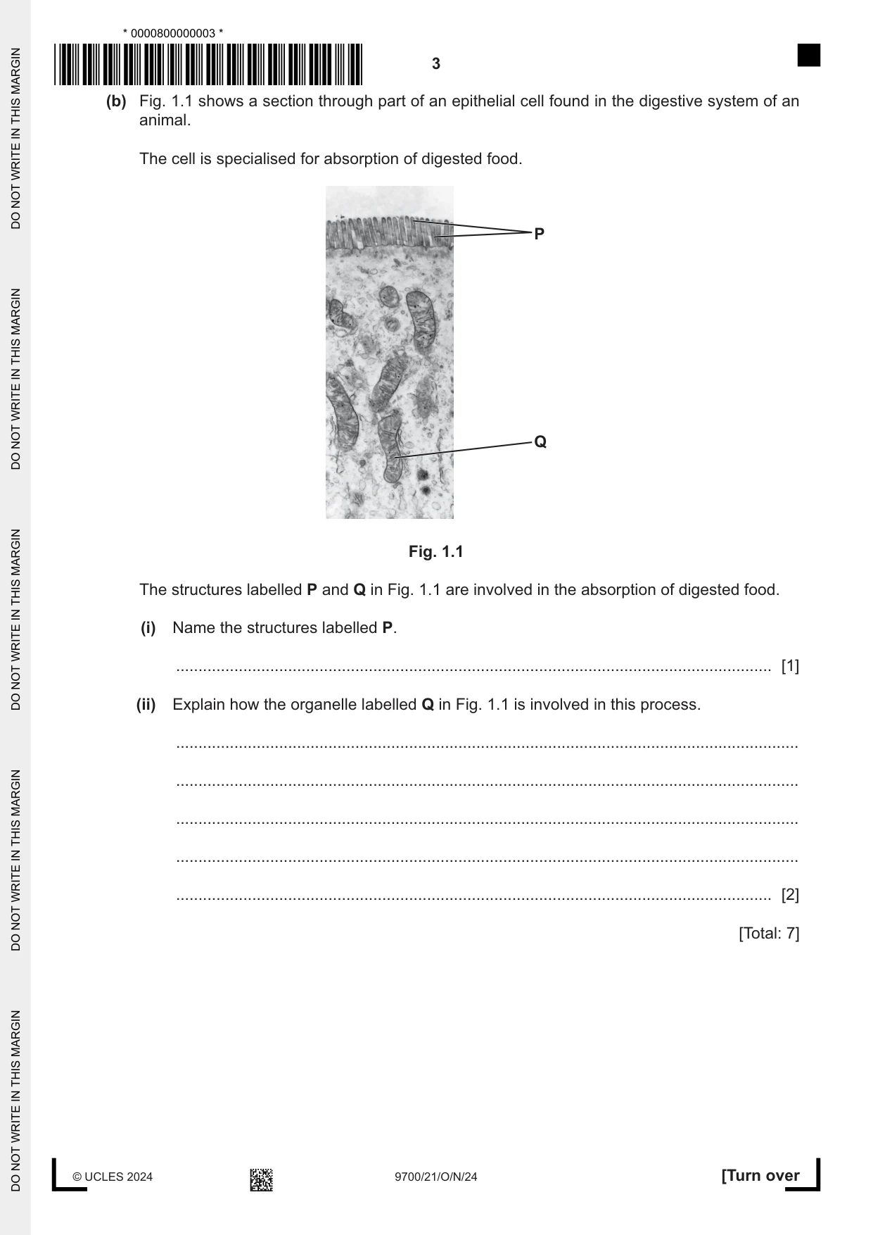
1 (a) Animal cells, plant cells and prokaryotic cells have similarities and differences in their structure. Table 1.1 lists five organelles found in cells. Complete Table 1.1 by placing a tick (3) to show whether the organelle is present in animal cells, plant cells and prokaryotic cells or a cross (✗) if the organelle is absent. Put a tick (3) or a cross (✗) in every box. The first row has been completed for you. Table 1.1 organelle cell type animal cells plant cells prokaryotic cells nucleus 3 3 ✗ large permanent vacuole rough endoplasmic reticulum Golgi body centrioles [4] (b) Fig. 1.1 shows a section through part of an epithelial cell found in the digestive system of an animal. The cell is specialised for absorption of digested food. P Q Fig. 1.1 The structures labelled P and Q in Fig. 1.1 are involved in the absorption of digested food. (i) Name the structures labelled P. ..................................................................................................................................... [1] (ii) Explain how the organelle labelled Q in Fig. 1.1 is involved in this process. ........................................................................................................................................... ........................................................................................................................................... ........................................................................................................................................... ........................................................................................................................................... ..................................................................................................................................... [2] [Total: 7]
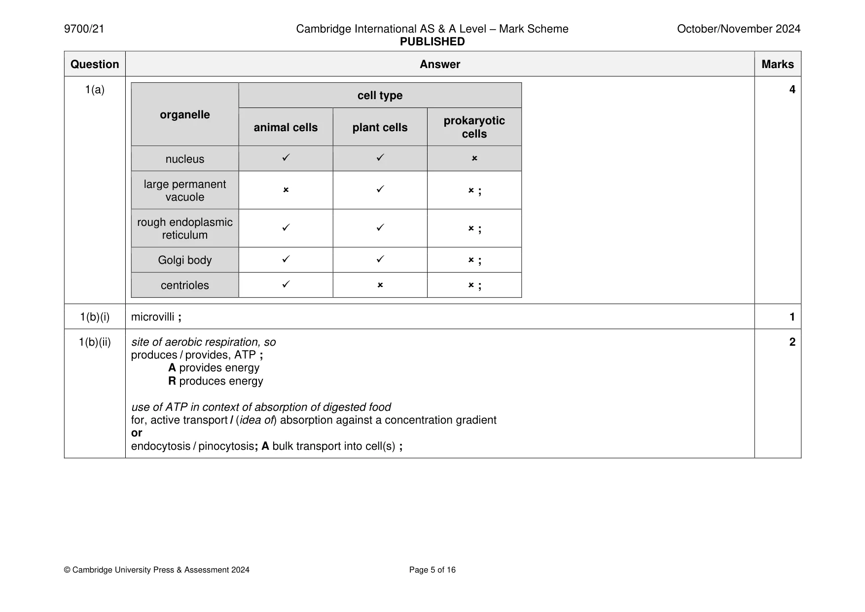
Mark scheme: Question Answer Marks 1(a) 4 cell type organelle prokaryotic animal cells plant cells cells nucleus ✓ ✓ large permanent ✓ ; vacuole rough endoplasmic ✓ ✓ ; reticulum Golgi body ✓ ✓ ; centrioles ✓ ; 1(b)(i) microvilli ; 1 1(b)(ii) site of aerobic respiration, so 2 produces / provides, ATP ; A provides energy R produces energy use of ATP in context of absorption of digested food for, active transport / (idea of) absorption against a concentration gradient or endocytosis / pinocytosis; A bulk transport into cell(s) ;
More questions on Cells as the basic units of living organisms
Q2 · In the mammalian circulatory system, red blood cells travel through different types of…
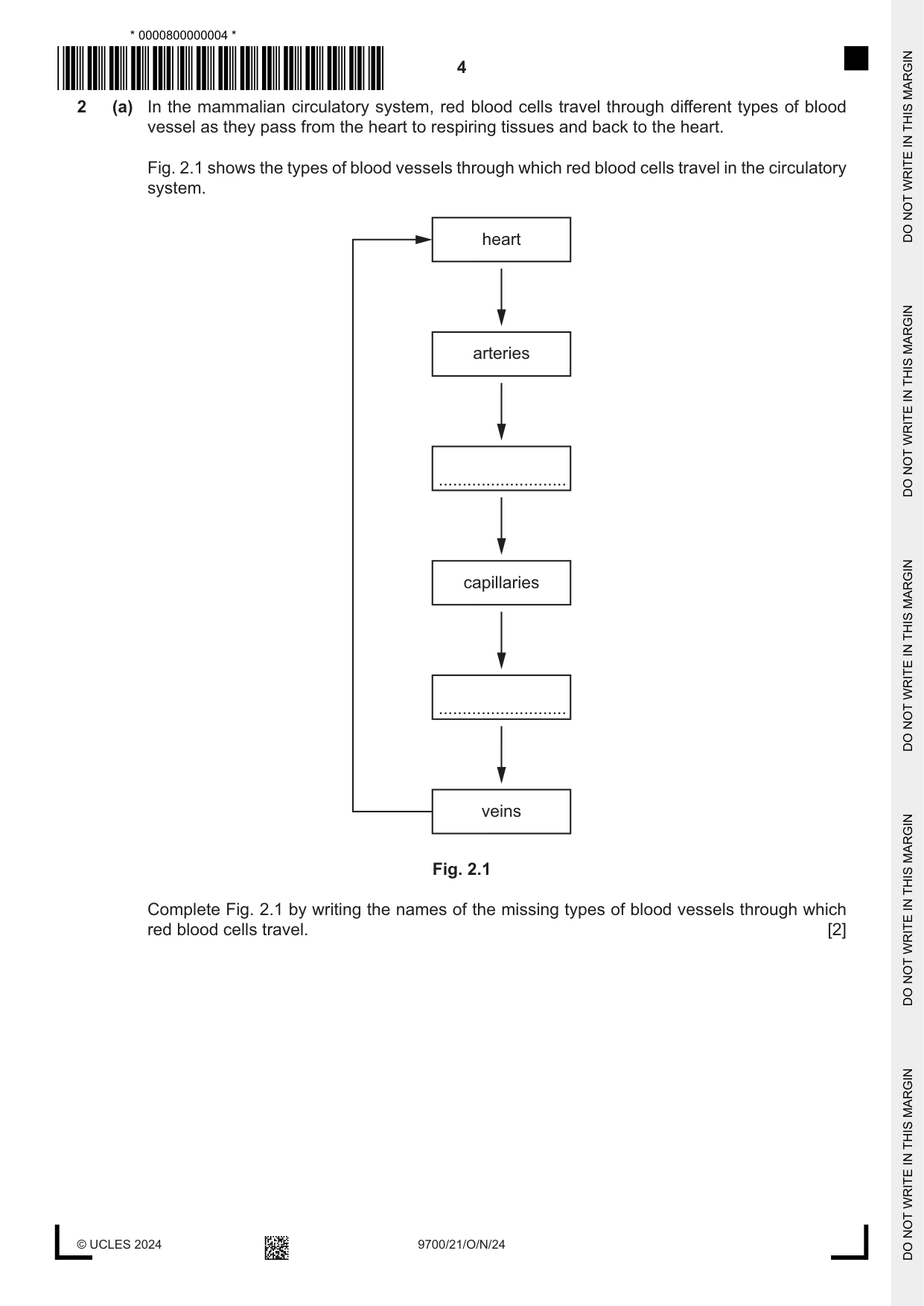
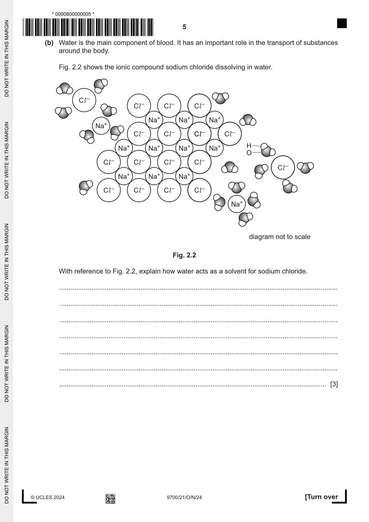
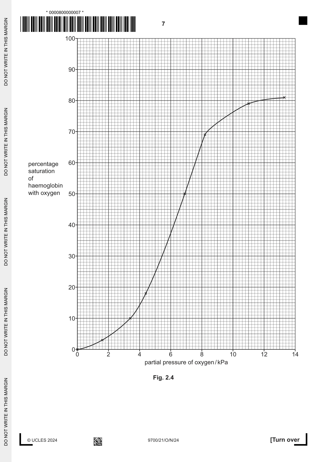
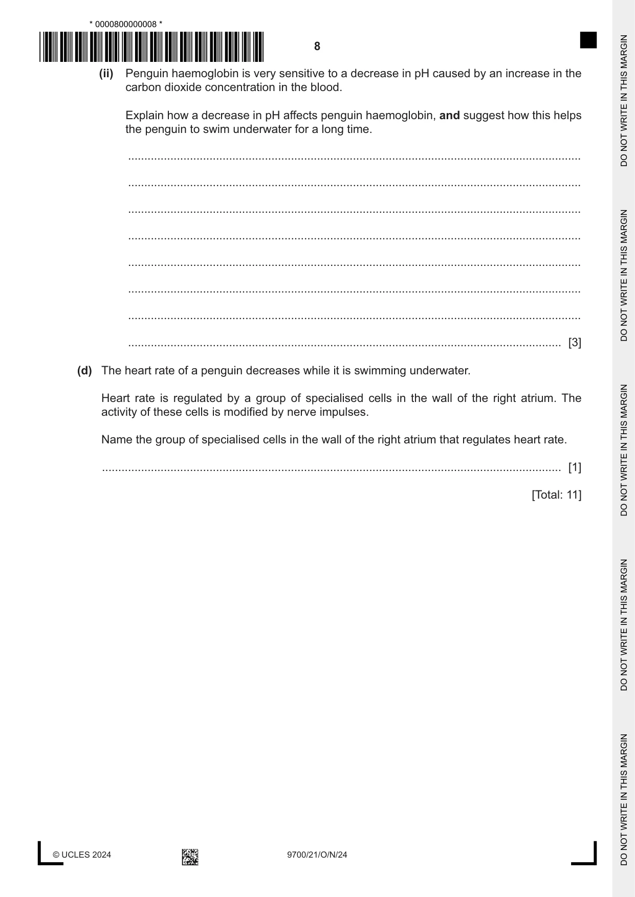
2 (a) In the mammalian circulatory system, red blood cells travel through different types of blood vessel as they pass from the heart to respiring tissues and back to the heart. Fig. 2.1 shows the types of blood vessels through which red blood cells travel in the circulatory system. heart arteries ........................... capillaries ........................... veins Fig. 2.1 Complete Fig. 2.1 by writing the names of the missing types of blood vessels through which red blood cells travel. [2] (b) Water is the main component of blood. It has an important role in the transport of substances around the body. Fig. 2.2 shows the ionic compound sodium chloride dissolving in water. Cl – Cl – Cl – Cl – Na+ Na+ Na+ Na+ Cl – Cl – ClCl –– Cl – Na+ Na+ Na+ Na+ H O Cl – Cl – Cl – Cl – Cl – Na+ Na+ Na+ Cl – Cl – Cl – Cl – Na+ diagram not to scale Fig. 2.2 With reference to Fig. 2.2, explain how water acts as a solvent for sodium chloride. ................................................................................................................................................... ................................................................................................................................................... ................................................................................................................................................... ................................................................................................................................................... ................................................................................................................................................... ................................................................................................................................................... ............................................................................................................................................. [3] (c) Fig. 2.3 shows a Galapagos penguin, Spheniscus mendiculus, swimming in the water. Fig. 2.3 Penguins are birds that live on land but spend a lot of time swimming underwater hunting for food. Penguins can remain underwater for up to twenty minutes. During this time they do not breathe but their tissues continue to respire. Haemoglobin in the red blood cells of penguins has a higher affinity for oxygen than haemoglobin in other birds that do not swim underwater. Fig. 2.4 shows the oxygen dissociation curve for a bird that does not swim underwater. (i) Draw a line on Fig. 2.4 to suggest the position of the oxygen dissociation curve for penguin haemoglobin. [2] (ii) Penguin haemoglobin is very sensitive to a decrease in pH caused by an increase in the carbon dioxide concentration in the blood. Explain how a decrease in pH affects penguin haemoglobin, and suggest how this helps the penguin to swim underwater for a long time. ........................................................................................................................................... ........................................................................................................................................... ........................................................................................................................................... ........................................................................................................................................... ........................................................................................................................................... ........................................................................................................................................... ........................................................................................................................................... ..................................................................................................................................... [3] (d) The heart rate of a penguin decreases while it is swimming underwater. Heart rate is regulated by a group of specialised cells in the wall of the right atrium. The activity of these cells is modified by nerve impulses. Name the group of specialised cells in the wall of the right atrium that regulates heart rate. ............................................................................................................................................. [1] [Total: 11]
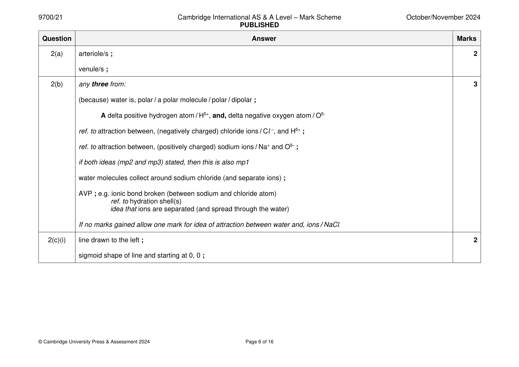
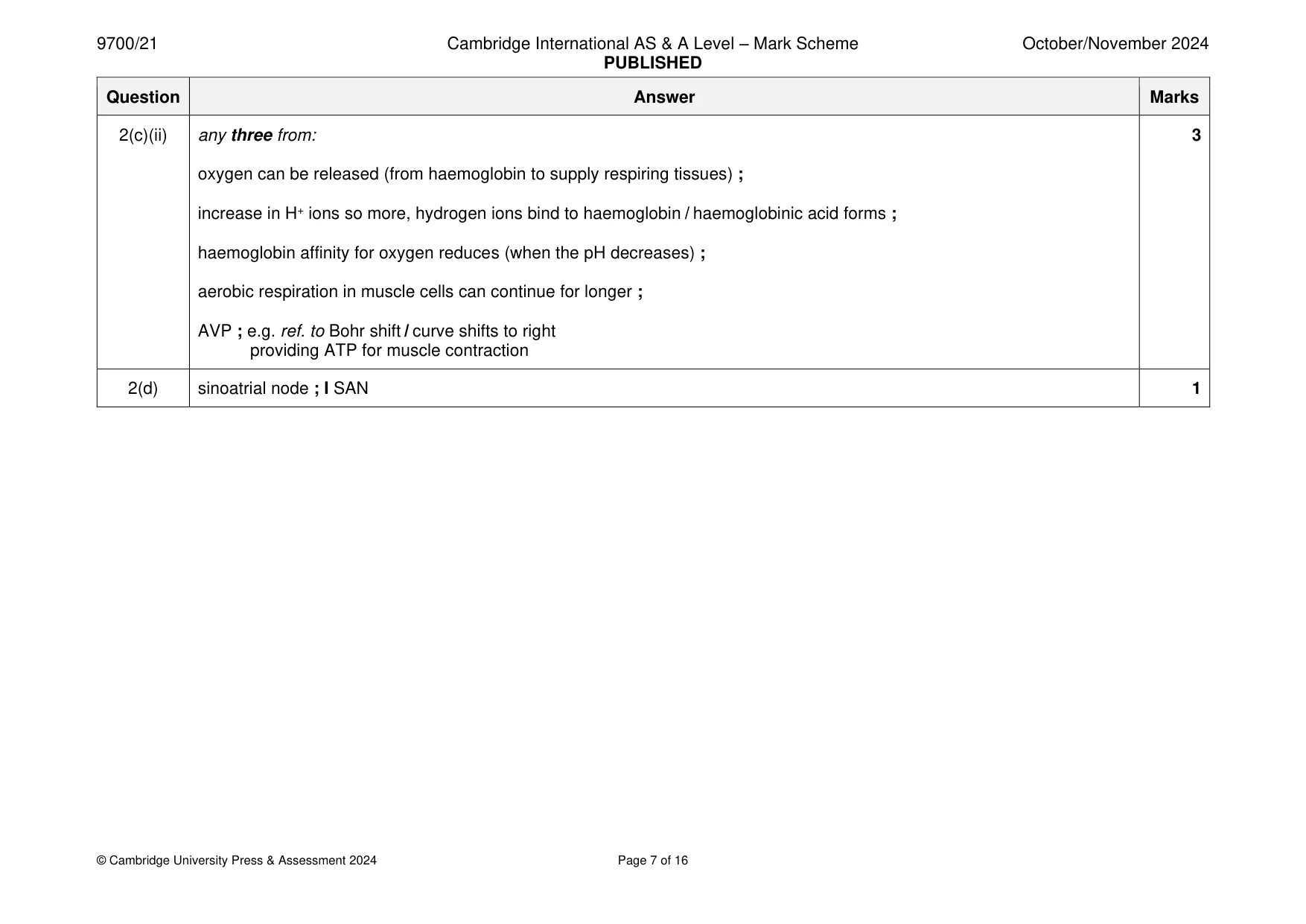
Mark scheme: 2(a) arteriole/s ; 2 venule/s ; 2(b) any three from: 3 (because) water is, polar / a polar molecule / polar / dipolar ; A delta positive hydrogen atom / Hδ+, and, delta negative oxygen atom / Oδ- ref. to attraction between, (negatively charged) chloride ions / Cl –, and Hδ+ ; ref. to attraction between, (positively charged) sodium ions / Na+ and Oδ– ; if both ideas (mp2 and mp3) stated, then this is also mp1 water molecules collect around sodium chloride (and separate ions) ; AVP ; e.g. ionic bond broken (between sodium and chloride atom) ref. to hydration shell(s) idea that ions are separated (and spread through the water) If no marks gained allow one mark for idea of attraction between water and, ions / NaCl 2(c)(i) line drawn to the left ; 2 sigmoid shape of line and starting at 0, 0 ; 2(c)(ii) any three from: 3 oxygen can be released (from haemoglobin to supply respiring tissues) ; increase in H+ ions so more, hydrogen ions bind to haemoglobin / haemoglobinic acid forms ; haemoglobin affinity for oxygen reduces (when the pH decreases) ; aerobic respiration in muscle cells can continue for longer ; AVP ; e.g. ref. to Bohr shift / curve shifts to right providing ATP for muscle contraction 2(d) sinoatrial node ; I SAN 1
Q3 · A photomicrograph of a transverse section through a region of the wall of the bronchus in…
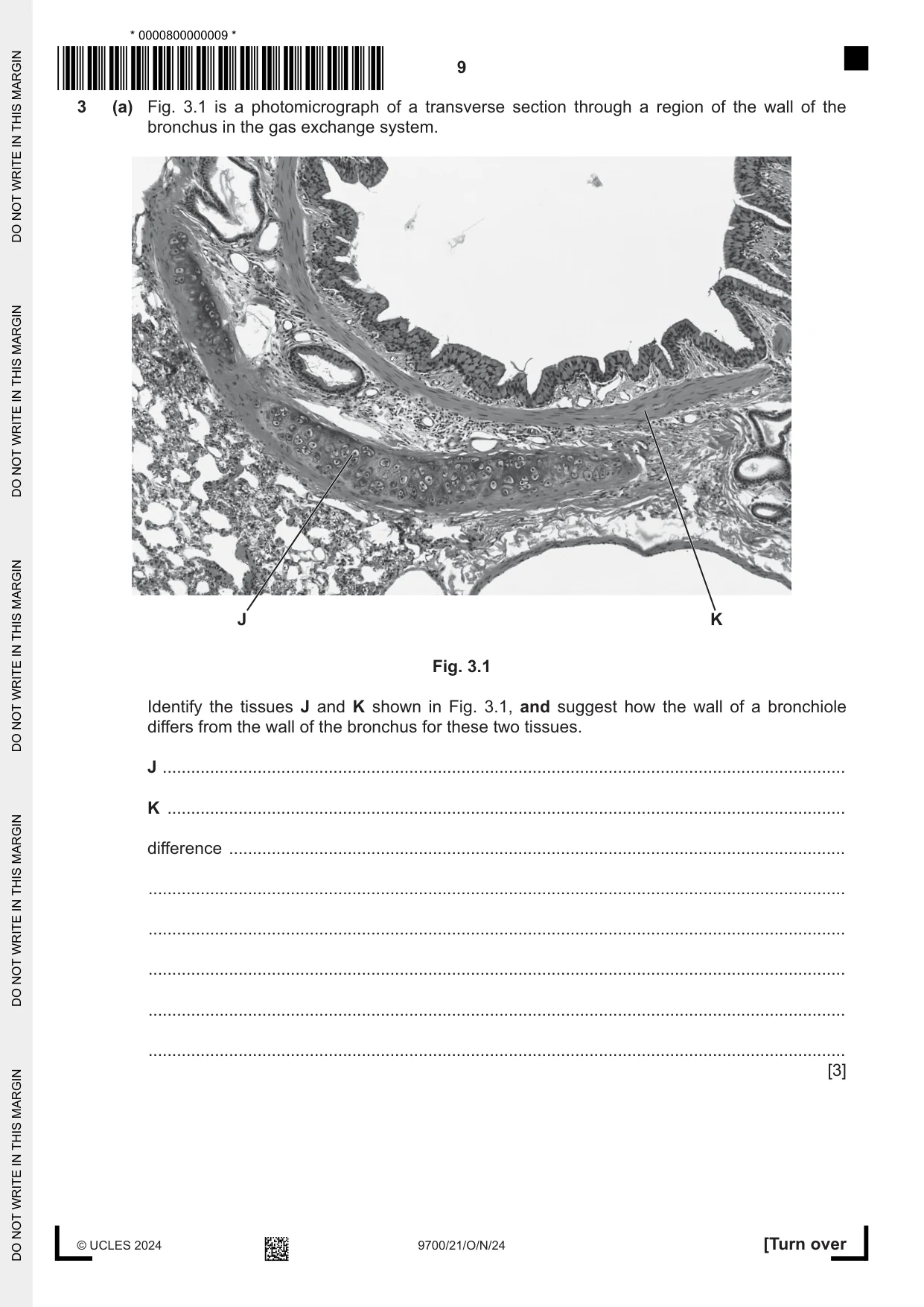
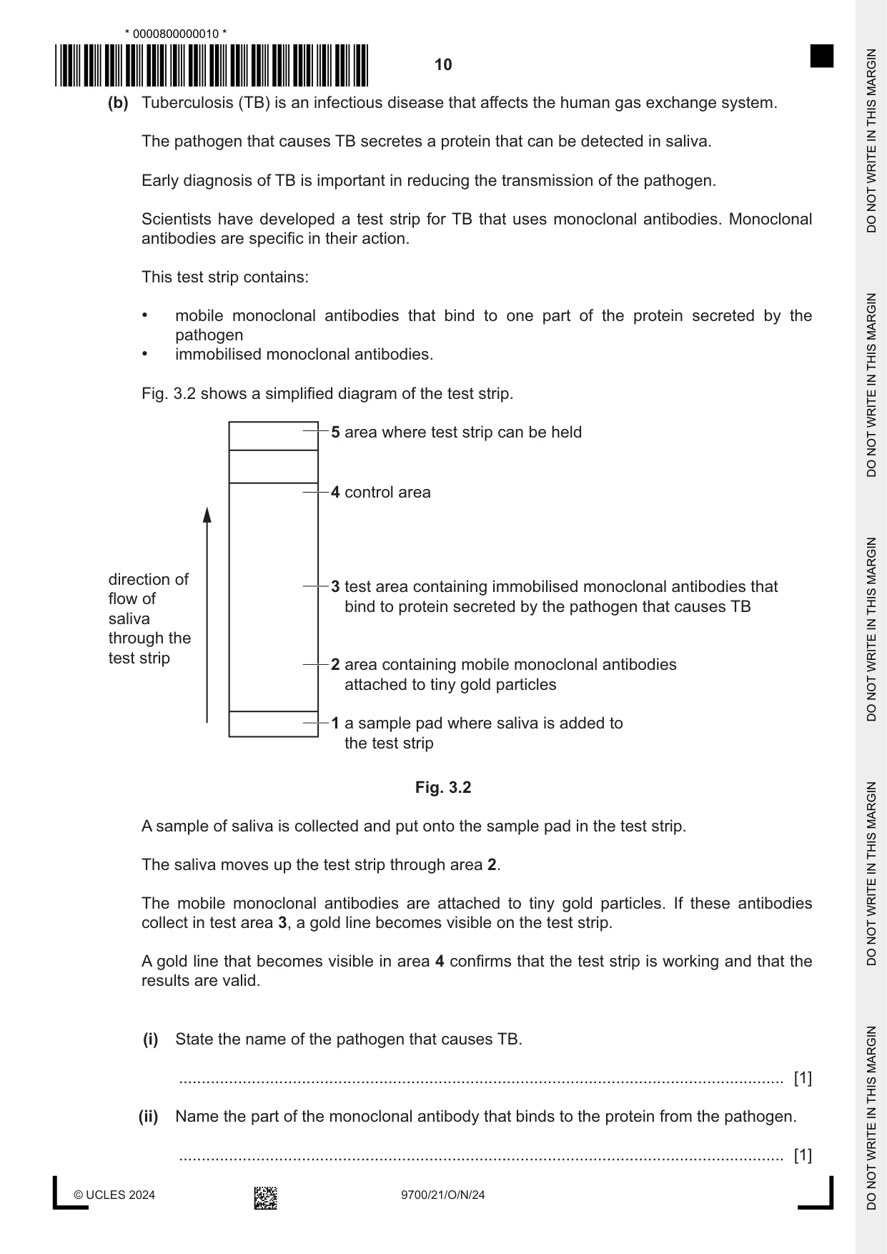
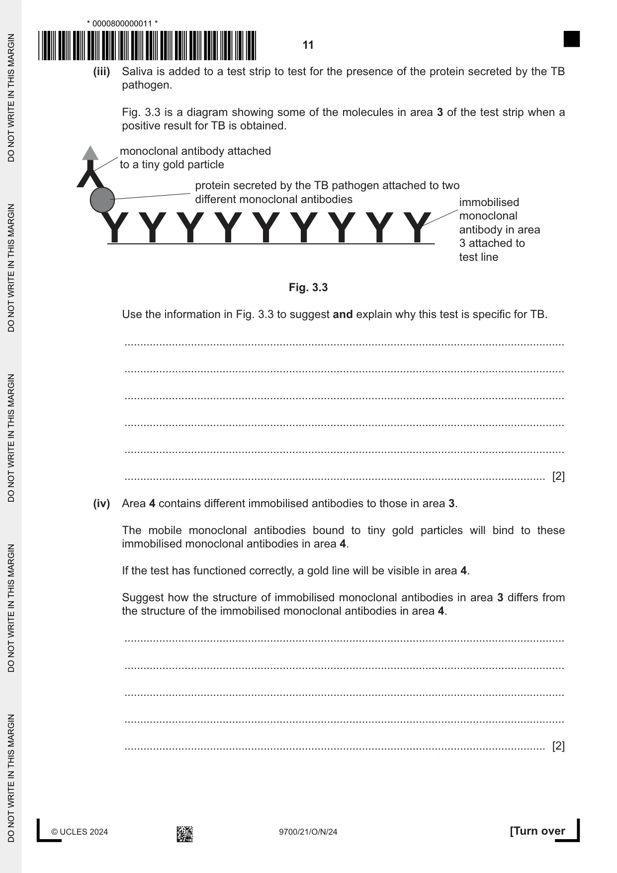
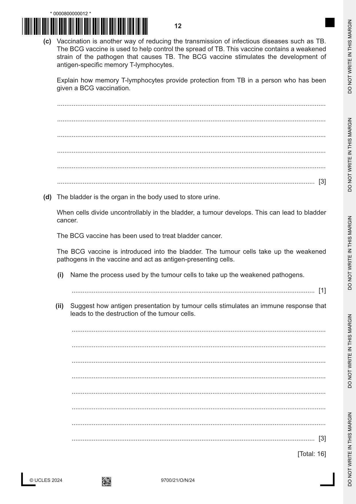
3 (a) Fig. 3.1 is a photomicrograph of a transverse section through a region of the wall of the bronchus in the gas exchange system. J K Fig. 3.1 Identify the tissues J and K shown in Fig. 3.1, and suggest how the wall of a bronchiole differs from the wall of the bronchus for these two tissues. J ................................................................................................................................................ K ............................................................................................................................................... difference .................................................................................................................................. ................................................................................................................................................... ................................................................................................................................................... ................................................................................................................................................... ................................................................................................................................................... ................................................................................................................................................... [3] (b) Tuberculosis (TB) is an infectious disease that affects the human gas exchange system. The pathogen that causes TB secretes a protein that can be detected in saliva. Early diagnosis of TB is important in reducing the transmission of the pathogen. Scientists have developed a test strip for TB that uses monoclonal antibodies. Monoclonal antibodies are specific in their action. This test strip contains: • mobile monoclonal antibodies that bind to one part of the protein secreted by the pathogen • immobilised monoclonal antibodies. Fig. 3.2 shows a simplified diagram of the test strip. 5 area where test strip can be held 4 control area direction of 3 test area containing immobilised monoclonal antibodies that flow of bind to protein secreted by the pathogen that causes TB saliva through the test strip 2 area containing mobile monoclonal antibodies attached to tiny gold particles 1 a sample pad where saliva is added to the test strip Fig. 3.2 A sample of saliva is collected and put onto the sample pad in the test strip. The saliva moves up the test strip through area 2. The mobile monoclonal antibodies are attached to tiny gold particles. If these antibodies collect in test area 3, a gold line becomes visible on the test strip. A gold line that becomes visible in area 4 confirms that the test strip is working and that the results are valid. (i) State the name of the pathogen that causes TB. ..................................................................................................................................... [1] (ii) Name the part of the monoclonal antibody that binds to the protein from the pathogen. ..................................................................................................................................... [1] (iii) Saliva is added to a test strip to test for the presence of the protein secreted by the TB pathogen. Fig. 3.3 is a diagram showing some of the molecules in area 3 of the test strip when a positive result for TB is obtained. monoclonal antibody attached to a tiny gold particle protein secreted by the TB pathogen attached to two different monoclonal antibodies immobilised monoclonal antibody in area 3 attached to test line Fig. 3.3 Use the information in Fig. 3.3 to suggest and explain why this test is specific for TB. ........................................................................................................................................... ........................................................................................................................................... ........................................................................................................................................... ........................................................................................................................................... ........................................................................................................................................... ..................................................................................................................................... [2] (iv) Area 4 contains different immobilised antibodies to those in area 3. The mobile monoclonal antibodies bound to tiny gold particles will bind to these immobilised monoclonal antibodies in area 4. If the test has functioned correctly, a gold line will be visible in area 4. Suggest how the structure of immobilised monoclonal antibodies in area 3 differs from the structure of the immobilised monoclonal antibodies in area 4. ........................................................................................................................................... ........................................................................................................................................... ........................................................................................................................................... ........................................................................................................................................... ..................................................................................................................................... [2] (c) Vaccination is another way of reducing the transmission of infectious diseases such as TB. The BCG vaccine is used to help control the spread of TB. This vaccine contains a weakened strain of the pathogen that causes TB. The BCG vaccine stimulates the development of antigen-specific memory T-lymphocytes. Explain how memory T-lymphocytes provide protection from TB in a person who has been given a BCG vaccination. ................................................................................................................................................... ................................................................................................................................................... ................................................................................................................................................... ................................................................................................................................................... ................................................................................................................................................... ............................................................................................................................................. [3] (d) The bladder is the organ in the body used to store urine. When cells divide uncontrollably in the bladder, a tumour develops. This can lead to bladder cancer. The BCG vaccine has been used to treat bladder cancer. The BCG vaccine is introduced into the bladder. The tumour cells take up the weakened pathogens in the vaccine and act as antigen-presenting cells. (i) Name the process used by the tumour cells to take up the weakened pathogens. ..................................................................................................................................... [1] (ii) Suggest how antigen presentation by tumour cells stimulates an immune response that leads to the destruction of the tumour cells. ........................................................................................................................................... ........................................................................................................................................... ........................................................................................................................................... ........................................................................................................................................... ........................................................................................................................................... ........................................................................................................................................... ........................................................................................................................................... ..................................................................................................................................... [3] [Total: 16]
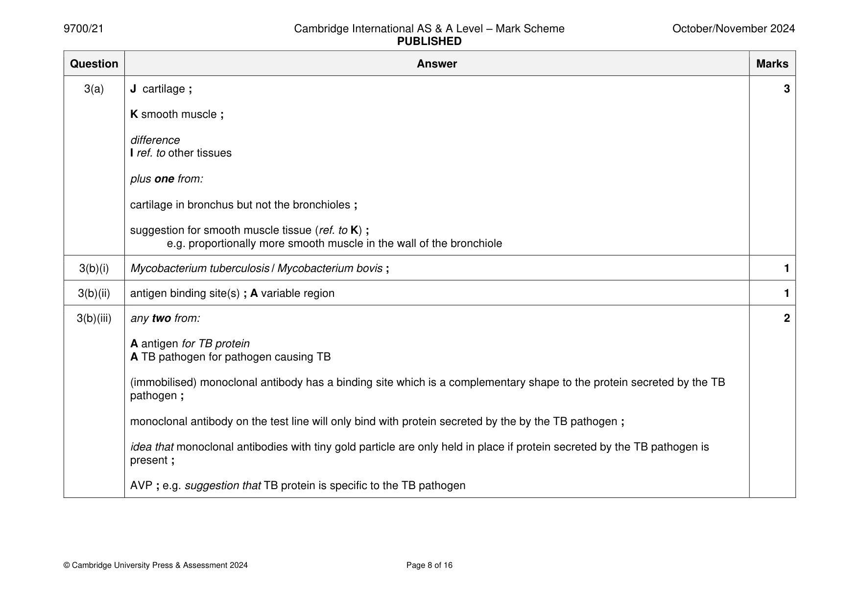
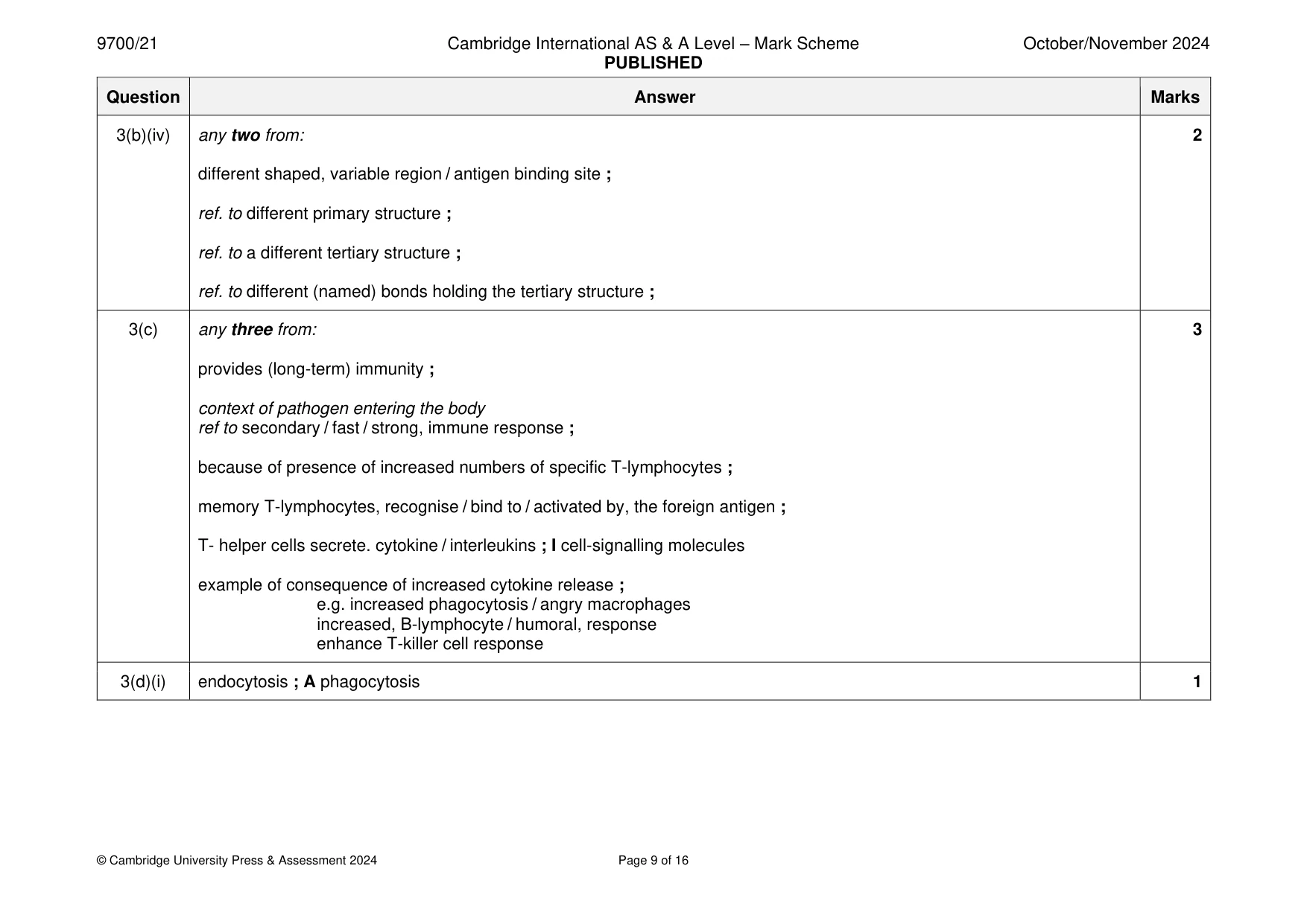
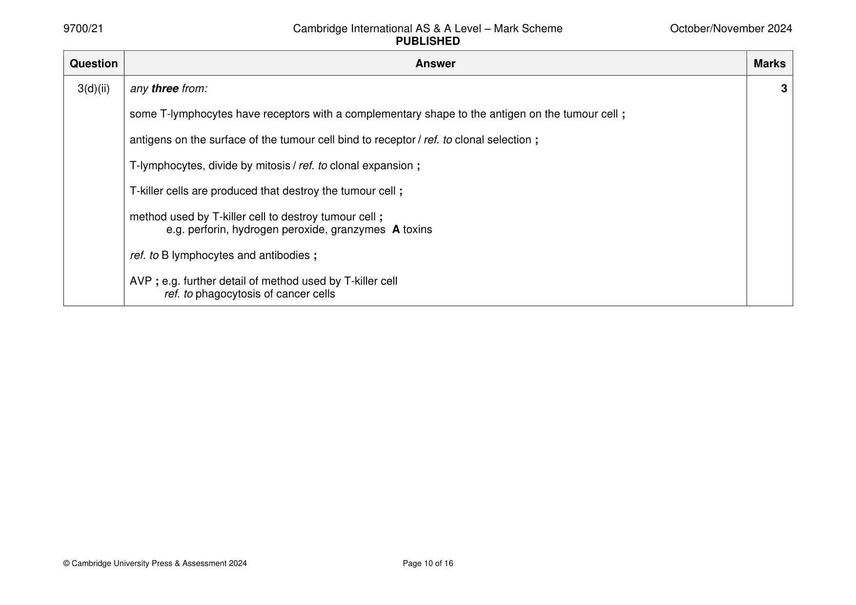
Mark scheme: 3(a) J cartilage ; 3 K smooth muscle ; difference I ref. to other tissues plus one from: cartilage in bronchus but not the bronchioles ; suggestion for smooth muscle tissue (ref. to K) ; e.g. proportionally more smooth muscle in the wall of the bronchiole 3(b)(i) Mycobacterium tuberculosis / Mycobacterium bovis ; 1 3(b)(ii) antigen binding site(s) ; A variable region 1 3(b)(iii) any two from: 2 A antigen for TB protein A TB pathogen for pathogen causing TB (immobilised) monoclonal antibody has a binding site which is a complementary shape to the protein secreted by the TB pathogen ; monoclonal antibody on the test line will only bind with protein secreted by the by the TB pathogen ; idea that monoclonal antibodies with tiny gold particle are only held in place if protein secreted by the TB pathogen is present ; AVP ; e.g. suggestion that TB protein is specific to the TB pathogen 3(b)(iv) any two from: 2 different shaped, variable region / antigen binding site ; ref. to different primary structure ; ref. to a different tertiary structure ; ref. to different (named) bonds holding the tertiary structure ; 3(c) any three from: 3 provides (long-term) immunity ; context of pathogen entering the body ref to secondary / fast / strong, immune response ; because of presence of increased numbers of specific T-lymphocytes ; memory T-lymphocytes, recognise / bind to / activated by, the foreign antigen ; T- helper cells secrete. cytokine / interleukins ; I cell-signalling molecules example of consequence of increased cytokine release ; e.g. increased phagocytosis / angry macrophages increased, B-lymphocyte / humoral, response enhance T-killer cell response 3(d)(i) endocytosis ; A phagocytosis 1 3(d)(ii) any three from: 3 some T-lymphocytes have receptors with a complementary shape to the antigen on the tumour cell ; antigens on the surface of the tumour cell bind to receptor / ref. to clonal selection ; T-lymphocytes, divide by mitosis / ref. to clonal expansion ; T-killer cells are produced that destroy the tumour cell ; method used by T-killer cell to destroy tumour cell ; e.g. perforin, hydrogen peroxide, granzymes A toxins ref. to B lymphocytes and antibodies ; AVP ; e.g. further detail of method used by T-killer cell ref. to phagocytosis of cancer cells
Q4 · The structure of sucrose, a disaccharide produced by plant cells
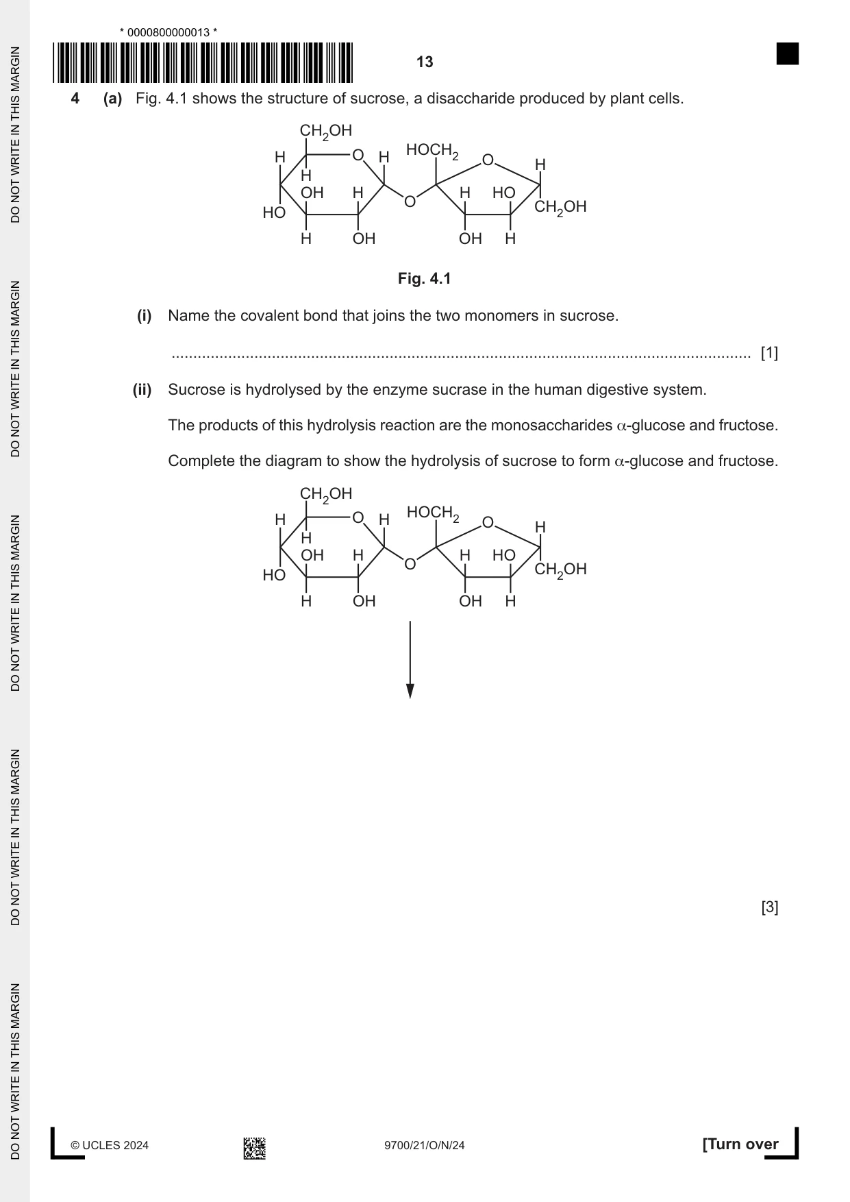
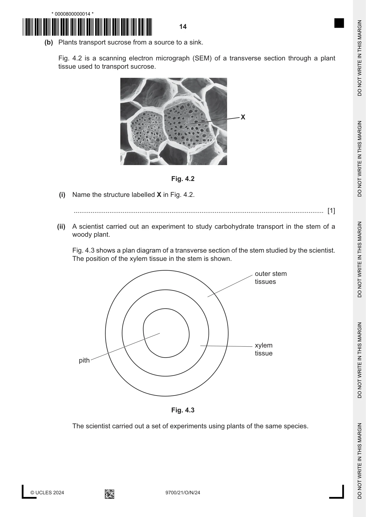
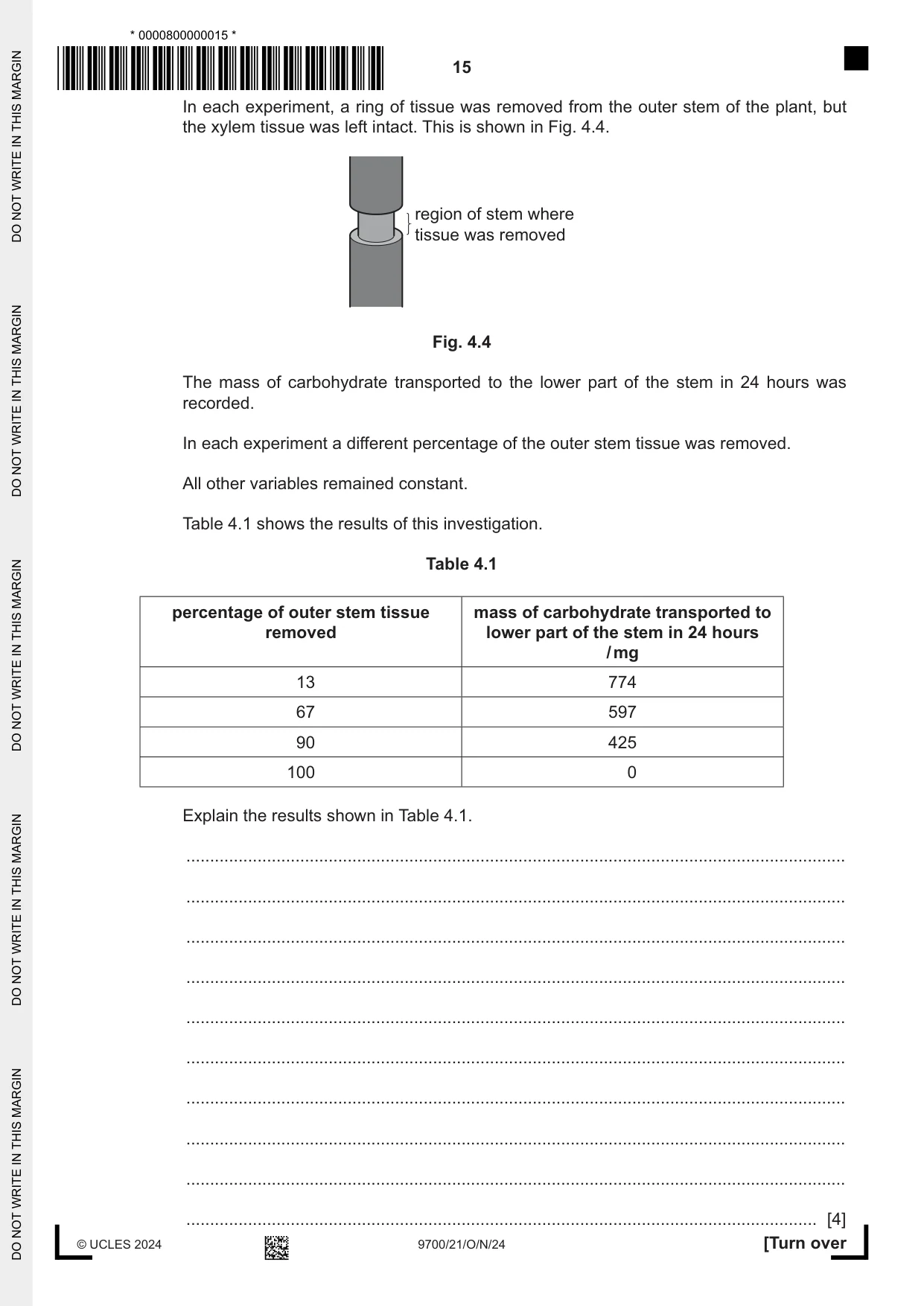
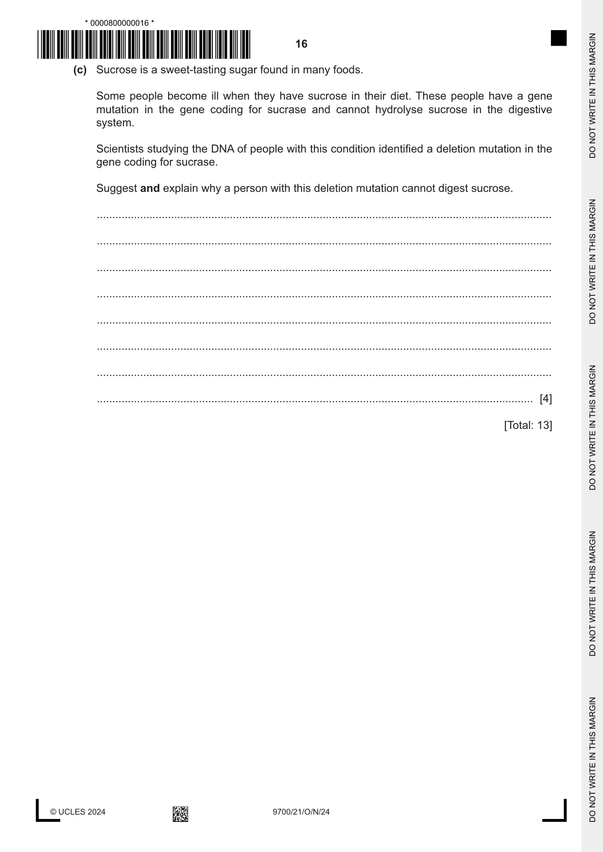
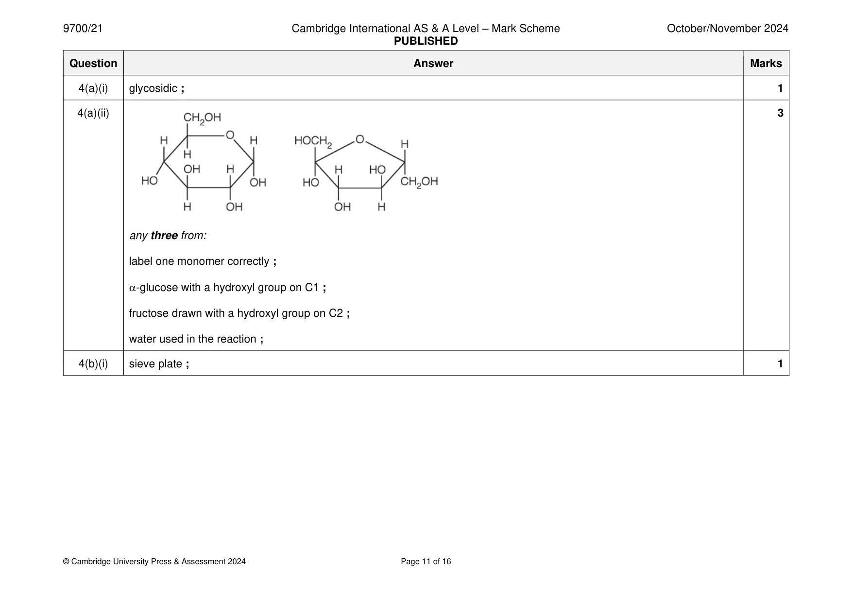
4 (a) Fig. 4.1 shows the structure of sucrose, a disaccharide produced by plant cells. CH2OH H O H HOCH2 O H H OH H H HO O HO CH2OH H OH OH H Fig. 4.1 (i) Name the covalent bond that joins the two monomers in sucrose. ..................................................................................................................................... [1] (ii) Sucrose is hydrolysed by the enzyme sucrase in the human digestive system. The products of this hydrolysis reaction are the monosaccharides α-glucose and fructose. Complete the diagram to show the hydrolysis of sucrose to form α-glucose and fructose. CH2OH H O H HOCH2 O H H OH H H HO O HO CH2OH H OH OH H [3] (b) Plants transport sucrose from a source to a sink. Fig. 4.2 is a scanning electron micrograph (SEM) of a transverse section through a plant tissue used to transport sucrose. X Fig. 4.2 (i) Name the structure labelled X in Fig. 4.2. ..................................................................................................................................... [1] (ii) A scientist carried out an experiment to study carbohydrate transport in the stem of a woody plant. Fig. 4.3 shows a plan diagram of a transverse section of the stem studied by the scientist. The position of the xylem tissue in the stem is shown. outer stem tissues xylem tissue pith Fig. 4.3 The scientist carried out a set of experiments using plants of the same species. In each experiment, a ring of tissue was removed from the outer stem of the plant, but the xylem tissue was left intact. This is shown in Fig. 4.4. region of stem where tissue was removed Fig. 4.4 The mass of carbohydrate transported to the lower part of the stem in 24 hours was recorded. In each experiment a different percentage of the outer stem tissue was removed. All other variables remained constant. Table 4.1 shows the results of this investigation. Table 4.1 percentage of outer stem tissue mass of carbohydrate transported to removed lower part of the stem in 24 hours / mg 13 774 67 597 90 425 100 0 Explain the results shown in Table 4.1. ........................................................................................................................................... ........................................................................................................................................... ........................................................................................................................................... ........................................................................................................................................... ........................................................................................................................................... ........................................................................................................................................... ........................................................................................................................................... ........................................................................................................................................... ........................................................................................................................................... ..................................................................................................................................... [4] (c) Sucrose is a sweet-tasting sugar found in many foods. Some people become ill when they have sucrose in their diet. These people have a gene mutation in the gene coding for sucrase and cannot hydrolyse sucrose in the digestive system. Scientists studying the DNA of people with this condition identified a deletion mutation in the gene coding for sucrase. Suggest and explain why a person with this deletion mutation cannot digest sucrose. ................................................................................................................................................... ................................................................................................................................................... ................................................................................................................................................... ................................................................................................................................................... ................................................................................................................................................... ................................................................................................................................................... ................................................................................................................................................... ............................................................................................................................................. [4] [Total: 13] Question 5 starts on page 18
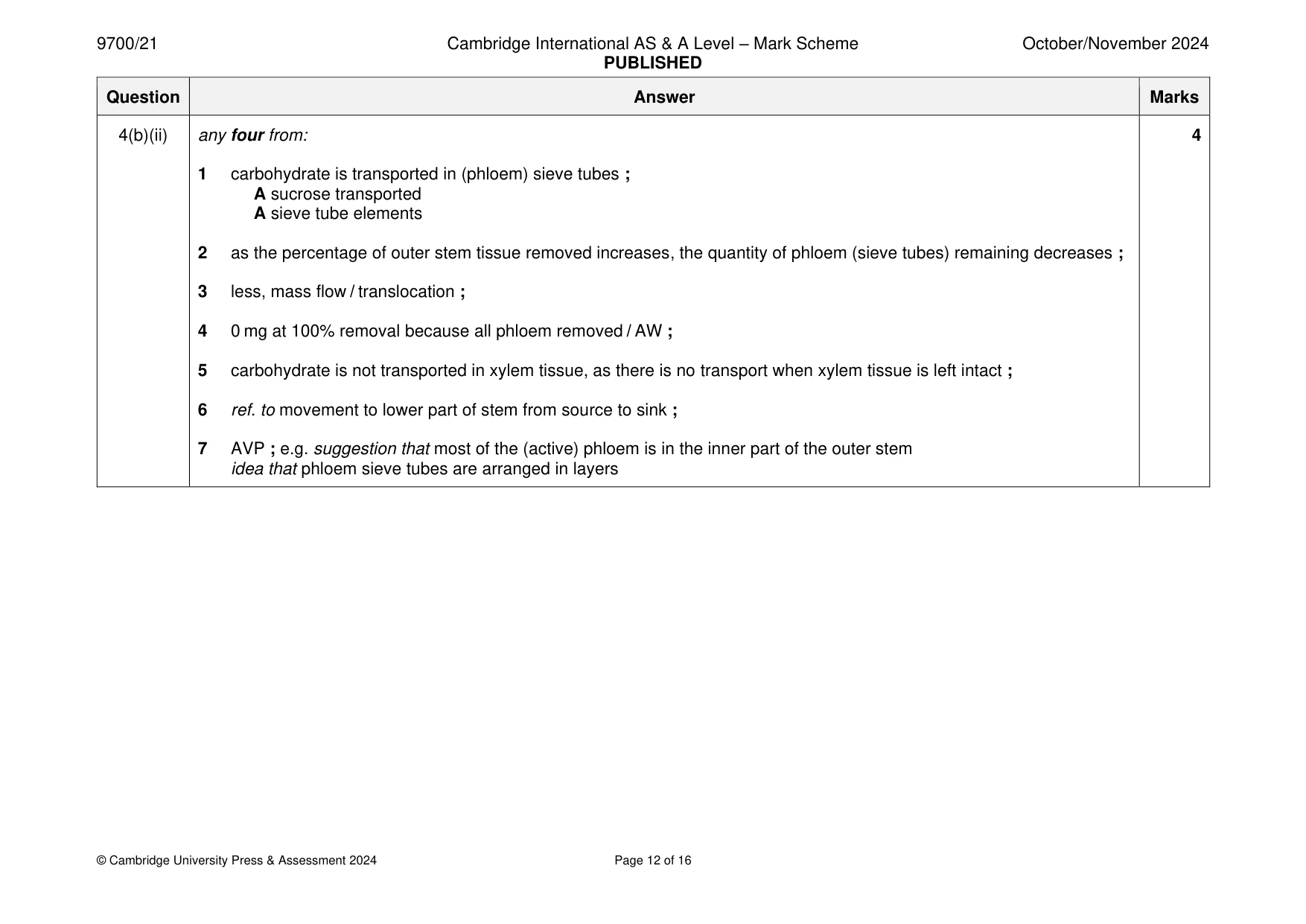
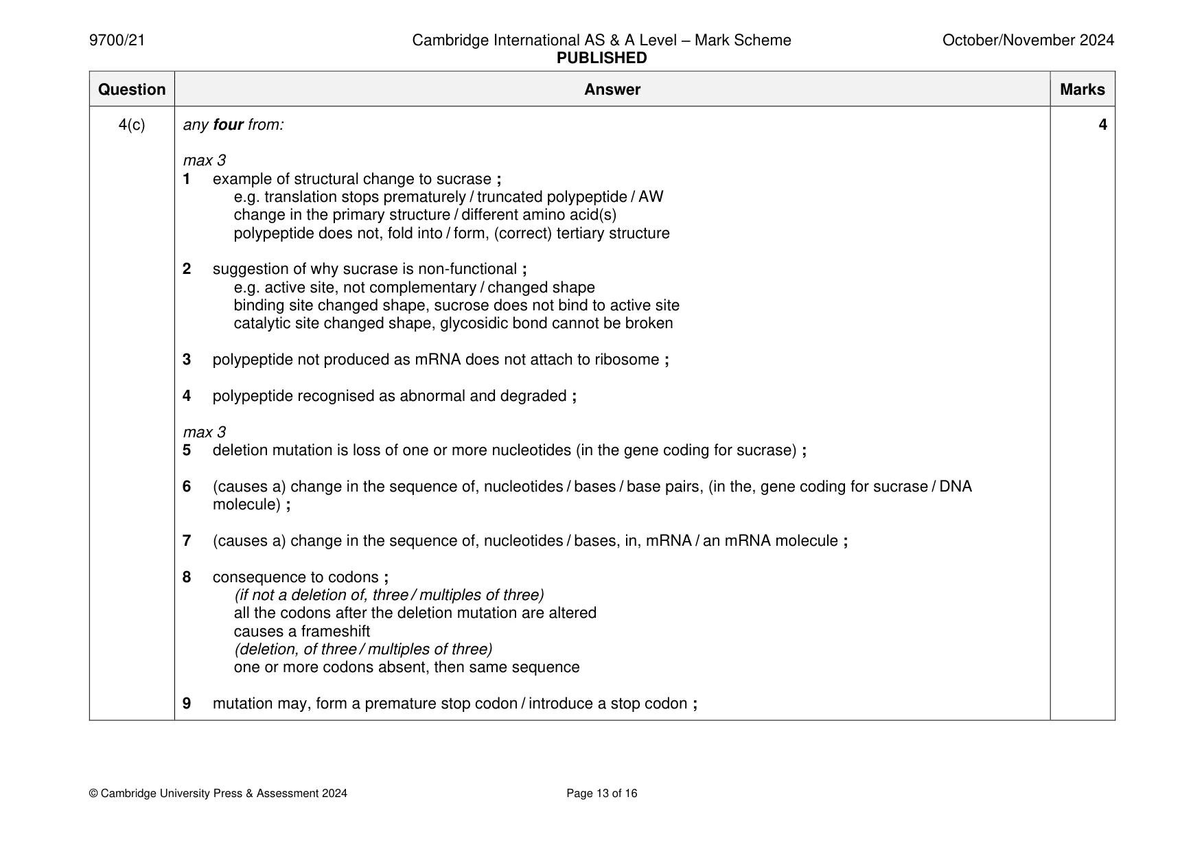
Mark scheme: 4(a)(i) glycosidic ; 1 4(a)(ii) 3 any three from: label one monomer correctly ; -glucose with a hydroxyl group on C1 ; fructose drawn with a hydroxyl group on C2 ; water used in the reaction ; 4(b)(i) sieve plate ; 1 4(b)(ii) any four from: 4 1 carbohydrate is transported in (phloem) sieve tubes ; A sucrose transported A sieve tube elements 2 as the percentage of outer stem tissue removed increases, the quantity of phloem (sieve tubes) remaining decreases ; 3 less, mass flow / translocation ; 4 0 mg at 100% removal because all phloem removed / AW ; 5 carbohydrate is not transported in xylem tissue, as there is no transport when xylem tissue is left intact ; 6 ref. to movement to lower part of stem from source to sink ; 7 AVP ; e.g. suggestion that most of the (active) phloem is in the inner part of the outer stem idea that phloem sieve tubes are arranged in layers 4(c) any four from: 4 max 3 1 example of structural change to sucrase ; e.g. translation stops prematurely / truncated polypeptide / AW change in the primary structure / different amino acid(s) polypeptide does not, fold into / form, (correct) tertiary structure 2 suggestion of why sucrase is non-functional ; e.g. active site, not complementary / changed shape binding site changed shape, sucrose does not bind to active site catalytic site changed shape, glycosidic bond cannot be broken 3 polypeptide not produced as mRNA does not attach to ribosome ; 4 polypeptide recognised as abnormal and degraded ; max 3 5 deletion mutation is loss of one or more nucleotides (in the gene coding for sucrase) ; 6 (causes a) change in the sequence of, nucleotides / bases / base pairs, (in the, gene coding for sucrase / DNA molecule) ; 7 (causes a) change in the sequence of, nucleotides / bases, in, mRNA / an mRNA molecule ; 8 consequence to codons ; (if not a deletion of, three / multiples of three) all the codons after the deletion mutation are altered causes a frameshift (deletion, of three / multiples of three) one or more codons absent, then same sequence 9 mutation may, form a premature stop codon / introduce a stop codon ;
Q5 · Trypsin is an enzyme which catalyses the hydrolysis of casein, a protein found in milk
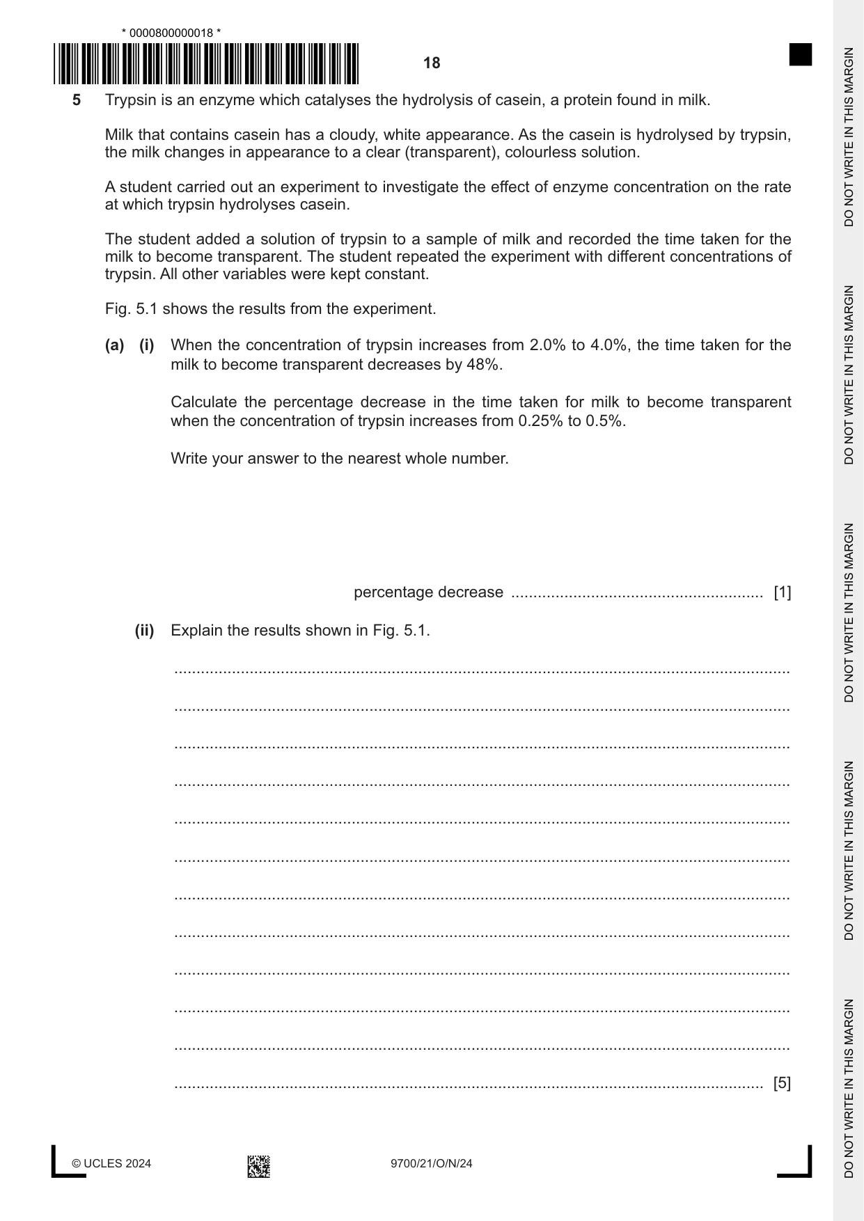
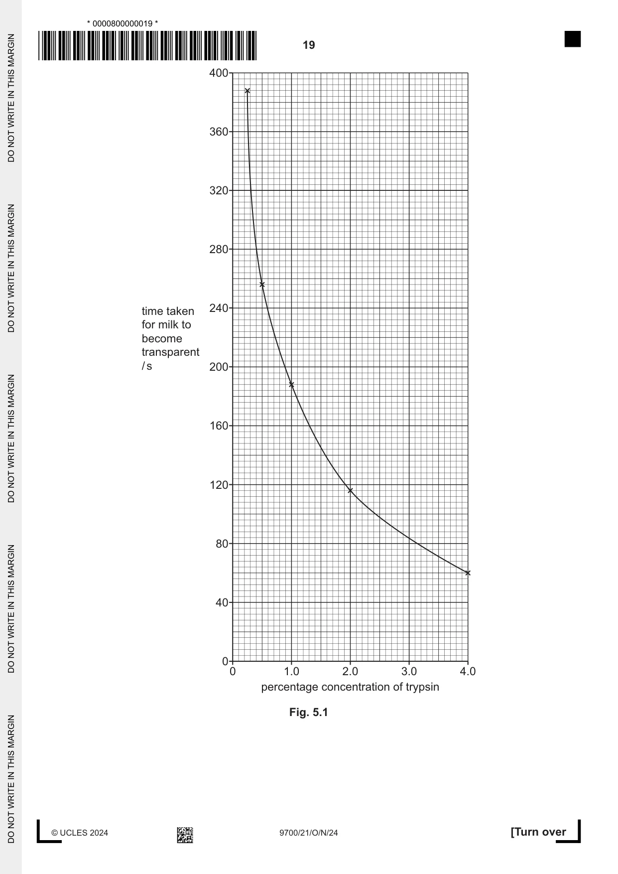
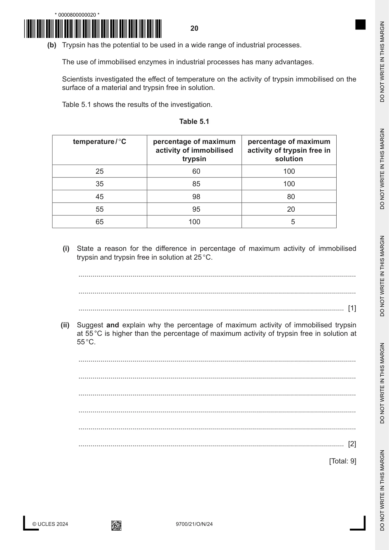
5 Trypsin is an enzyme which catalyses the hydrolysis of casein, a protein found in milk. Milk that contains casein has a cloudy, white appearance. As the casein is hydrolysed by trypsin, the milk changes in appearance to a clear (transparent), colourless solution. A student carried out an experiment to investigate the effect of enzyme concentration on the rate at which trypsin hydrolyses casein. The student added a solution of trypsin to a sample of milk and recorded the time taken for the milk to become transparent. The student repeated the experiment with different concentrations of trypsin. All other variables were kept constant. Fig. 5.1 shows the results from the experiment. (a) (i) When the concentration of trypsin increases from 2.0% to 4.0%, the time taken for the milk to become transparent decreases by 48%. Calculate the percentage decrease in the time taken for milk to become transparent when the concentration of trypsin increases from 0.25% to 0.5%. Write your answer to the nearest whole number. percentage decrease ......................................................... [1] (ii) Explain the results shown in Fig. 5.1. ........................................................................................................................................... ........................................................................................................................................... ........................................................................................................................................... ........................................................................................................................................... ........................................................................................................................................... ........................................................................................................................................... ........................................................................................................................................... ........................................................................................................................................... ........................................................................................................................................... ........................................................................................................................................... ........................................................................................................................................... ..................................................................................................................................... [5] (b) Trypsin has the potential to be used in a wide range of industrial processes. The use of immobilised enzymes in industrial processes has many advantages. Scientists investigated the effect of temperature on the activity of trypsin immobilised on the surface of a material and trypsin free in solution. Table 5.1 shows the results of the investigation. Table 5.1 temperature / °C percentage of maximum percentage of maximum activity of immobilised activity of trypsin free in trypsin solution 25 60 100 35 85 100 45 98 80 55 95 20 65 100 5 (i) State a reason for the difference in percentage of maximum activity of immobilised trypsin and trypsin free in solution at 25 °C. ........................................................................................................................................... ........................................................................................................................................... ..................................................................................................................................... [1] (ii) Suggest and explain why the percentage of maximum activity of immobilised trypsin at 55 °C is higher than the percentage of maximum activity of trypsin free in solution at 55 °C. ........................................................................................................................................... ........................................................................................................................................... ........................................................................................................................................... ........................................................................................................................................... ........................................................................................................................................... ..................................................................................................................................... [2] [Total: 9]
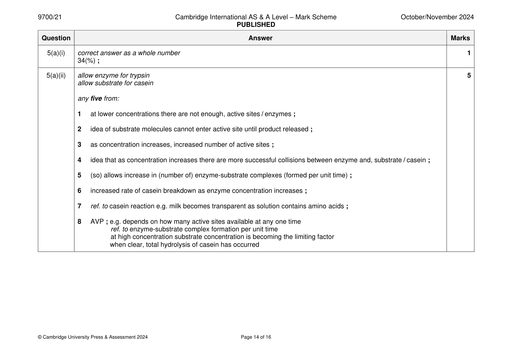
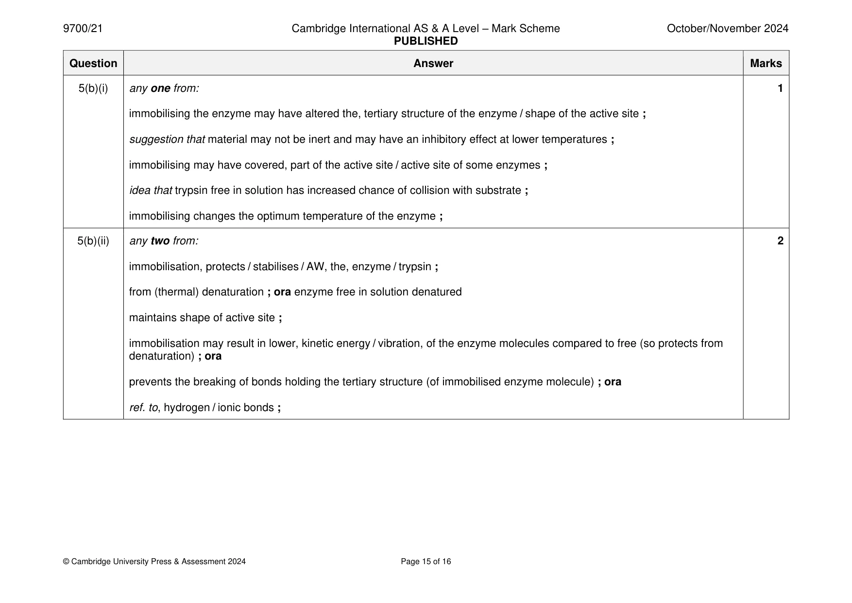
Mark scheme: 5(a)(i) correct answer as a whole number 1 34(%) ; 5(a)(ii) allow enzyme for trypsin 5 allow substrate for casein any five from: 1 at lower concentrations there are not enough, active sites / enzymes ; 2 idea of substrate molecules cannot enter active site until product released ; 3 as concentration increases, increased number of active sites ; 4 idea that as concentration increases there are more successful collisions between enzyme and, substrate / casein ; 5 (so) allows increase in (number of) enzyme-substrate complexes (formed per unit time) ; 6 increased rate of casein breakdown as enzyme concentration increases ; 7 ref. to casein reaction e.g. milk becomes transparent as solution contains amino acids ; 8 AVP ; e.g. depends on how many active sites available at any one time ref. to enzyme-substrate complex formation per unit time at high concentration substrate concentration is becoming the limiting factor when clear, total hydrolysis of casein has occurred 5(b)(i) any one from: 1 immobilising the enzyme may have altered the, tertiary structure of the enzyme / shape of the active site ; suggestion that material may not be inert and may have an inhibitory effect at lower temperatures ; immobilising may have covered, part of the active site / active site of some enzymes ; idea that trypsin free in solution has increased chance of collision with substrate ; immobilising changes the optimum temperature of the enzyme ; 5(b)(ii) any two from: 2 immobilisation, protects / stabilises / AW, the, enzyme / trypsin ; from (thermal) denaturation ; ora enzyme free in solution denatured maintains shape of active site ; immobilisation may result in lower, kinetic energy / vibration, of the enzyme molecules compared to free (so protects from denaturation) ; ora prevents the breaking of bonds holding the tertiary structure (of immobilised enzyme molecule) ; ora ref. to, hydrogen / ionic bonds ;
Q6 · A plant cell in a stage of mitosis
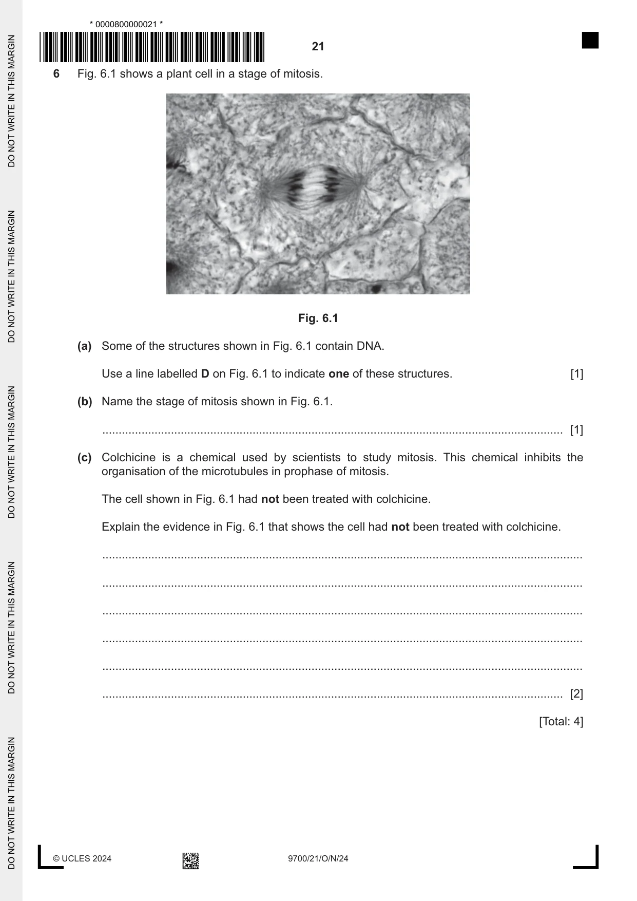
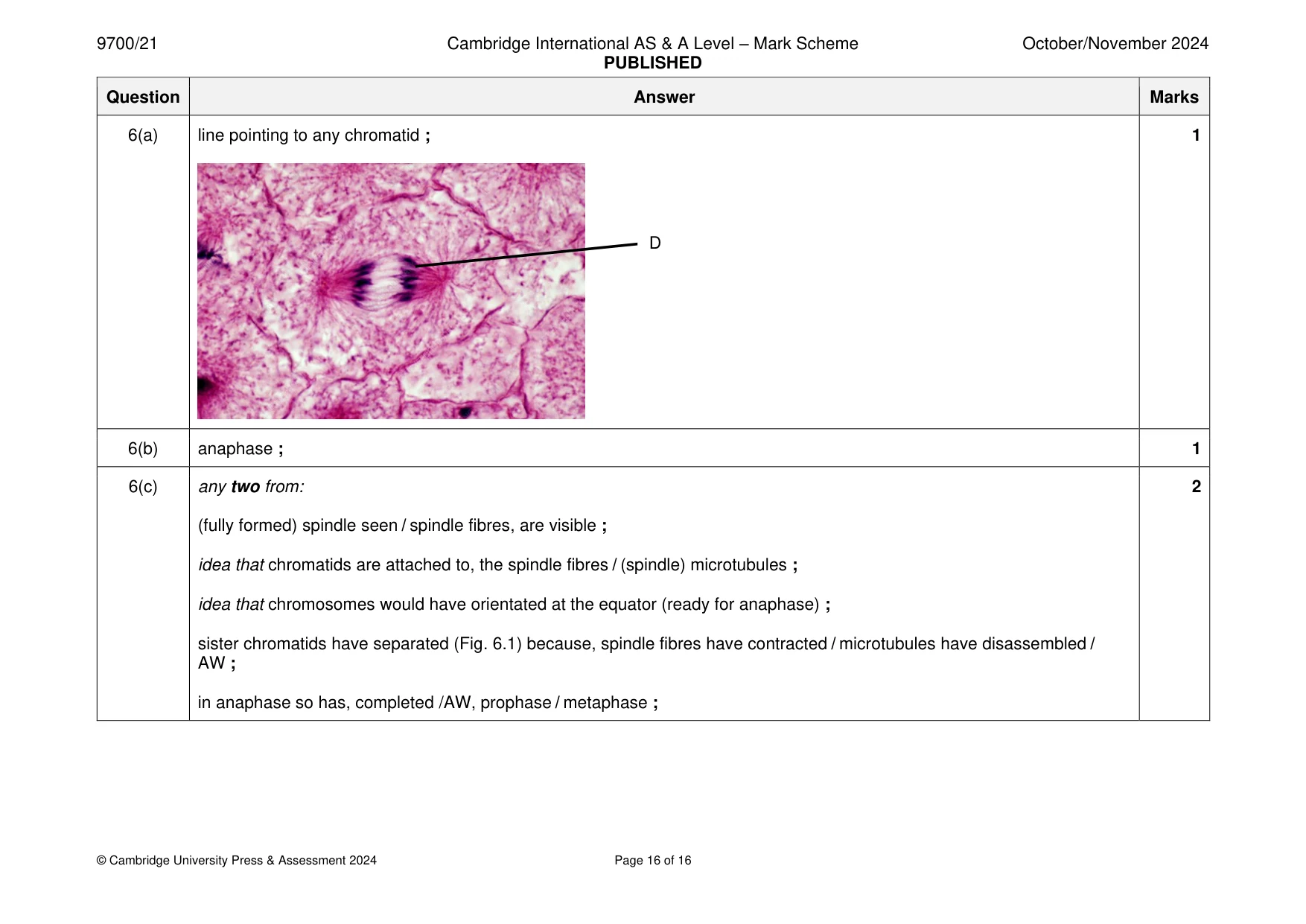
6 Fig. 6.1 shows a plant cell in a stage of mitosis. Fig. 6.1 (a) Some of the structures shown in Fig. 6.1 contain DNA. Use a line labelled D on Fig. 6.1 to indicate one of these structures. [1] (b) Name the stage of mitosis shown in Fig. 6.1. ............................................................................................................................................. [1] (c) Colchicine is a chemical used by scientists to study mitosis. This chemical inhibits the organisation of the microtubules in prophase of mitosis. The cell shown in Fig. 6.1 had not been treated with colchicine. Explain the evidence in Fig. 6.1 that shows the cell had not been treated with colchicine. ................................................................................................................................................... ................................................................................................................................................... ................................................................................................................................................... ................................................................................................................................................... ................................................................................................................................................... ............................................................................................................................................. [2] [Total: 4]
Mark scheme: 6(a) line pointing to any chromatid ; 1 D 6(b) anaphase ; 1 6(c) any two from: 2 (fully formed) spindle seen / spindle fibres, are visible ; idea that chromatids are attached to, the spindle fibres / (spindle) microtubules ; idea that chromosomes would have orientated at the equator (ready for anaphase) ; sister chromatids have separated (Fig. 6.1) because, spindle fibres have contracted / microtubules have disassembled / AW ; in anaphase so has, completed /AW, prophase / metaphase ;
What was in this paper
The subtopics covered by these 6 questions, and how many questions each got. Open one in a new tab to see every Cambridge question on it.
What you needed in this session
Cambridge’s own grade thresholds for 2024 Oct/Nov, Paper 2 · Variant 1. A higher threshold means an easier paper — the bar moves with how the cohort did.