Cambridge A Level Biology 9700 — 2023 Oct/Nov Paper 2 · Variant 1
9700/21/O/N/23 · 6 questions · 60 marks · ≈68 min
The question paper and its mark scheme, free to read here and free to download. This is Cambridge’s own paper, exactly as it was sat.
Question paper20 pages


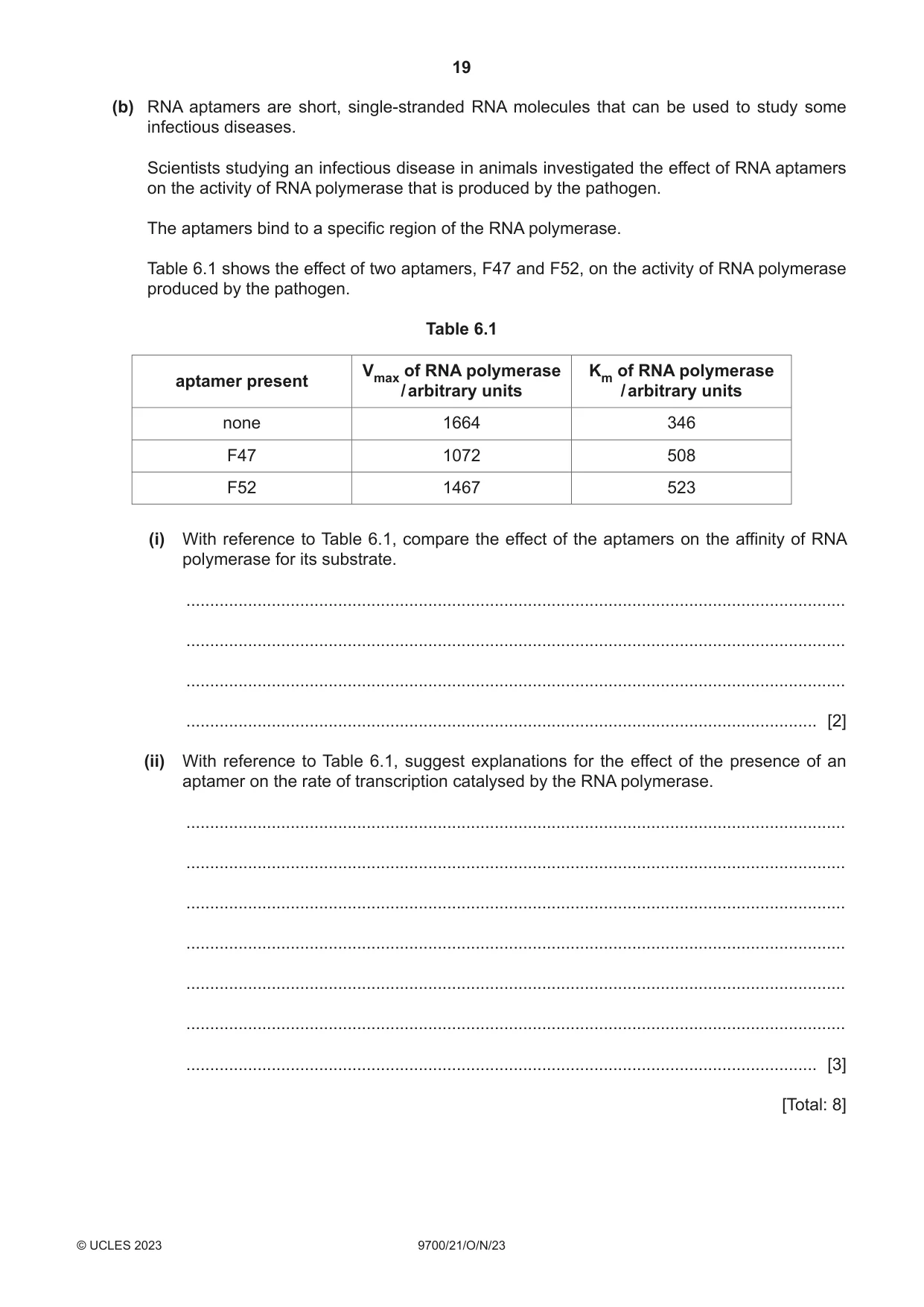
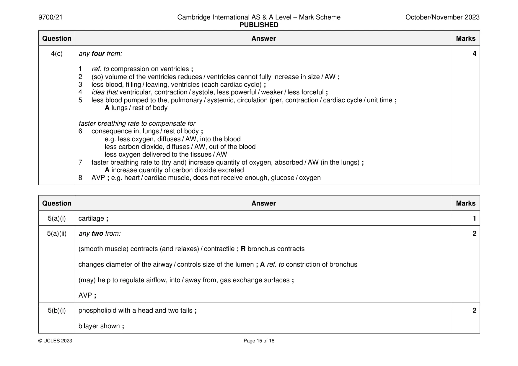
Mark scheme18 pages
Answers below. Sit the paper first if you are practising.
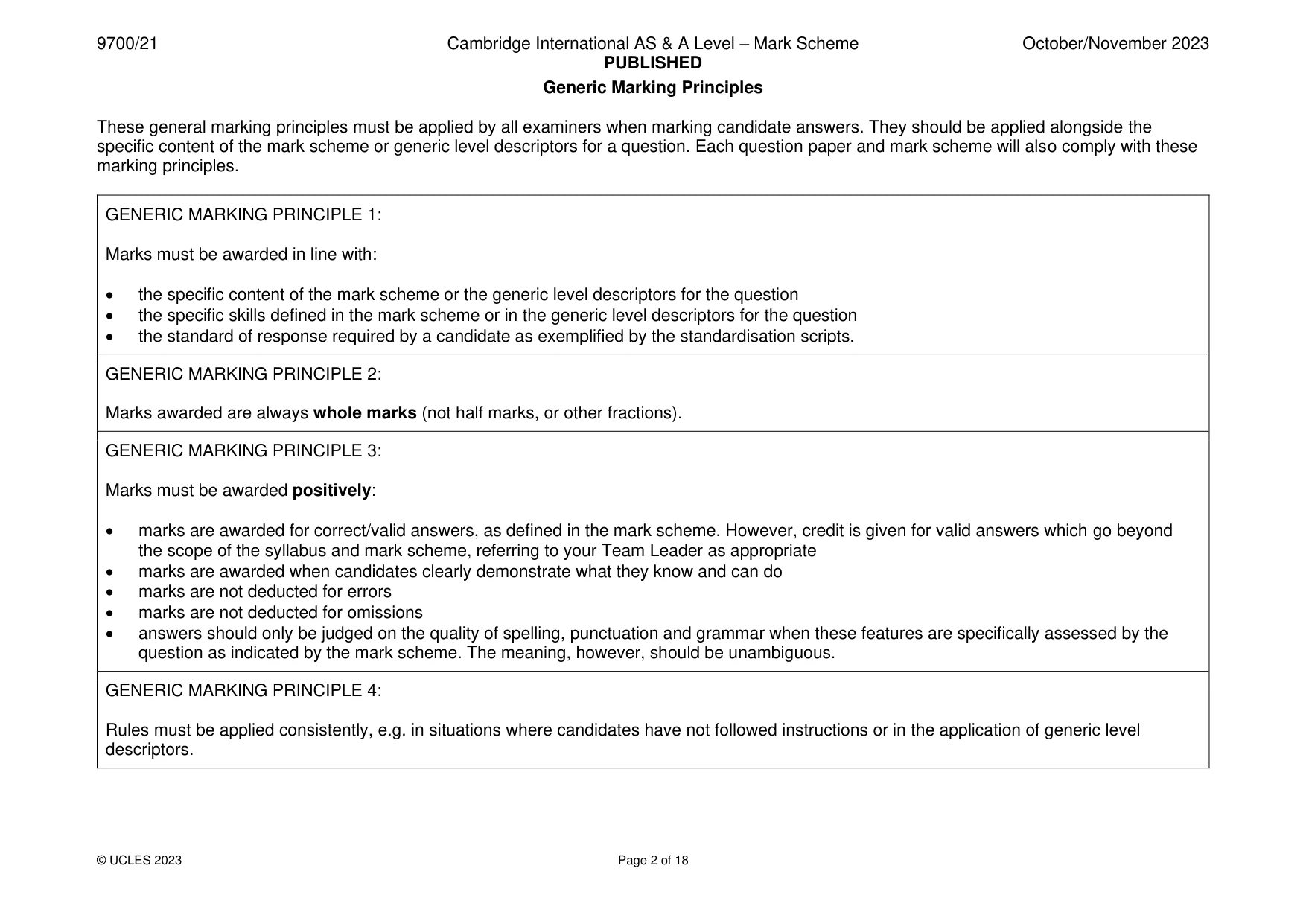
Questions as text
Q1 · Scientists measured the concentration of sodium ions and potassium ions in the red blood…
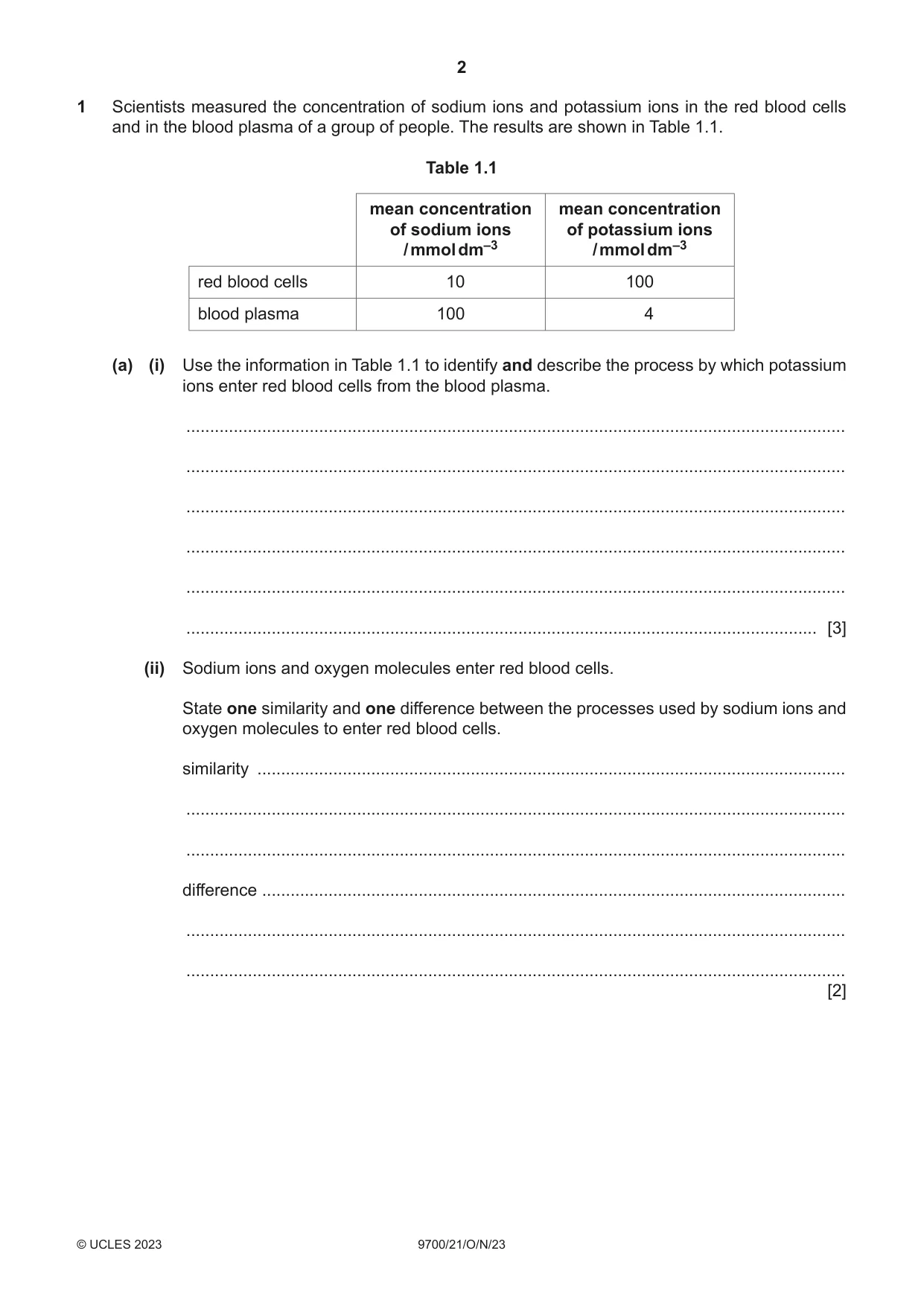
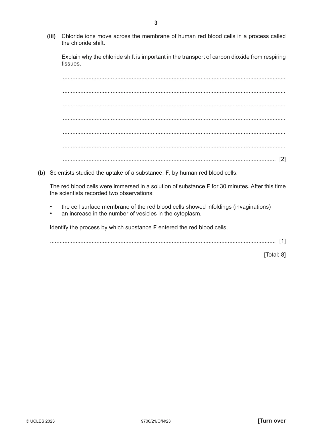
1 Scientists measured the concentration of sodium ions and potassium ions in the red blood cells and in the blood plasma of a group of people. The results are shown in Table 1.1. Table 1.1 mean concentration mean concentration of sodium ions of potassium ions / mmol dm–3 / mmol dm–3 red blood cells 10 100 blood plasma 100 4 (a) (i) Use the information in Table 1.1 to identify and describe the process by which potassium ions enter red blood cells from the blood plasma. ........................................................................................................................................... ........................................................................................................................................... ........................................................................................................................................... ........................................................................................................................................... ........................................................................................................................................... ..................................................................................................................................... [3] (ii) Sodium ions and oxygen molecules enter red blood cells. State one similarity and one difference between the processes used by sodium ions and oxygen molecules to enter red blood cells. similarity ............................................................................................................................ ........................................................................................................................................... ........................................................................................................................................... difference ........................................................................................................................... ........................................................................................................................................... ........................................................................................................................................... [2] (iii) Chloride ions move across the membrane of human red blood cells in a process called the chloride shift. Explain why the chloride shift is important in the transport of carbon dioxide from respiring tissues. ........................................................................................................................................... ........................................................................................................................................... ........................................................................................................................................... ........................................................................................................................................... ........................................................................................................................................... ........................................................................................................................................... ..................................................................................................................................... [2] (b) Scientists studied the uptake of a substance, F, by human red blood cells. The red blood cells were immersed in a solution of substance F for 30 minutes. After this time the scientists recorded two observations: • the cell surface membrane of the red blood cells showed infoldings (invaginations) • an increase in the number of vesicles in the cytoplasm. Identify the process by which substance F entered the red blood cells. ............................................................................................................................................. [1] [Total: 8]
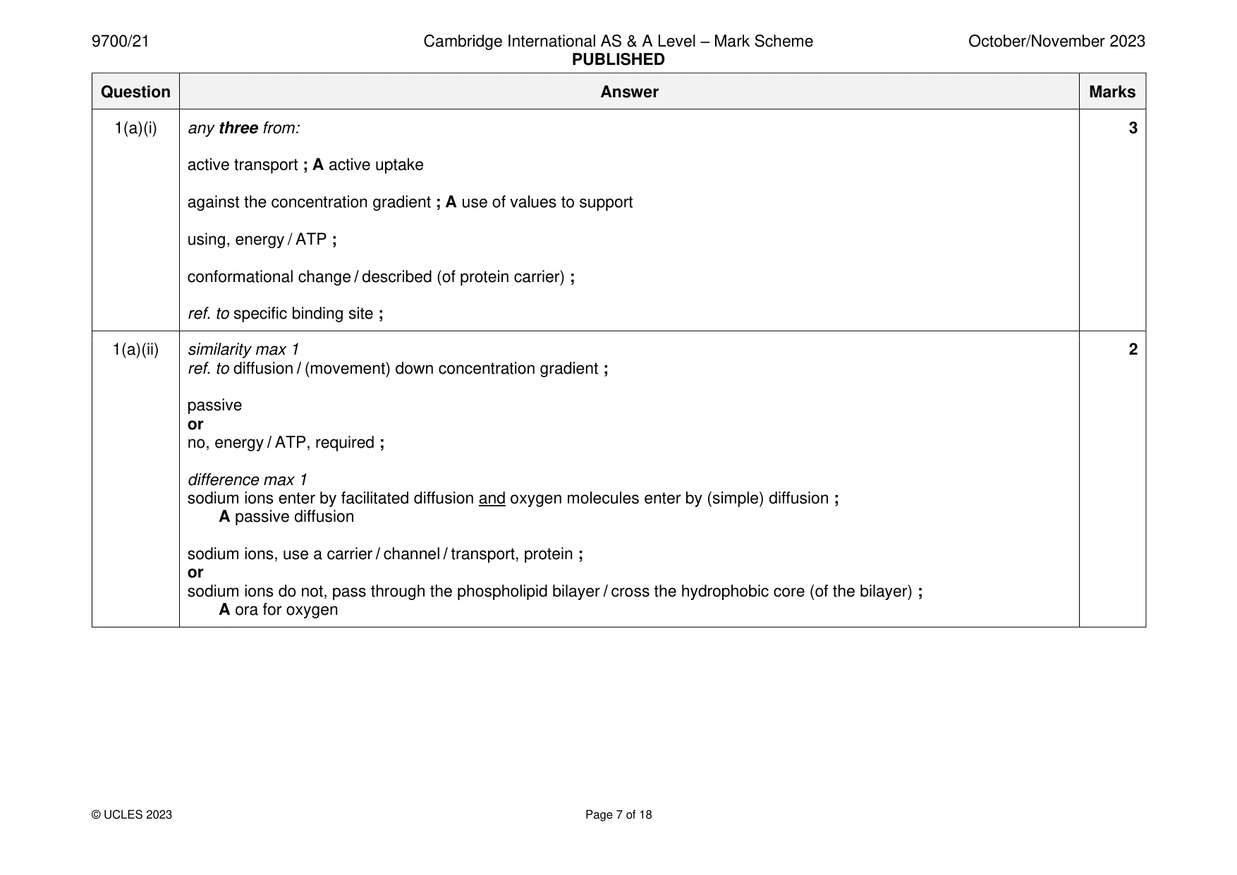
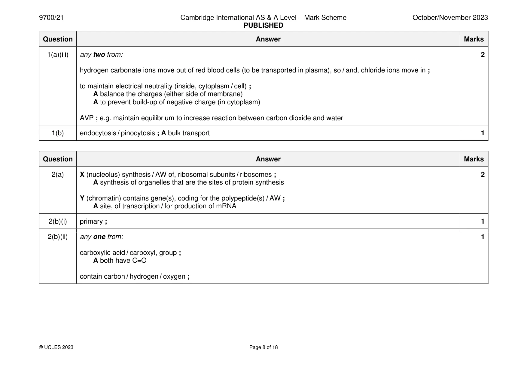
Mark scheme: Question Answer Marks 1(a)(i) any three from: 3 active transport ; A active uptake against the concentration gradient ; A use of values to support using, energy / ATP ; conformational change / described (of protein carrier) ; ref. to specific binding site ; 1(a)(ii) similarity max 1 2 ref. to diffusion / (movement) down concentration gradient ; passive or no, energy / ATP, required ; difference max 1 sodium ions enter by facilitated diffusion and oxygen molecules enter by (simple) diffusion ; A passive diffusion sodium ions, use a carrier / channel / transport, protein ; or sodium ions do not, pass through the phospholipid bilayer / cross the hydrophobic core (of the bilayer) ; A ora for oxygen 1(a)(iii) any two from: 2 hydrogen carbonate ions move out of red blood cells (to be transported in plasma), so / and, chloride ions move in ; to maintain electrical neutrality (inside, cytoplasm / cell) ; A balance the charges (either side of membrane) A to prevent build-up of negative charge (in cytoplasm) AVP ; e.g. maintain equilibrium to increase reaction between carbon dioxide and water 1(b) endocytosis / pinocytosis ; A bulk transport 1
Q2 · A transmission electron micrograph showing a section of a specialised epithelial cell…
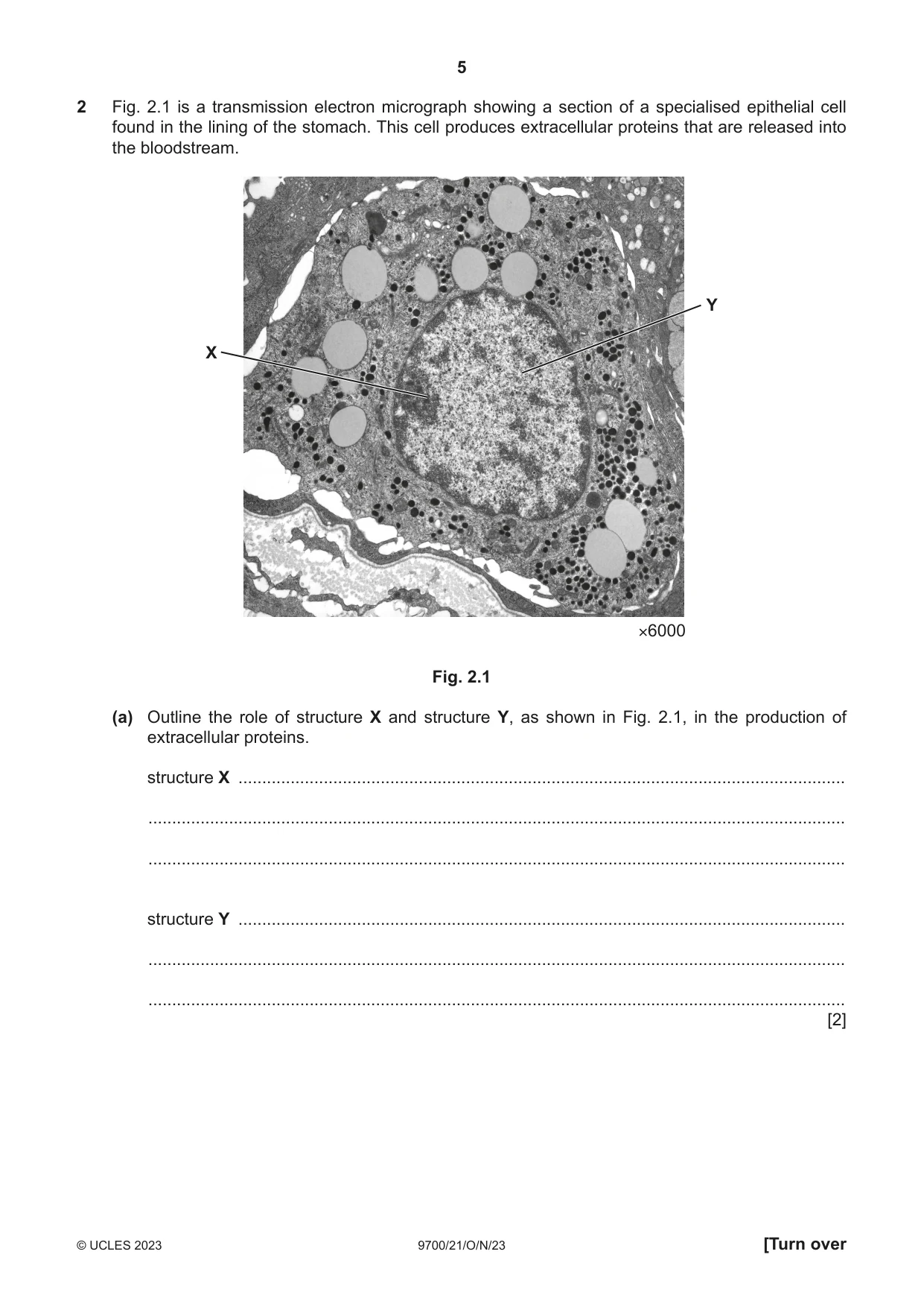
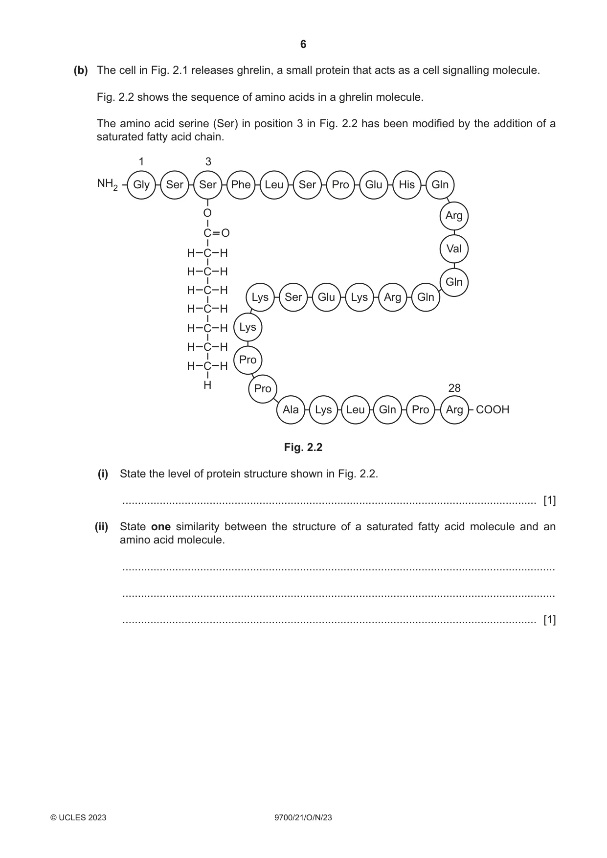
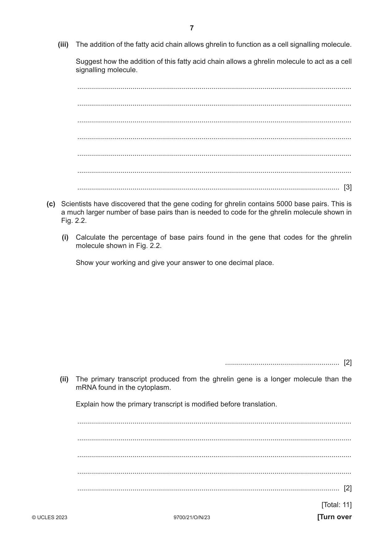
2 Fig. 2.1 is a transmission electron micrograph showing a section of a specialised epithelial cell found in the lining of the stomach. This cell produces extracellular proteins that are released into the bloodstream. Y X ×6000 Fig. 2.1 (a) Outline the role of structure X and structure Y, as shown in Fig. 2.1, in the production of extracellular proteins. structure X ................................................................................................................................ ................................................................................................................................................... ................................................................................................................................................... structure Y ................................................................................................................................ ................................................................................................................................................... ................................................................................................................................................... [2] (b) The cell in Fig. 2.1 releases ghrelin, a small protein that acts as a cell signalling molecule. Fig. 2.2 shows the sequence of amino acids in a ghrelin molecule. The amino acid serine (Ser) in position 3 in Fig. 2.2 has been modified by the addition of a saturated fatty acid chain. 1 3 NH2 Gly Ser Ser Phe Leu Ser Pro Glu His Gln O Arg C O Val H C H H C H Gln H C H Lys Ser Glu Lys Arg Gln H C H H C H Lys H C H Pro H C H H Pro 28 Ala Lys Leu Gln Pro Arg COOH Fig. 2.2 (i) State the level of protein structure shown in Fig. 2.2. ..................................................................................................................................... [1] (ii) State one similarity between the structure of a saturated fatty acid molecule and an amino acid molecule. ........................................................................................................................................... ........................................................................................................................................... ..................................................................................................................................... [1] (iii) The addition of the fatty acid chain allows ghrelin to function as a cell signalling molecule. Suggest how the addition of this fatty acid chain allows a ghrelin molecule to act as a cell signalling molecule. ........................................................................................................................................... ........................................................................................................................................... ........................................................................................................................................... ........................................................................................................................................... ........................................................................................................................................... ........................................................................................................................................... ..................................................................................................................................... [3] (c) Scientists have discovered that the gene coding for ghrelin contains 5000 base pairs. This is a much larger number of base pairs than is needed to code for the ghrelin molecule shown in Fig. 2.2. (i) Calculate the percentage of base pairs found in the gene that codes for the ghrelin molecule shown in Fig. 2.2. Show your working and give your answer to one decimal place. .......................................................... [2] (ii) The primary transcript produced from the ghrelin gene is a longer molecule than the mRNA found in the cytoplasm. Explain how the primary transcript is modified before translation. ........................................................................................................................................... ........................................................................................................................................... ........................................................................................................................................... ........................................................................................................................................... ..................................................................................................................................... [2] [Total: 11]
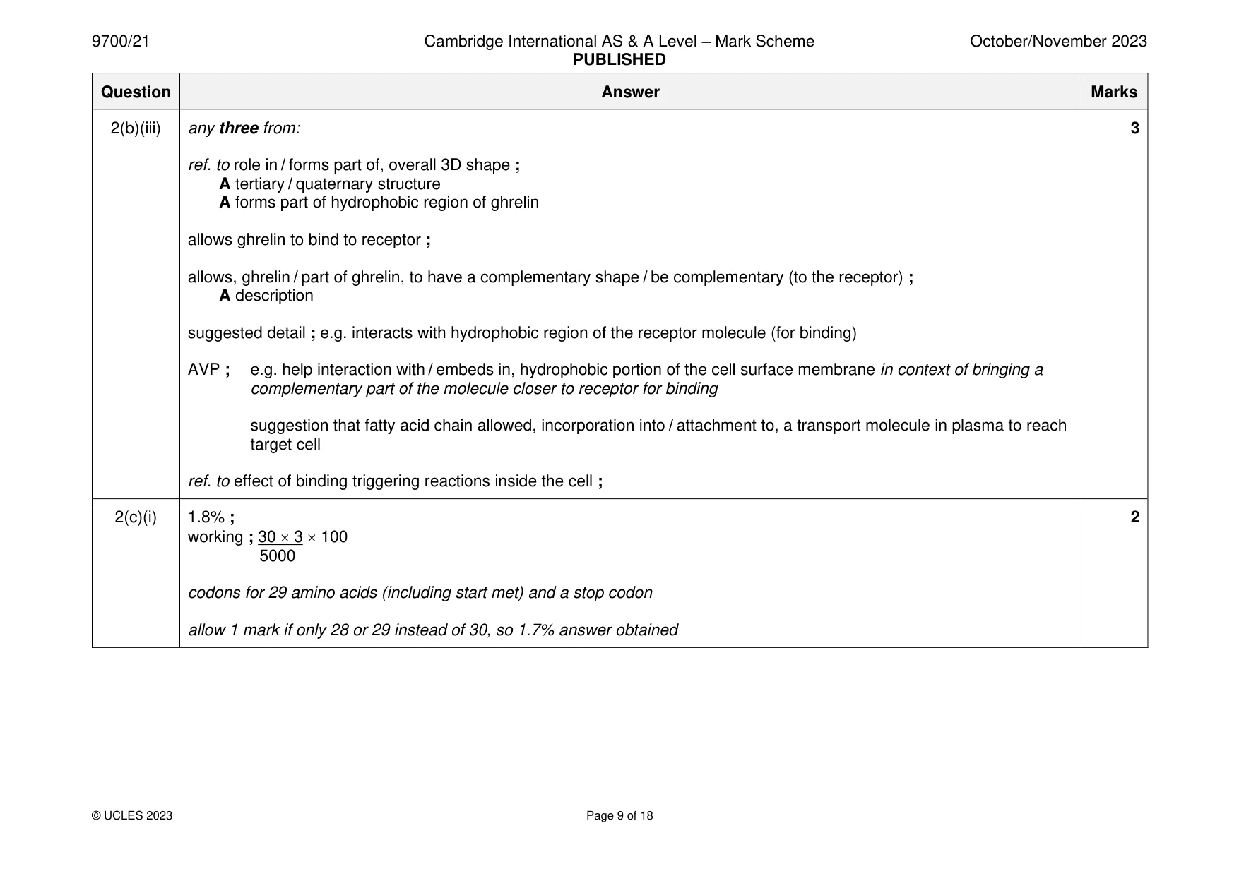
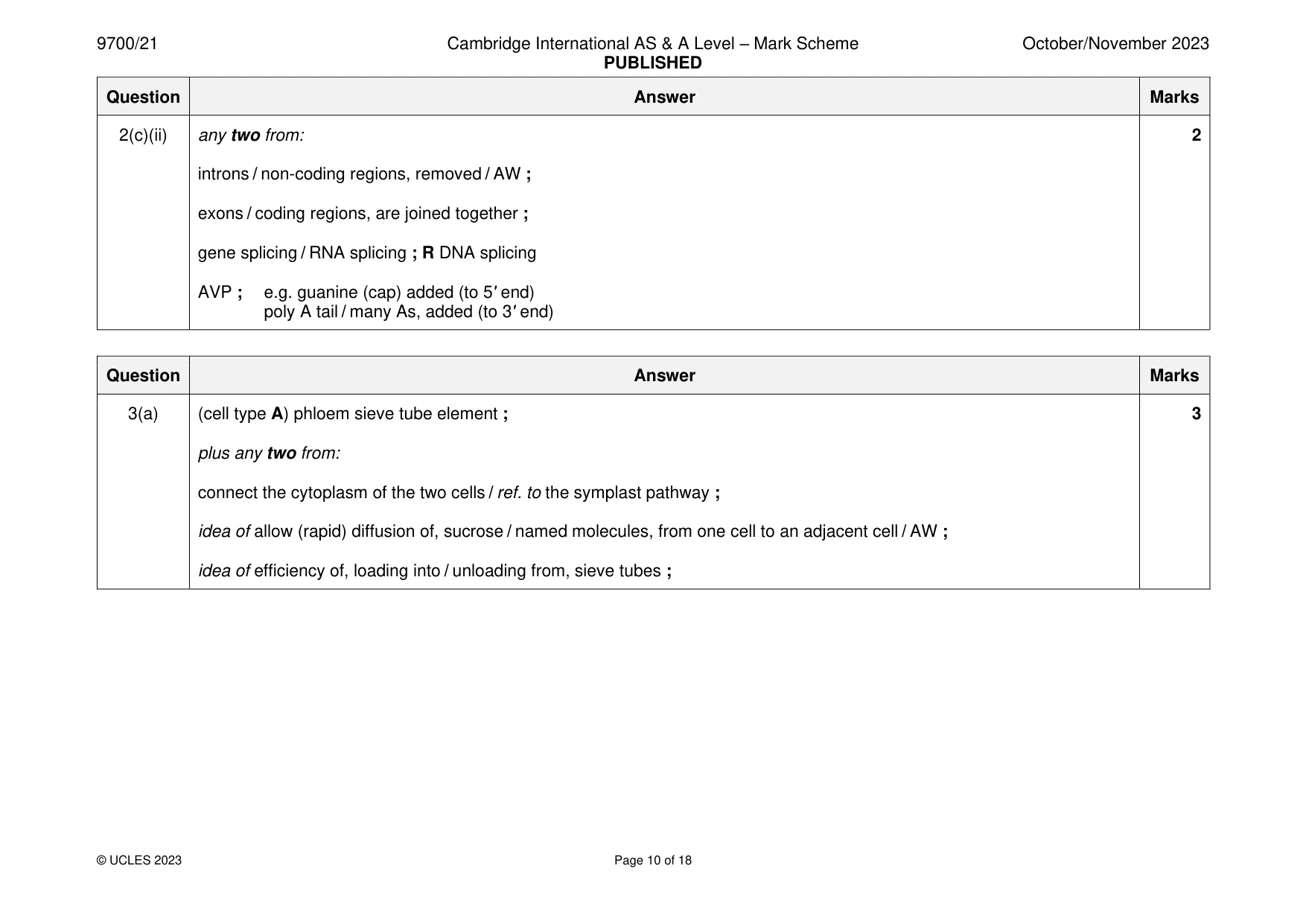
Mark scheme: 2(a) X (nucleolus) synthesis / AW of, ribosomal subunits / ribosomes ; 2 A synthesis of organelles that are the sites of protein synthesis Y (chromatin) contains gene(s), coding for the polypeptide(s) / AW ; A site, of transcription / for production of mRNA 2(b)(i) primary ; 1 2(b)(ii) any one from: 1 carboxylic acid / carboxyl, group ; A both have C=O contain carbon / hydrogen / oxygen ; 2(b)(iii) any three from: 3 ref. to role in / forms part of, overall 3D shape ; A tertiary / quaternary structure A forms part of hydrophobic region of ghrelin allows ghrelin to bind to receptor ; allows, ghrelin / part of ghrelin, to have a complementary shape / be complementary (to the receptor) ; A description suggested detail ; e.g. interacts with hydrophobic region of the receptor molecule (for binding) AVP ; e.g. help interaction with / embeds in, hydrophobic portion of the cell surface membrane in context of bringing a complementary part of the molecule closer to receptor for binding suggestion that fatty acid chain allowed, incorporation into / attachment to, a transport molecule in plasma to reach target cell ref. to effect of binding triggering reactions inside the cell ; 2(c)(i) 1.8% ; 2 working ; 30 3 100 5000 codons for 29 amino acids (including start met) and a stop codon allow 1 mark if only 28 or 29 instead of 30, so 1.7% answer obtained 2(c)(ii) any two from: 2 introns / non-coding regions, removed / AW ; exons / coding regions, are joined together ; gene splicing / RNA splicing ; R DNA splicing AVP ; e.g. guanine (cap) added (to 5ʹ end) poly A tail / many As, added (to 3ʹ end)
Q3 · Plants have specialised cells for the efficient transport of assimilates
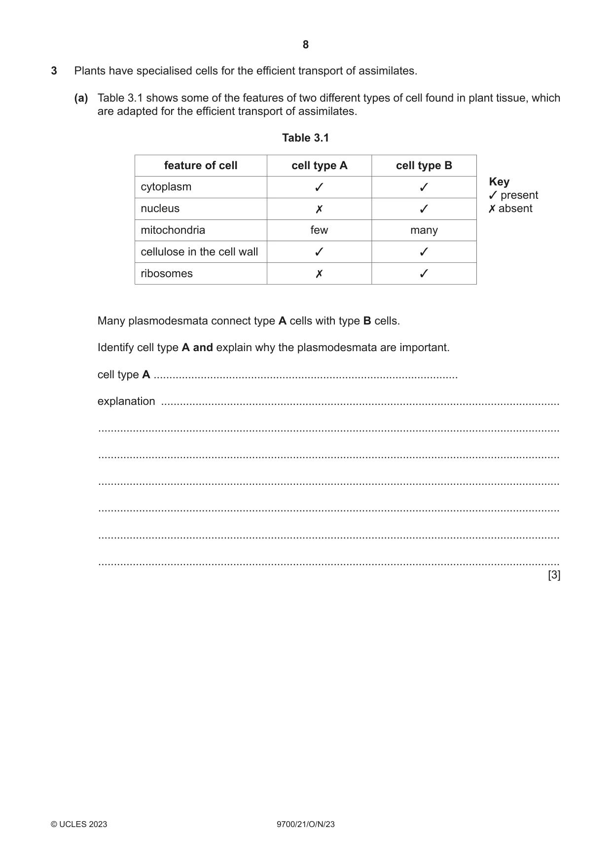
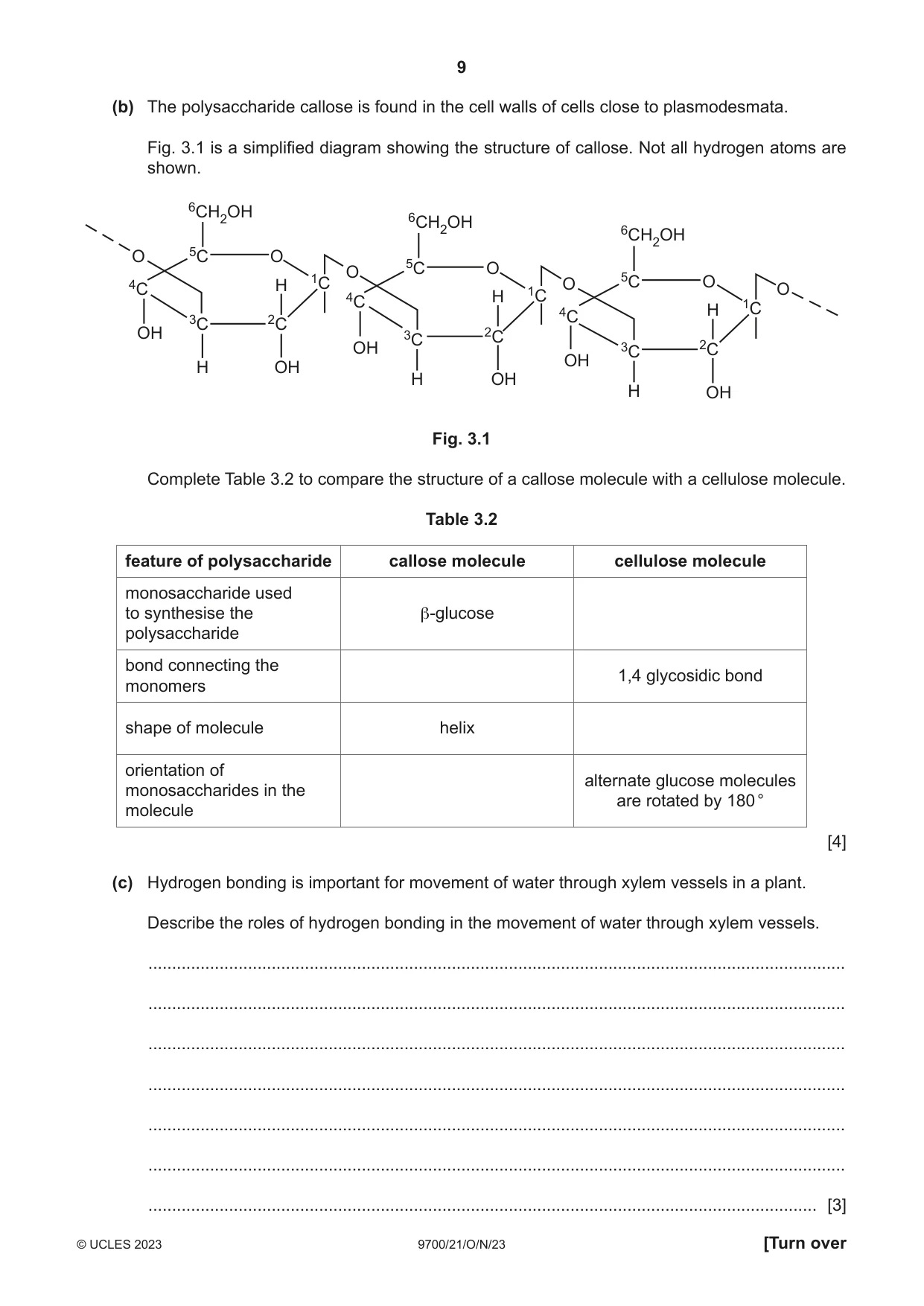
3 Plants have specialised cells for the efficient transport of assimilates. (a) Table 3.1 shows some of the features of two different types of cell found in plant tissue, which are adapted for the efficient transport of assimilates. Table 3.1 feature of cell cell type A cell type B Key cytoplasm ✓ ✓ ✓ present nucleus ✗ ✓ ✗ absent mitochondria few many cellulose in the cell wall ✓ ✓ ribosomes ✗ ✓ Many plasmodesmata connect type A cells with type B cells. Identify cell type A and explain why the plasmodesmata are important. cell type A ................................................................................................. explanation ............................................................................................................................... ................................................................................................................................................... ................................................................................................................................................... ................................................................................................................................................... ................................................................................................................................................... ................................................................................................................................................... ................................................................................................................................................... [3] (b) The polysaccharide callose is found in the cell walls of cells close to plasmodesmata. Fig. 3.1 is a simplified diagram showing the structure of callose. Not all hydrogen atoms are shown. 6CH2OH 6CH2OH 6CH2OH O 5C O O 5C O 4C H 1C O 5C O O H 1C 4C H 1C 4C 3C 2C OH 3C 2C OH 3C 2C OH H OH H OH H OH Fig. 3.1 Complete Table 3.2 to compare the structure of a callose molecule with a cellulose molecule. Table 3.2 feature of polysaccharide callose molecule cellulose molecule monosaccharide used to synthesise the β-glucose polysaccharide bond connecting the 1,4 glycosidic bond monomers shape of molecule helix orientation of alternate glucose molecules monosaccharides in the are rotated by 180 ° molecule [4] (c) Hydrogen bonding is important for movement of water through xylem vessels in a plant. Describe the roles of hydrogen bonding in the movement of water through xylem vessels. ................................................................................................................................................... ................................................................................................................................................... ................................................................................................................................................... ................................................................................................................................................... ................................................................................................................................................... ................................................................................................................................................... ............................................................................................................................................. [3] (d) Water that has travelled through xylem vessels reaches the leaves. Cooling of the leaf occurs as a result of the evaporation of water during transpiration. Water has a high latent heat of vaporisation because water molecules form hydrogen bonds. With reference to hydrogen bonding, suggest why cooling of the leaf occurs as a result of evaporation of water during transpiration. ................................................................................................................................................... ................................................................................................................................................... ................................................................................................................................................... ................................................................................................................................................... ............................................................................................................................................. [2] [Total: 12] Question 4 starts on page 12
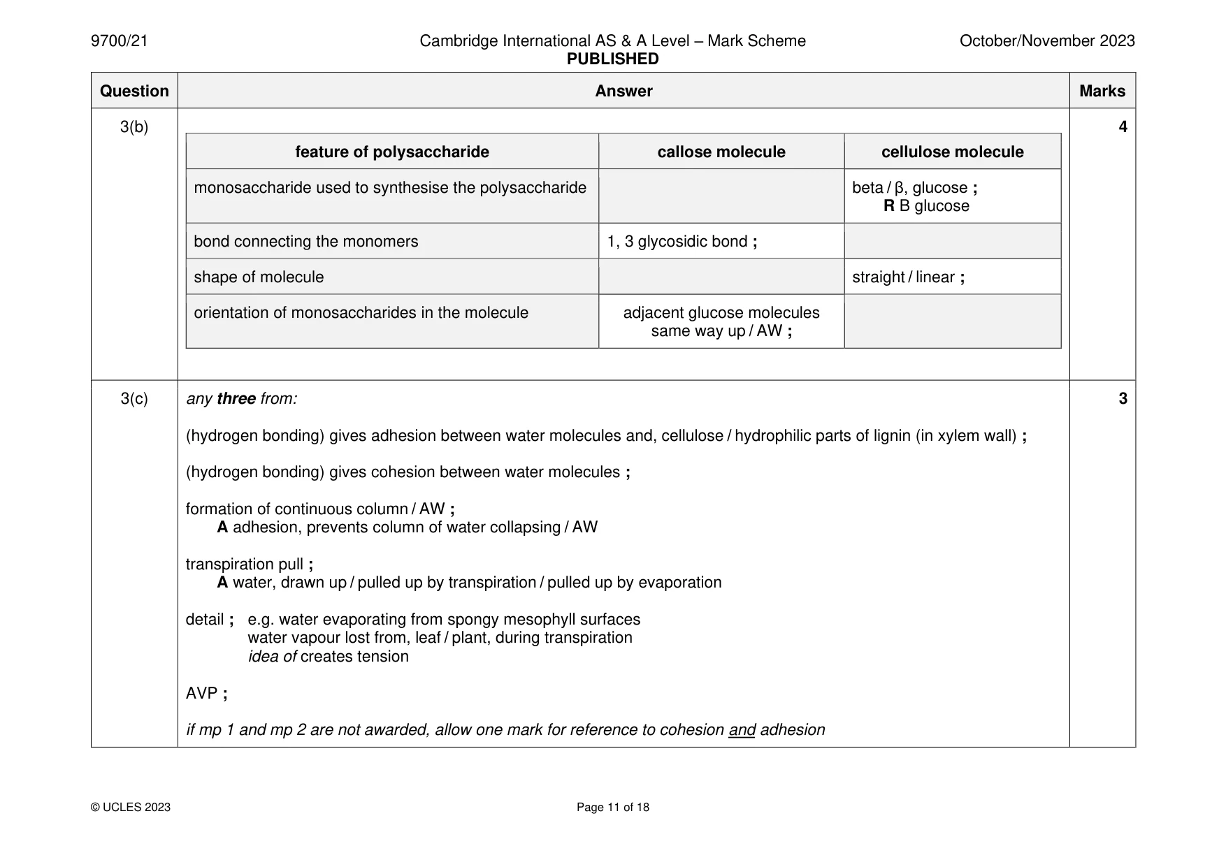
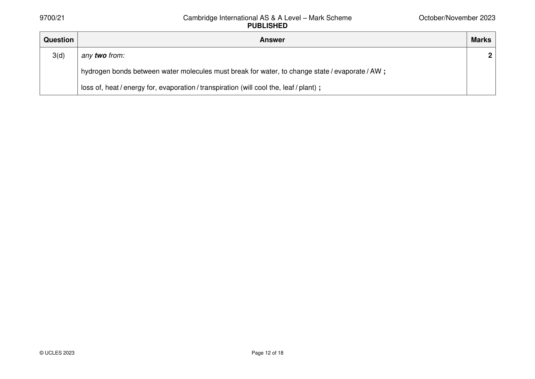
Mark scheme: 3(a) (cell type A) phloem sieve tube element ; 3 plus any two from: connect the cytoplasm of the two cells / ref. to the symplast pathway ; idea of allow (rapid) diffusion of, sucrose / named molecules, from one cell to an adjacent cell / AW ; idea of efficiency of, loading into / unloading from, sieve tubes ; 3(b) 4 feature of polysaccharide callose molecule cellulose molecule monosaccharide used to synthesise the polysaccharide beta / β, glucose ; R B glucose bond connecting the monomers 1, 3 glycosidic bond ; shape of molecule straight / linear ; orientation of monosaccharides in the molecule adjacent glucose molecules same way up / AW ; 3(c) any three from: 3 (hydrogen bonding) gives adhesion between water molecules and, cellulose / hydrophilic parts of lignin (in xylem wall) ; (hydrogen bonding) gives cohesion between water molecules ; formation of continuous column / AW ; A adhesion, prevents column of water collapsing / AW transpiration pull ; A water, drawn up / pulled up by transpiration / pulled up by evaporation detail ; e.g. water evaporating from spongy mesophyll surfaces water vapour lost from, leaf / plant, during transpiration idea of creates tension AVP ; if mp 1 and mp 2 are not awarded, allow one mark for reference to cohesion and adhesion 3(d) any two from: 2 hydrogen bonds between water molecules must break for water, to change state / evaporate / AW ; loss of, heat / energy for, evaporation / transpiration (will cool the, leaf / plant) ;
Q4 · Some people who are infected with HIV develop HIV/AIDS
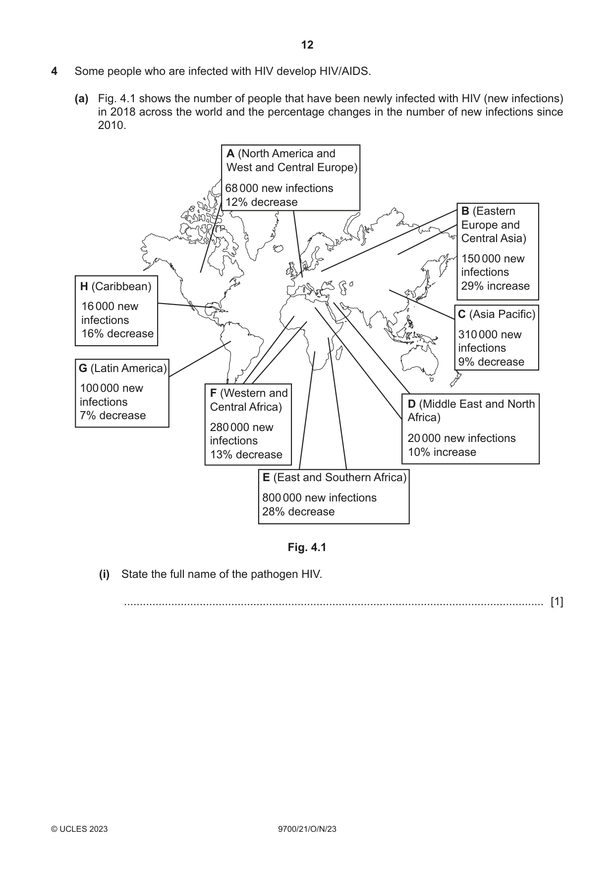
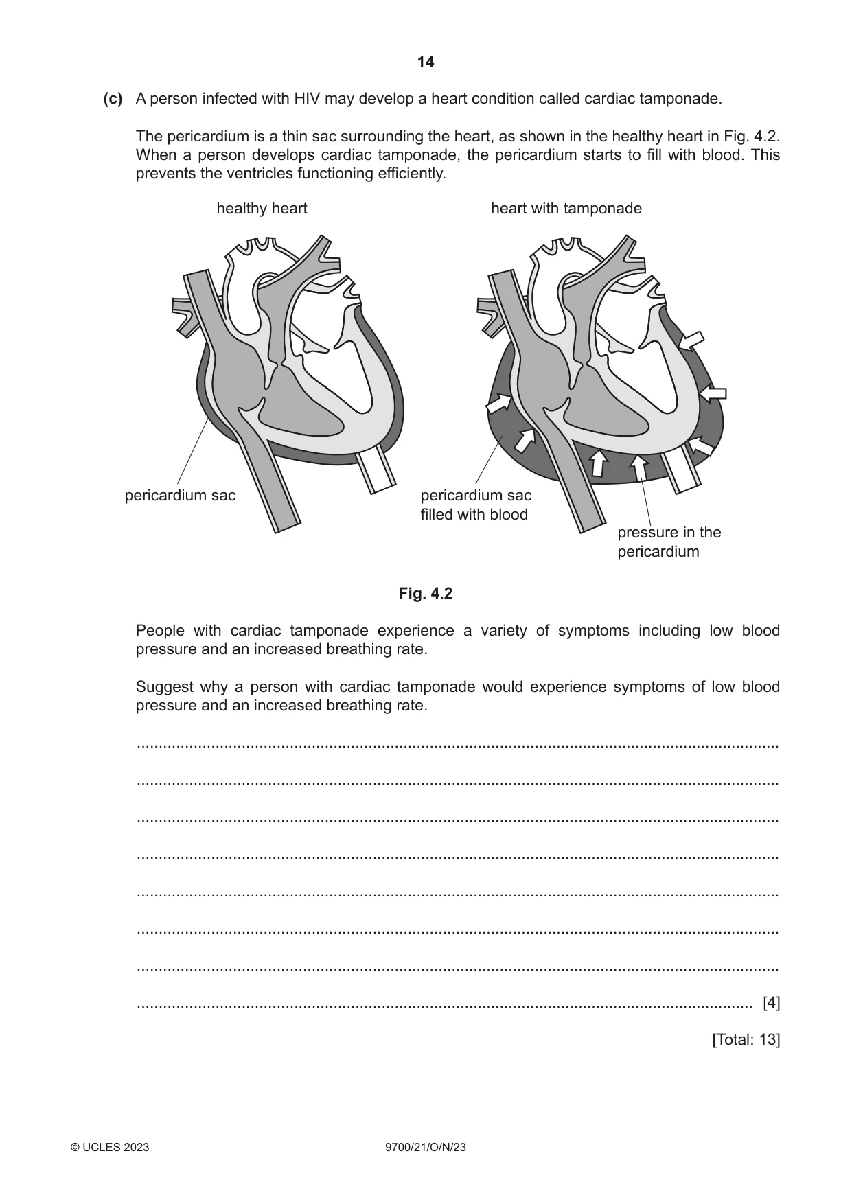
4 Some people who are infected with HIV develop HIV/AIDS. (a) Fig. 4.1 shows the number of people that have been newly infected with HIV (new infections) in 2018 across the world and the percentage changes in the number of new infections since 2010. A (North America and West and Central Europe) 68 000 new infections 12% decrease B (Eastern Europe and Central Asia) 150 000 new infections H (Caribbean) 29% increase 16 000 new C (Asia Pacific) infections 16% decrease 310 000 new infections 9% decrease G (Latin America) 100 000 new F (Western and infections Central Africa) D (Middle East and North 7% decrease Africa) 280 000 new infections 20 000 new infections 13% decrease 10% increase E (East and Southern Africa) 800 000 new infections 28% decrease Fig. 4.1 (i) State the full name of the pathogen HIV. ..................................................................................................................................... [1] (ii) Using only the data in Fig. 4.1, state what can be concluded about the change in number of new HIV infections across the world between 2010 and 2018. You may use the letters in Fig. 4.1 to identify the regions of the world. ........................................................................................................................................... ........................................................................................................................................... ........................................................................................................................................... ........................................................................................................................................... ........................................................................................................................................... ........................................................................................................................................... ........................................................................................................................................... ........................................................................................................................................... ........................................................................................................................................... ..................................................................................................................................... [4] (b) People who develop HIV/AIDS have fewer T-helper cells as the pathogen destroys these cells. This makes them more susceptible to tuberculosis (TB). Explain why people with HIV/AIDS are more likely to develop TB. ................................................................................................................................................... ................................................................................................................................................... ................................................................................................................................................... ................................................................................................................................................... ................................................................................................................................................... ................................................................................................................................................... ................................................................................................................................................... ................................................................................................................................................... ................................................................................................................................................... ............................................................................................................................................. [4] (c) A person infected with HIV may develop a heart condition called cardiac tamponade. The pericardium is a thin sac surrounding the heart, as shown in the healthy heart in Fig. 4.2. When a person develops cardiac tamponade, the pericardium starts to fill with blood. This prevents the ventricles functioning efficiently. healthy heart heart with tamponade pericardium sac pericardium sac filled with blood pressure in the pericardium Fig. 4.2 People with cardiac tamponade experience a variety of symptoms including low blood pressure and an increased breathing rate. Suggest why a person with cardiac tamponade would experience symptoms of low blood pressure and an increased breathing rate. ................................................................................................................................................... ................................................................................................................................................... ................................................................................................................................................... ................................................................................................................................................... ................................................................................................................................................... ................................................................................................................................................... ................................................................................................................................................... ............................................................................................................................................. [4] [Total: 13]
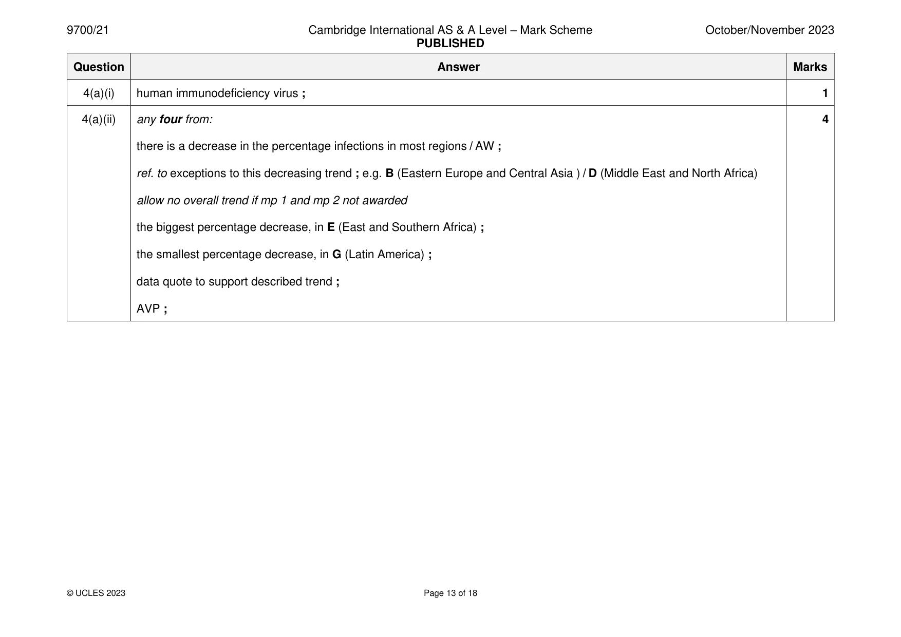
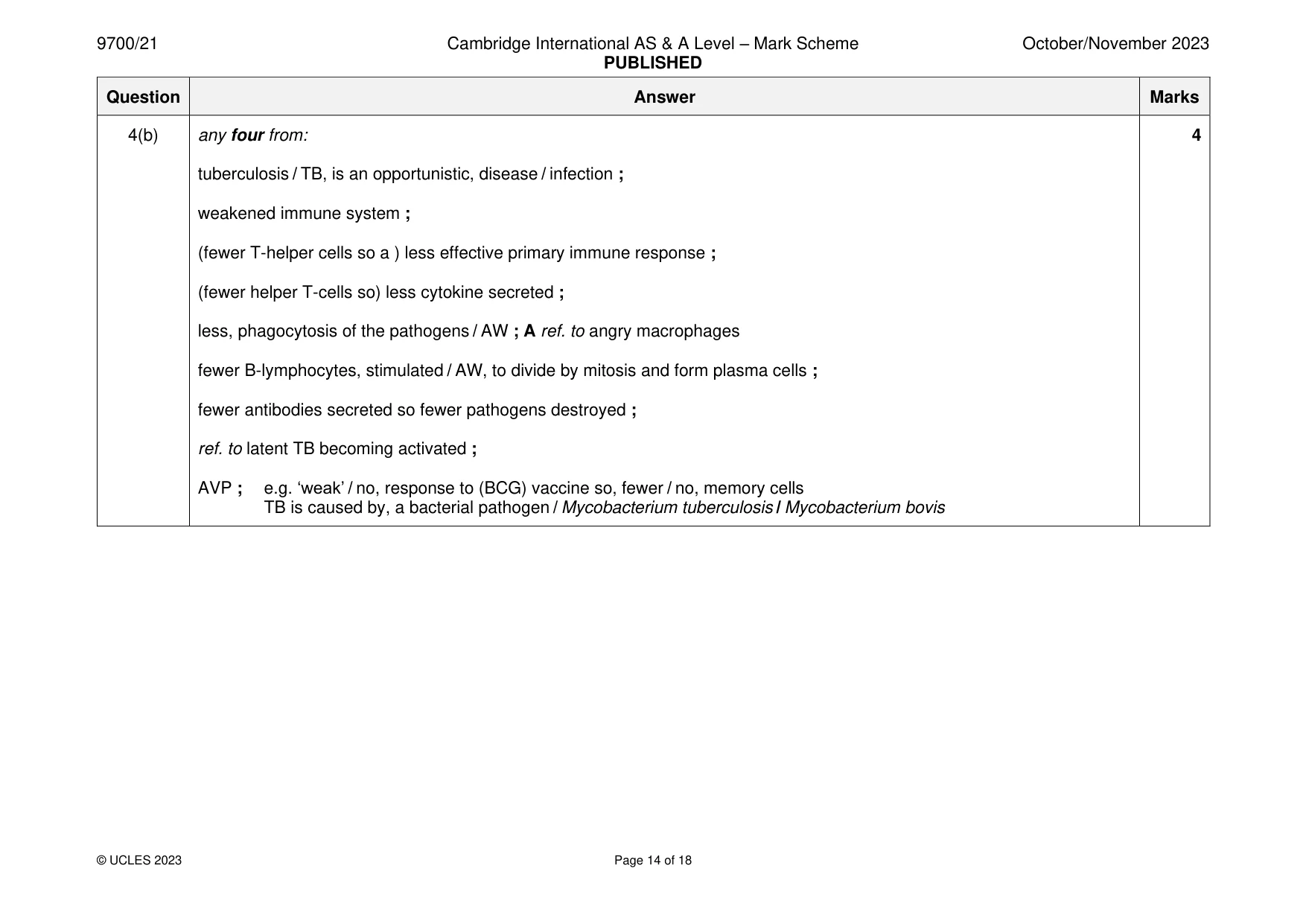
Mark scheme: 4(a)(i) human immunodeficiency virus ; 1 4(a)(ii) any four from: 4 there is a decrease in the percentage infections in most regions / AW ; ref. to exceptions to this decreasing trend ; e.g. B (Eastern Europe and Central Asia ) / D (Middle East and North Africa) allow no overall trend if mp 1 and mp 2 not awarded the biggest percentage decrease, in E (East and Southern Africa) ; the smallest percentage decrease, in G (Latin America) ; data quote to support described trend ; AVP ; 4(b) any four from: 4 tuberculosis / TB, is an opportunistic, disease / infection ; weakened immune system ; (fewer T-helper cells so a ) less effective primary immune response ; (fewer helper T-cells so) less cytokine secreted ; less, phagocytosis of the pathogens / AW ; A ref. to angry macrophages fewer B-lymphocytes, stimulated / AW, to divide by mitosis and form plasma cells ; fewer antibodies secreted so fewer pathogens destroyed ; ref. to latent TB becoming activated ; AVP ; e.g. ‘weak’ / no, response to (BCG) vaccine so, fewer / no, memory cells TB is caused by, a bacterial pathogen / Mycobacterium tuberculosis / Mycobacterium bovis 4(c) any four from: 4 1 ref. to compression on ventricles ; 2 (so) volume of the ventricles reduces / ventricles cannot fully increase in size / AW ; 3 less blood, filling / leaving, ventricles (each cardiac cycle) ; 4 idea that ventricular, contraction / systole, less powerful / weaker / less forceful ; 5 less blood pumped to the, pulmonary / systemic, circulation (per, contraction / cardiac cycle / unit time ; A lungs / rest of body faster breathing rate to compensate for 6 consequence in, lungs / rest of body ; e.g. less oxygen, diffuses / AW, into the blood less carbon dioxide, diffuses / AW, out of the blood less oxygen delivered to the tissues / AW 7 faster breathing rate to (try and) increase quantity of oxygen, absorbed / AW (in the lungs) ; A increase quantity of carbon dioxide excreted 8 AVP ; e.g. heart / cardiac muscle, does not receive enough, glucose / oxygen
Q5 · A photomicrograph of a transverse section of a bronchus in the lungs
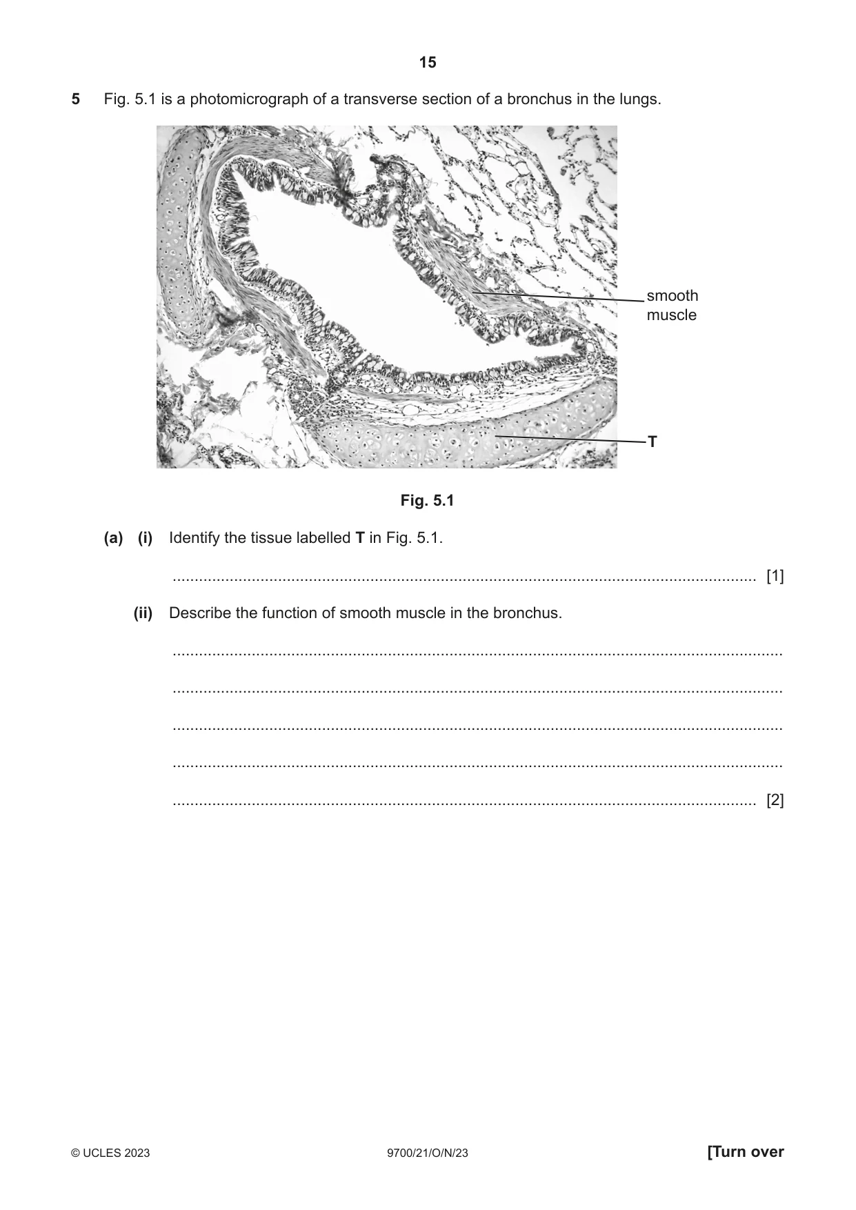
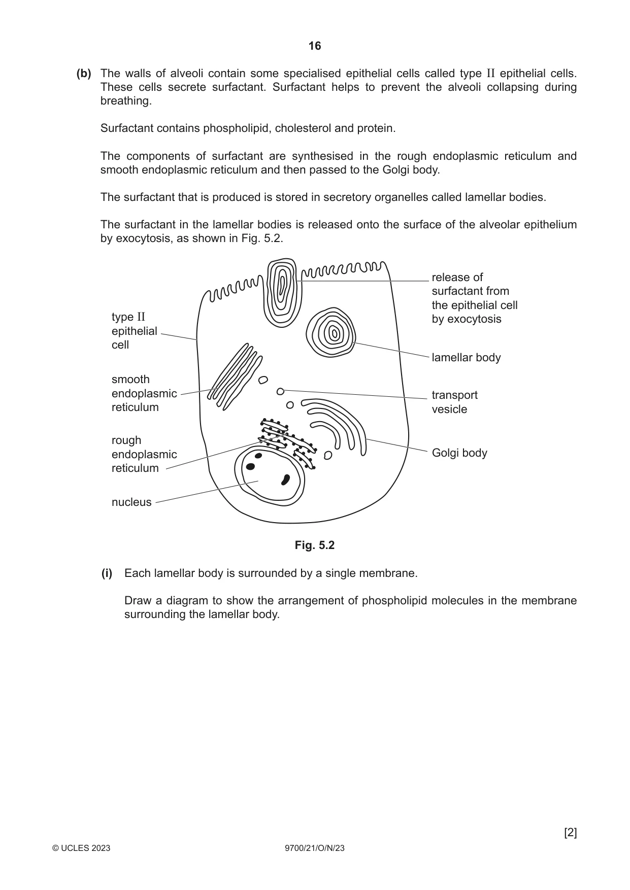
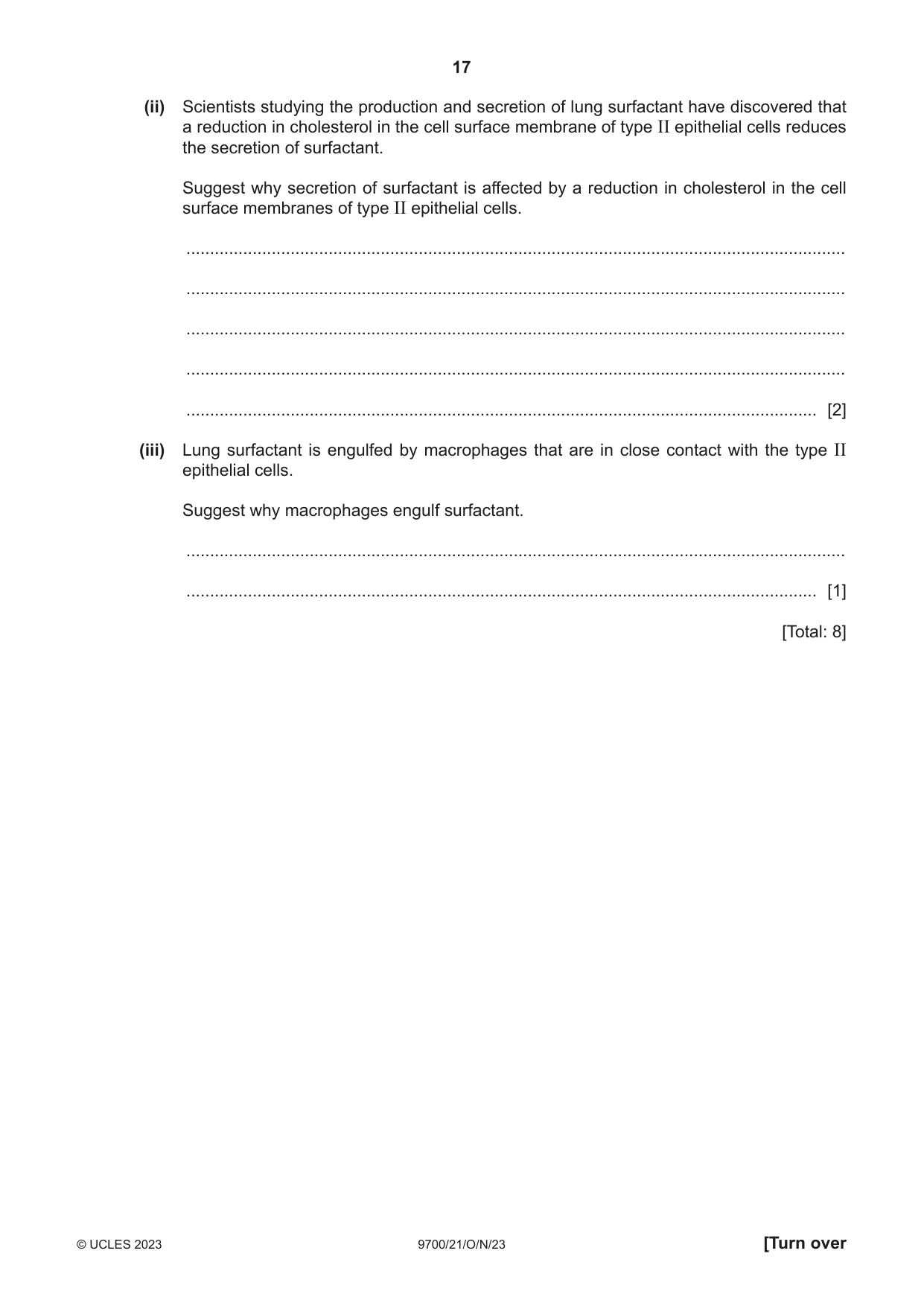
5 Fig. 5.1 is a photomicrograph of a transverse section of a bronchus in the lungs. smooth muscle T Fig. 5.1 (a) (i) Identify the tissue labelled T in Fig. 5.1. ..................................................................................................................................... [1] (ii) Describe the function of smooth muscle in the bronchus. ........................................................................................................................................... ........................................................................................................................................... ........................................................................................................................................... ........................................................................................................................................... ..................................................................................................................................... [2] (b) The walls of alveoli contain some specialised epithelial cells called type II epithelial cells. These cells secrete surfactant. Surfactant helps to prevent the alveoli collapsing during breathing. Surfactant contains phospholipid, cholesterol and protein. The components of surfactant are synthesised in the rough endoplasmic reticulum and smooth endoplasmic reticulum and then passed to the Golgi body. The surfactant that is produced is stored in secretory organelles called lamellar bodies. The surfactant in the lamellar bodies is released onto the surface of the alveolar epithelium by exocytosis, as shown in Fig. 5.2. release of surfactant from the epithelial cell type II by exocytosis epithelial cell lamellar body smooth endoplasmic transport reticulum vesicle rough endoplasmic Golgi body reticulum nucleus Fig. 5.2 (i) Each lamellar body is surrounded by a single membrane. Draw a diagram to show the arrangement of phospholipid molecules in the membrane surrounding the lamellar body. [2] (ii) Scientists studying the production and secretion of lung surfactant have discovered that a reduction in cholesterol in the cell surface membrane of type II epithelial cells reduces the secretion of surfactant. Suggest why secretion of surfactant is affected by a reduction in cholesterol in the cell surface membranes of type II epithelial cells. ........................................................................................................................................... ........................................................................................................................................... ........................................................................................................................................... ........................................................................................................................................... ..................................................................................................................................... [2] (iii) Lung surfactant is engulfed by macrophages that are in close contact with the type II epithelial cells. Suggest why macrophages engulf surfactant. ........................................................................................................................................... ..................................................................................................................................... [1] [Total: 8]
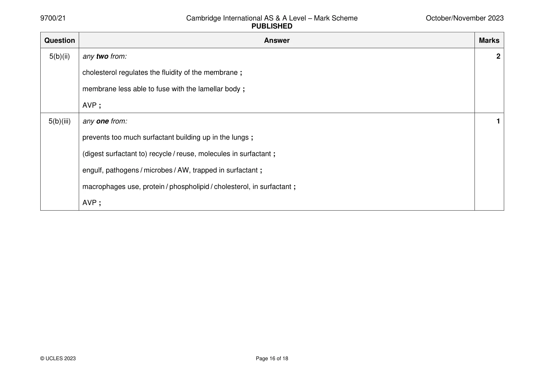
Mark scheme: 5(a)(i) cartilage ; 1 5(a)(ii) any two from: 2 (smooth muscle) contracts (and relaxes) / contractile ; R bronchus contracts changes diameter of the airway / controls size of the lumen ; A ref. to constriction of bronchus (may) help to regulate airflow, into / away from, gas exchange surfaces ; AVP ; 5(b)(i) phospholipid with a head and two tails ; 2 bilayer shown ; 5(b)(ii) any two from: 2 cholesterol regulates the fluidity of the membrane ; membrane less able to fuse with the lamellar body ; AVP ; 5(b)(iii) any one from: 1 prevents too much surfactant building up in the lungs ; (digest surfactant to) recycle / reuse, molecules in surfactant ; engulf, pathogens / microbes / AW, trapped in surfactant ; macrophages use, protein / phospholipid / cholesterol, in surfactant ; AVP ;
Q6 · State the name of the phase of the mitotic cell cycle during which DNA replication occurs
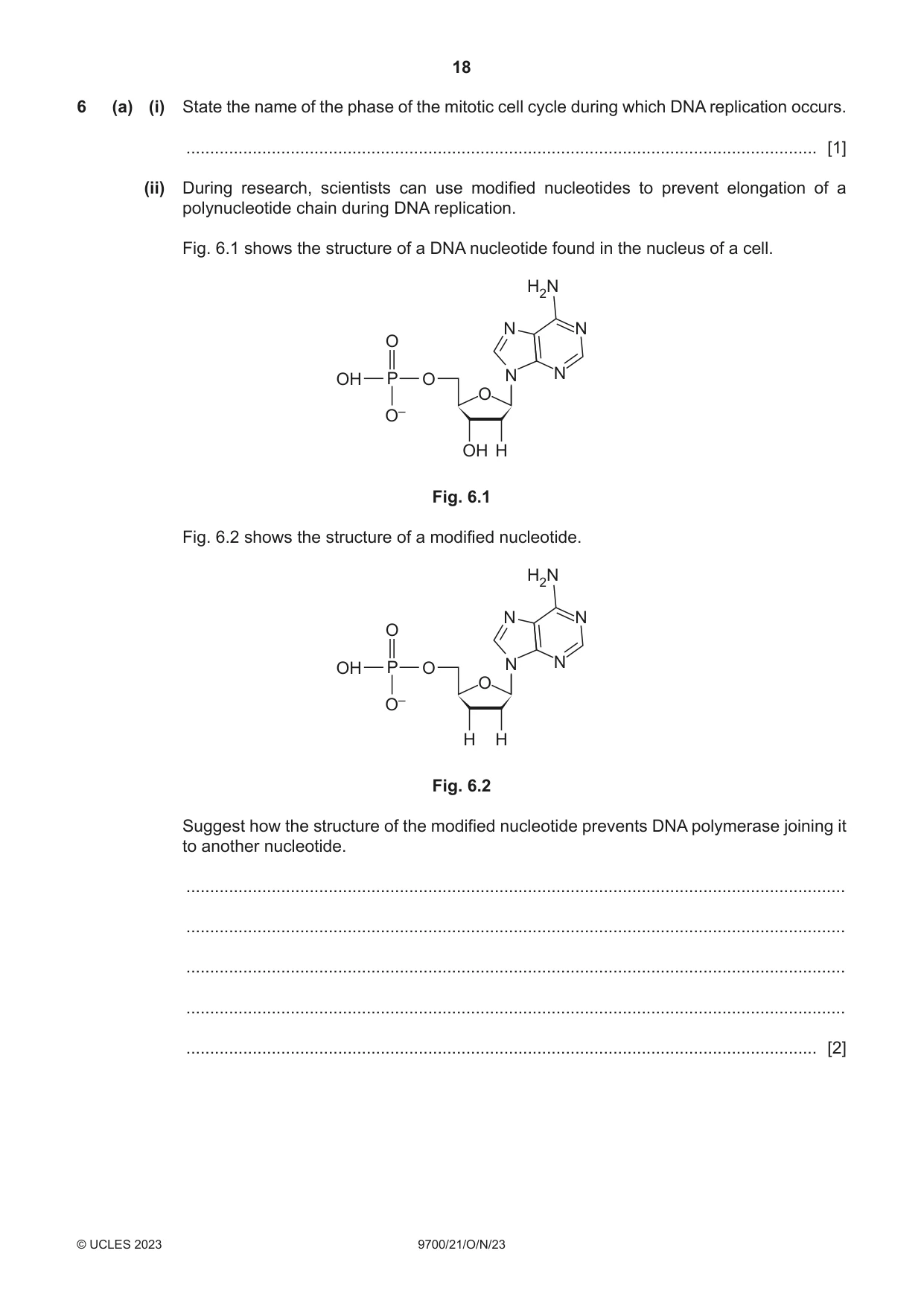
6 (a) (i) State the name of the phase of the mitotic cell cycle during which DNA replication occurs. ..................................................................................................................................... [1] (ii) During research, scientists can use modified nucleotides to prevent elongation of a polynucleotide chain during DNA replication. Fig. 6.1 shows the structure of a DNA nucleotide found in the nucleus of a cell. H2N N N O OH P O N N O O– OH H Fig. 6.1 Fig. 6.2 shows the structure of a modified nucleotide. H2N N N O OH P O N N O O– H H Fig. 6.2 Suggest how the structure of the modified nucleotide prevents DNA polymerase joining it to another nucleotide. ........................................................................................................................................... ........................................................................................................................................... ........................................................................................................................................... ........................................................................................................................................... ..................................................................................................................................... [2] (b) RNA aptamers are short, single-stranded RNA molecules that can be used to study some infectious diseases. Scientists studying an infectious disease in animals investigated the effect of RNA aptamers on the activity of RNA polymerase that is produced by the pathogen. The aptamers bind to a specific region of the RNA polymerase. Table 6.1 shows the effect of two aptamers, F47 and F52, on the activity of RNA polymerase produced by the pathogen. Table 6.1 Vmax of RNA polymerase Km of RNA polymerase aptamer present / arbitrary units / arbitrary units none 1664 346 F47 1072 508 F52 1467 523 (i) With reference to Table 6.1, compare the effect of the aptamers on the affinity of RNA polymerase for its substrate. ........................................................................................................................................... ........................................................................................................................................... ........................................................................................................................................... ..................................................................................................................................... [2] (ii) With reference to Table 6.1, suggest explanations for the effect of the presence of an aptamer on the rate of transcription catalysed by the RNA polymerase. ........................................................................................................................................... ........................................................................................................................................... ........................................................................................................................................... ........................................................................................................................................... ........................................................................................................................................... ........................................................................................................................................... ..................................................................................................................................... [3] [Total: 8]
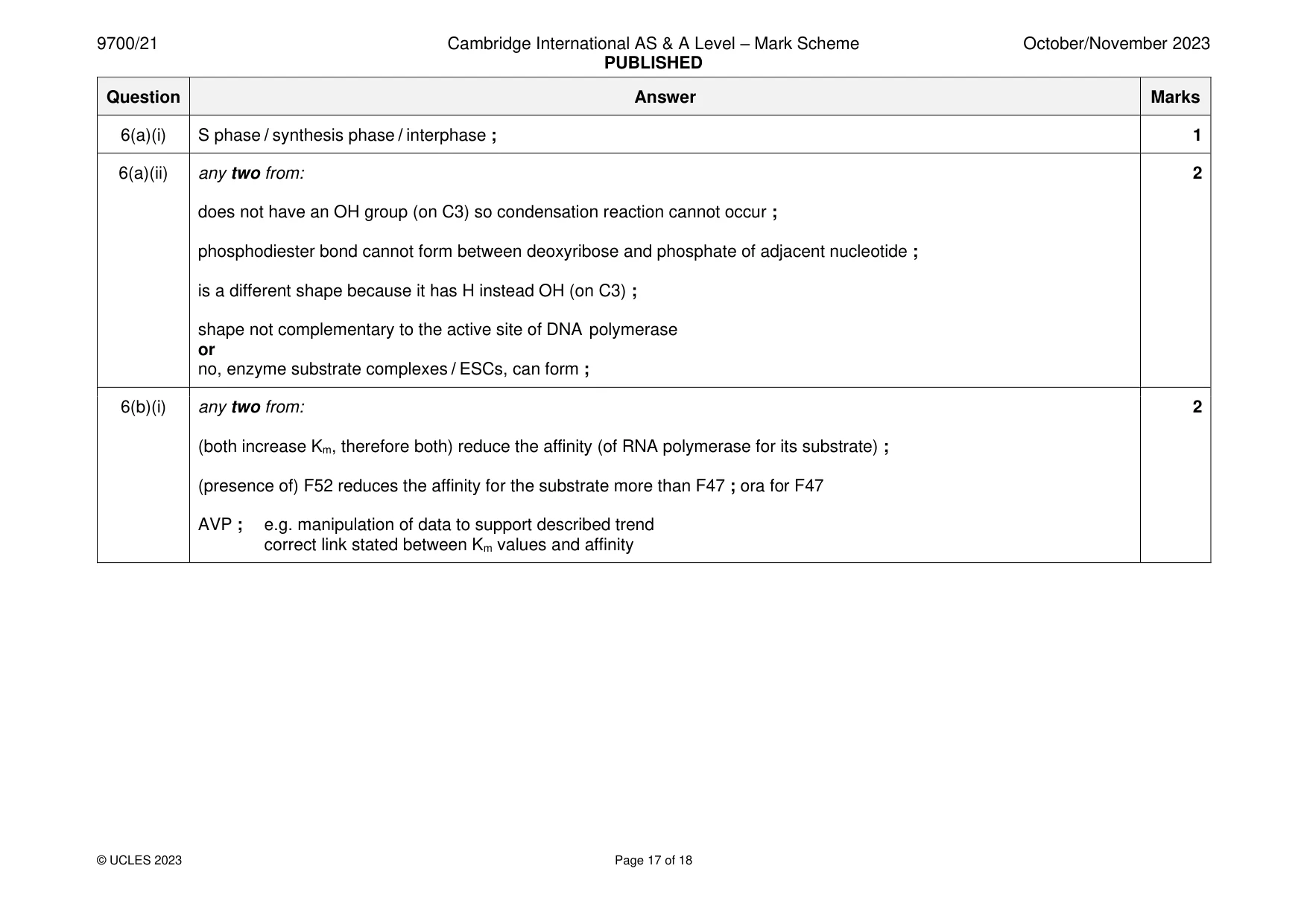
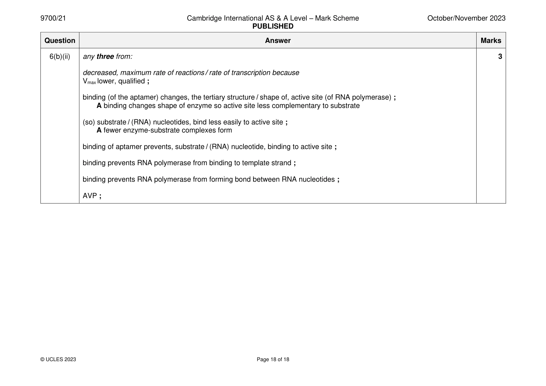
Mark scheme: 6(a)(i) S phase / synthesis phase / interphase ; 1 6(a)(ii) any two from: 2 does not have an OH group (on C3) so condensation reaction cannot occur ; phosphodiester bond cannot form between deoxyribose and phosphate of adjacent nucleotide ; is a different shape because it has H instead OH (on C3) ; shape not complementary to the active site of DNA polymerase or no, enzyme substrate complexes / ESCs, can form ; 6(b)(i) any two from: 2 (both increase Km, therefore both) reduce the affinity (of RNA polymerase for its substrate) ; (presence of) F52 reduces the affinity for the substrate more than F47 ; ora for F47 AVP ; e.g. manipulation of data to support described trend correct link stated between Km values and affinity 6(b)(ii) any three from: 3 decreased, maximum rate of reactions / rate of transcription because Vmax lower, qualified ; binding (of the aptamer) changes, the tertiary structure / shape of, active site (of RNA polymerase) ; A binding changes shape of enzyme so active site less complementary to substrate (so) substrate / (RNA) nucleotides, bind less easily to active site ; A fewer enzyme-substrate complexes form binding of aptamer prevents, substrate / (RNA) nucleotide, binding to active site ; binding prevents RNA polymerase from binding to template strand ; binding prevents RNA polymerase from forming bond between RNA nucleotides ; AVP ;
What was in this paper
The subtopics covered by these 6 questions, and how many questions each got. Open one in a new tab to see every Cambridge question on it.
What you needed in this session
Cambridge’s own grade thresholds for 2023 Oct/Nov, Paper 2 · Variant 1. A higher threshold means an easier paper — the bar moves with how the cohort did.