Cambridge A Level Biology 9700 — 2024 May/June Paper 2 · Variant 1
9700/21/M/J/24 · 6 questions · 60 marks · ≈68 min
The question paper and its mark scheme, free to read here and free to download. This is Cambridge’s own paper, exactly as it was sat.
Question paper20 pages

Mark scheme15 pages
Answers below. Sit the paper first if you are practising.
Questions as text
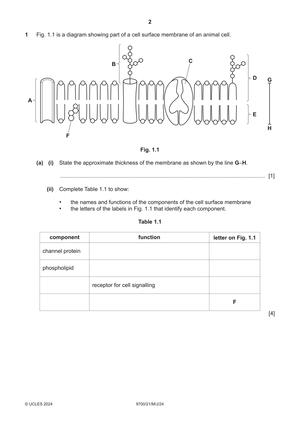
Q1 · A diagram showing part of a cell surface membrane of an animal cell
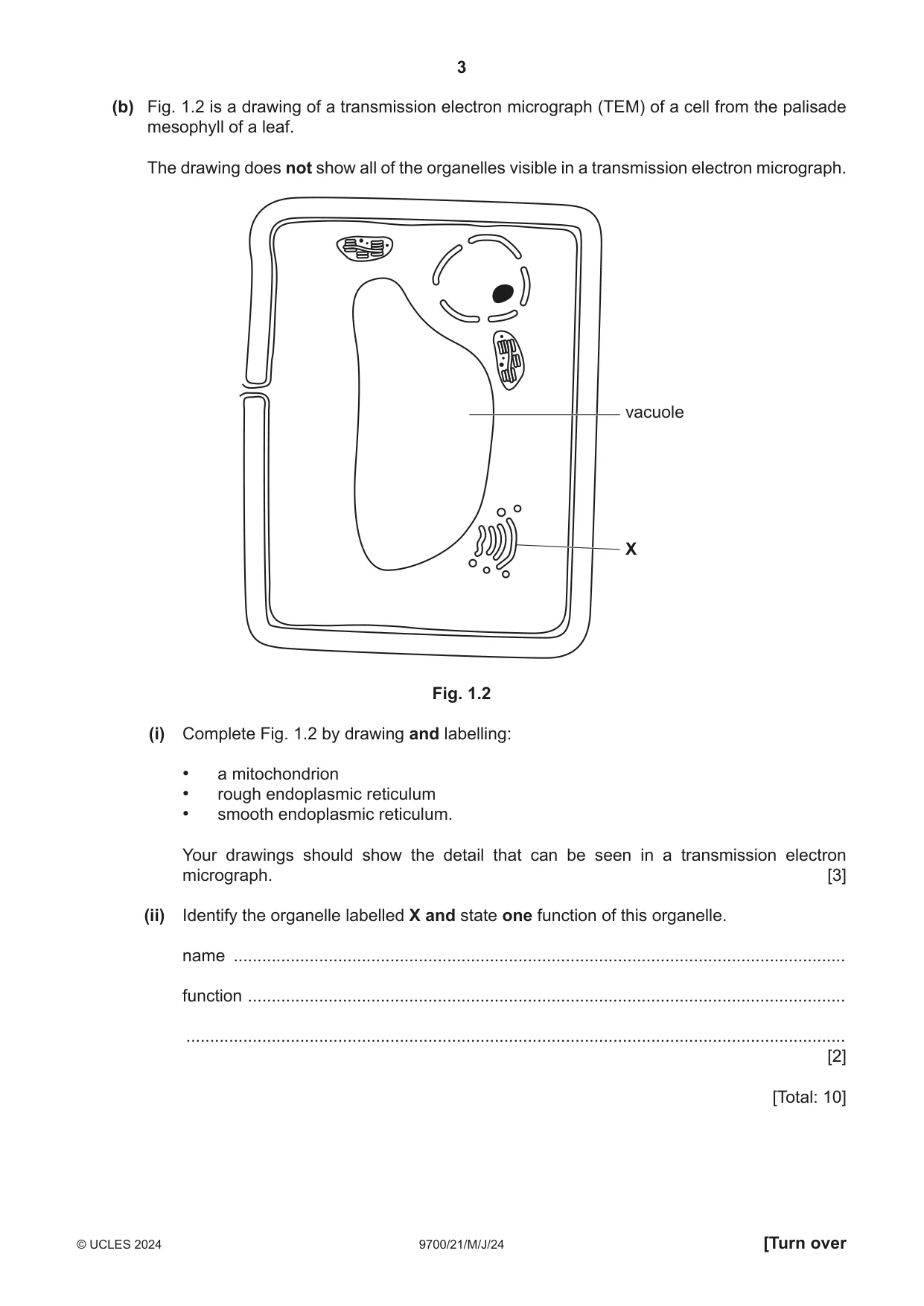
1 Fig. 1.1 is a diagram showing part of a cell surface membrane of an animal cell. C B D G A E H F Fig. 1.1 (a) (i) State the approximate thickness of the membrane as shown by the line G–H. ..................................................................................................................................... [1] (ii) Complete Table 1.1 to show: • the names and functions of the components of the cell surface membrane • the letters of the labels in Fig. 1.1 that identify each component. Table 1.1 component function letter on Fig. 1.1 channel protein phospholipid receptor for cell signalling F [4] (b) Fig. 1.2 is a drawing of a transmission electron micrograph (TEM) of a cell from the palisade mesophyll of a leaf. The drawing does not show all of the organelles visible in a transmission electron micrograph. vacuole X Fig. 1.2 (i) Complete Fig. 1.2 by drawing and labelling: • a mitochondrion • rough endoplasmic reticulum • smooth endoplasmic reticulum. Your drawings should show the detail that can be seen in a transmission electron micrograph. [3] (ii) Identify the organelle labelled X and state one function of this organelle. name ................................................................................................................................. function .............................................................................................................................. ........................................................................................................................................... [2] [Total: 10]
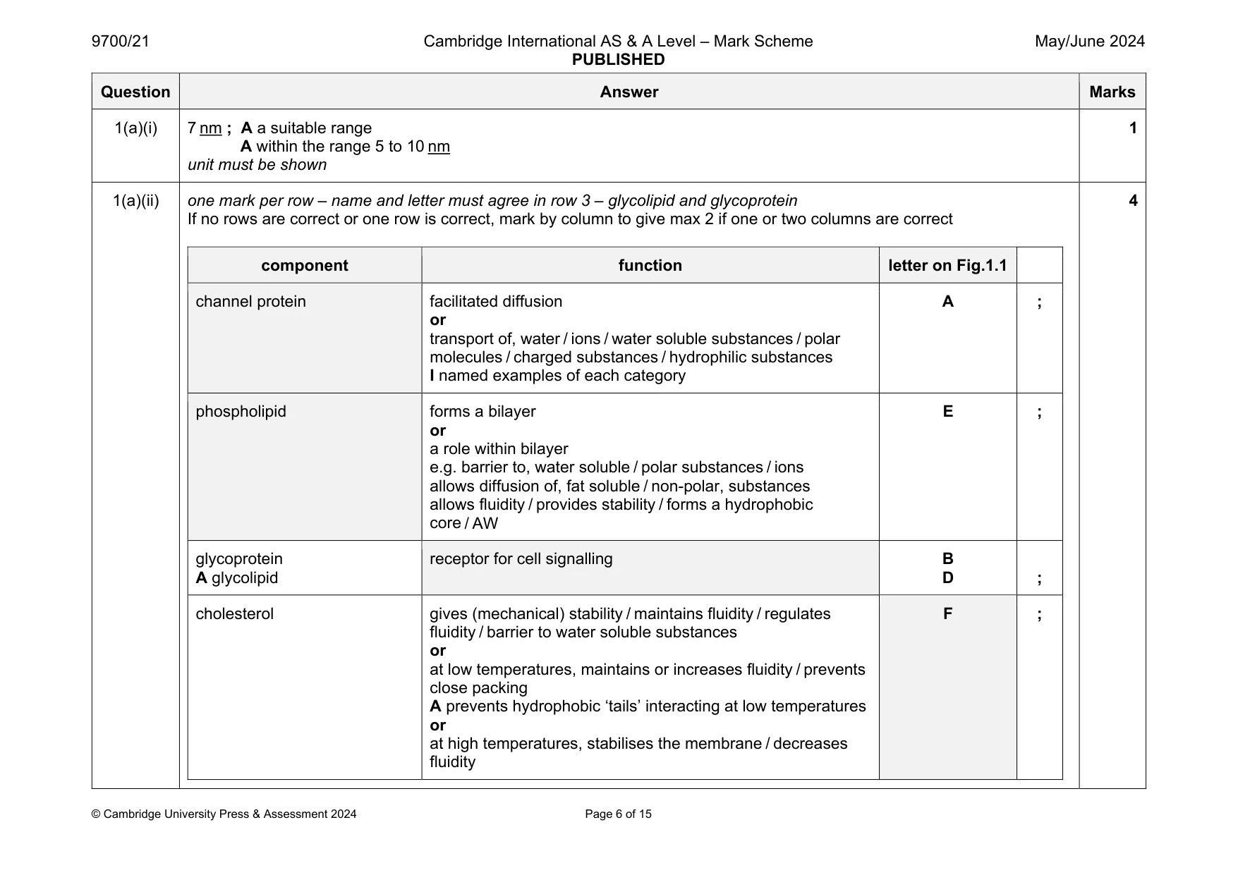
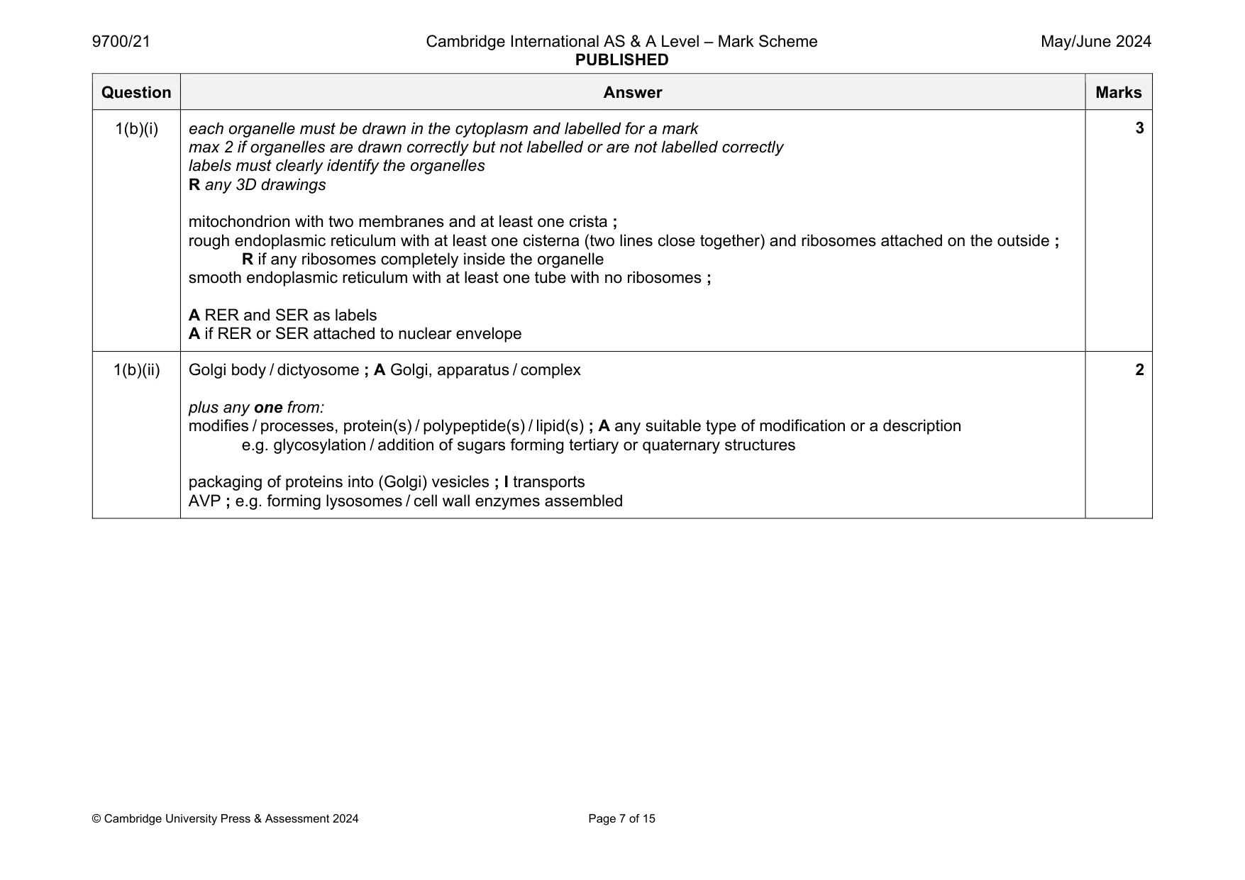
Mark scheme: 1(a)(i) 7 nm ; A a suitable range A within the range 5 to 10 nm unit must be shown 1 1(a)(ii) one mark per row – name and letter must agree in row 3 – glycolipid and glycoprotein If no rows are correct or one row is correct, mark by column to give max 2 if one or two columns are correct component function letter on Fig.1.1 channel protein facilitated diffusion or transport of, water / ions / water soluble substances / polar molecules / charged substances / hydrophilic substances I named examples of each category A ; phospholipid forms a bilayer or a role within bilayer e.g. barrier to, water soluble / polar substances / ions allows diffusion of, fat soluble / non-polar, substances allows fluidity / provides stability / forms a hydrophobic core / AW E ; glycoprotein A glycolipid receptor for cell signalling B D ; cholesterol gives (mechanical) stability / maintains fluidity / regulates fluidity / barrier to water soluble substances or at low temperatures, maintains or increases fluidity / prevents close packing A prevents hydrophobic ‘tails’ interacting at low temperatures or at high temperatures, stabilises the membrane / decreases fluidity F ; 4 Question Answer Marks 1(b)(i) each organelle must be drawn in the cytoplasm and labelled for a mark max 2 if organelles are drawn correctly but not labelled or are not labelled correctly labels must clearly identify the organelles R any 3D drawings mitochondrion with two membranes and at least one crista ; rough endoplasmic reticulum with at least one cisterna (two lines close together) and ribosomes attached on the outside ; R if any ribosomes completely inside the organelle smooth endoplasmic reticulum with at least one tube with no ribosomes ; A RER and SER as labels A if RER or SER attached to nuclear envelope 3 1(b)(ii) Golgi body / dictyosome ; A Golgi, apparatus / complex plus any one from: modifies / processes, protein(s) / polypeptide(s) / lipid(s) ; A any suitable type of modification or a description e.g. glycosylation / addition of sugars forming tertiary or quaternary structures packaging of proteins into (Golgi) vesicles ; I transports AVP ; e.g. forming lysosomes / cell wall enzymes assembled 2
Q2 · Water is the main component of blood
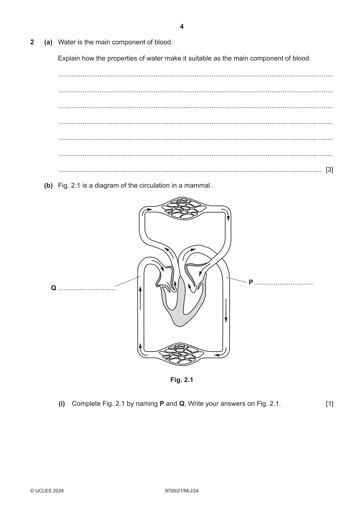
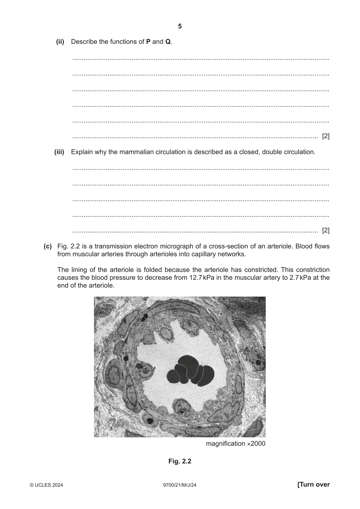
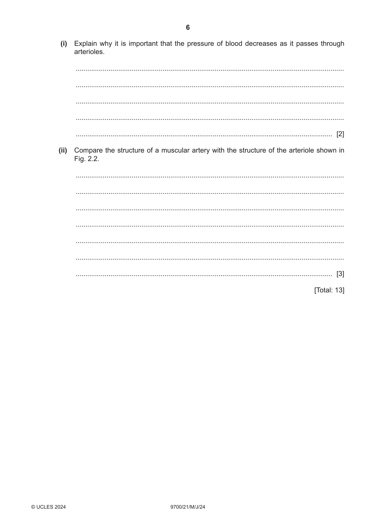
2 (a) Water is the main component of blood. Explain how the properties of water make it suitable as the main component of blood. ................................................................................................................................................... ................................................................................................................................................... ................................................................................................................................................... ................................................................................................................................................... ................................................................................................................................................... ................................................................................................................................................... ............................................................................................................................................. [3] (b) Fig. 2.1 is a diagram of the circulation in a mammal. P ............................... Q ............................... Fig. 2.1 (i) Complete Fig. 2.1 by naming P and Q. Write your answers on Fig. 2.1. [1] (ii) Describe the functions of P and Q. ........................................................................................................................................... ........................................................................................................................................... ........................................................................................................................................... ........................................................................................................................................... ........................................................................................................................................... ..................................................................................................................................... [2] (iii) Explain why the mammalian circulation is described as a closed, double circulation. ........................................................................................................................................... ........................................................................................................................................... ........................................................................................................................................... ........................................................................................................................................... ..................................................................................................................................... [2] (c) Fig. 2.2 is a transmission electron micrograph of a cross‑section of an arteriole. Blood flows from muscular arteries through arterioles into capillary networks. The lining of the arteriole is folded because the arteriole has constricted. This constriction causes the blood pressure to decrease from 12.7 kPa in the muscular artery to 2.7 kPa at the end of the arteriole. magnification ×2000 Fig. 2.2 (i) Explain why it is important that the pressure of blood decreases as it passes through arterioles. ........................................................................................................................................... ........................................................................................................................................... ........................................................................................................................................... ........................................................................................................................................... ..................................................................................................................................... [2] (ii) Compare the structure of a muscular artery with the structure of the arteriole shown in Fig. 2.2. ........................................................................................................................................... ........................................................................................................................................... ........................................................................................................................................... ........................................................................................................................................... ........................................................................................................................................... ........................................................................................................................................... ..................................................................................................................................... [3] [Total: 13]
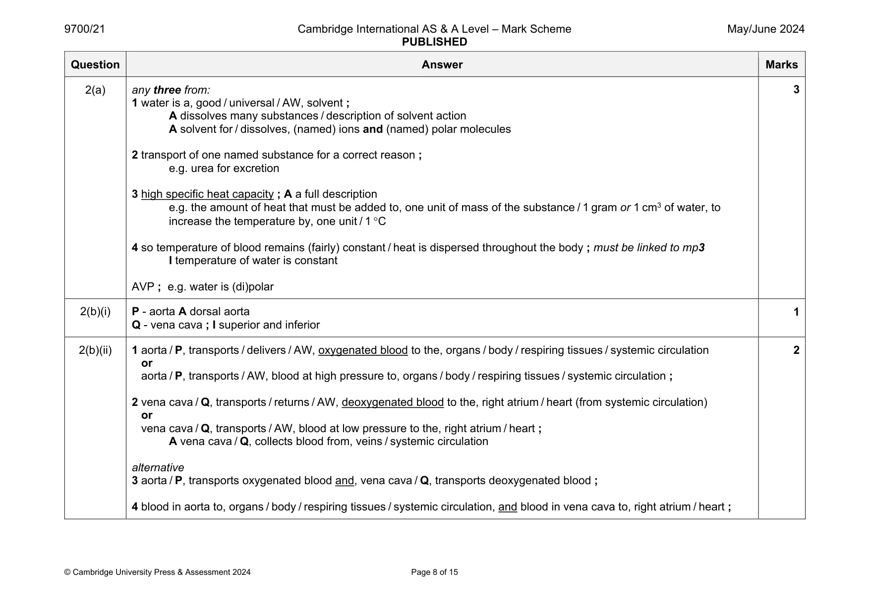
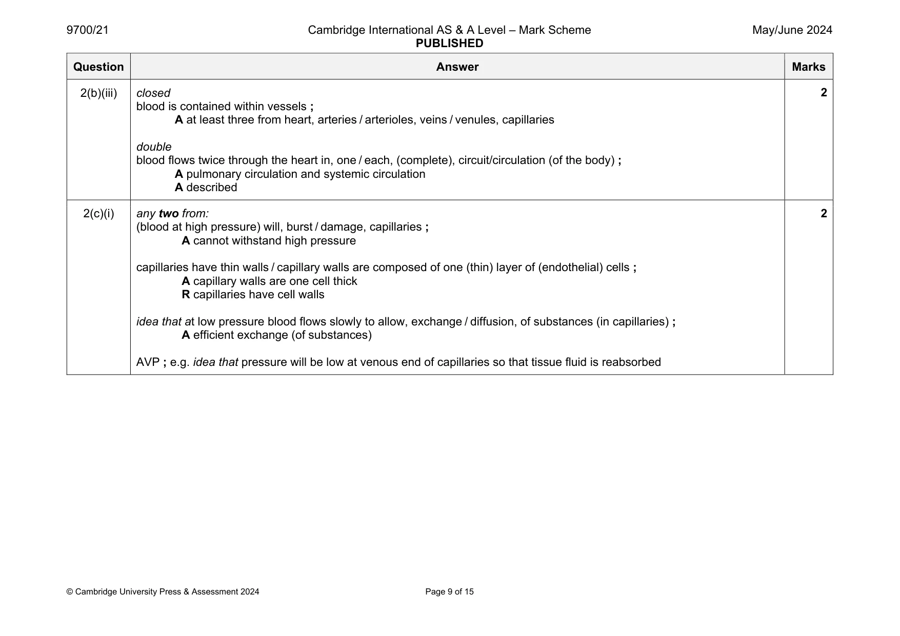
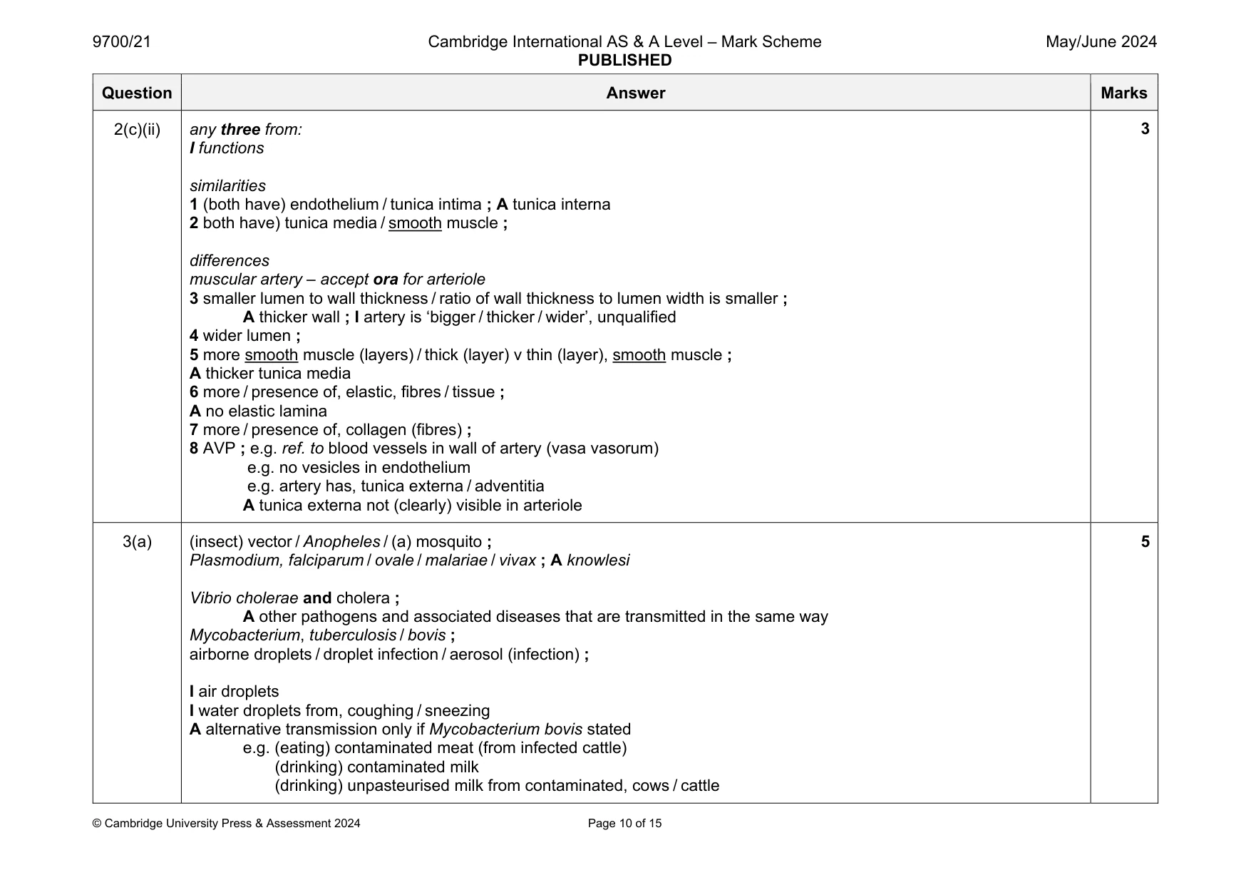
Mark scheme: 2(a) any three from: 1 water is a, good / universal / AW, solvent ; A dissolves many substances / description of solvent action A solvent for / dissolves, (named) ions and (named) polar molecules 2 transport of one named substance for a correct reason ; e.g. urea for excretion 3 high specific heat capacity ; A a full description e.g. the amount of heat that must be added to, one unit of mass of the substance / 1 gram or 1 cm3 of water, to increase the temperature by, one unit / 1 C 4 so temperature of blood remains (fairly) constant / heat is dispersed throughout the body ; must be linked to mp3 I temperature of water is constant AVP ; e.g. water is (di)polar 2(b)(i) P - aorta A dorsal aorta Q - vena cava ; I superior and inferior 1 2(b)(ii) 1 aorta / P, transports / delivers / AW, oxygenated blood to the, organs / body / respiring tissues / systemic circulation or aorta / P, transports / AW, blood at high pressure to, organs / body / respiring tissues / systemic circulation ; 2 vena cava / Q, transports / returns / AW, deoxygenated blood to the, right atrium / heart (from systemic circulation) or vena cava / Q, transports / AW, blood at low pressure to the, right atrium / heart ; A vena cava / Q, collects blood from, veins / systemic circulation alternative 3 aorta / P, transports oxygenated blood and, vena cava / Q, transports deoxygenated blood ; 4 blood in aorta to, organs / body / respiring tissues / systemic circulation, and blood in vena cava to, right atrium / heart ; 2 Question Answer Marks 2(b)(iii) closed blood is contained within vessels ; A at least three from heart, arteries / arterioles, veins / venules, capillaries double blood flows twice through the heart in, one / each, (complete), circuit/circulation (of the body) ; A pulmonary circulation and systemic circulation A described 2 2(c)(i) any two from: (blood at high pressure) will, burst / damage, capillaries ; A cannot withstand high pressure capillaries have thin walls / capillary walls are composed of one (thin) layer of (endothelial) cells ; A capillary walls are one cell thick R capillaries have cell walls idea that at low pressure blood flows slowly to allow, exchange / diffusion, of substances (in capillaries) ; A efficient exchange (of substances) AVP ; e.g. idea that pressure will be low at venous end of capillaries so that tissue fluid is reabsorbed 2 Question Answer Marks 2(c)(ii) any three from: I functions similarities 1 (both have) endothelium / tunica intima ; A tunica interna 2 both have) tunica media / smooth muscle ; differences muscular artery – accept ora for arteriole 3 smaller lumen to wall thickness / ratio of wall thickness to lumen width is smaller ; A thicker wall ; I artery is ‘bigger / thicker / wider’, unqualified 4 wider lumen ; 5 more smooth muscle (layers) / thick (layer) v thin (layer), smooth muscle ; A thicker tunica media 6 more / presence of, elastic, fibres / tissue ; A no elastic lamina 7 more / presence of, collagen (fibres) ; 8 AVP ; e.g. ref. to blood vessels in wall of artery (vasa vasorum) e.g. no vesicles in endothelium e.g. artery has, tunica externa / adventitia A tunica externa not (clearly) visible in arteriole 3
Q3 · A class of students was studying the features of some human pathogens
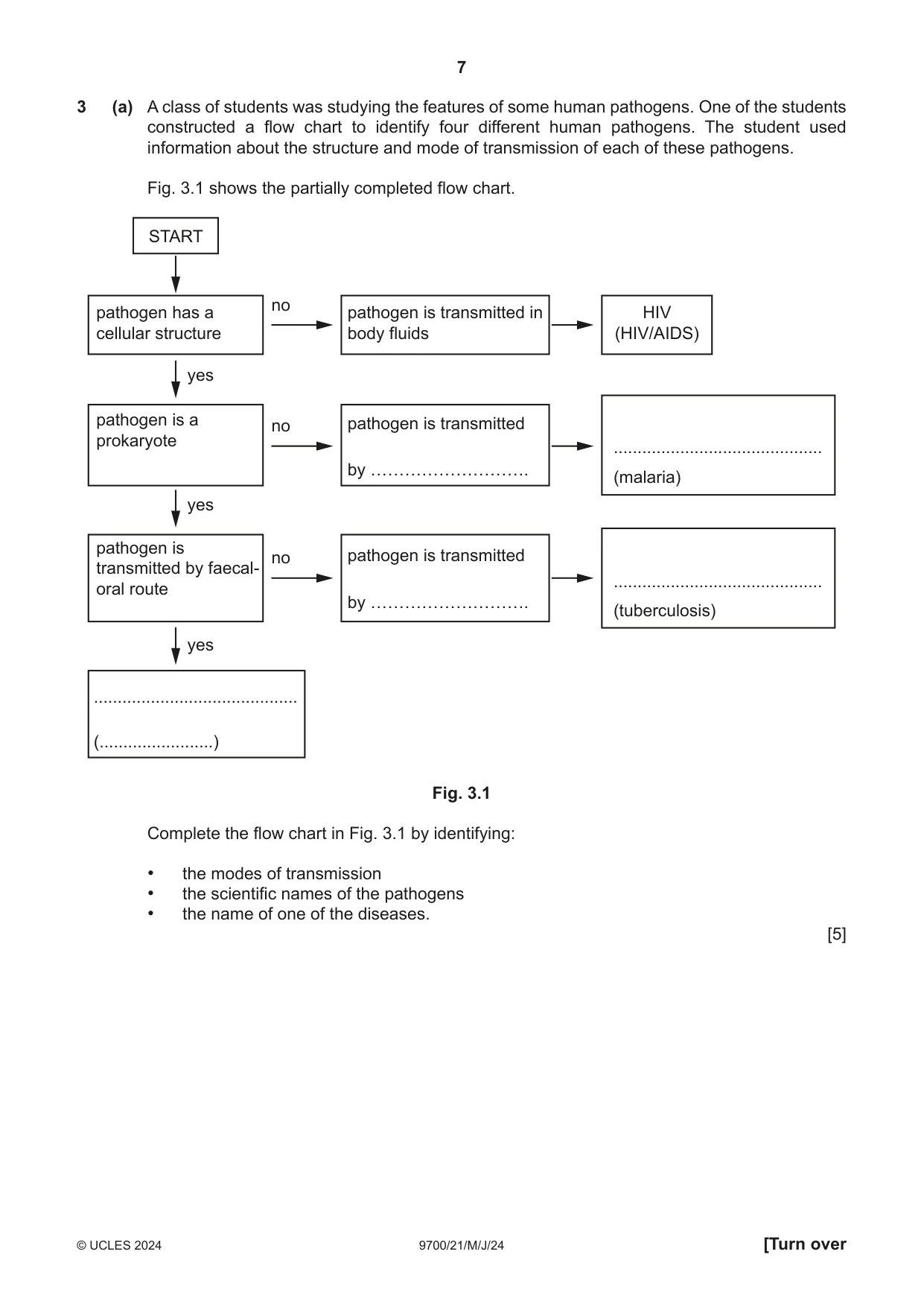
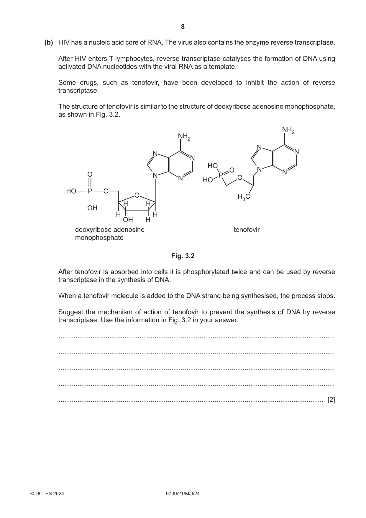
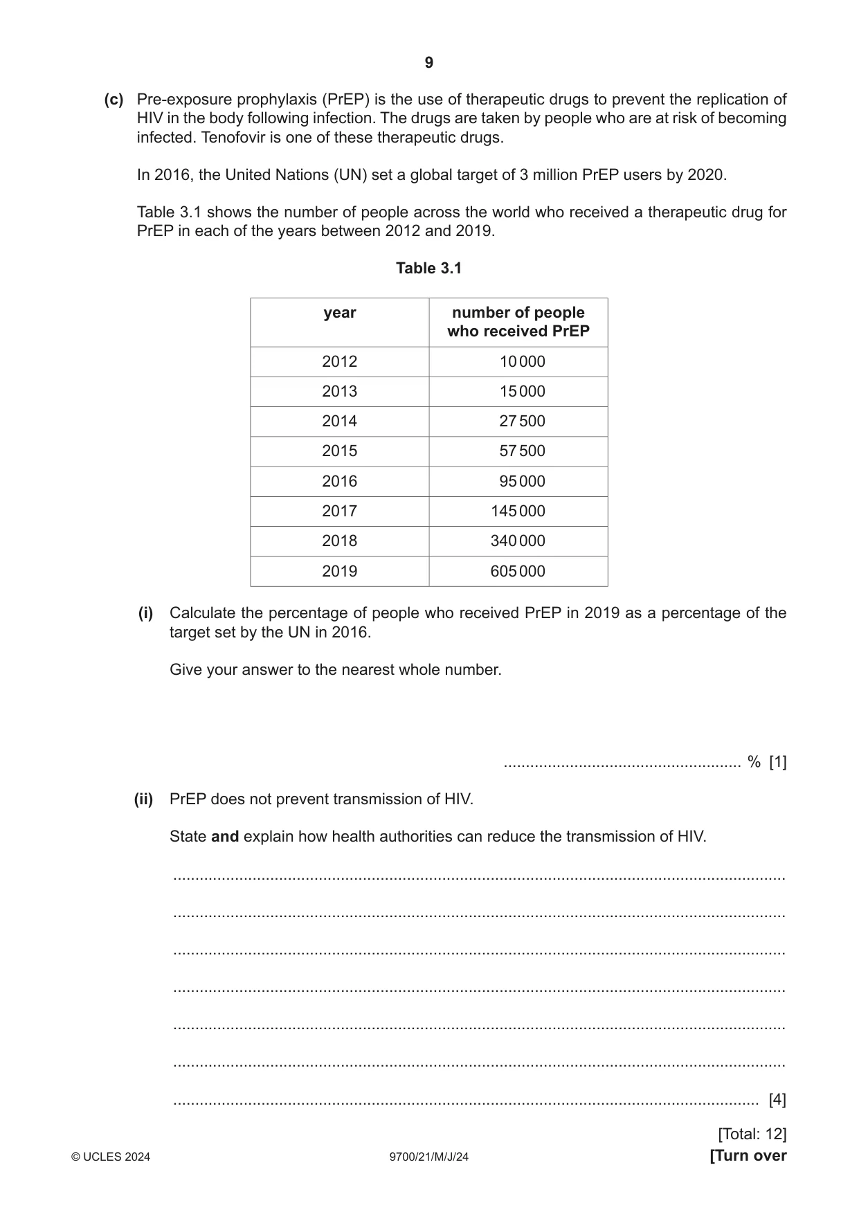
3 (a) A class of students was studying the features of some human pathogens. One of the students constructed a flow chart to identify four different human pathogens. The student used information about the structure and mode of transmission of each of these pathogens. Fig. 3.1 shows the partially completed flow chart. START no pathogen has a pathogen is transmitted in HIV cellular structure body fluids (HIV/AIDS) yes pathogen is a no pathogen is transmitted prokaryote ............................................ by ………………………. (malaria) yes pathogen is no pathogen is transmitted transmitted by faecal- ............................................ oral route by ………………………. (tuberculosis) yes ........................................... (........................) Fig. 3.1 Complete the flow chart in Fig. 3.1 by identifying: • the modes of transmission • the scientific names of the pathogens • the name of one of the diseases. [5] (b) HIV has a nucleic acid core of RNA. The virus also contains the enzyme reverse transcriptase. After HIV enters T‑lymphocytes, reverse transcriptase catalyses the formation of DNA using activated DNA nucleotides with the viral RNA as a template. Some drugs, such as tenofovir, have been developed to inhibit the action of reverse transcriptase. The structure of tenofovir is similar to the structure of deoxyribose adenosine monophosphate, as shown in Fig. 3.2. NH2 NH2 N N N N HO O N N O N P N O HO HO P O O H3C H H OH H H OH H deoxyribose adenosine tenofovir monophosphate Fig. 3.2 After tenofovir is absorbed into cells it is phosphorylated twice and can be used by reverse transcriptase in the synthesis of DNA. When a tenofovir molecule is added to the DNA strand being synthesised, the process stops. Suggest the mechanism of action of tenofovir to prevent the synthesis of DNA by reverse transcriptase. Use the information in Fig. 3.2 in your answer. ................................................................................................................................................... ................................................................................................................................................... ................................................................................................................................................... ................................................................................................................................................... ............................................................................................................................................. [2] (c) Pre‑exposure prophylaxis (PrEP) is the use of therapeutic drugs to prevent the replication of HIV in the body following infection. The drugs are taken by people who are at risk of becoming infected. Tenofovir is one of these therapeutic drugs. In 2016, the United Nations (UN) set a global target of 3 million PrEP users by 2020. Table 3.1 shows the number of people across the world who received a therapeutic drug for PrEP in each of the years between 2012 and 2019. Table 3.1 year number of people who received PrEP 2012 10 000 2013 15 000 2014 27 500 2015 57 500 2016 95 000 2017 145 000 2018 340 000 2019 605 000 (i) Calculate the percentage of people who received PrEP in 2019 as a percentage of the target set by the UN in 2016. Give your answer to the nearest whole number. ...................................................... % [1] (ii) PrEP does not prevent transmission of HIV. State and explain how health authorities can reduce the transmission of HIV. ........................................................................................................................................... ........................................................................................................................................... ........................................................................................................................................... ........................................................................................................................................... ........................................................................................................................................... ........................................................................................................................................... ..................................................................................................................................... [4] [Total: 12]
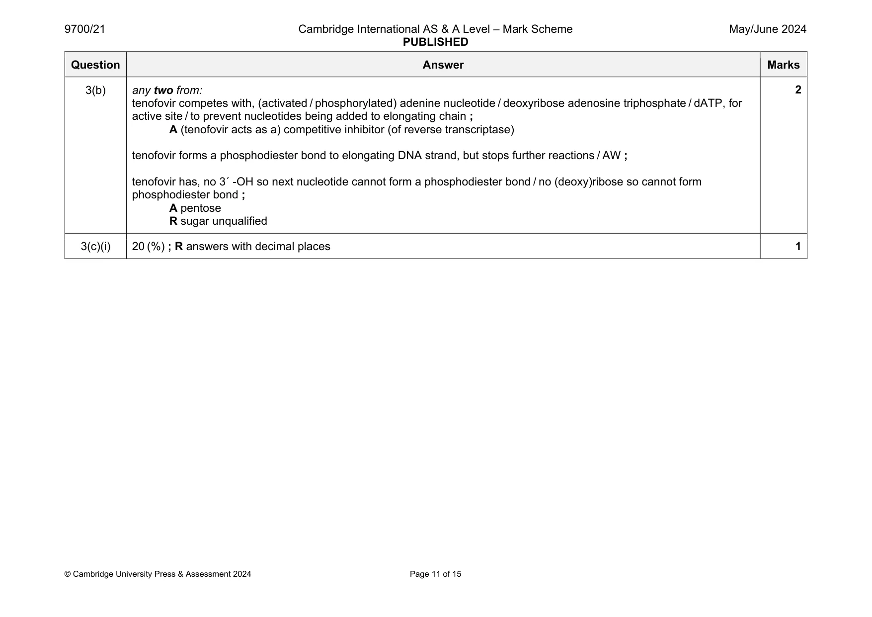
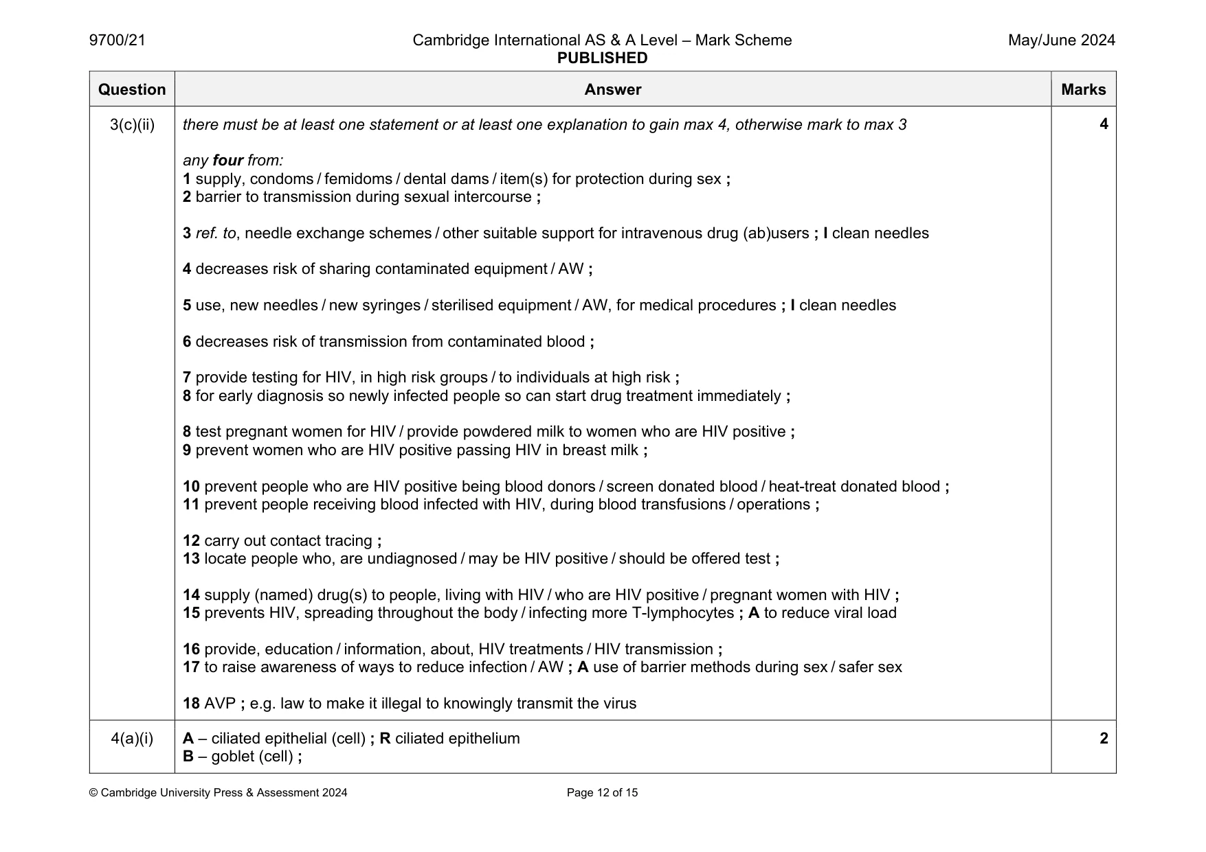
Mark scheme: 3(a) (insect) vector / Anopheles / (a) mosquito ; Plasmodium, falciparum / ovale / malariae / vivax ; A knowlesi Vibrio cholerae and cholera ; A other pathogens and associated diseases that are transmitted in the same way Mycobacterium, tuberculosis / bovis ; airborne droplets / droplet infection / aerosol (infection) ; I air droplets I water droplets from, coughing / sneezing A alternative transmission only if Mycobacterium bovis stated e.g. (eating) contaminated meat (from infected cattle) (drinking) contaminated milk (drinking) unpasteurised milk from contaminated, cows / cattle 5 Question Answer Marks 3(b) any two from: tenofovir competes with, (activated / phosphorylated) adenine nucleotide / deoxyribose adenosine triphosphate / dATP, for active site / to prevent nucleotides being added to elongating chain ; A (tenofovir acts as a) competitive inhibitor (of reverse transcriptase) tenofovir forms a phosphodiester bond to elongating DNA strand, but stops further reactions / AW ; tenofovir has, no 3´ -OH so next nucleotide cannot form a phosphodiester bond / no (deoxy)ribose so cannot form phosphodiester bond ; A pentose R sugar unqualified 2 3(c)(i) 20 (%) ; R answers with decimal places 1 Question Answer Marks 3(c)(ii) there must be at least one statement or at least one explanation to gain max 4, otherwise mark to max 3 any four from: 1 supply, condoms / femidoms / dental dams / item(s) for protection during sex ; 2 barrier to transmission during sexual intercourse ; 3 ref. to, needle exchange schemes / other suitable support for intravenous drug (ab)users ; I clean needles 4 decreases risk of sharing contaminated equipment / AW ; 5 use, new needles / new syringes / sterilised equipment / AW, for medical procedures ; I clean needles 6 decreases risk of transmission from contaminated blood ; 7 provide testing for HIV, in high risk groups / to individuals at high risk ; 8 for early diagnosis so newly infected people so can start drug treatment immediately ; 8 test pregnant women for HIV / provide powdered milk to women who are HIV positive ; 9 prevent women who are HIV positive passing HIV in breast milk ; 10 prevent people who are HIV positive being blood donors / screen donated blood / heat-treat donated blood ; 11 prevent people receiving blood infected with HIV, during blood transfusions / operations ; 12 carry out contact tracing ; 13 locate people who, are undiagnosed / may be HIV positive / should be offered test ; 14 supply (named) drug(s) to people, living with HIV / who are HIV positive / pregnant women with HIV ; 15 prevents HIV, spreading throughout the body / infecting more T-lymphocytes ; A to reduce viral load 16 provide, education / information, about, HIV treatments / HIV transmission ; 17 to raise awareness of ways to reduce infection / AW ; A use of barrier methods during sex / safer sex 18 AVP ; e.g. law to make it illegal to knowingly transmit the virus 4
Q4 · A scanning electron micrograph showing the tissue that lines the bronchi in the gas…
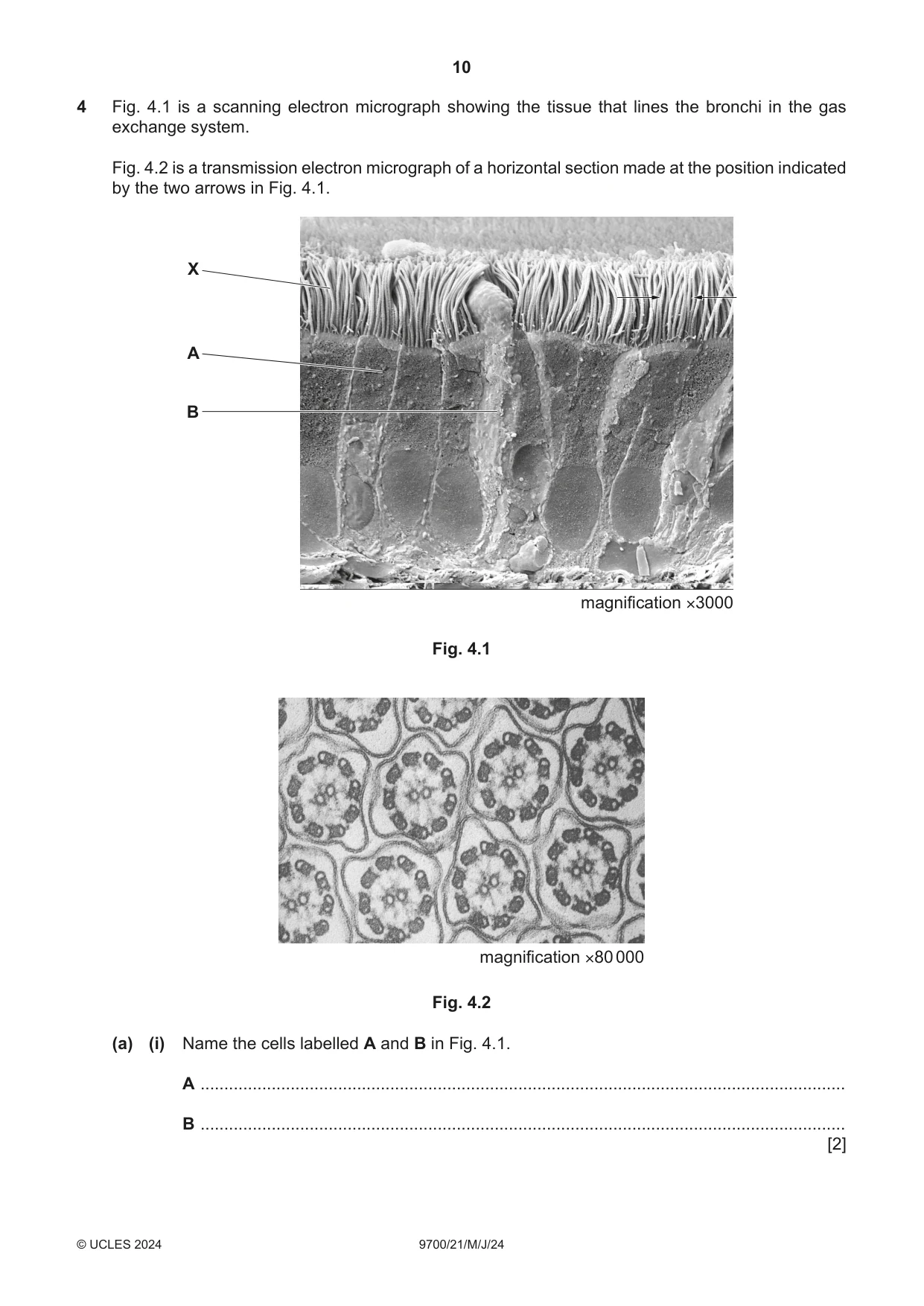
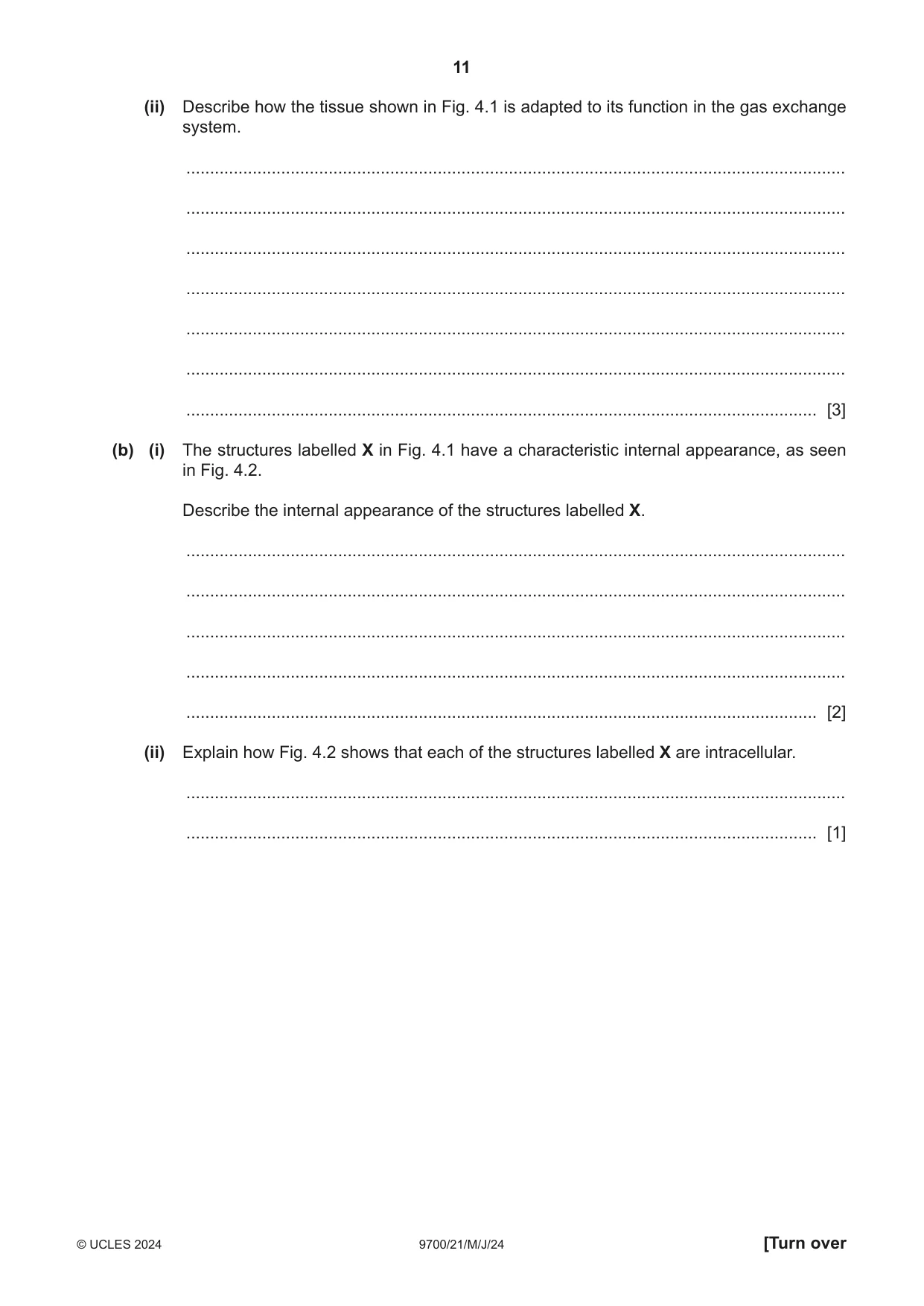
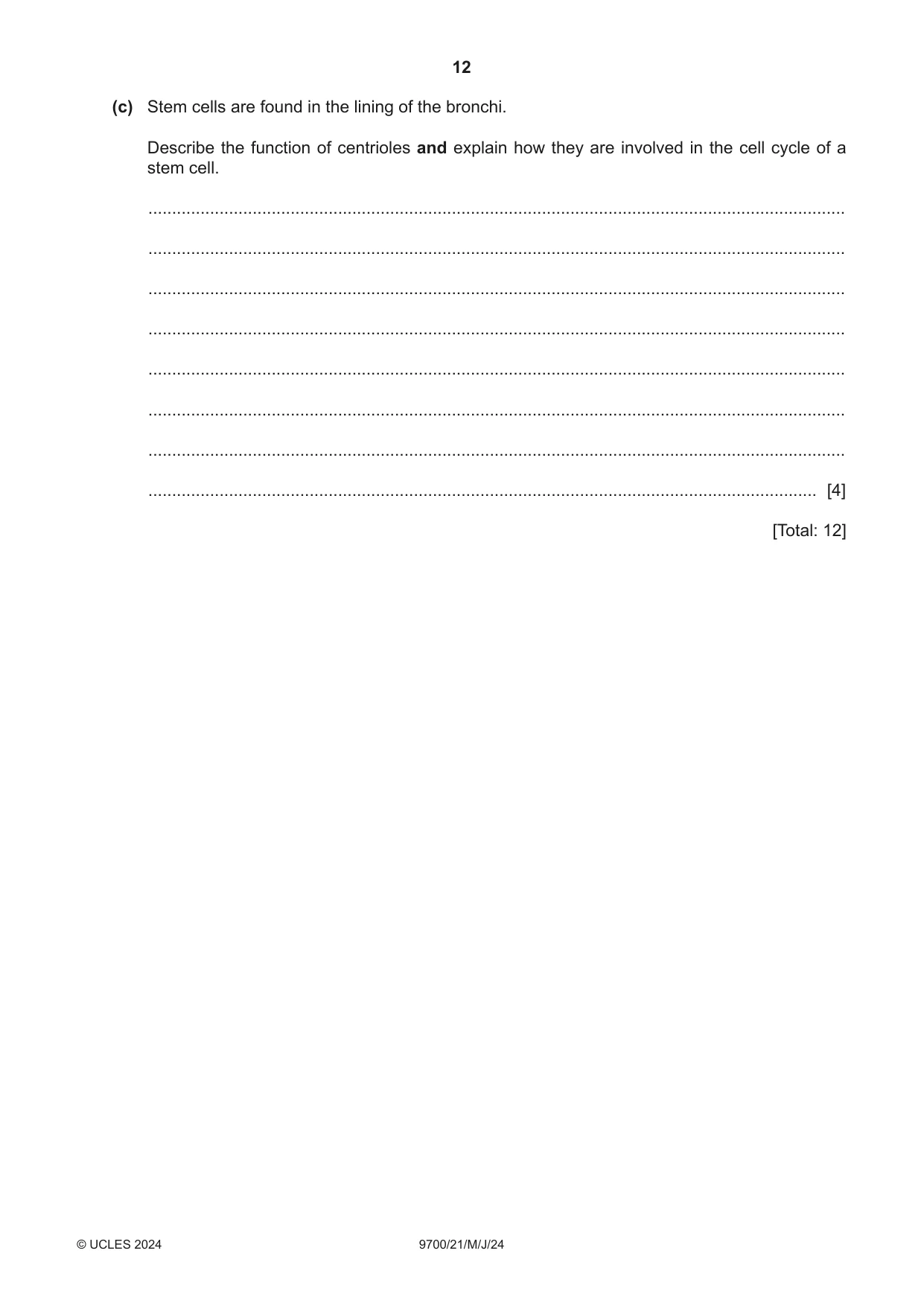
4 Fig. 4.1 is a scanning electron micrograph showing the tissue that lines the bronchi in the gas exchange system. Fig. 4.2 is a transmission electron micrograph of a horizontal section made at the position indicated by the two arrows in Fig. 4.1. X A B magnification ×3000 Fig. 4.1 magnification ×80 000 Fig. 4.2 (a) (i) Name the cells labelled A and B in Fig. 4.1. A ........................................................................................................................................ B ........................................................................................................................................ [2] (ii) Describe how the tissue shown in Fig. 4.1 is adapted to its function in the gas exchange system. ........................................................................................................................................... ........................................................................................................................................... ........................................................................................................................................... ........................................................................................................................................... ........................................................................................................................................... ........................................................................................................................................... ..................................................................................................................................... [3] (b) (i) The structures labelled X in Fig. 4.1 have a characteristic internal appearance, as seen in Fig. 4.2. Describe the internal appearance of the structures labelled X. ........................................................................................................................................... ........................................................................................................................................... ........................................................................................................................................... ........................................................................................................................................... ..................................................................................................................................... [2] (ii) Explain how Fig. 4.2 shows that each of the structures labelled X are intracellular. ........................................................................................................................................... ..................................................................................................................................... [1] (c) Stem cells are found in the lining of the bronchi. Describe the function of centrioles and explain how they are involved in the cell cycle of a stem cell. ................................................................................................................................................... ................................................................................................................................................... ................................................................................................................................................... ................................................................................................................................................... ................................................................................................................................................... ................................................................................................................................................... ................................................................................................................................................... ............................................................................................................................................. [4] [Total: 12]
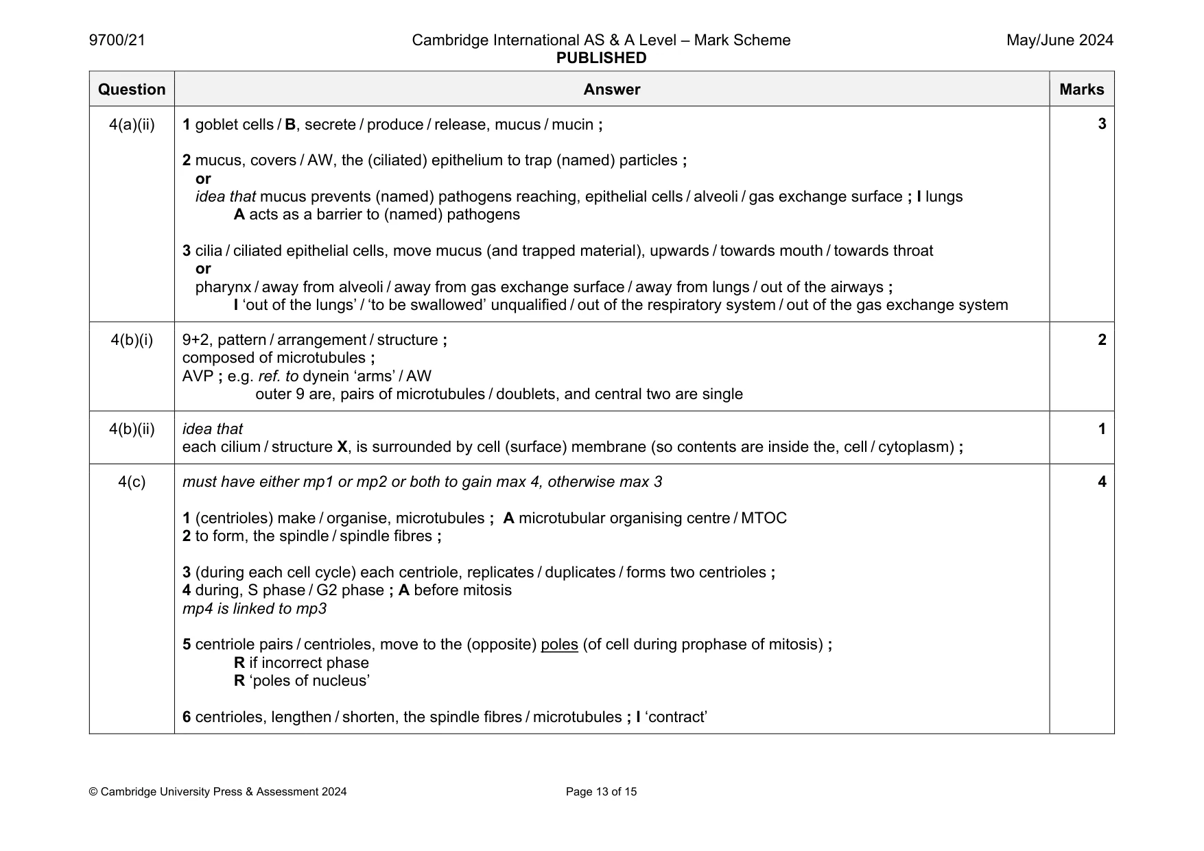
Mark scheme: 4(a)(i) A – ciliated epithelial (cell) ; R ciliated epithelium B – goblet (cell) ; 2 Question Answer Marks 4(a)(ii) 1 goblet cells / B, secrete / produce / release, mucus / mucin ; 2 mucus, covers / AW, the (ciliated) epithelium to trap (named) particles ; or idea that mucus prevents (named) pathogens reaching, epithelial cells / alveoli / gas exchange surface ; I lungs A acts as a barrier to (named) pathogens 3 cilia / ciliated epithelial cells, move mucus (and trapped material), upwards / towards mouth / towards throat or pharynx / away from alveoli / away from gas exchange surface / away from lungs / out of the airways ; I ‘out of the lungs’ / ‘to be swallowed’ unqualified / out of the respiratory system / out of the gas exchange system 3 4(b)(i) 9+2, pattern / arrangement / structure ; composed of microtubules ; AVP ; e.g. ref. to dynein ‘arms’ / AW outer 9 are, pairs of microtubules / doublets, and central two are single 2 4(b)(ii) idea that each cilium / structure X, is surrounded by cell (surface) membrane (so contents are inside the, cell / cytoplasm) ; 1 4(c) must have either mp1 or mp2 or both to gain max 4, otherwise max 3 1 (centrioles) make / organise, microtubules ; A microtubular organising centre / MTOC 2 to form, the spindle / spindle fibres ; 3 (during each cell cycle) each centriole, replicates / duplicates / forms two centrioles ; 4 during, S phase / G2 phase ; A before mitosis mp4 is linked to mp3 5 centriole pairs / centrioles, move to the (opposite) poles (of cell during prophase of mitosis) ; R if incorrect phase R ‘poles of nucleus’ 6 centrioles, lengthen / shorten, the spindle fibres / microtubules ; I ‘contract’ 4
Q5 · The pressure of water vapour inside and outside leaves can be measured
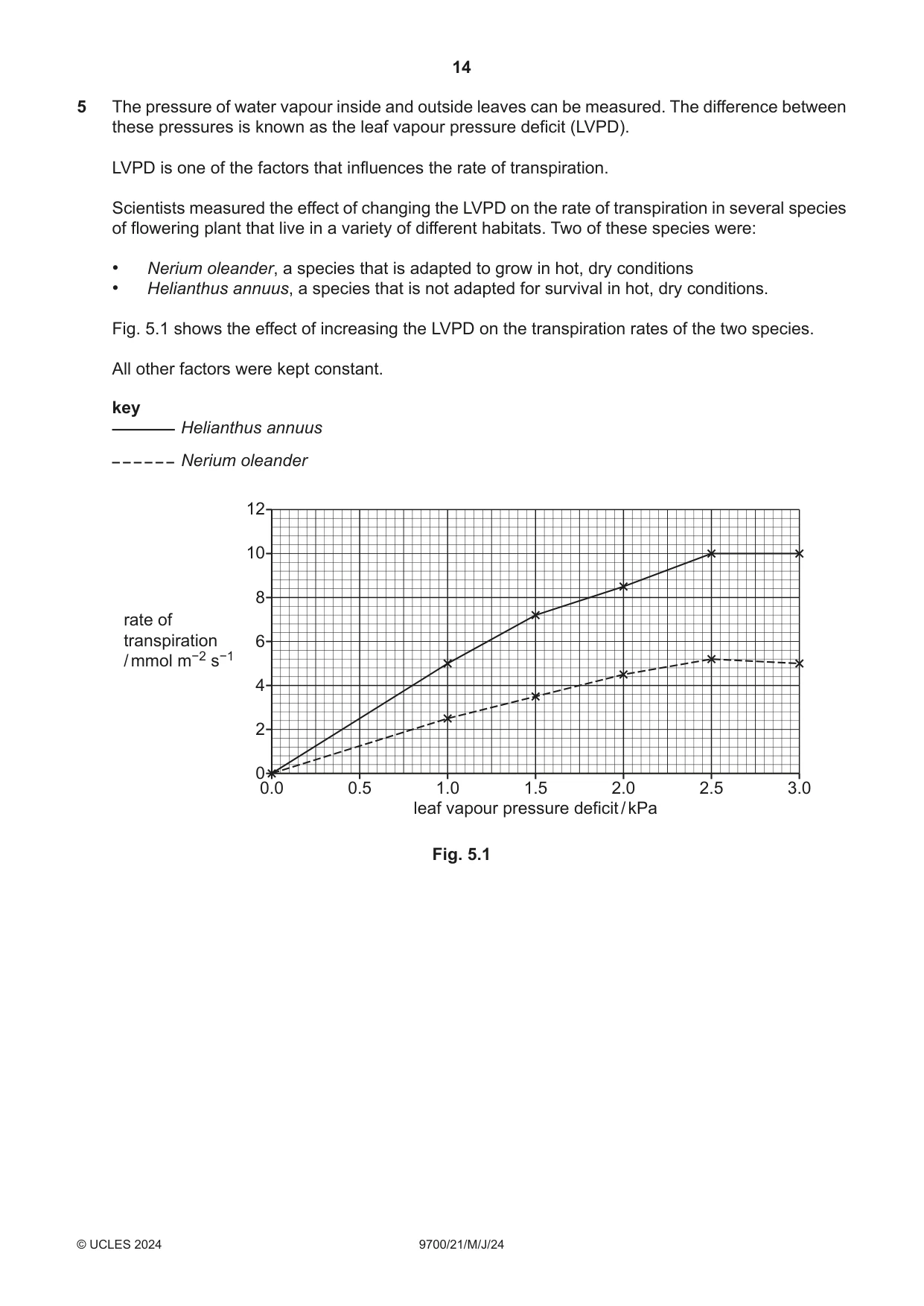
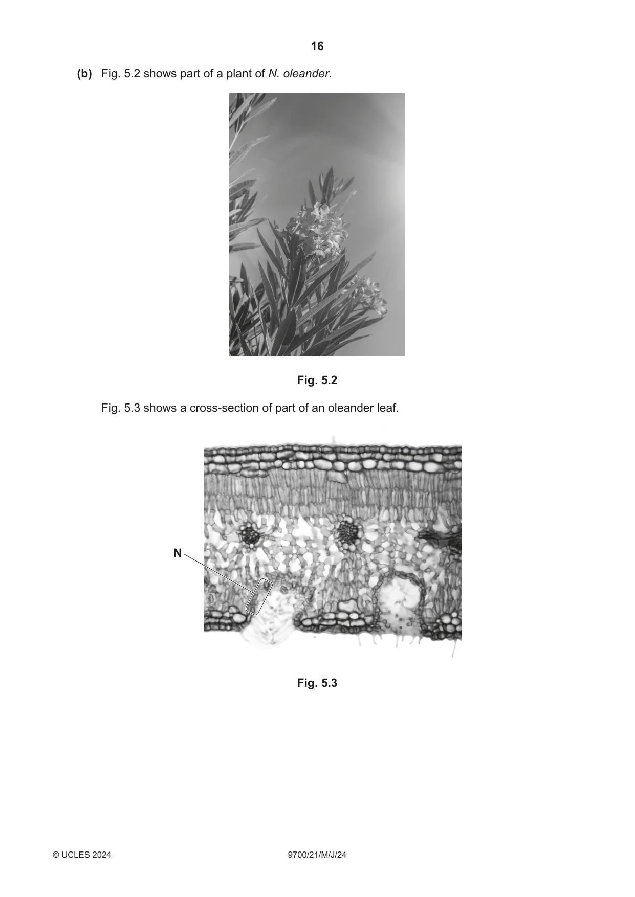
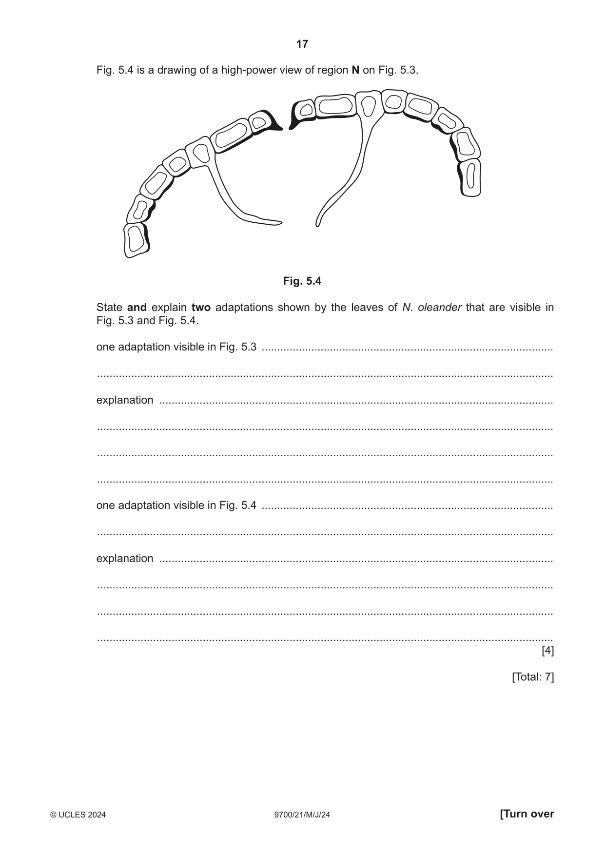
5 The pressure of water vapour inside and outside leaves can be measured. The difference between these pressures is known as the leaf vapour pressure deficit (LVPD). LVPD is one of the factors that influences the rate of transpiration. Scientists measured the effect of changing the LVPD on the rate of transpiration in several species of flowering plant that live in a variety of different habitats. Two of these species were: • Nerium oleander, a species that is adapted to grow in hot, dry conditions • Helianthus annuus, a species that is not adapted for survival in hot, dry conditions. Fig. 5.1 shows the effect of increasing the LVPD on the transpiration rates of the two species. All other factors were kept constant. key Helianthus annuus Nerium oleander 12 10 8 rate of transpiration 6 / mmol m−2 s−1 4 2 0 0.0 0.5 1.0 1.5 2.0 2.5 3.0 leaf vapour pressure deficit / kPa Fig. 5.1 (a) Compare the results of the two species shown in Fig. 5.1. ................................................................................................................................................... ................................................................................................................................................... ................................................................................................................................................... ................................................................................................................................................... ................................................................................................................................................... ................................................................................................................................................... ................................................................................................................................................... ............................................................................................................................................. [3] (b) Fig. 5.2 shows part of a plant of N. oleander. Fig. 5.2 Fig. 5.3 shows a cross‑section of part of an oleander leaf. N Fig. 5.3 Fig. 5.4 is a drawing of a high‑power view of region N on Fig. 5.3. Fig. 5.4 State and explain two adaptations shown by the leaves of N. oleander that are visible in Fig. 5.3 and Fig. 5.4. one adaptation visible in Fig. 5.3 .............................................................................................. ................................................................................................................................................... explanation ............................................................................................................................... ................................................................................................................................................... ................................................................................................................................................... ................................................................................................................................................... one adaptation visible in Fig. 5.4 .............................................................................................. ................................................................................................................................................... explanation ............................................................................................................................... ................................................................................................................................................... ................................................................................................................................................... ................................................................................................................................................... [4] [Total: 7]
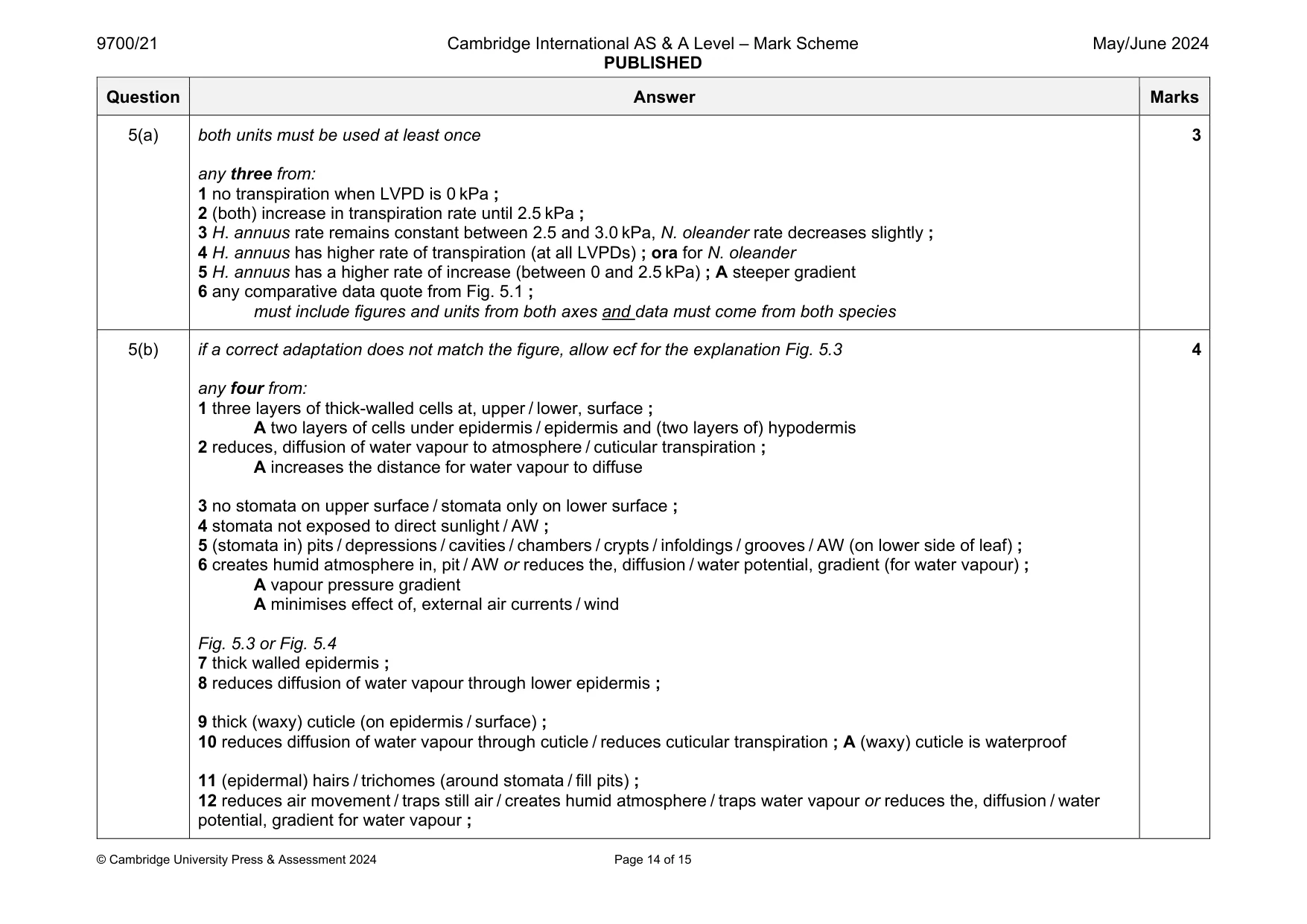
Mark scheme: 5(a) both units must be used at least once any three from: 1 no transpiration when LVPD is 0 kPa ; 2 (both) increase in transpiration rate until 2.5 kPa ; 3 H. annuus rate remains constant between 2.5 and 3.0 kPa, N. oleander rate decreases slightly ; 4 H. annuus has higher rate of transpiration (at all LVPDs) ; ora for N. oleander 5 H. annuus has a higher rate of increase (between 0 and 2.5 kPa) ; A steeper gradient 6 any comparative data quote from Fig. 5.1 ; must include figures and units from both axes and data must come from both species 3 5(b) if a correct adaptation does not match the figure, allow ecf for the explanation Fig. 5.3 any four from: 1 three layers of thick-walled cells at, upper / lower, surface ; A two layers of cells under epidermis / epidermis and (two layers of) hypodermis 2 reduces, diffusion of water vapour to atmosphere / cuticular transpiration ; A increases the distance for water vapour to diffuse 3 no stomata on upper surface / stomata only on lower surface ; 4 stomata not exposed to direct sunlight / AW ; 5 (stomata in) pits / depressions / cavities / chambers / crypts / infoldings / grooves / AW (on lower side of leaf) ; 6 creates humid atmosphere in, pit / AW or reduces the, diffusion / water potential, gradient (for water vapour) ; A vapour pressure gradient A minimises effect of, external air currents / wind Fig. 5.3 or Fig. 5.4 7 thick walled epidermis ; 8 reduces diffusion of water vapour through lower epidermis ; 9 thick (waxy) cuticle (on epidermis / surface) ; 10 reduces diffusion of water vapour through cuticle / reduces cuticular transpiration ; A (waxy) cuticle is waterproof 11 (epidermal) hairs / trichomes (around stomata / fill pits) ; 12 reduces air movement / traps still air / creates humid atmosphere / traps water vapour or reduces the, diffusion / water potential, gradient for water vapour ; 4
Q6 · Antibodies are produced by plasma cells
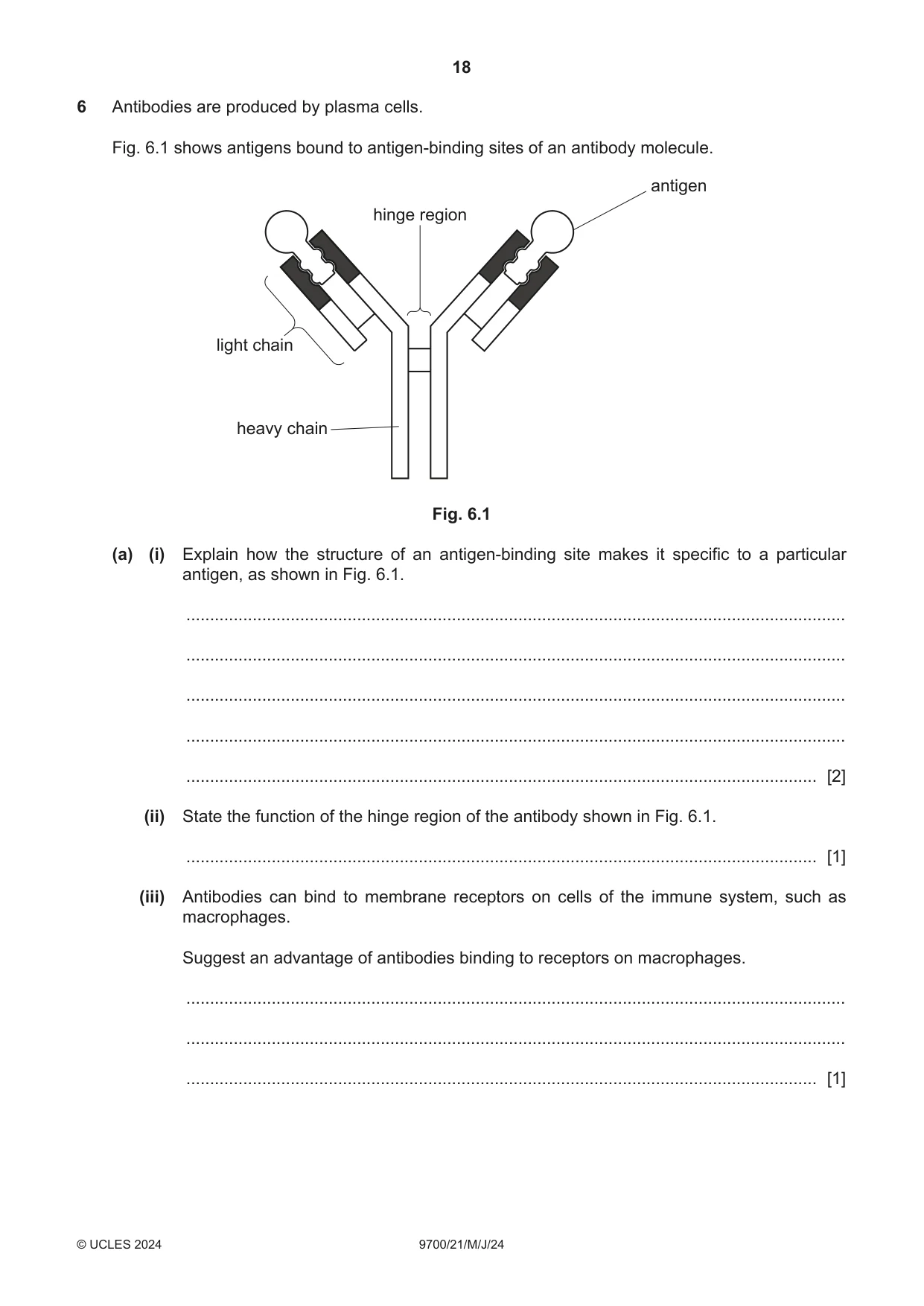
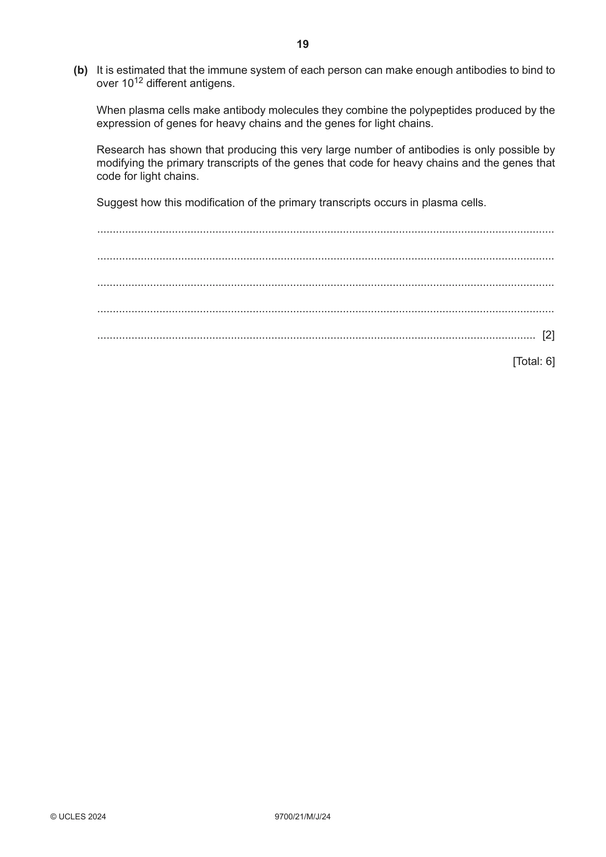
6 Antibodies are produced by plasma cells. Fig. 6.1 shows antigens bound to antigen‑binding sites of an antibody molecule. antigen hinge region light chain heavy chain Fig. 6.1 (a) (i) Explain how the structure of an antigen‑binding site makes it specific to a particular antigen, as shown in Fig. 6.1. ........................................................................................................................................... ........................................................................................................................................... ........................................................................................................................................... ........................................................................................................................................... ..................................................................................................................................... [2] (ii) State the function of the hinge region of the antibody shown in Fig. 6.1. ..................................................................................................................................... [1] (iii) Antibodies can bind to membrane receptors on cells of the immune system, such as macrophages. Suggest an advantage of antibodies binding to receptors on macrophages. ........................................................................................................................................... ........................................................................................................................................... ..................................................................................................................................... [1] (b) It is estimated that the immune system of each person can make enough antibodies to bind to over 1012 different antigens. When plasma cells make antibody molecules they combine the polypeptides produced by the expression of genes for heavy chains and the genes for light chains. Research has shown that producing this very large number of antibodies is only possible by modifying the primary transcripts of the genes that code for heavy chains and the genes that code for light chains. Suggest how this modification of the primary transcripts occurs in plasma cells. ................................................................................................................................................... ................................................................................................................................................... ................................................................................................................................................... ................................................................................................................................................... ............................................................................................................................................. [2] [Total: 6]
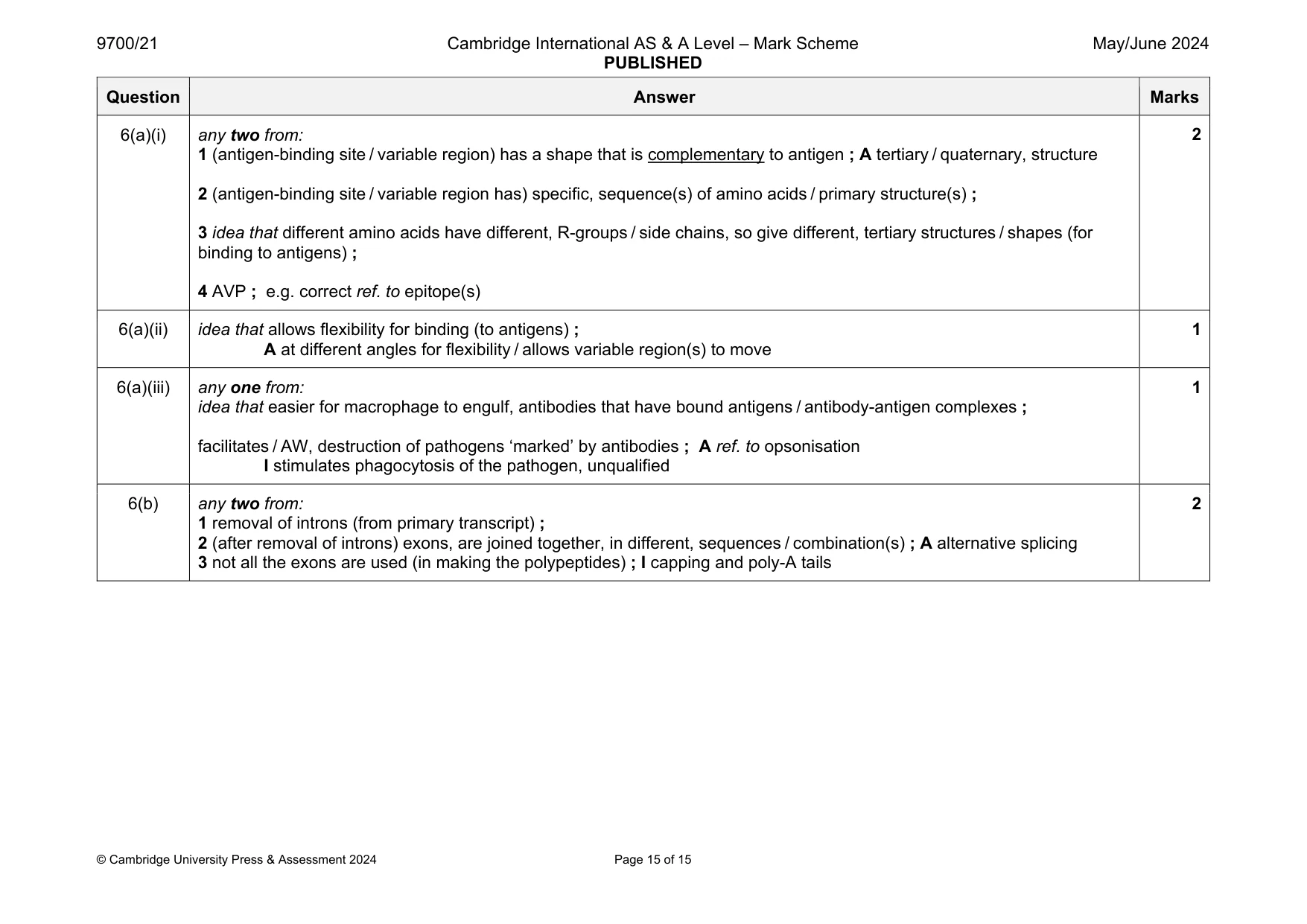
Mark scheme: 6(a)(i) any two from: 1 (antigen-binding site / variable region) has a shape that is complementary to antigen ; A tertiary / quaternary, structure 2 (antigen-binding site / variable region has) specific, sequence(s) of amino acids / primary structure(s) ; 3 idea that different amino acids have different, R-groups / side chains, so give different, tertiary structures / shapes (for binding to antigens) ; 4 AVP ; e.g. correct ref. to epitope(s) 6(a)(ii) idea that allows flexibility for binding (to antigens) ; A at different angles for flexibility / allows variable region(s) to move 1 6(a)(iii) any one from: idea that easier for macrophage to engulf, antibodies that have bound antigens / antibody-antigen complexes ; facilitates / AW, destruction of pathogens ‘marked’ by antibodies ; A ref. to opsonisation I stimulates phagocytosis of the pathogen, unqualified 1 6(b) any two from: 1 removal of introns (from primary transcript) ; 2 (after removal of introns) exons, are joined together, in different, sequences / combination(s) ; A alternative splicing 3 not all the exons are used (in making the polypeptides) ; I capping and poly-A tails 2
What was in this paper
The subtopics covered by these 6 questions, and how many questions each got. Open one in a new tab to see every Cambridge question on it.
What you needed in this session
Cambridge’s own grade thresholds for 2024 May/June, Paper 2 · Variant 1. A higher threshold means an easier paper — the bar moves with how the cohort did.