Cambridge A Level Biology 9700 — 2022 May/June Paper 2 · Variant 1
9700/21/M/J/22 · 6 questions · 60 marks · ≈68 min
The question paper and its mark scheme, free to read here and free to download. This is Cambridge’s own paper, exactly as it was sat.
Question paper20 pages


Mark scheme13 pages
Answers below. Sit the paper first if you are practising.
Questions as text
Question 1
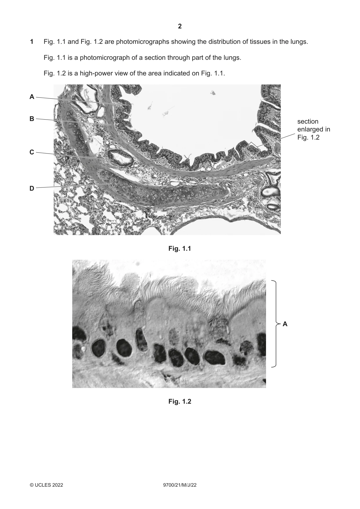
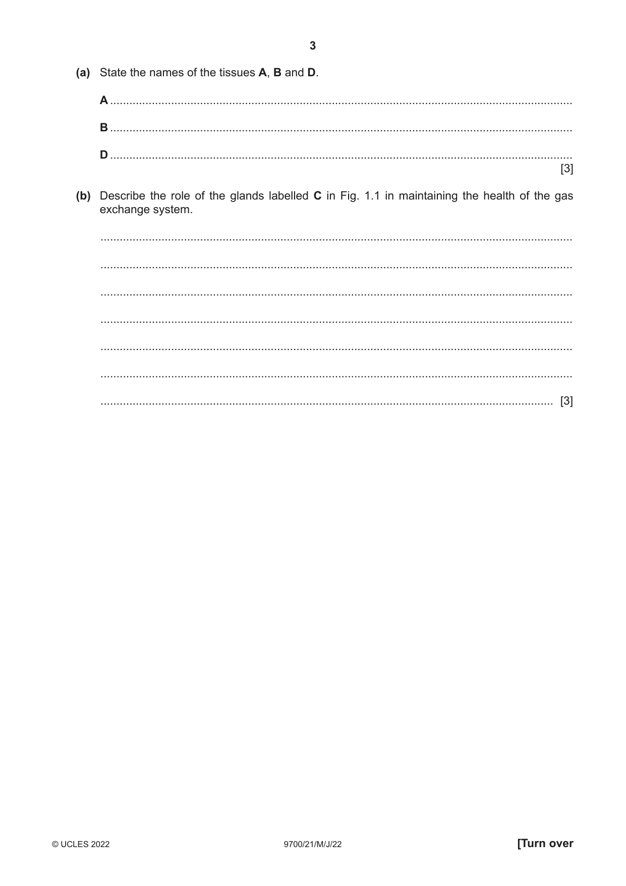
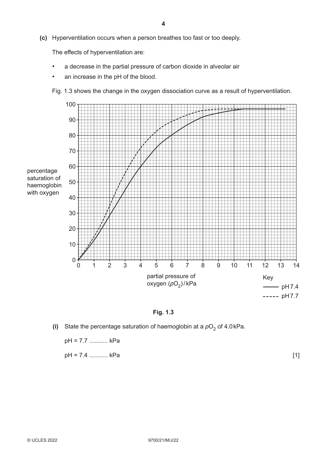
1 Fig. 1.1 and Fig. 1.2 are photomicrographs showing the distribution of tissues in the lungs. Fig. 1.1 is a photomicrograph of a section through part of the lungs. Fig. 1.2 is a high-power view of the area indicated on Fig. 1.1. A B section enlarged in Fig. 1.2 C D Fig. 1.1 A Fig. 1.2 (a) State the names of the tissues A, B and D. A ................................................................................................................................................ B ................................................................................................................................................ D ................................................................................................................................................ [3] (b) Describe the role of the glands labelled C in Fig. 1.1 in maintaining the health of the gas exchange system. ................................................................................................................................................... ................................................................................................................................................... ................................................................................................................................................... ................................................................................................................................................... ................................................................................................................................................... ................................................................................................................................................... ............................................................................................................................................. [3] (c) Hyperventilation occurs when a person breathes too fast or too deeply. The effects of hyperventilation are: • a decrease in the partial pressure of carbon dioxide in alveolar air • an increase in the pH of the blood. Fig. 1.3 shows the change in the oxygen dissociation curve as a result of hyperventilation. 100 90 80 70 60 percentage saturation of 50haemoglobin with oxygen 40 30 20 10 0 0 1 2 3 4 5 6 7 8 9 10 11 12 13 14 partial pressure of Key oxygen (pO2) / kPa pH 7.4 pH 7.7 Fig. 1.3 (i) State the percentage saturation of haemoglobin at a pO2 of 4.0 kPa. pH = 7.7 ........... kPa pH = 7.4 ........... kPa [1] (ii) Use the information in Fig. 1.3 to state and explain the effect of hyperventilation on the supply of oxygen to the respiring tissues. ........................................................................................................................................... ........................................................................................................................................... ........................................................................................................................................... ........................................................................................................................................... ..................................................................................................................................... [2] [Total: 9]
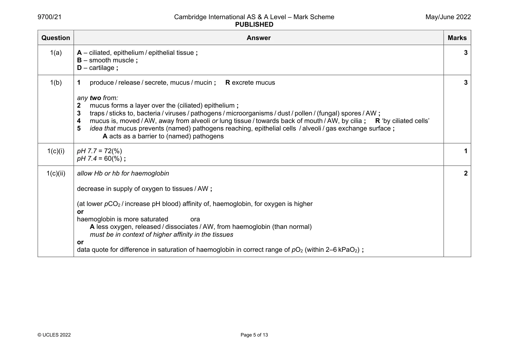
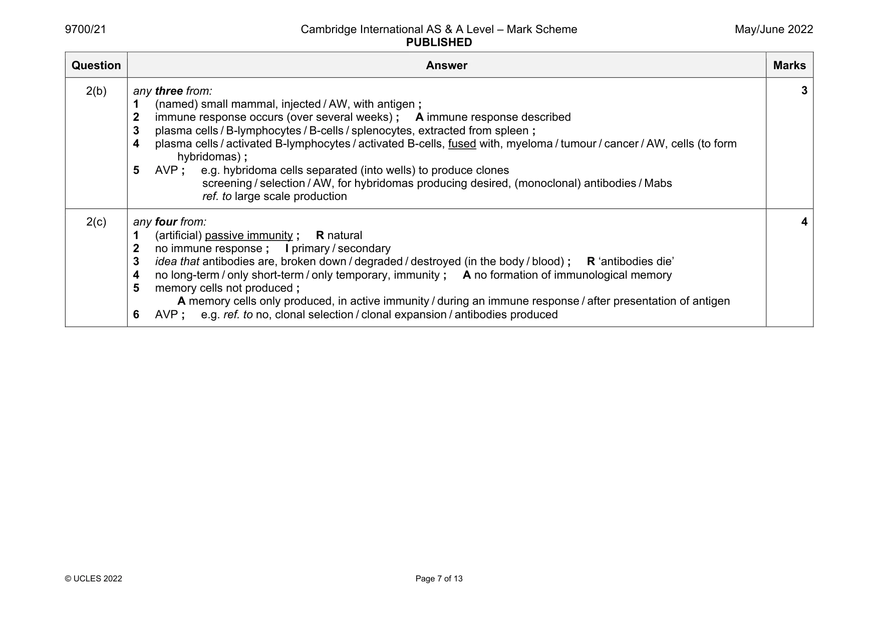
Mark scheme: 1(a) A – ciliated, epithelium / epithelial tissue ; B – smooth muscle ; D – cartilage ; 3 1(b) 1 produce / release / secrete, mucus / mucin ; R excrete mucus any two from: 2 mucus forms a layer over the (ciliated) epithelium ; 3 traps / sticks to, bacteria / viruses / pathogens / microorganisms / dust / pollen / (fungal) spores / AW ; 4 mucus is, moved / AW, away from alveoli or lung tissue / towards back of mouth / AW, by cilia ; R ‘by ciliated cells’ 5 idea that mucus prevents (named) pathogens reaching, epithelial cells / alveoli / gas exchange surface ; A acts as a barrier to (named) pathogens 3 1(c)(i) pH 7.7 = 72(%) pH 7.4 = 60(%) ; 1 1(c)(ii) allow Hb or hb for haemoglobin decrease in supply of oxygen to tissues / AW ; (at lower pCO2 / increase pH blood) affinity of, haemoglobin, for oxygen is higher or haemoglobin is more saturated ora A less oxygen, released / dissociates / AW, from haemoglobin (than normal) must be in context of higher affinity in the tissues or data quote for difference in saturation of haemoglobin in correct range of pO2 (within 2–6 kPaO2) ; 2
Q2 · B-lymphocytes are activated to form plasma cells during immune responses
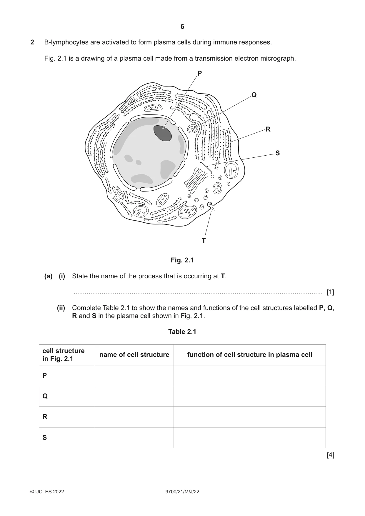
2 B-lymphocytes are activated to form plasma cells during immune responses. Fig. 2.1 is a drawing of a plasma cell made from a transmission electron micrograph. P Q R S T Fig. 2.1 (a) (i) State the name of the process that is occurring at T. ..................................................................................................................................... [1] (ii) Complete Table 2.1 to show the names and functions of the cell structures labelled P, Q, R and S in the plasma cell shown in Fig. 2.1. Table 2.1 cell structure name of cell structure function of cell structure in plasma cell in Fig. 2.1 P Q R S [4] (b) Plasma cells can be used in the commercial production of some monoclonal antibodies. The method of production is known as the hybridoma method. Outline the steps in the production of monoclonal antibodies by the hybridoma method. ................................................................................................................................................... ................................................................................................................................................... ................................................................................................................................................... ................................................................................................................................................... ................................................................................................................................................... ................................................................................................................................................... ............................................................................................................................................. [3] (c) Antibodies can be collected from human blood donors and used to treat people that may have been infected with a pathogen. This prevents them becoming ill with the disease. Explain why this treatment does not prevent people becoming ill if they are infected again with the same pathogen. ................................................................................................................................................... ................................................................................................................................................... ................................................................................................................................................... ................................................................................................................................................... ................................................................................................................................................... ................................................................................................................................................... ................................................................................................................................................... ................................................................................................................................................... ............................................................................................................................................. [4] [Total: 12]
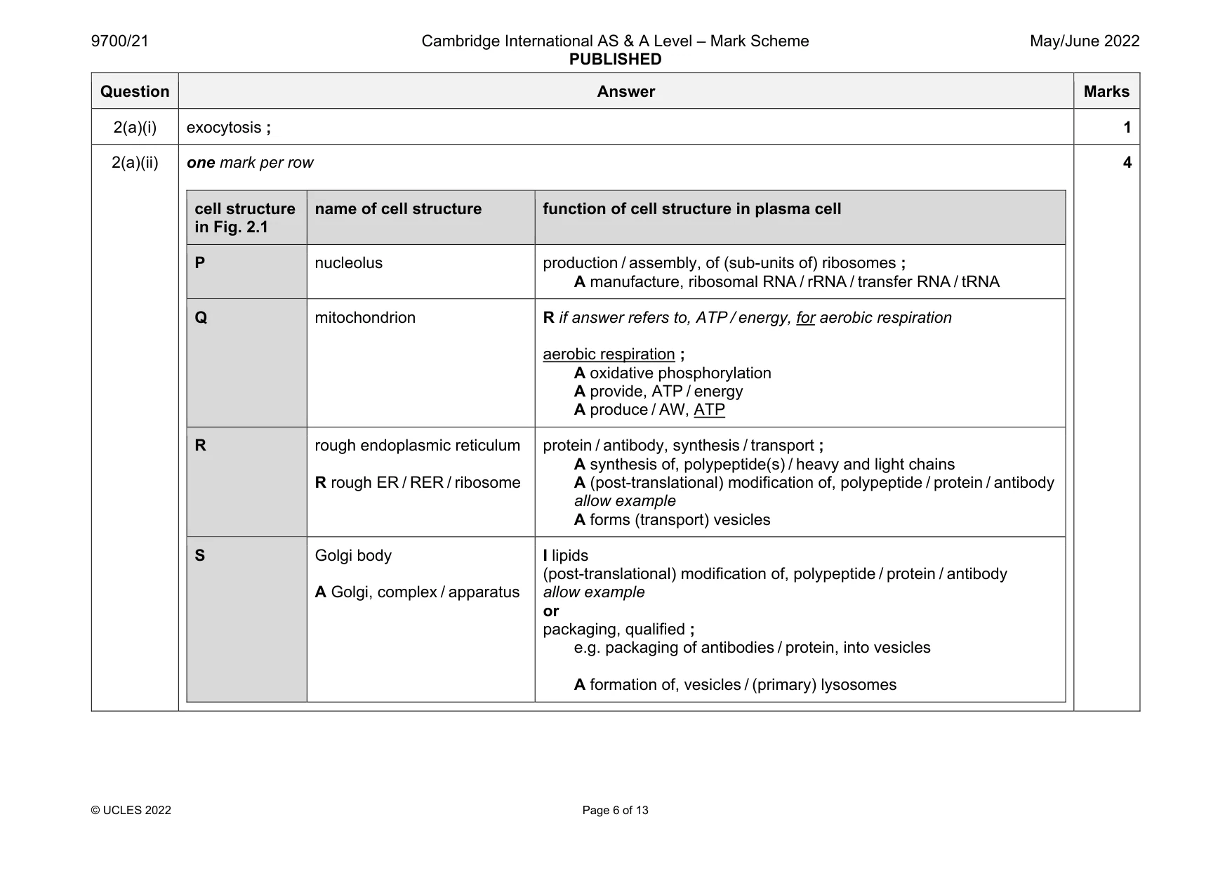
Mark scheme: 2(a)(i) exocytosis ; 1 2(a)(ii) one mark per row cell structure in Fig. 2.1 name of cell structure function of cell structure in plasma cell P nucleolus production / assembly, of (sub-units of) ribosomes ; A manufacture, ribosomal RNA / rRNA / transfer RNA / tRNA Q mitochondrion R if answer refers to, ATP / energy, for aerobic respiration aerobic respiration ; A oxidative phosphorylation A provide, ATP / energy A produce / AW, ATP R rough endoplasmic reticulum R rough ER / RER / ribosome protein / antibody, synthesis / transport ; A synthesis of, polypeptide(s) / heavy and light chains A (post-translational) modification of, polypeptide / protein / antibody allow example A forms (transport) vesicles S Golgi body A Golgi, complex / apparatus I lipids (post-translational) modification of, polypeptide / protein / antibody allow example or packaging, qualified ; e.g. packaging of antibodies / protein, into vesicles A formation of, vesicles / (primary) lysosomes 4 Question Answer Marks 2(b) any three from: 1 (named) small mammal, injected / AW, with antigen ; 2 immune response occurs (over several weeks) ; A immune response described 3 plasma cells / B-lymphocytes / B-cells / splenocytes, extracted from spleen ; 4 plasma cells / activated B-lymphocytes / activated B-cells, fused with, myeloma / tumour / cancer / AW, cells (to form hybridomas) ; 5 AVP ; e.g. hybridoma cells separated (into wells) to produce clones screening / selection / AW, for hybridomas producing desired, (monoclonal) antibodies / Mabs ref. to large scale production 3 2(c) any four from: 1 (artificial) passive immunity ; R natural 2 no immune response ; I primary / secondary 3 idea that antibodies are, broken down / degraded / destroyed (in the body / blood) ; R ‘antibodies die’ 4 no long-term / only short-term / only temporary, immunity ; A no formation of immunological memory 5 memory cells not produced ; A memory cells only produced, in active immunity / during an immune response / after presentation of antigen 6 AVP ; e.g. ref. to no, clonal selection / clonal expansion / antibodies produced 4
Q3 · A tyrosine kinase receptor (TKR) is a protein complex found in the cell surface membrane…
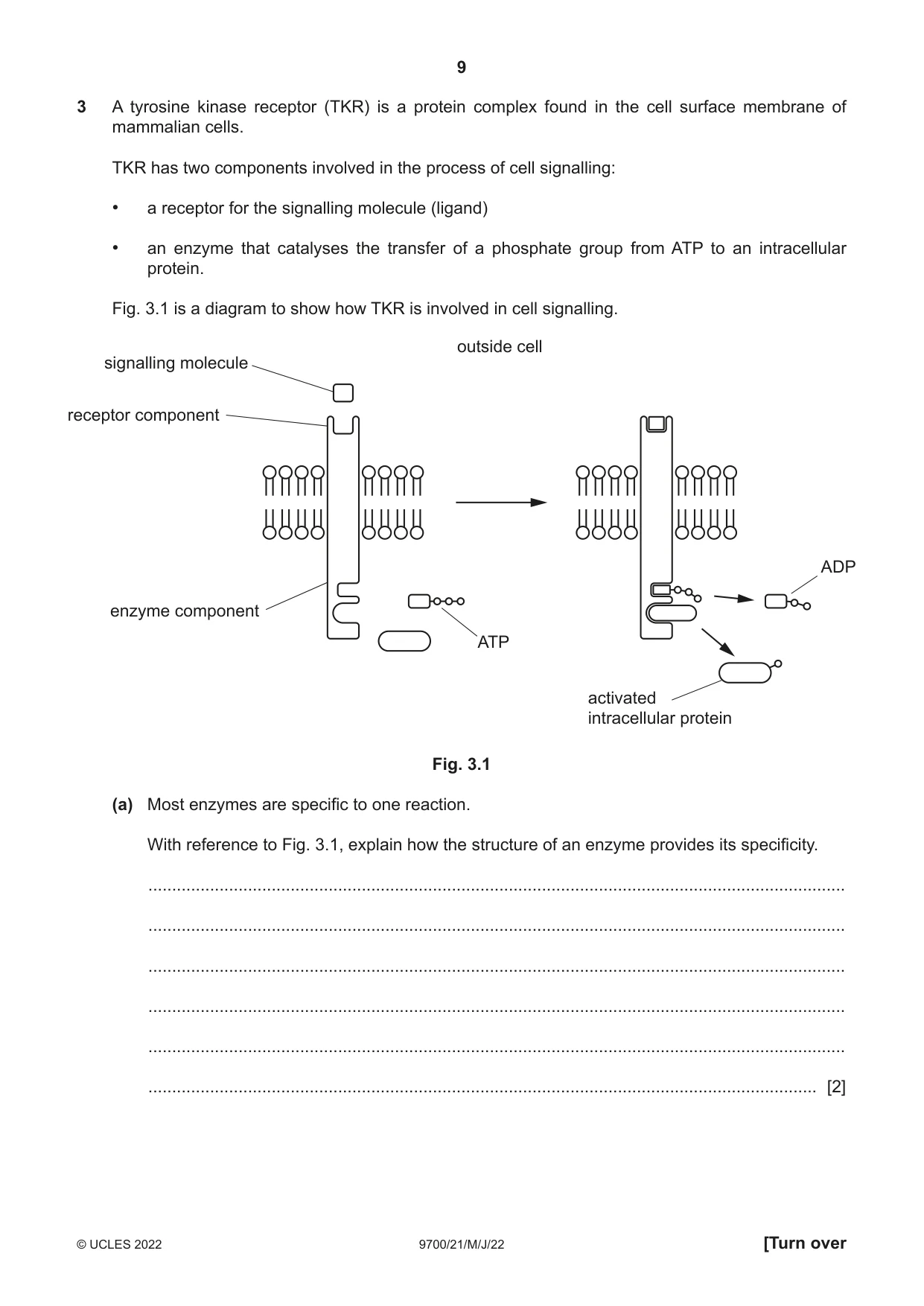
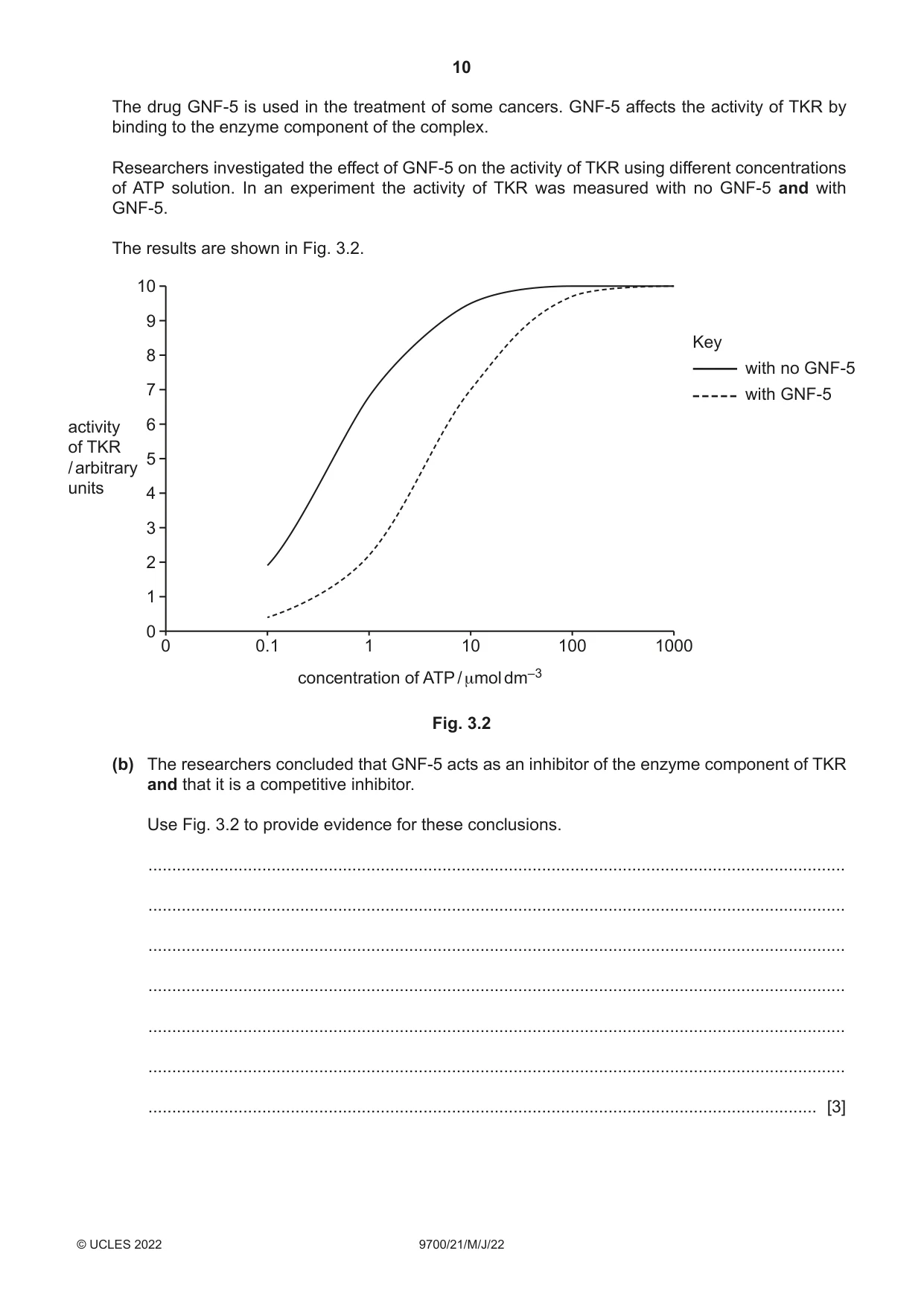
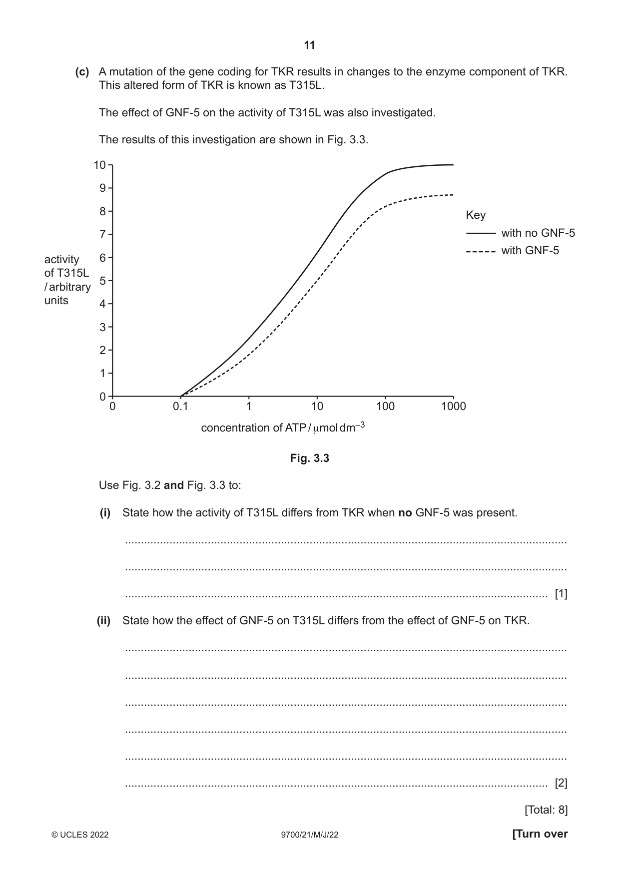
3 A tyrosine kinase receptor (TKR) is a protein complex found in the cell surface membrane of mammalian cells. TKR has two components involved in the process of cell signalling: • a receptor for the signalling molecule (ligand) • an enzyme that catalyses the transfer of a phosphate group from ATP to an intracellular protein. Fig. 3.1 is a diagram to show how TKR is involved in cell signalling. outside cell signalling molecule receptor component ADP enzyme component ATP activated intracellular protein Fig. 3.1 (a) Most enzymes are specific to one reaction. With reference to Fig. 3.1, explain how the structure of an enzyme provides its specificity. ................................................................................................................................................... ................................................................................................................................................... ................................................................................................................................................... ................................................................................................................................................... ................................................................................................................................................... ............................................................................................................................................. [2] The drug GNF-5 is used in the treatment of some cancers. GNF-5 affects the activity of TKR by binding to the enzyme component of the complex. Researchers investigated the effect of GNF-5 on the activity of TKR using different concentrations of ATP solution. In an experiment the activity of TKR was measured with no GNF-5 and with GNF-5. The results are shown in Fig. 3.2. 10 9 Key 8 with no GNF-5 7 with GNF-5 activity 6 of TKR 5/ arbitrary units 4 3 2 1 0 0 0.1 1 10 100 1000 concentration of ATP / μmol dm–3 Fig. 3.2 (b) The researchers concluded that GNF-5 acts as an inhibitor of the enzyme component of TKR and that it is a competitive inhibitor. Use Fig. 3.2 to provide evidence for these conclusions. ................................................................................................................................................... ................................................................................................................................................... ................................................................................................................................................... ................................................................................................................................................... ................................................................................................................................................... ................................................................................................................................................... ............................................................................................................................................. [3] (c) A mutation of the gene coding for TKR results in changes to the enzyme component of TKR. This altered form of TKR is known as T315L. The effect of GNF-5 on the activity of T315L was also investigated. The results of this investigation are shown in Fig. 3.3. 10 9 8 Key 7 with no GNF-5 with GNF-5 activity 6 of T315L 5/ arbitrary units 4 3 2 1 0 0 0.1 1 10 100 1000 concentration of ATP / μmol dm–3 Fig. 3.3 Use Fig. 3.2 and Fig. 3.3 to: (i) State how the activity of T315L differs from TKR when no GNF-5 was present. ........................................................................................................................................... ........................................................................................................................................... ..................................................................................................................................... [1] (ii) State how the effect of GNF-5 on T315L differs from the effect of GNF-5 on TKR. ........................................................................................................................................... ........................................................................................................................................... ........................................................................................................................................... ........................................................................................................................................... ........................................................................................................................................... ..................................................................................................................................... [2] [Total: 8]
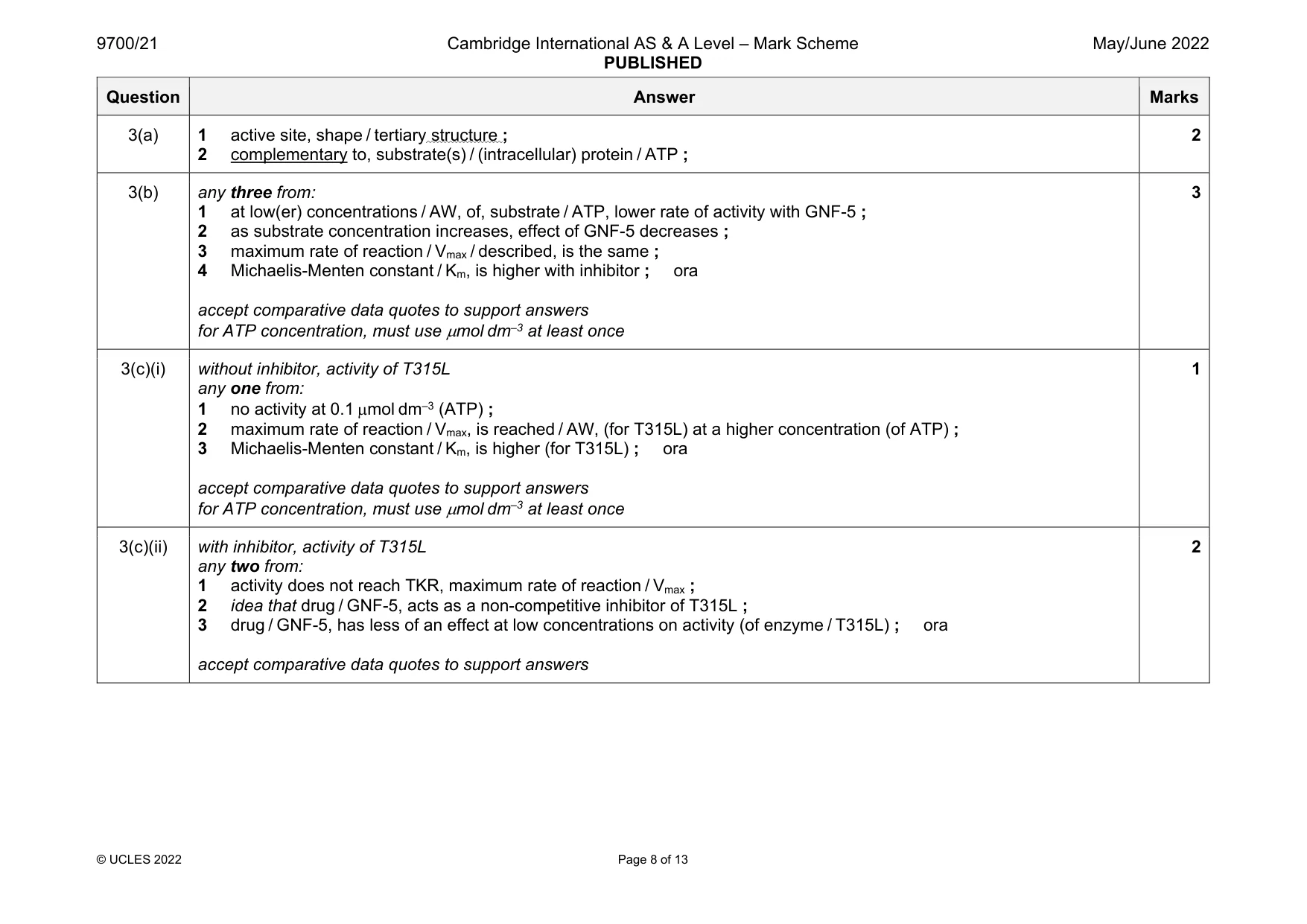
Mark scheme: 3(a) 1 active site, shape / tertiary structure ; 2 complementary to, substrate(s) / (intracellular) protein / ATP ; 2 3(b) any three from: 1 at low(er) concentrations / AW, of, substrate / ATP, lower rate of activity with GNF-5 ; 2 as substrate concentration increases, effect of GNF-5 decreases ; 3 maximum rate of reaction / Vmax / described, is the same ; 4 Michaelis-Menten constant / Km, is higher with inhibitor ; ora accept comparative data quotes to support answers for ATP concentration, must use mol dm–3 at least once 3 3(c)(i) without inhibitor, activity of T315L any one from: 1 no activity at 0.1 mol dm–3 (ATP) ; 2 maximum rate of reaction / Vmax, is reached / AW, (for T315L) at a higher concentration (of ATP) ; 3 Michaelis-Menten constant / Km, is higher (for T315L) ; ora accept comparative data quotes to support answers for ATP concentration, must use mol dm–3 at least once 1 3(c)(ii) with inhibitor, activity of T315L any two from: 1 activity does not reach TKR, maximum rate of reaction / Vmax ; 2 idea that drug / GNF-5, acts as a non-competitive inhibitor of T315L ; 3 drug / GNF-5, has less of an effect at low concentrations on activity (of enzyme / T315L) ; ora accept comparative data quotes to support answers 2
Q4 · Adipocytes are cells found in adipose tissue in mammals
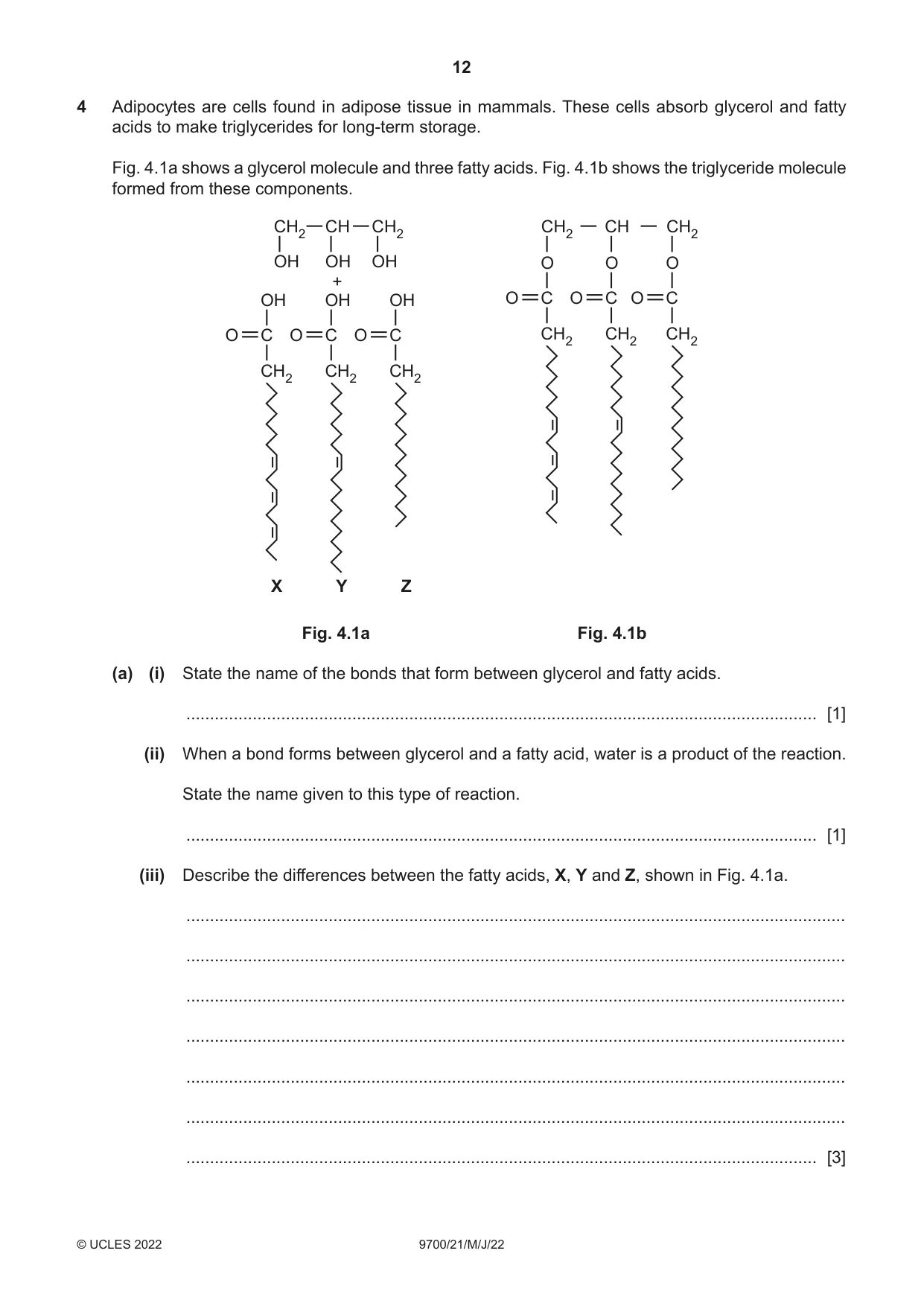
4 Adipocytes are cells found in adipose tissue in mammals. These cells absorb glycerol and fatty acids to make triglycerides for long-term storage. Fig. 4.1a shows a glycerol molecule and three fatty acids. Fig. 4.1b shows the triglyceride molecule formed from these components. CH2 CH CH2 CH2 CH CH2 OH OH OH O O O + OH OH OH O C O C O C O C O C O C CH2 CH2 CH2 CH2 CH2 CH2 X Y Z Fig. 4.1a Fig. 4.1b (a) (i) State the name of the bonds that form between glycerol and fatty acids. ..................................................................................................................................... [1] (ii) When a bond forms between glycerol and a fatty acid, water is a product of the reaction. State the name given to this type of reaction. ..................................................................................................................................... [1] (iii) Describe the differences between the fatty acids, X, Y and Z, shown in Fig. 4.1a. ........................................................................................................................................... ........................................................................................................................................... ........................................................................................................................................... ........................................................................................................................................... ........................................................................................................................................... ........................................................................................................................................... ..................................................................................................................................... [3] (b) (i) State reasons why triglycerides are described as hydrophobic. ........................................................................................................................................... ........................................................................................................................................... ........................................................................................................................................... ........................................................................................................................................... ..................................................................................................................................... [2] (ii) Explain why triglycerides are not suitable as a component of cell surface membranes. ........................................................................................................................................... ........................................................................................................................................... ........................................................................................................................................... ........................................................................................................................................... ..................................................................................................................................... [2] [Total: 9]
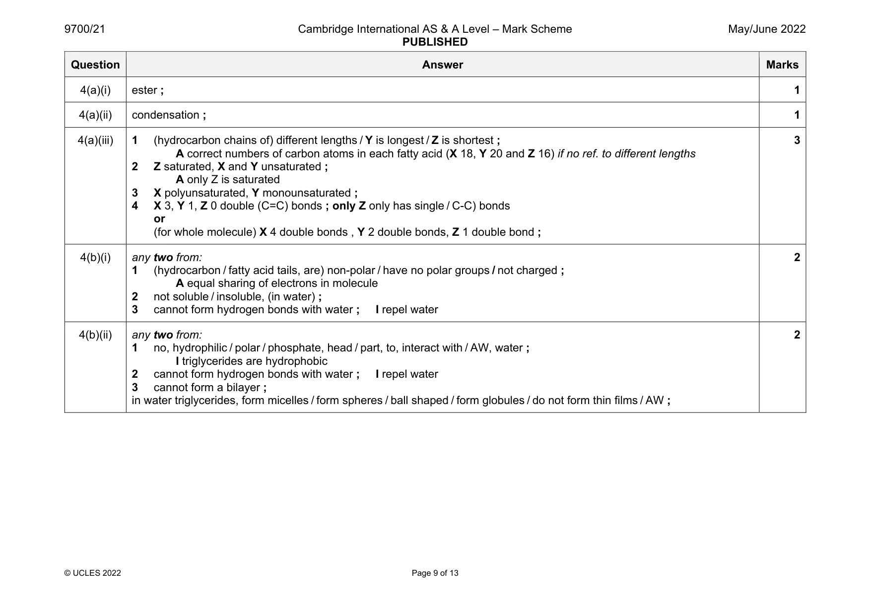
Mark scheme: 4(a)(i) ester ; 1 4(a)(ii) condensation ; 1 4(a)(iii) 1 (hydrocarbon chains of) different lengths / Y is longest / Z is shortest ; A correct numbers of carbon atoms in each fatty acid (X 18, Y 20 and Z 16) if no ref. to different lengths 2 Z saturated, X and Y unsaturated ; A only Z is saturated 3 X polyunsaturated, Y monounsaturated ; 4 X 3, Y 1, Z 0 double (C=C) bonds ; only Z only has single / C-C) bonds or (for whole molecule) X 4 double bonds , Y 2 double bonds, Z 1 double bond ; 3 4(b)(i) any two from: 1 (hydrocarbon / fatty acid tails, are) non-polar / have no polar groups / not charged ; A equal sharing of electrons in molecule 2 not soluble / insoluble, (in water) ; 3 cannot form hydrogen bonds with water ; I repel water 2 4(b)(ii) any two from: 1 no, hydrophilic / polar / phosphate, head / part, to, interact with / AW, water ; I triglycerides are hydrophobic 2 cannot form hydrogen bonds with water ; I repel water 3 cannot form a bilayer ; in water triglycerides, form micelles / form spheres / ball shaped / form globules / do not form thin films / AW ; 2
Q5 · Infectious diseases are caused by pathogens and are described as transmissible diseases
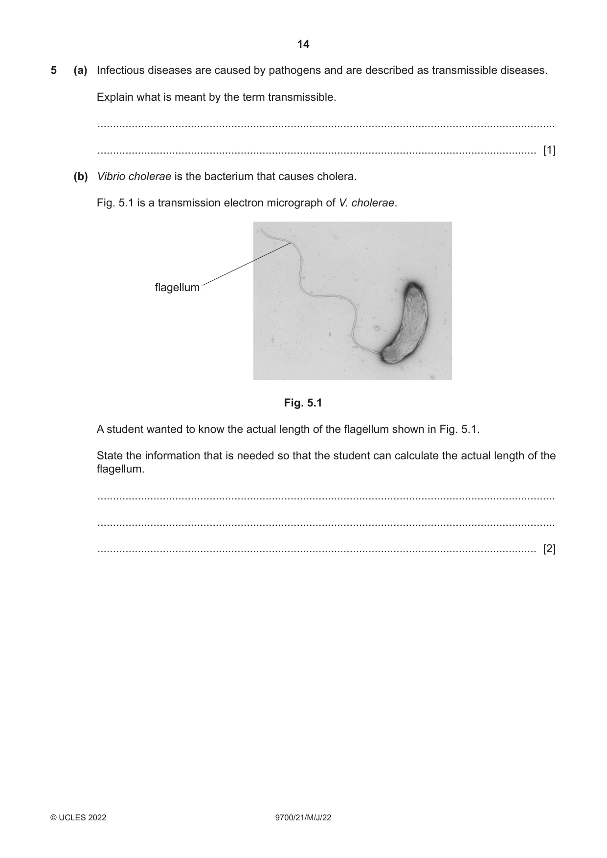
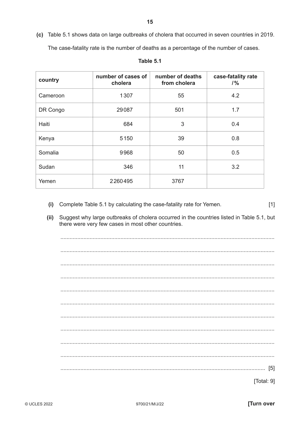
5 (a) Infectious diseases are caused by pathogens and are described as transmissible diseases. Explain what is meant by the term transmissible. ................................................................................................................................................... ............................................................................................................................................. [1] (b) Vibrio cholerae is the bacterium that causes cholera. Fig. 5.1 is a transmission electron micrograph of V. cholerae. flagellum Fig. 5.1 A student wanted to know the actual length of the flagellum shown in Fig. 5.1. State the information that is needed so that the student can calculate the actual length of the flagellum. ................................................................................................................................................... ................................................................................................................................................... ............................................................................................................................................. [2] (c) Table 5.1 shows data on large outbreaks of cholera that occurred in seven countries in 2019. The case-fatality rate is the number of deaths as a percentage of the number of cases. Table 5.1 number of cases of number of deaths case-fatality rate country cholera from cholera / % Cameroon 1 307 55 4.2 DR Congo 29 087 501 1.7 Haiti 684 3 0.4 Kenya 5 150 39 0.8 Somalia 9 968 50 0.5 Sudan 346 11 3.2 Yemen 2 260 495 3767 (i) Complete Table 5.1 by calculating the case-fatality rate for Yemen. [1] (ii) Suggest why large outbreaks of cholera occurred in the countries listed in Table 5.1, but there were very few cases in most other countries. ........................................................................................................................................... ........................................................................................................................................... ........................................................................................................................................... ........................................................................................................................................... ........................................................................................................................................... ........................................................................................................................................... ........................................................................................................................................... ........................................................................................................................................... ........................................................................................................................................... ........................................................................................................................................... ..................................................................................................................................... [5] [Total: 9]
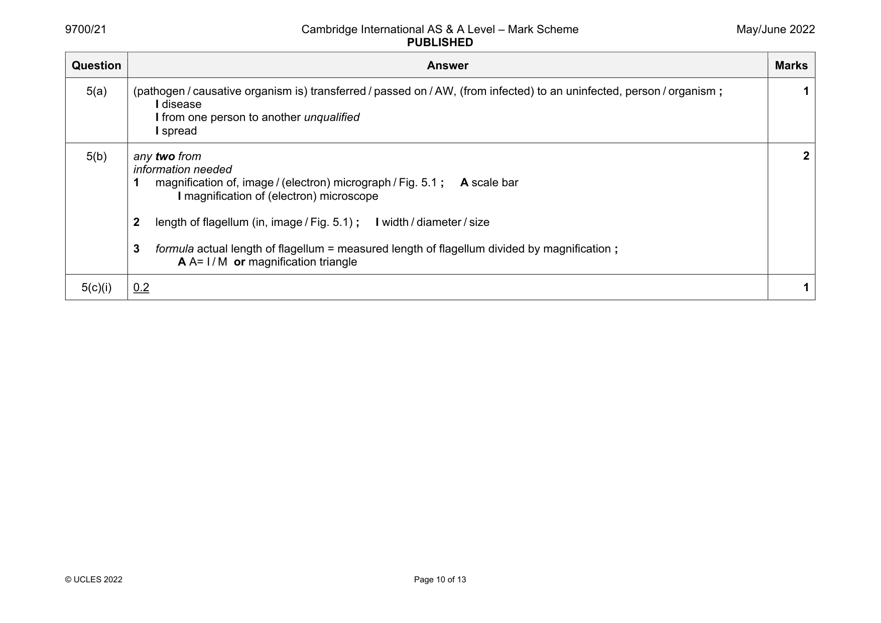
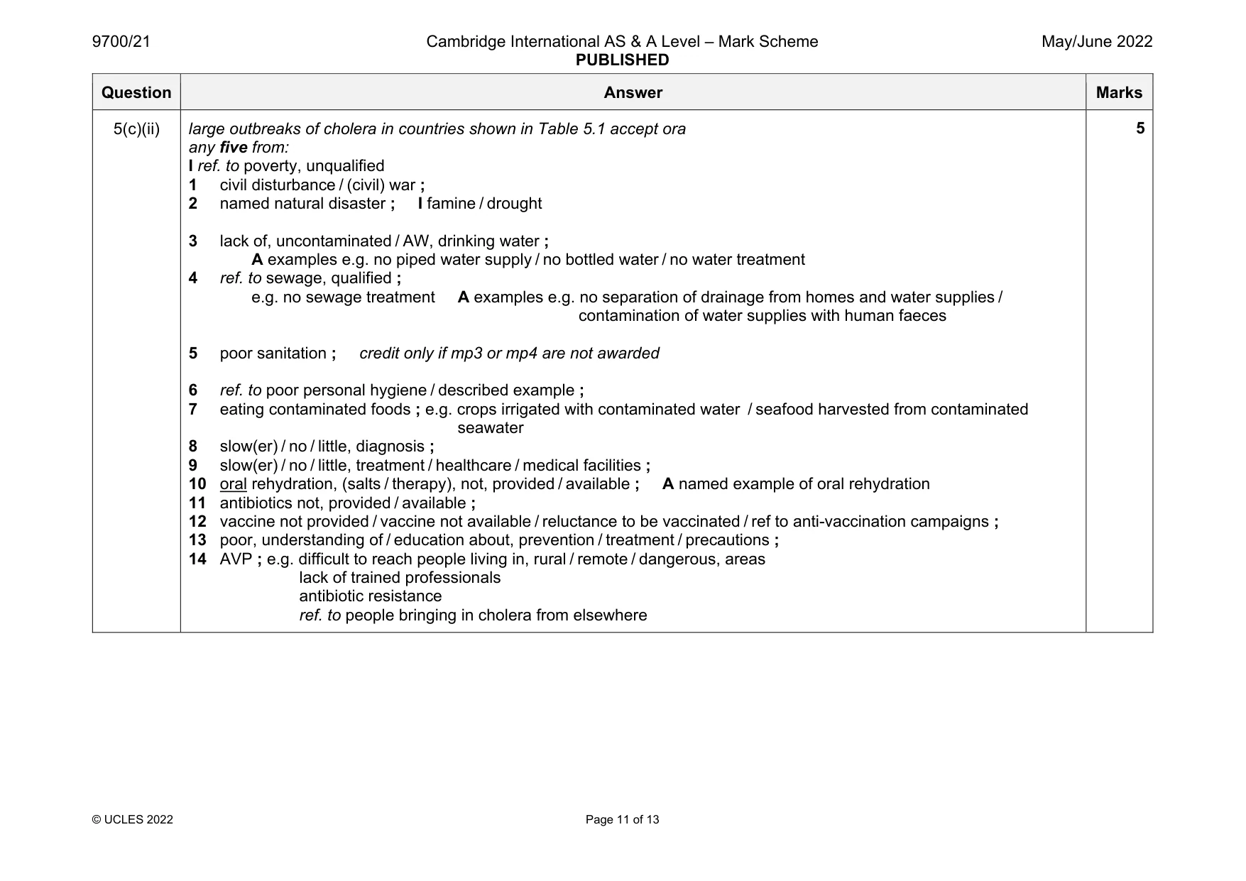
Mark scheme: 5(a) (pathogen / causative organism is) transferred / passed on / AW, (from infected) to an uninfected, person / organism ; I disease I from one person to another unqualified I spread 1 5(b) any two from information needed 1 magnification of, image / (electron) micrograph / Fig. 5.1 ; A scale bar I magnification of (electron) microscope 2 length of flagellum (in, image / Fig. 5.1) ; I width / diameter / size 3 formula actual length of flagellum = measured length of flagellum divided by magnification ; A A= I / M or magnification triangle 2 5(c)(i) 0.2 1 Question Answer Marks 5(c)(ii) large outbreaks of cholera in countries shown in Table 5.1 accept ora any five from: I ref. to poverty, unqualified 1 civil disturbance / (civil) war ; 2 named natural disaster ; I famine / drought 3 lack of, uncontaminated / AW, drinking water ; A examples e.g. no piped water supply / no bottled water / no water treatment 4 ref. to sewage, qualified ; e.g. no sewage treatment A examples e.g. no separation of drainage from homes and water supplies / contamination of water supplies with human faeces 5 poor sanitation ; credit only if mp3 or mp4 are not awarded 6 ref. to poor personal hygiene / described example ; 7 eating contaminated foods ; e.g. crops irrigated with contaminated water / seafood harvested from contaminated seawater 8 slow(er) / no / little, diagnosis ; 9 slow(er) / no / little, treatment / healthcare / medical facilities ; 10 oral rehydration, (salts / therapy), not, provided / available ; A named example of oral rehydration 11 antibiotics not, provided / available ; 12 vaccine not provided / vaccine not available / reluctance to be vaccinated / ref to anti-vaccination campaigns ; 13 poor, understanding of / education about, prevention / treatment / precautions ; 14 AVP ; e.g. difficult to reach people living in, rural / remote / dangerous, areas lack of trained professionals antibiotic resistance ref. to people bringing in cholera from elsewhere 5
Q6 · Cotransporter proteins are membrane proteins found in companion cells of phloem tissue
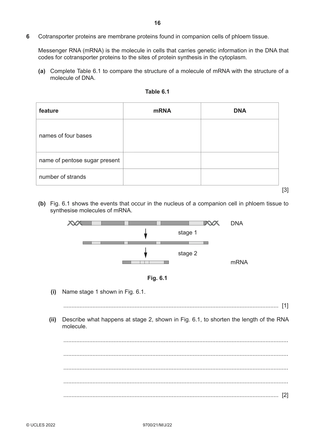
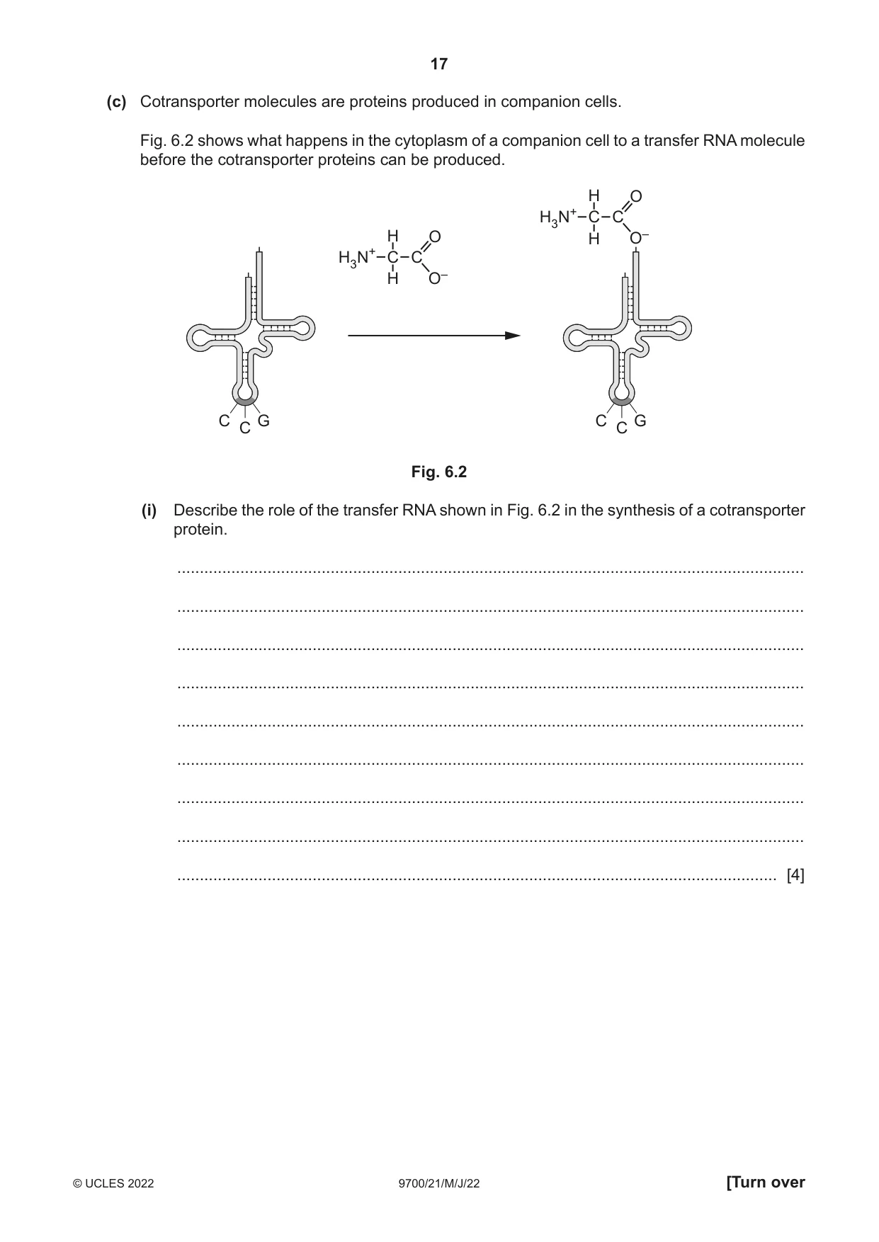
6 Cotransporter proteins are membrane proteins found in companion cells of phloem tissue. Messenger RNA (mRNA) is the molecule in cells that carries genetic information in the DNA that codes for cotransporter proteins to the sites of protein synthesis in the cytoplasm. (a) Complete Table 6.1 to compare the structure of a molecule of mRNA with the structure of a molecule of DNA. Table 6.1 feature mRNA DNA names of four bases name of pentose sugar present number of strands [3] (b) Fig. 6.1 shows the events that occur in the nucleus of a companion cell in phloem tissue to synthesise molecules of mRNA. DNA stage 1 stage 2 mRNA Fig. 6.1 (i) Name stage 1 shown in Fig. 6.1. ..................................................................................................................................... [1] (ii) Describe what happens at stage 2, shown in Fig. 6.1, to shorten the length of the RNA molecule. ........................................................................................................................................... ........................................................................................................................................... ........................................................................................................................................... ........................................................................................................................................... ..................................................................................................................................... [2] (c) Cotransporter molecules are proteins produced in companion cells. Fig. 6.2 shows what happens in the cytoplasm of a companion cell to a transfer RNA molecule before the cotransporter proteins can be produced. H O H3N+ C C H O H O– H3N+ C C H O– C G C G C C Fig. 6.2 (i) Describe the role of the transfer RNA shown in Fig. 6.2 in the synthesis of a cotransporter protein. ........................................................................................................................................... ........................................................................................................................................... ........................................................................................................................................... ........................................................................................................................................... ........................................................................................................................................... ........................................................................................................................................... ........................................................................................................................................... ........................................................................................................................................... ..................................................................................................................................... [4] (ii) Outline the role of cotransporter proteins in companion cells. ........................................................................................................................................... ........................................................................................................................................... ........................................................................................................................................... ........................................................................................................................................... ........................................................................................................................................... ........................................................................................................................................... ........................................................................................................................................... ........................................................................................................................................... ..................................................................................................................................... [3] [Total: 13]
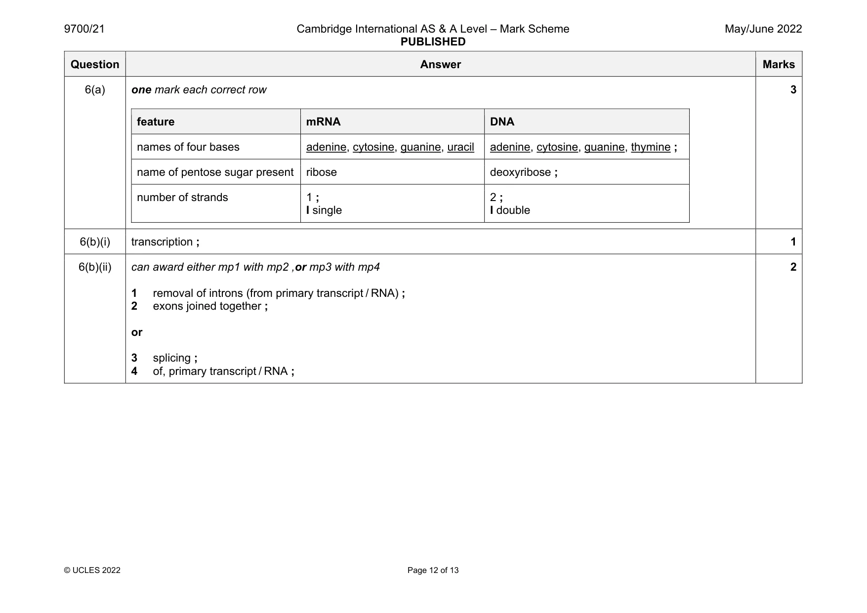
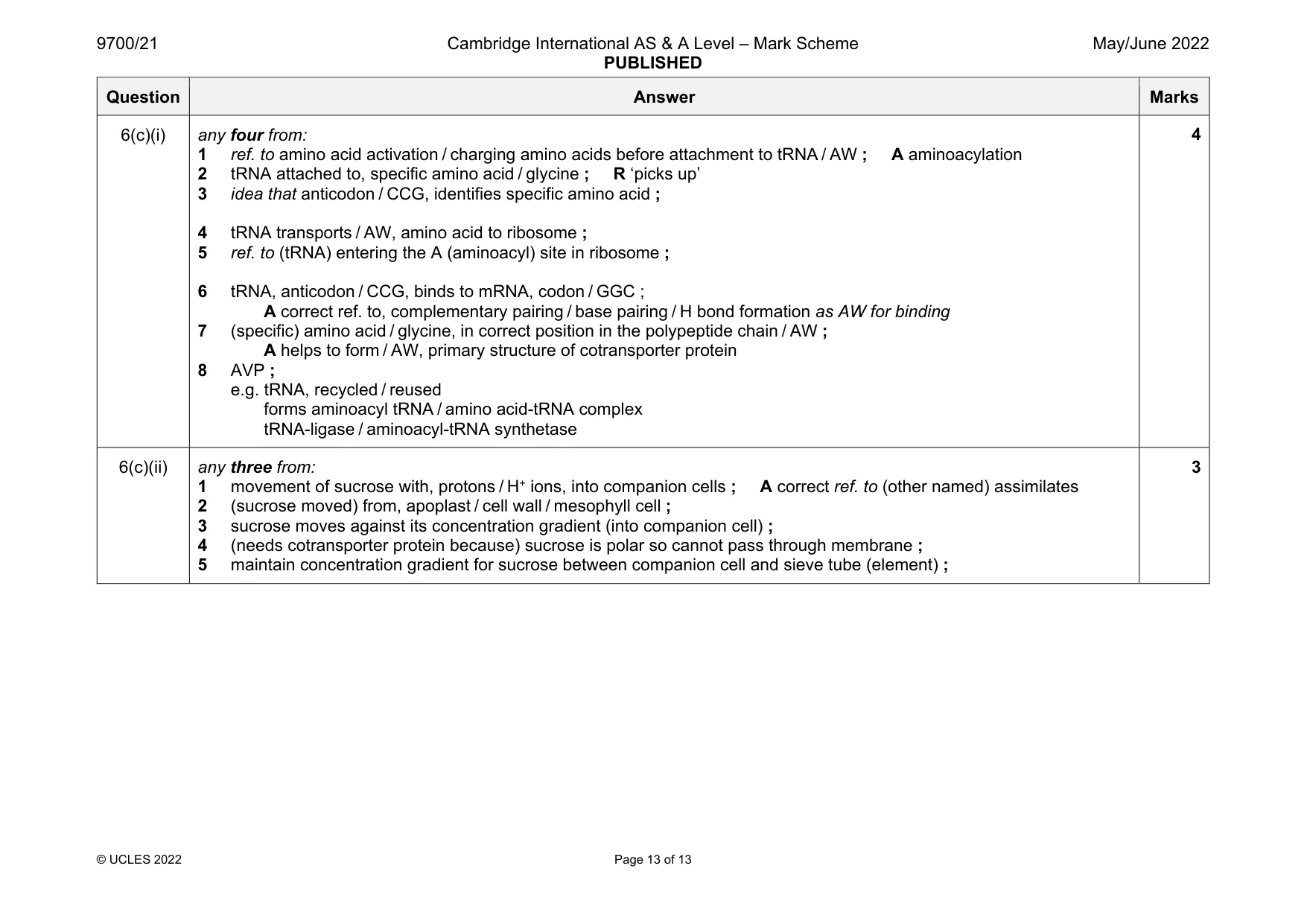
Mark scheme: 6(a) one mark each correct row feature mRNA DNA names of four bases adenine, cytosine, guanine, uracil adenine, cytosine, guanine, thymine ; name of pentose sugar present ribose deoxyribose ; number of strands 1 ; I single 2 ; I double 3 6(b)(i) transcription ; 1 6(b)(ii) can award either mp1 with mp2 ,or mp3 with mp4 1 removal of introns (from primary transcript / RNA) ; 2 exons joined together ; or 3 splicing ; 4 of, primary transcript / RNA ; 2 Question Answer Marks 6(c)(i) any four from: 1 ref. to amino acid activation / charging amino acids before attachment to tRNA / AW ; A aminoacylation 2 tRNA attached to, specific amino acid / glycine ; R ‘picks up’ 3 idea that anticodon / CCG, identifies specific amino acid ; 4 tRNA transports / AW, amino acid to ribosome ; 5 ref. to (tRNA) entering the A (aminoacyl) site in ribosome ; 6 tRNA, anticodon / CCG, binds to mRNA, codon / GGC ; A correct ref. to, complementary pairing / base pairing / H bond formation as AW for binding 7 (specific) amino acid / glycine, in correct position in the polypeptide chain / AW ; A helps to form / AW, primary structure of cotransporter protein 8 AVP ; e.g. tRNA, recycled / reused forms aminoacyl tRNA / amino acid-tRNA complex tRNA-ligase / aminoacyl-tRNA synthetase 4 6(c)(ii) any three from: 1 movement of sucrose with, protons / H+ ions, into companion cells ; A correct ref. to (other named) assimilates 2 (sucrose moved) from, apoplast / cell wall / mesophyll cell ; 3 sucrose moves against its concentration gradient (into companion cell) ; 4 (needs cotransporter protein because) sucrose is polar so cannot pass through membrane ; 5 maintain concentration gradient for sucrose between companion cell and sieve tube (element) ; 3
What was in this paper
The subtopics covered by these 6 questions, and how many questions each got. Open one in a new tab to see every Cambridge question on it.
What you needed in this session
Cambridge’s own grade thresholds for 2022 May/June, Paper 2 · Variant 1. A higher threshold means an easier paper — the bar moves with how the cohort did.