Cambridge A Level Biology 9700 — 2023 May/June Paper 2 · Variant 1
9700/21/M/J/23 · 6 questions · 60 marks · ≈68 min
The question paper and its mark scheme, free to read here and free to download. This is Cambridge’s own paper, exactly as it was sat.
Question paper20 pages


Mark scheme18 pages
Answers below. Sit the paper first if you are practising.
Questions as text
Q1 · A transmission electron micrograph of a cell from the stem of sago pondweed, Stuckenia…
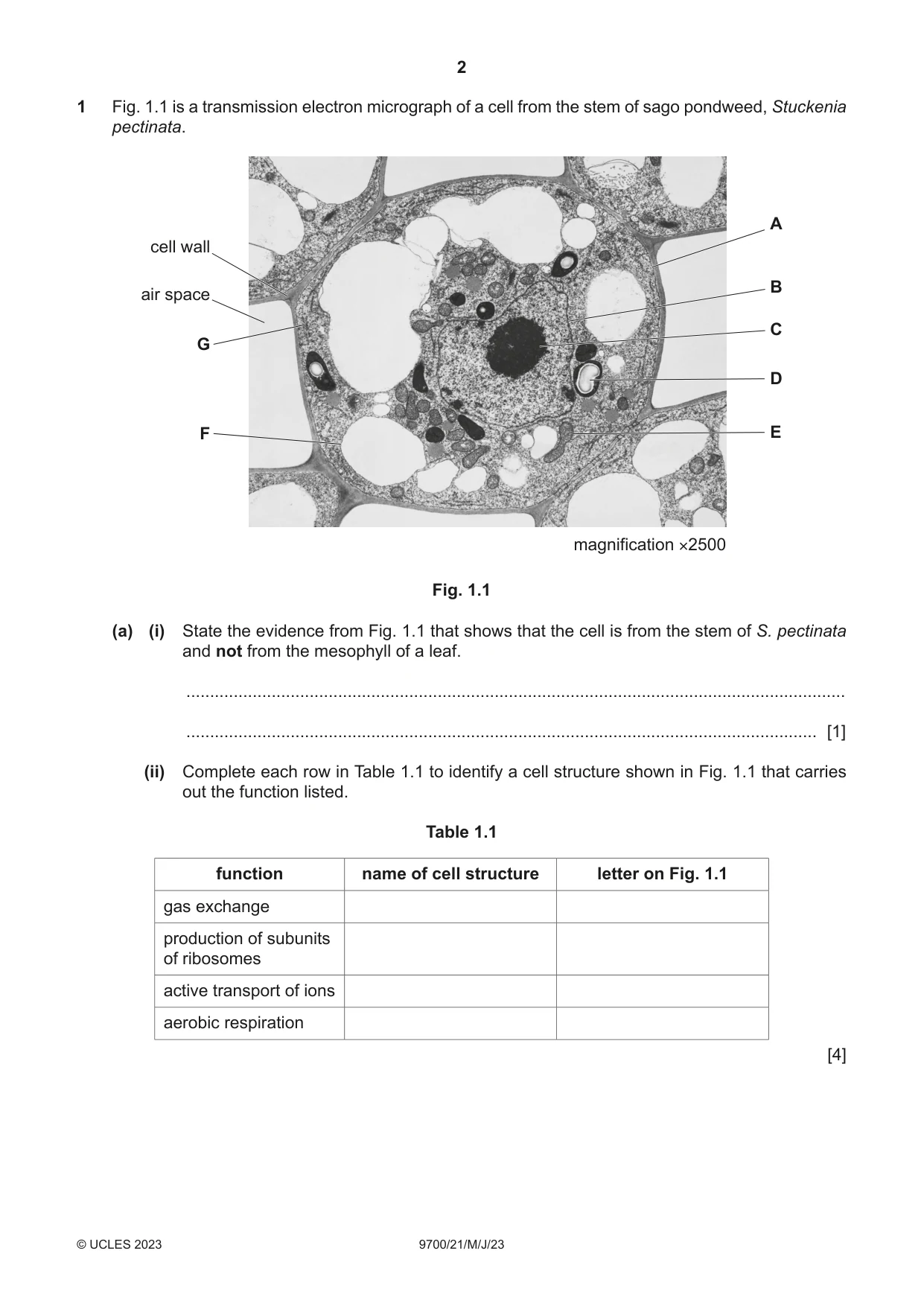
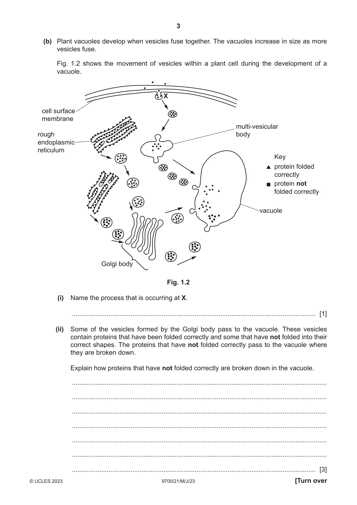
1 Fig. 1.1 is a transmission electron micrograph of a cell from the stem of sago pondweed, Stuckenia pectinata. A cell wall B air space C G D F E magnification ×2500 Fig. 1.1 (a) (i) State the evidence from Fig. 1.1 that shows that the cell is from the stem of S. pectinata and not from the mesophyll of a leaf. ........................................................................................................................................... ..................................................................................................................................... [1] (ii) Complete each row in Table 1.1 to identify a cell structure shown in Fig. 1.1 that carries out the function listed. Table 1.1 function name of cell structure letter on Fig. 1.1 gas exchange production of subunits of ribosomes active transport of ions aerobic respiration [4] (b) Plant vacuoles develop when vesicles fuse together. The vacuoles increase in size as more vesicles fuse. Fig. 1.2 shows the movement of vesicles within a plant cell during the development of a vacuole. X cell surface membrane multi-vesicular rough body endoplasmic reticulum Key protein folded correctly protein not folded correctly vacuole Golgi body Fig. 1.2 (i) Name the process that is occurring at X. ..................................................................................................................................... [1] (ii) Some of the vesicles formed by the Golgi body pass to the vacuole. These vesicles contain proteins that have been folded correctly and some that have not folded into their correct shapes. The proteins that have not folded correctly pass to the vacuole where they are broken down. Explain how proteins that have not folded correctly are broken down in the vacuole. ........................................................................................................................................... ........................................................................................................................................... ........................................................................................................................................... ........................................................................................................................................... ........................................................................................................................................... ........................................................................................................................................... ..................................................................................................................................... [3] (c) Small vacuoles in S. pectinata may have roles similar to lysosomes in animal cells. Describe the role of lysosomes in animal cells in defence against pathogens. ................................................................................................................................................... ................................................................................................................................................... ................................................................................................................................................... ................................................................................................................................................... ................................................................................................................................................... ............................................................................................................................................. [2] [Total: 11] Question 2 is on page 6.
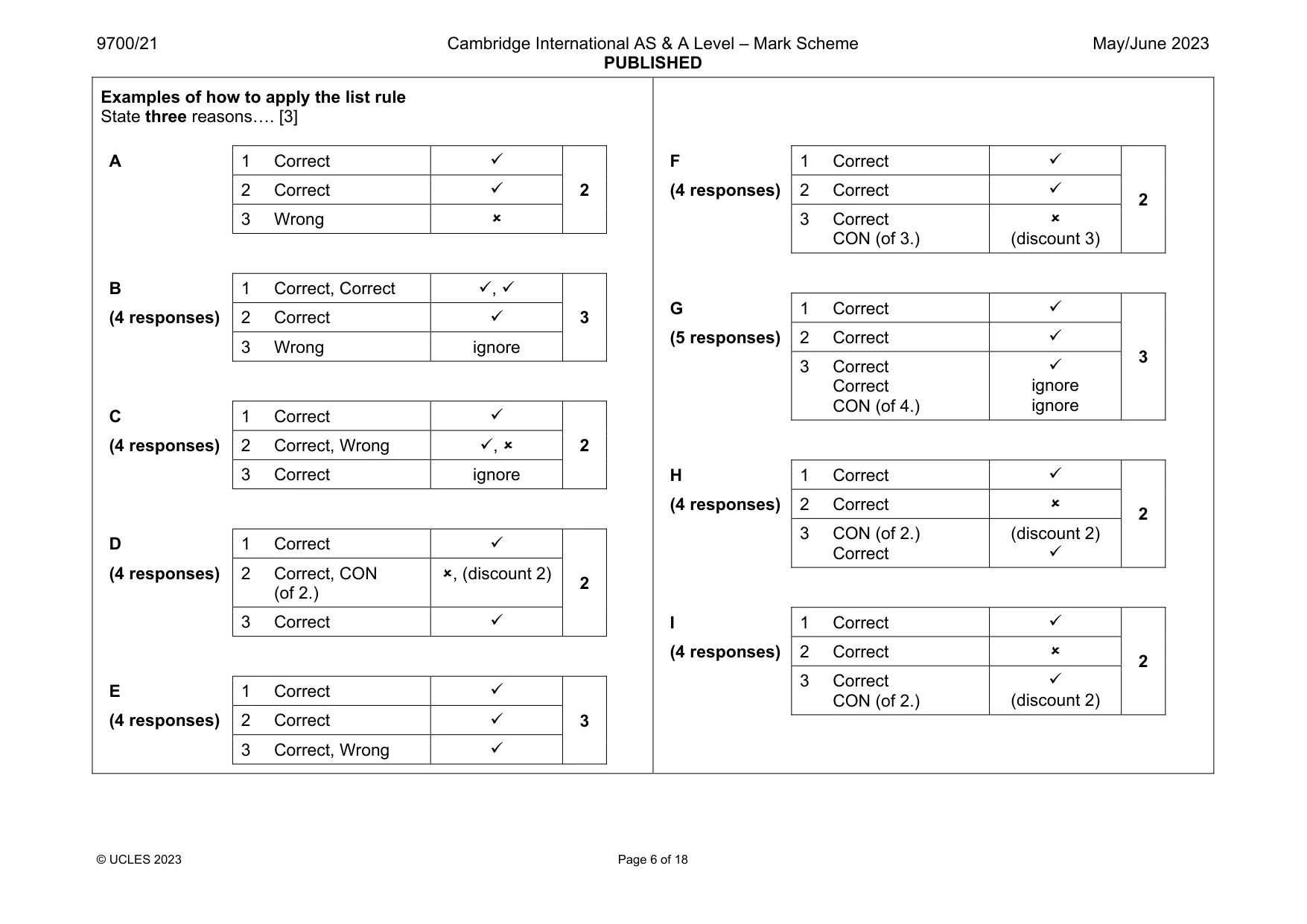
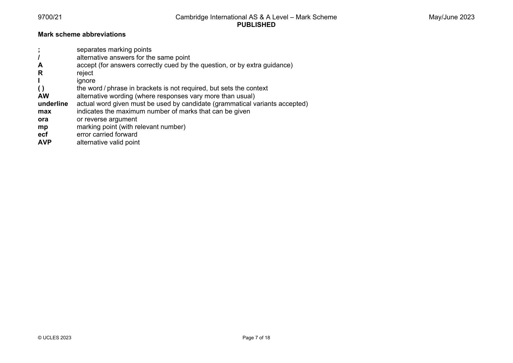
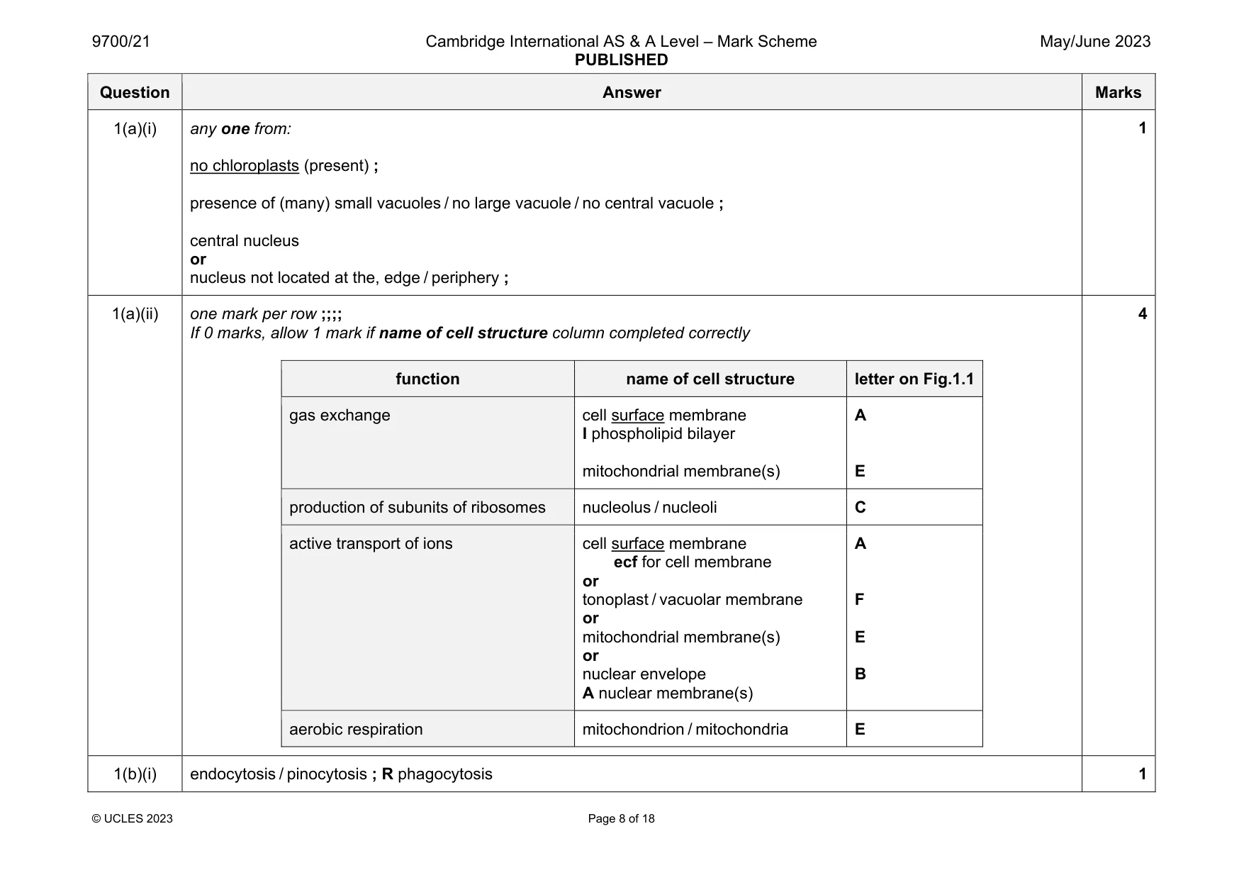
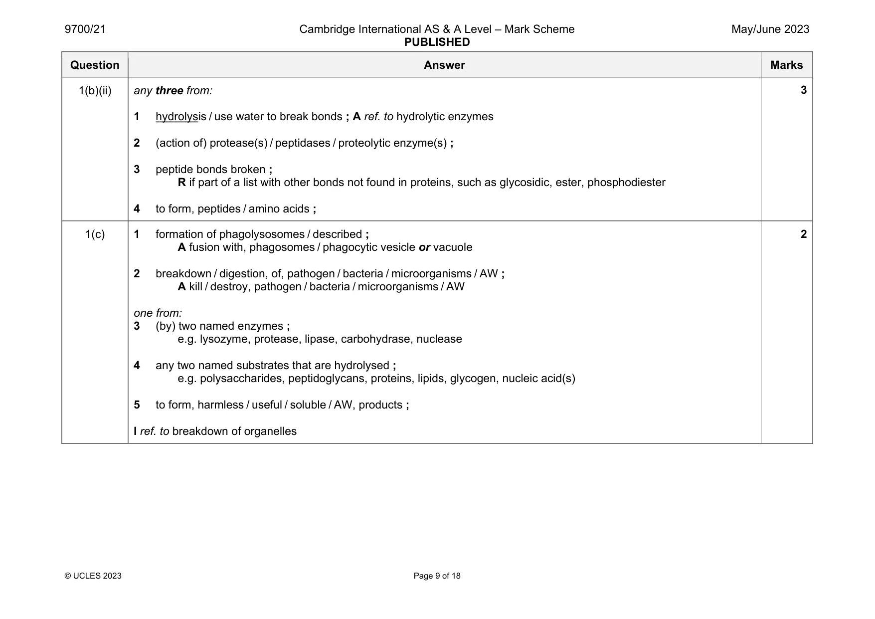
Mark scheme: 1(a)(i) any one from: no chloroplasts (present) ; presence of (many) small vacuoles / no large vacuole / no central vacuole ; central nucleus or nucleus not located at the, edge / periphery ; 1(a)(ii) one mark per row ;;;; If 0 marks, allow 1 mark if name of cell structure column completed correctly function name of cell structure letter on Fig.1.1 gas exchange cell surface membrane I phospholipid bilayer mitochondrial membrane(s) A E production of subunits of ribosomes nucleolus / nucleoli C active transport of ions cell surface membrane ecf for cell membrane or tonoplast / vacuolar membrane or mitochondrial membrane(s) or nuclear envelope A nuclear membrane(s) A F E B aerobic respiration mitochondrion / mitochondria E 4 1(b)(i) endocytosis / pinocytosis ; R phagocytosis 1 Question Answer Marks 1(b)(ii) any three from: 1 hydrolysis / use water to break bonds ; A ref. to hydrolytic enzymes 2 (action of) protease(s) / peptidases / proteolytic enzyme(s) ; 3 peptide bonds broken ; R if part of a list with other bonds not found in proteins, such as glycosidic, ester, phosphodiester 4 to form, peptides / amino acids ; 3 1(c) 1 formation of phagolysosomes / described ; A fusion with, phagosomes / phagocytic vesicle or vacuole 2 breakdown / digestion, of, pathogen / bacteria / microorganisms / AW ; A kill / destroy, pathogen / bacteria / microorganisms / AW one from: 3 (by) two named enzymes ; e.g. lysozyme, protease, lipase, carbohydrase, nuclease 4 any two named substrates that are hydrolysed ; e.g. polysaccharides, peptidoglycans, proteins, lipids, glycogen, nucleic acid(s) 5 to form, harmless / useful / soluble / AW, products ; I ref. to breakdown of organelles 2
More questions on Cells as the basic units of living organisms
Q2 · Glycogen and cellulose are polymers
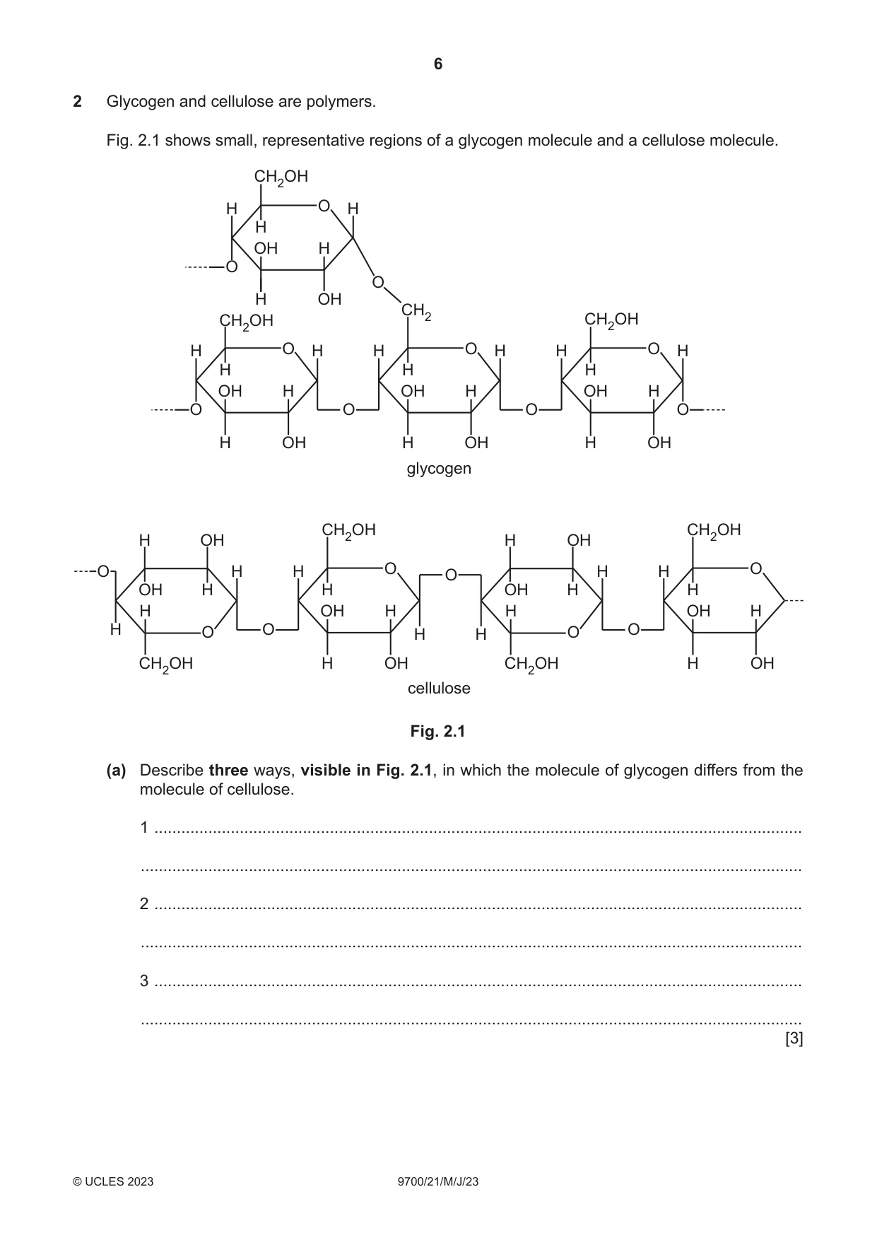
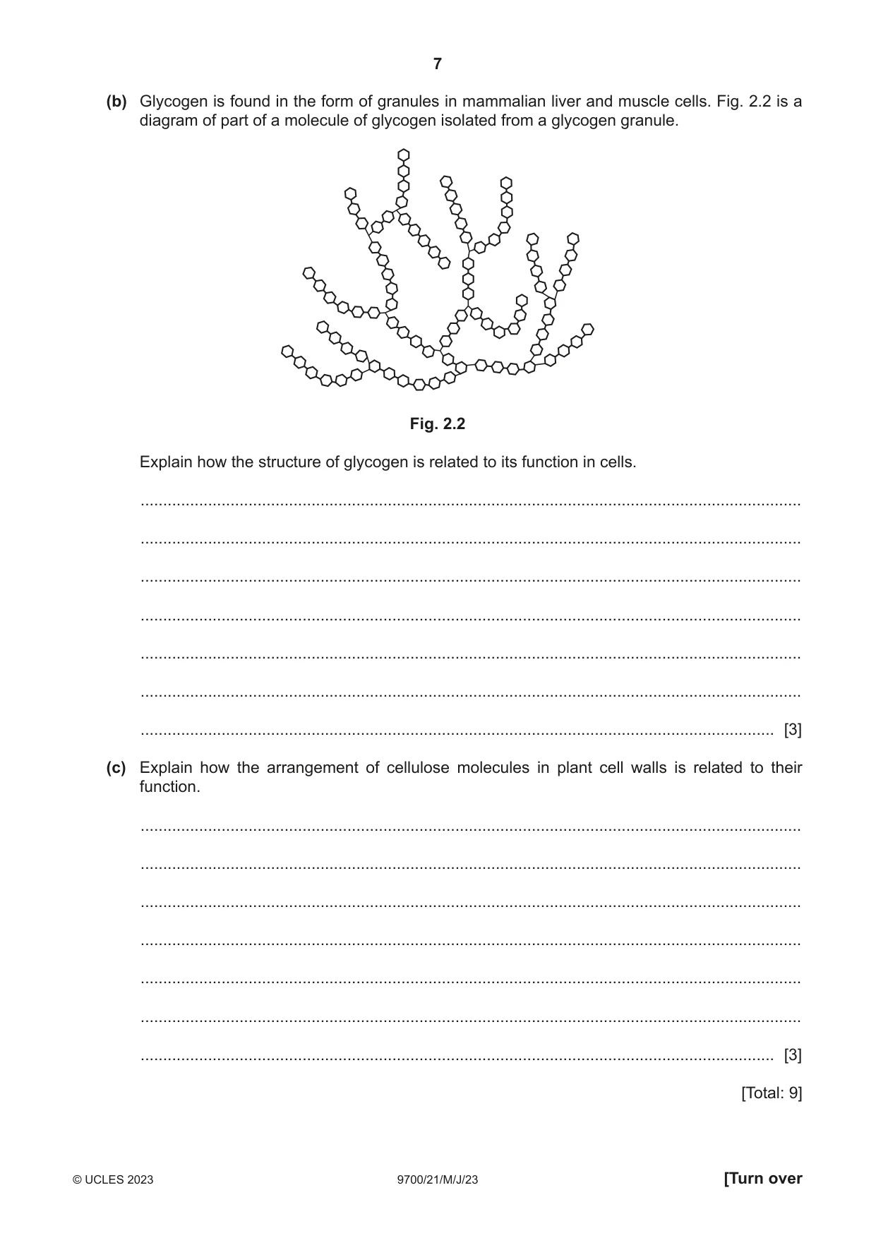
2 Glycogen and cellulose are polymers. Fig. 2.1 shows small, representative regions of a glycogen molecule and a cellulose molecule. CH2OH H O H H OH H O O H OH CH2 CH2OH CH2OH H O H H O H H O H H H H OH H OH H OH H O O O O H OH H OH H OH glycogen CH2OH CH2OH H OH H OH O H H O O H H O OH H H OH H H H OH H H OH H H O O H H O O CH2OH H OH CH2OH H OH cellulose Fig. 2.1 (a) Describe three ways, visible in Fig. 2.1, in which the molecule of glycogen differs from the molecule of cellulose. 1 ................................................................................................................................................ ................................................................................................................................................... 2 ................................................................................................................................................ ................................................................................................................................................... 3 ................................................................................................................................................ ................................................................................................................................................... [3] (b) Glycogen is found in the form of granules in mammalian liver and muscle cells. Fig. 2.2 is a diagram of part of a molecule of glycogen isolated from a glycogen granule. Fig. 2.2 Explain how the structure of glycogen is related to its function in cells. ................................................................................................................................................... ................................................................................................................................................... ................................................................................................................................................... ................................................................................................................................................... ................................................................................................................................................... ................................................................................................................................................... ............................................................................................................................................. [3] (c) Explain how the arrangement of cellulose molecules in plant cell walls is related to their function. ................................................................................................................................................... ................................................................................................................................................... ................................................................................................................................................... ................................................................................................................................................... ................................................................................................................................................... ................................................................................................................................................... ............................................................................................................................................. [3] [Total: 9]
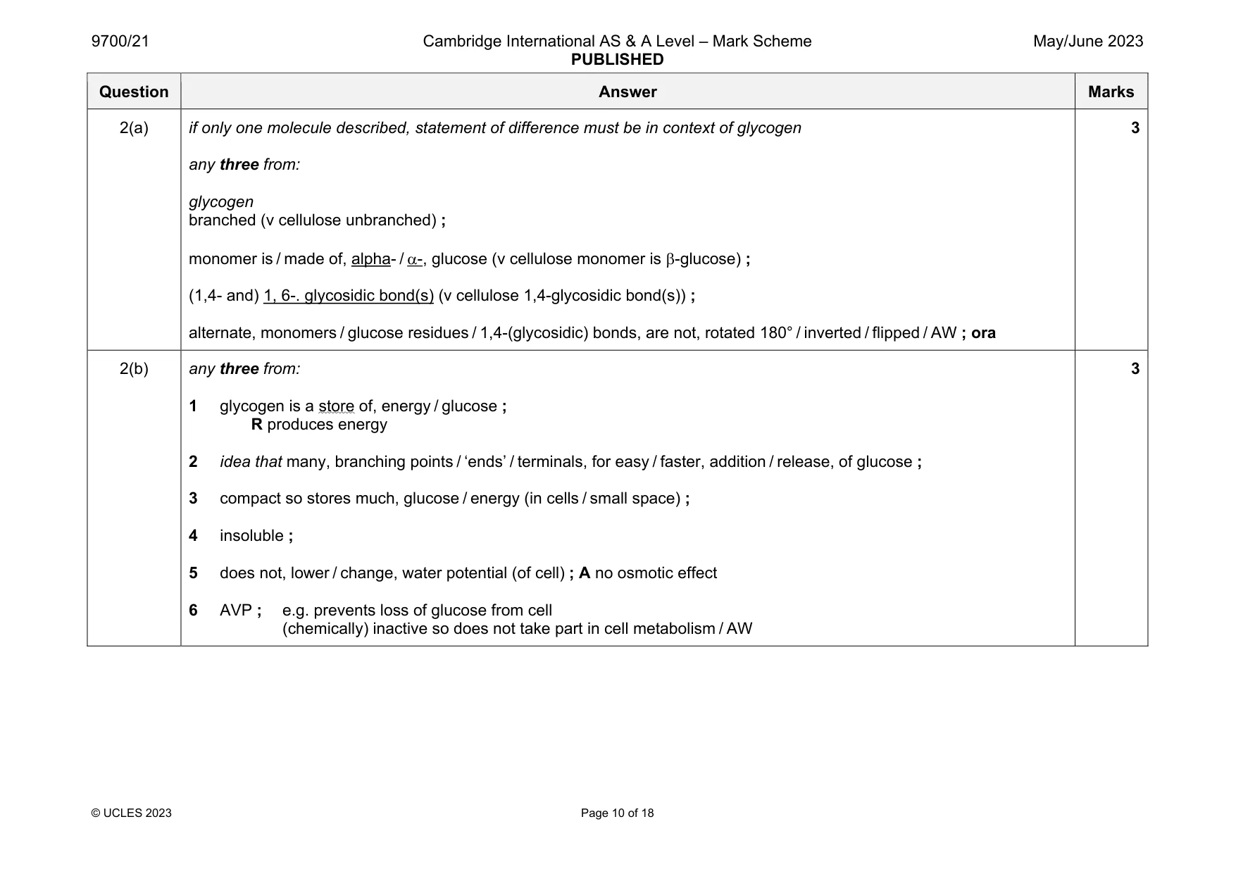
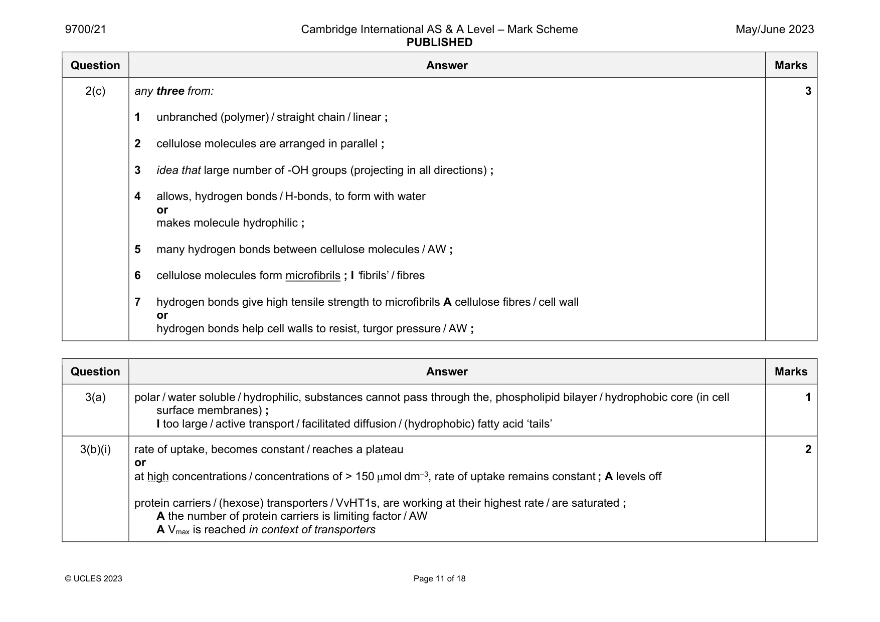
Mark scheme: 2(a) if only one molecule described, statement of difference must be in context of glycogen any three from: glycogen branched (v cellulose unbranched) ; monomer is / made of, alpha- / -, glucose (v cellulose monomer is -glucose) ; (1,4- and) 1, 6-. glycosidic bond(s) (v cellulose 1,4-glycosidic bond(s)) ; alternate, monomers / glucose residues / 1,4-(glycosidic) bonds, are not, rotated 180° / inverted / flipped / AW ; ora 3 2(b) any three from: 1 glycogen is a store of, energy / glucose ; R produces energy 2 idea that many, branching points / ‘ends’ / terminals, for easy / faster, addition / release, of glucose ; 3 compact so stores much, glucose / energy (in cells / small space) ; 4 insoluble ; 5 does not, lower / change, water potential (of cell) ; A no osmotic effect 6 AVP ; e.g. prevents loss of glucose from cell (chemically) inactive so does not take part in cell metabolism / AW 3 Question Answer Marks 2(c) any three from: 1 unbranched (polymer) / straight chain / linear ; 2 cellulose molecules are arranged in parallel ; 3 idea that large number of -OH groups (projecting in all directions) ; 4 allows, hydrogen bonds / H-bonds, to form with water or makes molecule hydrophilic ; 5 many hydrogen bonds between cellulose molecules / AW ; 6 cellulose molecules form microfibrils ; I ‘fibrils’ / fibres 7 hydrogen bonds give high tensile strength to microfibrils A cellulose fibres / cell wall or hydrogen bonds help cell walls to resist, turgor pressure / AW ; 3
Q3 · Some fruits (grapes) of the grapevine, Vitis vinifera
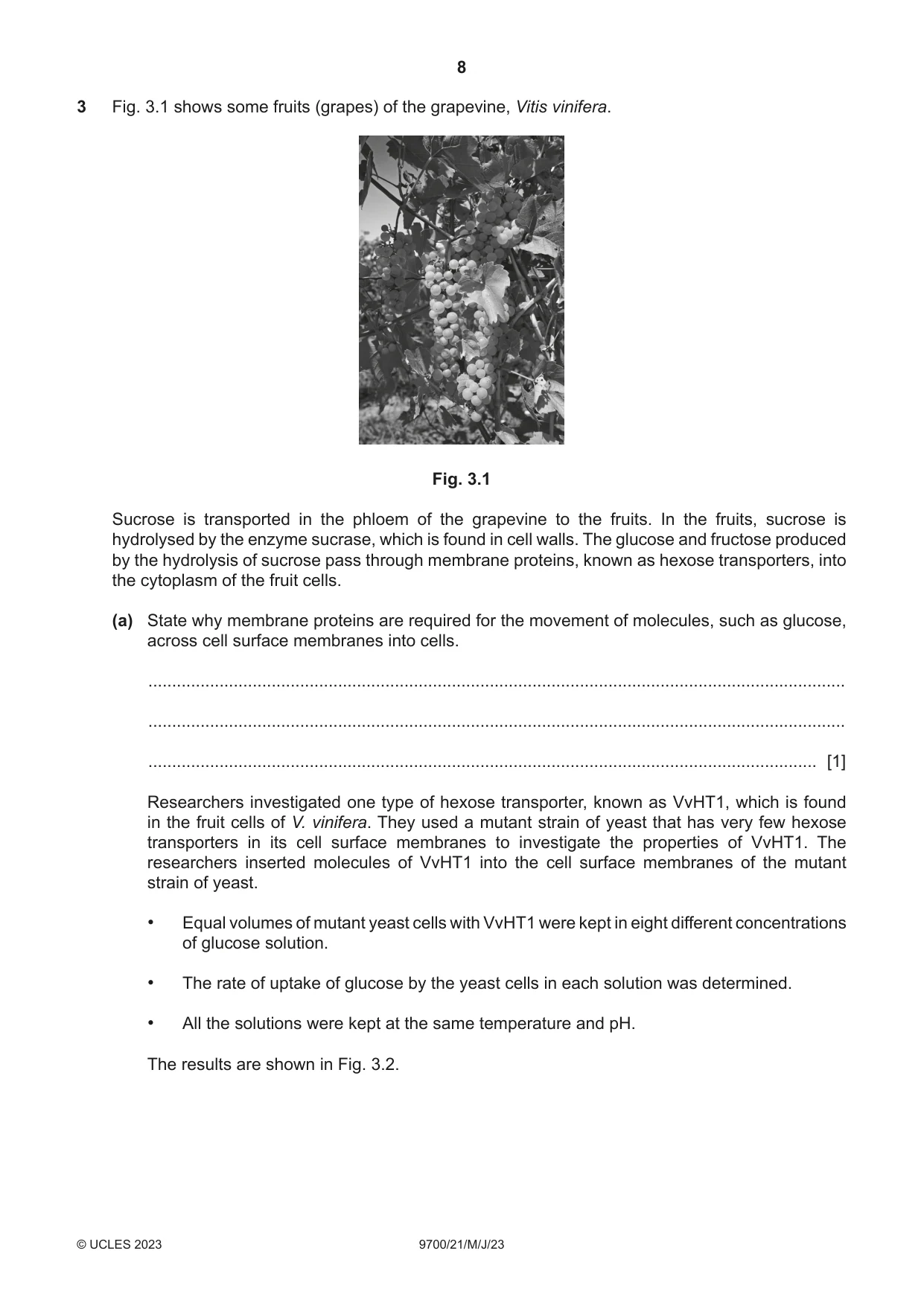
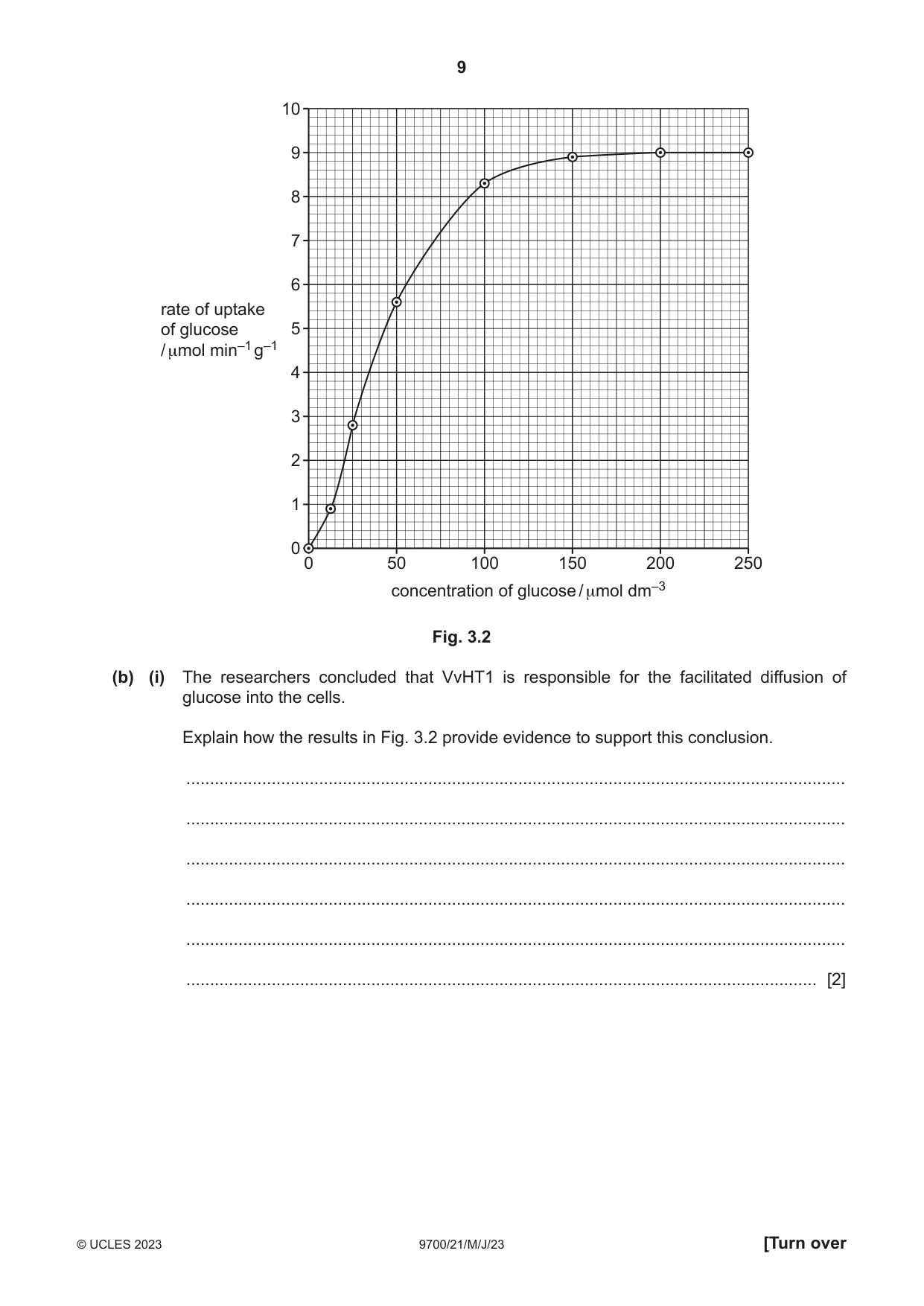
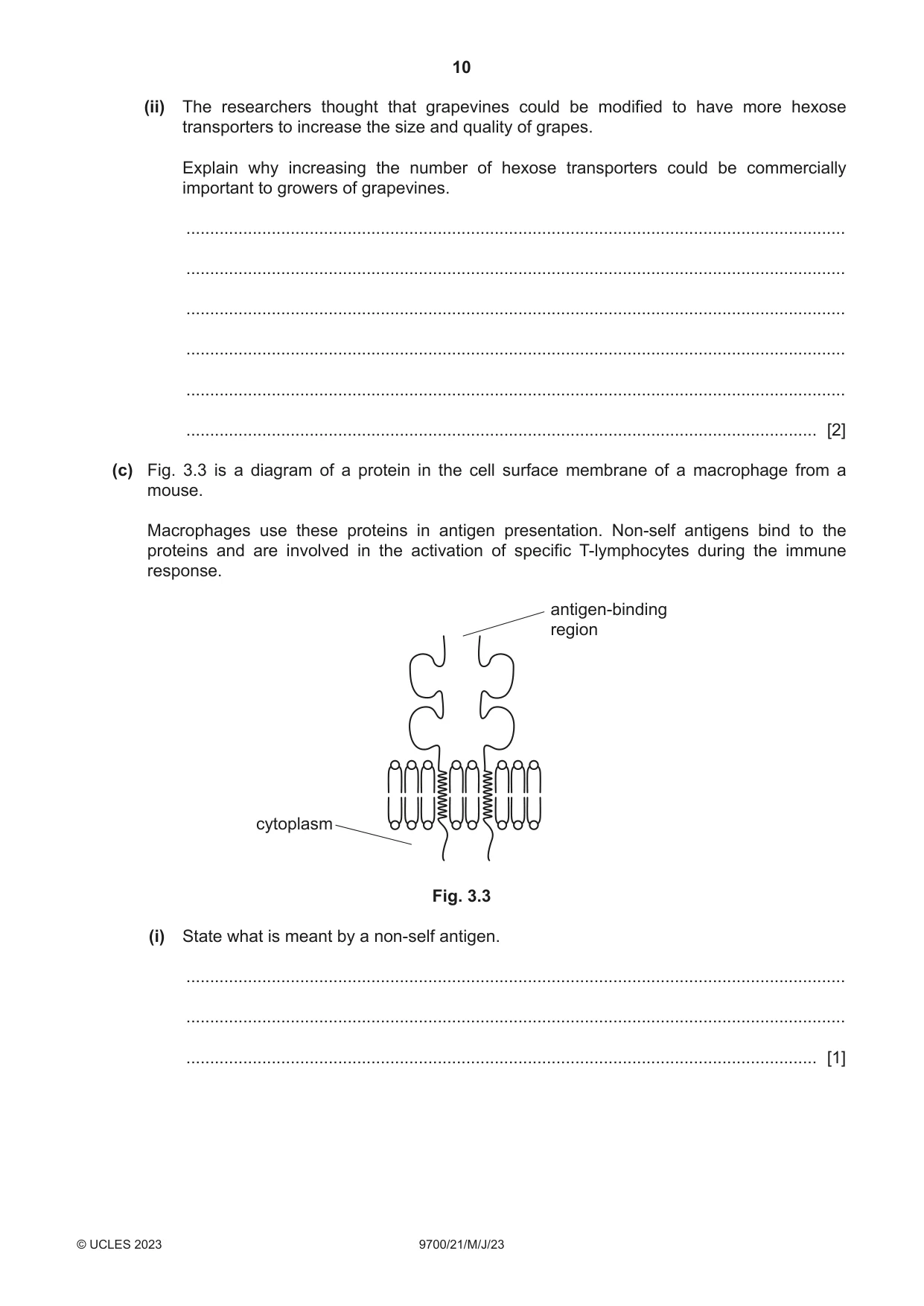
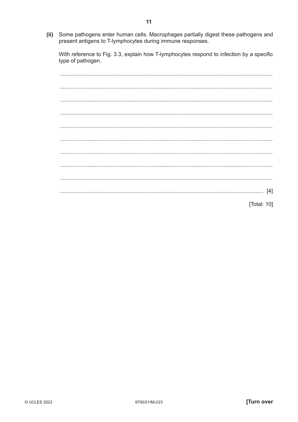
3 Fig. 3.1 shows some fruits (grapes) of the grapevine, Vitis vinifera. Fig. 3.1 Sucrose is transported in the phloem of the grapevine to the fruits. In the fruits, sucrose is hydrolysed by the enzyme sucrase, which is found in cell walls. The glucose and fructose produced by the hydrolysis of sucrose pass through membrane proteins, known as hexose transporters, into the cytoplasm of the fruit cells. (a) State why membrane proteins are required for the movement of molecules, such as glucose, across cell surface membranes into cells. ................................................................................................................................................... ................................................................................................................................................... ............................................................................................................................................. [1] Researchers investigated one type of hexose transporter, known as VvHT1, which is found in the fruit cells of V. vinifera. They used a mutant strain of yeast that has very few hexose transporters in its cell surface membranes to investigate the properties of VvHT1. The researchers inserted molecules of VvHT1 into the cell surface membranes of the mutant strain of yeast. • Equal volumes of mutant yeast cells with VvHT1 were kept in eight different concentrations of glucose solution. • The rate of uptake of glucose by the yeast cells in each solution was determined. • All the solutions were kept at the same temperature and pH. The results are shown in Fig. 3.2. 10 9 8 7 6 rate of uptake of glucose 5 / μmol min–1 g–1 4 3 2 1 0 0 50 100 150 200 250 concentration of glucose / μmol dm–3 Fig. 3.2 (b) (i) The researchers concluded that VvHT1 is responsible for the facilitated diffusion of glucose into the cells. Explain how the results in Fig. 3.2 provide evidence to support this conclusion. ........................................................................................................................................... ........................................................................................................................................... ........................................................................................................................................... ........................................................................................................................................... ........................................................................................................................................... ..................................................................................................................................... [2] (ii) The researchers thought that grapevines could be modified to have more hexose transporters to increase the size and quality of grapes. Explain why increasing the number of hexose transporters could be commercially important to growers of grapevines. ........................................................................................................................................... ........................................................................................................................................... ........................................................................................................................................... ........................................................................................................................................... ........................................................................................................................................... ..................................................................................................................................... [2] (c) Fig. 3.3 is a diagram of a protein in the cell surface membrane of a macrophage from a mouse. Macrophages use these proteins in antigen presentation. Non-self antigens bind to the proteins and are involved in the activation of specific T-lymphocytes during the immune response. antigen-binding region cytoplasm Fig. 3.3 (i) State what is meant by a non-self antigen. ........................................................................................................................................... ........................................................................................................................................... ..................................................................................................................................... [1] (ii) Some pathogens enter human cells. Macrophages partially digest these pathogens and present antigens to T-lymphocytes during immune responses. With reference to Fig. 3.3, explain how T-lymphocytes respond to infection by a specific type of pathogen. ........................................................................................................................................... ........................................................................................................................................... ........................................................................................................................................... ........................................................................................................................................... ........................................................................................................................................... ........................................................................................................................................... ........................................................................................................................................... ........................................................................................................................................... ........................................................................................................................................... ..................................................................................................................................... [4] [Total: 10]
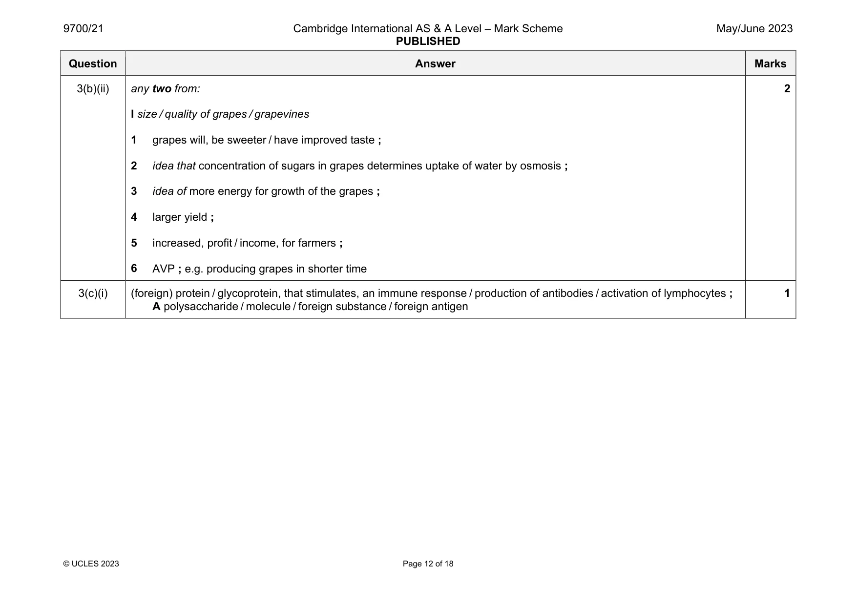
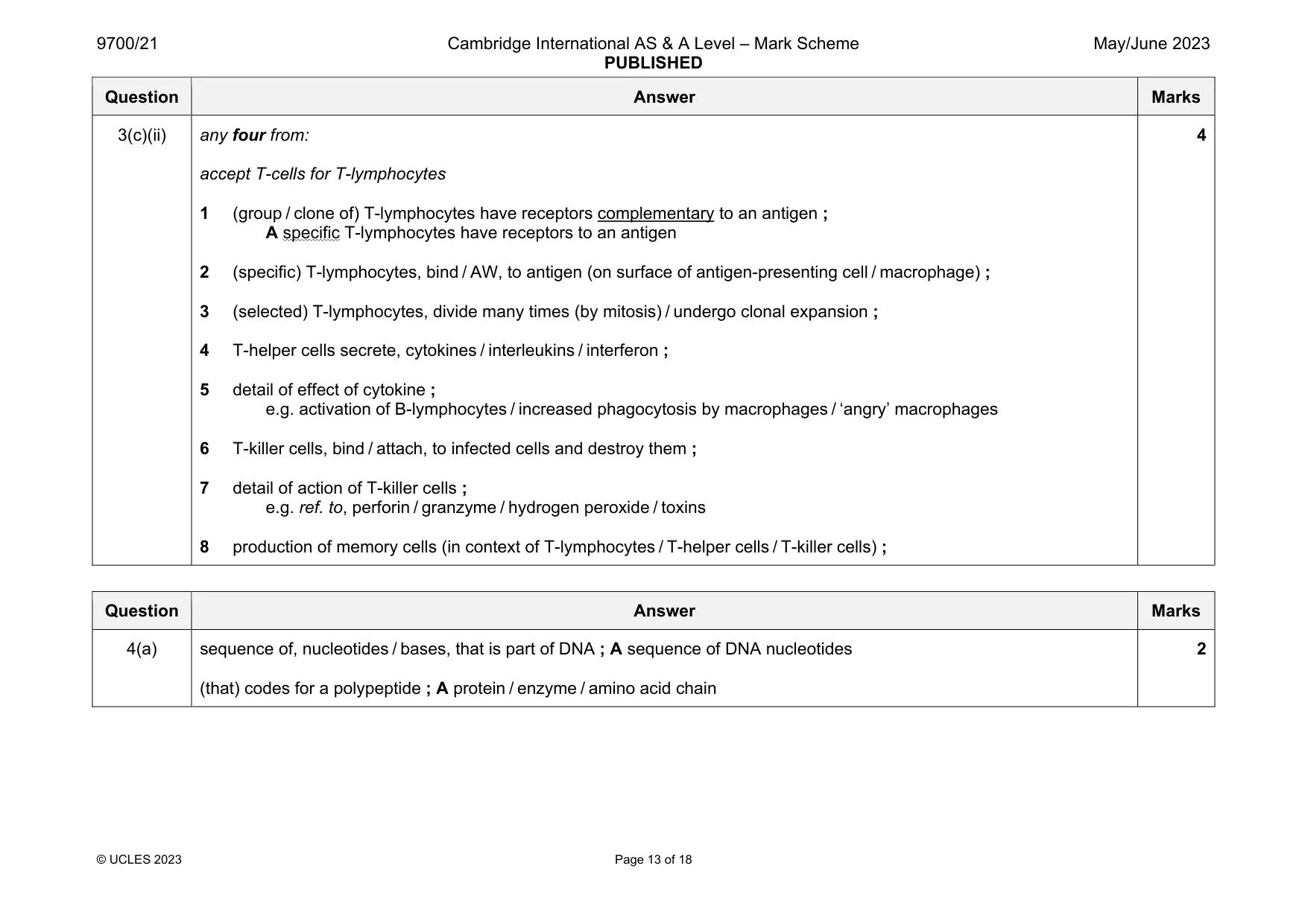
Mark scheme: 3(a) polar / water soluble / hydrophilic, substances cannot pass through the, phospholipid bilayer / hydrophobic core (in cell surface membranes) ; I too large / active transport / facilitated diffusion / (hydrophobic) fatty acid ‘tails’ 1 3(b)(i) rate of uptake, becomes constant / reaches a plateau or at high concentrations / concentrations of > 150 mol dm–3, rate of uptake remains constant ; A levels off protein carriers / (hexose) transporters / VvHT1s, are working at their highest rate / are saturated ; A the number of protein carriers is limiting factor / AW A Vmax is reached in context of transporters 2 Question Answer Marks 3(b)(ii) any two from: I size / quality of grapes / grapevines 1 grapes will, be sweeter / have improved taste ; 2 idea that concentration of sugars in grapes determines uptake of water by osmosis ; 3 idea of more energy for growth of the grapes ; 4 larger yield ; 5 increased, profit / income, for farmers ; 6 AVP ; e.g. producing grapes in shorter time 2 3(c)(i) (foreign) protein / glycoprotein, that stimulates, an immune response / production of antibodies / activation of lymphocytes ; A polysaccharide / molecule / foreign substance / foreign antigen 1 Question Answer Marks 3(c)(ii) any four from: accept T-cells for T-lymphocytes 1 (group / clone of) T-lymphocytes have receptors complementary to an antigen ; A specific T-lymphocytes have receptors to an antigen 2 (specific) T-lymphocytes, bind / AW, to antigen (on surface of antigen-presenting cell / macrophage) ; 3 (selected) T-lymphocytes, divide many times (by mitosis) / undergo clonal expansion ; 4 T-helper cells secrete, cytokines / interleukins / interferon ; 5 detail of effect of cytokine ; e.g. activation of B-lymphocytes / increased phagocytosis by macrophages / ‘angry’ macrophages 6 T-killer cells, bind / attach, to infected cells and destroy them ; 7 detail of action of T-killer cells ; e.g. ref. to, perforin / granzyme / hydrogen peroxide / toxins 8 production of memory cells (in context of T-lymphocytes / T-helper cells / T-killer cells) ; 4
Q4 · The gene for the enzyme catalase is on chromosome 11 in humans
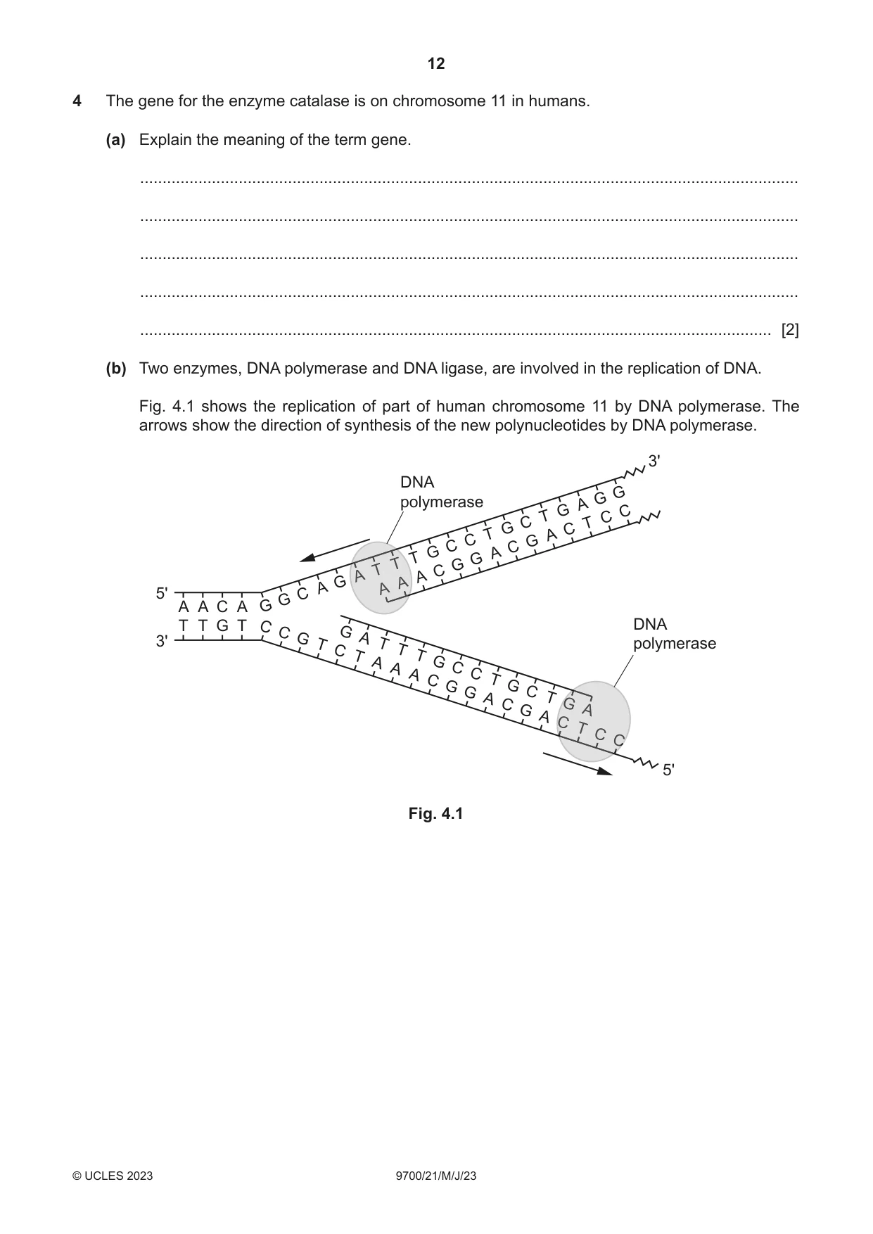
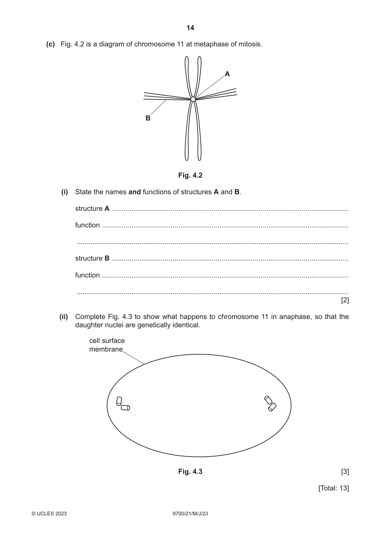
4 The gene for the enzyme catalase is on chromosome 11 in humans. (a) Explain the meaning of the term gene. ................................................................................................................................................... ................................................................................................................................................... ................................................................................................................................................... ................................................................................................................................................... ............................................................................................................................................. [2] (b) Two enzymes, DNA polymerase and DNA ligase, are involved in the replication of DNA. Fig. 4.1 shows the replication of part of human chromosome 11 by DNA polymerase. The arrows show the direction of synthesis of the new polynucleotides by DNA polymerase. 3' DNA G G polymerase A G C T C C T G C T A C G C C G A T G TT G TT C AA A G AA A AA 5' C G A A C A G T T G T C DNA C G A 3' G T polymerase T C T T T A G A C A C C T G G G C A T C GGG G AA A CCC TT CC CCC 5' Fig. 4.1 (i) Describe the roles of DNA polymerase and DNA ligase in the replication of DNA. ........................................................................................................................................... ........................................................................................................................................... ........................................................................................................................................... ........................................................................................................................................... ........................................................................................................................................... ........................................................................................................................................... ........................................................................................................................................... ........................................................................................................................................... ........................................................................................................................................... ........................................................................................................................................... ..................................................................................................................................... [5] (ii) State the name of the stage of interphase in the cell cycle when DNA replication occurs. ..................................................................................................................................... [1] (c) Fig. 4.2 is a diagram of chromosome 11 at metaphase of mitosis. A B Fig. 4.2 (i) State the names and functions of structures A and B. structure A ......................................................................................................................... function .............................................................................................................................. ........................................................................................................................................... structure B ......................................................................................................................... function .............................................................................................................................. ........................................................................................................................................... [2] (ii) Complete Fig. 4.3 to show what happens to chromosome 11 in anaphase, so that the daughter nuclei are genetically identical. cell surface membrane Fig. 4.3 [3] [Total: 13] Question 5 is on page 16.
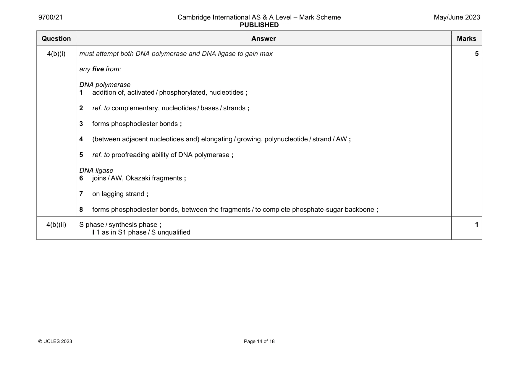
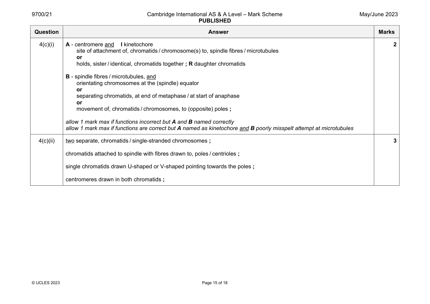
Mark scheme: 4(a) sequence of, nucleotides / bases, that is part of DNA ; A sequence of DNA nucleotides (that) codes for a polypeptide ; A protein / enzyme / amino acid chain 2 Question Answer Marks 4(b)(i) must attempt both DNA polymerase and DNA ligase to gain max any five from: DNA polymerase 1 addition of, activated / phosphorylated, nucleotides ; 2 ref. to complementary, nucleotides / bases / strands ; 3 forms phosphodiester bonds ; 4 (between adjacent nucleotides and) elongating / growing, polynucleotide / strand / AW ; 5 ref. to proofreading ability of DNA polymerase ; DNA ligase 6 joins / AW, Okazaki fragments ; 7 on lagging strand ; 8 forms phosphodiester bonds, between the fragments / to complete phosphate-sugar backbone ; 5 4(b)(ii) S phase / synthesis phase ; I 1 as in S1 phase / S unqualified 1 Question Answer Marks 4(c)(i) A - centromere and I kinetochore site of attachment of, chromatids / chromosome(s) to, spindle fibres / microtubules or holds, sister / identical, chromatids together ; R daughter chromatids B - spindle fibres / microtubules, and orientating chromosomes at the (spindle) equator or separating chromatids, at end of metaphase / at start of anaphase or movement of, chromatids / chromosomes, to (opposite) poles ; allow 1 mark max if functions incorrect but A and B named correctly allow 1 mark max if functions are correct but A named as kinetochore and B poorly misspelt attempt at microtubules 2 4(c)(ii) two separate, chromatids / single-stranded chromosomes ; chromatids attached to spindle with fibres drawn to, poles / centrioles ; single chromatids drawn U-shaped or V-shaped pointing towards the poles ; centromeres drawn in both chromatids ; 3
More questions on Structure of nucleic acids and replication of DNA
Q5 · A longitudinal section of a capillary in muscle tissue as viewed with a transmission…
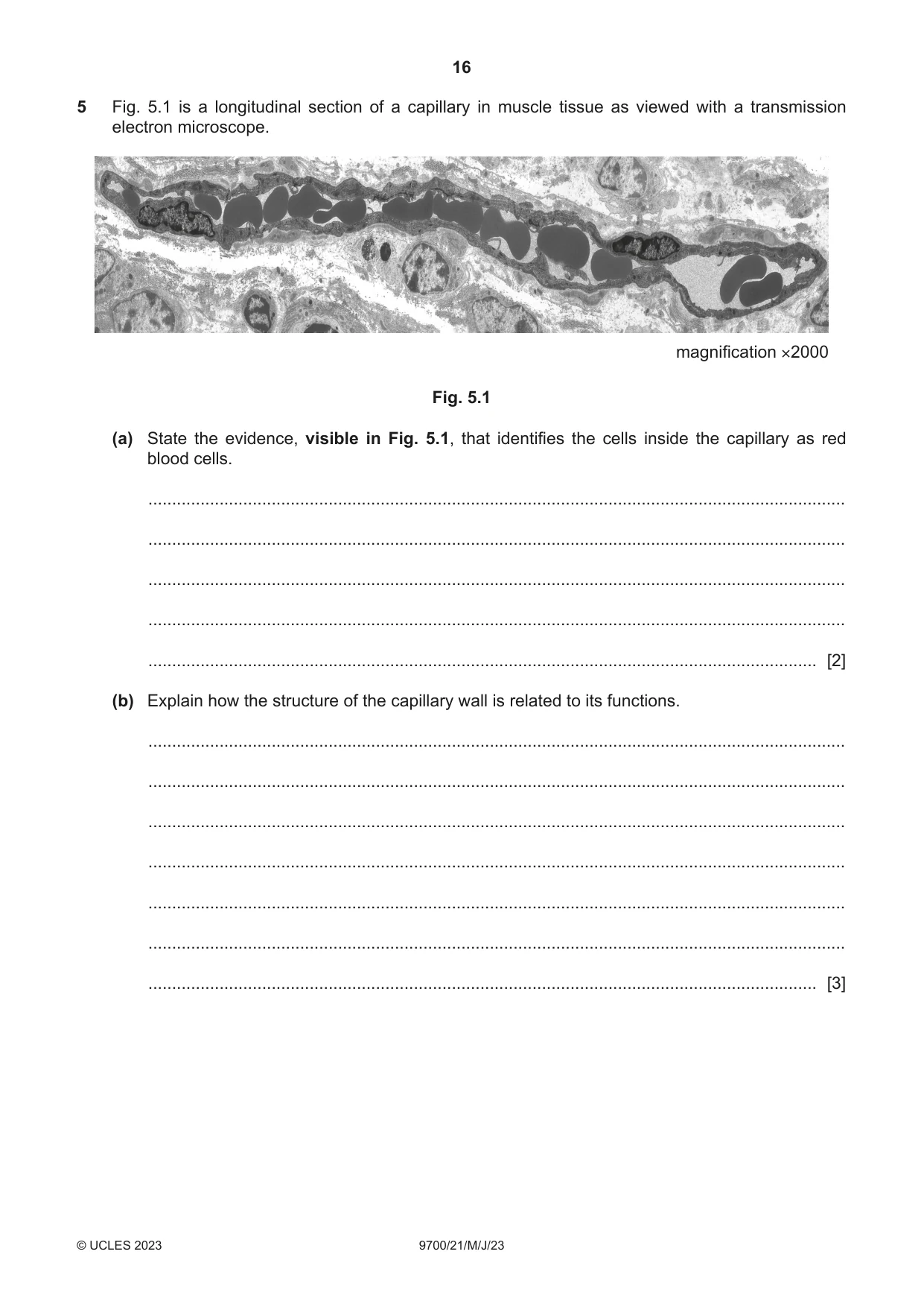
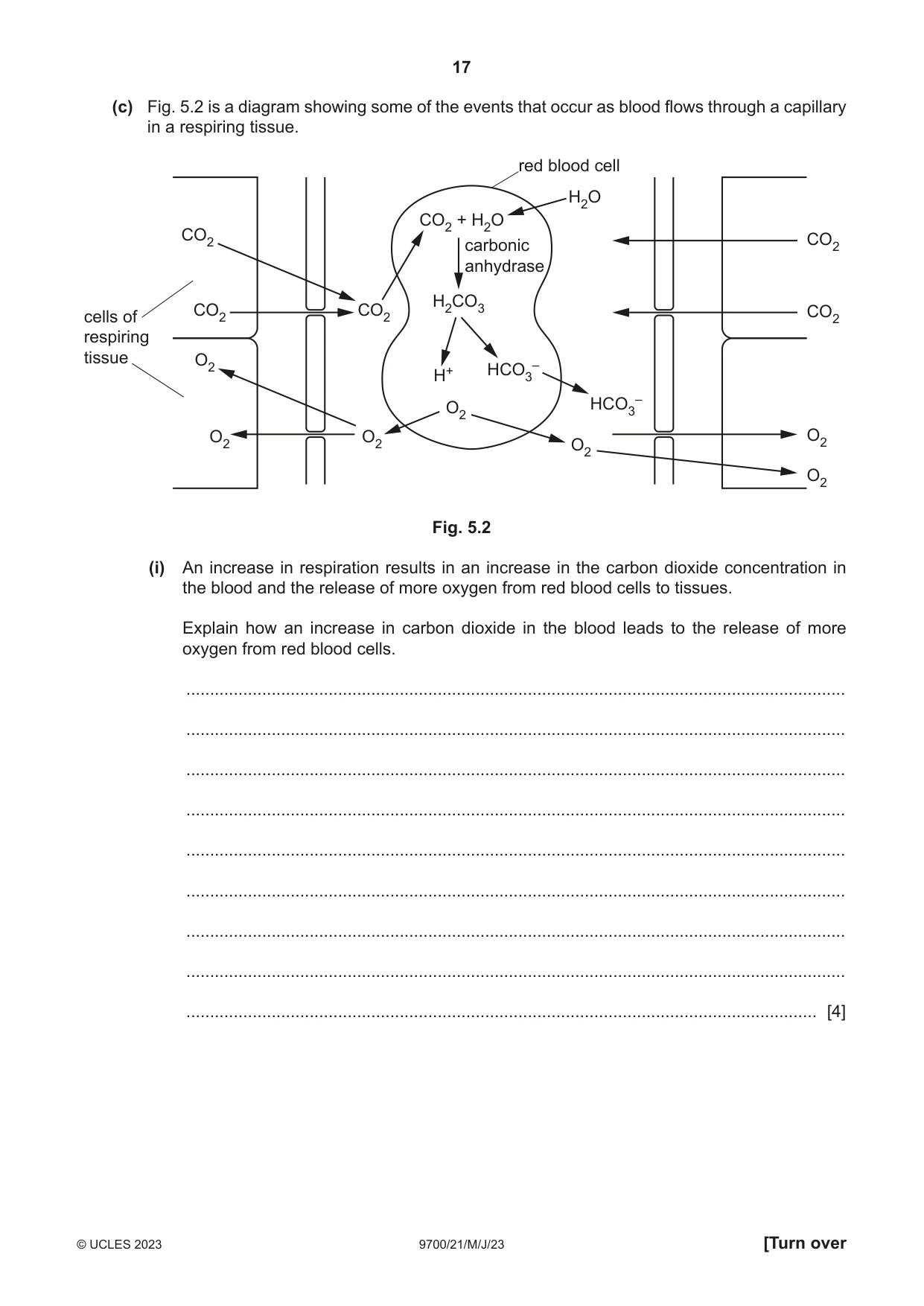
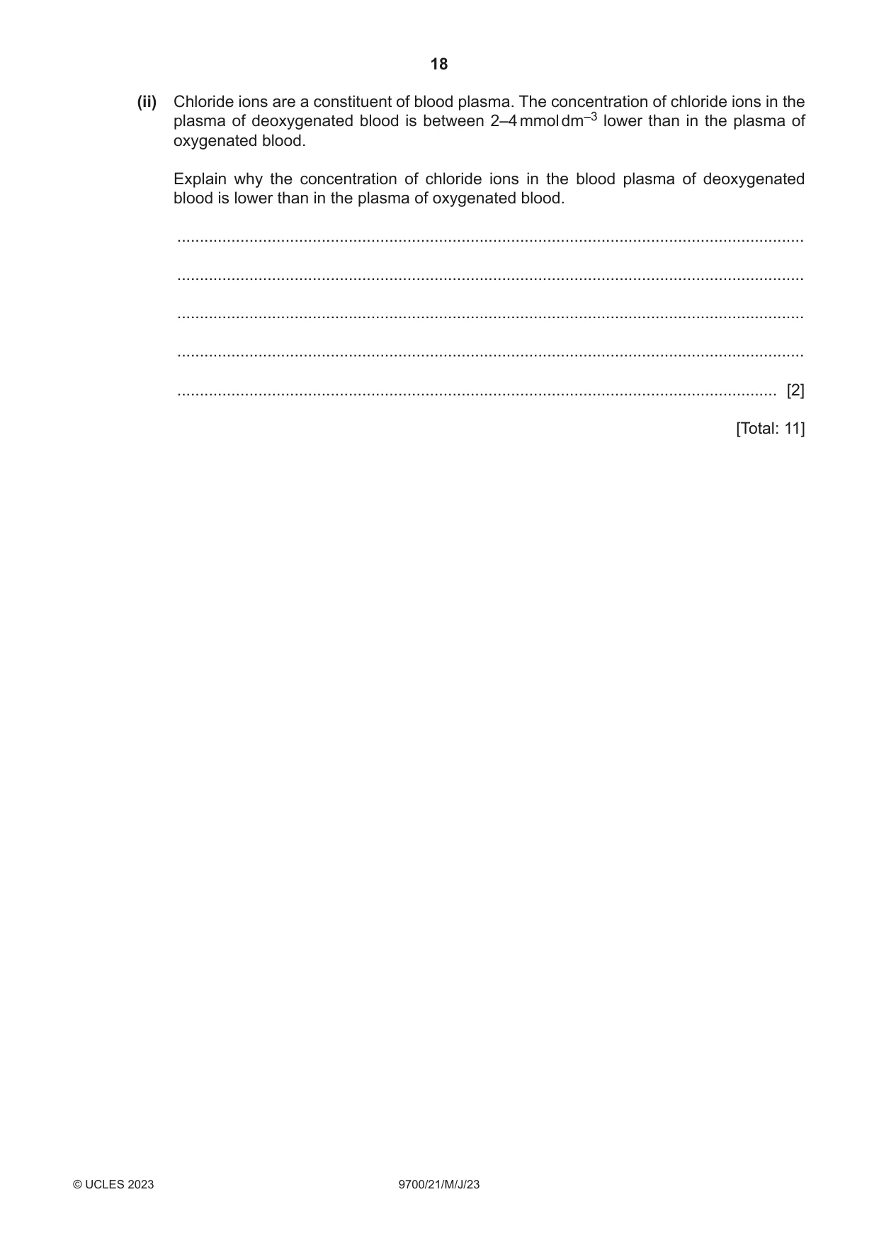
5 Fig. 5.1 is a longitudinal section of a capillary in muscle tissue as viewed with a transmission electron microscope. magnification ×2000 Fig. 5.1 (a) State the evidence, visible in Fig. 5.1, that identifies the cells inside the capillary as red blood cells. ................................................................................................................................................... ................................................................................................................................................... ................................................................................................................................................... ................................................................................................................................................... ............................................................................................................................................. [2] (b) Explain how the structure of the capillary wall is related to its functions. ................................................................................................................................................... ................................................................................................................................................... ................................................................................................................................................... ................................................................................................................................................... ................................................................................................................................................... ................................................................................................................................................... ............................................................................................................................................. [3] (c) Fig. 5.2 is a diagram showing some of the events that occur as blood flows through a capillary in a respiring tissue. red blood cell H2O CO2 + H2O CO2 carbonic CO2 anhydrase H2CO3cells of CO2 CO2 CO2 respiring tissue O2 – H+ HCO3 O2 HCO3 – O2 O2 O2 O2 O2 Fig. 5.2 (i) An increase in respiration results in an increase in the carbon dioxide concentration in the blood and the release of more oxygen from red blood cells to tissues. Explain how an increase in carbon dioxide in the blood leads to the release of more oxygen from red blood cells. ........................................................................................................................................... ........................................................................................................................................... ........................................................................................................................................... ........................................................................................................................................... ........................................................................................................................................... ........................................................................................................................................... ........................................................................................................................................... ........................................................................................................................................... ..................................................................................................................................... [4] (ii) Chloride ions are a constituent of blood plasma. The concentration of chloride ions in the plasma of deoxygenated blood is between 2–4 mmol dm–3 lower than in the plasma of oxygenated blood. Explain why the concentration of chloride ions in the blood plasma of deoxygenated blood is lower than in the plasma of oxygenated blood. ........................................................................................................................................... ........................................................................................................................................... ........................................................................................................................................... ........................................................................................................................................... ..................................................................................................................................... [2] [Total: 11]
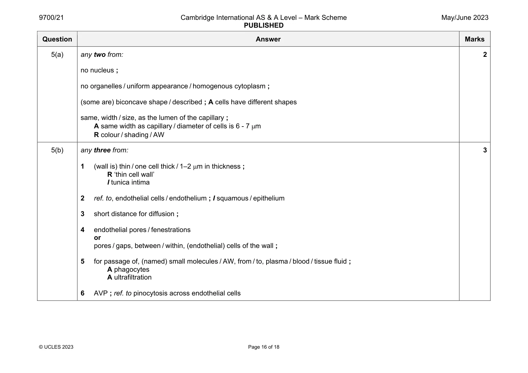
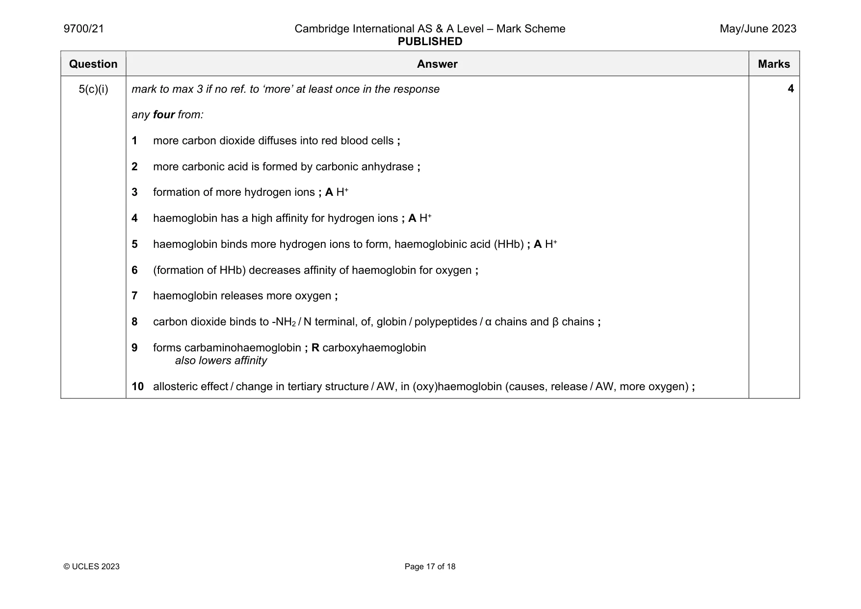
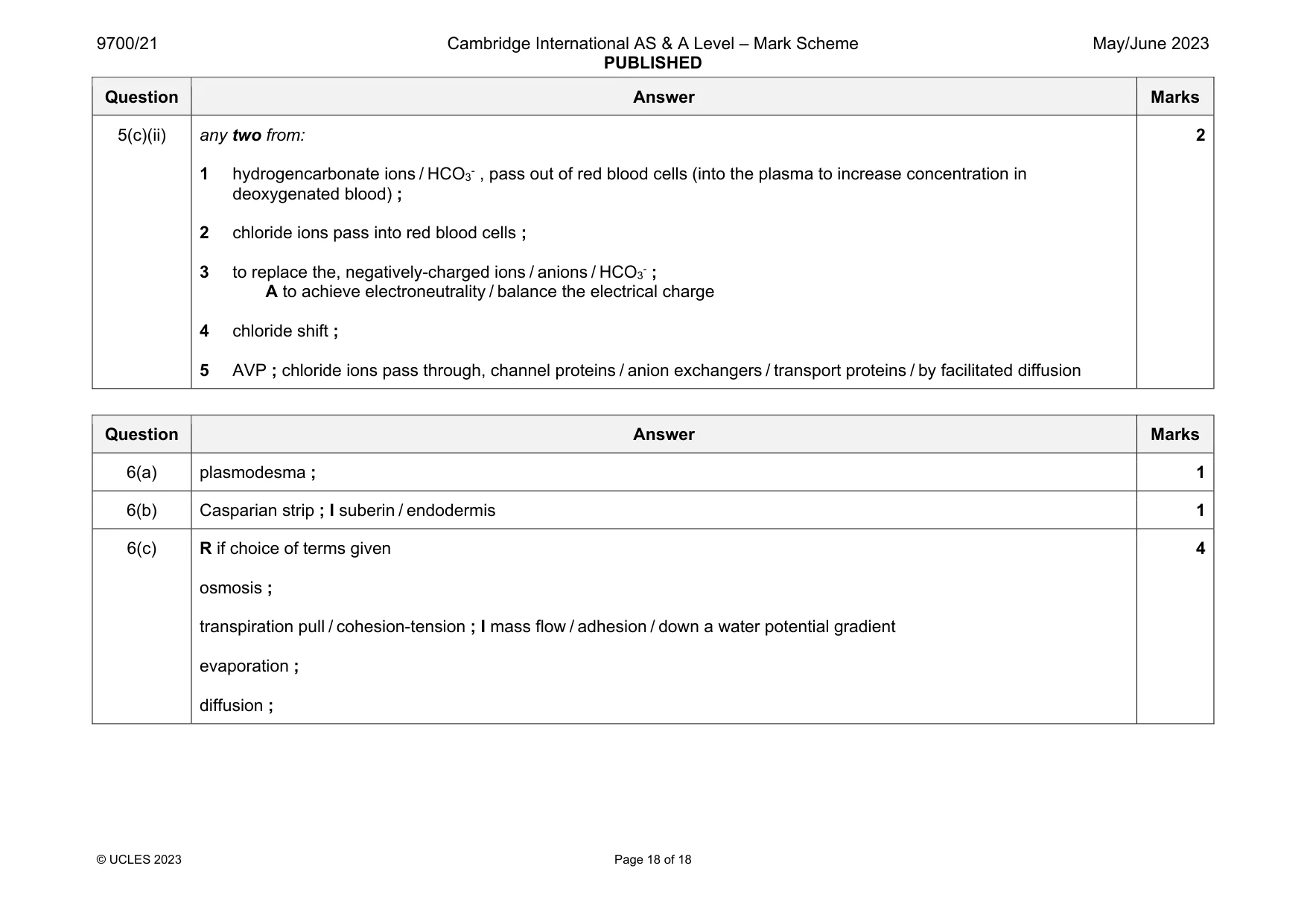
Mark scheme: 5(a) any two from: no nucleus ; no organelles / uniform appearance / homogenous cytoplasm ; (some are) biconcave shape / described ; A cells have different shapes same, width / size, as the lumen of the capillary ; A same width as capillary / diameter of cells is 6 - 7 μm R colour / shading / AW 2 5(b) any three from: 1 (wall is) thin / one cell thick / 1–2 m in thickness ; R ‘thin cell wall’ I tunica intima 2 ref. to, endothelial cells / endothelium ; I squamous / epithelium 3 short distance for diffusion ; 4 endothelial pores / fenestrations or pores / gaps, between / within, (endothelial) cells of the wall ; 5 for passage of, (named) small molecules / AW, from / to, plasma / blood / tissue fluid ; A phagocytes A ultrafiltration 6 AVP ; ref. to pinocytosis across endothelial cells 3 Question Answer Marks 5(c)(i) mark to max 3 if no ref. to ‘more’ at least once in the response any four from: 1 more carbon dioxide diffuses into red blood cells ; 2 more carbonic acid is formed by carbonic anhydrase ; 3 formation of more hydrogen ions ; A H+ 4 haemoglobin has a high affinity for hydrogen ions ; A H+ 5 haemoglobin binds more hydrogen ions to form, haemoglobinic acid (HHb) ; A H+ 6 (formation of HHb) decreases affinity of haemoglobin for oxygen ; 7 haemoglobin releases more oxygen ; 8 carbon dioxide binds to -NH2 / N terminal, of, globin / polypeptides / α chains and β chains ; 9 forms carbaminohaemoglobin ; R carboxyhaemoglobin also lowers affinity 10 allosteric effect / change in tertiary structure / AW, in (oxy)haemoglobin (causes, release / AW, more oxygen) ; 4 Question Answer Marks 5(c)(ii) any two from: 1 hydrogencarbonate ions / HCO3- , pass out of red blood cells (into the plasma to increase concentration in deoxygenated blood) ; 2 chloride ions pass into red blood cells ; 3 to replace the, negatively-charged ions / anions / HCO3- ; A to achieve electroneutrality / balance the electrical charge 4 chloride shift ; 5 AVP ; chloride ions pass through, channel proteins / anion exchangers / transport proteins / by facilitated diffusion 2
Q6 · A diagram showing the passage of water through the tissues of a flowering plant from the…
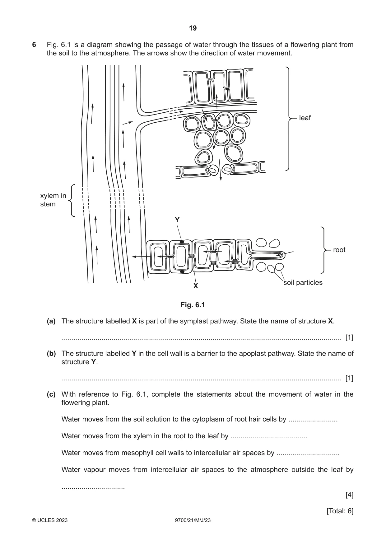
6 Fig. 6.1 is a diagram showing the passage of water through the tissues of a flowering plant from the soil to the atmosphere. The arrows show the direction of water movement. leaf xylem in stem Y root soil particles X Fig. 6.1 (a) The structure labelled X is part of the symplast pathway. State the name of structure X. ............................................................................................................................................. [1] (b) The structure labelled Y in the cell wall is a barrier to the apoplast pathway. State the name of structure Y. ............................................................................................................................................. [1] (c) With reference to Fig. 6.1, complete the statements about the movement of water in the flowering plant. Water moves from the soil solution to the cytoplasm of root hair cells by ......................... Water moves from the xylem in the root to the leaf by ....................................... Water moves from mesophyll cell walls to intercellular air spaces by ................................ Water vapour moves from intercellular air spaces to the atmosphere outside the leaf by ................................ [4] [Total: 6]
Mark scheme: 6(a) plasmodesma ; 1 6(b) Casparian strip ; I suberin / endodermis 1 6(c) R if choice of terms given osmosis ; transpiration pull / cohesion-tension ; I mass flow / adhesion / down a water potential gradient evaporation ; diffusion ; 4
What was in this paper
The subtopics covered by these 6 questions, and how many questions each got. Open one in a new tab to see every Cambridge question on it.
What you needed in this session
Cambridge’s own grade thresholds for 2023 May/June, Paper 2 · Variant 1. A higher threshold means an easier paper — the bar moves with how the cohort did.