Cambridge A Level Biology 9700 — 2023 Oct/Nov Paper 2 · Variant 2
9700/22/O/N/23 · 6 questions · 60 marks · ≈68 min
The question paper and its mark scheme, free to read here and free to download. This is Cambridge’s own paper, exactly as it was sat.
Question paper16 pages
Mark scheme25 pages
Answers below. Sit the paper first if you are practising.
Questions as text
Q1 · During interphase and mitosis of the cell cycle, the chromosomes within a cell go through…
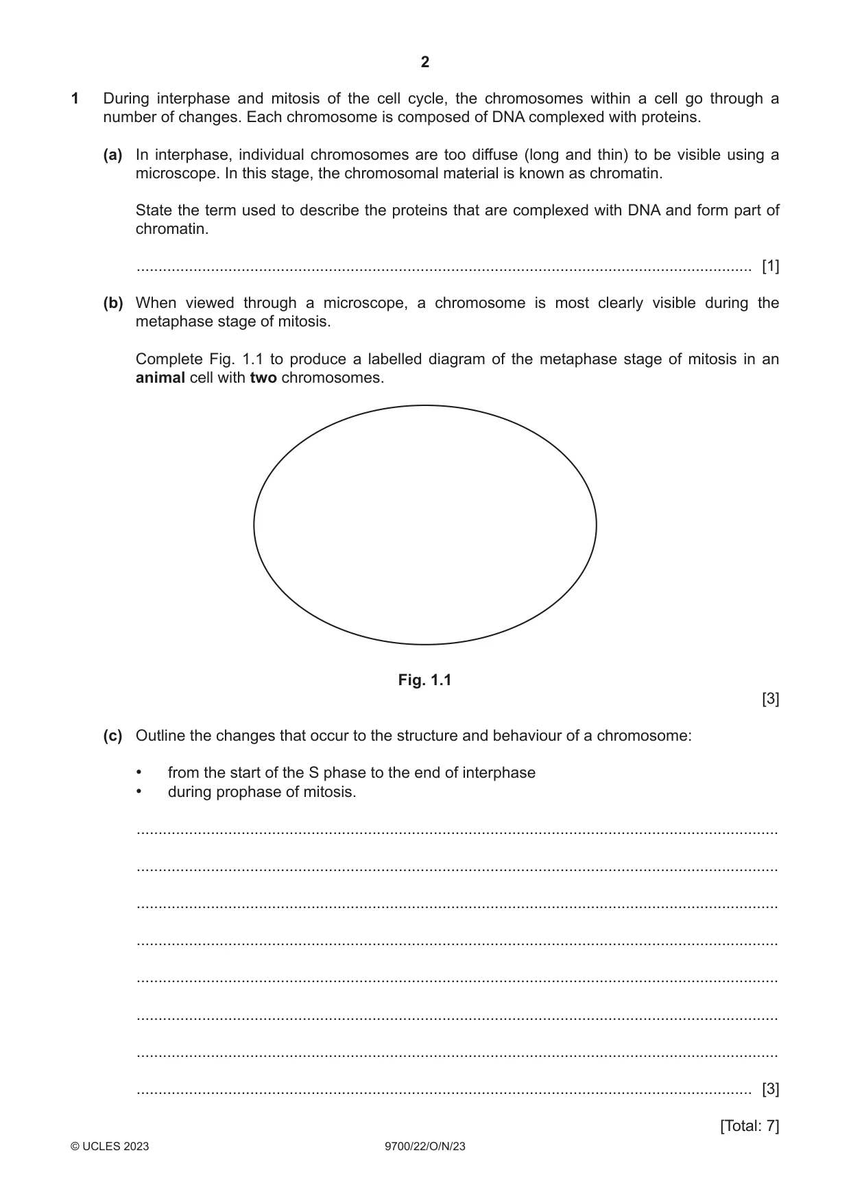
1 During interphase and mitosis of the cell cycle, the chromosomes within a cell go through a number of changes. Each chromosome is composed of DNA complexed with proteins. (a) In interphase, individual chromosomes are too diffuse (long and thin) to be visible using a microscope. In this stage, the chromosomal material is known as chromatin. State the term used to describe the proteins that are complexed with DNA and form part of chromatin. ............................................................................................................................................. [1] (b) When viewed through a microscope, a chromosome is most clearly visible during the metaphase stage of mitosis. Complete Fig. 1.1 to produce a labelled diagram of the metaphase stage of mitosis in an animal cell with two chromosomes. Fig. 1.1 [3] (c) Outline the changes that occur to the structure and behaviour of a chromosome: • from the start of the S phase to the end of interphase • during prophase of mitosis. ................................................................................................................................................... ................................................................................................................................................... ................................................................................................................................................... ................................................................................................................................................... ................................................................................................................................................... ................................................................................................................................................... ................................................................................................................................................... ............................................................................................................................................. [3] [Total: 7]
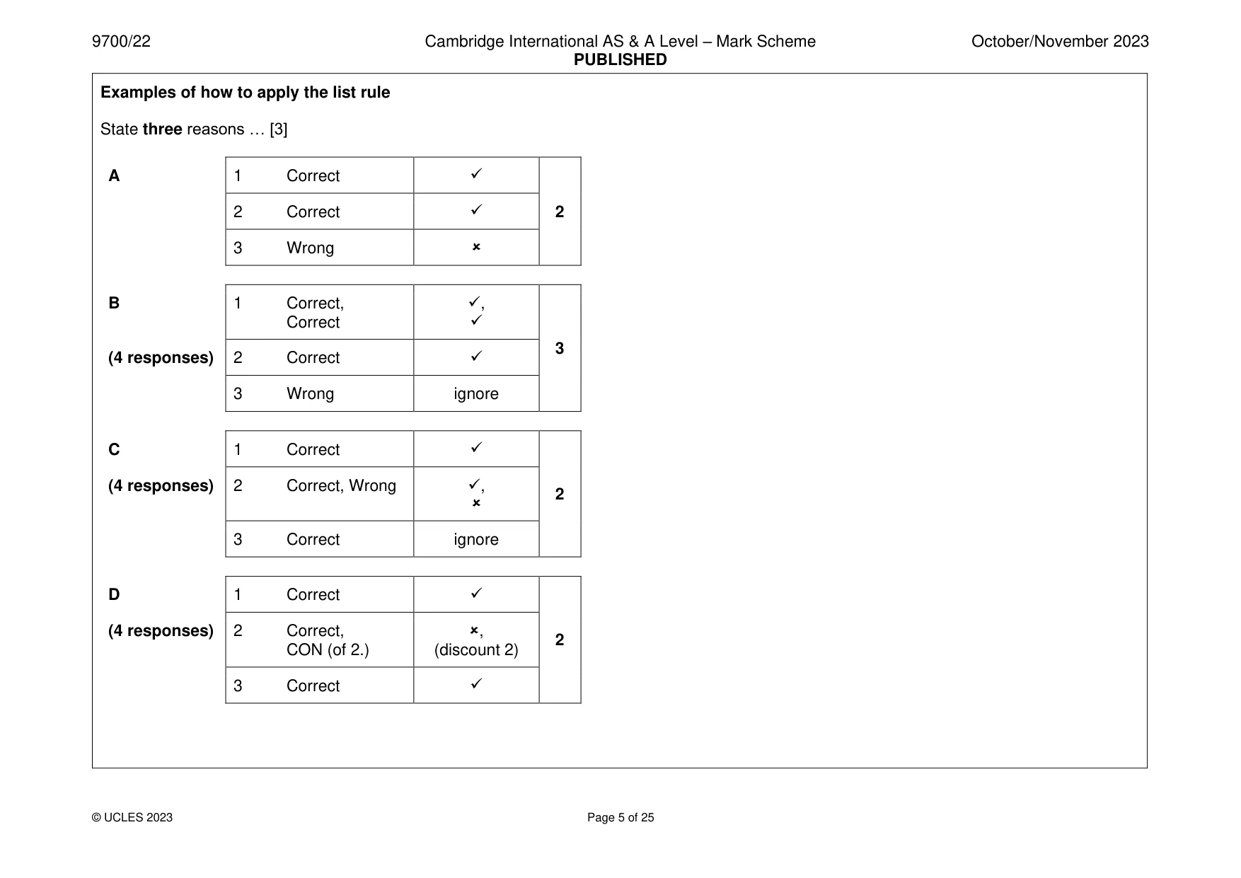
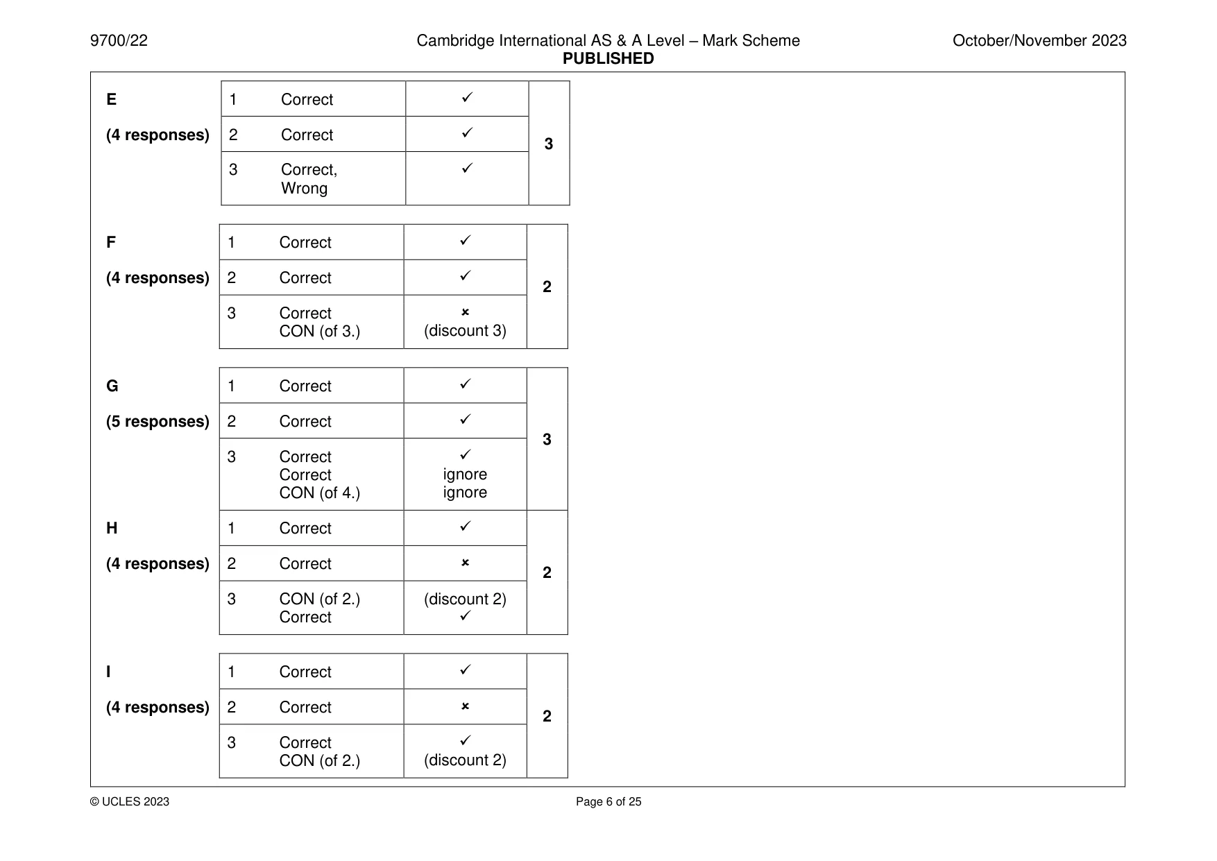
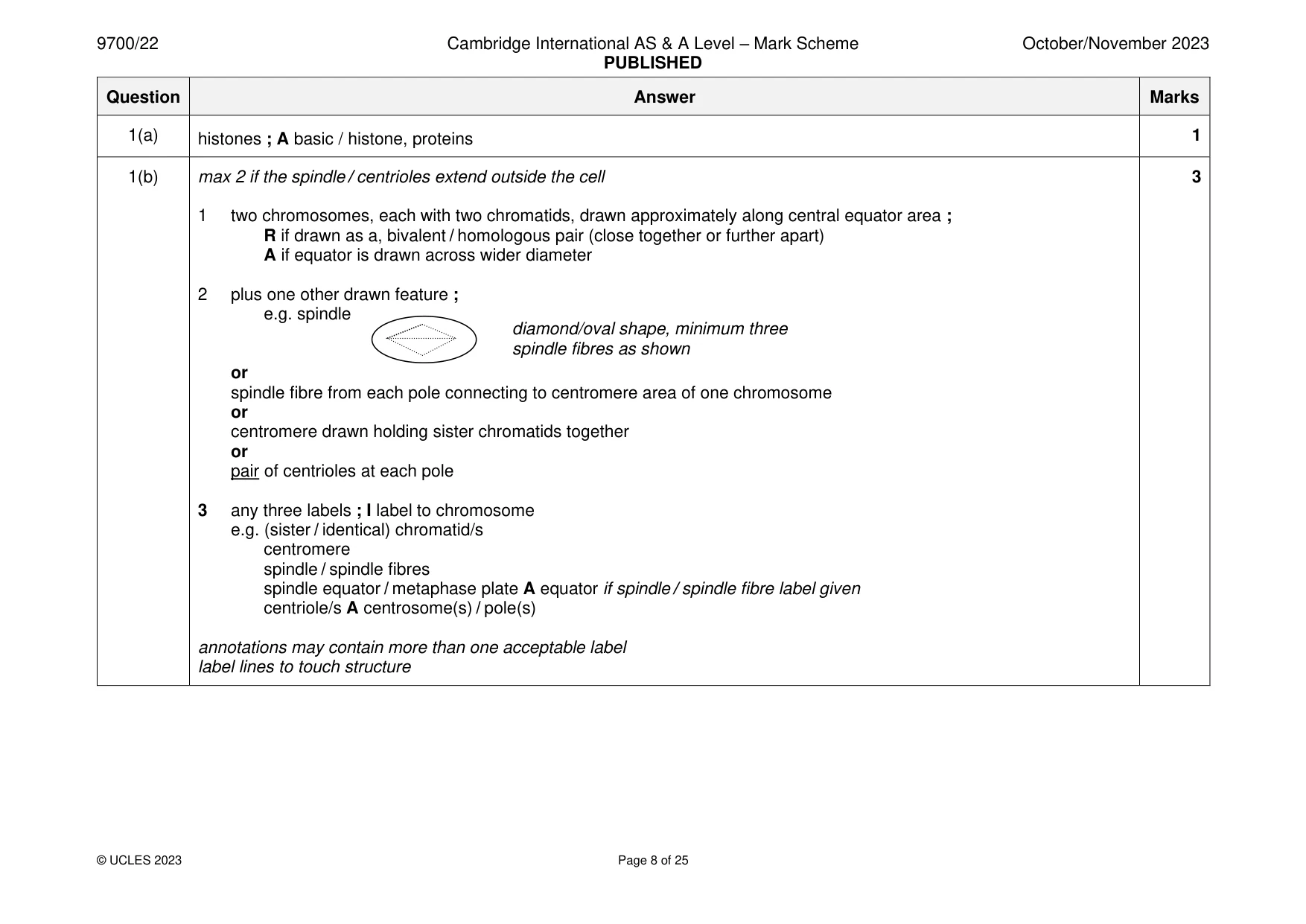
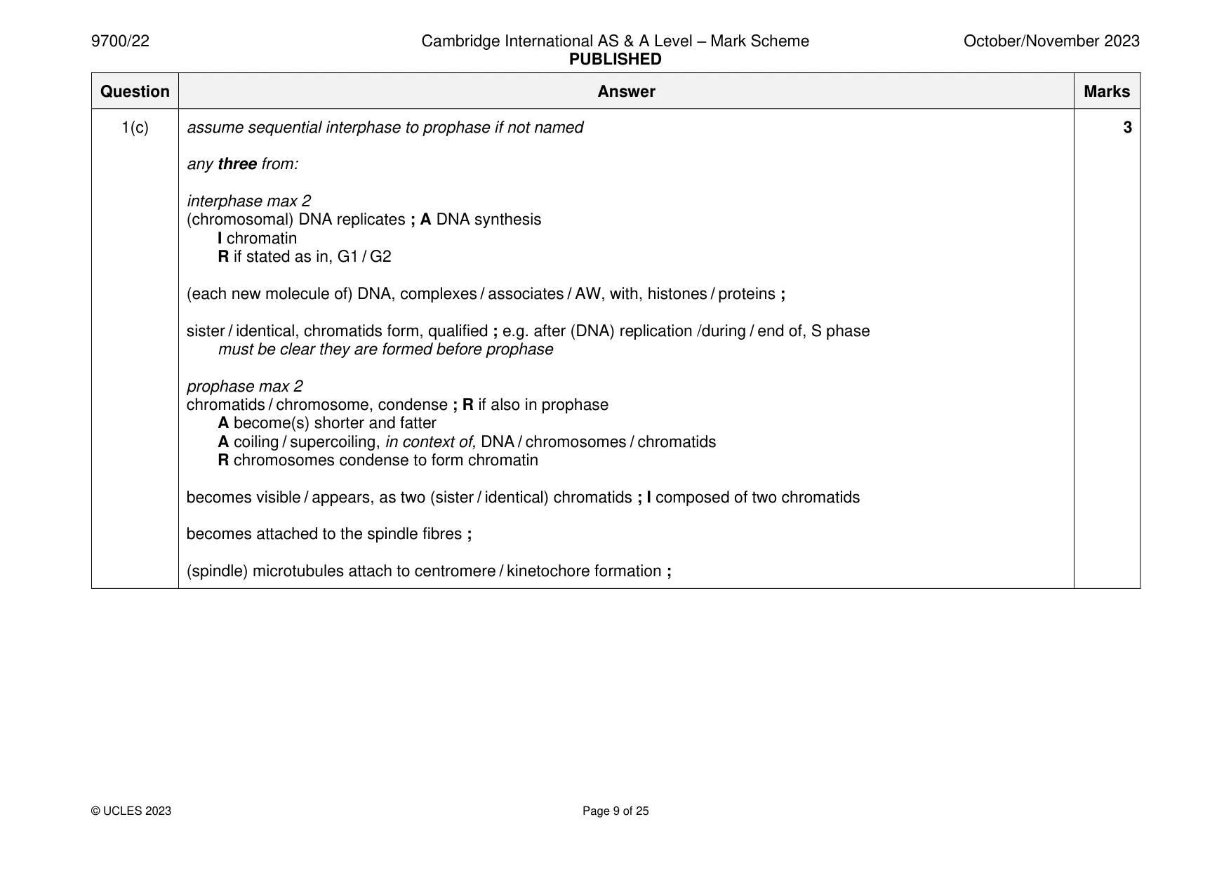
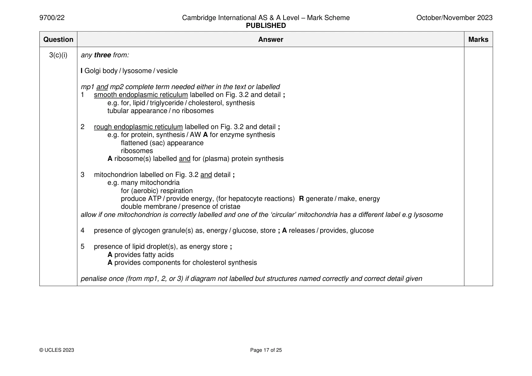
Mark scheme: Question Answer Marks 1(a) histones ; A basic / histone, proteins 1 1(b) max 2 if the spindle / centrioles extend outside the cell 3 1 two chromosomes, each with two chromatids, drawn approximately along central equator area ; R if drawn as a, bivalent / homologous pair (close together or further apart) A if equator is drawn across wider diameter 2 plus one other drawn feature ; e.g. spindle diamond/oval shape, minimum three spindle fibres as shown or spindle fibre from each pole connecting to centromere area of one chromosome or centromere drawn holding sister chromatids together or pair of centrioles at each pole 3 any three labels ; I label to chromosome e.g. (sister / identical) chromatid/s centromere spindle / spindle fibres spindle equator / metaphase plate A equator if spindle / spindle fibre label given centriole/s A centrosome(s) / pole(s) annotations may contain more than one acceptable label label lines to touch structure 1(c) assume sequential interphase to prophase if not named 3 any three from: interphase max 2 (chromosomal) DNA replicates ; A DNA synthesis I chromatin R if stated as in, G1 / G2 (each new molecule of) DNA, complexes / associates / AW, with, histones / proteins ; sister / identical, chromatids form, qualified ; e.g. after (DNA) replication /during / end of, S phase must be clear they are formed before prophase prophase max 2 chromatids / chromosome, condense ; R if also in prophase A become(s) shorter and fatter A coiling / supercoiling, in context of, DNA / chromosomes / chromatids R chromosomes condense to form chromatin becomes visible / appears, as two (sister / identical) chromatids ; I composed of two chromatids becomes attached to the spindle fibres ; (spindle) microtubules attach to centromere / kinetochore formation ;
Q2 · Keratin is the structural protein in feathers of birds
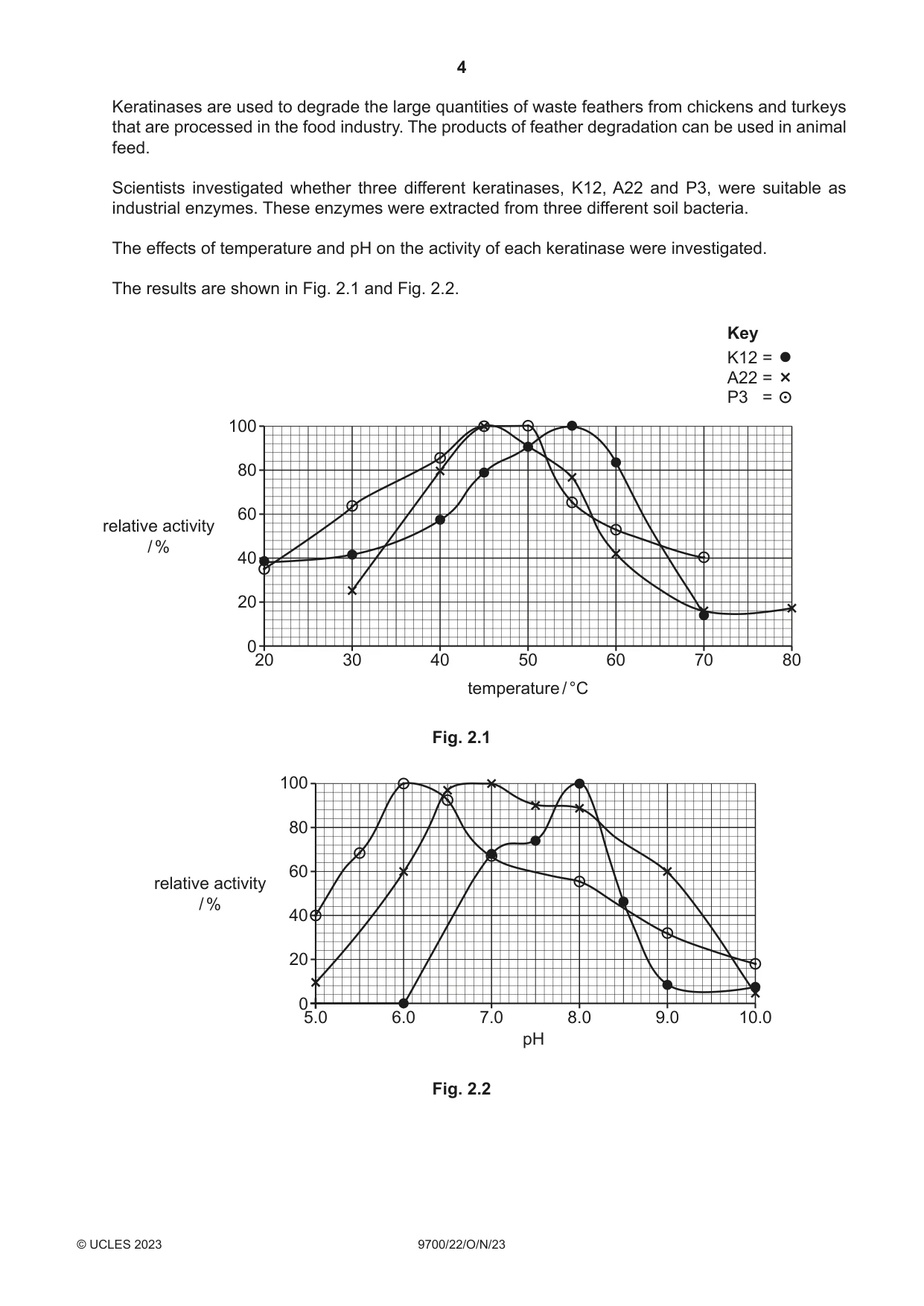
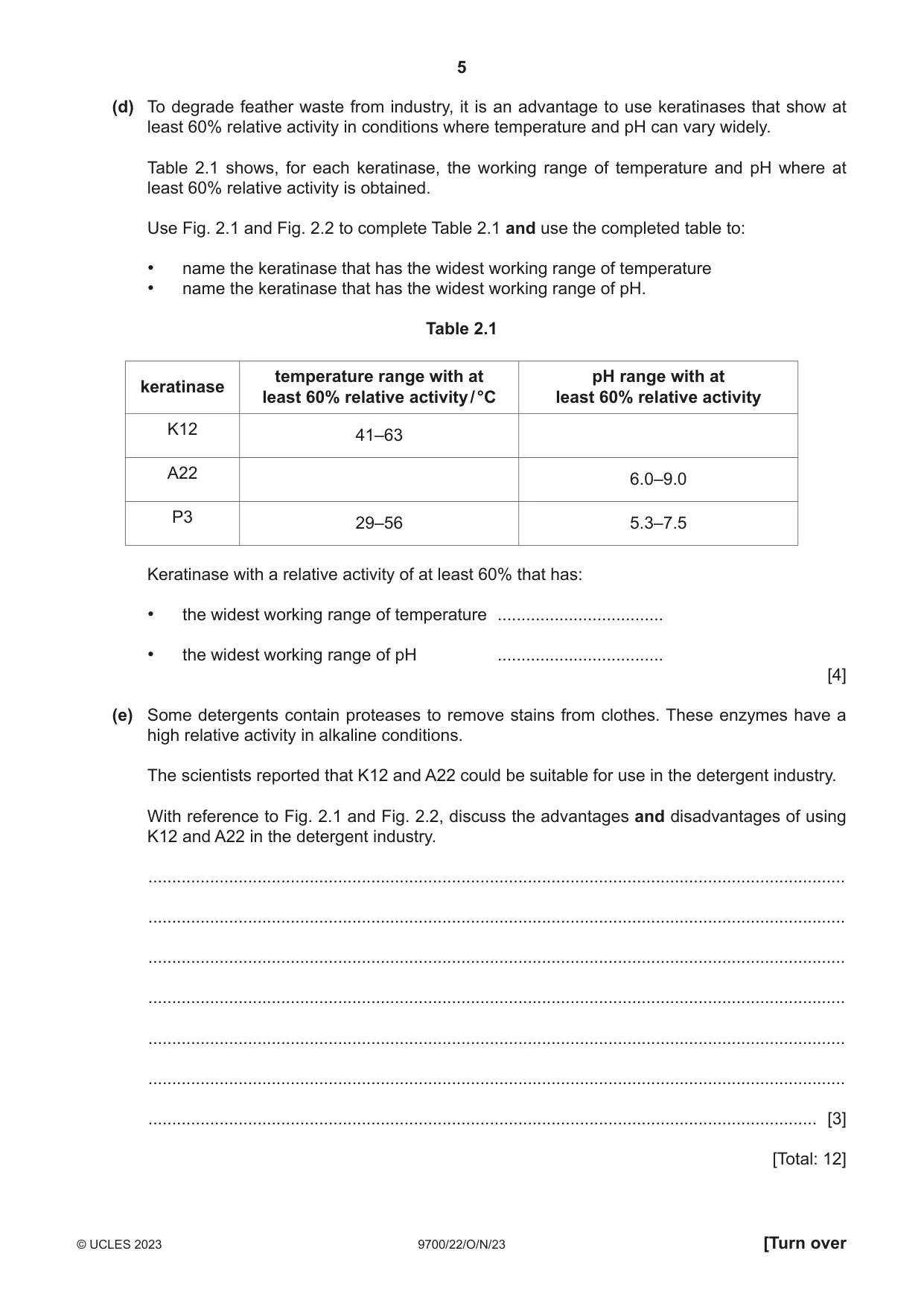
2 Keratin is the structural protein in feathers of birds. Keratin polypeptides are composed of a high proportion of cysteine amino acids, which have sulfur-containing R groups. Keratin polypeptides form filaments. The two main types of keratin in feathers are α-keratin, which consists of many α-helices, and β-keratin, consisting of many β-pleated sheets. (a) Keratin can be classified as α-keratin or β-keratin based on a study of protein structure. Suggest the level of protein structure used to classify a protein as α-keratin or β-keratin. ............................................................................................................................................. [1] (b) Protease enzymes hydrolyse proteins. Many proteases are able to hydrolyse more than one type of protein. Suggest why it is possible for a protease to act on different types of protein. ................................................................................................................................................... ................................................................................................................................................... ............................................................................................................................................. [1] (c) Proteases known as keratinases vary in the extent to which they can hydrolyse keratin. Feathers are not easily degraded (broken down) because keratin is a very stable protein. Suggest features of keratin structure that contribute to its stability. ................................................................................................................................................... ................................................................................................................................................... ................................................................................................................................................... ................................................................................................................................................... ................................................................................................................................................... ................................................................................................................................................... ................................................................................................................................................... ............................................................................................................................................. [3] Keratinases are used to degrade the large quantities of waste feathers from chickens and turkeys that are processed in the food industry. The products of feather degradation can be used in animal feed. Scientists investigated whether three different keratinases, K12, A22 and P3, were suitable as industrial enzymes. These enzymes were extracted from three different soil bacteria. The effects of temperature and pH on the activity of each keratinase were investigated. The results are shown in Fig. 2.1 and Fig. 2.2. Key K12 = A22 = P3 = 100 80 60 relative activity / % 40 20 0 20 30 40 50 60 70 80 temperature / °C Fig. 2.1 100 80 60 relative activity / % 40 20 0 5.0 6.0 7.0 8.0 9.0 10.0 pH Fig. 2.2 (d) To degrade feather waste from industry, it is an advantage to use keratinases that show at least 60% relative activity in conditions where temperature and pH can vary widely. Table 2.1 shows, for each keratinase, the working range of temperature and pH where at least 60% relative activity is obtained. Use Fig. 2.1 and Fig. 2.2 to complete Table 2.1 and use the completed table to: • name the keratinase that has the widest working range of temperature • name the keratinase that has the widest working range of pH. Table 2.1 temperature range with at pH range with at keratinase least 60% relative activity / °C least 60% relative activity K12 41–63 A22 6.0–9.0 P3 29–56 5.3–7.5 Keratinase with a relative activity of at least 60% that has: • the widest working range of temperature ................................... • the widest working range of pH ................................... [4] (e) Some detergents contain proteases to remove stains from clothes. These enzymes have a high relative activity in alkaline conditions. The scientists reported that K12 and A22 could be suitable for use in the detergent industry. With reference to Fig. 2.1 and Fig. 2.2, discuss the advantages and disadvantages of using K12 and A22 in the detergent industry. ................................................................................................................................................... ................................................................................................................................................... ................................................................................................................................................... ................................................................................................................................................... ................................................................................................................................................... ................................................................................................................................................... ............................................................................................................................................. [3] [Total: 12]
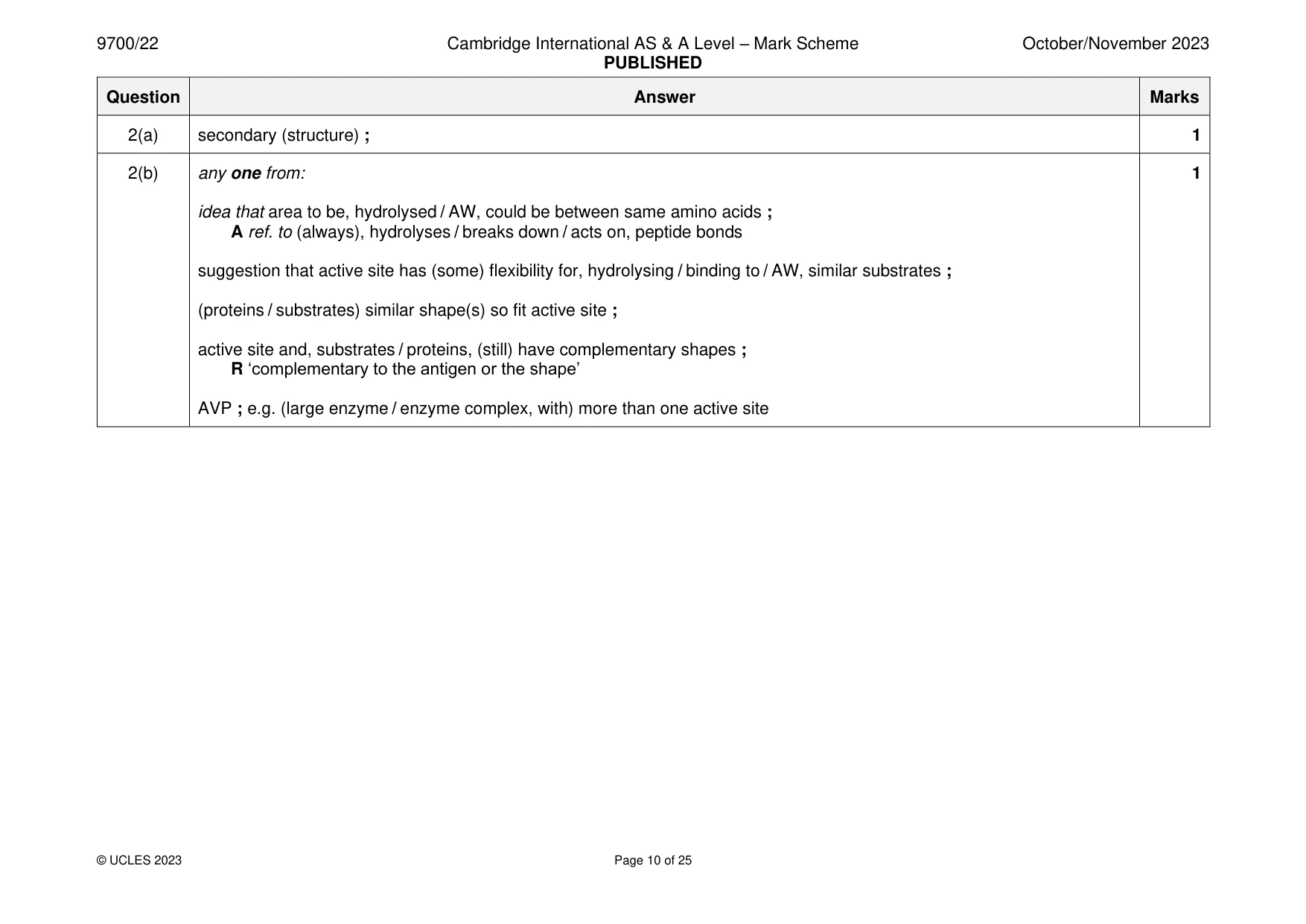
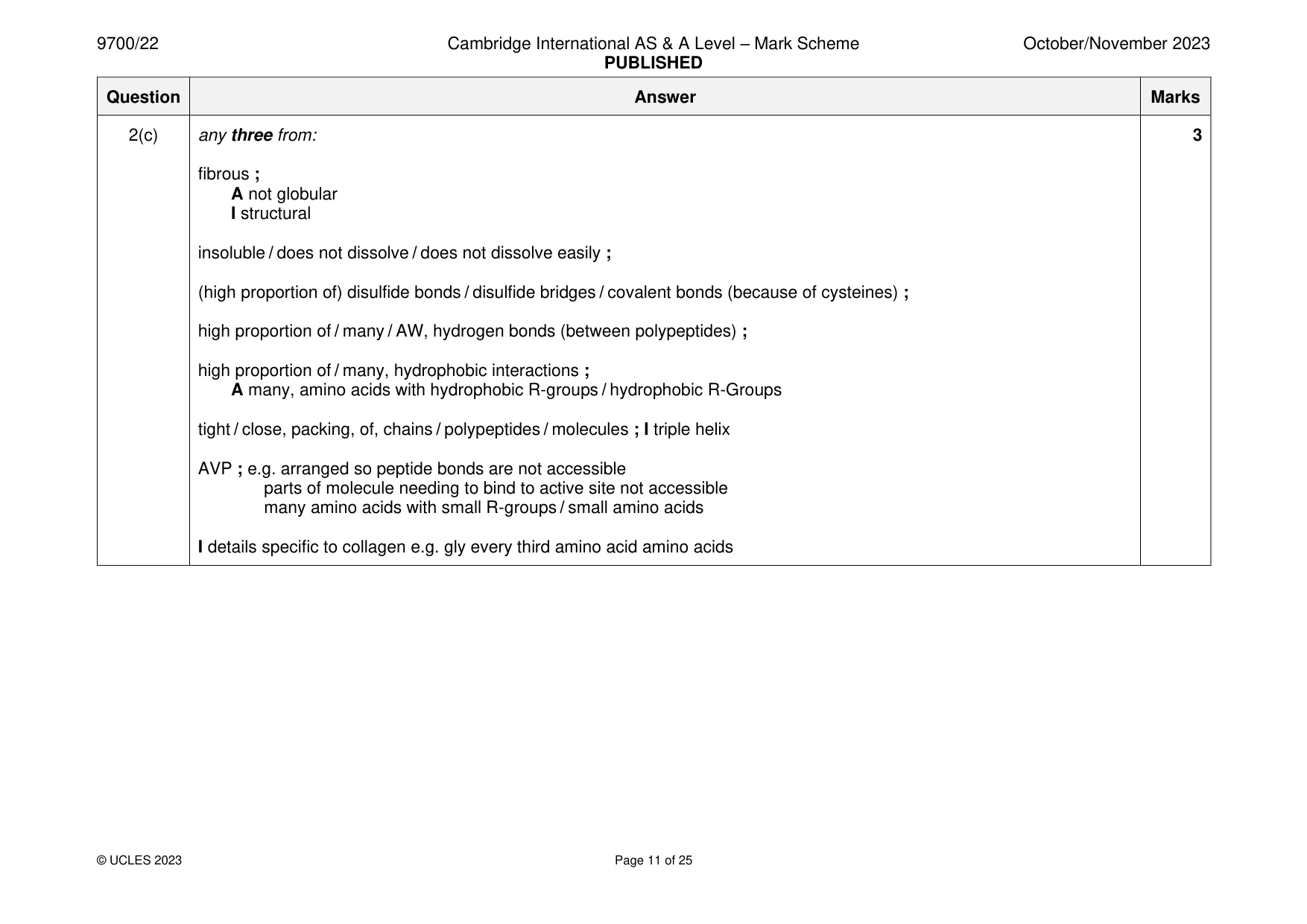
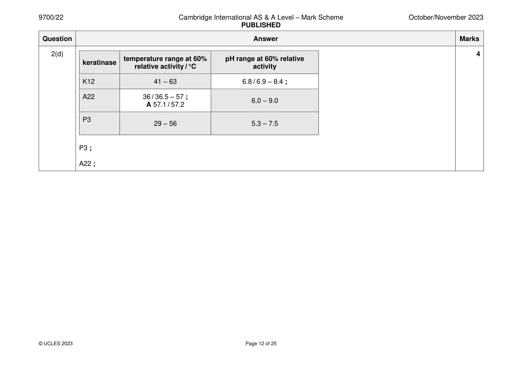
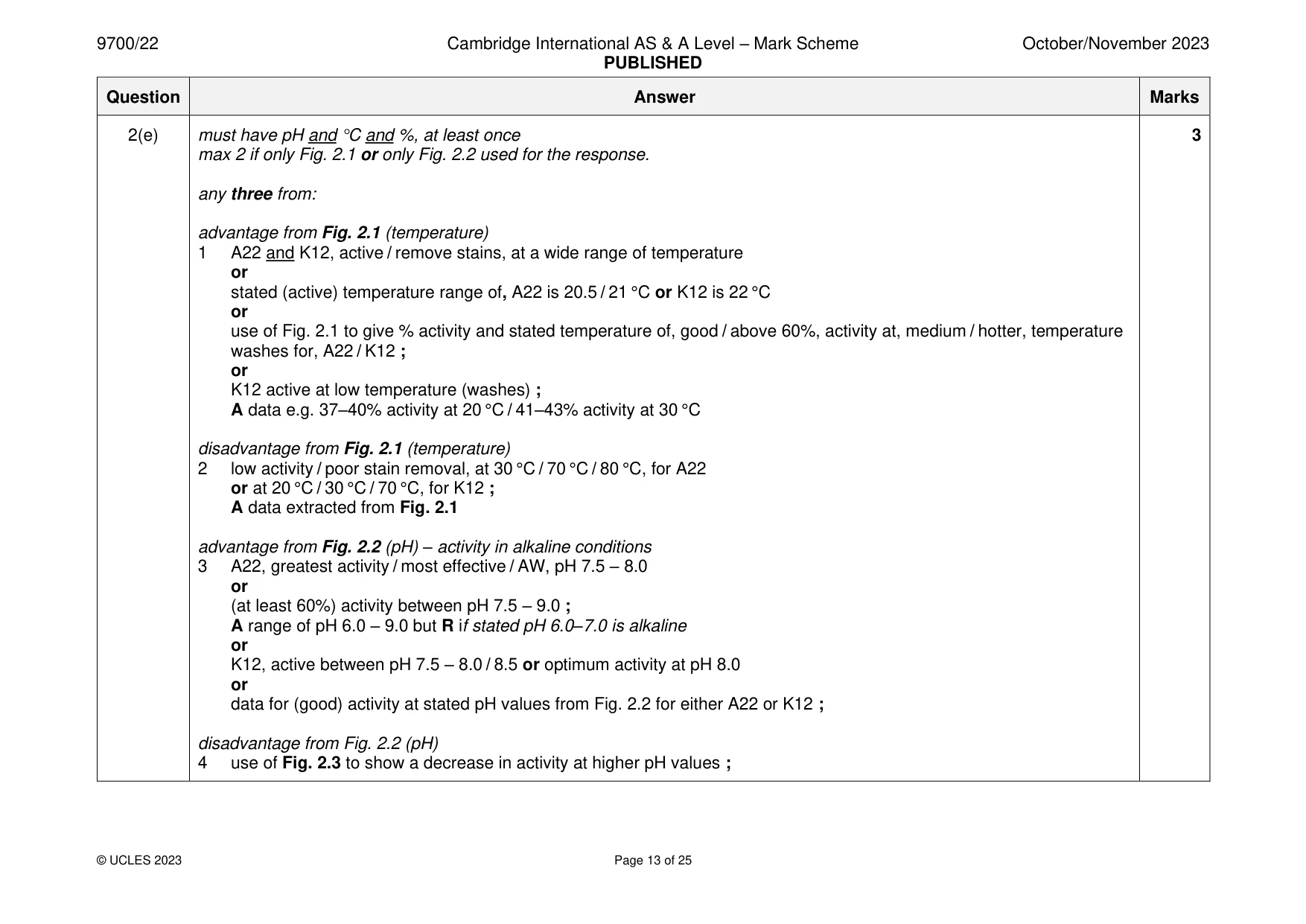
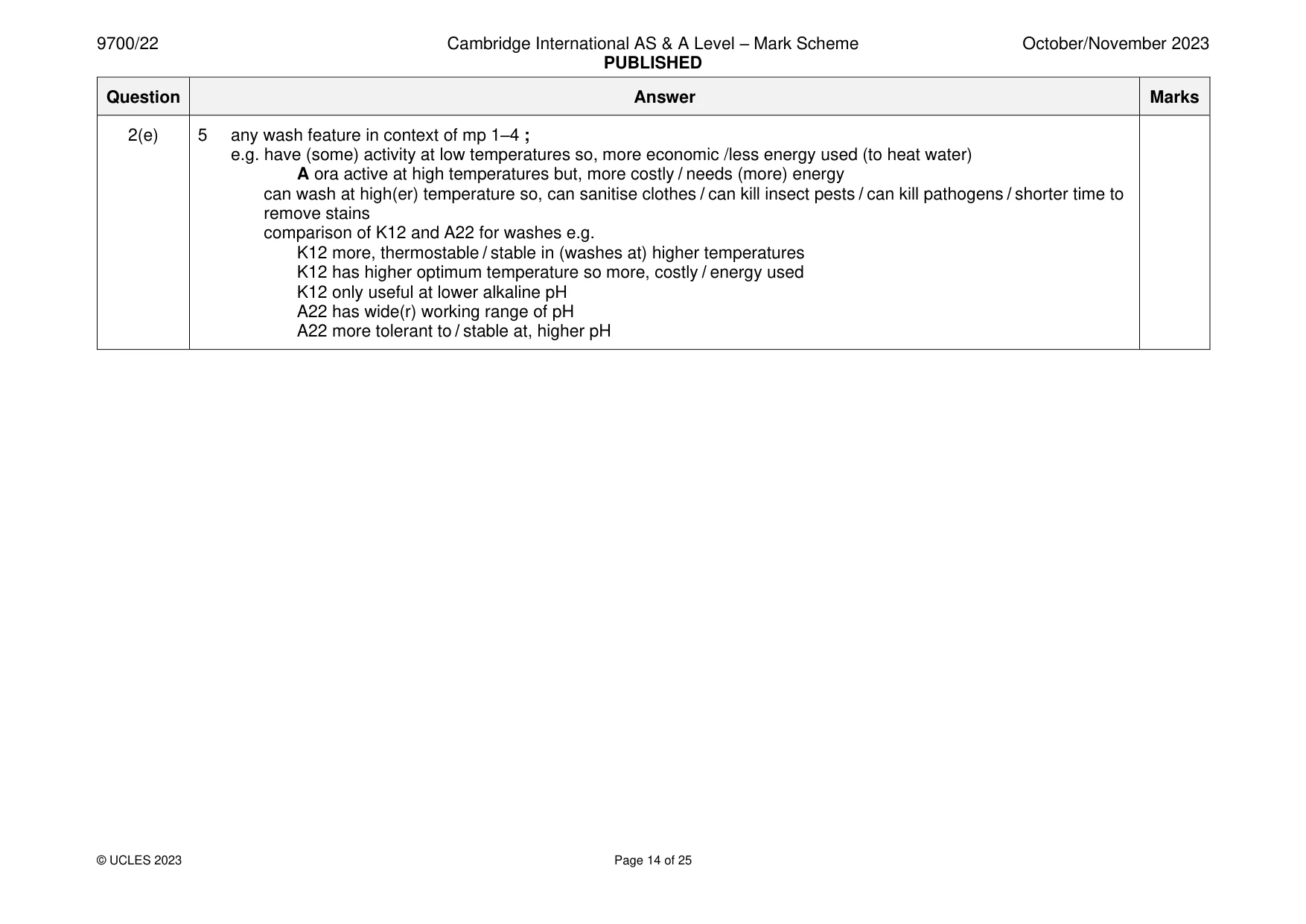
Mark scheme: 2(a) secondary (structure) ; 1 2(b) any one from: 1 idea that area to be, hydrolysed / AW, could be between same amino acids ; A ref. to (always), hydrolyses / breaks down / acts on, peptide bonds suggestion that active site has (some) flexibility for, hydrolysing / binding to / AW, similar substrates ; (proteins / substrates) similar shape(s) so fit active site ; active site and, substrates / proteins, (still) have complementary shapes ; R ‘complementary to the antigen or the shape’ AVP ; e.g. (large enzyme / enzyme complex, with) more than one active site 2(c) any three from: 3 fibrous ; A not globular I structural insoluble / does not dissolve / does not dissolve easily ; (high proportion of) disulfide bonds / disulfide bridges / covalent bonds (because of cysteines) ; high proportion of / many / AW, hydrogen bonds (between polypeptides) ; high proportion of / many, hydrophobic interactions ; A many, amino acids with hydrophobic R-groups / hydrophobic R-Groups tight / close, packing, of, chains / polypeptides / molecules ; I triple helix AVP ; e.g. arranged so peptide bonds are not accessible parts of molecule needing to bind to active site not accessible many amino acids with small R-groups / small amino acids I details specific to collagen e.g. gly every third amino acid amino acids 2(d) 4 temperature range at 60% pH range at 60% relative keratinase relative activity / °C activity K12 41 – 63 6.8 / 6.9 – 8.4 ; A22 36 / 36.5 – 57 ; 6.0 – 9.0 A 57.1 / 57.2 P3 29 – 56 5.3 – 7.5 P3 ; A22 ; 2(e) must have pH and °C and %, at least once 3 max 2 if only Fig. 2.1 or only Fig. 2.2 used for the response. any three from: advantage from Fig. 2.1 (temperature) 1 A22 and K12, active / remove stains, at a wide range of temperature or stated (active) temperature range of, A22 is 20.5 / 21 °C or K12 is 22 °C or use of Fig. 2.1 to give % activity and stated temperature of, good / above 60%, activity at, medium / hotter, temperature washes for, A22 / K12 ; or K12 active at low temperature (washes) ; A data e.g. 37–40% activity at 20 °C / 41–43% activity at 30 °C disadvantage from Fig. 2.1 (temperature) 2 low activity / poor stain removal, at 30 °C / 70 °C / 80 °C, for A22 or at 20 °C / 30 °C / 70 °C, for K12 ; A data extracted from Fig. 2.1 advantage from Fig. 2.2 (pH) – activity in alkaline conditions 3 A22, greatest activity / most effective / AW, pH 7.5 – 8.0 or (at least 60%) activity between pH 7.5 – 9.0 ; A range of pH 6.0 – 9.0 but R if stated pH 6.0–7.0 is alkaline or K12, active between pH 7.5 – 8.0 / 8.5 or optimum activity at pH 8.0 or data for (good) activity at stated pH values from Fig. 2.2 for either A22 or K12 ; disadvantage from Fig. 2.2 (pH) 4 use of Fig. 2.3 to show a decrease in activity at higher pH values ; 2(e) 5 any wash feature in context of mp 1–4 ; e.g. have (some) activity at low temperatures so, more economic /less energy used (to heat water) A ora active at high temperatures but, more costly / needs (more) energy can wash at high(er) temperature so, can sanitise clothes / can kill insect pests / can kill pathogens / shorter time to remove stains comparison of K12 and A22 for washes e.g. K12 more, thermostable / stable in (washes at) higher temperatures K12 has higher optimum temperature so more, costly / energy used K12 only useful at lower alkaline pH A22 has wide(r) working range of pH A22 more tolerant to / stable at, higher pH
Q3 · The liver receives blood from the hepatic artery and from the hepatic portal vein
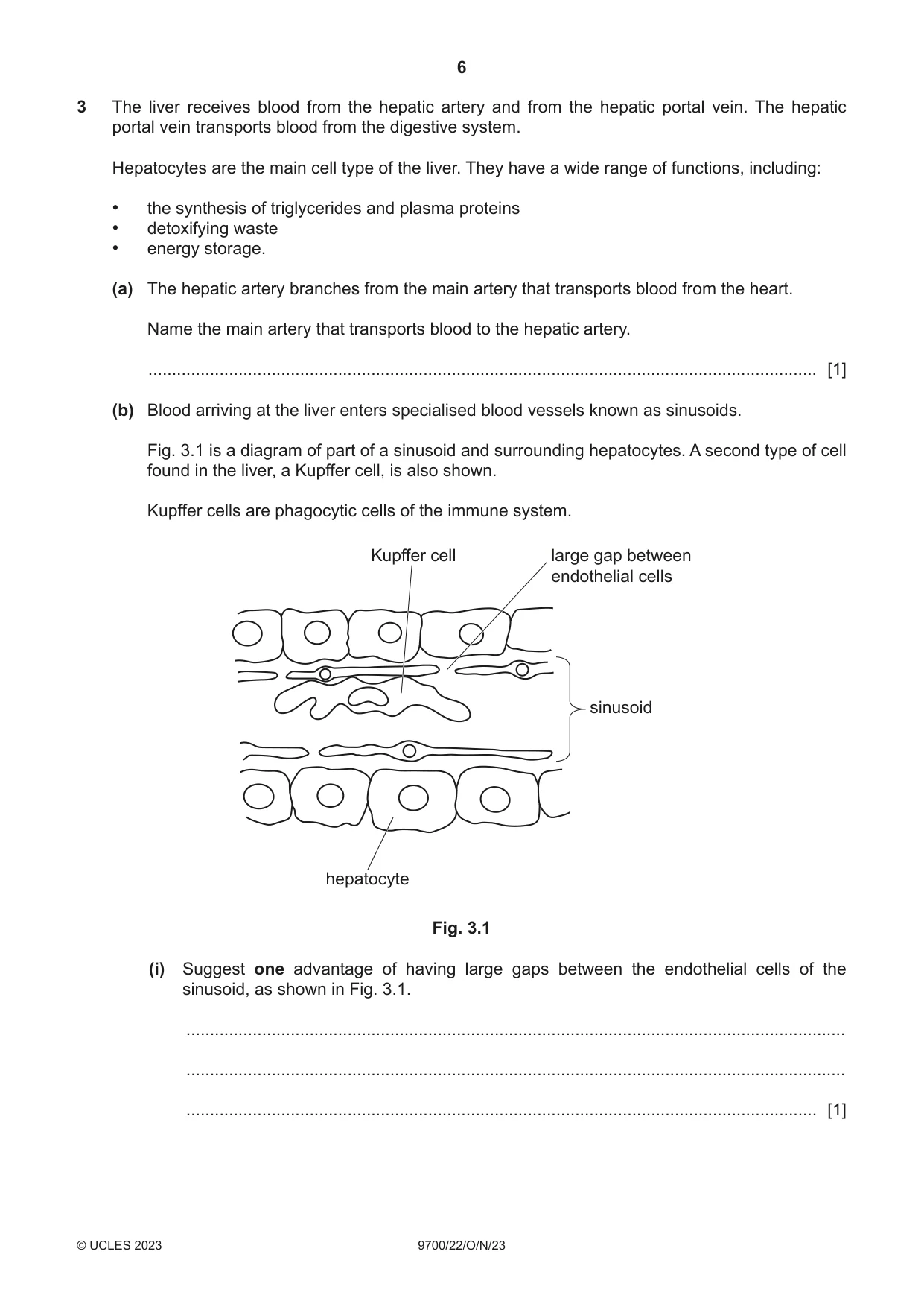
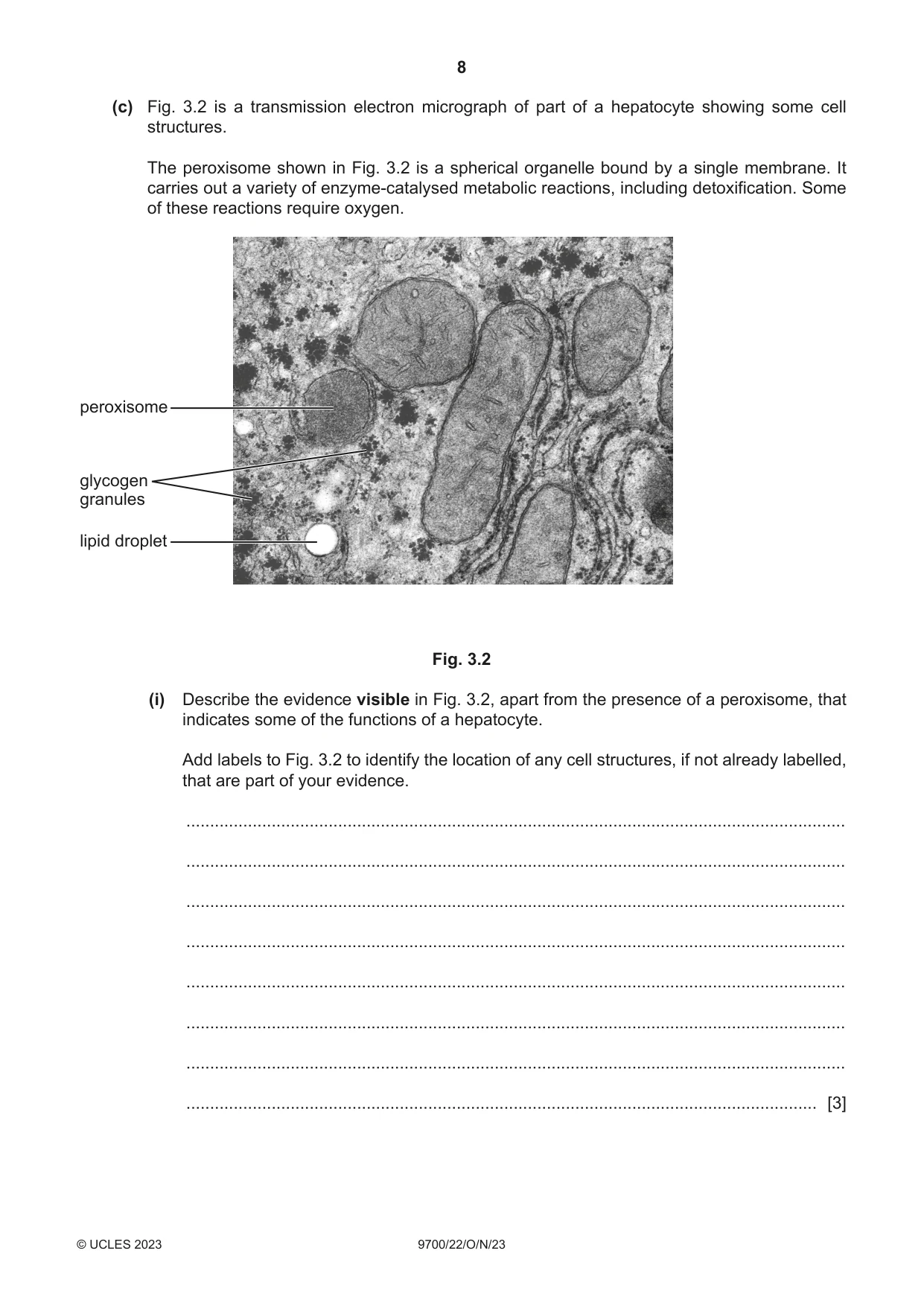
3 The liver receives blood from the hepatic artery and from the hepatic portal vein. The hepatic portal vein transports blood from the digestive system. Hepatocytes are the main cell type of the liver. They have a wide range of functions, including: • the synthesis of triglycerides and plasma proteins • detoxifying waste • energy storage. (a) The hepatic artery branches from the main artery that transports blood from the heart. Name the main artery that transports blood to the hepatic artery. ............................................................................................................................................. [1] (b) Blood arriving at the liver enters specialised blood vessels known as sinusoids. Fig. 3.1 is a diagram of part of a sinusoid and surrounding hepatocytes. A second type of cell found in the liver, a Kupffer cell, is also shown. Kupffer cells are phagocytic cells of the immune system. Kupffer cell large gap between endothelial cells sinusoid hepatocyte Fig. 3.1 (i) Suggest one advantage of having large gaps between the endothelial cells of the sinusoid, as shown in Fig. 3.1. ........................................................................................................................................... ........................................................................................................................................... ..................................................................................................................................... [1] (ii) In addition to removing bacteria present in the blood inside the sinusoid, Kupffer cells are also able to remove old or damaged red blood cells. Describe the mode of action of a Kupffer cell in removing and breaking down a damaged red blood cell. ........................................................................................................................................... ........................................................................................................................................... ........................................................................................................................................... ........................................................................................................................................... ........................................................................................................................................... ........................................................................................................................................... ........................................................................................................................................... ........................................................................................................................................... ........................................................................................................................................... ..................................................................................................................................... [4] (c) Fig. 3.2 is a transmission electron micrograph of part of a hepatocyte showing some cell structures. The peroxisome shown in Fig. 3.2 is a spherical organelle bound by a single membrane. It carries out a variety of enzyme-catalysed metabolic reactions, including detoxification. Some of these reactions require oxygen. peroxisome glycogen granules lipid droplet Fig. 3.2 (i) Describe the evidence visible in Fig. 3.2, apart from the presence of a peroxisome, that indicates some of the functions of a hepatocyte. Add labels to Fig. 3.2 to identify the location of any cell structures, if not already labelled, that are part of your evidence. ........................................................................................................................................... ........................................................................................................................................... ........................................................................................................................................... ........................................................................................................................................... ........................................................................................................................................... ........................................................................................................................................... ........................................................................................................................................... ..................................................................................................................................... [3] (ii) The mitochondria in Fig. 3.2 are larger than the peroxisome. State one other difference, visible in Fig. 3.2, between a peroxisome and a mitochondrion. ........................................................................................................................................... ........................................................................................................................................... ........................................................................................................................................... ..................................................................................................................................... [1] (iii) Some of the enzymes used within mitochondria can be synthesised by the organelle. Peroxisomes cannot synthesise any of the enzymes that they contain. Suggest why a mitochondrion can synthesise enzymes, but a peroxisome cannot synthesise enzymes. ........................................................................................................................................... ........................................................................................................................................... ........................................................................................................................................... ........................................................................................................................................... ..................................................................................................................................... [2] (iv) One of the enzymes present in peroxisomes is catalase. This enzyme catalyses the breakdown of hydrogen peroxide to harmless products. Suggest why it is useful to the cell for this reaction to take place within peroxisomes. ........................................................................................................................................... ........................................................................................................................................... ........................................................................................................................................... ........................................................................................................................................... ........................................................................................................................................... ..................................................................................................................................... [2] [Total: 14]
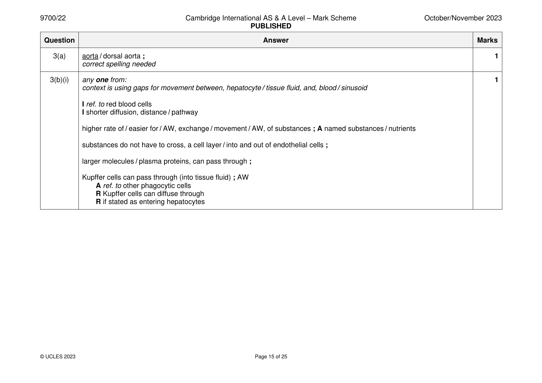
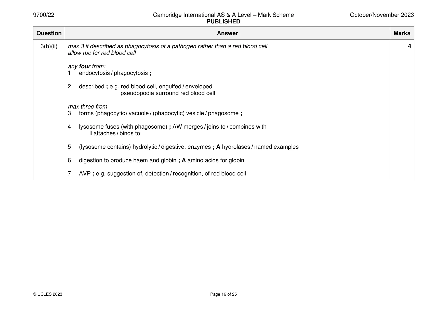
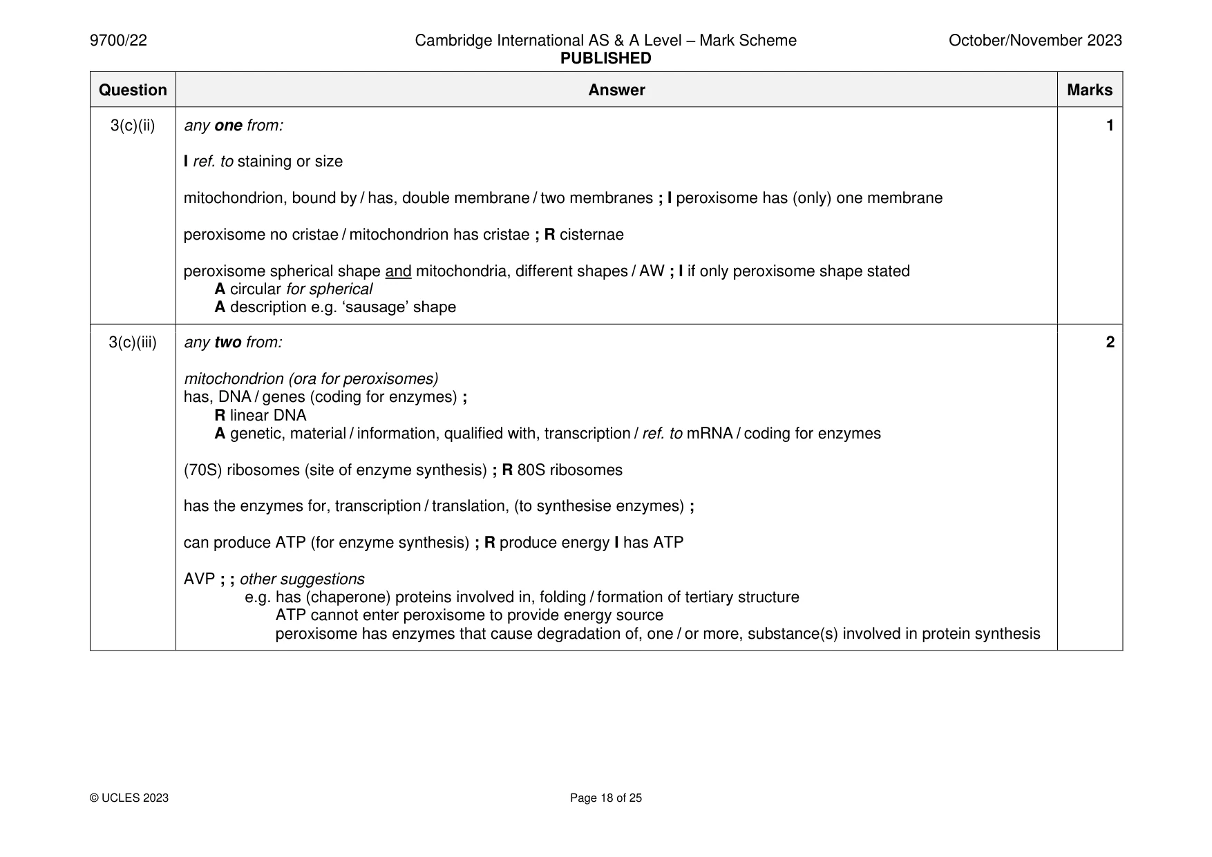
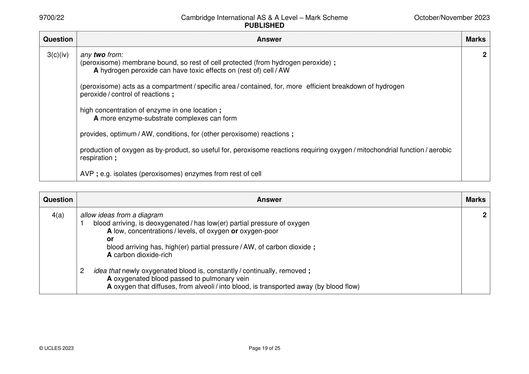
Mark scheme: 3(a) aorta / dorsal aorta ; 1 correct spelling needed 3(b)(i) any one from: 1 context is using gaps for movement between, hepatocyte / tissue fluid, and, blood / sinusoid I ref. to red blood cells I shorter diffusion, distance / pathway higher rate of / easier for / AW, exchange / movement / AW, of substances ; A named substances / nutrients substances do not have to cross, a cell layer / into and out of endothelial cells ; larger molecules / plasma proteins, can pass through ; Kupffer cells can pass through (into tissue fluid) ; AW A ref. to other phagocytic cells R Kupffer cells can diffuse through R if stated as entering hepatocytes 3(b)(ii) max 3 if described as phagocytosis of a pathogen rather than a red blood cell 4 allow rbc for red blood cell any four from: 1 endocytosis / phagocytosis ; 2 described ; e.g. red blood cell, engulfed / enveloped pseudopodia surround red blood cell max three from 3 forms (phagocytic) vacuole / (phagocytic) vesicle / phagosome ; 4 lysosome fuses (with phagosome) ; AW merges / joins to / combines with I attaches / binds to 5 (lysosome contains) hydrolytic / digestive, enzymes ; A hydrolases / named examples 6 digestion to produce haem and globin ; A amino acids for globin 7 AVP ; e.g. suggestion of, detection / recognition, of red blood cell 3(c)(i) any three from: I Golgi body / lysosome / vesicle mp1 and mp2 complete term needed either in the text or labelled 1 smooth endoplasmic reticulum labelled on Fig. 3.2 and detail ; e.g. for, lipid / triglyceride / cholesterol, synthesis tubular appearance / no ribosomes 2 rough endoplasmic reticulum labelled on Fig. 3.2 and detail ; e.g. for protein, synthesis / AW A for enzyme synthesis flattened (sac) appearance ribosomes A ribosome(s) labelled and for (plasma) protein synthesis 3 mitochondrion labelled on Fig. 3.2 and detail ; e.g. many mitochondria for (aerobic) respiration produce ATP / provide energy, (for hepatocyte reactions) R generate / make, energy double membrane / presence of cristae allow if one mitochondrion is correctly labelled and one of the ‘circular’ mitochondria has a different label e.g lysosome 4 presence of glycogen granule(s) as, energy / glucose, store ; A releases / provides, glucose 5 presence of lipid droplet(s), as energy store ; A provides fatty acids A provides components for cholesterol synthesis penalise once (from mp1, 2, or 3) if diagram not labelled but structures named correctly and correct detail given 3(c)(ii) any one from: 1 I ref. to staining or size mitochondrion, bound by / has, double membrane / two membranes ; I peroxisome has (only) one membrane peroxisome no cristae / mitochondrion has cristae ; R cisternae peroxisome spherical shape and mitochondria, different shapes / AW ; I if only peroxisome shape stated A circular for spherical A description e.g. ‘sausage’ shape 3(c)(iii) any two from: 2 mitochondrion (ora for peroxisomes) has, DNA / genes (coding for enzymes) ; R linear DNA A genetic, material / information, qualified with, transcription / ref. to mRNA / coding for enzymes (70S) ribosomes (site of enzyme synthesis) ; R 80S ribosomes has the enzymes for, transcription / translation, (to synthesise enzymes) ; can produce ATP (for enzyme synthesis) ; R produce energy I has ATP AVP ; ; other suggestions e.g. has (chaperone) proteins involved in, folding / formation of tertiary structure ATP cannot enter peroxisome to provide energy source peroxisome has enzymes that cause degradation of, one / or more, substance(s) involved in protein synthesis 3(c)(iv) any two from: 2 (peroxisome) membrane bound, so rest of cell protected (from hydrogen peroxide) ; A hydrogen peroxide can have toxic effects on (rest of) cell / AW (peroxisome) acts as a compartment / specific area / contained, for, more efficient breakdown of hydrogen peroxide / control of reactions ; high concentration of enzyme in one location ; A more enzyme-substrate complexes can form provides, optimum / AW, conditions, for (other peroxisome) reactions ; production of oxygen as by-product, so useful for, peroxisome reactions requiring oxygen / mitochondrial function / aerobic respiration ; AVP ; e.g. isolates (peroxisomes) enzymes from rest of cell
More questions on Cells as the basic units of living organisms
Q4 · The alveoli of the lungs are the main gas exchange surface in humans
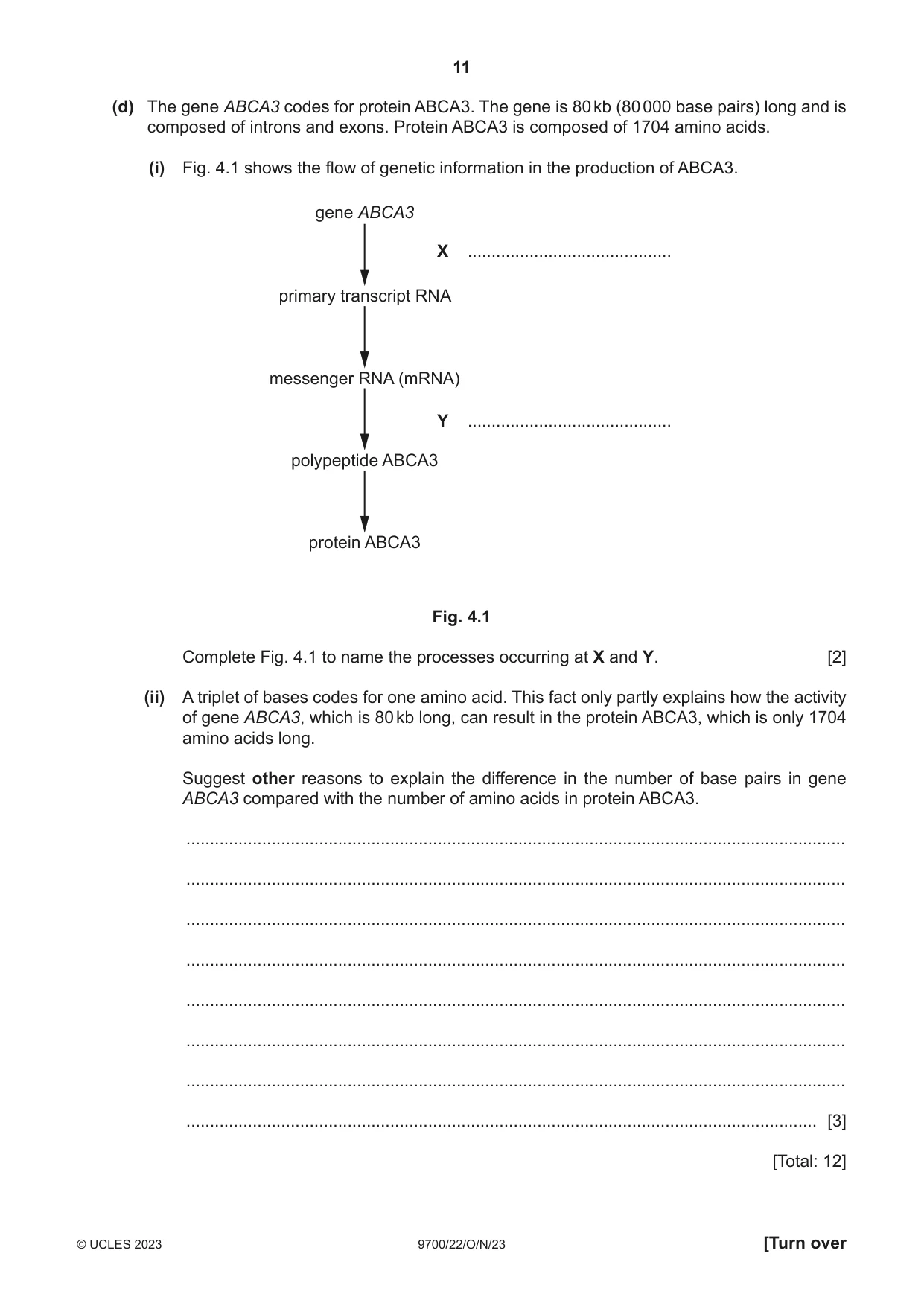
4 The alveoli of the lungs are the main gas exchange surface in humans. (a) Explain how blood flow through the alveolar capillaries helps to maintain steep diffusion gradients for gas exchange. ................................................................................................................................................... ................................................................................................................................................... ................................................................................................................................................... ................................................................................................................................................... ............................................................................................................................................. [2] (b) Ventilation of the lungs is the process of inhalation and exhalation. Ventilation helps to maintain steep diffusion gradients. Explain the role of elastic fibres in the alveolar wall during ventilation. ................................................................................................................................................... ................................................................................................................................................... ................................................................................................................................................... ................................................................................................................................................... ............................................................................................................................................. [2] (c) Some cells in the alveolar wall are specialised to secrete surfactant to prevent collapse of the alveoli at the end of exhalation. In these cells, surfactant is stored in membrane-bound organelles known as lamellar bodies. Surfactant is a mixture of lipids, mainly phospholipids, and some proteins. A protein known as ATP-binding cassette transporter A3 (ABCA3) is needed to move surfactant phospholipids into lamellar bodies from the surrounding cytosol (fluid part of cytoplasm). Suggest and explain the features of protein ABCA3 that make it suited to its function. ................................................................................................................................................... ................................................................................................................................................... ................................................................................................................................................... ................................................................................................................................................... ................................................................................................................................................... ................................................................................................................................................... ............................................................................................................................................. [3] (d) The gene ABCA3 codes for protein ABCA3. The gene is 80 kb (80 000 base pairs) long and is composed of introns and exons. Protein ABCA3 is composed of 1704 amino acids. (i) Fig. 4.1 shows the flow of genetic information in the production of ABCA3. gene ABCA3 X ........................................... primary transcript RNA messenger RNA (mRNA) Y ........................................... polypeptide ABCA3 protein ABCA3 Fig. 4.1 Complete Fig. 4.1 to name the processes occurring at X and Y. [2] (ii) A triplet of bases codes for one amino acid. This fact only partly explains how the activity of gene ABCA3, which is 80 kb long, can result in the protein ABCA3, which is only 1704 amino acids long. Suggest other reasons to explain the difference in the number of base pairs in gene ABCA3 compared with the number of amino acids in protein ABCA3. ........................................................................................................................................... ........................................................................................................................................... ........................................................................................................................................... ........................................................................................................................................... ........................................................................................................................................... ........................................................................................................................................... ........................................................................................................................................... ..................................................................................................................................... [3] [Total: 12]
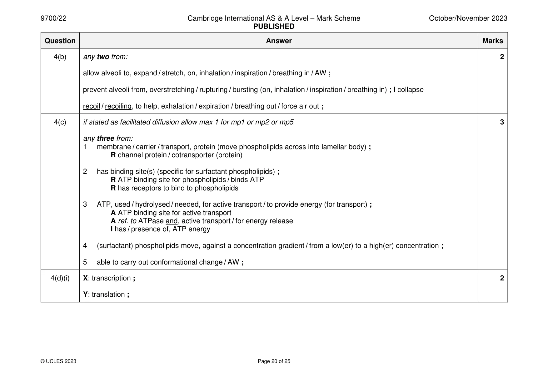
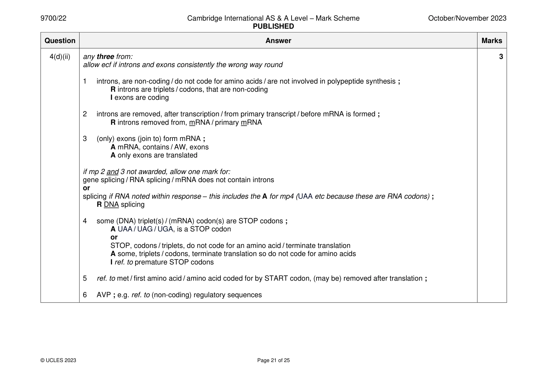
Mark scheme: 4(a) allow ideas from a diagram 2 1 blood arriving, is deoxygenated / has low(er) partial pressure of oxygen A low, concentrations / levels, of oxygen or oxygen-poor or blood arriving has, high(er) partial pressure / AW, of carbon dioxide ; A carbon dioxide-rich 2 idea that newly oxygenated blood is, constantly / continually, removed ; A oxygenated blood passed to pulmonary vein A oxygen that diffuses, from alveoli / into blood, is transported away (by blood flow) 4(b) any two from: 2 allow alveoli to, expand / stretch, on, inhalation / inspiration / breathing in / AW ; prevent alveoli from, overstretching / rupturing / bursting (on, inhalation / inspiration / breathing in) ; I collapse recoil / recoiling, to help, exhalation / expiration / breathing out / force air out ; 4(c) if stated as facilitated diffusion allow max 1 for mp1 or mp2 or mp5 3 any three from: 1 membrane / carrier / transport, protein (move phospholipids across into lamellar body) ; R channel protein / cotransporter (protein) 2 has binding site(s) (specific for surfactant phospholipids) ; R ATP binding site for phospholipids / binds ATP R has receptors to bind to phospholipids 3 ATP, used / hydrolysed / needed, for active transport / to provide energy (for transport) ; A ATP binding site for active transport A ref. to ATPase and, active transport / for energy release I has / presence of, ATP energy 4 (surfactant) phospholipids move, against a concentration gradient / from a low(er) to a high(er) concentration ; 5 able to carry out conformational change / AW ; 4(d)(i) X: transcription ; 2 Y: translation ; 4(d)(ii) any three from: 3 allow ecf if introns and exons consistently the wrong way round 1 introns, are non-coding / do not code for amino acids / are not involved in polypeptide synthesis ; R introns are triplets / codons, that are non-coding I exons are coding 2 introns are removed, after transcription / from primary transcript / before mRNA is formed ; R introns removed from, mRNA / primary mRNA 3 (only) exons (join to) form mRNA ; A mRNA, contains / AW, exons A only exons are translated if mp 2 and 3 not awarded, allow one mark for: gene splicing / RNA splicing / mRNA does not contain introns or splicing if RNA noted within response – this includes the A for mp4 (UAA etc because these are RNA codons) ; R DNA splicing 4 some (DNA) triplet(s) / (mRNA) codon(s) are STOP codons ; A UAA / UAG / UGA, is a STOP codon or STOP, codons / triplets, do not code for an amino acid / terminate translation A some, triplets / codons, terminate translation so do not code for amino acids I ref. to premature STOP codons 5 ref. to met / first amino acid / amino acid coded for by START codon, (may be) removed after translation ; 6 AVP ; e.g. ref. to (non-coding) regulatory sequences
Q5 · Tuberculosis (TB) is an infectious disease caused by a bacterium
5 Tuberculosis (TB) is an infectious disease caused by a bacterium. In the majority of people, only the lungs are affected. In most cases, the transmission of TB from an infected person to an uninfected person involves Mycobacterium tuberculosis. A different species of bacterium is involved in the transmission of TB from cattle, such as dairy cows, to humans. (a) Name the species of bacterium causing the transmission of TB from dairy cows to humans. ............................................................................................................................................. [1] (b) In some areas, cattle cannot be regularly tested or treated for TB. In these areas, milk and dairy products from infected dairy cattle may enter the human food chain. Outline a control measure that can be taken to protect people that consume milk and dairy products from these infected cattle. ................................................................................................................................................... ............................................................................................................................................. [1] (c) Cattle are not usually affected by M. tuberculosis, but the pathogen can cause disease in other animals. A few cases of transmission of TB from people to animals have been reported. Explain the most likely mode of transmission of TB from an infected person to an animal. ................................................................................................................................................... ................................................................................................................................................... ................................................................................................................................................... ................................................................................................................................................... ............................................................................................................................................. [2] (d) In most people, the response of the immune system to the infection of lung tissue by M. tuberculosis can prevent the spread of the bacterium to other organs of the body. The bacterium is contained in the lungs in a dormant state. This is known as latent TB. Outline the treatment that is used to kill M. tuberculosis in latent TB infections. ................................................................................................................................................... ................................................................................................................................................... ............................................................................................................................................. [2] (e) M. tuberculosis can spread in the blood and lymph to other organs in the body. In very rare cases, a disease known as mycotic aneurysm can be caused by infection of the arterial wall, particularly in elastic arteries. The damage caused by the pathogen can lead to a rupture (bursting) of the artery. With reference to the structure of the wall of elastic arteries, suggest how damage caused by M. tuberculosis infection can lead to the rupture of the artery. You may draw a diagram if you wish. ................................................................................................................................................... ................................................................................................................................................... ................................................................................................................................................... ................................................................................................................................................... ................................................................................................................................................... ................................................................................................................................................... ................................................................................................................................................... ................................................................................................................................................... ................................................................................................................................................... Space for diagram [3] [Total: 9]
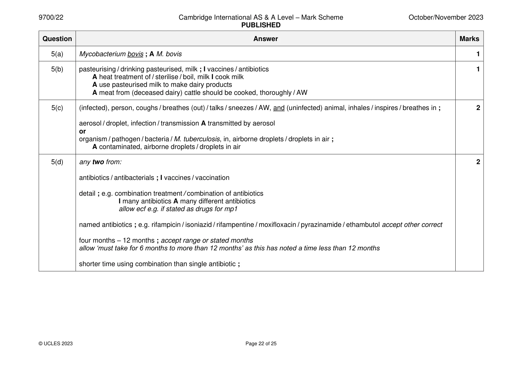
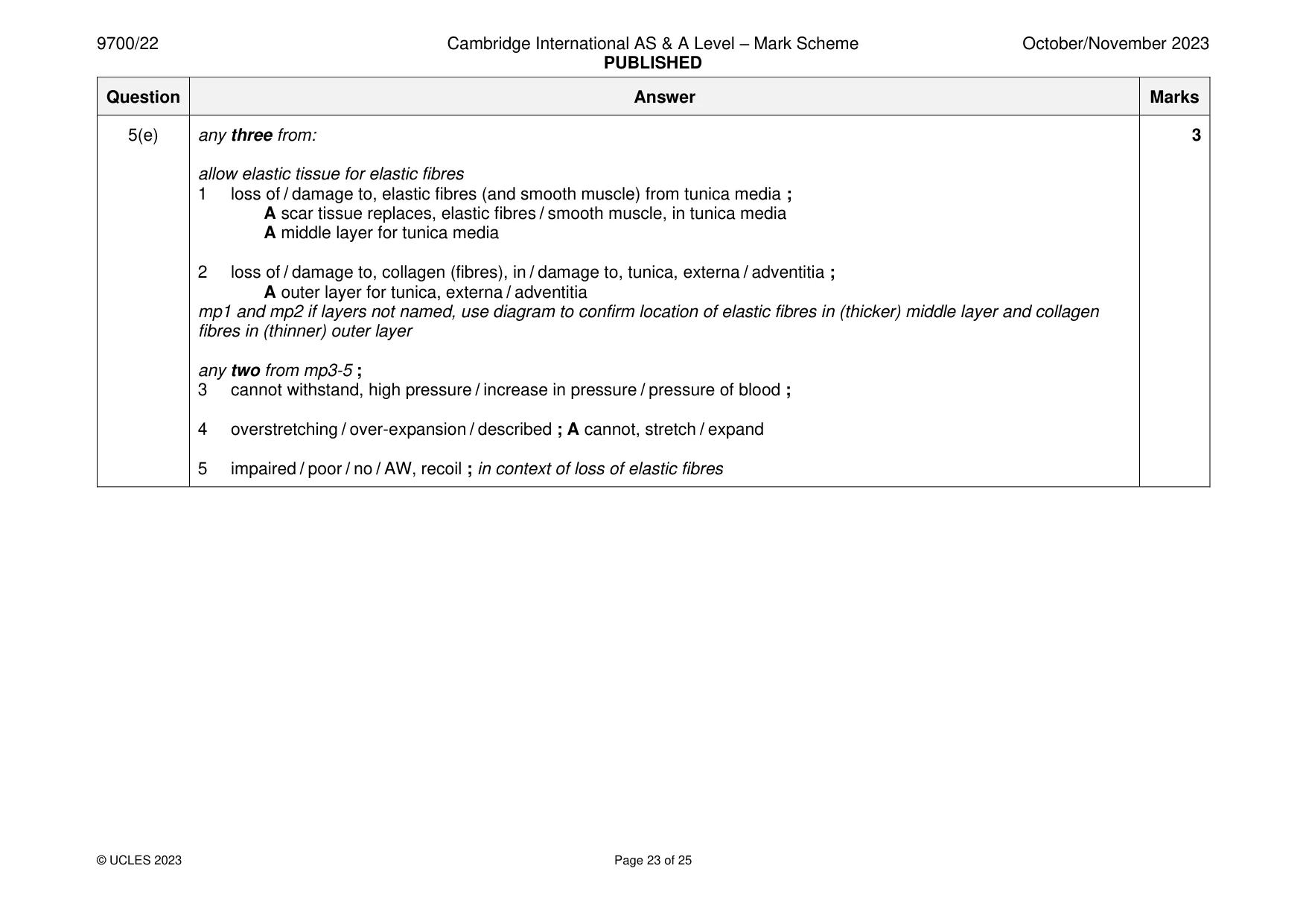
Mark scheme: 5(a) Mycobacterium bovis ; A M. bovis 1 5(b) pasteurising / drinking pasteurised, milk ; I vaccines / antibiotics 1 A heat treatment of / sterilise / boil, milk I cook milk A use pasteurised milk to make dairy products A meat from (deceased dairy) cattle should be cooked, thoroughly / AW 5(c) (infected), person, coughs / breathes (out) / talks / sneezes / AW, and (uninfected) animal, inhales / inspires / breathes in ; 2 aerosol / droplet, infection / transmission A transmitted by aerosol or organism / pathogen / bacteria / M. tuberculosis, in, airborne droplets / droplets in air ; A contaminated, airborne droplets / droplets in air 5(d) any two from: 2 antibiotics / antibacterials ; I vaccines / vaccination detail ; e.g. combination treatment / combination of antibiotics I many antibiotics A many different antibiotics allow ecf e.g. if stated as drugs for mp1 named antibiotics ; e.g. rifampicin / isoniazid / rifampentine / moxifloxacin / pyrazinamide / ethambutol accept other correct four months – 12 months ; accept range or stated months allow ‘must take for 6 months to more than 12 months’ as this has noted a time less than 12 months shorter time using combination than single antibiotic ; 5(e) any three from: 3 allow elastic tissue for elastic fibres 1 loss of / damage to, elastic fibres (and smooth muscle) from tunica media ; A scar tissue replaces, elastic fibres / smooth muscle, in tunica media A middle layer for tunica media 2 loss of / damage to, collagen (fibres), in / damage to, tunica, externa / adventitia ; A outer layer for tunica, externa / adventitia mp1 and mp2 if layers not named, use diagram to confirm location of elastic fibres in (thicker) middle layer and collagen fibres in (thinner) outer layer any two from mp3-5 ; 3 cannot withstand, high pressure / increase in pressure / pressure of blood ; 4 overstretching / over-expansion / described ; A cannot, stretch / expand 5 impaired / poor / no / AW, recoil ; in context of loss of elastic fibres
Q6 · Water that is absorbed from the soil solution by the roots of a plant enters xylem…
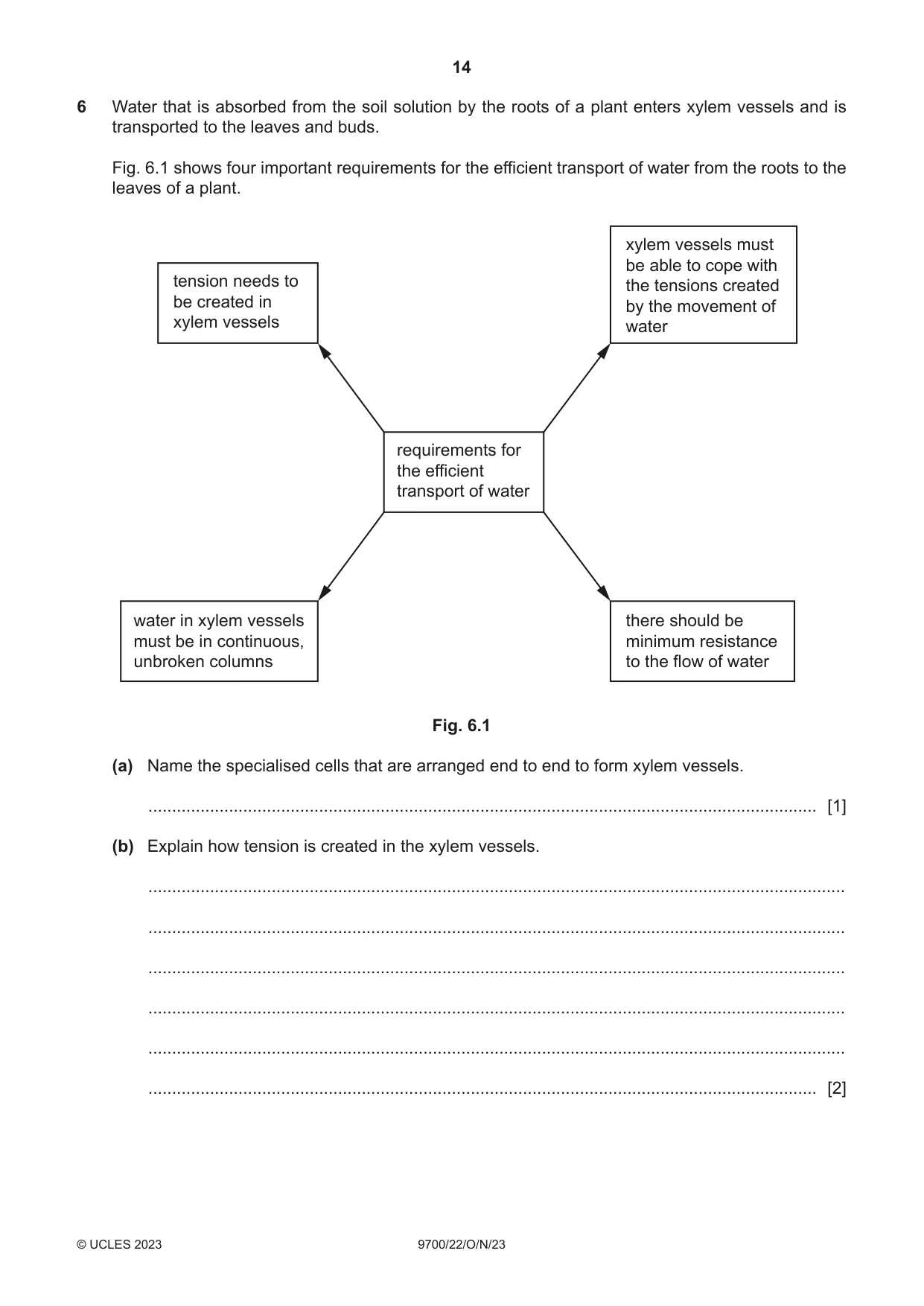
6 Water that is absorbed from the soil solution by the roots of a plant enters xylem vessels and is transported to the leaves and buds. Fig. 6.1 shows four important requirements for the efficient transport of water from the roots to the leaves of a plant. xylem vessels must be able to cope with tension needs to the tensions created be created in by the movement of xylem vessels water requirements for the efficient transport of water water in xylem vessels there should be must be in continuous, minimum resistance unbroken columns to the flow of water Fig. 6.1 (a) Name the specialised cells that are arranged end to end to form xylem vessels. ............................................................................................................................................. [1] (b) Explain how tension is created in the xylem vessels. ................................................................................................................................................... ................................................................................................................................................... ................................................................................................................................................... ................................................................................................................................................... ................................................................................................................................................... ............................................................................................................................................. [2] (c) Fig. 6.1 highlights how the structure of xylem vessels must be related to their function. This means that during the development of xylem vessels changes need to occur to the cells forming the vessels. (i) The walls of the cells forming the xylem vessel walls become lignified during development. Explain how this feature is important for the efficient transport of water. ........................................................................................................................................... ........................................................................................................................................... ........................................................................................................................................... ........................................................................................................................................... ..................................................................................................................................... [2] (ii) During the development of xylem vessels, the end walls of the cells forming the vessels break down. This contributes to minimising resistance to the flow of water. Describe one other main change that needs to occur to these cells so that their structure becomes suited to their function. ........................................................................................................................................... ........................................................................................................................................... ..................................................................................................................................... [1] [Total: 6]
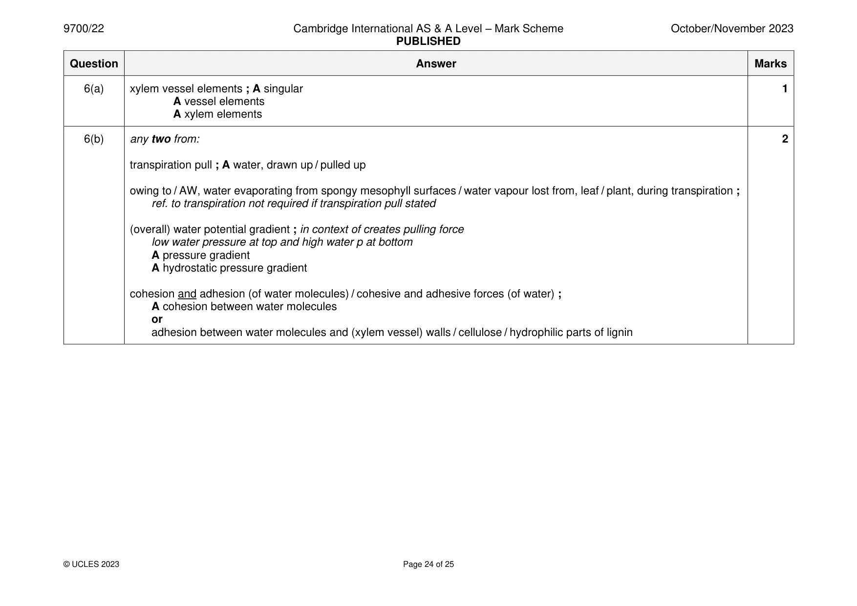
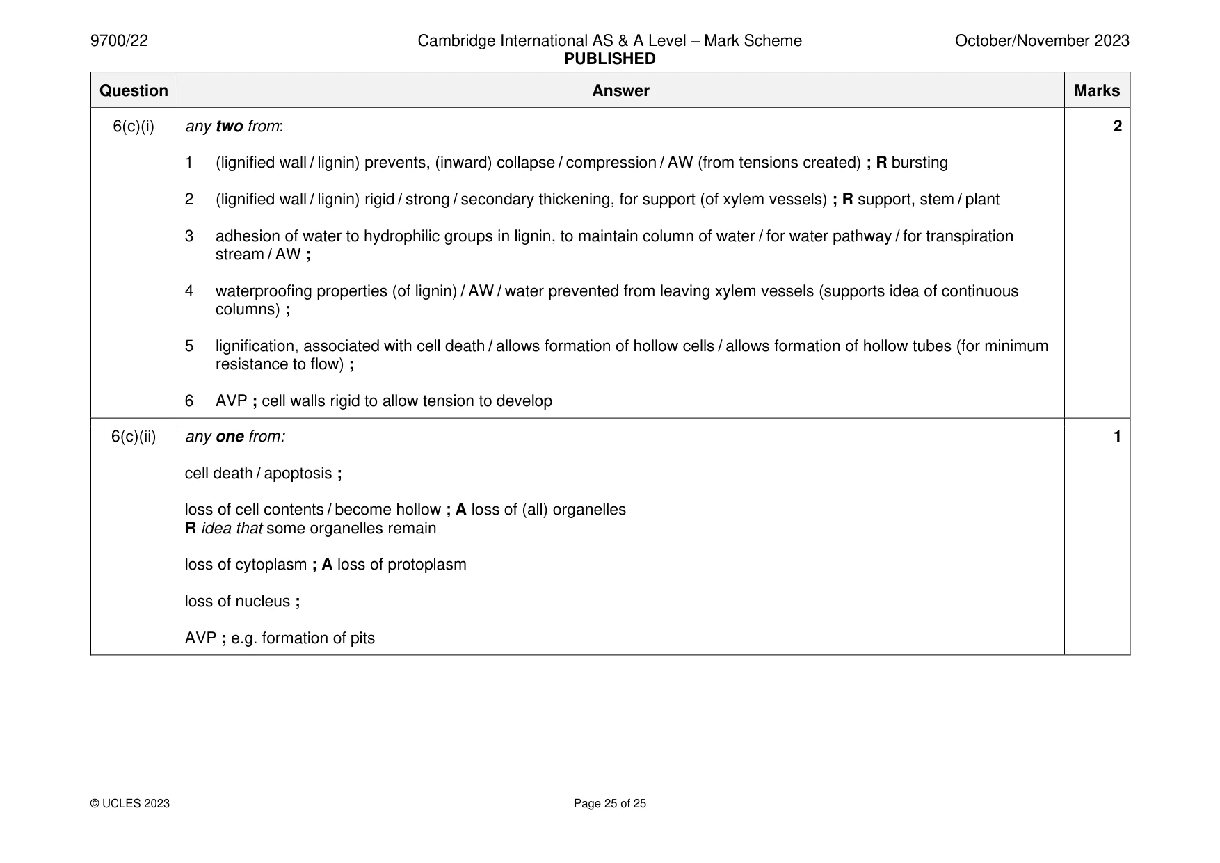
Mark scheme: 6(a) xylem vessel elements ; A singular 1 A vessel elements A xylem elements 6(b) any two from: 2 transpiration pull ; A water, drawn up / pulled up owing to / AW, water evaporating from spongy mesophyll surfaces / water vapour lost from, leaf / plant, during transpiration ; ref. to transpiration not required if transpiration pull stated (overall) water potential gradient ; in context of creates pulling force low water pressure at top and high water p at bottom A pressure gradient A hydrostatic pressure gradient cohesion and adhesion (of water molecules) / cohesive and adhesive forces (of water) ; A cohesion between water molecules or adhesion between water molecules and (xylem vessel) walls / cellulose / hydrophilic parts of lignin 6(c)(i) any two from: 2 1 (lignified wall / lignin) prevents, (inward) collapse / compression / AW (from tensions created) ; R bursting 2 (lignified wall / lignin) rigid / strong / secondary thickening, for support (of xylem vessels) ; R support, stem / plant 3 adhesion of water to hydrophilic groups in lignin, to maintain column of water / for water pathway / for transpiration stream / AW ; 4 waterproofing properties (of lignin) / AW / water prevented from leaving xylem vessels (supports idea of continuous columns) ; 5 lignification, associated with cell death / allows formation of hollow cells / allows formation of hollow tubes (for minimum resistance to flow) ; 6 AVP ; cell walls rigid to allow tension to develop 6(c)(ii) any one from: 1 cell death / apoptosis ; loss of cell contents / become hollow ; A loss of (all) organelles R idea that some organelles remain loss of cytoplasm ; A loss of protoplasm loss of nucleus ; AVP ; e.g. formation of pits
What was in this paper
The subtopics covered by these 6 questions, and how many questions each got. Open one in a new tab to see every Cambridge question on it.
What you needed in this session
Cambridge’s own grade thresholds for 2023 Oct/Nov, Paper 2 · Variant 2. A higher threshold means an easier paper — the bar moves with how the cohort did.