Cambridge A Level Biology 9700 — 2022 Oct/Nov Paper 2 · Variant 3
9700/23/O/N/22 · 6 questions · 60 marks · ≈68 min
The question paper and its mark scheme, free to read here and free to download. This is Cambridge’s own paper, exactly as it was sat.
Question paper16 pages

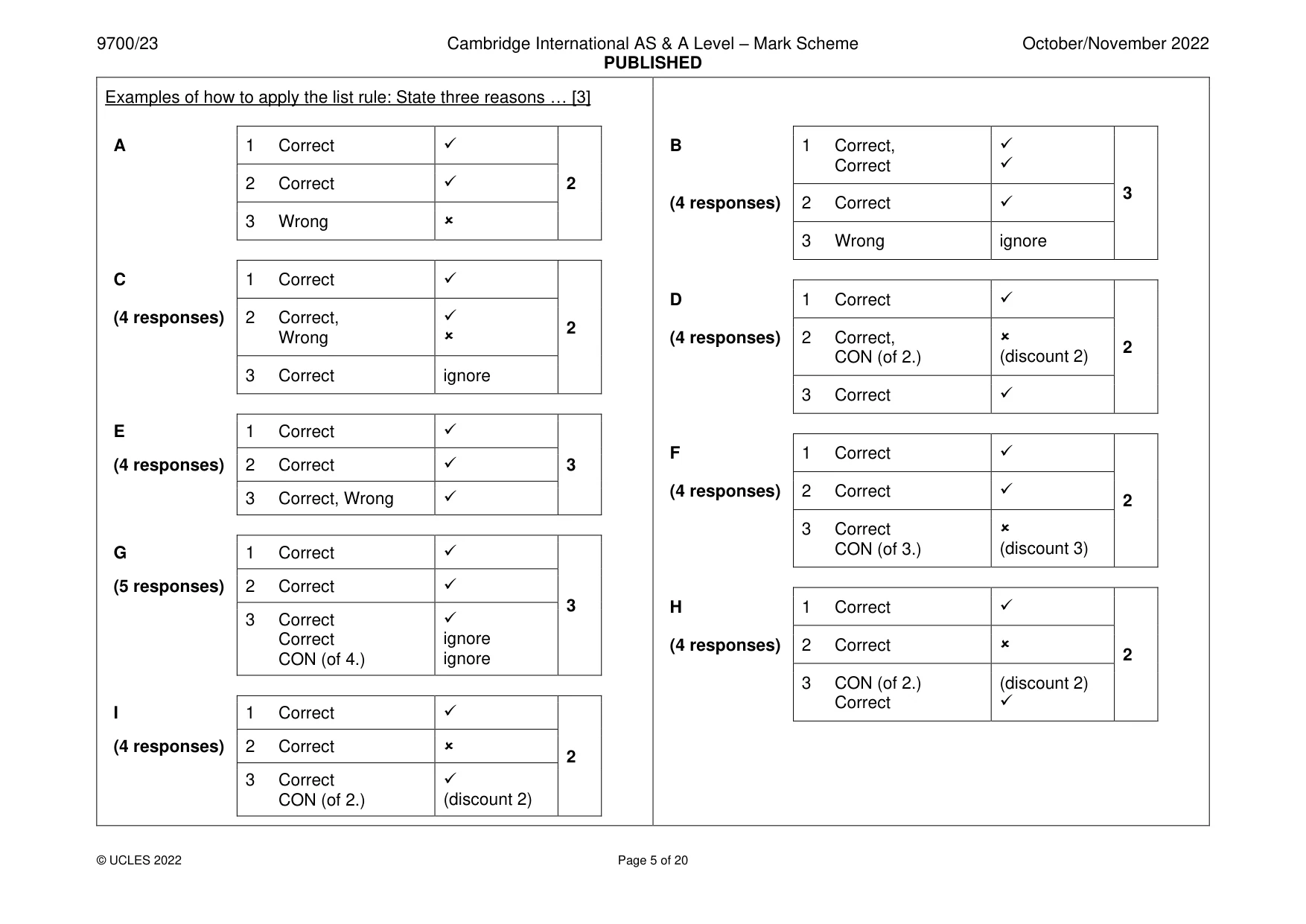
Mark scheme20 pages
Answers below. Sit the paper first if you are practising.
Questions as text
Q1 · During translation, a polypeptide is synthesised when amino acids are added to a growing…
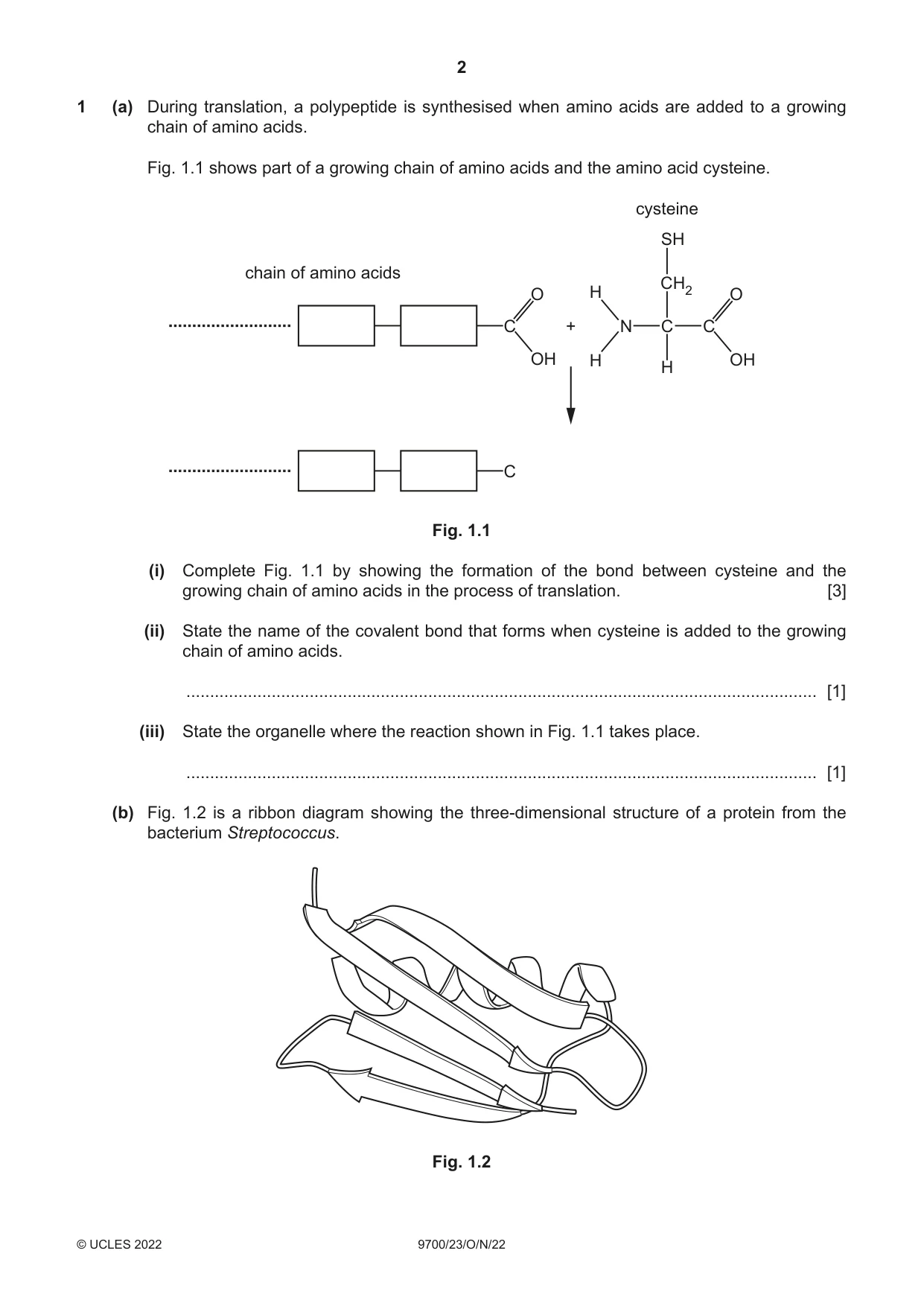
1 (a) During translation, a polypeptide is synthesised when amino acids are added to a growing chain of amino acids. Fig. 1.1 shows part of a growing chain of amino acids and the amino acid cysteine. cysteine SH chain of amino acids CH2 O H O .......................... C + N C C OH H OH H .......................... C Fig. 1.1 (i) Complete Fig. 1.1 by showing the formation of the bond between cysteine and the growing chain of amino acids in the process of translation. [3] (ii) State the name of the covalent bond that forms when cysteine is added to the growing chain of amino acids. ..................................................................................................................................... [1] (iii) State the organelle where the reaction shown in Fig. 1.1 takes place. ..................................................................................................................................... [1] (b) Fig. 1.2 is a ribbon diagram showing the three-dimensional structure of a protein from the bacterium Streptococcus. Fig. 1.2 (i) Describe the secondary structure of the protein shown in Fig. 1.2. ........................................................................................................................................... ........................................................................................................................................... ........................................................................................................................................... ........................................................................................................................................... ..................................................................................................................................... [2] (ii) Explain why the protein shown in Fig. 1.2 has tertiary structure, but not quaternary structure. ........................................................................................................................................... ........................................................................................................................................... ........................................................................................................................................... ........................................................................................................................................... ..................................................................................................................................... [2] (iii) An analysis of the amino acid composition of the protein in Fig. 1.2 showed that it does not contain any cysteine residues. Explain how the three-dimensional structure of the protein shown in Fig. 1.2 is held in place. ........................................................................................................................................... ........................................................................................................................................... ........................................................................................................................................... ........................................................................................................................................... ........................................................................................................................................... ........................................................................................................................................... ........................................................................................................................................... ..................................................................................................................................... [3] [Total: 12]
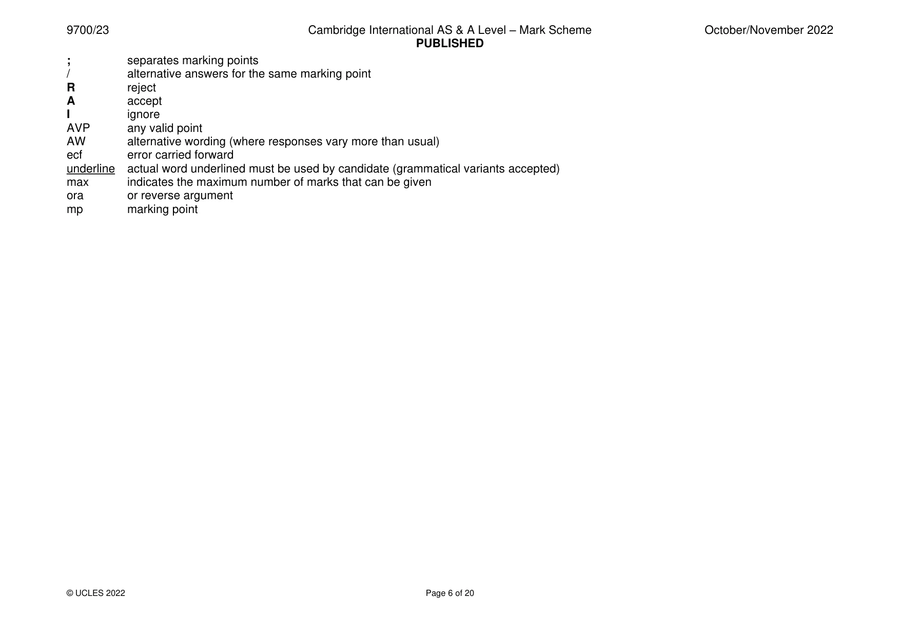
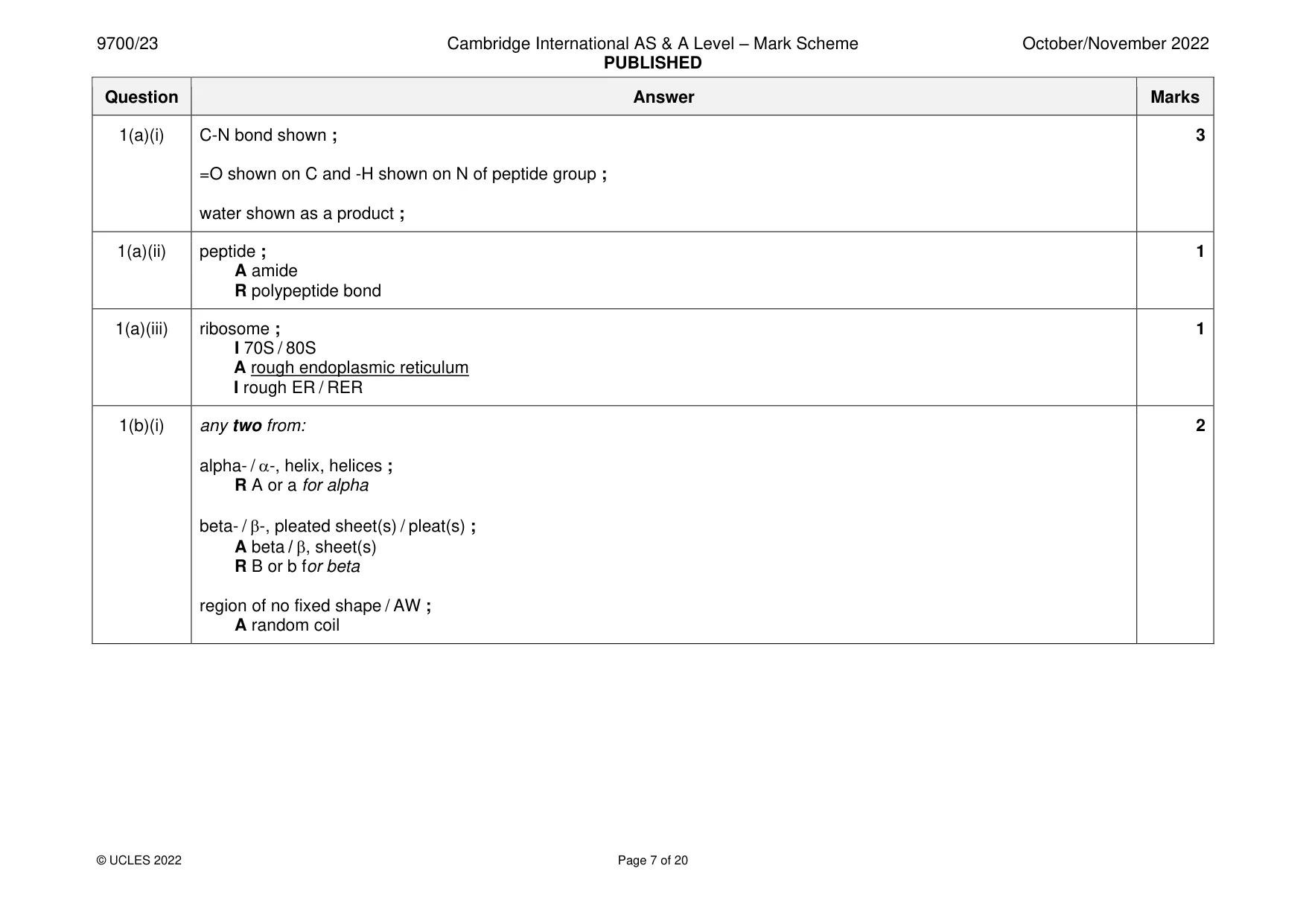
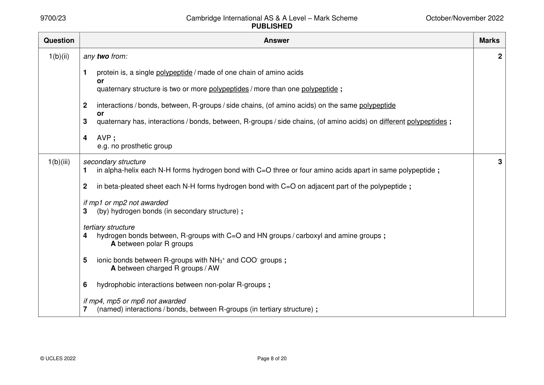
Mark scheme: Question Answer Marks 1(a)(i) C-N bond shown ; 3 =O shown on C and -H shown on N of peptide group ; water shown as a product ; 1(a)(ii) peptide ; 1 A amide R polypeptide bond 1(a)(iii) ribosome ; 1 I 70S / 80S A rough endoplasmic reticulum I rough ER / RER 1(b)(i) any two from: 2 alpha- / -, helix, helices ; R A or a for alpha beta- / -, pleated sheet(s) / pleat(s) ; A beta / , sheet(s) R B or b for beta region of no fixed shape / AW ; A random coil 1(b)(ii) any two from: 2 1 protein is, a single polypeptide / made of one chain of amino acids or quaternary structure is two or more polypeptides / more than one polypeptide ; 2 interactions / bonds, between, R-groups / side chains, (of amino acids) on the same polypeptide or 3 quaternary has, interactions / bonds, between, R-groups / side chains, (of amino acids) on different polypeptides ; 4 AVP ; e.g. no prosthetic group 1(b)(iii) secondary structure 3 1 in alpha-helix each N-H forms hydrogen bond with C=O three or four amino acids apart in same polypeptide ; 2 in beta-pleated sheet each N-H forms hydrogen bond with C=O on adjacent part of the polypeptide ; if mp1 or mp2 not awarded 3 (by) hydrogen bonds (in secondary structure) ; tertiary structure 4 hydrogen bonds between, R-groups with C=O and HN groups / carboxyl and amine groups ; A between polar R groups 5 ionic bonds between R-groups with NH3+ and COO- groups ; A between charged R groups / AW 6 hydrophobic interactions between non-polar R-groups ; if mp4, mp5 or mp6 not awarded 7 (named) interactions / bonds, between R-groups (in tertiary structure) ;
Q2 · A scanning electron micrograph showing a macrophage engulfing some bacteria
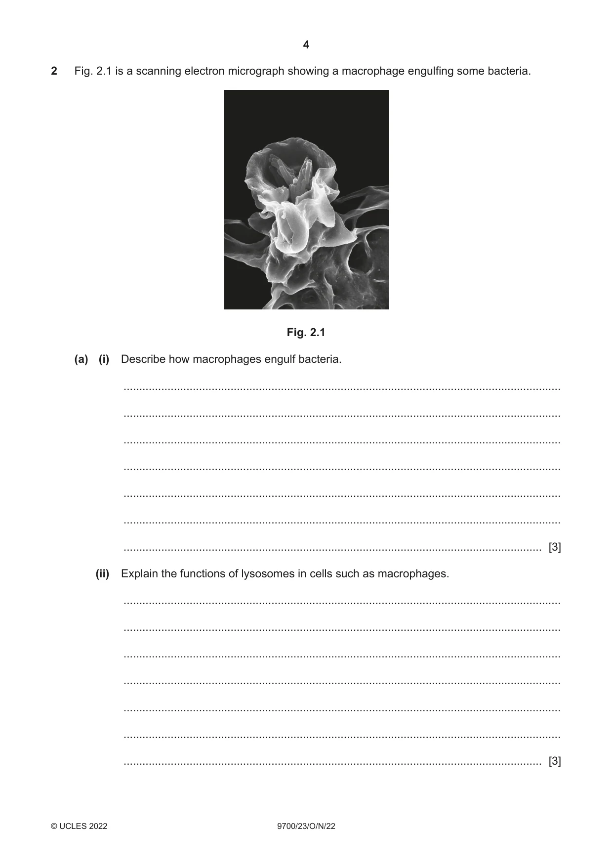
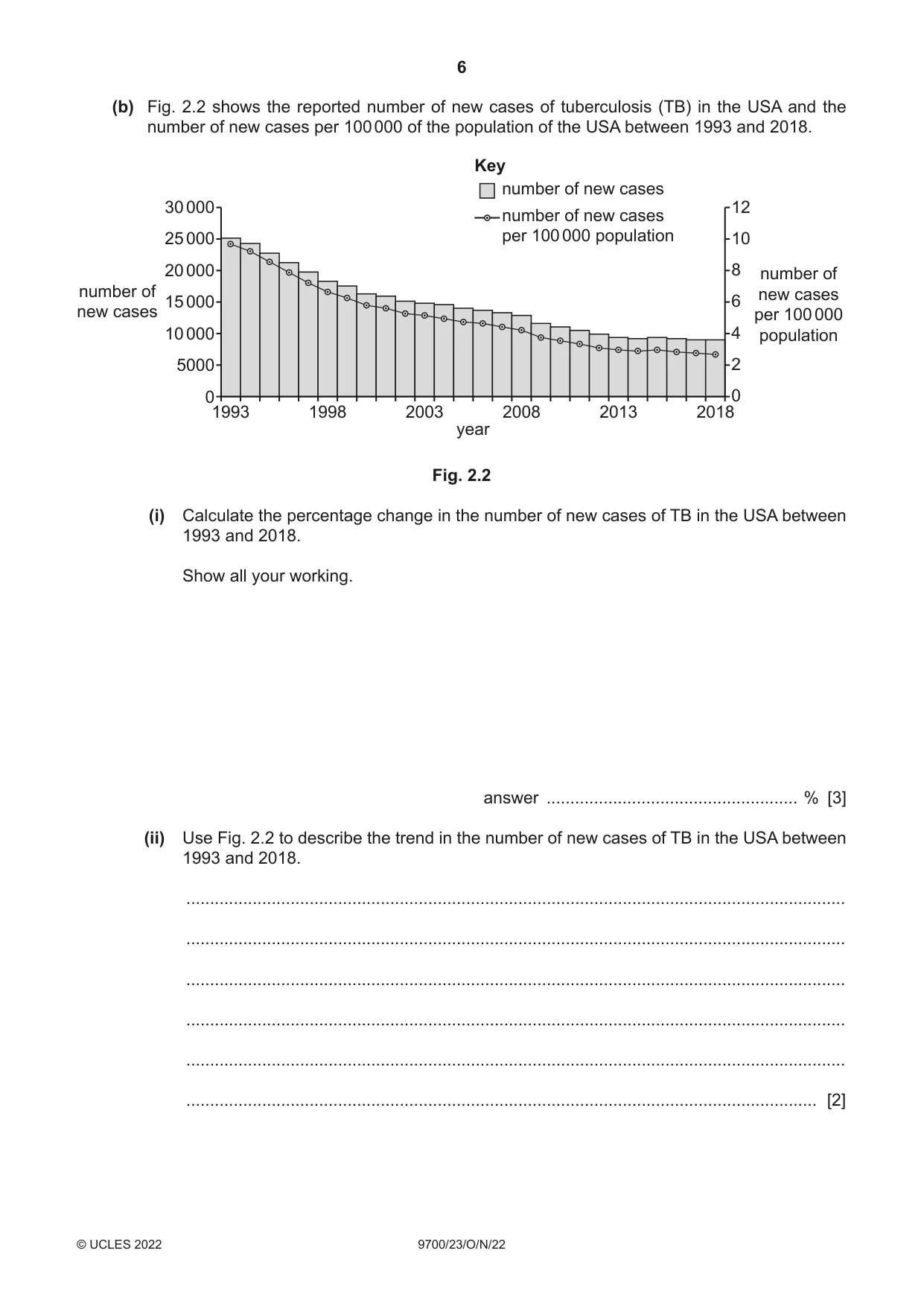
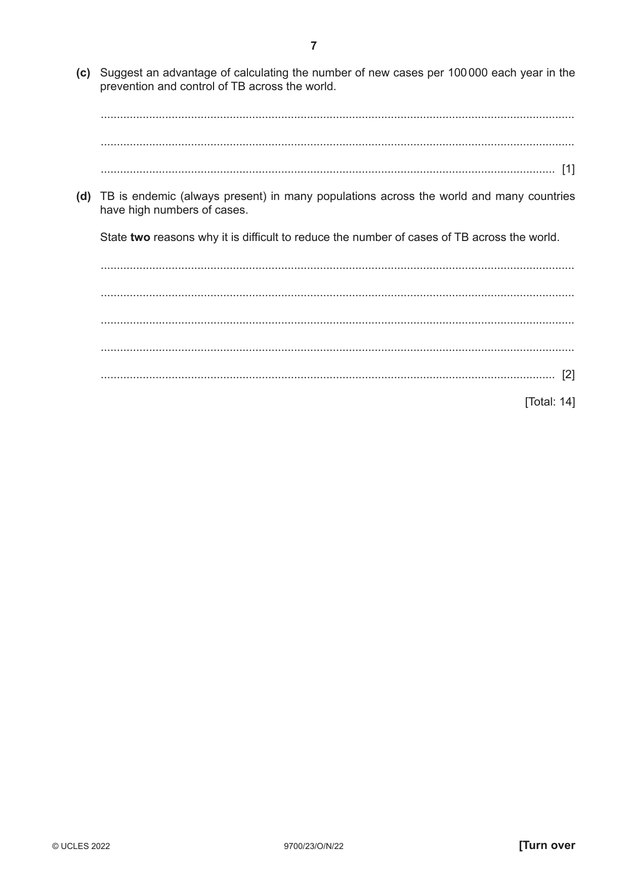
2 Fig. 2.1 is a scanning electron micrograph showing a macrophage engulfing some bacteria. Fig. 2.1 (a) (i) Describe how macrophages engulf bacteria. ........................................................................................................................................... ........................................................................................................................................... ........................................................................................................................................... ........................................................................................................................................... ........................................................................................................................................... ........................................................................................................................................... ..................................................................................................................................... [3] (ii) Explain the functions of lysosomes in cells such as macrophages. ........................................................................................................................................... ........................................................................................................................................... ........................................................................................................................................... ........................................................................................................................................... ........................................................................................................................................... ........................................................................................................................................... ..................................................................................................................................... [3] Question 2 continues on page 6. (b) Fig. 2.2 shows the reported number of new cases of tuberculosis (TB) in the USA and the number of new cases per 100 000 of the population of the USA between 1993 and 2018. Key number of new cases 30 000 12 number of new cases 25 000 per 100 000 population 10 20 000 8 number of number of new cases 15 000 6new cases per 100 000 10 000 4 population 5000 2 0 0 1993 1998 2003 2008 2013 2018 year Fig. 2.2 (i) Calculate the percentage change in the number of new cases of TB in the USA between 1993 and 2018. Show all your working. answer ..................................................... % [3] (ii) Use Fig. 2.2 to describe the trend in the number of new cases of TB in the USA between 1993 and 2018. ........................................................................................................................................... ........................................................................................................................................... ........................................................................................................................................... ........................................................................................................................................... ........................................................................................................................................... ..................................................................................................................................... [2] (c) Suggest an advantage of calculating the number of new cases per 100 000 each year in the prevention and control of TB across the world. ................................................................................................................................................... ................................................................................................................................................... ............................................................................................................................................. [1] (d) TB is endemic (always present) in many populations across the world and many countries have high numbers of cases. State two reasons why it is difficult to reduce the number of cases of TB across the world. ................................................................................................................................................... ................................................................................................................................................... ................................................................................................................................................... ................................................................................................................................................... ............................................................................................................................................. [2] [Total: 14]
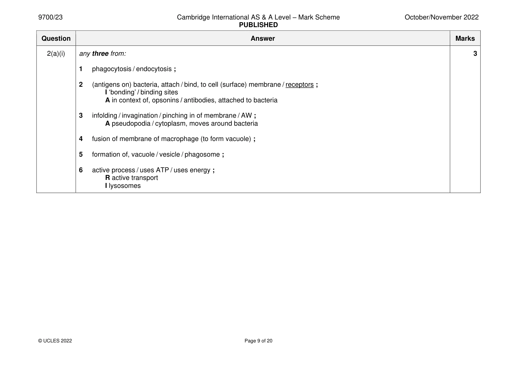
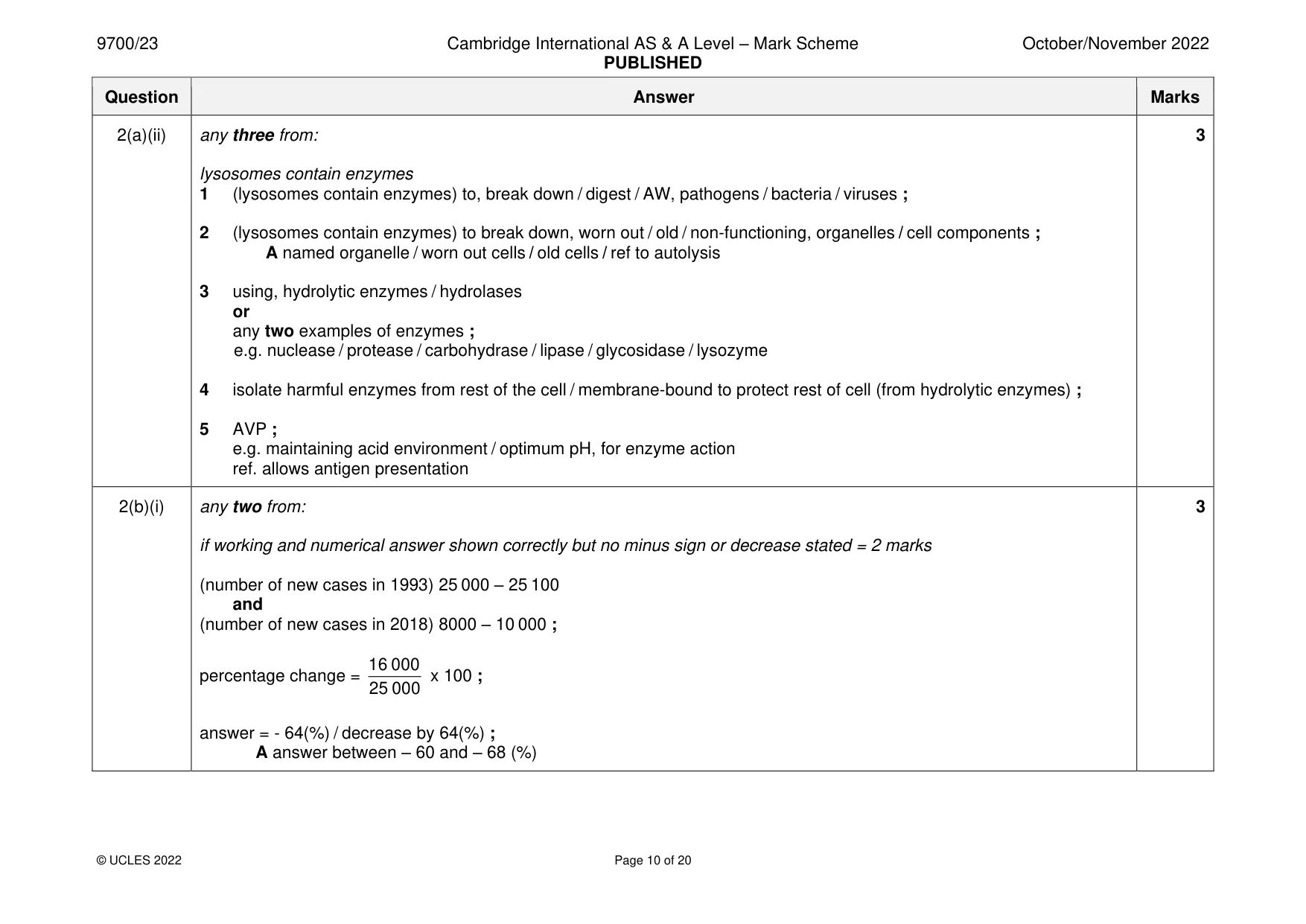
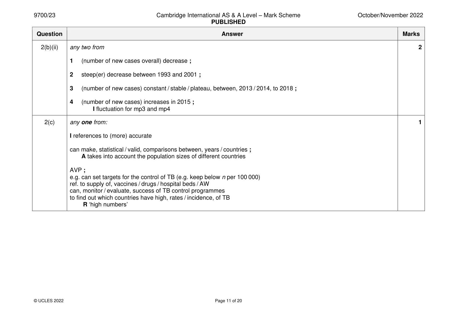
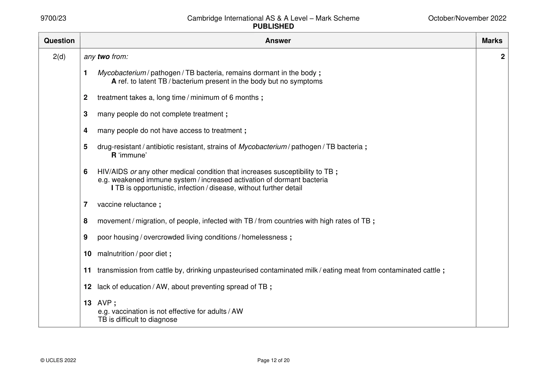
Mark scheme: 2(a)(i) any three from: 3 1 phagocytosis / endocytosis ; 2 (antigens on) bacteria, attach / bind, to cell (surface) membrane / receptors ; I ‘bonding’ / binding sites A in context of, opsonins / antibodies, attached to bacteria 3 infolding / invagination / pinching in of membrane / AW ; A pseudopodia / cytoplasm, moves around bacteria 4 fusion of membrane of macrophage (to form vacuole) ; 5 formation of, vacuole / vesicle / phagosome ; 6 active process / uses ATP / uses energy ; R active transport I lysosomes 2(a)(ii) any three from: 3 lysosomes contain enzymes 1 (lysosomes contain enzymes) to, break down / digest / AW, pathogens / bacteria / viruses ; 2 (lysosomes contain enzymes) to break down, worn out / old / non-functioning, organelles / cell components ; A named organelle / worn out cells / old cells / ref to autolysis 3 using, hydrolytic enzymes / hydrolases or any two examples of enzymes ; e.g. nuclease / protease / carbohydrase / lipase / glycosidase / lysozyme 4 isolate harmful enzymes from rest of the cell / membrane-bound to protect rest of cell (from hydrolytic enzymes) ; 5 AVP ; e.g. maintaining acid environment / optimum pH, for enzyme action ref. allows antigen presentation 2(b)(i) any two from: 3 if working and numerical answer shown correctly but no minus sign or decrease stated = 2 marks (number of new cases in 1993) 25 000 – 25 100 and (number of new cases in 2018) 8000 – 10 000 ; 16 000 percentage change = x 100 ; 25 000 answer = - 64(%) / decrease by 64(%) ; A answer between – 60 and – 68 (%) 2(b)(ii) any two from 2 1 (number of new cases overall) decrease ; 2 steep(er) decrease between 1993 and 2001 ; 3 (number of new cases) constant / stable / plateau, between, 2013 / 2014, to 2018 ; 4 (number of new cases) increases in 2015 ; I fluctuation for mp3 and mp4 2(c) any one from: 1 I references to (more) accurate can make, statistical / valid, comparisons between, years / countries ; A takes into account the population sizes of different countries AVP ; e.g. can set targets for the control of TB (e.g. keep below n per 100 000) ref. to supply of, vaccines / drugs / hospital beds / AW can, monitor / evaluate, success of TB control programmes to find out which countries have high, rates / incidence, of TB R ‘high numbers’ 2(d) any two from: 2 1 Mycobacterium / pathogen / TB bacteria, remains dormant in the body ; A ref. to latent TB / bacterium present in the body but no symptoms 2 treatment takes a, long time / minimum of 6 months ; 3 many people do not complete treatment ; 4 many people do not have access to treatment ; 5 drug-resistant / antibiotic resistant, strains of Mycobacterium / pathogen / TB bacteria ; R ‘immune’ 6 HIV/AIDS or any other medical condition that increases susceptibility to TB ; e.g. weakened immune system / increased activation of dormant bacteria I TB is opportunistic, infection / disease, without further detail 7 vaccine reluctance ; 8 movement / migration, of people, infected with TB / from countries with high rates of TB ; 9 poor housing / overcrowded living conditions / homelessness ; 10 malnutrition / poor diet ; 11 transmission from cattle by, drinking unpasteurised contaminated milk / eating meat from contaminated cattle ; 12 lack of education / AW, about preventing spread of TB ; 13 AVP ; e.g. vaccination is not effective for adults / AW TB is difficult to diagnose
Q3 · Explain how water moves up xylem vessels in the trunks of trees
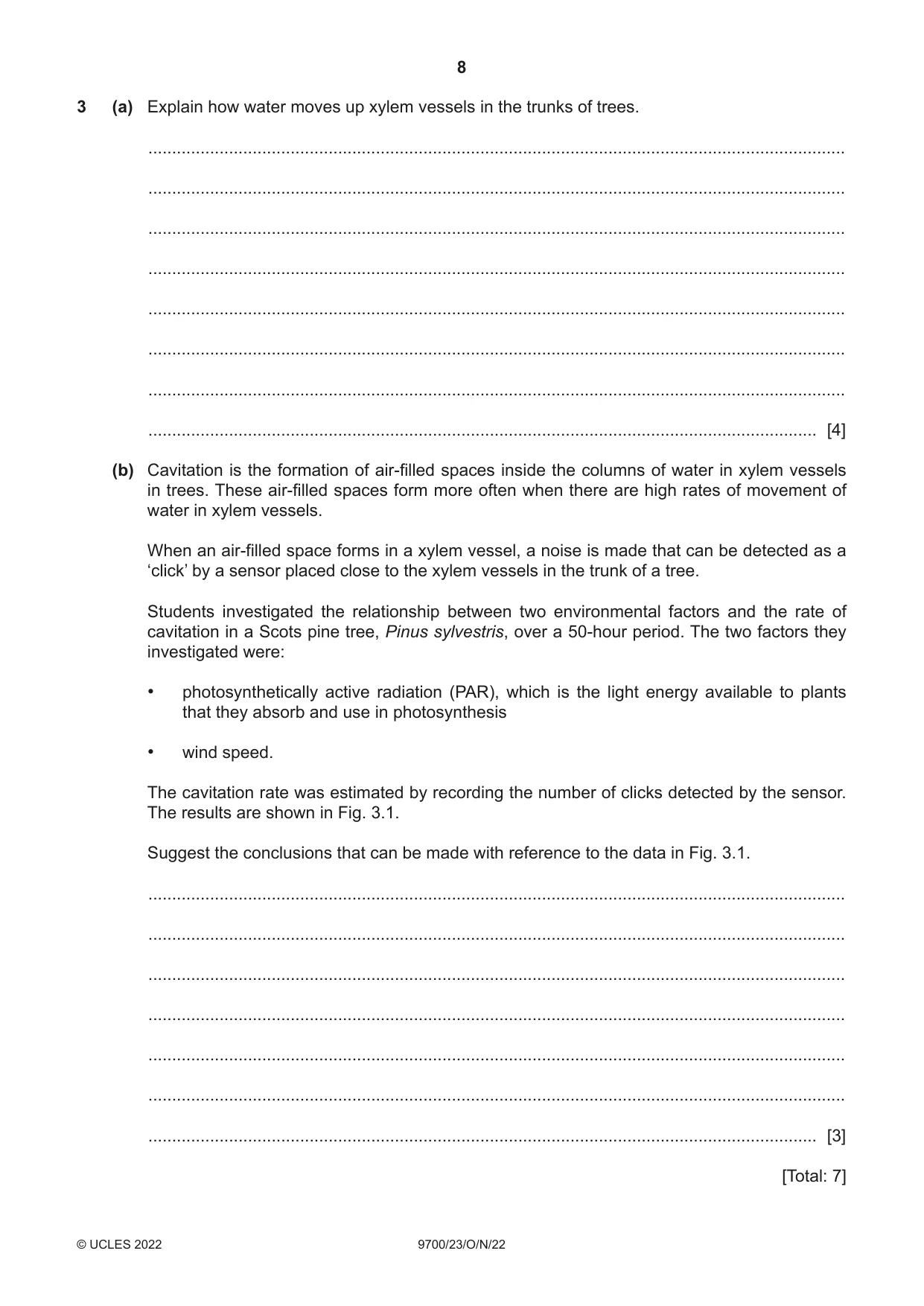
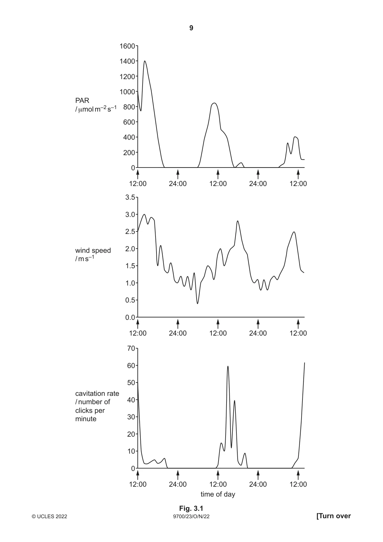
3 (a) Explain how water moves up xylem vessels in the trunks of trees. ................................................................................................................................................... ................................................................................................................................................... ................................................................................................................................................... ................................................................................................................................................... ................................................................................................................................................... ................................................................................................................................................... ................................................................................................................................................... ............................................................................................................................................. [4] (b) Cavitation is the formation of air-filled spaces inside the columns of water in xylem vessels in trees. These air-filled spaces form more often when there are high rates of movement of water in xylem vessels. When an air-filled space forms in a xylem vessel, a noise is made that can be detected as a ‘click’ by a sensor placed close to the xylem vessels in the trunk of a tree. Students investigated the relationship between two environmental factors and the rate of cavitation in a Scots pine tree, Pinus sylvestris, over a 50-hour period. The two factors they investigated were: • photosynthetically active radiation (PAR), which is the light energy available to plants that they absorb and use in photosynthesis • wind speed. The cavitation rate was estimated by recording the number of clicks detected by the sensor. The results are shown in Fig. 3.1. Suggest the conclusions that can be made with reference to the data in Fig. 3.1. ................................................................................................................................................... ................................................................................................................................................... ................................................................................................................................................... ................................................................................................................................................... ................................................................................................................................................... ................................................................................................................................................... ............................................................................................................................................. [3] [Total: 7] 1600 1400 1200 1000 PAR 800/ μmol m–2 s–1 600 400 200 0 12:00 24:00 12:00 24:00 12:00 3.5 3.0 2.5 wind speed 2.0 / m s–1 1.5 1.0 0.5 0.0 12:00 24:00 12:00 24:00 12:00 70 60 50 cavitation rate 40/ number of clicks per minute 30 20 10 0 12:00 24:00 12:00 24:00 12:00 time of day
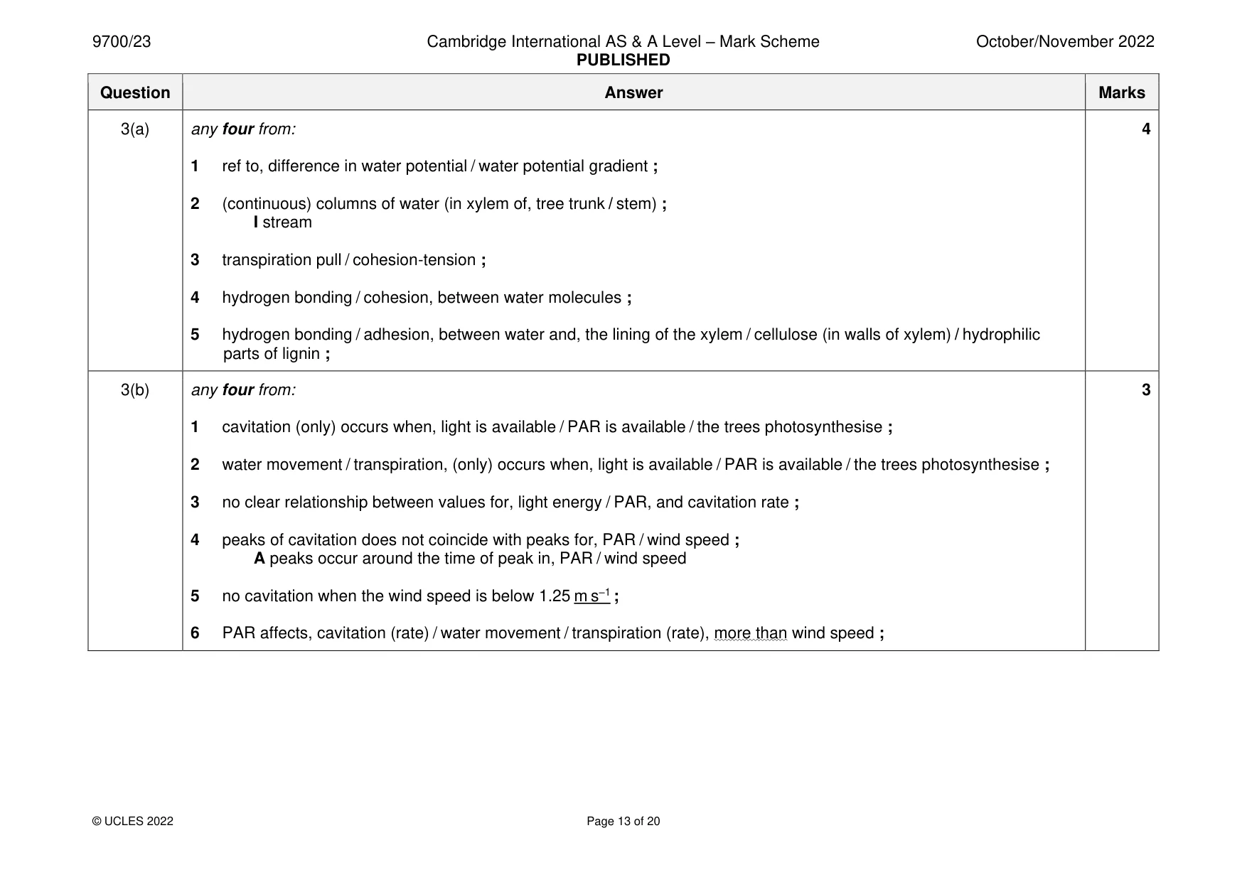
Mark scheme: 3(a) any four from: 4 1 ref to, difference in water potential / water potential gradient ; 2 (continuous) columns of water (in xylem of, tree trunk / stem) ; I stream 3 transpiration pull / cohesion-tension ; 4 hydrogen bonding / cohesion, between water molecules ; 5 hydrogen bonding / adhesion, between water and, the lining of the xylem / cellulose (in walls of xylem) / hydrophilic parts of lignin ; 3(b) any four from: 3 1 cavitation (only) occurs when, light is available / PAR is available / the trees photosynthesise ; 2 water movement / transpiration, (only) occurs when, light is available / PAR is available / the trees photosynthesise ; 3 no clear relationship between values for, light energy / PAR, and cavitation rate ; 4 peaks of cavitation does not coincide with peaks for, PAR / wind speed ; A peaks occur around the time of peak in, PAR / wind speed 5 no cavitation when the wind speed is below 1.25 m s–1 ; 6 PAR affects, cavitation (rate) / water movement / transpiration (rate), more than wind speed ;
Q4 · The early development of an animal involves divisions of the zygote and daughter cells by…
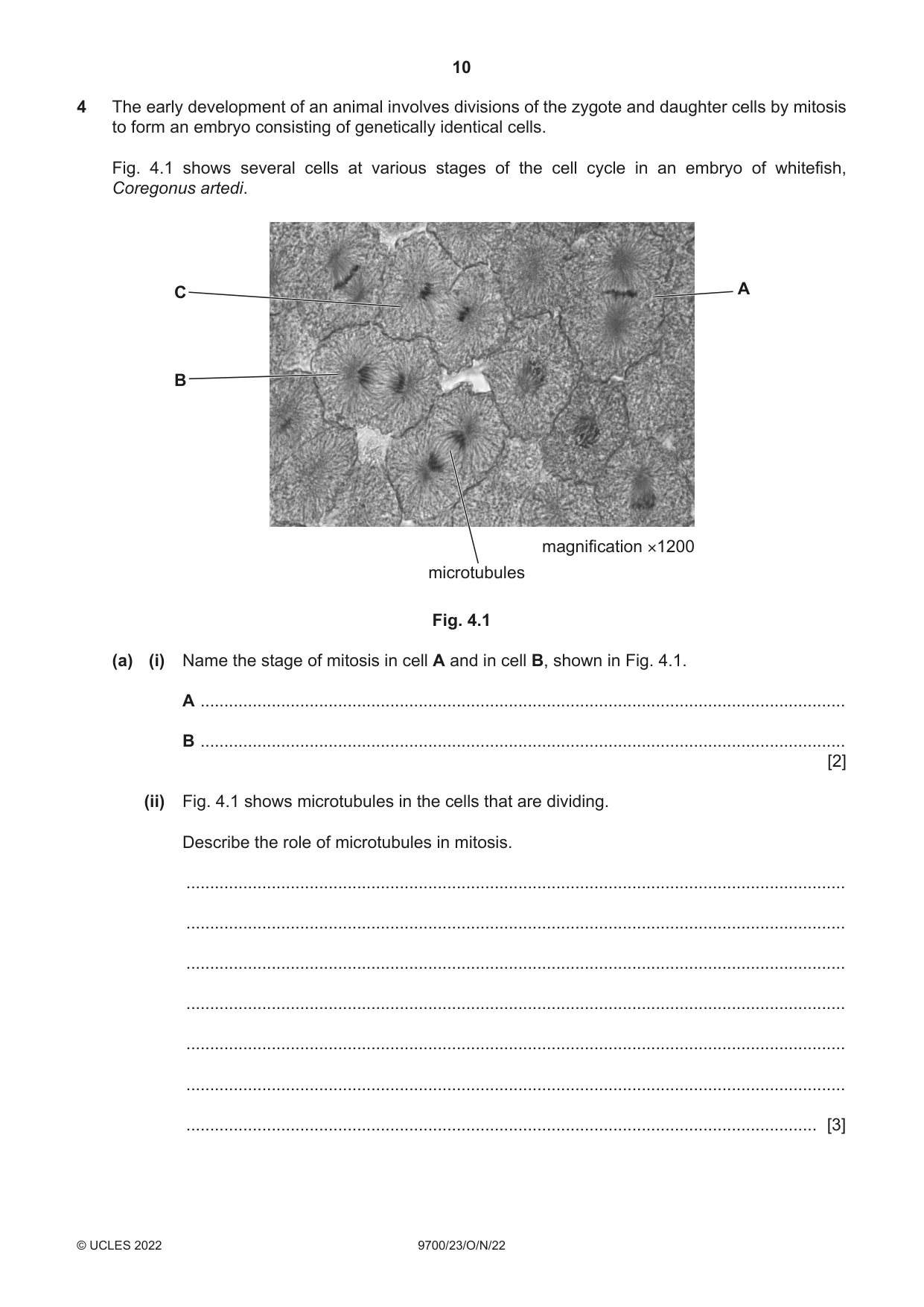
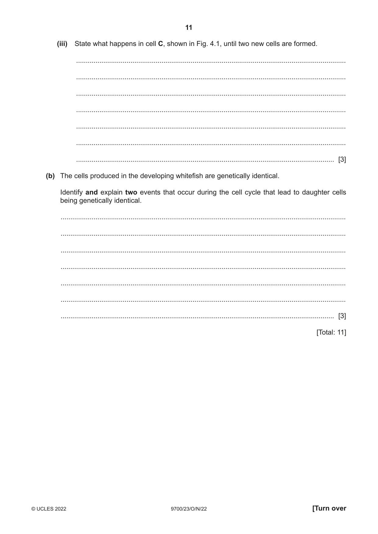
4 The early development of an animal involves divisions of the zygote and daughter cells by mitosis to form an embryo consisting of genetically identical cells. Fig. 4.1 shows several cells at various stages of the cell cycle in an embryo of whitefish, Coregonus artedi. C A B magnification ×1200 microtubules Fig. 4.1 (a) (i) Name the stage of mitosis in cell A and in cell B, shown in Fig. 4.1. A ........................................................................................................................................ B ........................................................................................................................................ [2] (ii) Fig. 4.1 shows microtubules in the cells that are dividing. Describe the role of microtubules in mitosis. ........................................................................................................................................... ........................................................................................................................................... ........................................................................................................................................... ........................................................................................................................................... ........................................................................................................................................... ........................................................................................................................................... ..................................................................................................................................... [3] (iii) State what happens in cell C, shown in Fig. 4.1, until two new cells are formed. ........................................................................................................................................... ........................................................................................................................................... ........................................................................................................................................... ........................................................................................................................................... ........................................................................................................................................... ........................................................................................................................................... ..................................................................................................................................... [3] (b) The cells produced in the developing whitefish are genetically identical. Identify and explain two events that occur during the cell cycle that lead to daughter cells being genetically identical. ................................................................................................................................................... ................................................................................................................................................... ................................................................................................................................................... ................................................................................................................................................... ................................................................................................................................................... ................................................................................................................................................... ............................................................................................................................................. [3] [Total: 11]
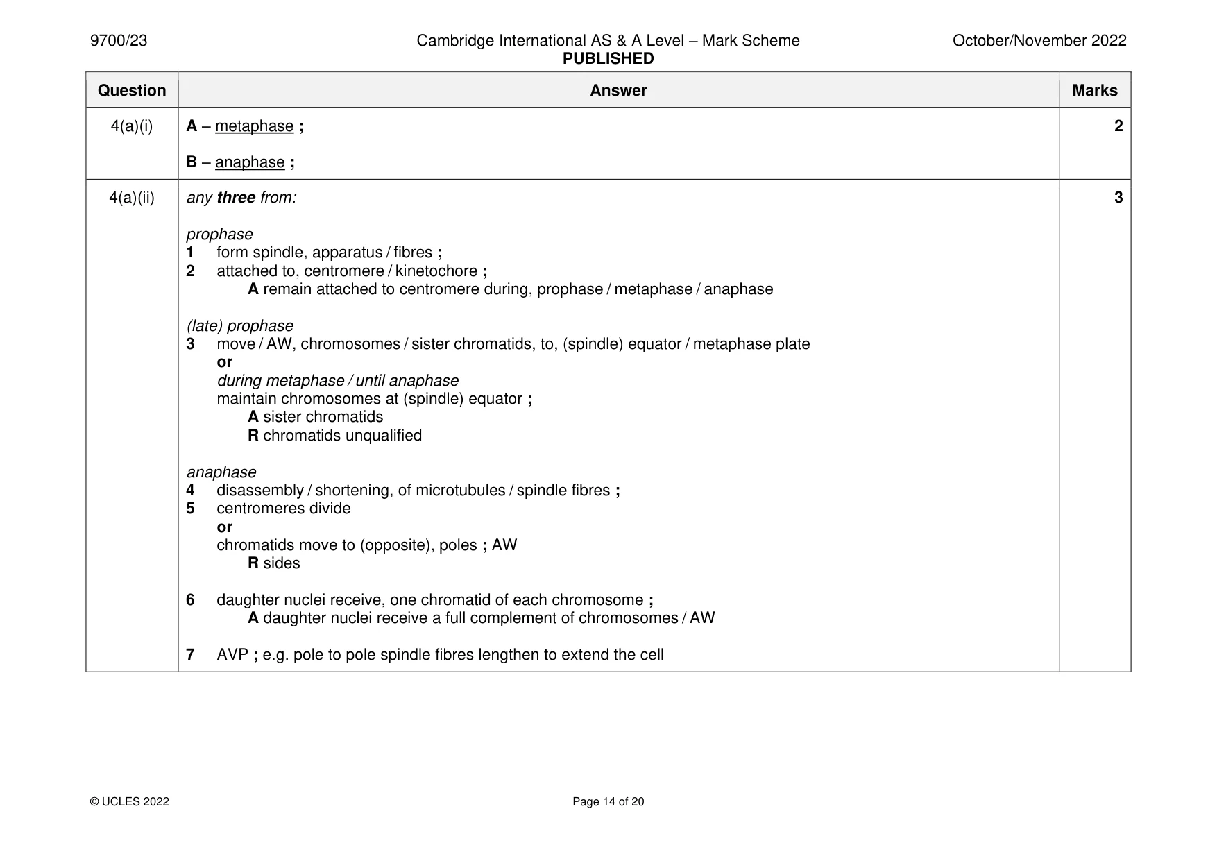
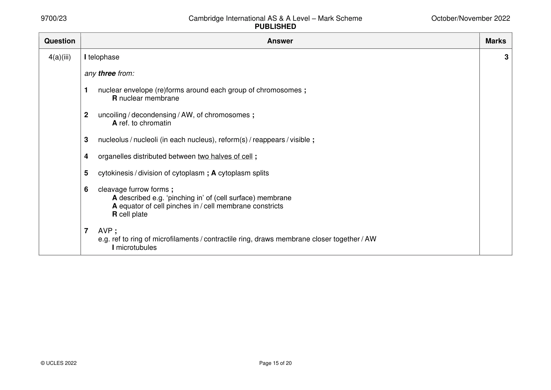
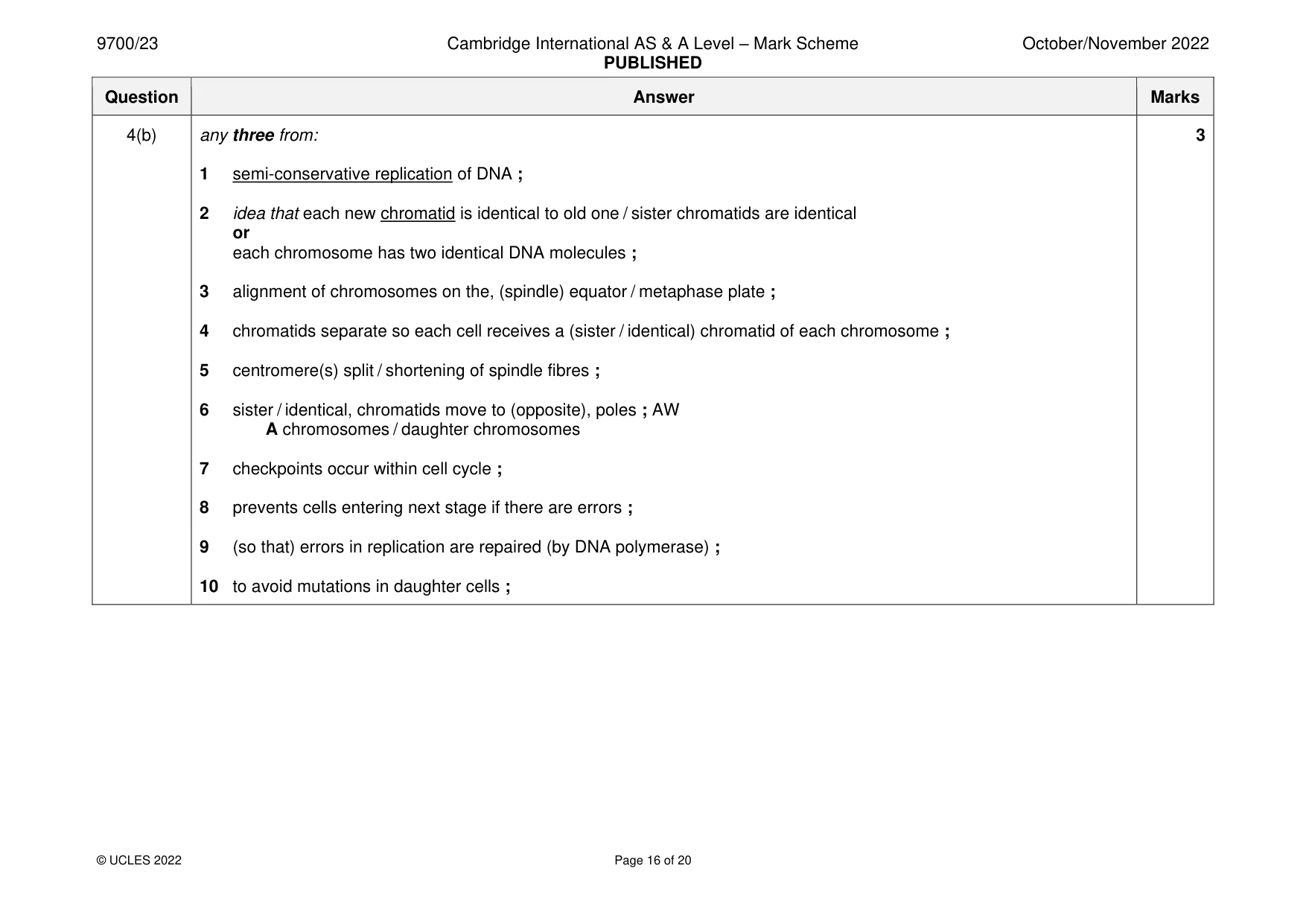
Mark scheme: 4(a)(i) A – metaphase ; 2 B – anaphase ; 4(a)(ii) any three from: 3 prophase 1 form spindle, apparatus / fibres ; 2 attached to, centromere / kinetochore ; A remain attached to centromere during, prophase / metaphase / anaphase (late) prophase 3 move / AW, chromosomes / sister chromatids, to, (spindle) equator / metaphase plate or during metaphase / until anaphase maintain chromosomes at (spindle) equator ; A sister chromatids R chromatids unqualified anaphase 4 disassembly / shortening, of microtubules / spindle fibres ; 5 centromeres divide or chromatids move to (opposite), poles ; AW R sides 6 daughter nuclei receive, one chromatid of each chromosome ; A daughter nuclei receive a full complement of chromosomes / AW 7 AVP ; e.g. pole to pole spindle fibres lengthen to extend the cell 4(a)(iii) I telophase 3 any three from: 1 nuclear envelope (re)forms around each group of chromosomes ; R nuclear membrane 2 uncoiling / decondensing / AW, of chromosomes ; A ref. to chromatin 3 nucleolus / nucleoli (in each nucleus), reform(s) / reappears / visible ; 4 organelles distributed between two halves of cell ; 5 cytokinesis / division of cytoplasm ; A cytoplasm splits 6 cleavage furrow forms ; A described e.g. ‘pinching in’ of (cell surface) membrane A equator of cell pinches in / cell membrane constricts R cell plate 7 AVP ; e.g. ref to ring of microfilaments / contractile ring, draws membrane closer together / AW I microtubules 4(b) any three from: 3 1 semi-conservative replication of DNA ; 2 idea that each new chromatid is identical to old one / sister chromatids are identical or each chromosome has two identical DNA molecules ; 3 alignment of chromosomes on the, (spindle) equator / metaphase plate ; 4 chromatids separate so each cell receives a (sister / identical) chromatid of each chromosome ; 5 centromere(s) split / shortening of spindle fibres ; 6 sister / identical, chromatids move to (opposite), poles ; AW A chromosomes / daughter chromosomes 7 checkpoints occur within cell cycle ; 8 prevents cells entering next stage if there are errors ; 9 (so that) errors in replication are repaired (by DNA polymerase) ; 10 to avoid mutations in daughter cells ;
Q5 · Cathelicidin LL-37 is a cell signalling compound that stimulates many different cells in…
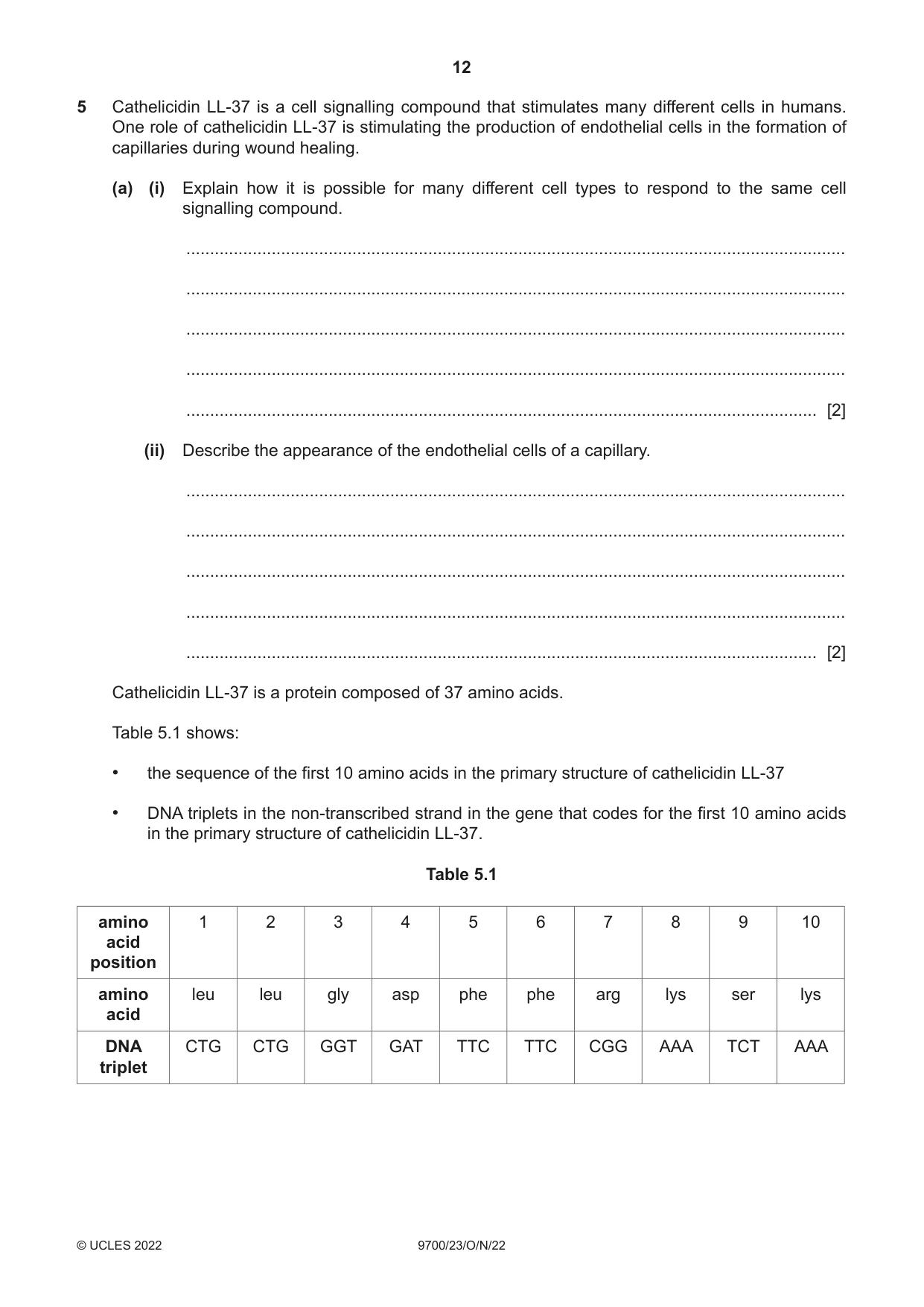
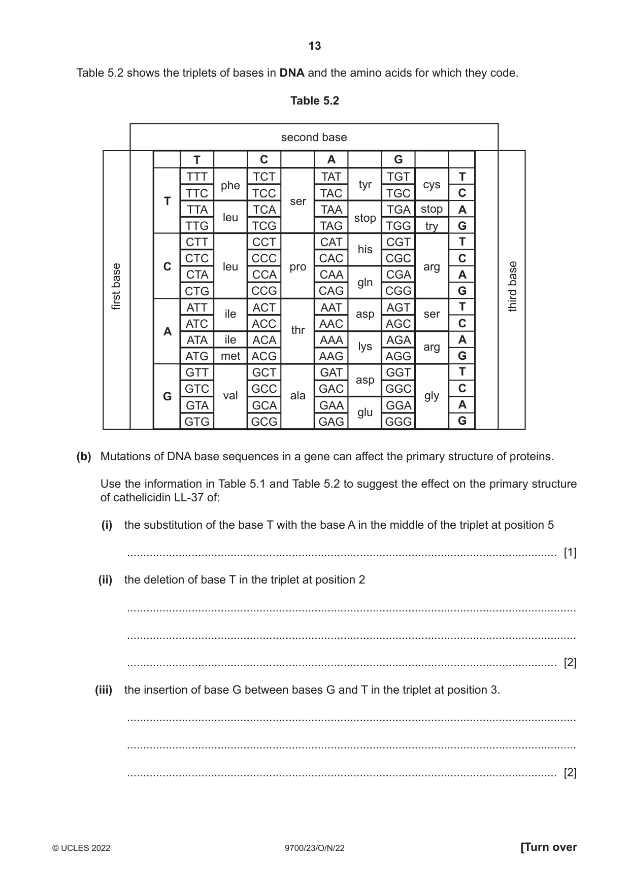
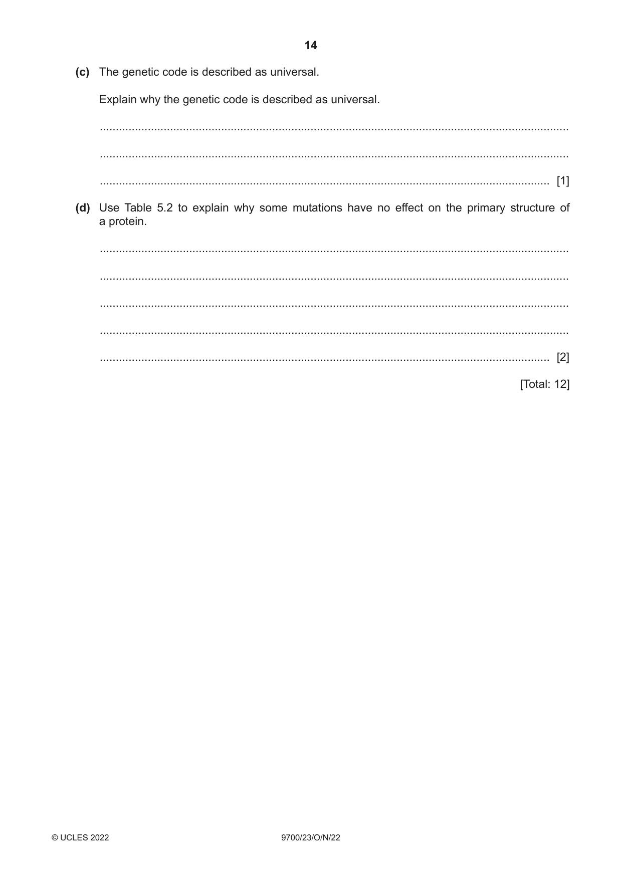
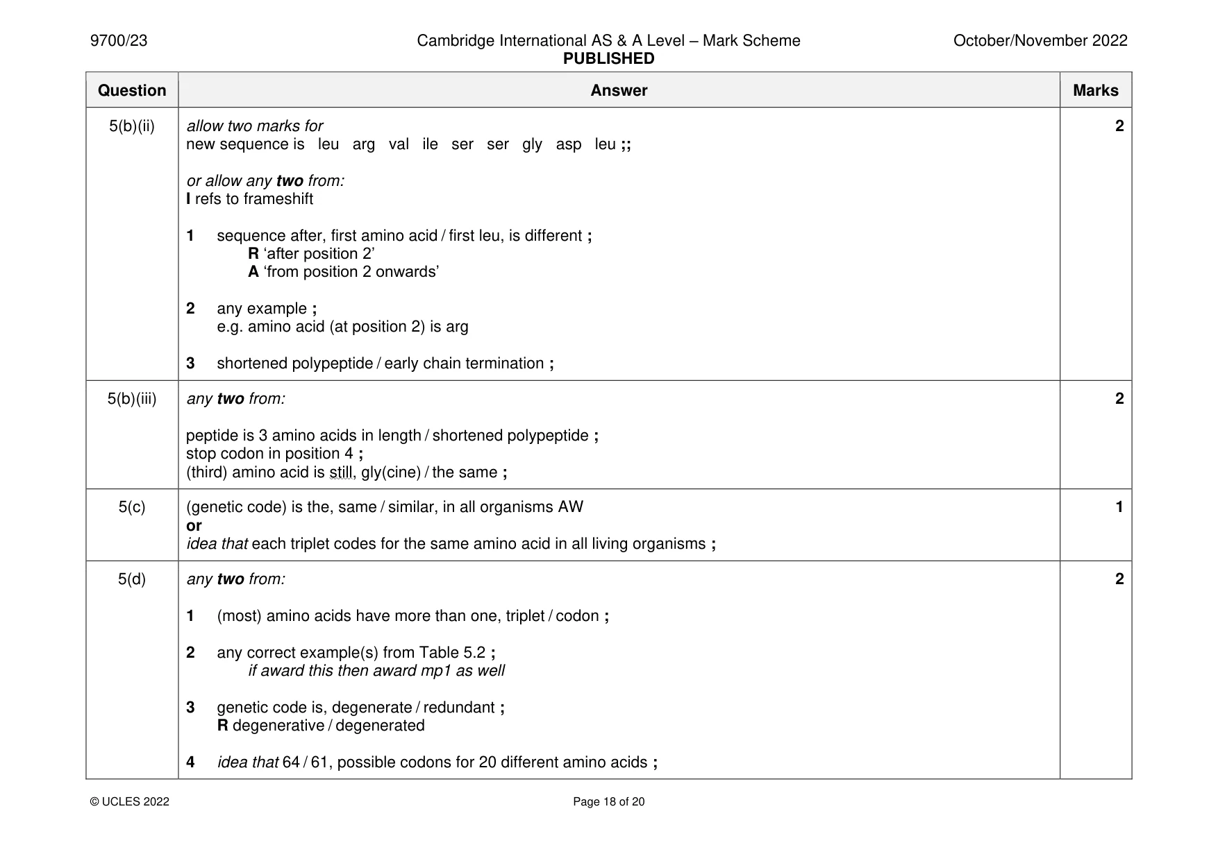
5 Cathelicidin LL-37 is a cell signalling compound that stimulates many different cells in humans. One role of cathelicidin LL-37 is stimulating the production of endothelial cells in the formation of capillaries during wound healing. (a) (i) Explain how it is possible for many different cell types to respond to the same cell signalling compound. ........................................................................................................................................... ........................................................................................................................................... ........................................................................................................................................... ........................................................................................................................................... ..................................................................................................................................... [2] (ii) Describe the appearance of the endothelial cells of a capillary. ........................................................................................................................................... ........................................................................................................................................... ........................................................................................................................................... ........................................................................................................................................... ..................................................................................................................................... [2] Cathelicidin LL-37 is a protein composed of 37 amino acids. Table 5.1 shows: • the sequence of the first 10 amino acids in the primary structure of cathelicidin LL-37 • DNA triplets in the non-transcribed strand in the gene that codes for the first 10 amino acids in the primary structure of cathelicidin LL-37. Table 5.1 amino 1 2 3 4 5 6 7 8 9 10 acid position amino leu leu gly asp phe phe arg lys ser lys acid DNA CTG CTG GGT GAT TTC TTC CGG AAA TCT AAA triplet Table 5.2 shows the triplets of bases in DNA and the amino acids for which they code. Table 5.2 second base T C A G TTT TCT TAT TGT T phe tyr cys TTC TCC TAC TGC C T ser TTA TCA TAA TGA stop A leu stop TTG TCG TAG TGG try G CTT CCT CAT CGT T his CTC CCC CAC CGC C C leu pro arg CTA CCA CAA CGA A base gln base CTG CCG CAG CGG G first third ATT ACT AAT AGT T ile asp ser ATC ACC AAC AGC C A thr ATA ile ACA AAA AGA A lys arg ATG met ACG AAG AGG G GTT GCT GAT GGT T asp GTC GCC GAC GGC C G val ala gly GTA GCA GAA GGA A glu GTG GCG GAG GGG G (b) Mutations of DNA base sequences in a gene can affect the primary structure of proteins. Use the information in Table 5.1 and Table 5.2 to suggest the effect on the primary structure of cathelicidin LL-37 of: (i) the substitution of the base T with the base A in the middle of the triplet at position 5 ..................................................................................................................................... [1] (ii) the deletion of base T in the triplet at position 2 ........................................................................................................................................... ........................................................................................................................................... ..................................................................................................................................... [2] (iii) the insertion of base G between bases G and T in the triplet at position 3. ........................................................................................................................................... ........................................................................................................................................... ..................................................................................................................................... [2] (c) The genetic code is described as universal. Explain why the genetic code is described as universal. ................................................................................................................................................... ................................................................................................................................................... ............................................................................................................................................. [1] (d) Use Table 5.2 to explain why some mutations have no effect on the primary structure of a protein. ................................................................................................................................................... ................................................................................................................................................... ................................................................................................................................................... ................................................................................................................................................... ............................................................................................................................................. [2] [Total: 12]
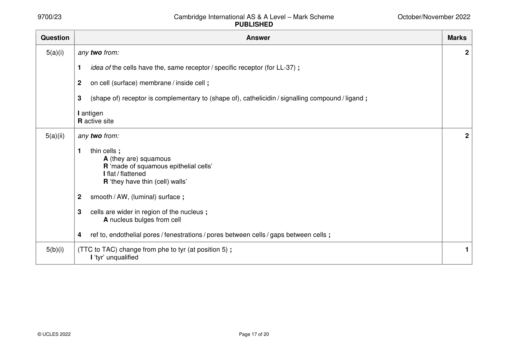
Mark scheme: 5(a)(i) any two from: 2 1 idea of the cells have the, same receptor / specific receptor (for LL-37) ; 2 on cell (surface) membrane / inside cell ; 3 (shape of) receptor is complementary to (shape of), cathelicidin / signalling compound / ligand ; I antigen R active site 5(a)(ii) any two from: 2 1 thin cells ; A (they are) squamous R ‘made of squamous epithelial cells’ I flat / flattened R ‘they have thin (cell) walls’ 2 smooth / AW, (luminal) surface ; 3 cells are wider in region of the nucleus ; A nucleus bulges from cell 4 ref to, endothelial pores / fenestrations / pores between cells / gaps between cells ; 5(b)(i) (TTC to TAC) change from phe to tyr (at position 5) ; 1 I ‘tyr’ unqualified 5(b)(ii) allow two marks for 2 new sequence is leu arg val ile ser ser gly asp leu ;; or allow any two from: I refs to frameshift 1 sequence after, first amino acid / first leu, is different ; R ‘after position 2’ A ‘from position 2 onwards’ 2 any example ; e.g. amino acid (at position 2) is arg 3 shortened polypeptide / early chain termination ; 5(b)(iii) any two from: 2 peptide is 3 amino acids in length / shortened polypeptide ; stop codon in position 4 ; (third) amino acid is still, gly(cine) / the same ; 5(c) (genetic code) is the, same / similar, in all organisms AW 1 or idea that each triplet codes for the same amino acid in all living organisms ; 5(d) any two from: 2 1 (most) amino acids have more than one, triplet / codon ; 2 any correct example(s) from Table 5.2 ; if award this then award mp1 as well 3 genetic code is, degenerate / redundant ; R degenerative / degenerated 4 idea that 64 / 61, possible codons for 20 different amino acids ;
Q6 · Some of the events that occur when a red blood cell flows through a capillary in the lungs
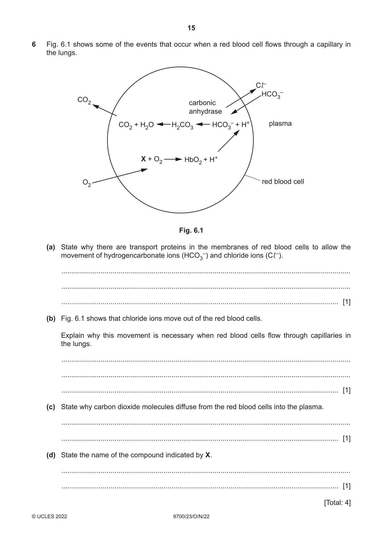
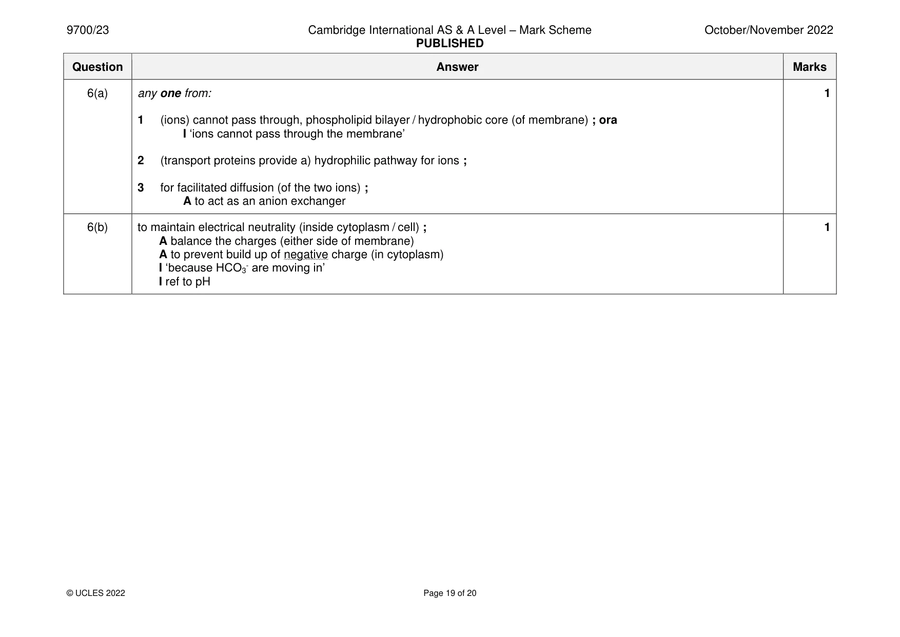
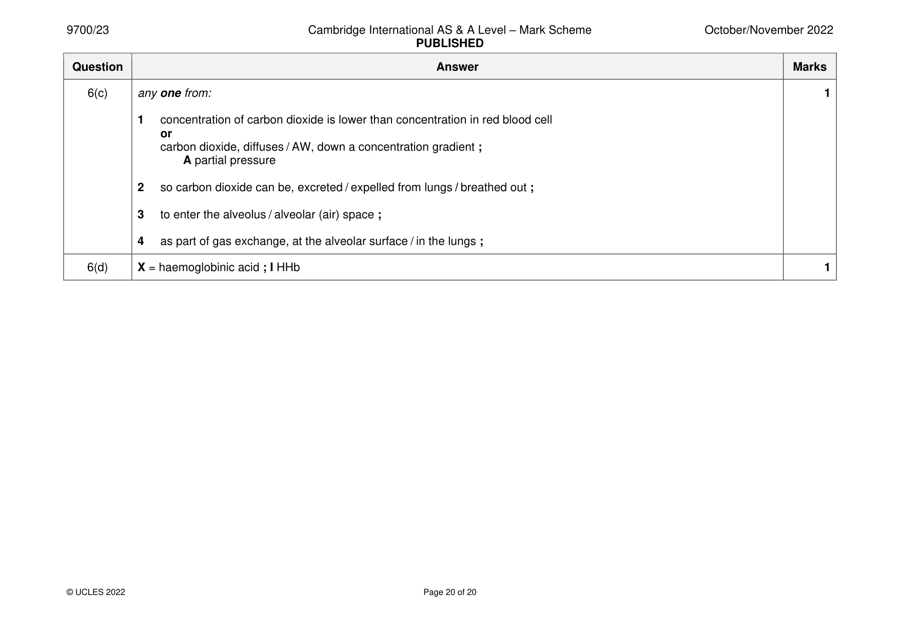
6 Fig. 6.1 shows some of the events that occur when a red blood cell flows through a capillary in the lungs. Cl – HCO3– CO2 carbonic anhydrase CO2 + H2O H2CO3 HCO3– + H+ plasma X + O2 HbO2 + H+ O2 red blood cell Fig. 6.1 (a) State why there are transport proteins in the membranes of red blood cells to allow the movement of hydrogencarbonate ions (HCO3–) and chloride ions (Cl –). ................................................................................................................................................... ................................................................................................................................................... ............................................................................................................................................. [1] (b) Fig. 6.1 shows that chloride ions move out of the red blood cells. Explain why this movement is necessary when red blood cells flow through capillaries in the lungs. ................................................................................................................................................... ................................................................................................................................................... ............................................................................................................................................. [1] (c) State why carbon dioxide molecules diffuse from the red blood cells into the plasma. ................................................................................................................................................... ............................................................................................................................................. [1] (d) State the name of the compound indicated by X. ................................................................................................................................................... ............................................................................................................................................. [1] [Total: 4]
Mark scheme: 6(a) any one from: 1 1 (ions) cannot pass through, phospholipid bilayer / hydrophobic core (of membrane) ; ora I ‘ions cannot pass through the membrane’ 2 (transport proteins provide a) hydrophilic pathway for ions ; 3 for facilitated diffusion (of the two ions) ; A to act as an anion exchanger 6(b) to maintain electrical neutrality (inside cytoplasm / cell) ; 1 A balance the charges (either side of membrane) A to prevent build up of negative charge (in cytoplasm) I ‘because HCO3- are moving in’ I ref to pH 6(c) any one from: 1 1 concentration of carbon dioxide is lower than concentration in red blood cell or carbon dioxide, diffuses / AW, down a concentration gradient ; A partial pressure 2 so carbon dioxide can be, excreted / expelled from lungs / breathed out ; 3 to enter the alveolus / alveolar (air) space ; 4 as part of gas exchange, at the alveolar surface / in the lungs ; 6(d) X = haemoglobinic acid ; I HHb 1
What was in this paper
The subtopics covered by these 6 questions, and how many questions each got. Open one in a new tab to see every Cambridge question on it.
What you needed in this session
Cambridge’s own grade thresholds for 2022 Oct/Nov, Paper 2 · Variant 3. A higher threshold means an easier paper — the bar moves with how the cohort did.