Cambridge A Level Biology 9700 — 2022 Oct/Nov Paper 2 · Variant 1
9700/21/O/N/22 · 6 questions · 60 marks · ≈68 min
The question paper and its mark scheme, free to read here and free to download. This is Cambridge’s own paper, exactly as it was sat.
Question paper20 pages


Mark scheme17 pages
Answers below. Sit the paper first if you are practising.
Questions as text
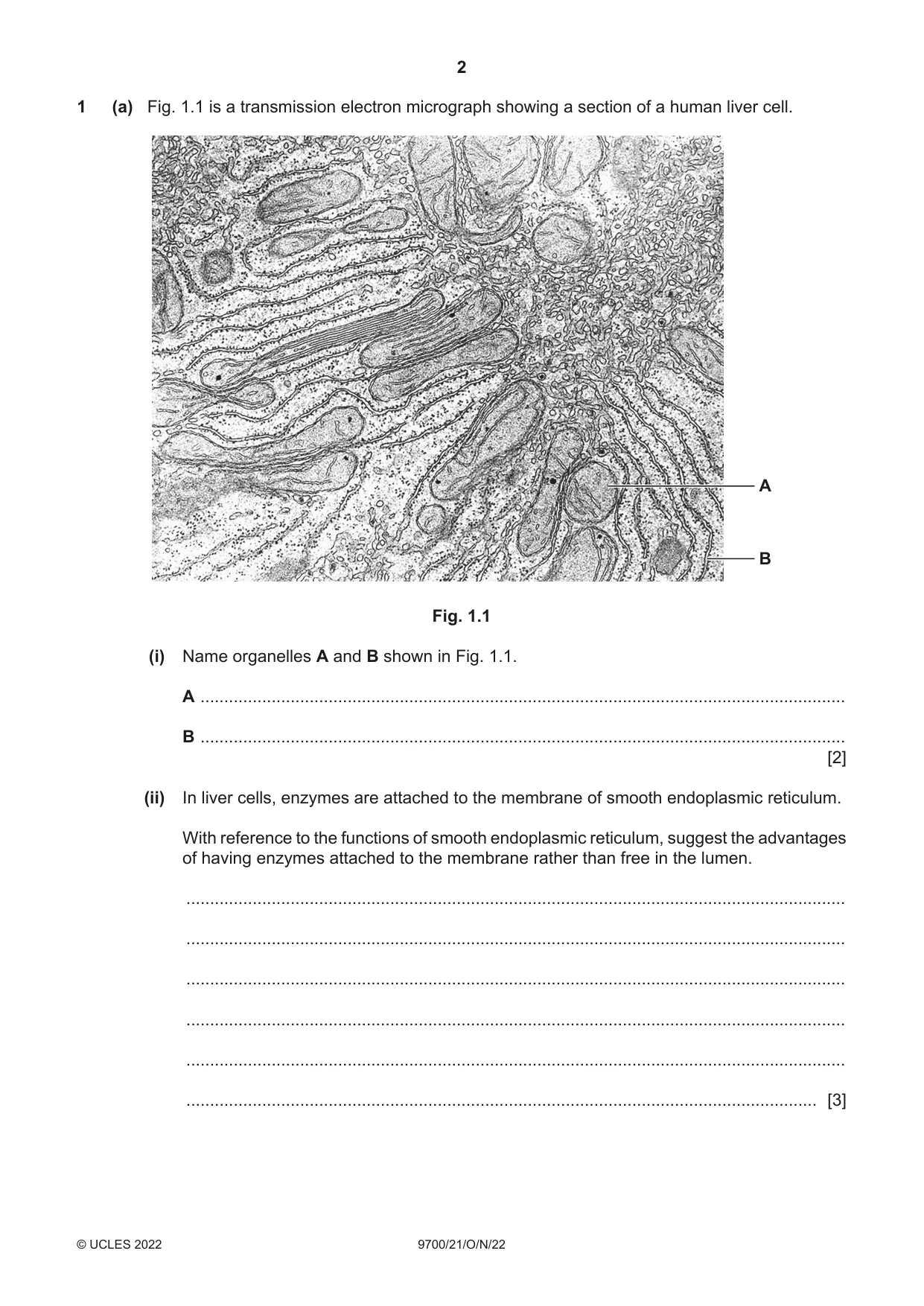
Q1 · A transmission electron micrograph showing a section of a human liver cell
1 (a) Fig. 1.1 is a transmission electron micrograph showing a section of a human liver cell. A B Fig. 1.1 (i) Name organelles A and B shown in Fig. 1.1. A ........................................................................................................................................ B ........................................................................................................................................ [2] (ii) In liver cells, enzymes are attached to the membrane of smooth endoplasmic reticulum. With reference to the functions of smooth endoplasmic reticulum, suggest the advantages of having enzymes attached to the membrane rather than free in the lumen. ........................................................................................................................................... ........................................................................................................................................... ........................................................................................................................................... ........................................................................................................................................... ........................................................................................................................................... ..................................................................................................................................... [3] (b) Explain the advantages of using a transmission electron microscope compared with a light microscope when viewing a liver cell. ................................................................................................................................................... ................................................................................................................................................... ................................................................................................................................................... ................................................................................................................................................... ................................................................................................................................................... ................................................................................................................................................... ............................................................................................................................................. [3] [Total: 8]
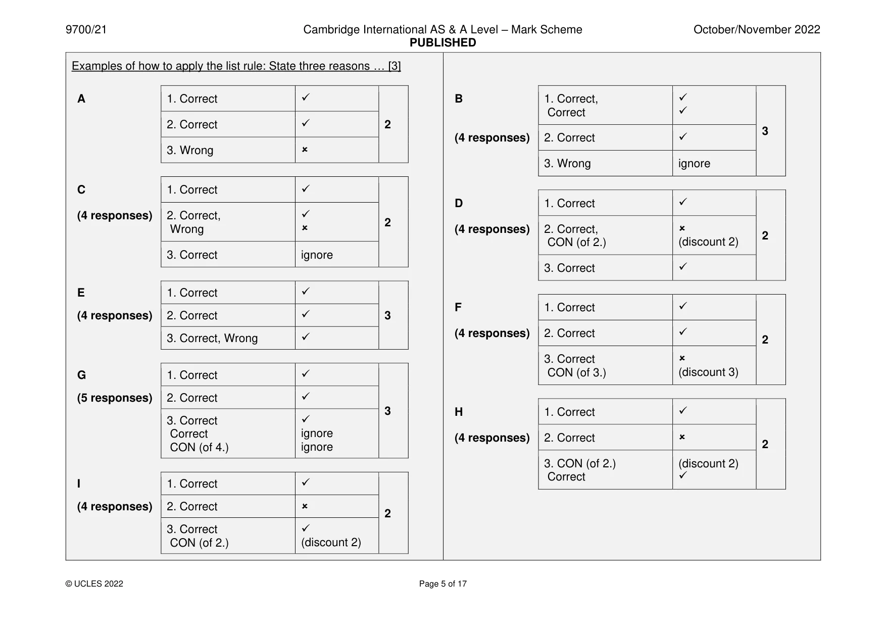
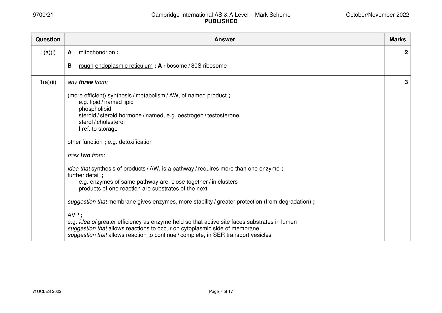
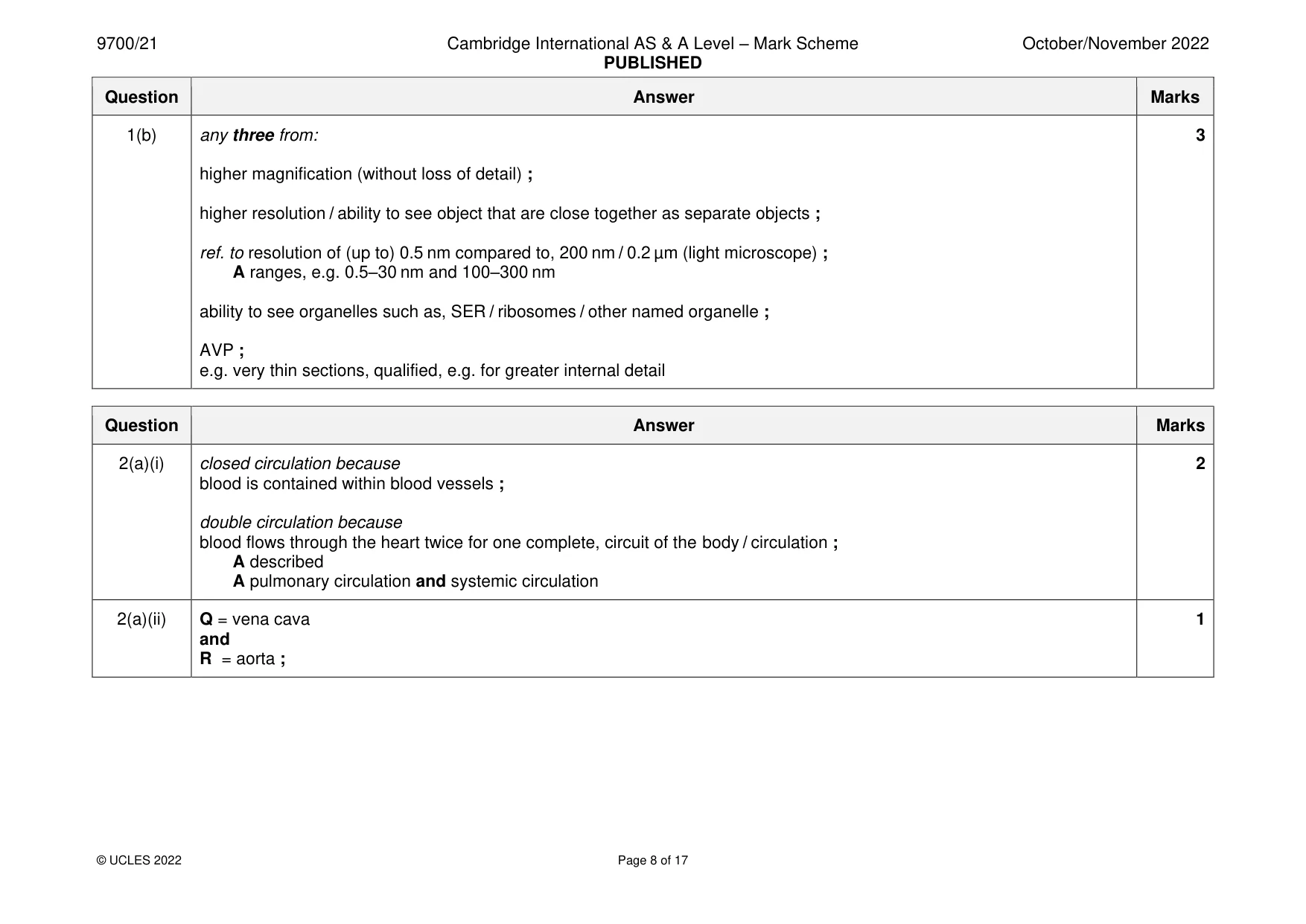
Mark scheme: Question Answer Marks 1(a)(i) A mitochondrion ; 2 B rough endoplasmic reticulum ; A ribosome / 80S ribosome 1(a)(ii) any three from: 3 (more efficient) synthesis / metabolism / AW, of named product ; e.g. lipid / named lipid phospholipid steroid / steroid hormone / named, e.g. oestrogen / testosterone sterol / cholesterol I ref. to storage other function ; e.g. detoxification max two from: idea that synthesis of products / AW, is a pathway / requires more than one enzyme ; further detail ; e.g. enzymes of same pathway are, close together / in clusters products of one reaction are substrates of the next suggestion that membrane gives enzymes, more stability / greater protection (from degradation) ; AVP ; e.g. idea of greater efficiency as enzyme held so that active site faces substrates in lumen suggestion that allows reactions to occur on cytoplasmic side of membrane suggestion that allows reaction to continue / complete, in SER transport vesicles 1(b) any three from: 3 higher magnification (without loss of detail) ; higher resolution / ability to see object that are close together as separate objects ; ref. to resolution of (up to) 0.5 nm compared to, 200 nm / 0.2 µm (light microscope) ; A ranges, e.g. 0.5–30 nm and 100–300 nm ability to see organelles such as, SER / ribosomes / other named organelle ; AVP ; e.g. very thin sections, qualified, e.g. for greater internal detail
More questions on Cells as the basic units of living organisms
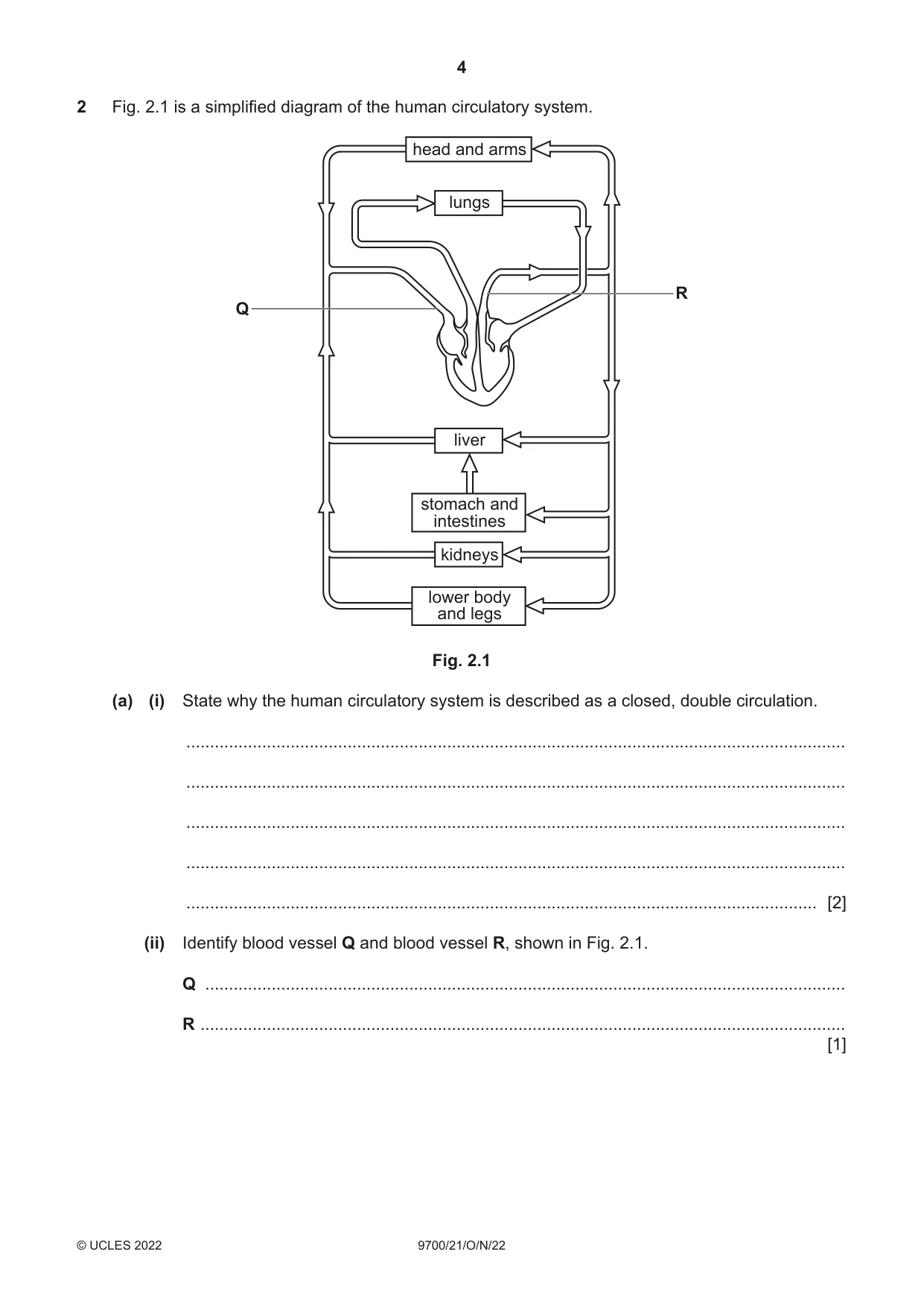
Q2 · A simplified diagram of the human circulatory system
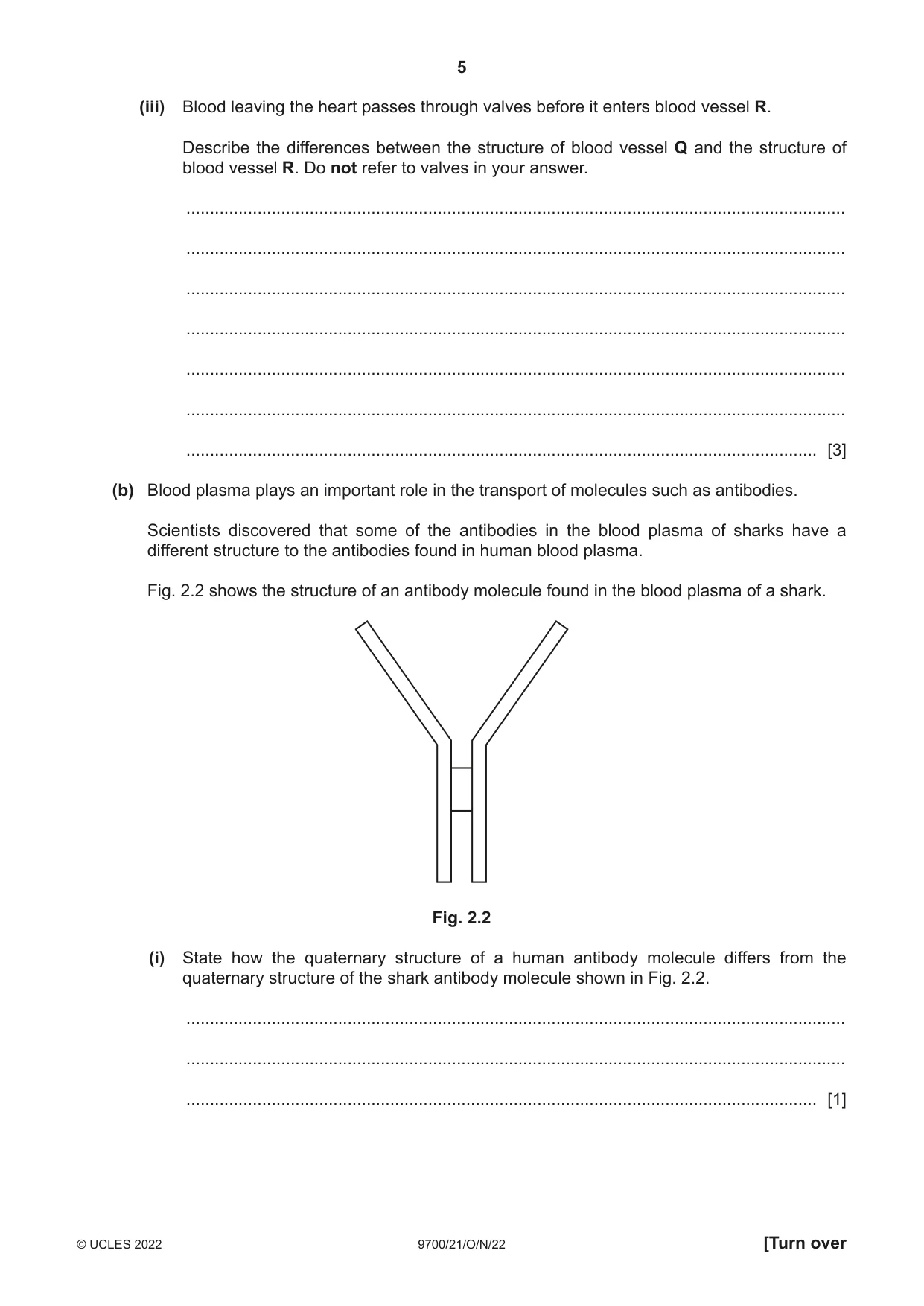
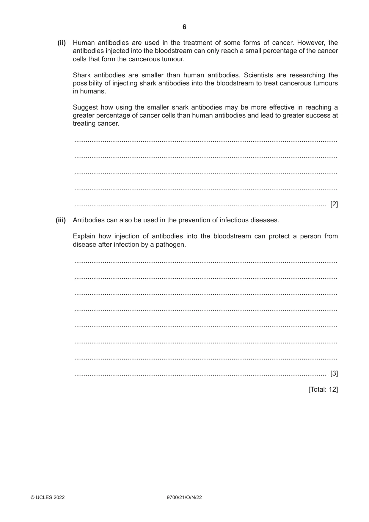
2 Fig. 2.1 is a simplified diagram of the human circulatory system. head and arms lungs R Q liver stomach and intestines kidneys lower body and legs Fig. 2.1 (a) (i) State why the human circulatory system is described as a closed, double circulation. ........................................................................................................................................... ........................................................................................................................................... ........................................................................................................................................... ........................................................................................................................................... ..................................................................................................................................... [2] (ii) Identify blood vessel Q and blood vessel R, shown in Fig. 2.1. Q ....................................................................................................................................... R ........................................................................................................................................ [1] (iii) Blood leaving the heart passes through valves before it enters blood vessel R. Describe the differences between the structure of blood vessel Q and the structure of blood vessel R. Do not refer to valves in your answer. ........................................................................................................................................... ........................................................................................................................................... ........................................................................................................................................... ........................................................................................................................................... ........................................................................................................................................... ........................................................................................................................................... ..................................................................................................................................... [3] (b) Blood plasma plays an important role in the transport of molecules such as antibodies. Scientists discovered that some of the antibodies in the blood plasma of sharks have a different structure to the antibodies found in human blood plasma. Fig. 2.2 shows the structure of an antibody molecule found in the blood plasma of a shark. Fig. 2.2 (i) State how the quaternary structure of a human antibody molecule differs from the quaternary structure of the shark antibody molecule shown in Fig. 2.2. ........................................................................................................................................... ........................................................................................................................................... ..................................................................................................................................... [1] (ii) Human antibodies are used in the treatment of some forms of cancer. However, the antibodies injected into the bloodstream can only reach a small percentage of the cancer cells that form the cancerous tumour. Shark antibodies are smaller than human antibodies. Scientists are researching the possibility of injecting shark antibodies into the bloodstream to treat cancerous tumours in humans. Suggest how using the smaller shark antibodies may be more effective in reaching a greater percentage of cancer cells than human antibodies and lead to greater success at treating cancer. ........................................................................................................................................... ........................................................................................................................................... ........................................................................................................................................... ........................................................................................................................................... ..................................................................................................................................... [2] (iii) Antibodies can also be used in the prevention of infectious diseases. Explain how injection of antibodies into the bloodstream can protect a person from disease after infection by a pathogen. ........................................................................................................................................... ........................................................................................................................................... ........................................................................................................................................... ........................................................................................................................................... ........................................................................................................................................... ........................................................................................................................................... ........................................................................................................................................... ..................................................................................................................................... [3] [Total: 12]
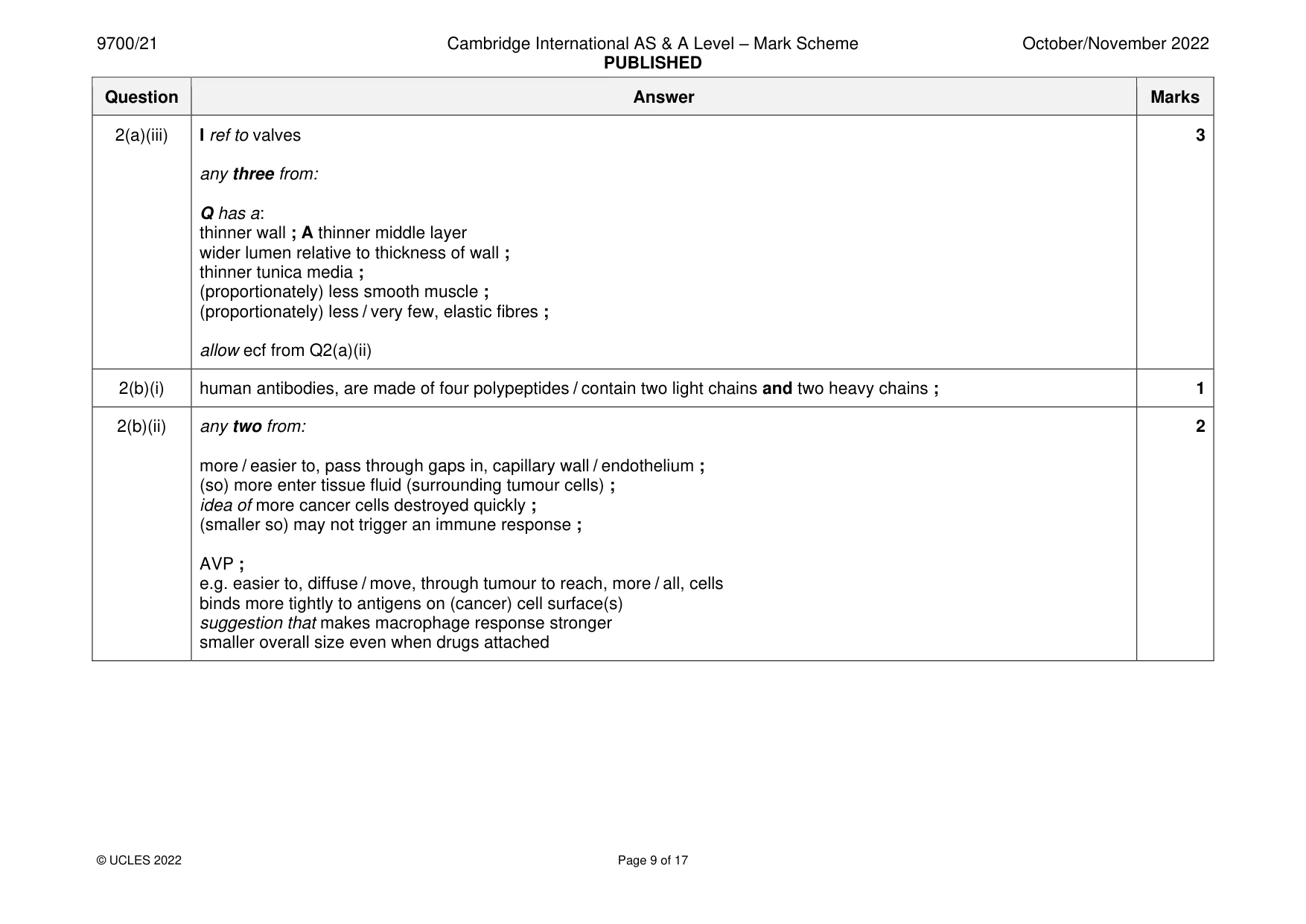
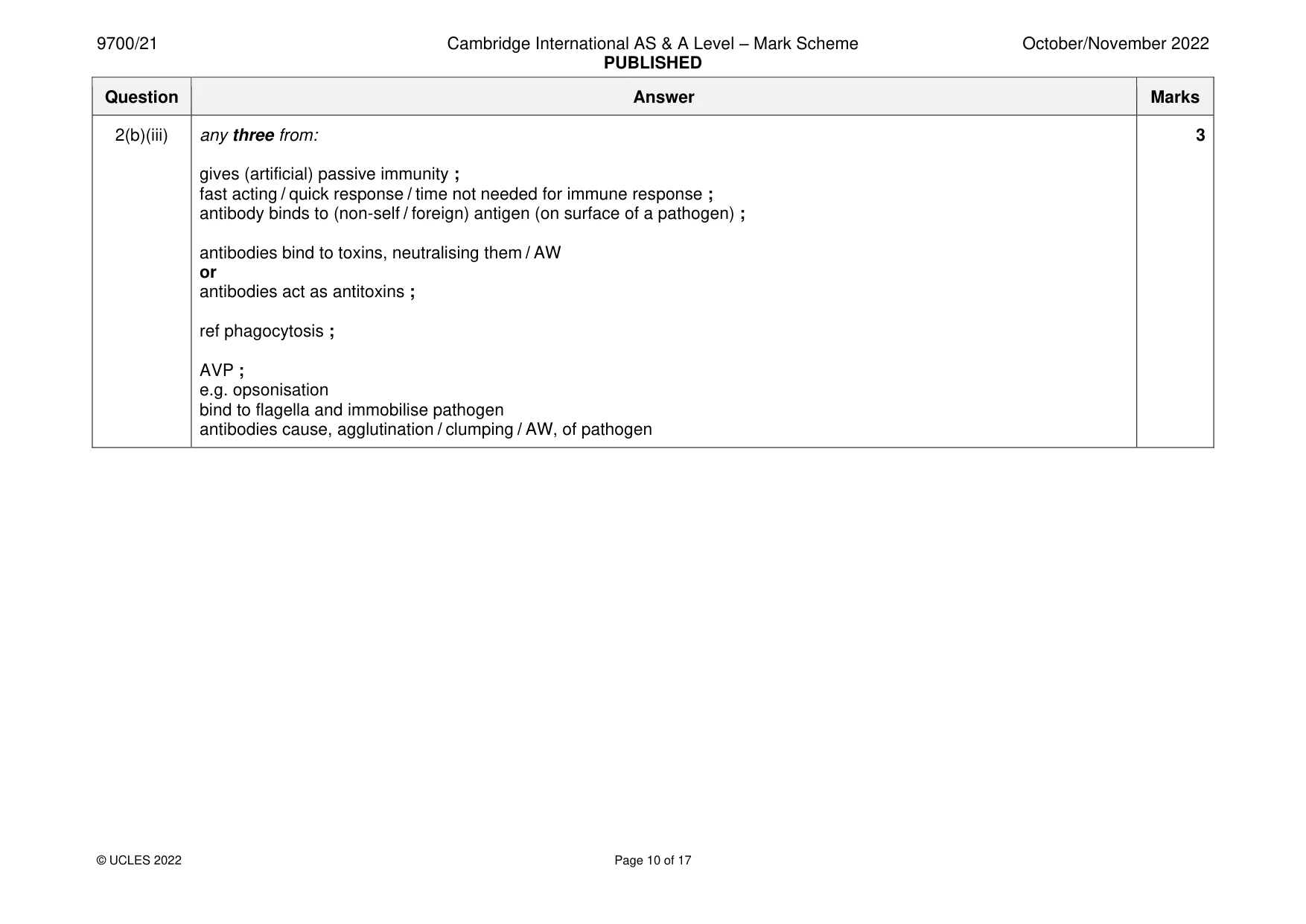
Mark scheme: 2(a)(i) closed circulation because 2 blood is contained within blood vessels ; double circulation because blood flows through the heart twice for one complete, circuit of the body / circulation ; A described A pulmonary circulation and systemic circulation 2(a)(ii) Q = vena cava 1 and R = aorta ; 2(a)(iii) I ref to valves 3 any three from: Q has a: thinner wall ; A thinner middle layer wider lumen relative to thickness of wall ; thinner tunica media ; (proportionately) less smooth muscle ; (proportionately) less / very few, elastic fibres ; allow ecf from Q2(a)(ii) 2(b)(i) human antibodies, are made of four polypeptides / contain two light chains and two heavy chains ; 1 2(b)(ii) any two from: 2 more / easier to, pass through gaps in, capillary wall / endothelium ; (so) more enter tissue fluid (surrounding tumour cells) ; idea of more cancer cells destroyed quickly ; (smaller so) may not trigger an immune response ; AVP ; e.g. easier to, diffuse / move, through tumour to reach, more / all, cells binds more tightly to antigens on (cancer) cell surface(s) suggestion that makes macrophage response stronger smaller overall size even when drugs attached 2(b)(iii) any three from: 3 gives (artificial) passive immunity ; fast acting / quick response / time not needed for immune response ; antibody binds to (non-self / foreign) antigen (on surface of a pathogen) ; antibodies bind to toxins, neutralising them / AW or antibodies act as antitoxins ; ref phagocytosis ; AVP ; e.g. opsonisation bind to flagella and immobilise pathogen antibodies cause, agglutination / clumping / AW, of pathogen
Q3 · Carbonic anhydrase is a globular protein found in red blood cells
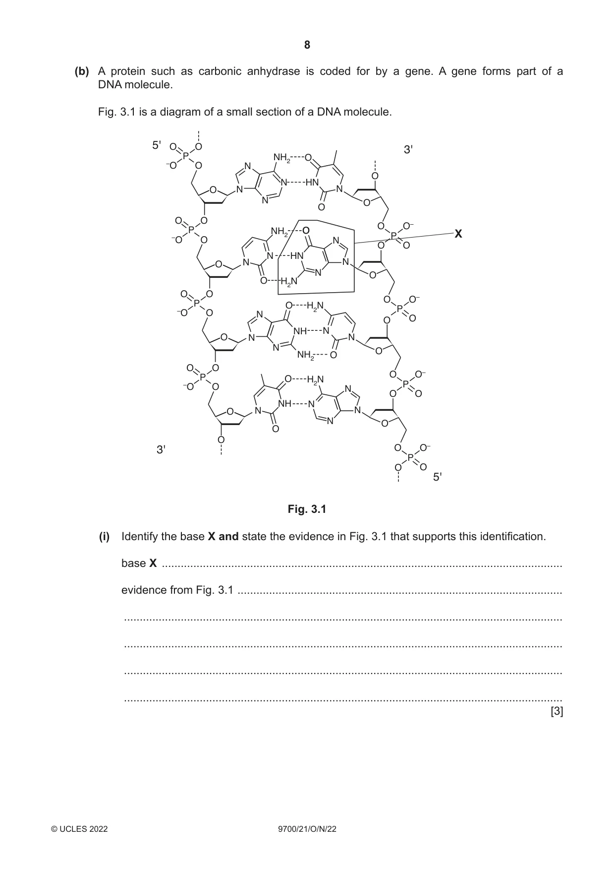
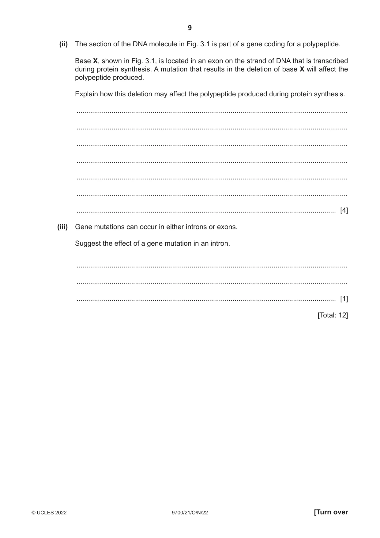
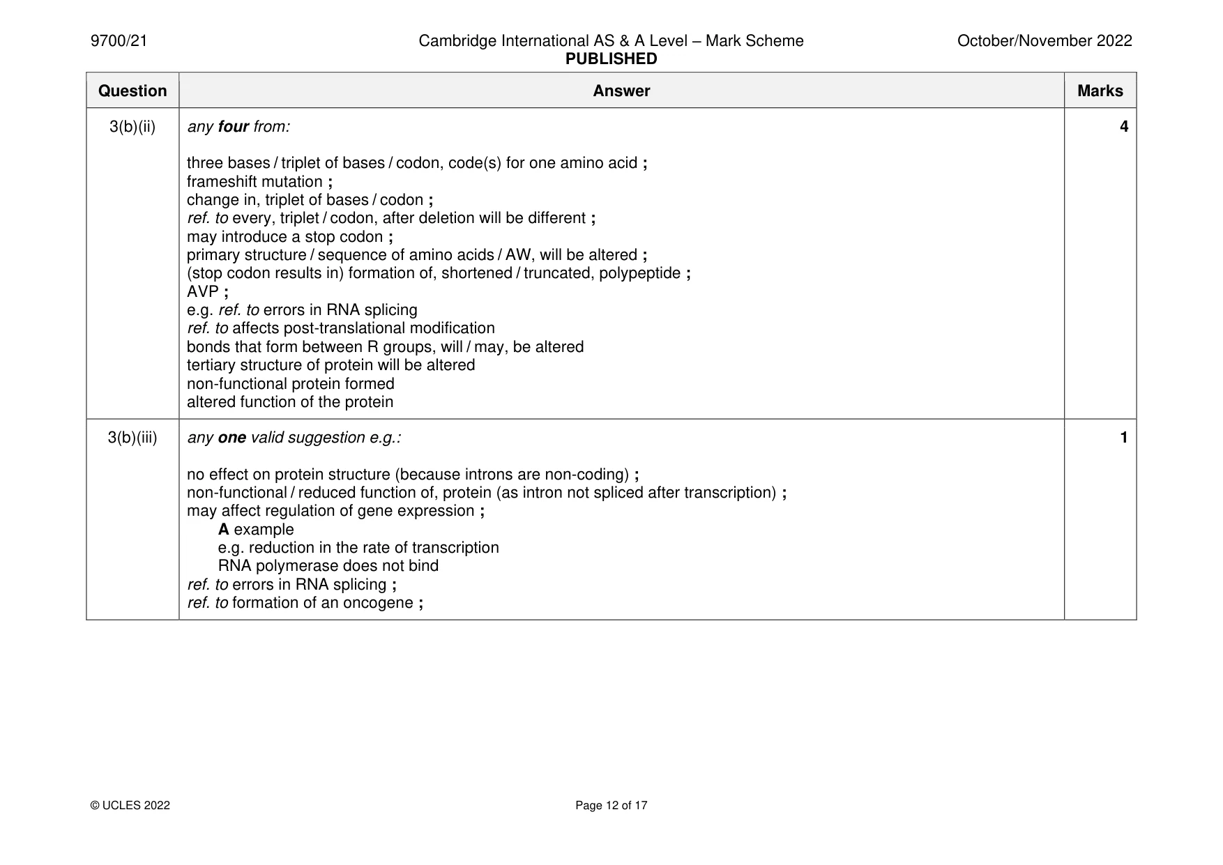
3 Carbonic anhydrase is a globular protein found in red blood cells. (a) (i) Explain why this protein is described as globular. ........................................................................................................................................... ........................................................................................................................................... ........................................................................................................................................... ........................................................................................................................................... ..................................................................................................................................... [2] (ii) State the function of carbonic anhydrase in red blood cells. ........................................................................................................................................... ........................................................................................................................................... ........................................................................................................................................... ........................................................................................................................................... ..................................................................................................................................... [2] (b) A protein such as carbonic anhydrase is coded for by a gene. A gene forms part of a DNA molecule. Fig. 3.1 is a diagram of a small section of a DNA molecule. 5' O O 3' P NH2 O –O O N O N HN O N N N O O O O O O– P NH2 O P X –O O N O O N HN O N N N O O H2N O O O O– P O H2N P –O O N O O NH N O N N N O NH2 O O O P O H2N O O– –O O N P O O NH N O N N N O O O 3' O O– P O O 5' Fig. 3.1 (i) Identify the base X and state the evidence in Fig. 3.1 that supports this identification. base X ............................................................................................................................... evidence from Fig. 3.1 ....................................................................................................... ........................................................................................................................................... ........................................................................................................................................... ........................................................................................................................................... ........................................................................................................................................... [3] (ii) The section of the DNA molecule in Fig. 3.1 is part of a gene coding for a polypeptide. Base X, shown in Fig. 3.1, is located in an exon on the strand of DNA that is transcribed during protein synthesis. A mutation that results in the deletion of base X will affect the polypeptide produced. Explain how this deletion may affect the polypeptide produced during protein synthesis. ........................................................................................................................................... ........................................................................................................................................... ........................................................................................................................................... ........................................................................................................................................... ........................................................................................................................................... ........................................................................................................................................... ..................................................................................................................................... [4] (iii) Gene mutations can occur in either introns or exons. Suggest the effect of a gene mutation in an intron. ........................................................................................................................................... ........................................................................................................................................... ..................................................................................................................................... [1] [Total: 12]
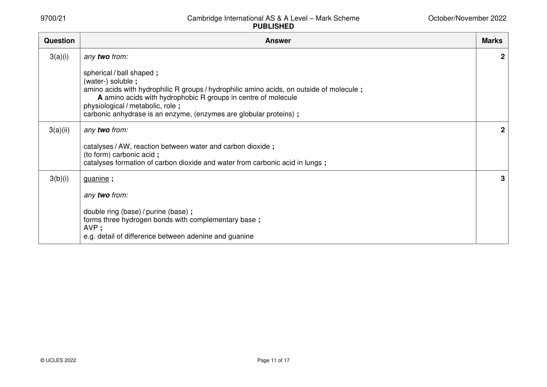
Mark scheme: 3(a)(i) any two from: 2 spherical / ball shaped ; (water-) soluble ; amino acids with hydrophilic R groups / hydrophilic amino acids, on outside of molecule ; A amino acids with hydrophobic R groups in centre of molecule physiological / metabolic, role ; carbonic anhydrase is an enzyme, (enzymes are globular proteins) ; 3(a)(ii) any two from: 2 catalyses / AW, reaction between water and carbon dioxide ; (to form) carbonic acid ; catalyses formation of carbon dioxide and water from carbonic acid in lungs ; 3(b)(i) guanine ; 3 any two from: double ring (base) / purine (base) ; forms three hydrogen bonds with complementary base ; AVP ; e.g. detail of difference between adenine and guanine 3(b)(ii) any four from: 4 three bases / triplet of bases / codon, code(s) for one amino acid ; frameshift mutation ; change in, triplet of bases / codon ; ref. to every, triplet / codon, after deletion will be different ; may introduce a stop codon ; primary structure / sequence of amino acids / AW, will be altered ; (stop codon results in) formation of, shortened / truncated, polypeptide ; AVP ; e.g. ref. to errors in RNA splicing ref. to affects post-translational modification bonds that form between R groups, will / may, be altered tertiary structure of protein will be altered non-functional protein formed altered function of the protein 3(b)(iii) any one valid suggestion e.g.: 1 no effect on protein structure (because introns are non-coding) ; non-functional / reduced function of, protein (as intron not spliced after transcription) ; may affect regulation of gene expression ; A example e.g. reduction in the rate of transcription RNA polymerase does not bind ref. to errors in RNA splicing ; ref. to formation of an oncogene ;
Q4 · Scientists have produced structures known as virosomes, which are used in certain vaccines
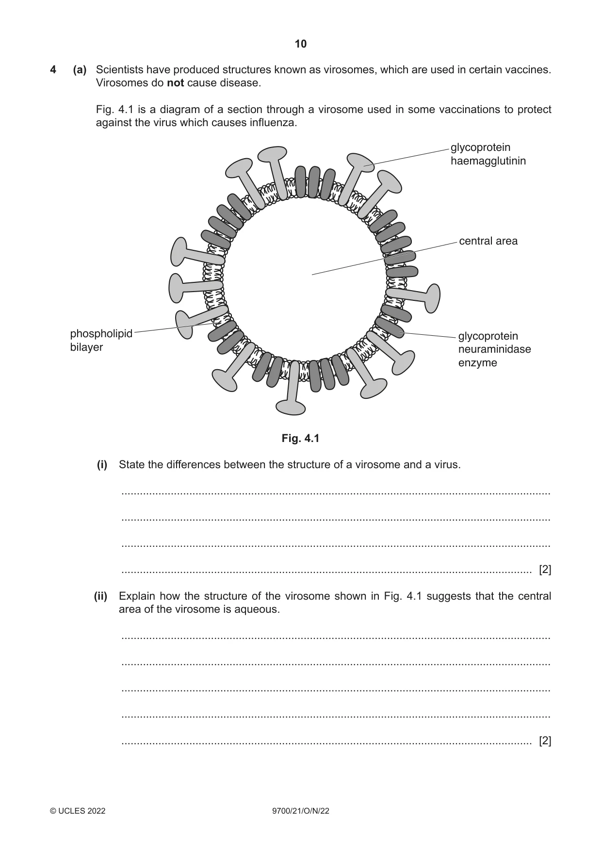
4 (a) Scientists have produced structures known as virosomes, which are used in certain vaccines. Virosomes do not cause disease. Fig. 4.1 is a diagram of a section through a virosome used in some vaccinations to protect against the virus which causes influenza. glycoprotein haemagglutinin central area phospholipid glycoprotein bilayer neuraminidase enzyme Fig. 4.1 (i) State the differences between the structure of a virosome and a virus. ........................................................................................................................................... ........................................................................................................................................... ........................................................................................................................................... ..................................................................................................................................... [2] (ii) Explain how the structure of the virosome shown in Fig. 4.1 suggests that the central area of the virosome is aqueous. ........................................................................................................................................... ........................................................................................................................................... ........................................................................................................................................... ........................................................................................................................................... ..................................................................................................................................... [2] (b) The glycoproteins haemagglutinin and neuraminidase are found in the influenza virus and in the virosomes used in a vaccine against the influenza virus. Haemagglutinin binds to a receptor in the cell surface membrane of phagocytes. Suggest why haemagglutinin is present in virosomes used in the vaccine for influenza. ................................................................................................................................................... ................................................................................................................................................... ................................................................................................................................................... ................................................................................................................................................... ................................................................................................................................................... ............................................................................................................................................. [2] (c) Different strains of the influenza virus have formed as a result of mutations. Each strain of the virus contains the enzyme neuraminidase. Neuraminidase helps the virus to leave host cells after the virus has replicated. In each strain of the influenza virus, the primary structure of the active site of the neuraminidase enzyme remains unchanged. Suggest why the primary structure of the active site of neuraminidase remains unchanged in each strain of the influenza virus. ................................................................................................................................................... ................................................................................................................................................... ................................................................................................................................................... ................................................................................................................................................... ............................................................................................................................................. [2] [Total: 8]
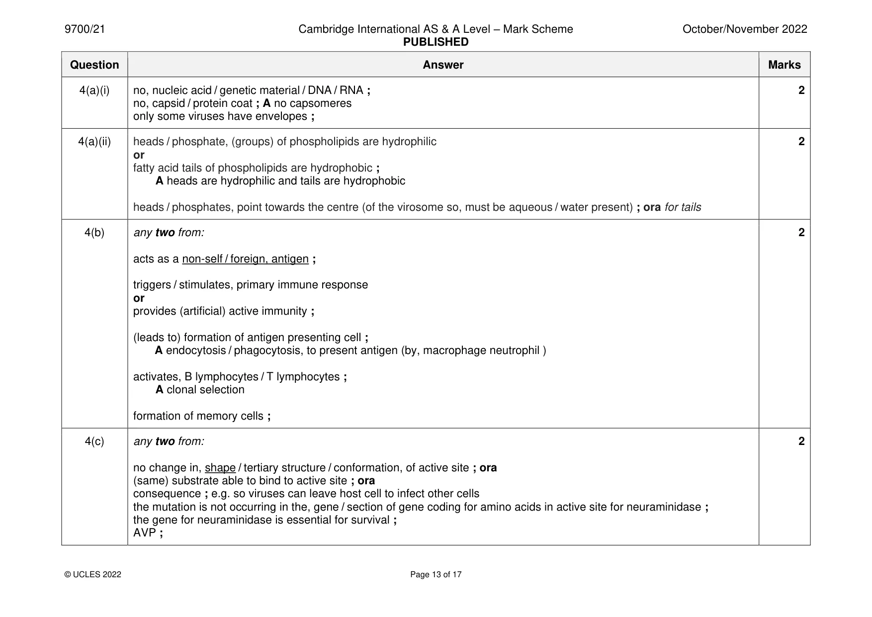
Mark scheme: 4(a)(i) no, nucleic acid / genetic material / DNA / RNA ; 2 no, capsid / protein coat ; A no capsomeres only some viruses have envelopes ; 4(a)(ii) heads / phosphate, (groups) of phospholipids are hydrophilic 2 or fatty acid tails of phospholipids are hydrophobic ; A heads are hydrophilic and tails are hydrophobic heads / phosphates, point towards the centre (of the virosome so, must be aqueous / water present) ; ora for tails 4(b) any two from: 2 acts as a non-self / foreign, antigen ; triggers / stimulates, primary immune response or provides (artificial) active immunity ; (leads to) formation of antigen presenting cell ; A endocytosis / phagocytosis, to present antigen (by, macrophage neutrophil ) activates, B lymphocytes / T lymphocytes ; A clonal selection formation of memory cells ; 4(c) any two from: 2 no change in, shape / tertiary structure / conformation, of active site ; ora (same) substrate able to bind to active site ; ora consequence ; e.g. so viruses can leave host cell to infect other cells the mutation is not occurring in the, gene / section of gene coding for amino acids in active site for neuraminidase ; the gene for neuraminidase is essential for survival ; AVP ;
Q5 · When water molecules enter a plant through the roots, the molecules cross the cortex and…
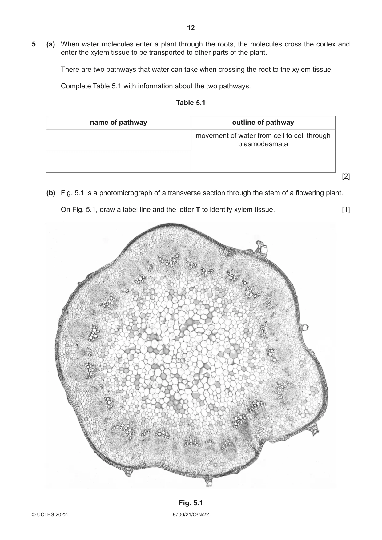
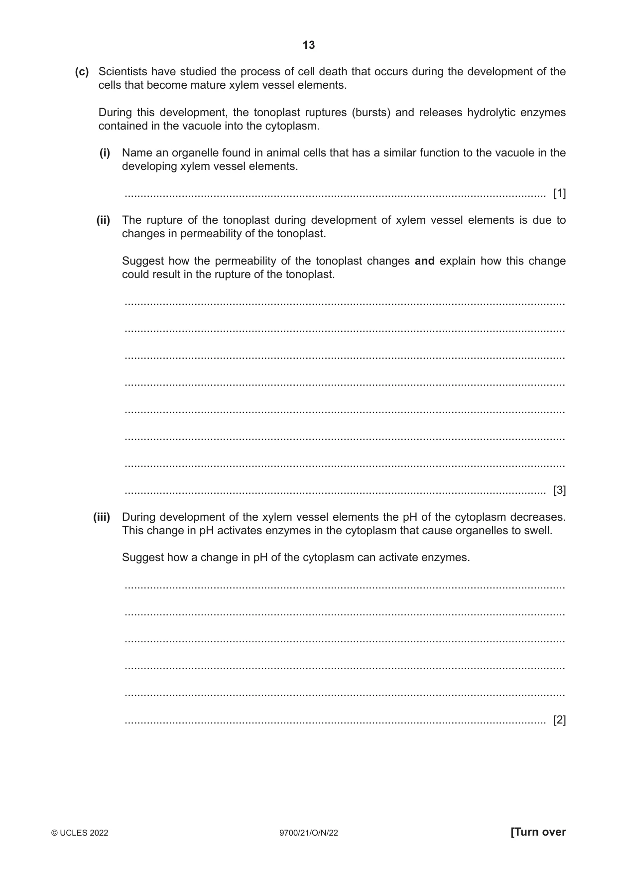
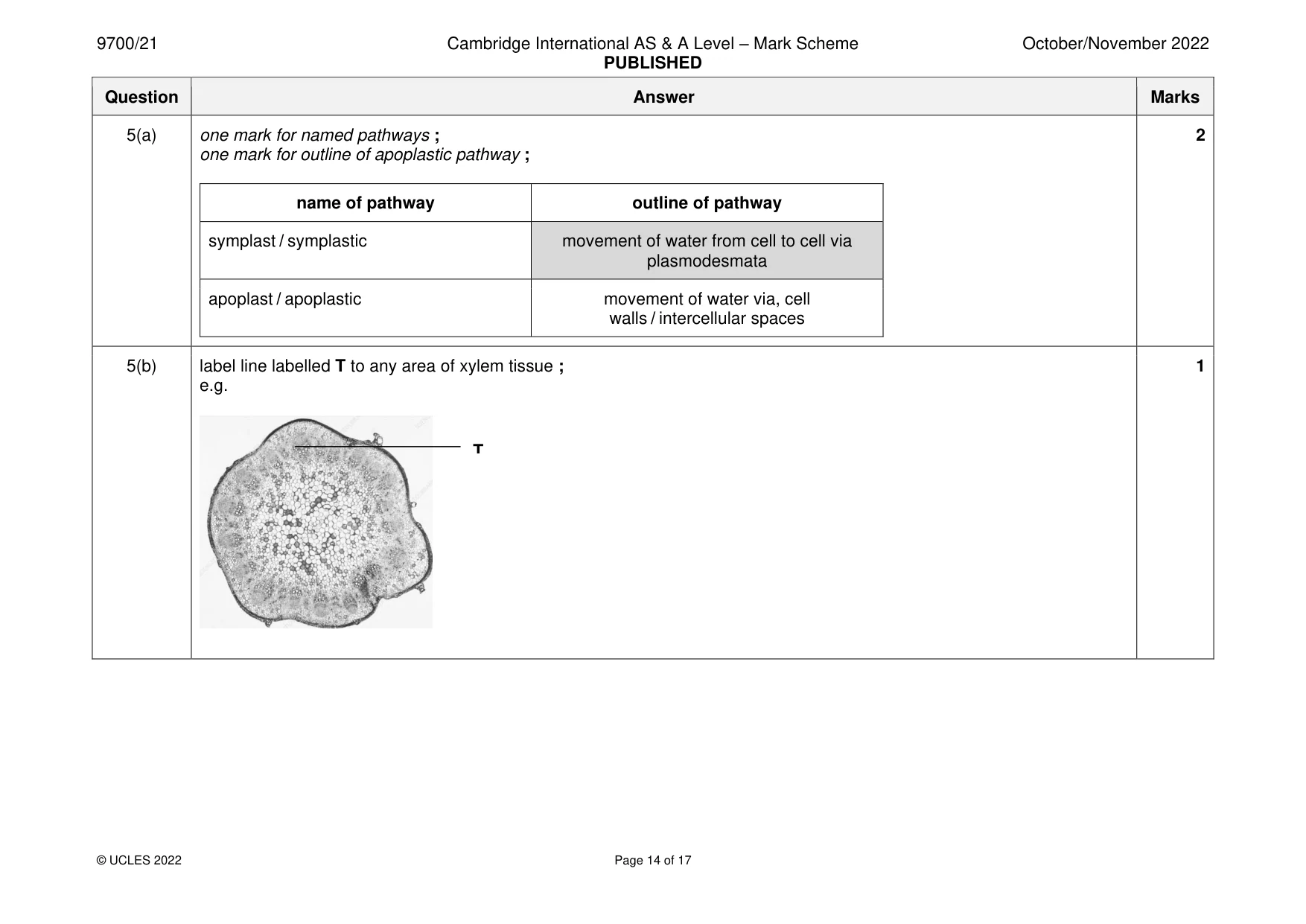
5 (a) When water molecules enter a plant through the roots, the molecules cross the cortex and enter the xylem tissue to be transported to other parts of the plant. There are two pathways that water can take when crossing the root to the xylem tissue. Complete Table 5.1 with information about the two pathways. Table 5.1 name of pathway outline of pathway movement of water from cell to cell through plasmodesmata [2] (b) Fig. 5.1 is a photomicrograph of a transverse section through the stem of a flowering plant. On Fig. 5.1, draw a label line and the letter T to identify xylem tissue. [1] Fig. 5.1 (c) Scientists have studied the process of cell death that occurs during the development of the cells that become mature xylem vessel elements. During this development, the tonoplast ruptures (bursts) and releases hydrolytic enzymes contained in the vacuole into the cytoplasm. (i) Name an organelle found in animal cells that has a similar function to the vacuole in the developing xylem vessel elements. ..................................................................................................................................... [1] (ii) The rupture of the tonoplast during development of xylem vessel elements is due to changes in permeability of the tonoplast. Suggest how the permeability of the tonoplast changes and explain how this change could result in the rupture of the tonoplast. ........................................................................................................................................... ........................................................................................................................................... ........................................................................................................................................... ........................................................................................................................................... ........................................................................................................................................... ........................................................................................................................................... ........................................................................................................................................... ..................................................................................................................................... [3] (iii) During development of the xylem vessel elements the pH of the cytoplasm decreases. This change in pH activates enzymes in the cytoplasm that cause organelles to swell. Suggest how a change in pH of the cytoplasm can activate enzymes. ........................................................................................................................................... ........................................................................................................................................... ........................................................................................................................................... ........................................................................................................................................... ........................................................................................................................................... ..................................................................................................................................... [2] (d) Some insects are vectors of disease. (i) When an insect feeds on the xylem fluid it can act as a vector of plant diseases such as Pierce’s disease. This bacterial disease affects many fruit trees causing the leaves to turn brown and drop from the plant, resulting in much less fruit being produced. Explain why Pierce’s disease can be described as an infectious disease. ........................................................................................................................................... ........................................................................................................................................... ........................................................................................................................................... ........................................................................................................................................... .......................................................................................................................................[2] (ii) Malaria is another infectious disease that also involves an insect vector. State the type of organism that causes malaria and name one organism that causes malaria in humans. type of organism ................................................................................................................ name ................................................................................................................................. [2] [Total: 13]
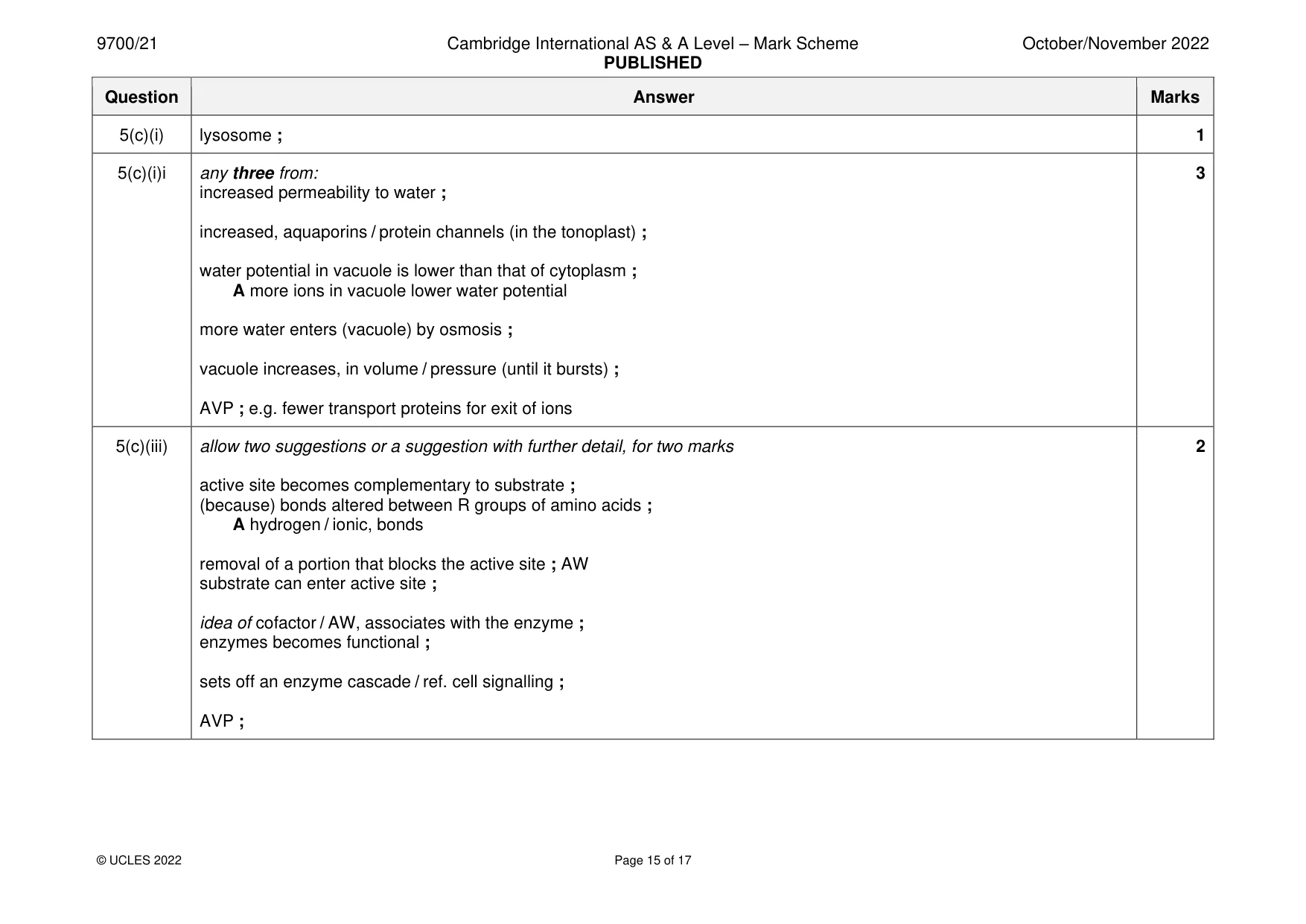
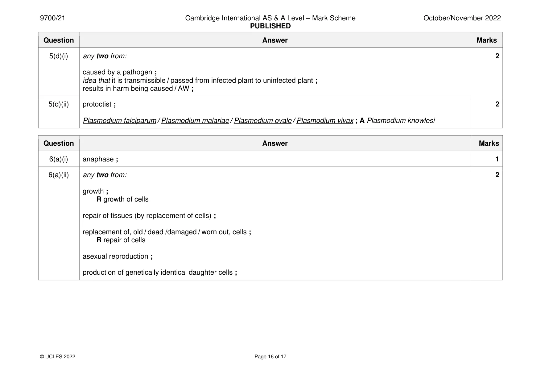
Mark scheme: 5(a) one mark for named pathways ; 2 one mark for outline of apoplastic pathway ; name of pathway outline of pathway symplast / symplastic movement of water from cell to cell via plasmodesmata apoplast / apoplastic movement of water via, cell walls / intercellular spaces 5(b) label line labelled T to any area of xylem tissue ; 1 e.g. T 5(c)(i) lysosome ; 1 5(c)(i)i any three from: 3 increased permeability to water ; increased, aquaporins / protein channels (in the tonoplast) ; water potential in vacuole is lower than that of cytoplasm ; A more ions in vacuole lower water potential more water enters (vacuole) by osmosis ; vacuole increases, in volume / pressure (until it bursts) ; AVP ; e.g. fewer transport proteins for exit of ions 5(c)(iii) allow two suggestions or a suggestion with further detail, for two marks 2 active site becomes complementary to substrate ; (because) bonds altered between R groups of amino acids ; A hydrogen / ionic, bonds removal of a portion that blocks the active site ; AW substrate can enter active site ; idea of cofactor / AW, associates with the enzyme ; enzymes becomes functional ; sets off an enzyme cascade / ref. cell signalling ; AVP ; 5(d)(i) any two from: 2 caused by a pathogen ; idea that it is transmissible / passed from infected plant to uninfected plant ; results in harm being caused / AW ; 5(d)(ii) protoctist ; 2 Plasmodium falciparum / Plasmodium malariae / Plasmodium ovale / Plasmodium vivax ; A Plasmodium knowlesi
More questions on Cells as the basic units of living organisms
Q6 · A photomicrograph of plant cells in stages of the mitotic cell cycle
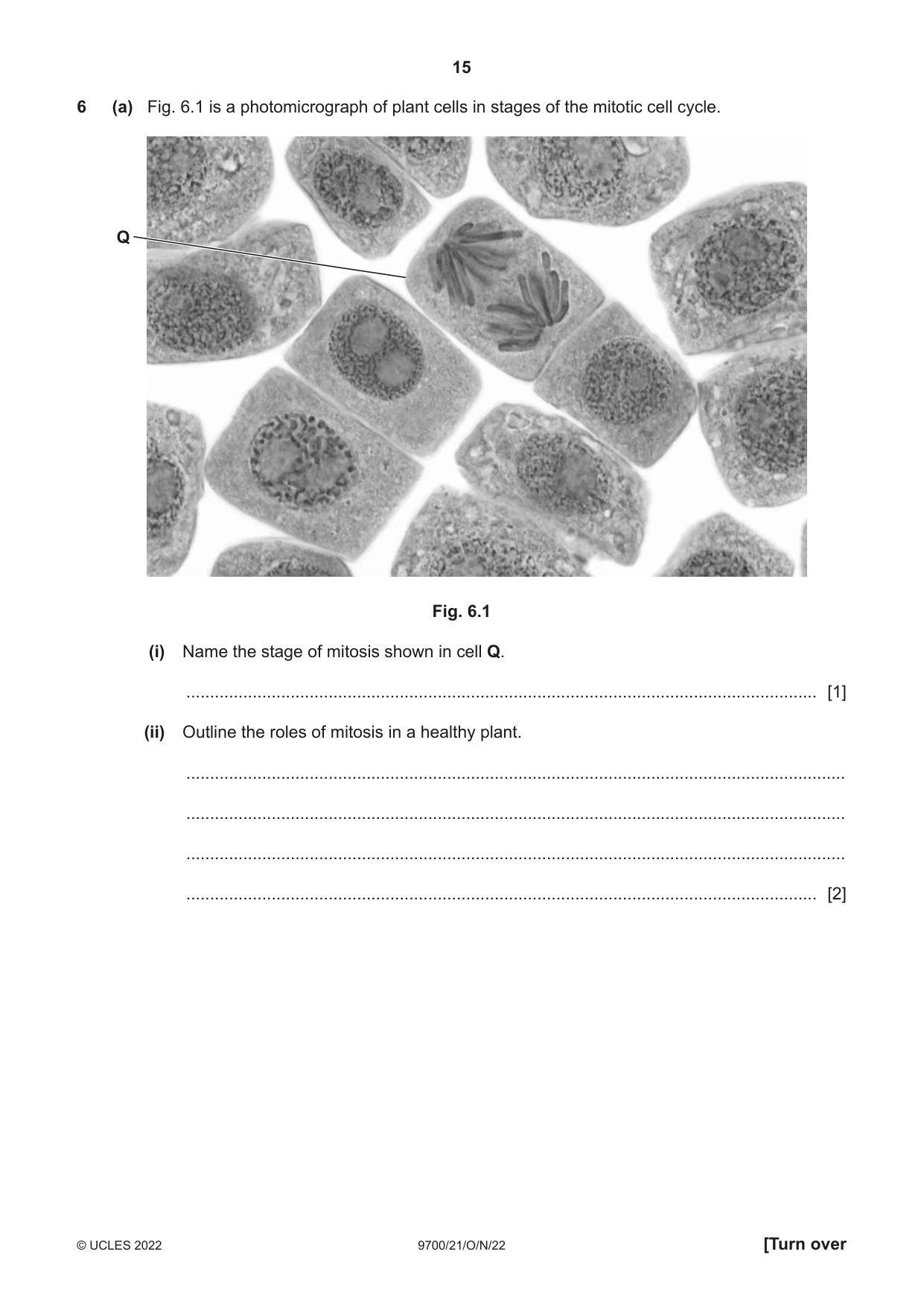
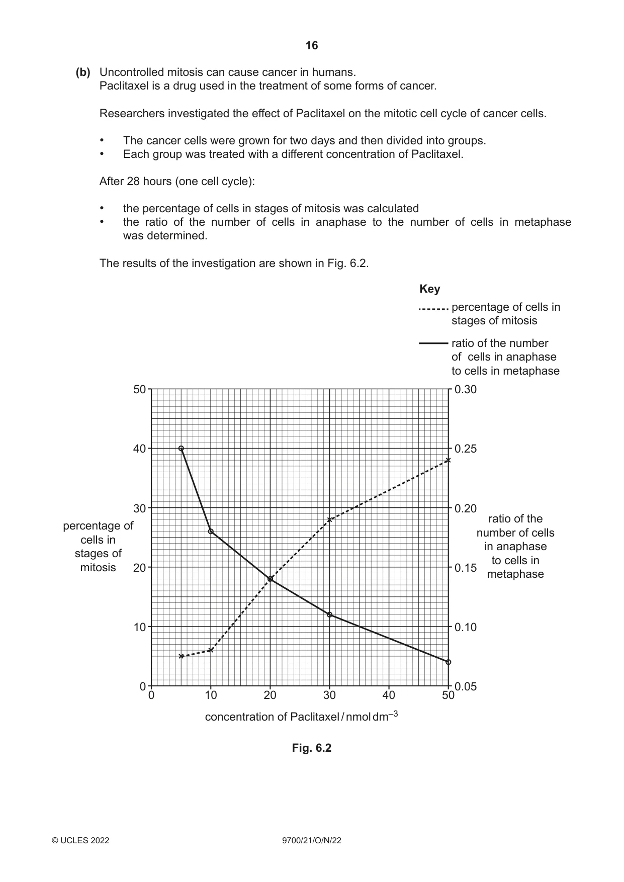
6 (a) Fig. 6.1 is a photomicrograph of plant cells in stages of the mitotic cell cycle. Q Fig. 6.1 (i) Name the stage of mitosis shown in cell Q. ..................................................................................................................................... [1] (ii) Outline the roles of mitosis in a healthy plant. ........................................................................................................................................... ........................................................................................................................................... ........................................................................................................................................... ..................................................................................................................................... [2] (b) Uncontrolled mitosis can cause cancer in humans. Paclitaxel is a drug used in the treatment of some forms of cancer. Researchers investigated the effect of Paclitaxel on the mitotic cell cycle of cancer cells. • The cancer cells were grown for two days and then divided into groups. • Each group was treated with a different concentration of Paclitaxel. After 28 hours (one cell cycle): • the percentage of cells in stages of mitosis was calculated • the ratio of the number of cells in anaphase to the number of cells in metaphase was determined. The results of the investigation are shown in Fig. 6.2. Key percentage of cells in stages of mitosis ratio of the number of cells in anaphase to cells in metaphase 50 0.30 40 0.25 30 0.20 ratio of thepercentage of number of cells cells in in anaphase stages of to cells in mitosis 20 0.15 metaphase 10 0.10 0 0.05 0 10 20 30 40 50 concentration of Paclitaxel / nmol dm–3 Fig. 6.2 With reference to Fig. 6.2, describe the results and suggest an explanation for the effect of Paclitaxel on the mitotic cell cycle. ................................................................................................................................................... ................................................................................................................................................... ................................................................................................................................................... ................................................................................................................................................... ................................................................................................................................................... ................................................................................................................................................... ................................................................................................................................................... ............................................................................................................................................. [4] [Total: 7]
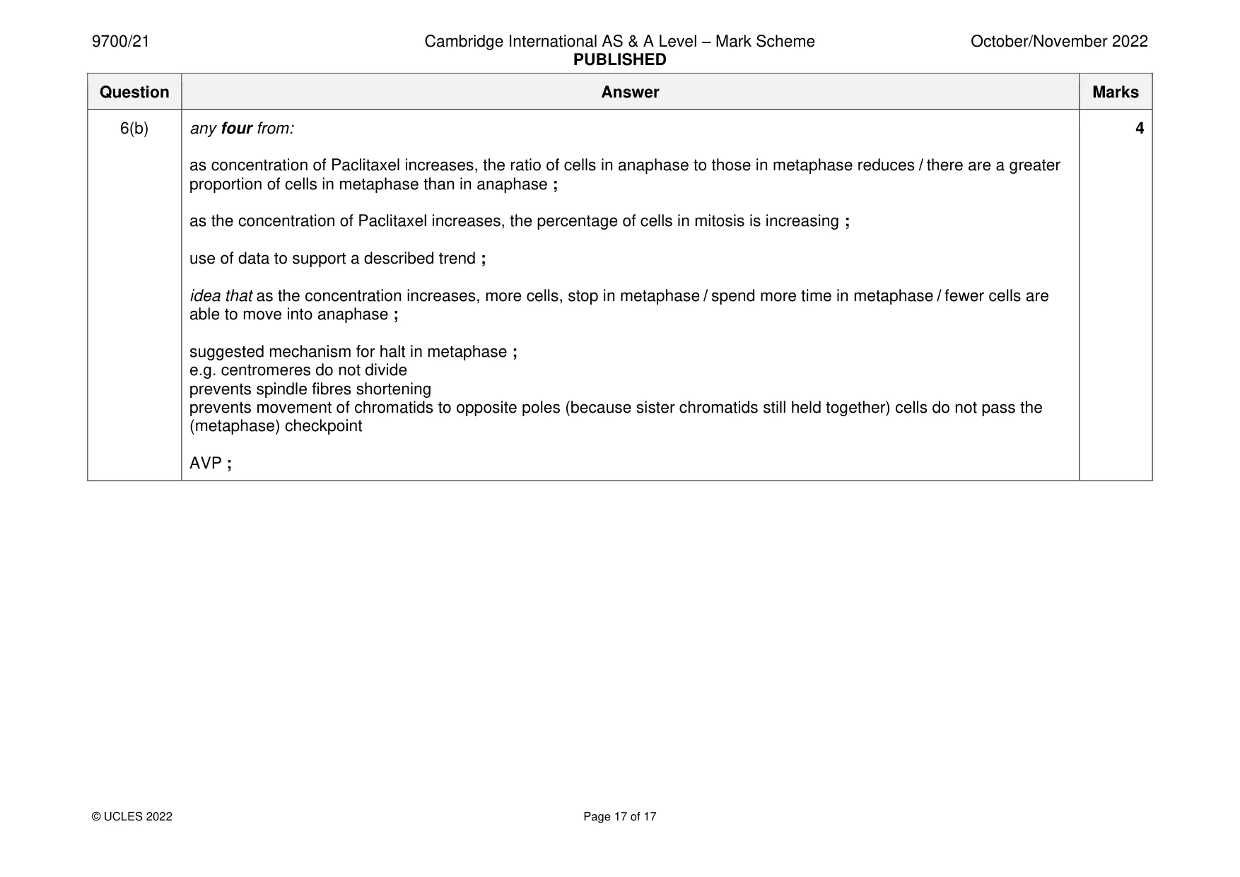
Mark scheme: 6(a)(i) anaphase ; 1 6(a)(ii) any two from: 2 growth ; R growth of cells repair of tissues (by replacement of cells) ; replacement of, old / dead /damaged / worn out, cells ; R repair of cells asexual reproduction ; production of genetically identical daughter cells ; 6(b) any four from: 4 as concentration of Paclitaxel increases, the ratio of cells in anaphase to those in metaphase reduces / there are a greater proportion of cells in metaphase than in anaphase ; as the concentration of Paclitaxel increases, the percentage of cells in mitosis is increasing ; use of data to support a described trend ; idea that as the concentration increases, more cells, stop in metaphase / spend more time in metaphase / fewer cells are able to move into anaphase ; suggested mechanism for halt in metaphase ; e.g. centromeres do not divide prevents spindle fibres shortening prevents movement of chromatids to opposite poles (because sister chromatids still held together) cells do not pass the (metaphase) checkpoint AVP ;
More questions on Replication and division of nuclei and cells
What was in this paper
The subtopics covered by these 6 questions, and how many questions each got. Open one in a new tab to see every Cambridge question on it.
What you needed in this session
Cambridge’s own grade thresholds for 2022 Oct/Nov, Paper 2 · Variant 1. A higher threshold means an easier paper — the bar moves with how the cohort did.