Cambridge A Level Biology 9700 — 2022 Oct/Nov Paper 2 · Variant 2
9700/22/O/N/22 · 6 questions · 60 marks · ≈68 min
The question paper and its mark scheme, free to read here and free to download. This is Cambridge’s own paper, exactly as it was sat.
Question paper16 pages

Mark scheme20 pages
Answers below. Sit the paper first if you are practising.
Questions as text
Q1 · The mammalian circulatory system is adapted for the long-distance transport of the…
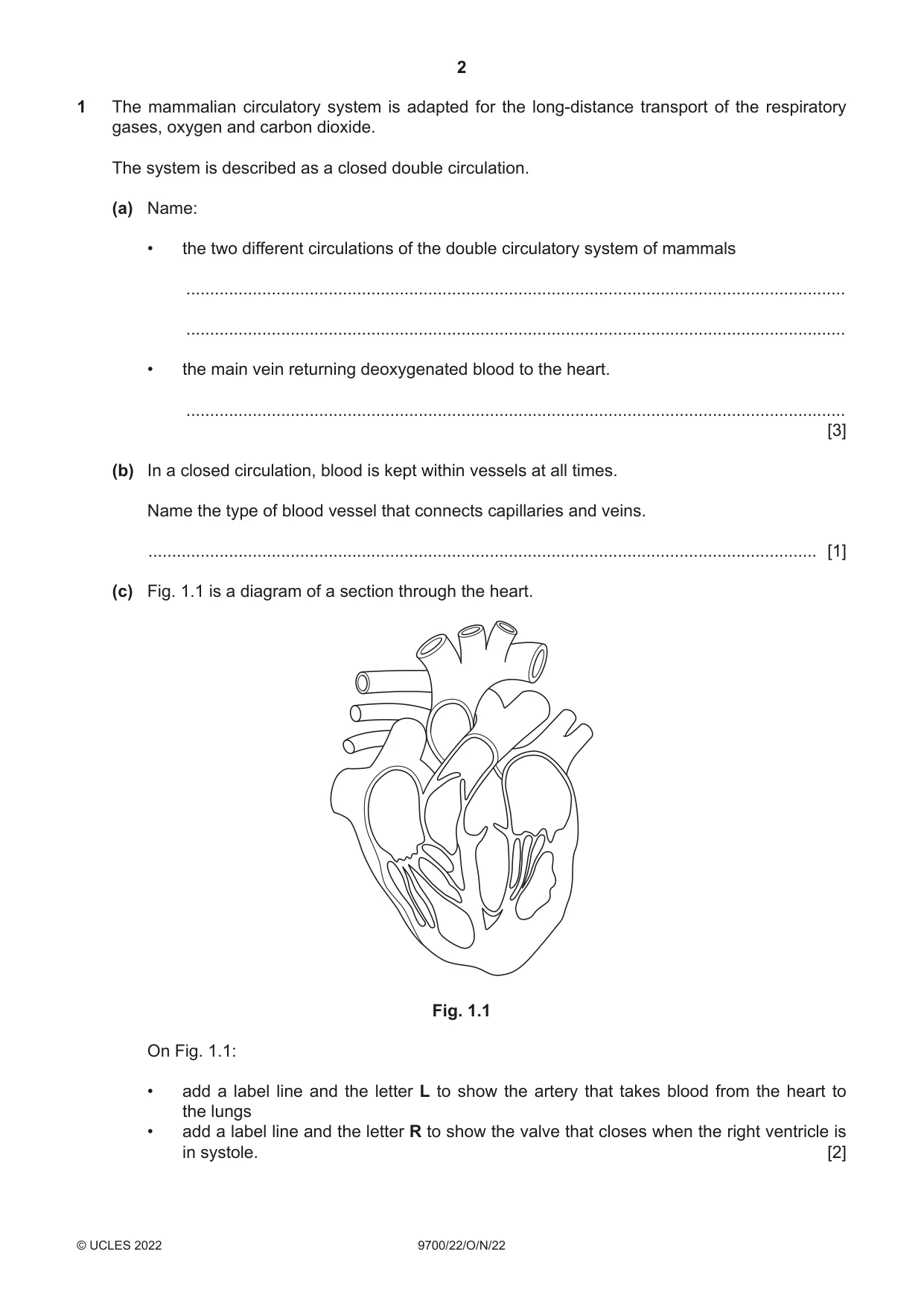
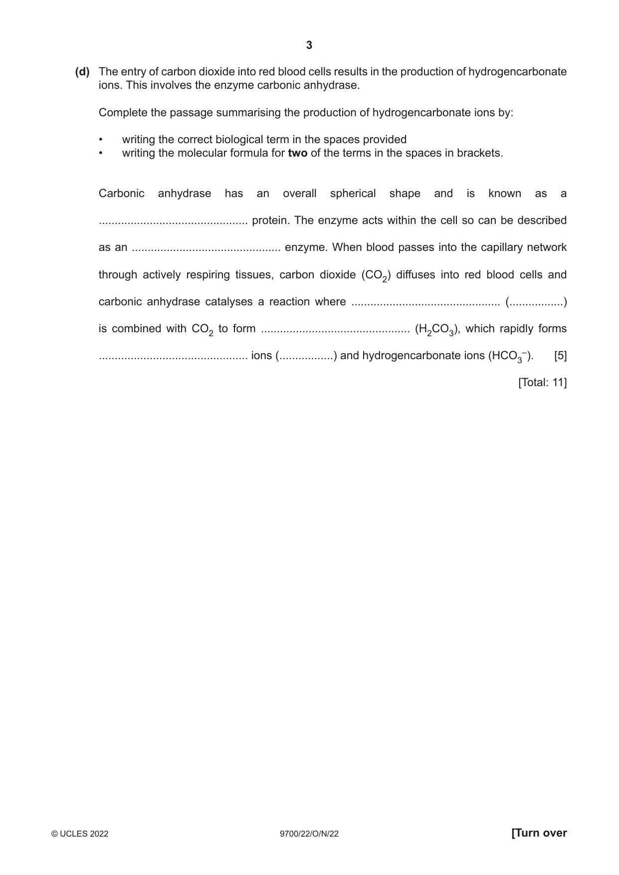
1 The mammalian circulatory system is adapted for the long-distance transport of the respiratory gases, oxygen and carbon dioxide. The system is described as a closed double circulation. (a) Name: • the two different circulations of the double circulatory system of mammals ........................................................................................................................................... ........................................................................................................................................... • the main vein returning deoxygenated blood to the heart. ........................................................................................................................................... [3] (b) In a closed circulation, blood is kept within vessels at all times. Name the type of blood vessel that connects capillaries and veins. ............................................................................................................................................. [1] (c) Fig. 1.1 is a diagram of a section through the heart. Fig. 1.1 On Fig. 1.1: • add a label line and the letter L to show the artery that takes blood from the heart to the lungs • add a label line and the letter R to show the valve that closes when the right ventricle is in systole. [2] (d) The entry of carbon dioxide into red blood cells results in the production of hydrogencarbonate ions. This involves the enzyme carbonic anhydrase. Complete the passage summarising the production of hydrogencarbonate ions by: • writing the correct biological term in the spaces provided • writing the molecular formula for two of the terms in the spaces in brackets. Carbonic anhydrase has an overall spherical shape and is known as a ............................................... protein. The enzyme acts within the cell so can be described as an ............................................... enzyme. When blood passes into the capillary network through actively respiring tissues, carbon dioxide (CO2) diffuses into red blood cells and carbonic anhydrase catalyses a reaction where ............................................... (.................) is combined with CO2 to form ............................................... (H2CO3), which rapidly forms ............................................... ions (.................) and hydrogencarbonate ions (HCO3–). [5] [Total: 11]
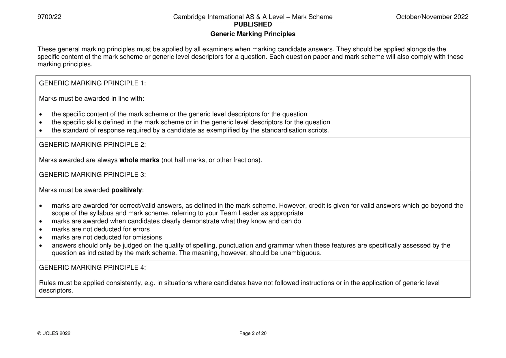
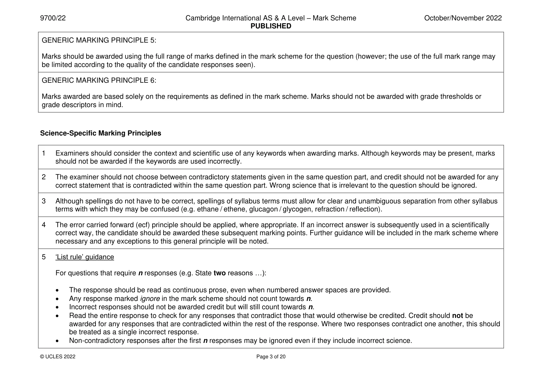
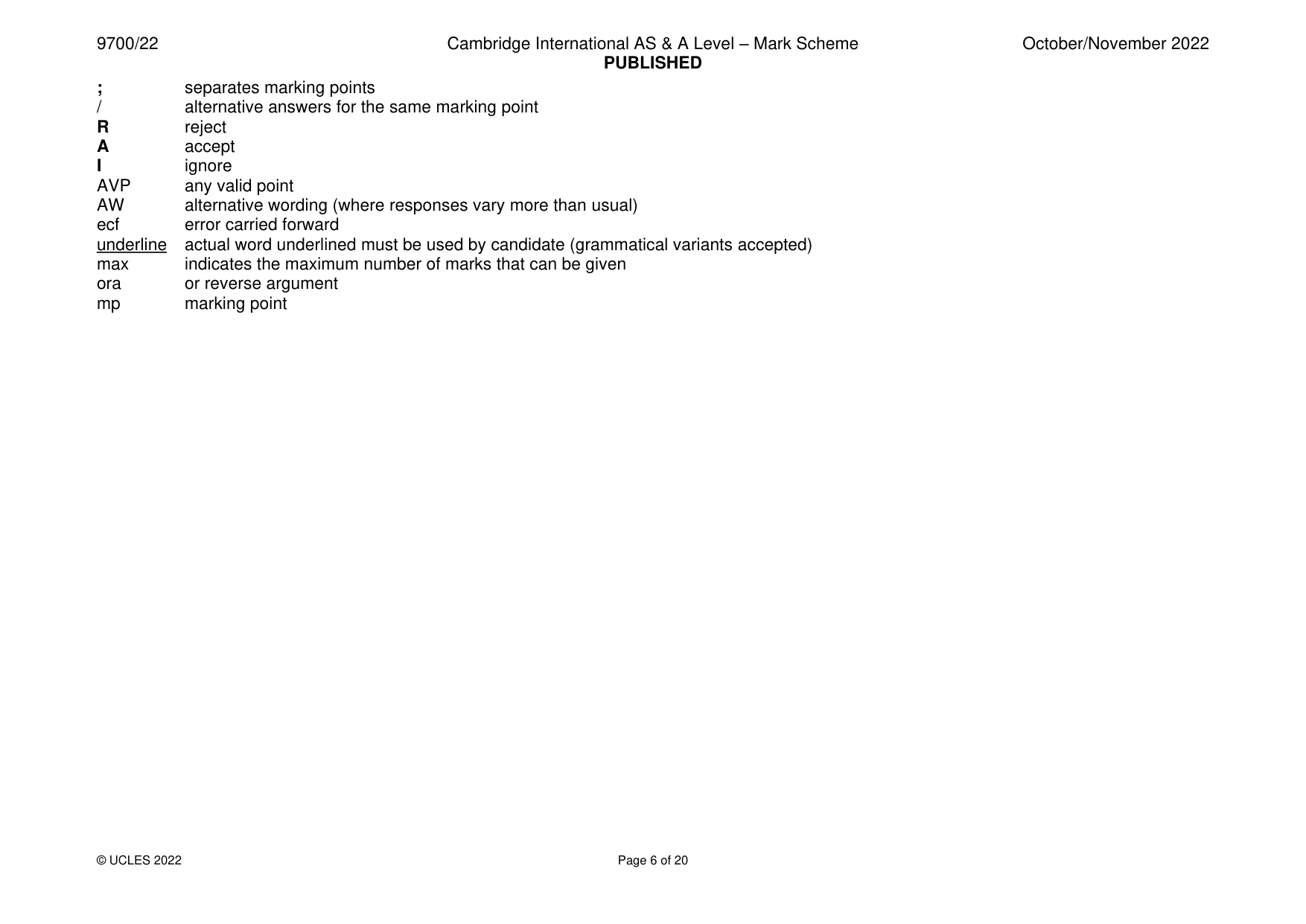
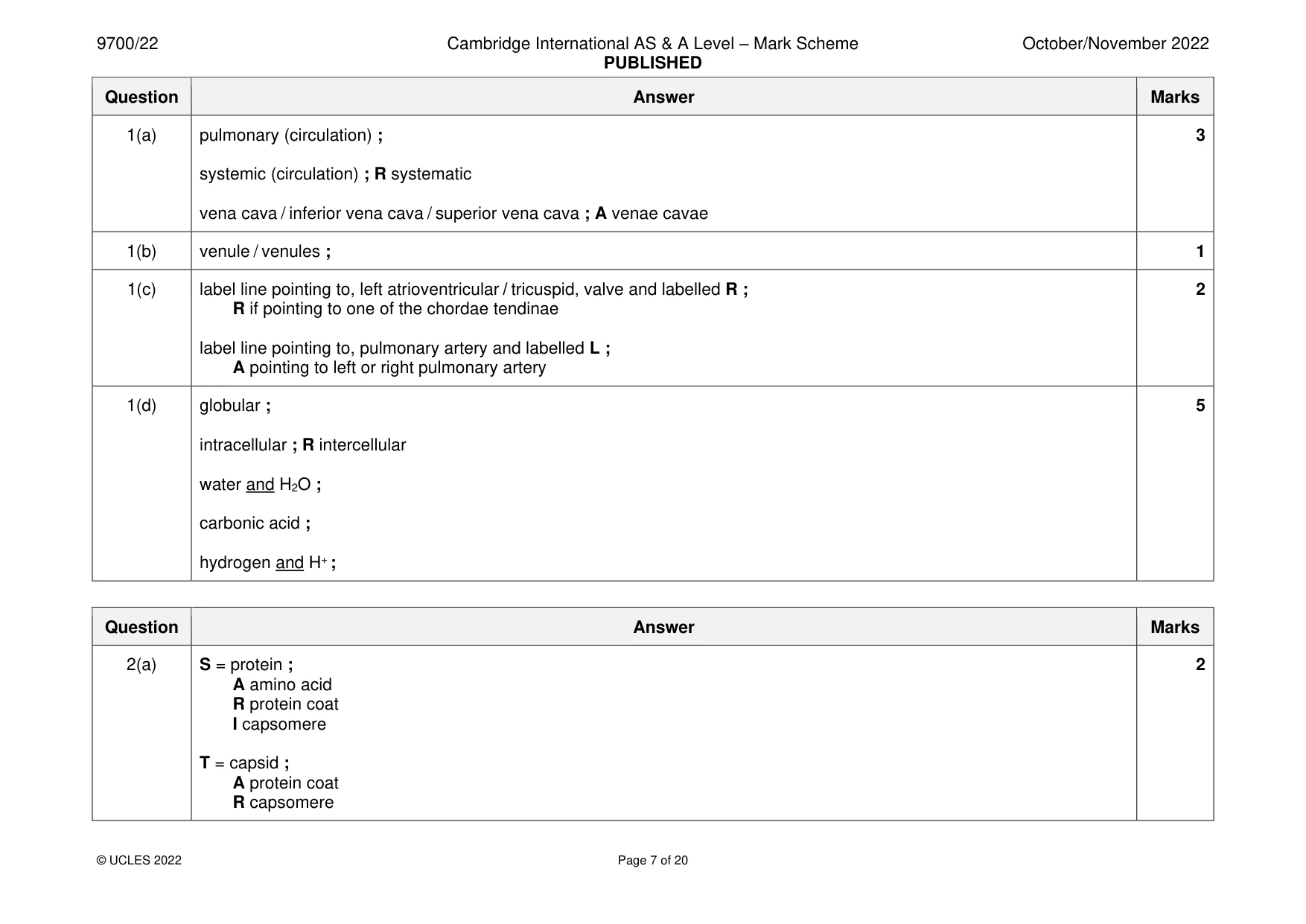
Mark scheme: Question Answer Marks 1(a) pulmonary (circulation) ; 3 systemic (circulation) ; R systematic vena cava / inferior vena cava / superior vena cava ; A venae cavae 1(b) venule / venules ; 1 1(c) label line pointing to, left atrioventricular / tricuspid, valve and labelled R ; 2 R if pointing to one of the chordae tendinae label line pointing to, pulmonary artery and labelled L ; A pointing to left or right pulmonary artery 1(d) globular ; 5 intracellular ; R intercellular water and H2O ; carbonic acid ; hydrogen and H+ ;
Q2 · Human cytomegalovirus (HCMV) is a common virus affecting humans
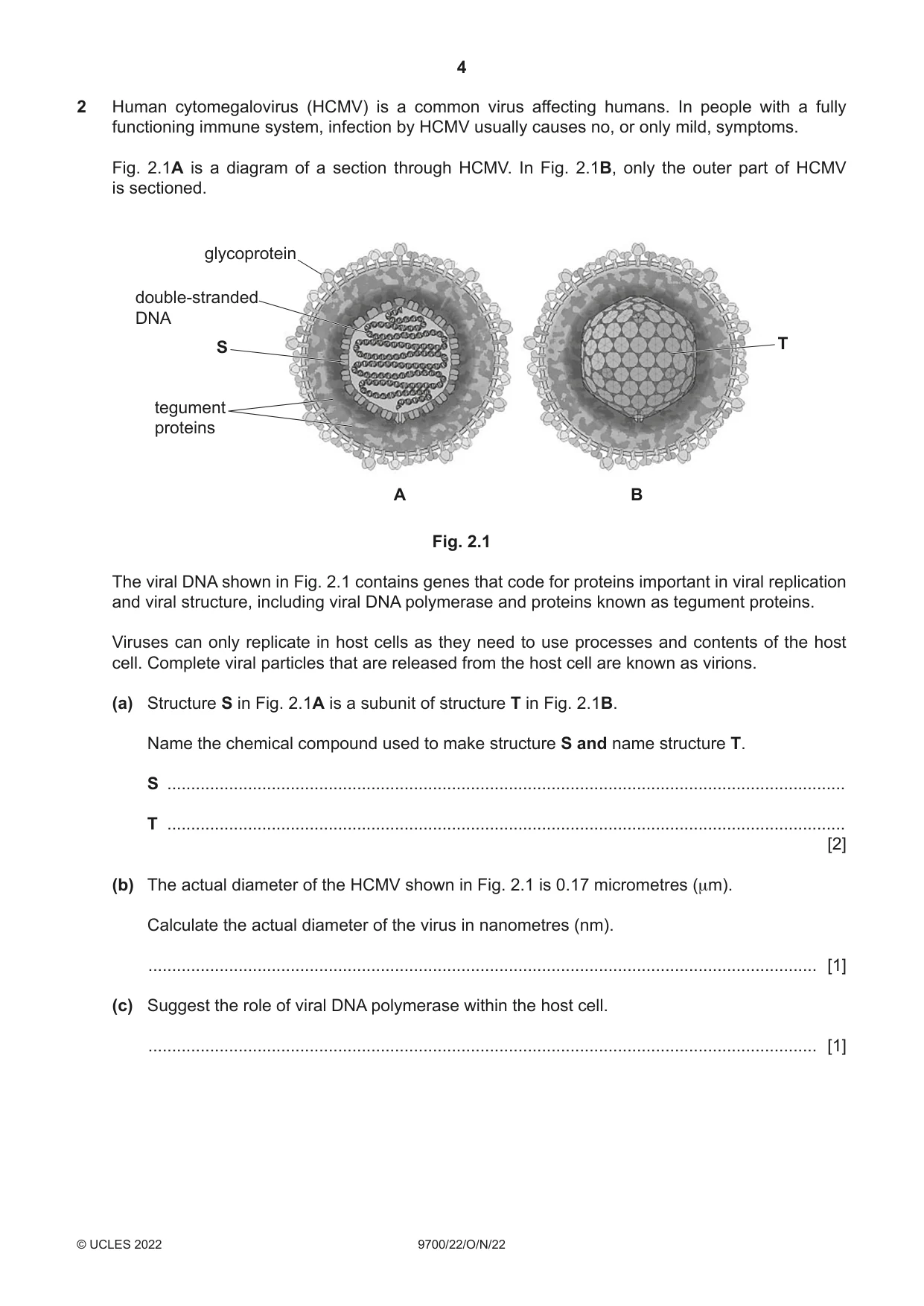
2 Human cytomegalovirus (HCMV) is a common virus affecting humans. In people with a fully functioning immune system, infection by HCMV usually causes no, or only mild, symptoms. Fig. 2.1A is a diagram of a section through HCMV. In Fig. 2.1B, only the outer part of HCMV is sectioned. glycoprotein double-stranded DNA S T tegument proteins A B Fig. 2.1 The viral DNA shown in Fig. 2.1 contains genes that code for proteins important in viral replication and viral structure, including viral DNA polymerase and proteins known as tegument proteins. Viruses can only replicate in host cells as they need to use processes and contents of the host cell. Complete viral particles that are released from the host cell are known as virions. (a) Structure S in Fig. 2.1A is a subunit of structure T in Fig. 2.1B. Name the chemical compound used to make structure S and name structure T. S ............................................................................................................................................... T ............................................................................................................................................... [2] (b) The actual diameter of the HCMV shown in Fig. 2.1 is 0.17 micrometres (µm). Calculate the actual diameter of the virus in nanometres (nm). ............................................................................................................................................. [1] (c) Suggest the role of viral DNA polymerase within the host cell. ............................................................................................................................................. [1] (d) The virus in Fig. 2.1 is drawn as a spherical shape. Structure T is always the same shape. However, electron micrographs show that HCMV virions are not all the same shape. Suggest how HCMV virions can be of different shapes. ................................................................................................................................................... ................................................................................................................................................... ................................................................................................................................................... ................................................................................................................................................... ............................................................................................................................................. [2] (e) With reference to Fig. 2.1A, state one similarity and one difference between the genetic material of HCMV and the genetic material of a typical bacterial cell. ................................................................................................................................................... ................................................................................................................................................... ................................................................................................................................................... ................................................................................................................................................... ............................................................................................................................................. [2] (f) HCMV is known to infect some types of human cell that carry out the mitotic cell cycle. Studies have shown that in the presence of one tegument protein, UL69, the cell cycle stops in the G1 stage. Outline the effects the presence of UL69 will have on the normal activity of the mitotic cell cycle. ................................................................................................................................................... ................................................................................................................................................... ................................................................................................................................................... ................................................................................................................................................... ................................................................................................................................................... ................................................................................................................................................... ................................................................................................................................................... ............................................................................................................................................. [3] (g) After a person has been infected with HCMV, the virus remains in a dormant state in the body for life. If the virus becomes active again (reactivates), the virus will only cause serious illness if the person has a weak immune system at that time. Explain why the response to reactivation of HCMV is more likely to cause serious illness in a person who has a weak immune system. ................................................................................................................................................... ................................................................................................................................................... ................................................................................................................................................... ................................................................................................................................................... ................................................................................................................................................... ................................................................................................................................................... ................................................................................................................................................... ............................................................................................................................................. [4] [Total: 15]
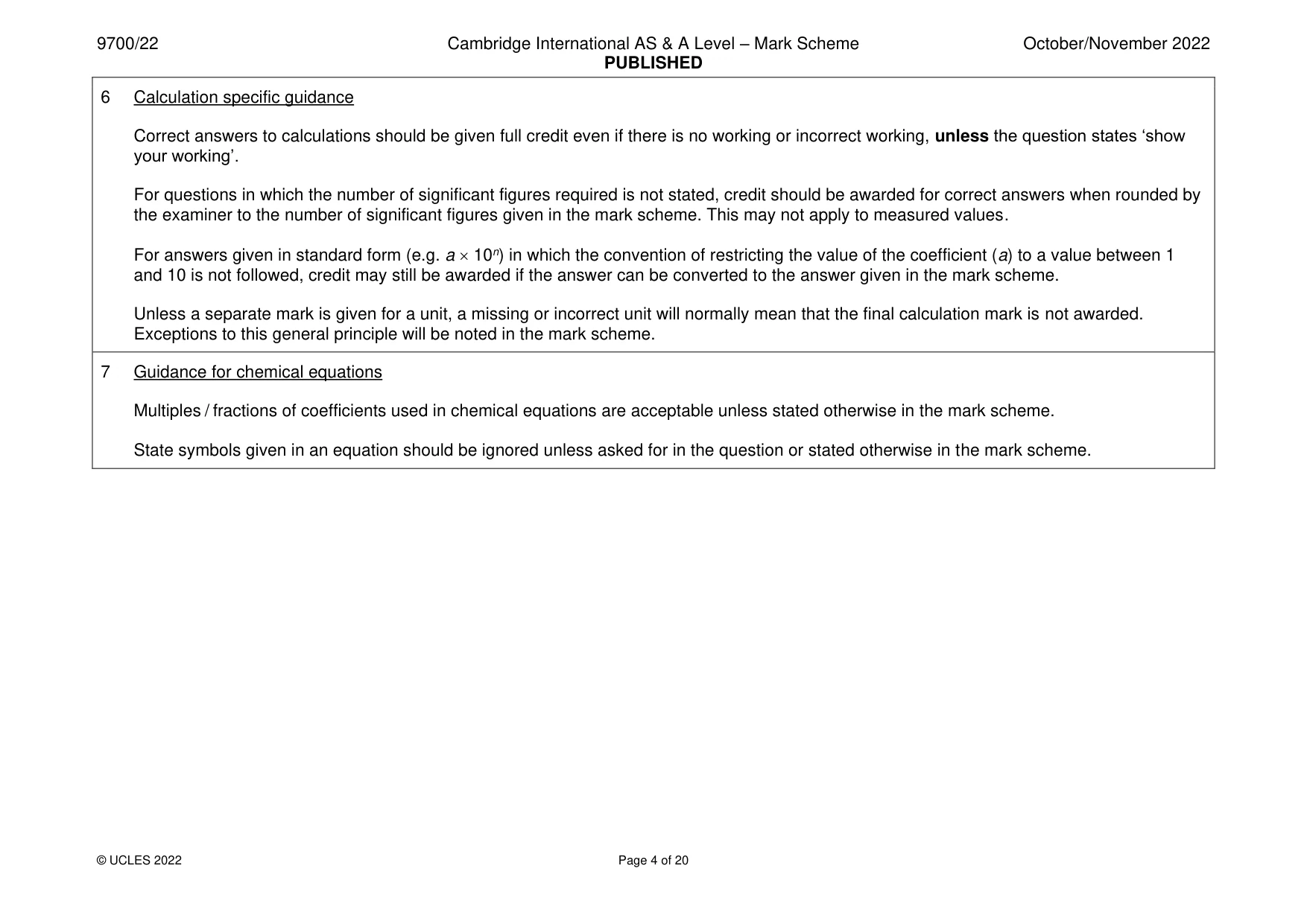
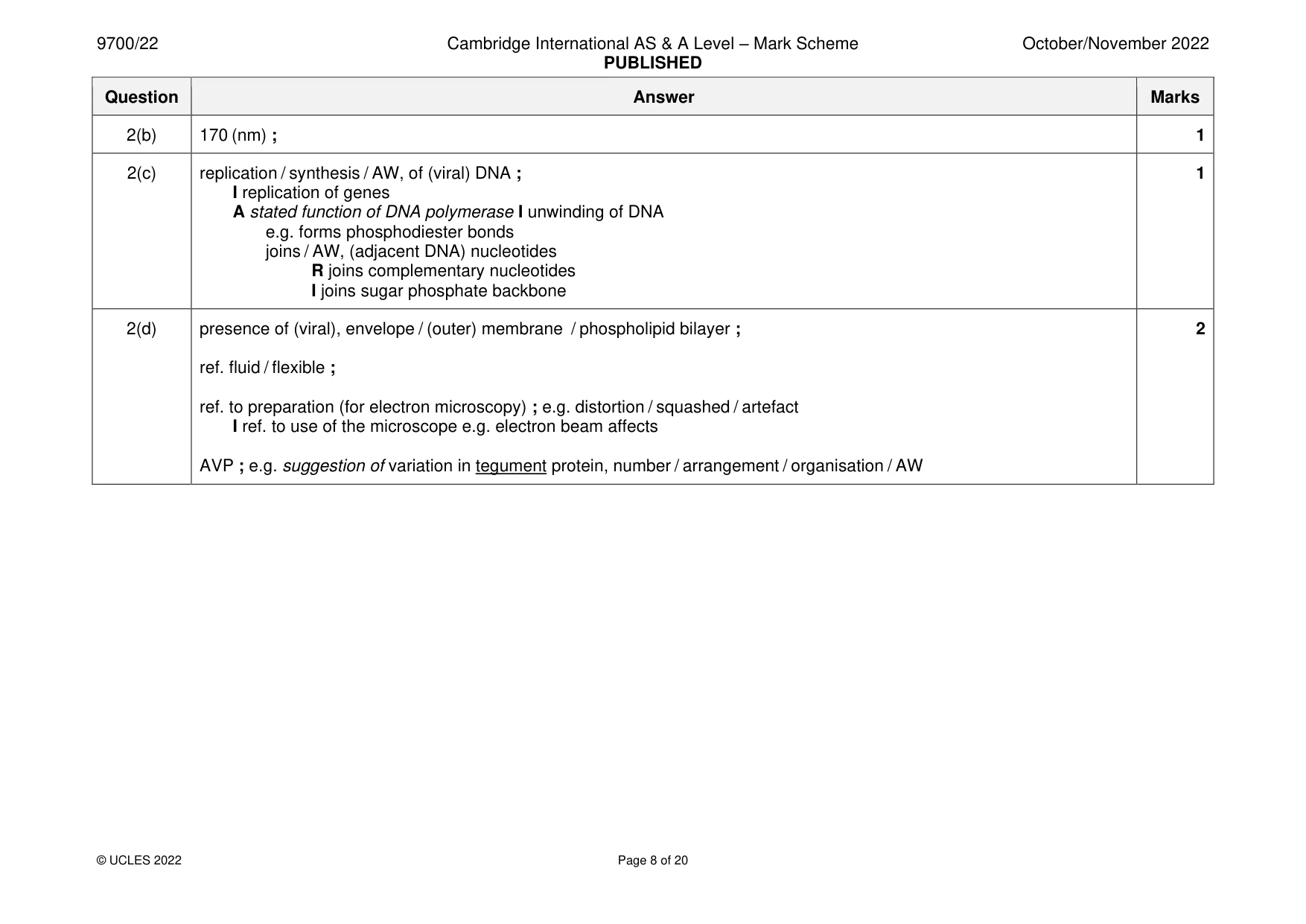
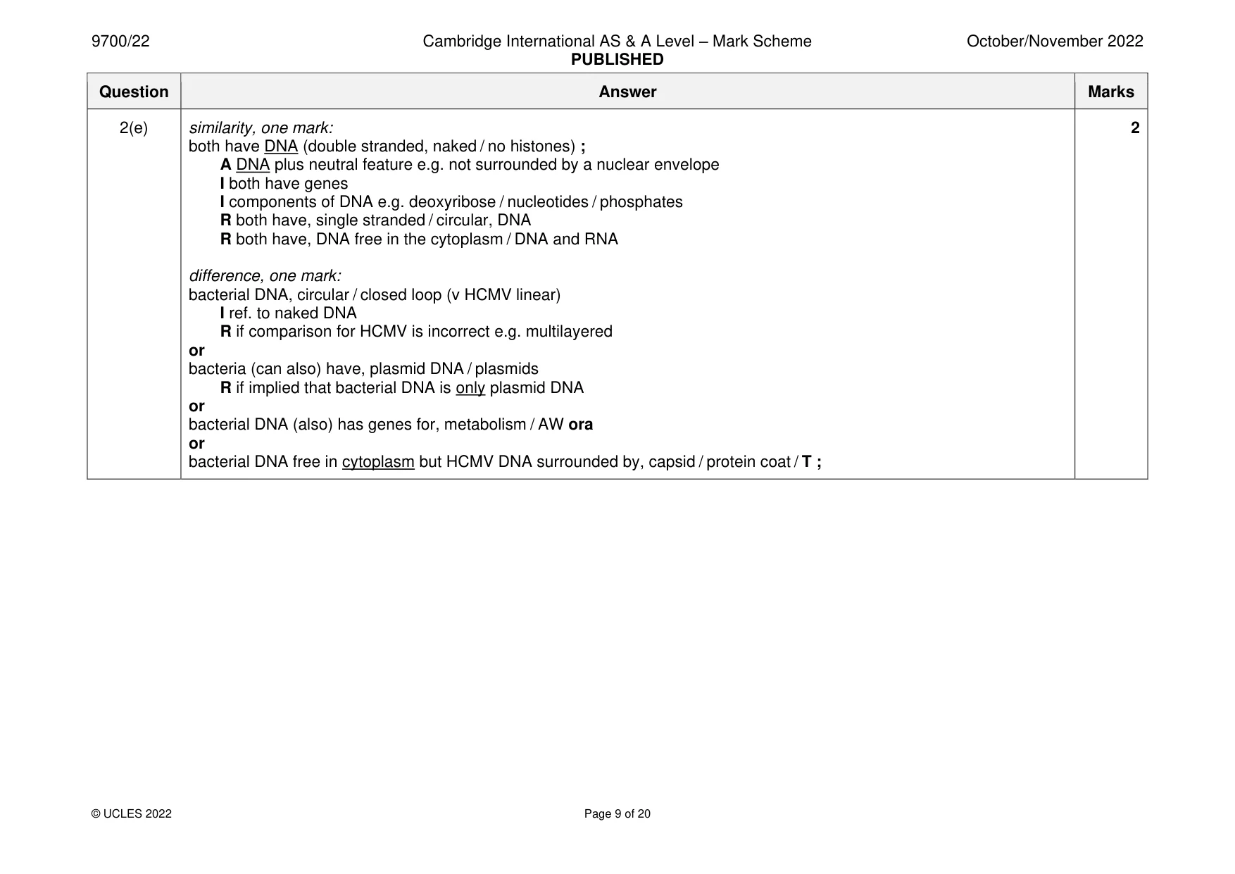
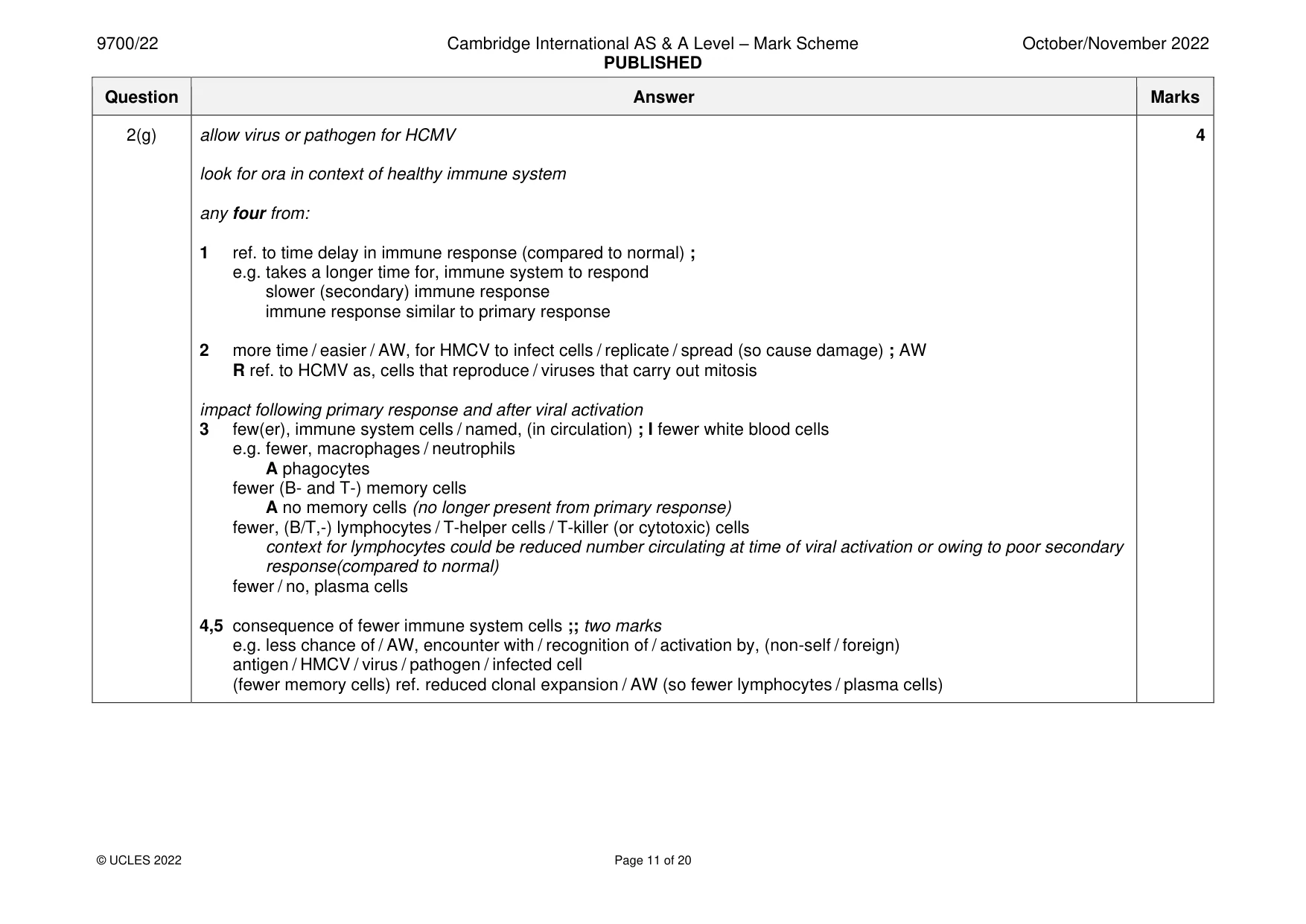
Mark scheme: 2(a) S = protein ; 2 A amino acid R protein coat I capsomere T = capsid ; A protein coat R capsomere 2(b) 170 (nm) ; 1 2(c) replication / synthesis / AW, of (viral) DNA ; 1 I replication of genes A stated function of DNA polymerase I unwinding of DNA e.g. forms phosphodiester bonds joins / AW, (adjacent DNA) nucleotides R joins complementary nucleotides I joins sugar phosphate backbone 2(d) presence of (viral), envelope / (outer) membrane / phospholipid bilayer ; 2 ref. fluid / flexible ; ref. to preparation (for electron microscopy) ; e.g. distortion / squashed / artefact I ref. to use of the microscope e.g. electron beam affects AVP ; e.g. suggestion of variation in tegument protein, number / arrangement / organisation / AW 2(e) similarity, one mark: 2 both have DNA (double stranded, naked / no histones) ; A DNA plus neutral feature e.g. not surrounded by a nuclear envelope I both have genes I components of DNA e.g. deoxyribose / nucleotides / phosphates R both have, single stranded / circular, DNA R both have, DNA free in the cytoplasm / DNA and RNA difference, one mark: bacterial DNA, circular / closed loop (v HCMV linear) I ref. to naked DNA R if comparison for HCMV is incorrect e.g. multilayered or bacteria (can also) have, plasmid DNA / plasmids R if implied that bacterial DNA is only plasmid DNA or bacterial DNA (also) has genes for, metabolism / AW ora or bacterial DNA free in cytoplasm but HCMV DNA surrounded by, capsid / protein coat / T ; 2(f) I transcription and translation stops 3 any three from: 1 no, S phase / G2 phase / mitosis / cytokinesis ; A remains in (G1) interphase A synthesis phase for S phase I named mitotic stages look for CON e.g. ‘no S-phase, so mitosis only has one chromatid’ 2 (semi-conservative) DNA, replication / synthesis, does not occur ; A sister / identical, chromatids, do not form R if a phase is stated and it is not given as the S-phase 3 failure to pass checkpoints / does not reach checkpoints ; A ref. to no error checking occurs (in context of checkpoints) I proofreading / error checking, (by DNA polymerase) during DNA replication 4 cell does not, grow / increase in size / increase cytoplasm ; (context of preparing for mitosis / cytokinesis) A grows to normal (non-dividing) size 5 idea that the cell does not, duplicate / synthesise, organelles / extra materials (context is for, cytokinesis / new cells) ; A centrioles do not replicate A microtubules not synthesised R if stated as occurring in S phase (even if e.g. G2 given as well) 2(g) allow virus or pathogen for HCMV 4 look for ora in context of healthy immune system any four from: 1 ref. to time delay in immune response (compared to normal) ; e.g. takes a longer time for, immune system to respond slower (secondary) immune response immune response similar to primary response 2 more time / easier / AW, for HMCV to infect cells / replicate / spread (so cause damage) ; AW R ref. to HCMV as, cells that reproduce / viruses that carry out mitosis impact following primary response and after viral activation 3 few(er), immune system cells / named, (in circulation) ; I fewer white blood cells e.g. fewer, macrophages / neutrophils A phagocytes fewer (B- and T-) memory cells A no memory cells (no longer present from primary response) fewer, (B/T,-) lymphocytes / T-helper cells / T-killer (or cytotoxic) cells context for lymphocytes could be reduced number circulating at time of viral activation or owing to poor secondary response(compared to normal) fewer / no, plasma cells 4,5 consequence of fewer immune system cells ;; two marks e.g. less chance of / AW, encounter with / recognition of / activation by, (non-self / foreign) antigen / HMCV / virus / pathogen / infected cell (fewer memory cells) ref. reduced clonal expansion / AW (so fewer lymphocytes / plasma cells) 2(g) (fewer plasma cells) lower concentration of antibodies A no antibodies (as no plasma cells) A longer time for antibody, production / concentration to build up (fewer T-helper cells) less cytokine released A effect of cytokine if spelling of cytokine rejected (fewer T-killer cells) fewer infected cells killed / A perforin / granzymes / hydrogen peroxide, not released (fewer phagocytes) less phagocytosis for antigen presentation
Q3 · Plasmodium falciparum is one species of Plasmodium that causes the life-threatening…
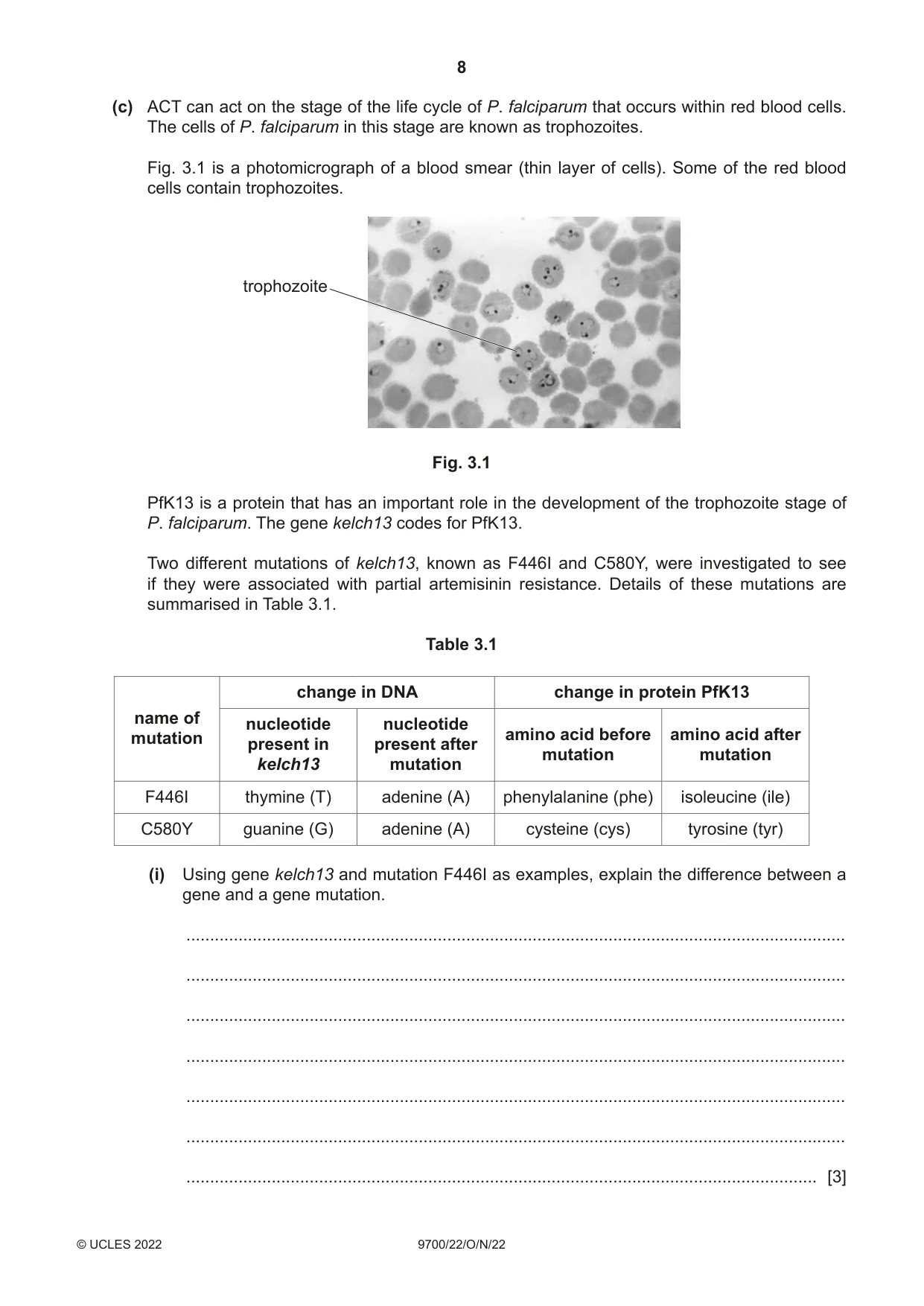
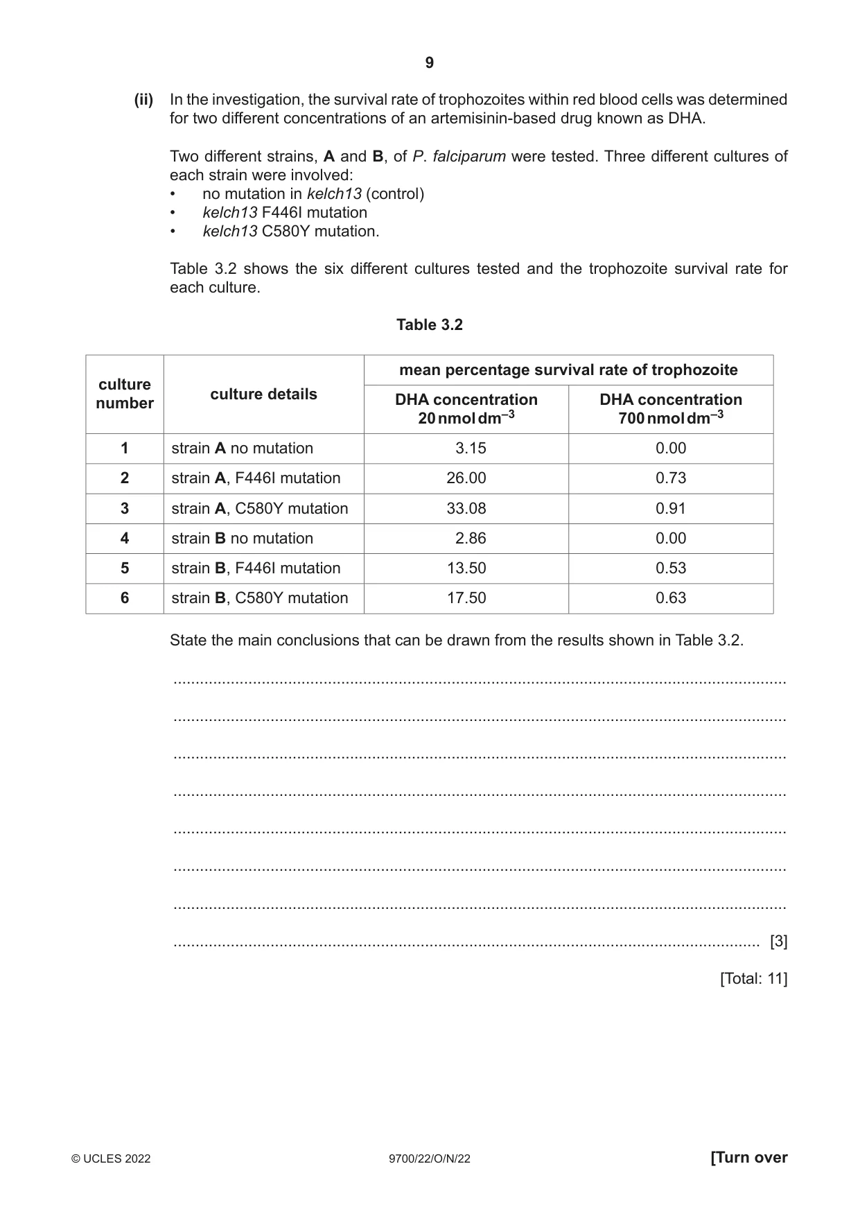
3 Plasmodium falciparum is one species of Plasmodium that causes the life-threatening disease malaria. With early diagnosis and the correct drug treatment, the pathogen can be eliminated from the body, particularly if the disease is not severe. (a) Name the type of pathogen that causes malaria. ............................................................................................................................................. [1] (b) To help prevent the development and spread of drug resistance in Plasmodium, the World Health Organization (WHO) recommends using a treatment known as artemisinin-based combination therapy (ACT). ACT involves two different types of drug: • a fast-acting drug derived from a compound known as artemisinin, which causes a rapid decrease in the number of P. falciparum • one or more longer-acting, non-artemisinin, drugs that eliminate any remaining pathogens. (i) Suggest why using ACT with the two different types of drug is more effective in preventing the development of drug resistance in Plasmodium than a treatment using only one type of drug. ........................................................................................................................................... ........................................................................................................................................... ........................................................................................................................................... ........................................................................................................................................... ..................................................................................................................................... [2] (ii) In some areas, partial artemisinin resistance has developed. This means ACT takes a longer time for the pathogen to be eliminated from the body. Explain why there is an increased risk of transmission of the pathogen to other people if a person is receiving ACT and the pathogen has partial artemisinin resistance. ........................................................................................................................................... ........................................................................................................................................... ........................................................................................................................................... ........................................................................................................................................... ..................................................................................................................................... [2] (c) ACT can act on the stage of the life cycle of P. falciparum that occurs within red blood cells. The cells of P. falciparum in this stage are known as trophozoites. Fig. 3.1 is a photomicrograph of a blood smear (thin layer of cells). Some of the red blood cells contain trophozoites. trophozoite Fig. 3.1 PfK13 is a protein that has an important role in the development of the trophozoite stage of P. falciparum. The gene kelch13 codes for PfK13. Two different mutations of kelch13, known as F446I and C580Y, were investigated to see if they were associated with partial artemisinin resistance. Details of these mutations are summarised in Table 3.1. Table 3.1 change in DNA change in protein PfK13 name of nucleotide nucleotide mutation amino acid before amino acid after present in present after mutation mutation kelch13 mutation F446I thymine (T) adenine (A) phenylalanine (phe) isoleucine (ile) C580Y guanine (G) adenine (A) cysteine (cys) tyrosine (tyr) (i) Using gene kelch13 and mutation F446I as examples, explain the difference between a gene and a gene mutation. ........................................................................................................................................... ........................................................................................................................................... ........................................................................................................................................... ........................................................................................................................................... ........................................................................................................................................... ........................................................................................................................................... ..................................................................................................................................... [3] (ii) In the investigation, the survival rate of trophozoites within red blood cells was determined for two different concentrations of an artemisinin-based drug known as DHA. Two different strains, A and B, of P. falciparum were tested. Three different cultures of each strain were involved: • no mutation in kelch13 (control) • kelch13 F446I mutation • kelch13 C580Y mutation. Table 3.2 shows the six different cultures tested and the trophozoite survival rate for each culture. Table 3.2 mean percentage survival rate of trophozoite culture culture detailsnumber DHA concentration DHA concentration 20 nmol dm–3 700 nmol dm–3 1 strain A no mutation 3.15 0.00 2 strain A, F446I mutation 26.00 0.73 3 strain A, C580Y mutation 33.08 0.91 4 strain B no mutation 2.86 0.00 5 strain B, F446I mutation 13.50 0.53 6 strain B, C580Y mutation 17.50 0.63 State the main conclusions that can be drawn from the results shown in Table 3.2. ........................................................................................................................................... ........................................................................................................................................... ........................................................................................................................................... ........................................................................................................................................... ........................................................................................................................................... ........................................................................................................................................... ........................................................................................................................................... ..................................................................................................................................... [3] [Total: 11]
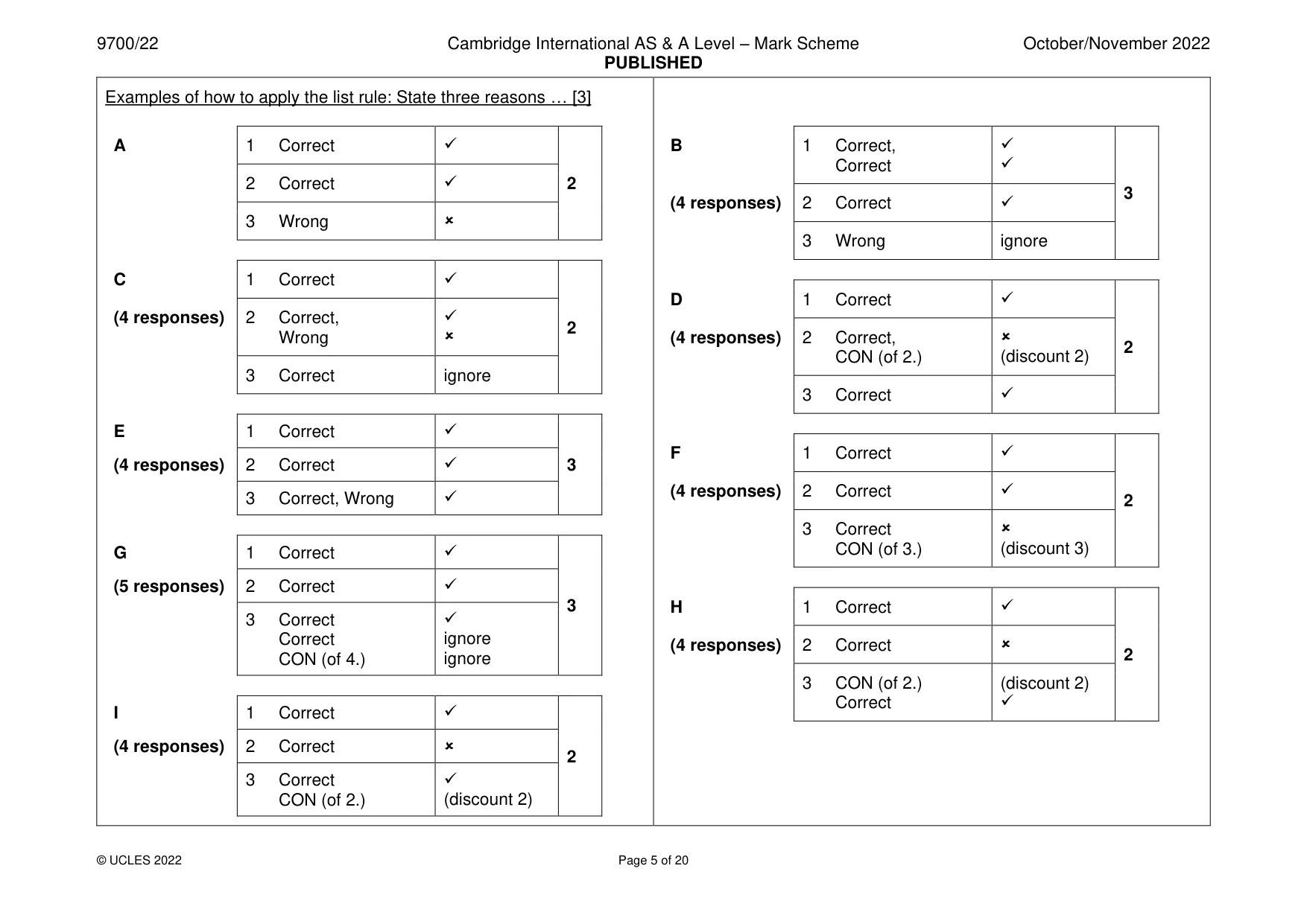
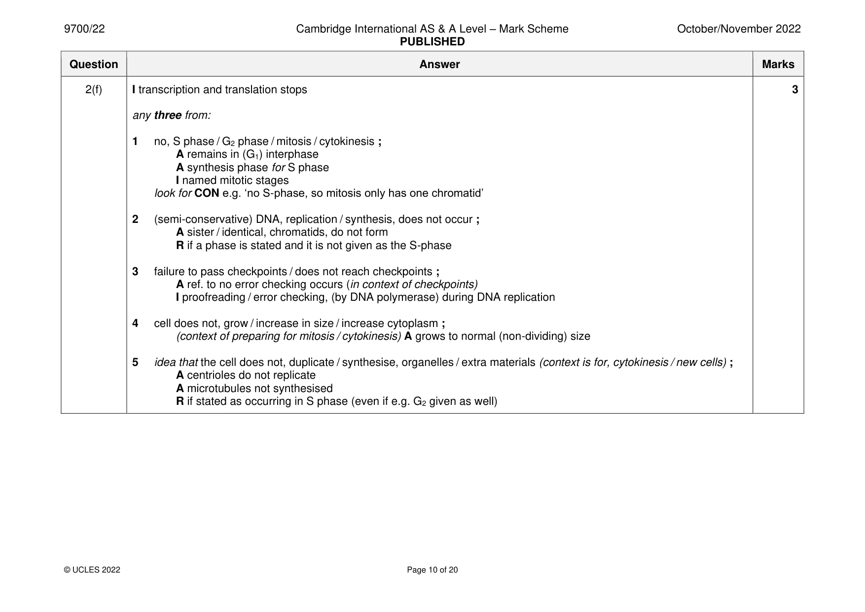
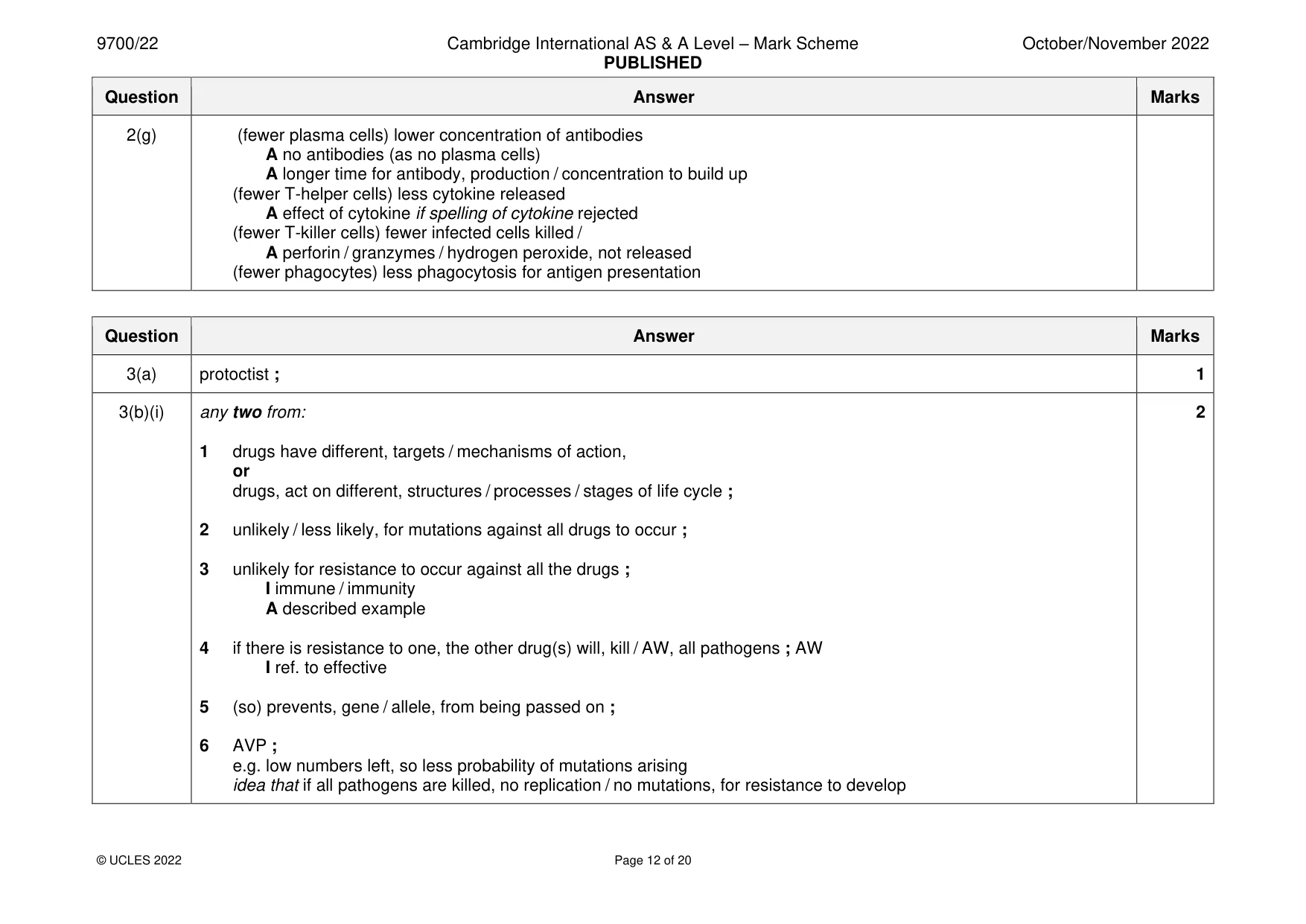
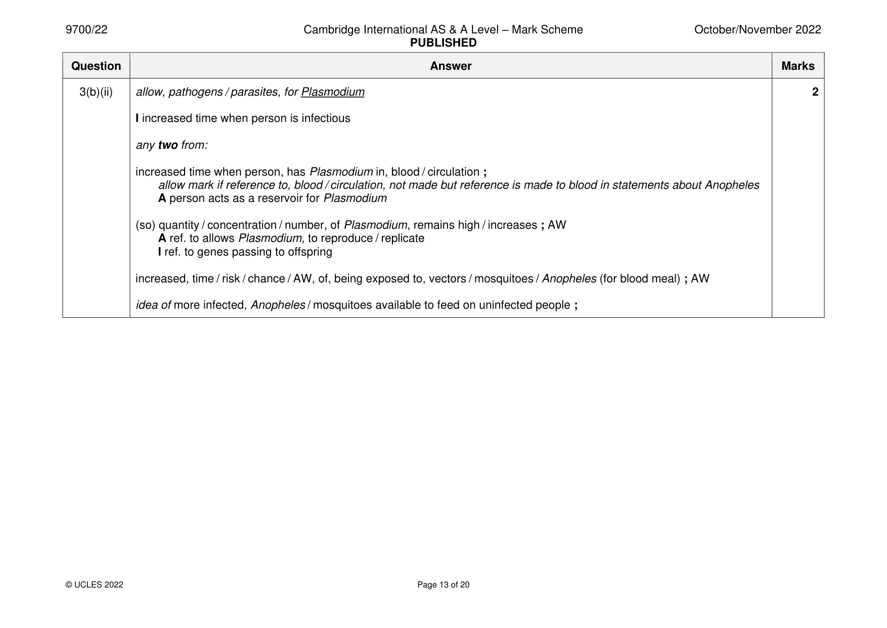
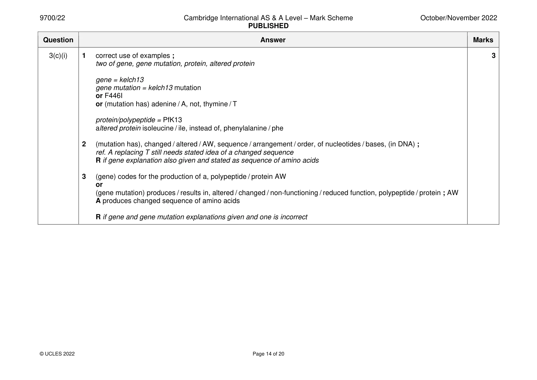
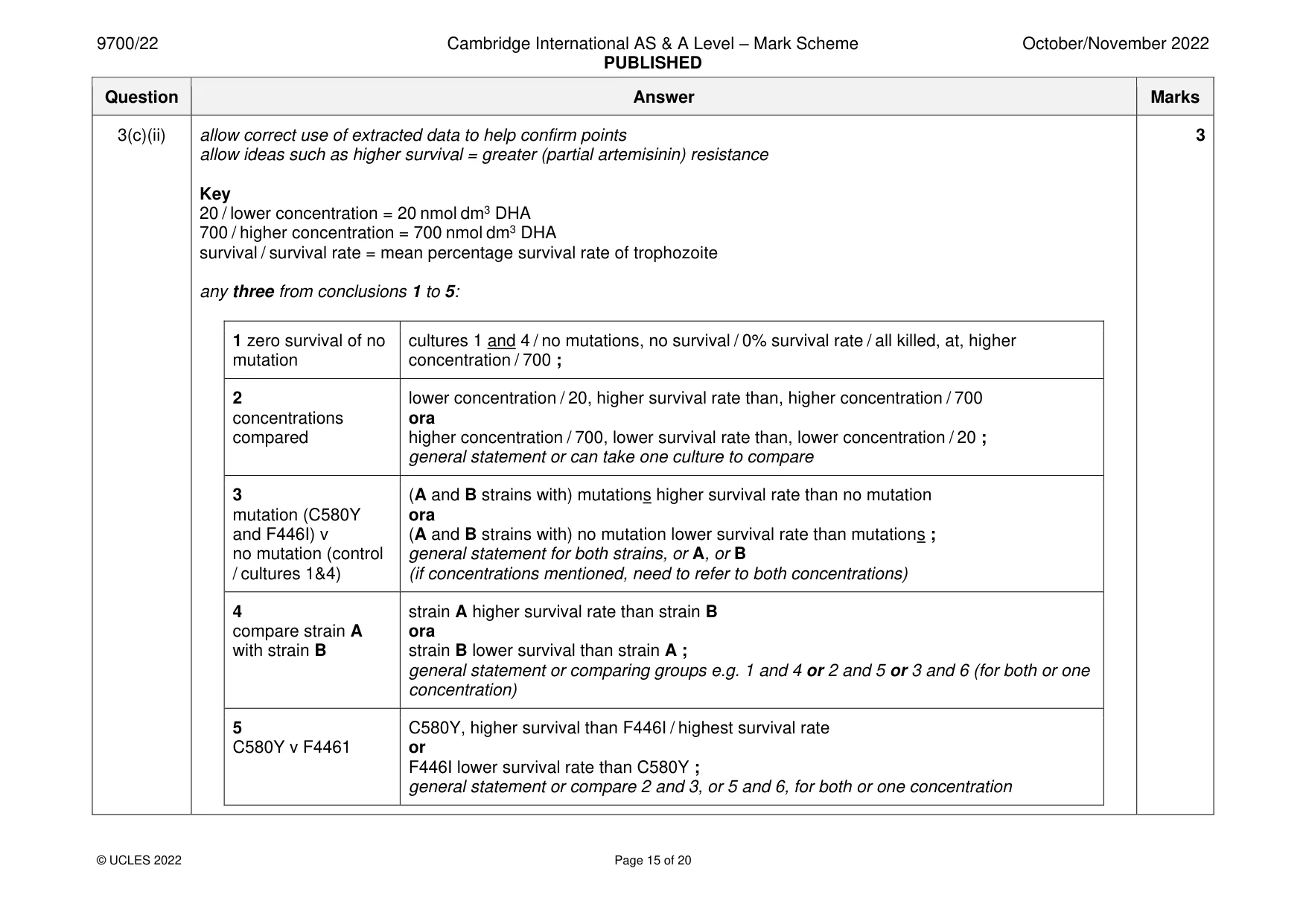
Mark scheme: 3(a) protoctist ; 1 3(b)(i) any two from: 2 1 drugs have different, targets / mechanisms of action, or drugs, act on different, structures / processes / stages of life cycle ; 2 unlikely / less likely, for mutations against all drugs to occur ; 3 unlikely for resistance to occur against all the drugs ; I immune / immunity A described example 4 if there is resistance to one, the other drug(s) will, kill / AW, all pathogens ; AW I ref. to effective 5 (so) prevents, gene / allele, from being passed on ; 6 AVP ; e.g. low numbers left, so less probability of mutations arising idea that if all pathogens are killed, no replication / no mutations, for resistance to develop 3(b)(ii) allow, pathogens / parasites, for Plasmodium 2 I increased time when person is infectious any two from: increased time when person, has Plasmodium in, blood / circulation ; allow mark if reference to, blood / circulation, not made but reference is made to blood in statements about Anopheles A person acts as a reservoir for Plasmodium (so) quantity / concentration / number, of Plasmodium, remains high / increases ; AW A ref. to allows Plasmodium, to reproduce / replicate I ref. to genes passing to offspring increased, time / risk / chance / AW, of, being exposed to, vectors / mosquitoes / Anopheles (for blood meal) ; AW idea of more infected, Anopheles / mosquitoes available to feed on uninfected people ; 3(c)(i) 1 correct use of examples ; 3 two of gene, gene mutation, protein, altered protein gene = kelch13 gene mutation = kelch13 mutation or F446I or (mutation has) adenine / A, not, thymine / T protein/polypeptide = PfK13 altered protein isoleucine / ile, instead of, phenylalanine / phe 2 (mutation has), changed / altered / AW, sequence / arrangement / order, of nucleotides / bases, (in DNA) ; ref. A replacing T still needs stated idea of a changed sequence R if gene explanation also given and stated as sequence of amino acids 3 (gene) codes for the production of a, polypeptide / protein AW or (gene mutation) produces / results in, altered / changed / non-functioning / reduced function, polypeptide / protein ; AW A produces changed sequence of amino acids R if gene and gene mutation explanations given and one is incorrect 3(c)(ii) allow correct use of extracted data to help confirm points 3 allow ideas such as higher survival = greater (partial artemisinin) resistance Key 20 / lower concentration = 20 nmol dm3 DHA 700 / higher concentration = 700 nmol dm3 DHA survival / survival rate = mean percentage survival rate of trophozoite any three from conclusions 1 to 5: 1 zero survival of no cultures 1 and 4 / no mutations, no survival / 0% survival rate / all killed, at, higher mutation concentration / 700 ; 2 lower concentration / 20, higher survival rate than, higher concentration / 700 concentrations ora compared higher concentration / 700, lower survival rate than, lower concentration / 20 ; general statement or can take one culture to compare 3 (A and B strains with) mutations higher survival rate than no mutation mutation (C580Y ora and F446I) v (A and B strains with) no mutation lower survival rate than mutations ; no mutation (control general statement for both strains, or A, or B / cultures 1&4) (if concentrations mentioned, need to refer to both concentrations) 4 strain A higher survival rate than strain B compare strain A ora with strain B strain B lower survival than strain A ; general statement or comparing groups e.g. 1 and 4 or 2 and 5 or 3 and 6 (for both or one concentration) 5 C580Y, higher survival than F446I / highest survival rate C580Y v F4461 or F446I lower survival rate than C580Y ; general statement or compare 2 and 3, or 5 and 6, for both or one concentration
Q4 · The airways of the gas exchange system are lined with epithelium
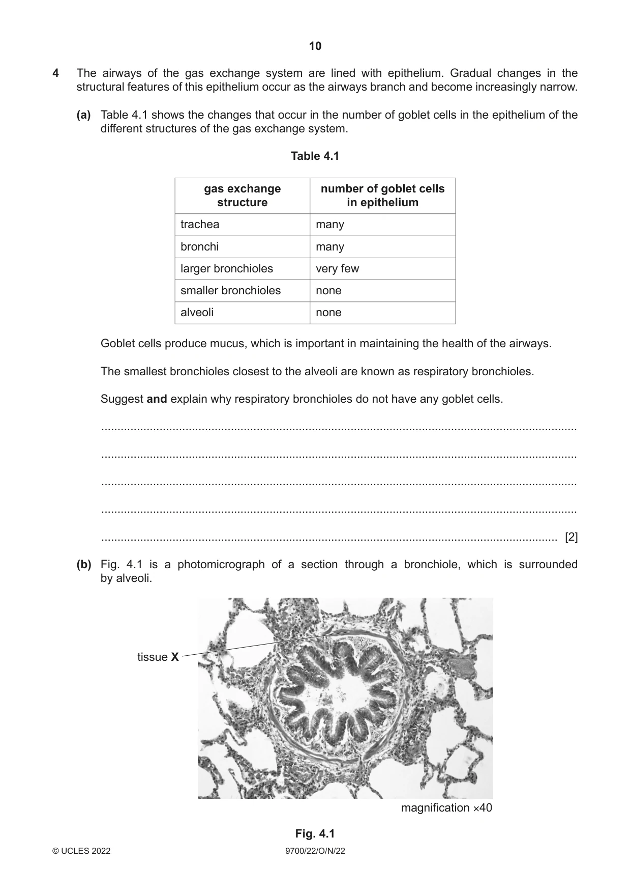
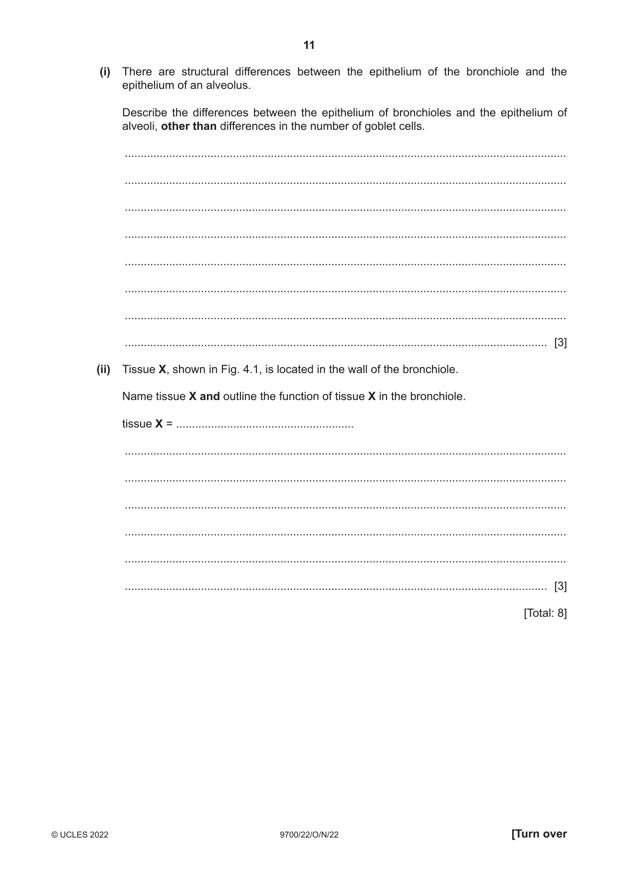
4 The airways of the gas exchange system are lined with epithelium. Gradual changes in the structural features of this epithelium occur as the airways branch and become increasingly narrow. (a) Table 4.1 shows the changes that occur in the number of goblet cells in the epithelium of the different structures of the gas exchange system. Table 4.1 gas exchange number of goblet cells structure in epithelium trachea many bronchi many larger bronchioles very few smaller bronchioles none alveoli none Goblet cells produce mucus, which is important in maintaining the health of the airways. The smallest bronchioles closest to the alveoli are known as respiratory bronchioles. Suggest and explain why respiratory bronchioles do not have any goblet cells. ................................................................................................................................................... ................................................................................................................................................... ................................................................................................................................................... ................................................................................................................................................... ............................................................................................................................................. [2] (b) Fig. 4.1 is a photomicrograph of a section through a bronchiole, which is surrounded by alveoli. tissue X magnification ×40 Fig. 4.1 (i) There are structural differences between the epithelium of the bronchiole and the epithelium of an alveolus. Describe the differences between the epithelium of bronchioles and the epithelium of alveoli, other than differences in the number of goblet cells. ........................................................................................................................................... ........................................................................................................................................... ........................................................................................................................................... ........................................................................................................................................... ........................................................................................................................................... ........................................................................................................................................... ........................................................................................................................................... ..................................................................................................................................... [3] (ii) Tissue X, shown in Fig. 4.1, is located in the wall of the bronchiole. Name tissue X and outline the function of tissue X in the bronchiole. tissue X = ........................................................ ........................................................................................................................................... ........................................................................................................................................... ........................................................................................................................................... ........................................................................................................................................... ........................................................................................................................................... ..................................................................................................................................... [3] [Total: 8]
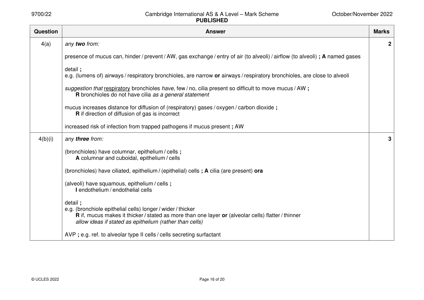
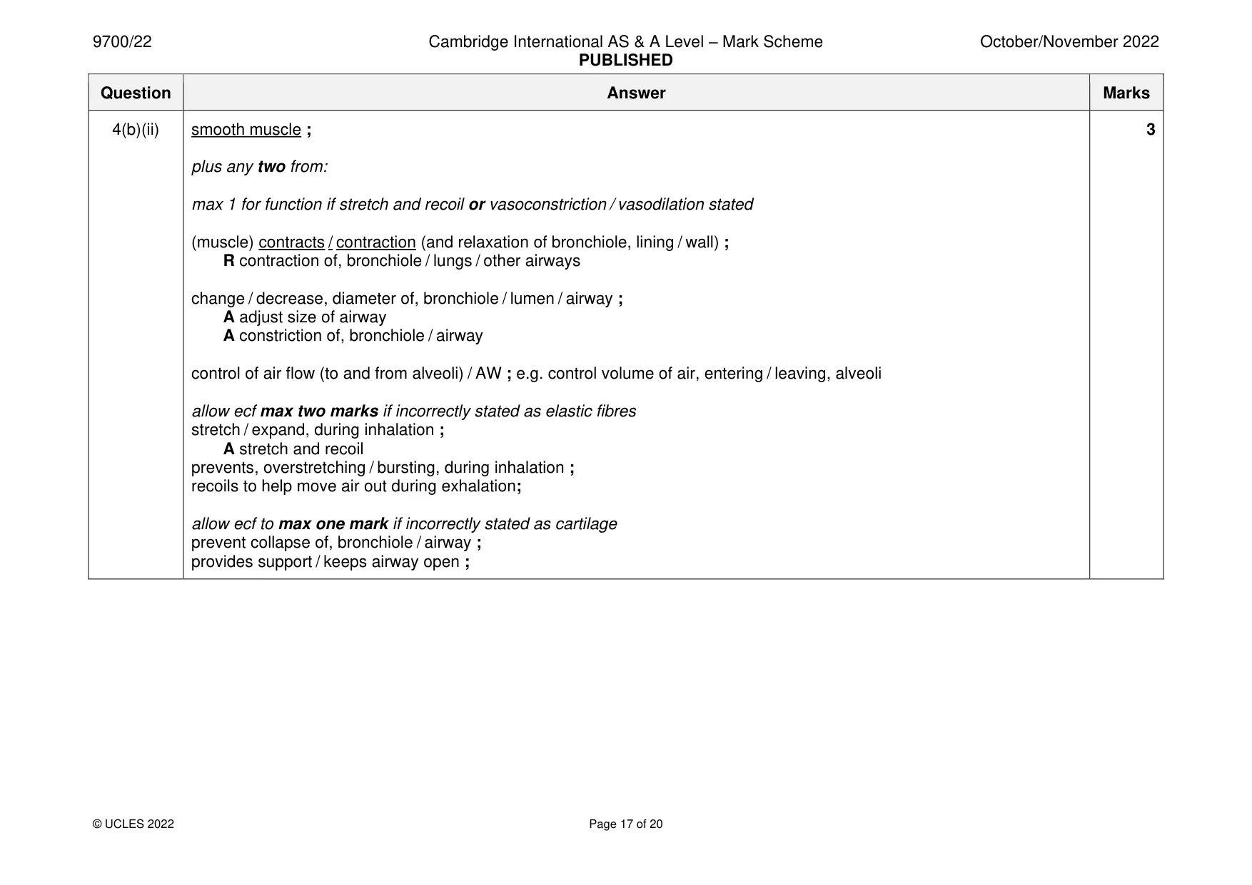
Mark scheme: 4(a) any two from: 2 presence of mucus can, hinder / prevent / AW, gas exchange / entry of air (to alveoli) / airflow (to alveoli) ; A named gases detail ; e.g. (lumens of) airways / respiratory bronchioles, are narrow or airways / respiratory bronchioles, are close to alveoli suggestion that respiratory bronchioles have, few / no, cilia present so difficult to move mucus / AW ; R bronchioles do not have cilia as a general statement mucus increases distance for diffusion of (respiratory) gases / oxygen / carbon dioxide ; R if direction of diffusion of gas is incorrect increased risk of infection from trapped pathogens if mucus present ; AW 4(b)(i) any three from: 3 (bronchioles) have columnar, epithelium / cells ; A columnar and cuboidal, epithelium / cells (bronchioles) have ciliated, epithelium / (epithelial) cells ; A cilia (are present) ora (alveoli) have squamous, epithelium / cells ; I endothelium / endothelial cells detail ; e.g. (bronchiole epithelial cells) longer / wider / thicker R if, mucus makes it thicker / stated as more than one layer or (alveolar cells) flatter / thinner allow ideas if stated as epithelium (rather than cells) AVP ; e.g. ref. to alveolar type II cells / cells secreting surfactant 4(b)(ii) smooth muscle ; 3 plus any two from: max 1 for function if stretch and recoil or vasoconstriction / vasodilation stated (muscle) contracts / contraction (and relaxation of bronchiole, lining / wall) ; R contraction of, bronchiole / lungs / other airways change / decrease, diameter of, bronchiole / lumen / airway ; A adjust size of airway A constriction of, bronchiole / airway control of air flow (to and from alveoli) / AW ; e.g. control volume of air, entering / leaving, alveoli allow ecf max two marks if incorrectly stated as elastic fibres stretch / expand, during inhalation ; A stretch and recoil prevents, overstretching / bursting, during inhalation ; recoils to help move air out during exhalation; allow ecf to max one mark if incorrectly stated as cartilage prevent collapse of, bronchiole / airway ; provides support / keeps airway open ;
Q5 · Fibroblasts are one of the cell types of connective tissue
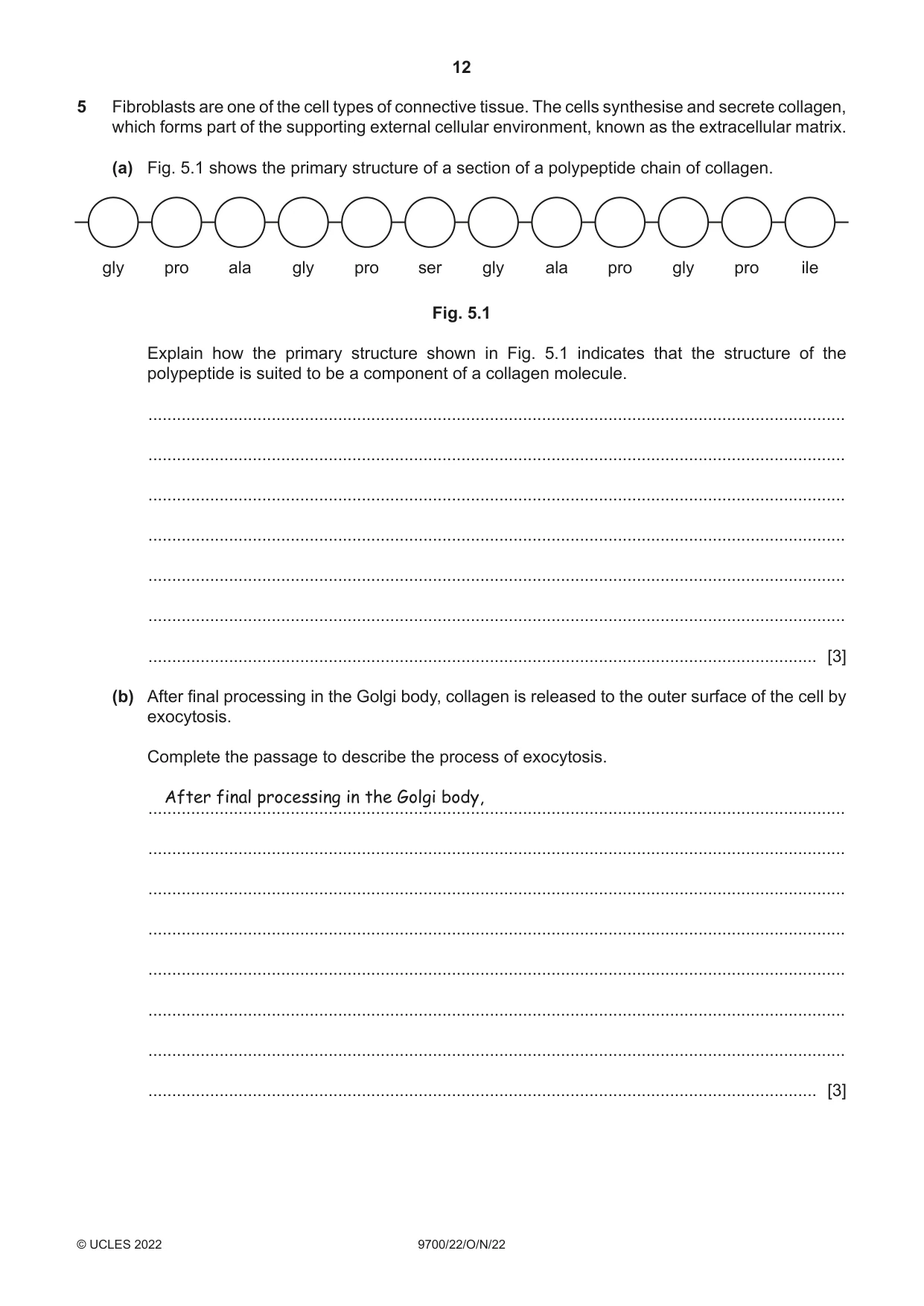
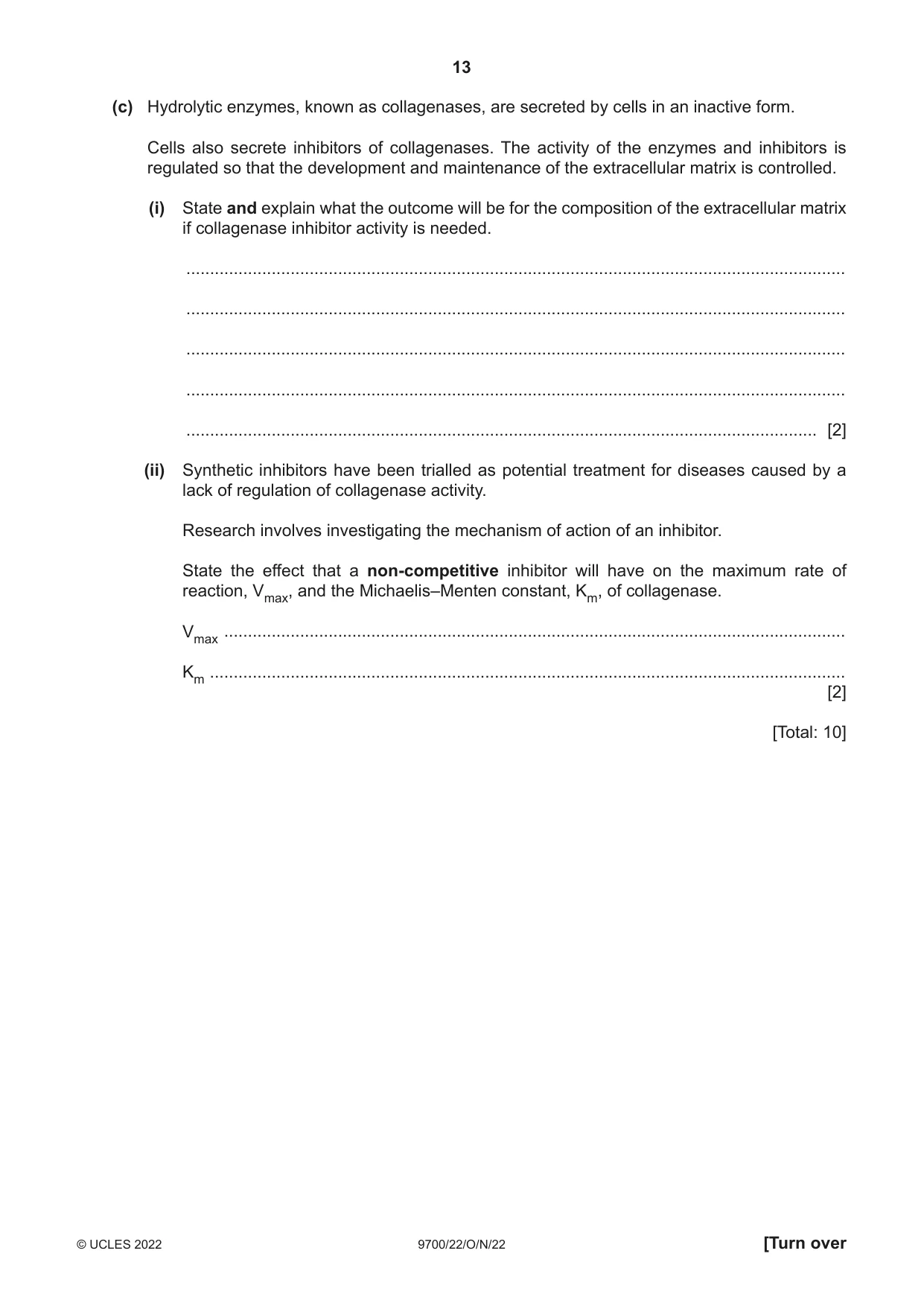
5 Fibroblasts are one of the cell types of connective tissue. The cells synthesise and secrete collagen, which forms part of the supporting external cellular environment, known as the extracellular matrix. (a) Fig. 5.1 shows the primary structure of a section of a polypeptide chain of collagen. gly pro ala gly pro ser gly ala pro gly pro ile Fig. 5.1 Explain how the primary structure shown in Fig. 5.1 indicates that the structure of the polypeptide is suited to be a component of a collagen molecule. ................................................................................................................................................... ................................................................................................................................................... ................................................................................................................................................... ................................................................................................................................................... ................................................................................................................................................... ................................................................................................................................................... ............................................................................................................................................. [3] (b) After final processing in the Golgi body, collagen is released to the outer surface of the cell by exocytosis. Complete the passage to describe the process of exocytosis. After final processing in the Golgi body, ................................................................................................................................................... ................................................................................................................................................... ................................................................................................................................................... ................................................................................................................................................... ................................................................................................................................................... ................................................................................................................................................... ................................................................................................................................................... ............................................................................................................................................. [3] (c) Hydrolytic enzymes, known as collagenases, are secreted by cells in an inactive form. Cells also secrete inhibitors of collagenases. The activity of the enzymes and inhibitors is regulated so that the development and maintenance of the extracellular matrix is controlled. (i) State and explain what the outcome will be for the composition of the extracellular matrix if collagenase inhibitor activity is needed. ........................................................................................................................................... ........................................................................................................................................... ........................................................................................................................................... ........................................................................................................................................... ..................................................................................................................................... [2] (ii) Synthetic inhibitors have been trialled as potential treatment for diseases caused by a lack of regulation of collagenase activity. Research involves investigating the mechanism of action of an inhibitor. State the effect that a non-competitive inhibitor will have on the maximum rate of reaction, Vmax, and the Michaelis–Menten constant, Km, of collagenase. Vmax ................................................................................................................................... Km ...................................................................................................................................... [2] [Total: 10]
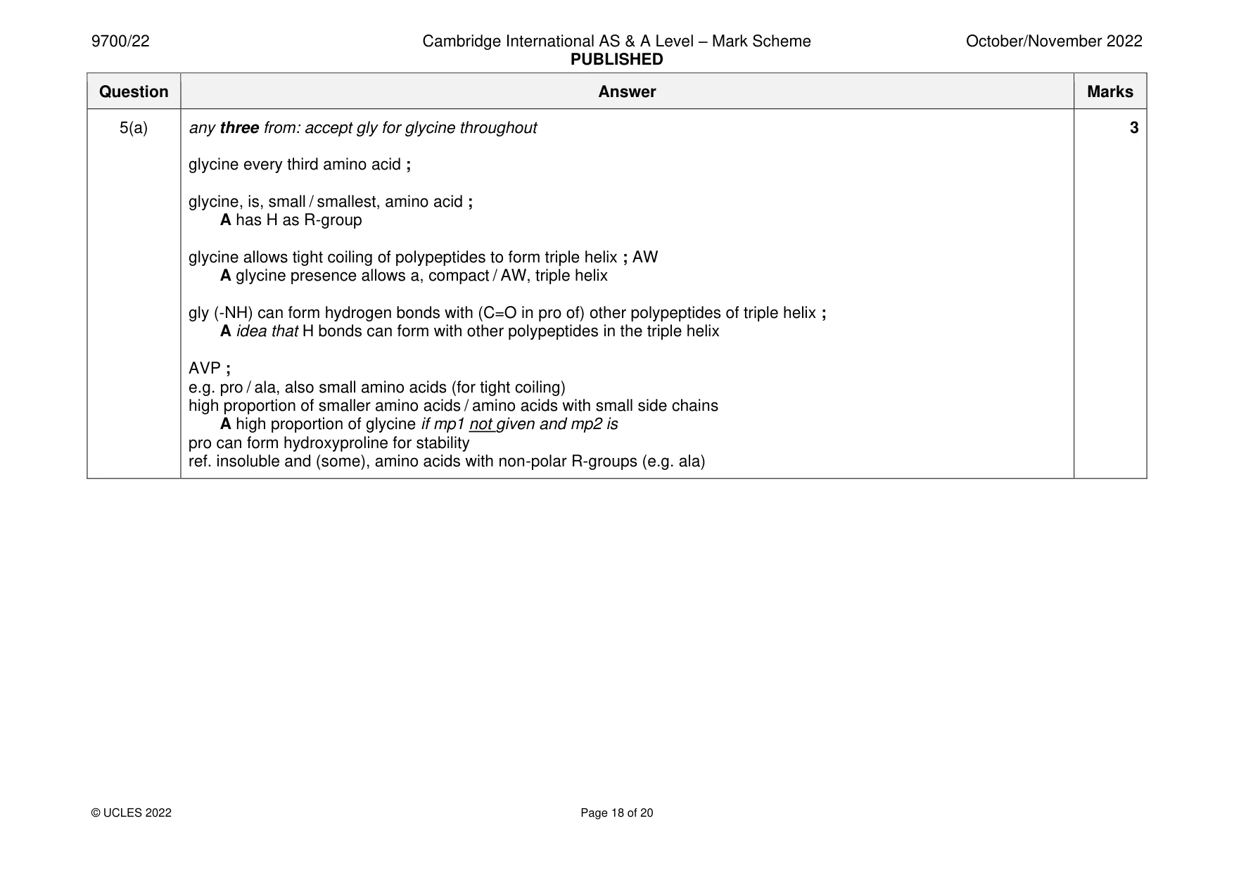
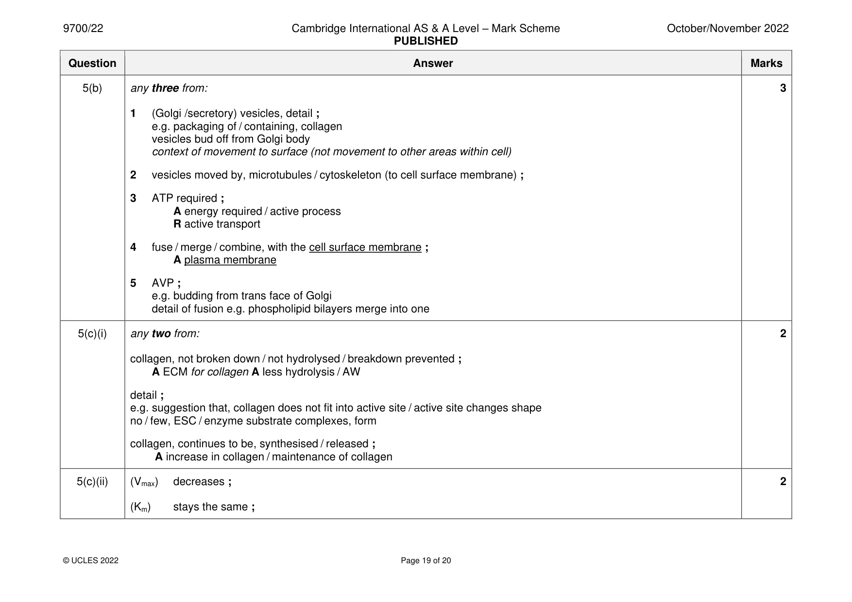
Mark scheme: 5(a) any three from: accept gly for glycine throughout 3 glycine every third amino acid ; glycine, is, small / smallest, amino acid ; A has H as R-group glycine allows tight coiling of polypeptides to form triple helix ; AW A glycine presence allows a, compact / AW, triple helix gly (-NH) can form hydrogen bonds with (C=O in pro of) other polypeptides of triple helix ; A idea that H bonds can form with other polypeptides in the triple helix AVP ; e.g. pro / ala, also small amino acids (for tight coiling) high proportion of smaller amino acids / amino acids with small side chains A high proportion of glycine if mp1 not given and mp2 is pro can form hydroxyproline for stability ref. insoluble and (some), amino acids with non-polar R-groups (e.g. ala) 5(b) any three from: 3 1 (Golgi /secretory) vesicles, detail ; e.g. packaging of / containing, collagen vesicles bud off from Golgi body context of movement to surface (not movement to other areas within cell) 2 vesicles moved by, microtubules / cytoskeleton (to cell surface membrane) ; 3 ATP required ; A energy required / active process R active transport 4 fuse / merge / combine, with the cell surface membrane ; A plasma membrane 5 AVP ; e.g. budding from trans face of Golgi detail of fusion e.g. phospholipid bilayers merge into one 5(c)(i) any two from: 2 collagen, not broken down / not hydrolysed / breakdown prevented ; A ECM for collagen A less hydrolysis / AW detail ; e.g. suggestion that, collagen does not fit into active site / active site changes shape no / few, ESC / enzyme substrate complexes, form collagen, continues to be, synthesised / released ; A increase in collagen / maintenance of collagen 5(c)(ii) (Vmax) decreases ; 2 (Km) stays the same ;
Q6 · Xylem and phloem are the transport tissues of plants
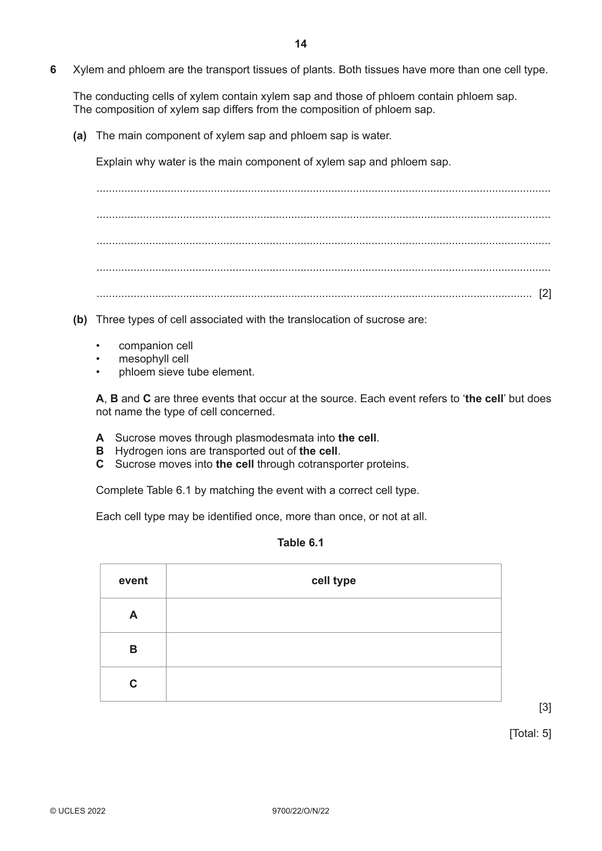
6 Xylem and phloem are the transport tissues of plants. Both tissues have more than one cell type. The conducting cells of xylem contain xylem sap and those of phloem contain phloem sap. The composition of xylem sap differs from the composition of phloem sap. (a) The main component of xylem sap and phloem sap is water. Explain why water is the main component of xylem sap and phloem sap. ................................................................................................................................................... ................................................................................................................................................... ................................................................................................................................................... ................................................................................................................................................... ............................................................................................................................................. [2] (b) Three types of cell associated with the translocation of sucrose are: • companion cell • mesophyll cell • phloem sieve tube element. A, B and C are three events that occur at the source. Each event refers to ‘the cell’ but does not name the type of cell concerned. A Sucrose moves through plasmodesmata into the cell. B Hydrogen ions are transported out of the cell. C Sucrose moves into the cell through cotransporter proteins. Complete Table 6.1 by matching the event with a correct cell type. Each cell type may be identified once, more than once, or not at all. Table 6.1 event cell type A B C [3] [Total: 5]
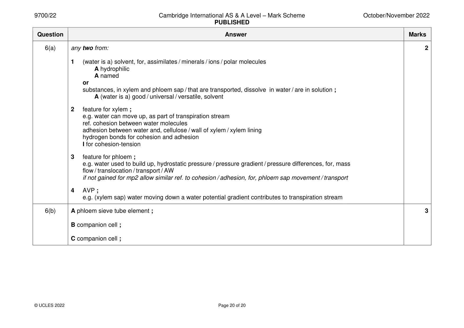
Mark scheme: 6(a) any two from: 2 1 (water is a) solvent, for, assimilates / minerals / ions / polar molecules A hydrophilic A named or substances, in xylem and phloem sap / that are transported, dissolve in water / are in solution ; A (water is a) good / universal / versatile, solvent 2 feature for xylem ; e.g. water can move up, as part of transpiration stream ref. cohesion between water molecules adhesion between water and, cellulose / wall of xylem / xylem lining hydrogen bonds for cohesion and adhesion I for cohesion-tension 3 feature for phloem ; e.g. water used to build up, hydrostatic pressure / pressure gradient / pressure differences, for, mass flow / translocation / transport / AW if not gained for mp2 allow similar ref. to cohesion / adhesion, for, phloem sap movement / transport 4 AVP ; e.g. (xylem sap) water moving down a water potential gradient contributes to transpiration stream 6(b) A phloem sieve tube element ; 3 B companion cell ; C companion cell ;
What was in this paper
The subtopics covered by these 6 questions, and how many questions each got. Open one in a new tab to see every Cambridge question on it.
What you needed in this session
Cambridge’s own grade thresholds for 2022 Oct/Nov, Paper 2 · Variant 2. A higher threshold means an easier paper — the bar moves with how the cohort did.