Cambridge A Level Biology 9700 — 2020 Oct/Nov Paper 2 · Variant 3
9700/23/O/N/20 · 6 questions · 60 marks · ≈68 min
The question paper and its mark scheme, free to read here and free to download. This is Cambridge’s own paper, exactly as it was sat.
Question paper16 pages
Mark scheme20 pages
Answers below. Sit the paper first if you are practising.
Questions as text
Q1 · A section through the human chest
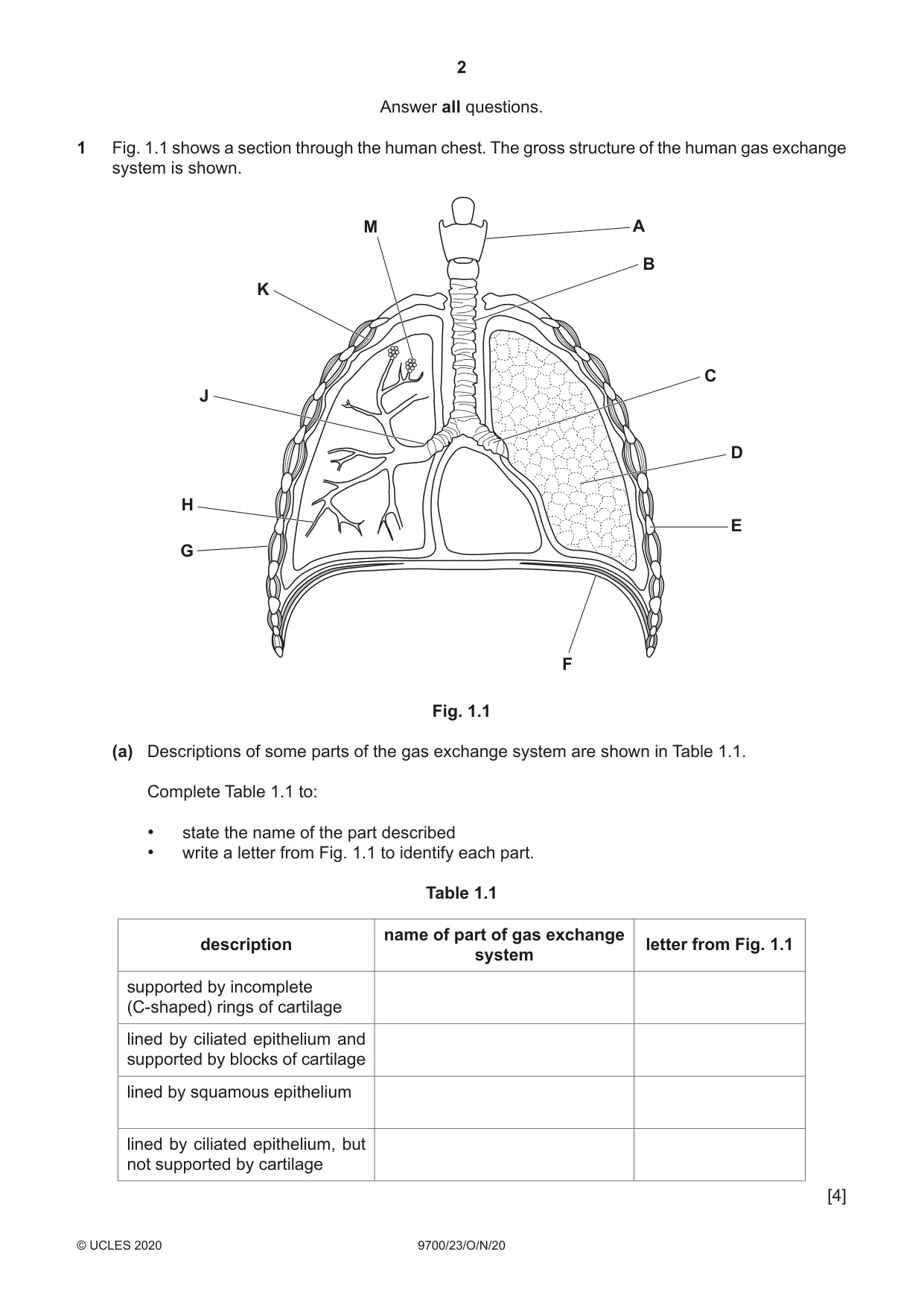
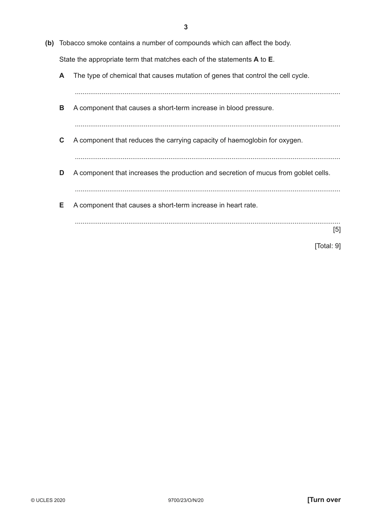
1 Fig. 1.1 shows a section through the human chest. The gross structure of the human gas exchange system is shown. M A B K C J D H E G F Fig. 1.1 (a) Descriptions of some parts of the gas exchange system are shown in Table 1.1. Complete Table 1.1 to: • state the name of the part described • write a letter from Fig. 1.1 to identify each part. Table 1.1 name of part of gas exchange description letter from Fig. 1.1 system supported by incomplete (C-shaped) rings of cartilage lined by ciliated epithelium and supported by blocks of cartilage lined by squamous epithelium lined by ciliated epithelium, but not supported by cartilage [4] (b) Tobacco smoke contains a number of compounds which can affect the body. State the appropriate term that matches each of the statements A to E. A The type of chemical that causes mutation of genes that control the cell cycle. ........................................................................................................................................... B A component that causes a short-term increase in blood pressure. ........................................................................................................................................... C A component that reduces the carrying capacity of haemoglobin for oxygen. ........................................................................................................................................... D A component that increases the production and secretion of mucus from goblet cells. ........................................................................................................................................... E A component that causes a short-term increase in heart rate. ........................................................................................................................................... [5] [Total: 9]
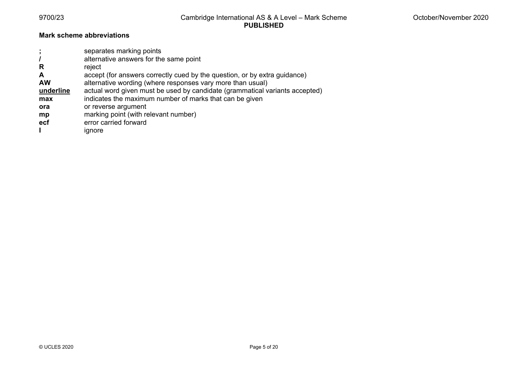
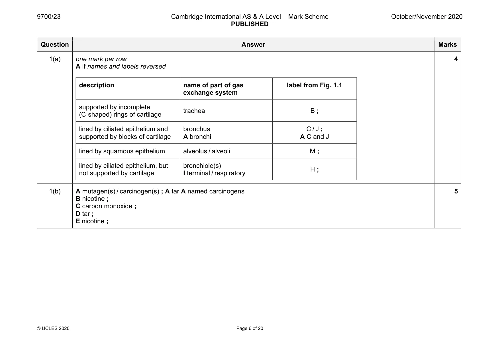
Mark scheme: 1(a) one mark per row A if names and labels reversed description name of part of gas exchange system label from Fig. 1.1 supported by incomplete (C-shaped) rings of cartilage trachea B ; lined by ciliated epithelium and supported by blocks of cartilage bronchus A bronchi C / J ; A C and J lined by squamous epithelium alveolus / alveoli M ; lined by ciliated epithelium, but not supported by cartilage bronchiole(s) I terminal / respiratory H ; 4 1(b) A mutagen(s) / carcinogen(s) ; A tar A named carcinogens B nicotine ; C carbon monoxide ; D tar ; E nicotine ; 5
Q2 · The veins of a leaf contain transport tissues
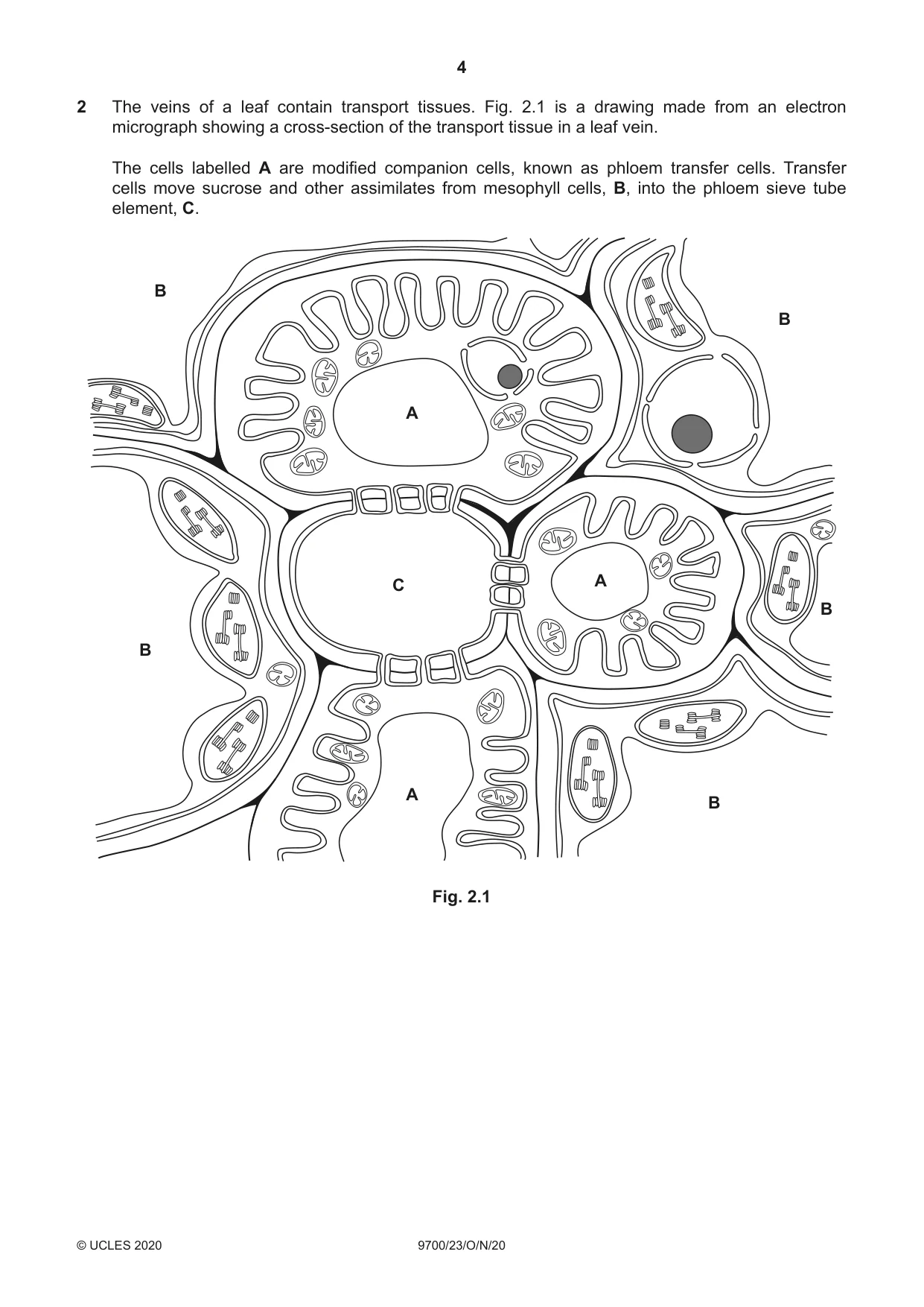
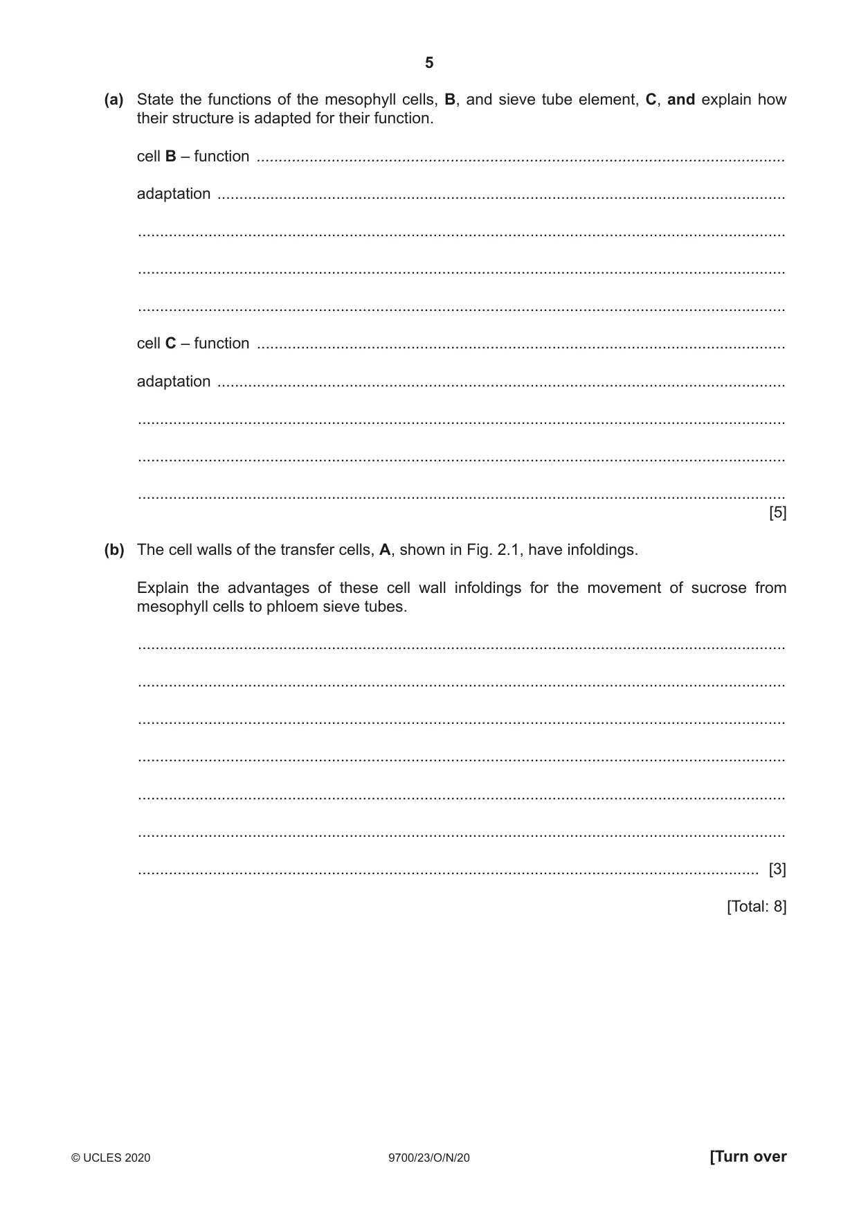
2 The veins of a leaf contain transport tissues. Fig. 2.1 is a drawing made from an electron micrograph showing a cross-section of the transport tissue in a leaf vein. The cells labelled A are modified companion cells, known as phloem transfer cells. Transfer cells move sucrose and other assimilates from mesophyll cells, B, into the phloem sieve tube element, C. B B A C A B B A B Fig. 2.1 (a) State the functions of the mesophyll cells, B, and sieve tube element, C, and explain how their structure is adapted for their function. cell B – function ........................................................................................................................ adaptation ................................................................................................................................. ................................................................................................................................................... ................................................................................................................................................... ................................................................................................................................................... cell C – function ........................................................................................................................ adaptation ................................................................................................................................. ................................................................................................................................................... ................................................................................................................................................... ................................................................................................................................................... [5] (b) The cell walls of the transfer cells, A, shown in Fig. 2.1, have infoldings. Explain the advantages of these cell wall infoldings for the movement of sucrose from mesophyll cells to phloem sieve tubes. ................................................................................................................................................... ................................................................................................................................................... ................................................................................................................................................... ................................................................................................................................................... ................................................................................................................................................... ................................................................................................................................................... ............................................................................................................................................. [3] [Total: 8]
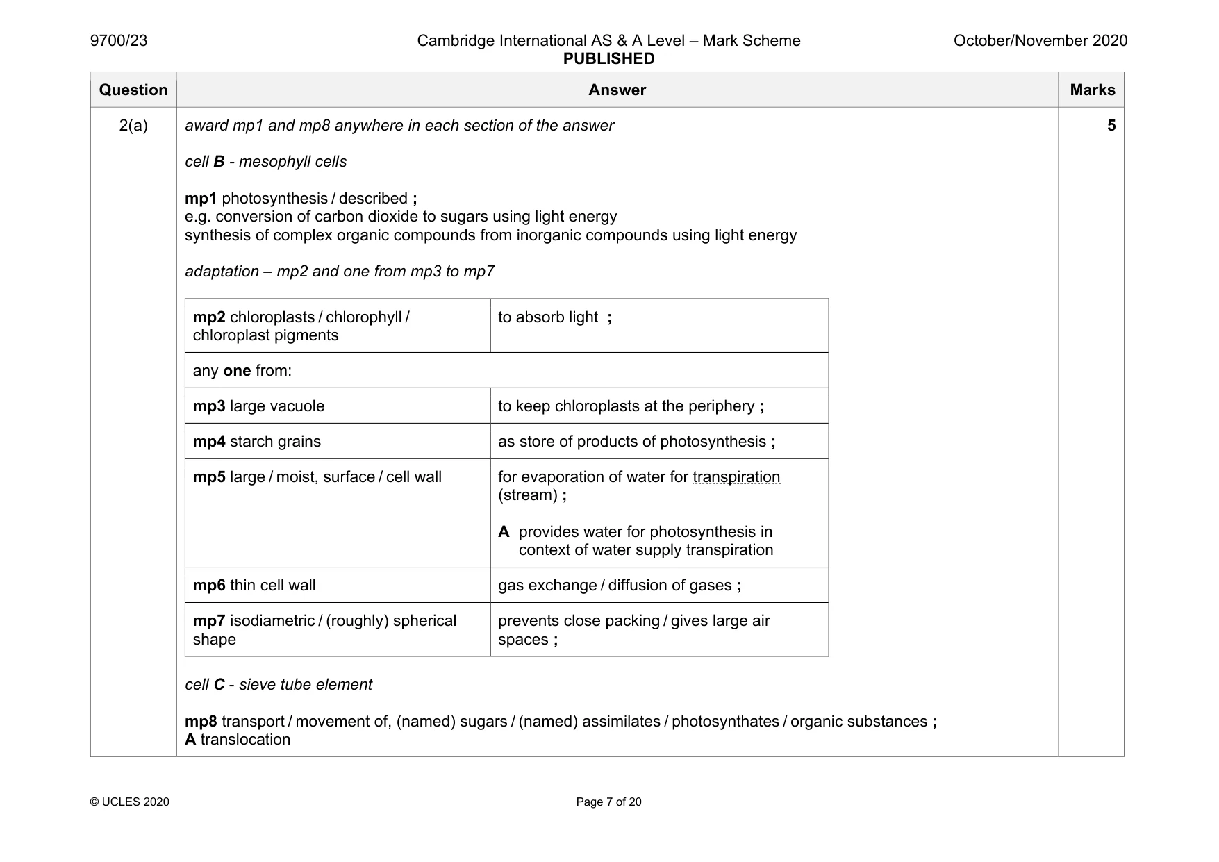
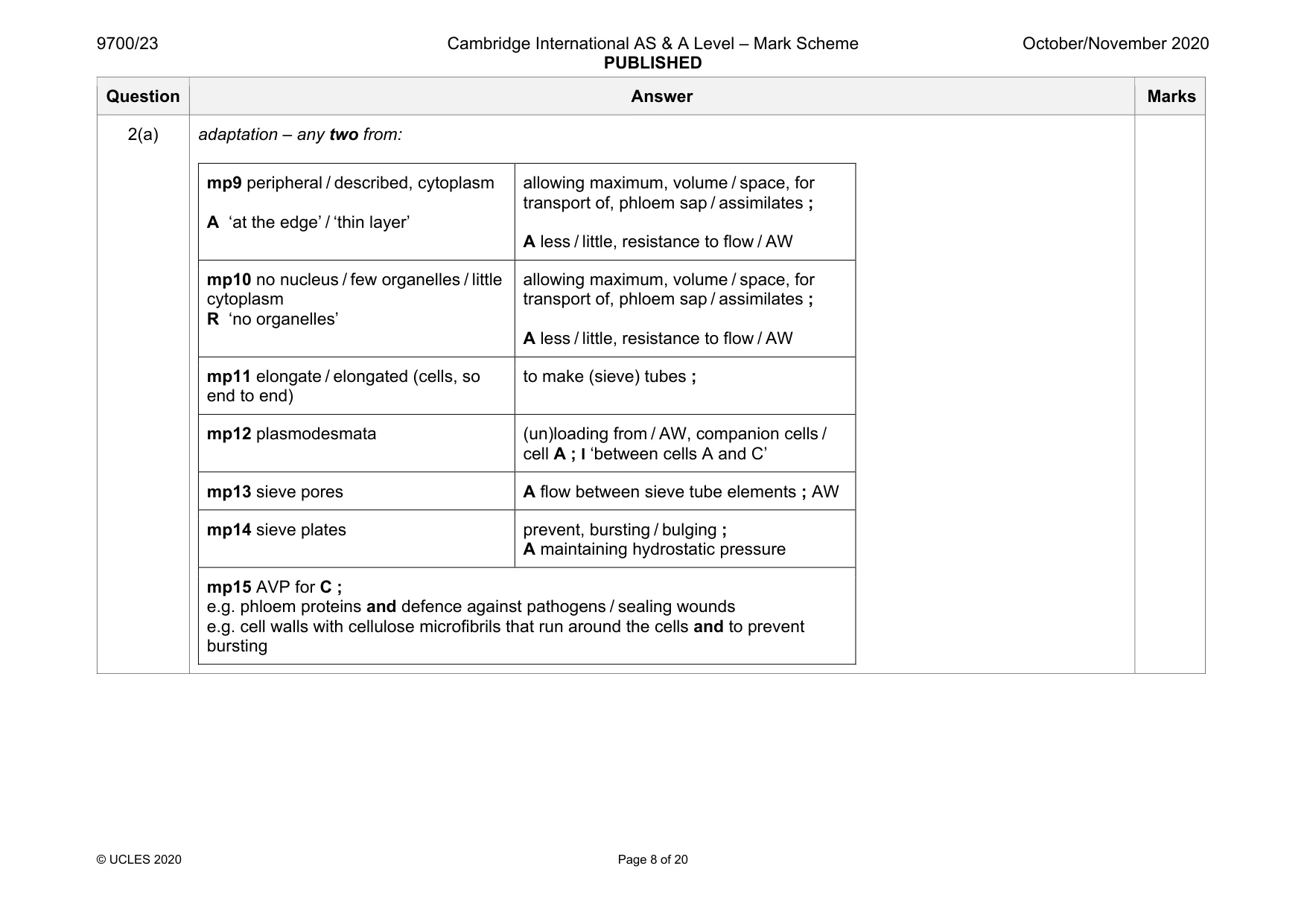
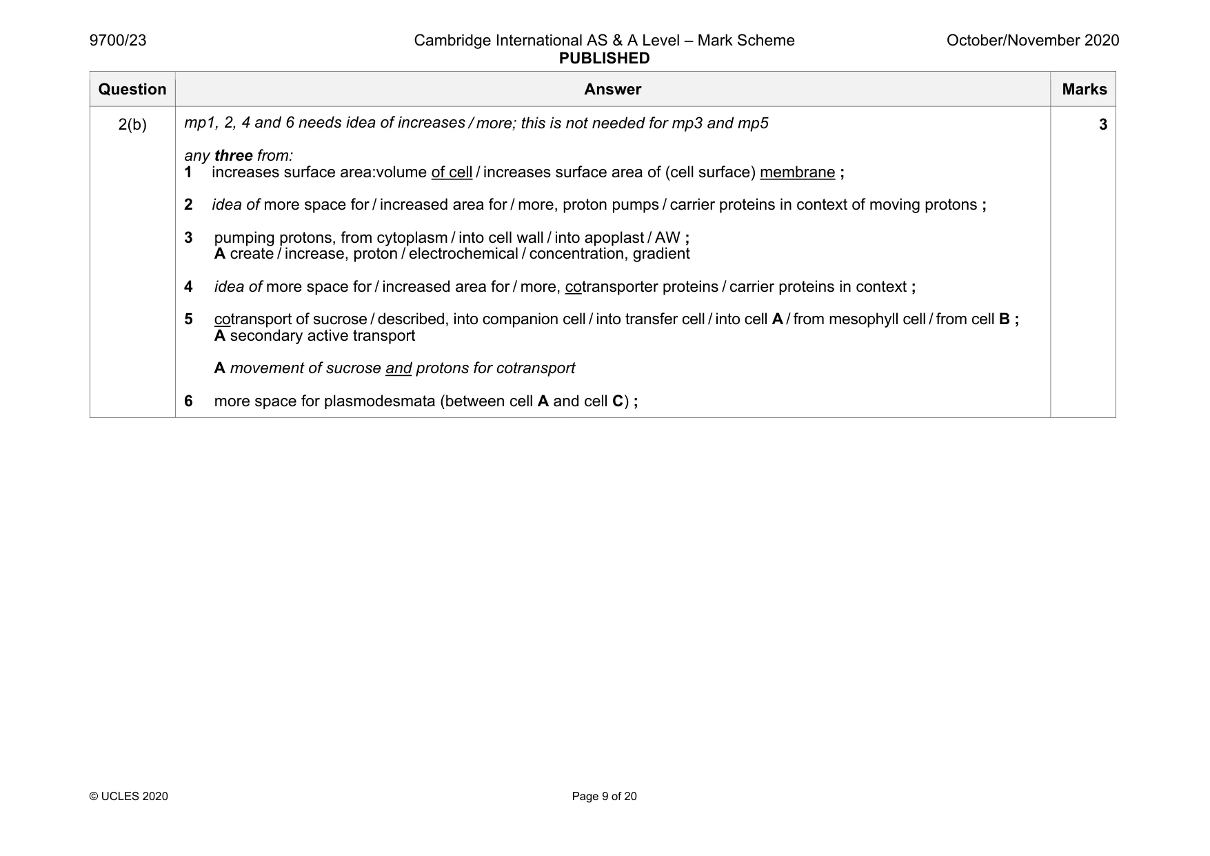
Mark scheme: 2(a) award mp1 and mp8 anywhere in each section of the answer cell B - mesophyll cells mp1 photosynthesis / described ; e.g. conversion of carbon dioxide to sugars using light energy synthesis of complex organic compounds from inorganic compounds using light energy adaptation – mp2 and one from mp3 to mp7 mp2 chloroplasts / chlorophyll / chloroplast pigments to absorb light ; any one from: mp3 large vacuole to keep chloroplasts at the periphery ; mp4 starch grains as store of products of photosynthesis ; mp5 large / moist, surface / cell wall for evaporation of water for transpiration (stream) ; A provides water for photosynthesis in context of water supply transpiration mp6 thin cell wall gas exchange / diffusion of gases ; mp7 isodiametric / (roughly) spherical shape prevents close packing / gives large air spaces ; cell C - sieve tube element mp8 transport / movement of, (named) sugars / (named) assimilates / photosynthates / organic substances ; A translocation Question Answer Marks 2(a) adaptation – any two from: mp9 peripheral / described, cytoplasm A ‘at the edge’ / ‘thin layer’ allowing maximum, volume / space, for transport of, phloem sap / assimilates ; A less / little, resistance to flow / AW mp10 no nucleus / few organelles / little cytoplasm R ‘no organelles’ allowing maximum, volume / space, for transport of, phloem sap / assimilates ; A less / little, resistance to flow / AW mp11 elongate / elongated (cells, so end to end) to make (sieve) tubes ; mp12 plasmodesmata (un)loading from / AW, companion cells / cell A ; I ‘between cells A and C’ mp13 sieve pores A flow between sieve tube elements ; AW mp14 sieve plates prevent, bursting / bulging ; A maintaining hydrostatic pressure mp15 AVP for C ; e.g. phloem proteins and defence against pathogens / sealing wounds e.g. cell walls with cellulose microfibrils that run around the cells and to prevent bursting Question Answer Marks 2(b) mp1, 2, 4 and 6 needs idea of increases / more; this is not needed for mp3 and mp5 any three from: 1 increases surface area:volume of cell / increases surface area of (cell surface) membrane ; 2 idea of more space for / increased area for / more, proton pumps / carrier proteins in context of moving protons ; 3 pumping protons, from cytoplasm / into cell wall / into apoplast / AW ; A create / increase, proton / electrochemical / concentration, gradient 4 idea of more space for / increased area for / more, cotransporter proteins / carrier proteins in context ; 5 cotransport of sucrose / described, into companion cell / into transfer cell / into cell A / from mesophyll cell / from cell B ; A secondary active transport A movement of sucrose and protons for cotransport 6 more space for plasmodesmata (between cell A and cell C) ; 3
Q3 · The nematode worm, Caenorhabditis elegans
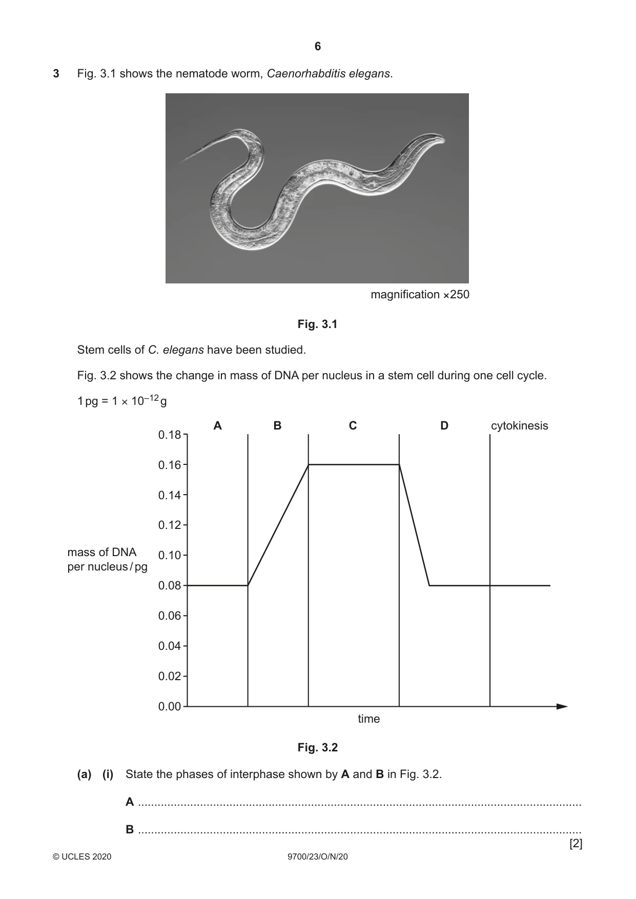
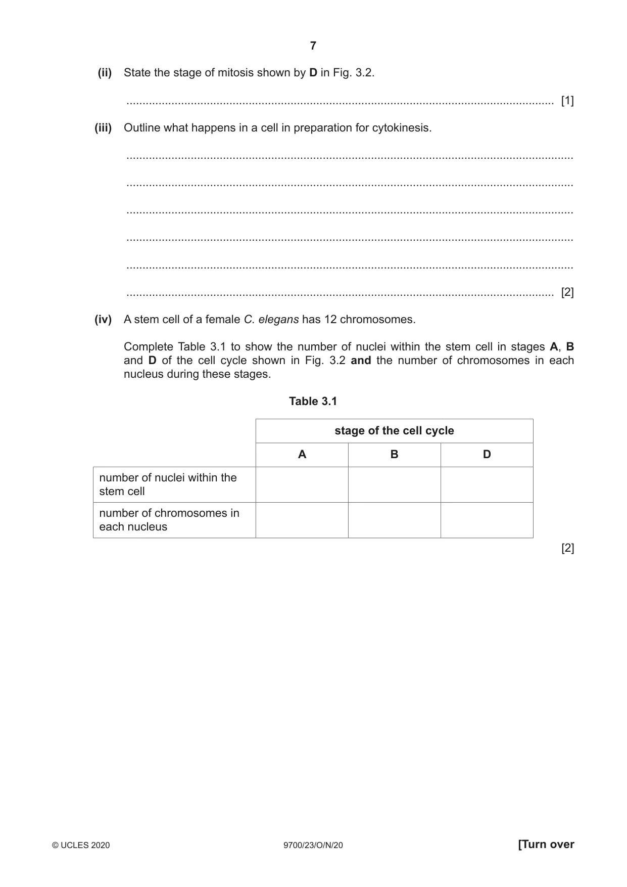
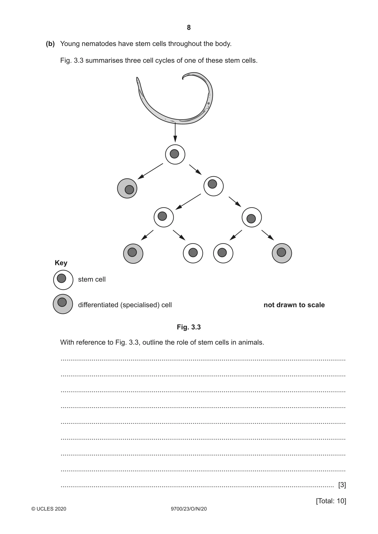
3 Fig. 3.1 shows the nematode worm, Caenorhabditis elegans. magnification ××250 Fig. 3.1 Stem cells of C. elegans have been studied. Fig. 3.2 shows the change in mass of DNA per nucleus in a stem cell during one cell cycle. 1 pg = 1 × 10–12 g A B C D cytokinesis 0.18 0.16 0.14 0.12 mass of DNA 0.10 per nucleus / pg 0.08 0.06 0.04 0.02 0.00 time Fig. 3.2 (a) (i) State the phases of interphase shown by A and B in Fig. 3.2. A ........................................................................................................................................ B ........................................................................................................................................ [2] (ii) State the stage of mitosis shown by D in Fig. 3.2. ..................................................................................................................................... [1] (iii) Outline what happens in a cell in preparation for cytokinesis. ........................................................................................................................................... ........................................................................................................................................... ........................................................................................................................................... ........................................................................................................................................... ........................................................................................................................................... ..................................................................................................................................... [2] (iv) A stem cell of a female C. elegans has 12 chromosomes. Complete Table 3.1 to show the number of nuclei within the stem cell in stages A, B and D of the cell cycle shown in Fig. 3.2 and the number of chromosomes in each nucleus during these stages. Table 3.1 stage of the cell cycle A B D number of nuclei within the stem cell number of chromosomes in each nucleus [2] (b) Young nematodes have stem cells throughout the body. Fig. 3.3 summarises three cell cycles of one of these stem cells. Key stem cell differentiated (specialised) cell not drawn to scale Fig. 3.3 With reference to Fig. 3.3, outline the role of stem cells in animals. ................................................................................................................................................... ................................................................................................................................................... ................................................................................................................................................... ................................................................................................................................................... ................................................................................................................................................... ................................................................................................................................................... ................................................................................................................................................... ................................................................................................................................................... ............................................................................................................................................. [3] [Total: 10]
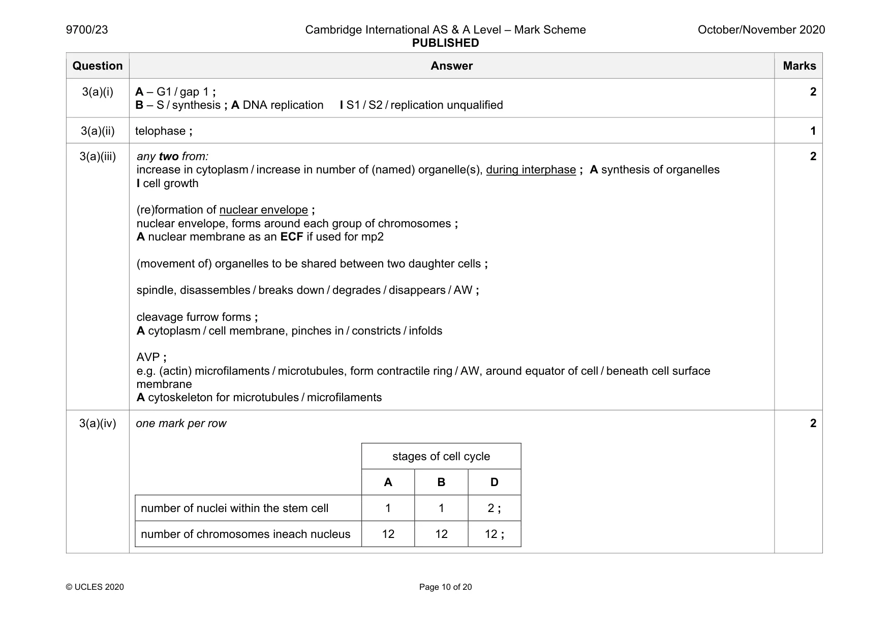
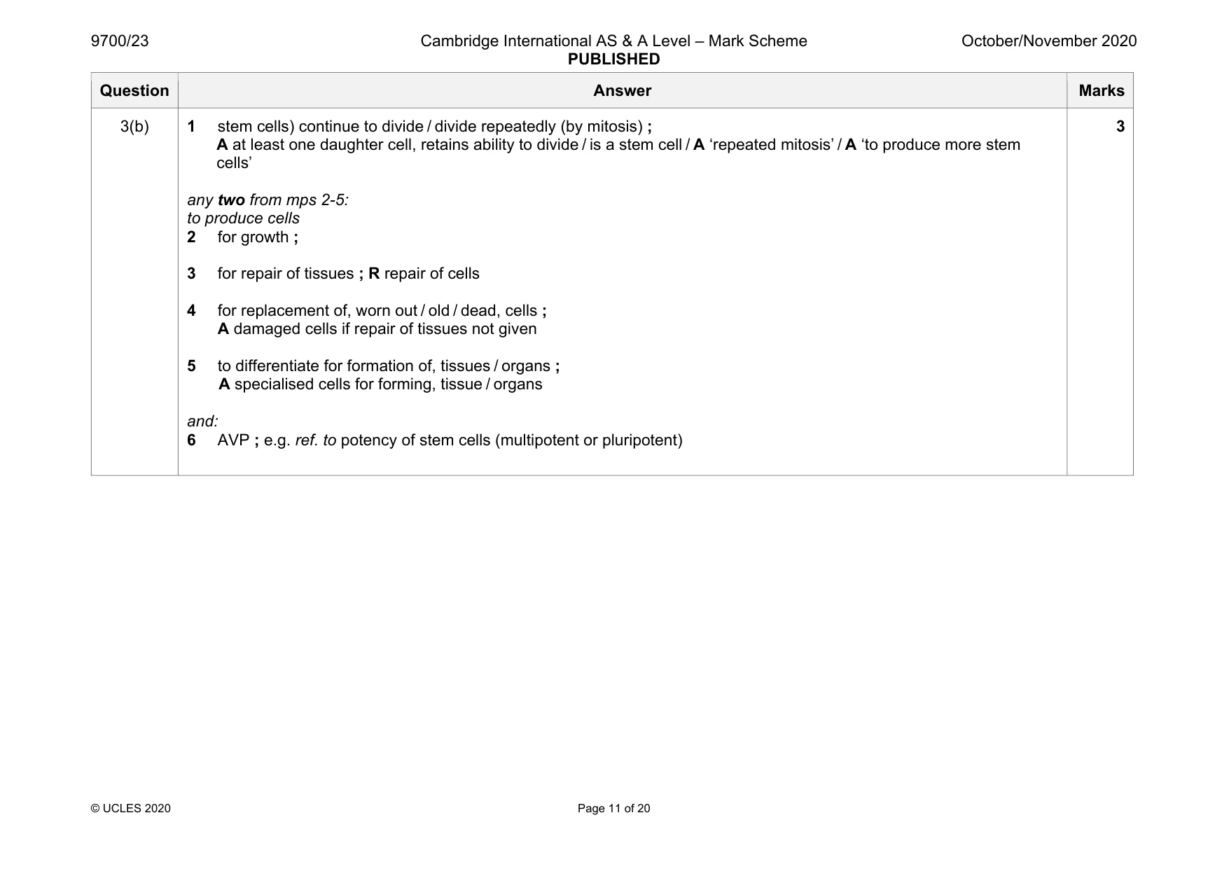
Mark scheme: 3(a)(i) A – G1 / gap 1 ; B – S / synthesis ; A DNA replication I S1 / S2 / replication unqualified 2 3(a)(ii) telophase ; 1 3(a)(iii) any two from: increase in cytoplasm / increase in number of (named) organelle(s), during interphase ; A synthesis of organelles I cell growth (re)formation of nuclear envelope ; nuclear envelope, forms around each group of chromosomes ; A nuclear membrane as an ECF if used for mp2 (movement of) organelles to be shared between two daughter cells ; spindle, disassembles / breaks down / degrades / disappears / AW ; cleavage furrow forms ; A cytoplasm / cell membrane, pinches in / constricts / infolds AVP ; e.g. (actin) microfilaments / microtubules, form contractile ring / AW, around equator of cell / beneath cell surface membrane A cytoskeleton for microtubules / microfilaments 2 3(a)(iv) one mark per row stages of cell cycle A B D number of nuclei within the stem cell 1 1 2 ; number of chromosomes ineach nucleus 12 12 12 ; 2 Question Answer Marks 3(b) 1 stem cells) continue to divide / divide repeatedly (by mitosis) ; A at least one daughter cell, retains ability to divide / is a stem cell / A ‘repeated mitosis’ / A ‘to produce more stem cells’ any two from mps 2-5: to produce cells 2 for growth ; 3 for repair of tissues ; R repair of cells 4 for replacement of, worn out / old / dead, cells ; A damaged cells if repair of tissues not given 5 to differentiate for formation of, tissues / organs ; A specialised cells for forming, tissue / organs and: 6 AVP ; e.g. ref. to potency of stem cells (multipotent or pluripotent) 3
More questions on Replication and division of nuclei and cells
Q4 · Antibiotic sensitivity tests can be carried out to choose appropriate antibiotics to use…
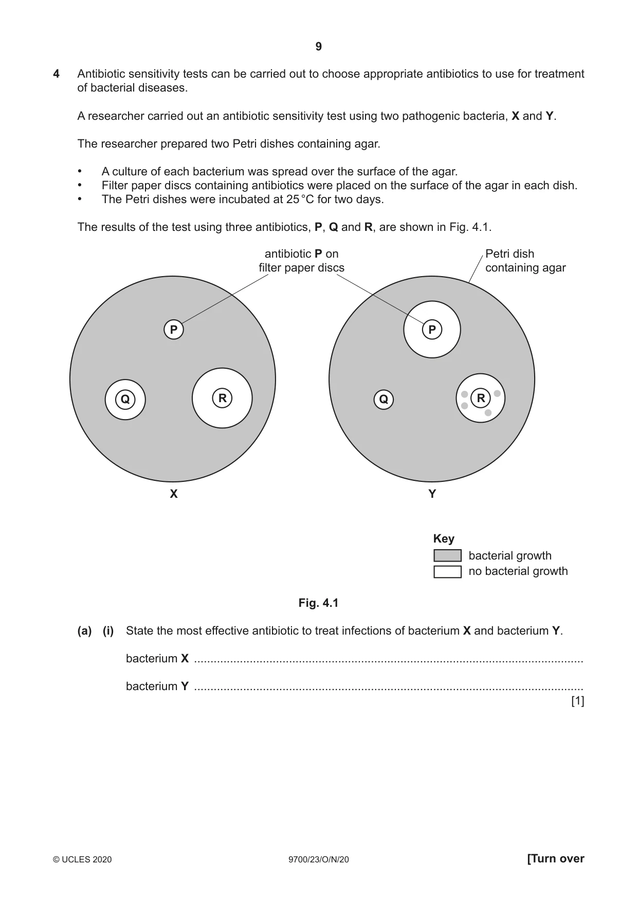
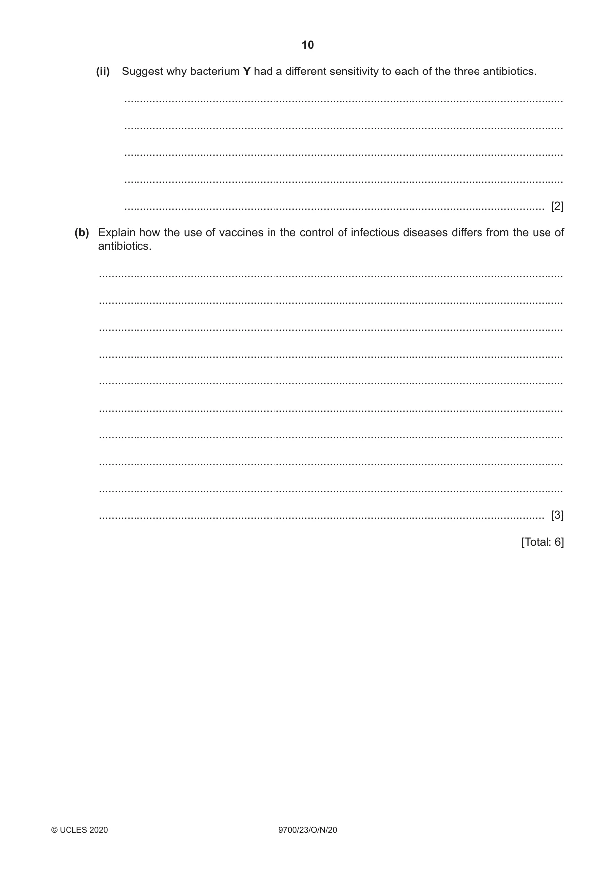
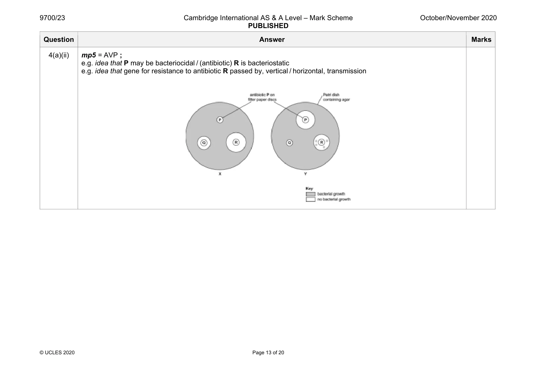
4 Antibiotic sensitivity tests can be carried out to choose appropriate antibiotics to use for treatment of bacterial diseases. A researcher carried out an antibiotic sensitivity test using two pathogenic bacteria, X and Y. The researcher prepared two Petri dishes containing agar. • A culture of each bacterium was spread over the surface of the agar. • Filter paper discs containing antibiotics were placed on the surface of the agar in each dish. • The Petri dishes were incubated at 25 °C for two days. The results of the test using three antibiotics, P, Q and R, are shown in Fig. 4.1. antibiotic P on Petri dish filter paper discs containing agar P P Q R Q R X Y Key bacterial growth no bacterial growth Fig. 4.1 (a) (i) State the most effective antibiotic to treat infections of bacterium X and bacterium Y. bacterium X ....................................................................................................................... bacterium Y ....................................................................................................................... [1] (ii) Suggest why bacterium Y had a different sensitivity to each of the three antibiotics. ........................................................................................................................................... ........................................................................................................................................... ........................................................................................................................................... ........................................................................................................................................... ..................................................................................................................................... [2] (b) Explain how the use of vaccines in the control of infectious diseases differs from the use of antibiotics. ................................................................................................................................................... ................................................................................................................................................... ................................................................................................................................................... ................................................................................................................................................... ................................................................................................................................................... ................................................................................................................................................... ................................................................................................................................................... ................................................................................................................................................... ................................................................................................................................................... ............................................................................................................................................. [3] [Total: 6]
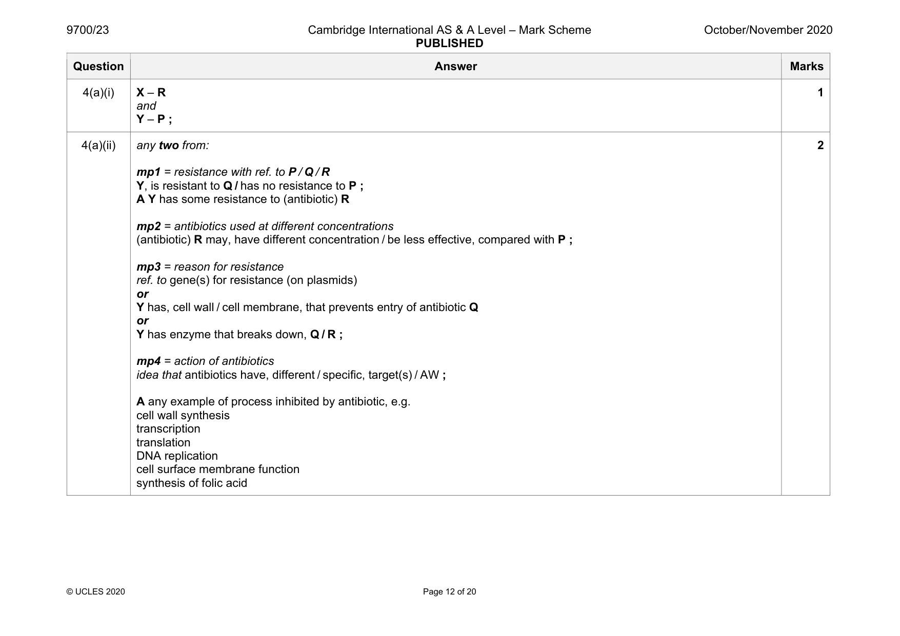
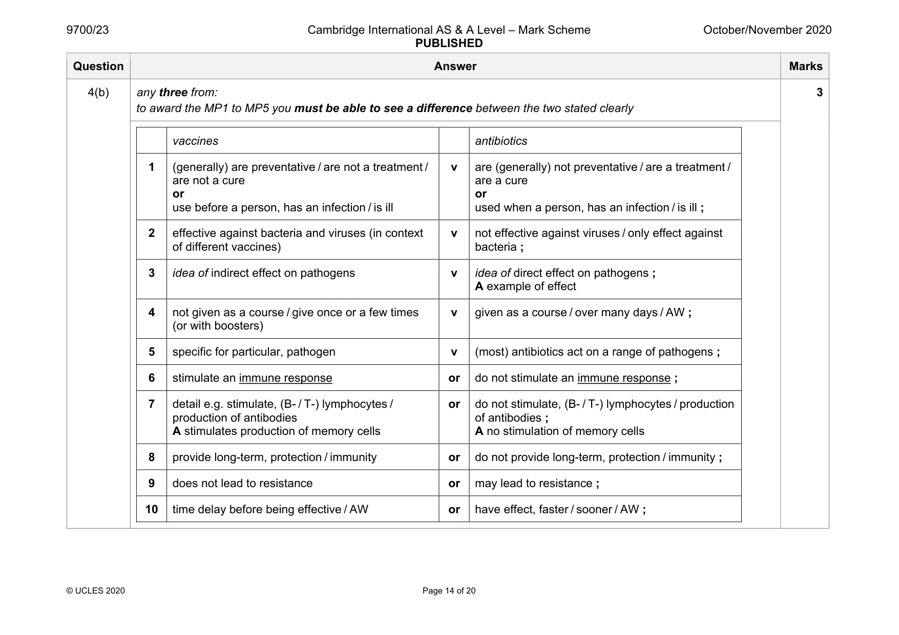
Mark scheme: 4(a)(i) X – R and Y – P ; 1 4(a)(ii) any two from: mp1 = resistance with ref. to P / Q / R Y, is resistant to Q / has no resistance to P ; A Y has some resistance to (antibiotic) R mp2 = antibiotics used at different concentrations (antibiotic) R may, have different concentration / be less effective, compared with P ; mp3 = reason for resistance ref. to gene(s) for resistance (on plasmids) or Y has, cell wall / cell membrane, that prevents entry of antibiotic Q or Y has enzyme that breaks down, Q / R ; mp4 = action of antibiotics idea that antibiotics have, different / specific, target(s) / AW ; A any example of process inhibited by antibiotic, e.g. cell wall synthesis transcription translation DNA replication cell surface membrane function synthesis of folic acid 2 Question Answer Marks 4(a)(ii) mp5 = AVP ; e.g. idea that P may be bacteriocidal / (antibiotic) R is bacteriostatic e.g. idea that gene for resistance to antibiotic R passed by, vertical / horizontal, transmission Question Answer Marks 4(b) any three from: to award the MP1 to MP5 you must be able to see a difference between the two stated clearly 3 vaccines antibiotics 1 (generally) are preventative / are not a treatment / are not a cure or use before a person, has an infection / is ill v are (generally) not preventative / are a treatment / are a cure or used when a person, has an infection / is ill ; 2 effective against bacteria and viruses (in context of different vaccines) v not effective against viruses / only effect against bacteria ; 3 idea of indirect effect on pathogens v idea of direct effect on pathogens ; A example of effect 4 not given as a course / give once or a few times (or with boosters) v given as a course / over many days / AW ; 5 specific for particular, pathogen v (most) antibiotics act on a range of pathogens ; 6 stimulate an immune response or do not stimulate an immune response ; 7 detail e.g. stimulate, (B- / T-) lymphocytes / production of antibodies A stimulates production of memory cells or do not stimulate, (B- / T-) lymphocytes / production of antibodies ; A no stimulation of memory cells 8 provide long-term, protection / immunity or do not provide long-term, protection / immunity ; 9 does not lead to resistance or may lead to resistance ; 10 time delay before being effective / AW or have effect, faster / sooner / AW ;
Q5 · A molecule of collagen consists of three identical polypeptides that form a triple helix
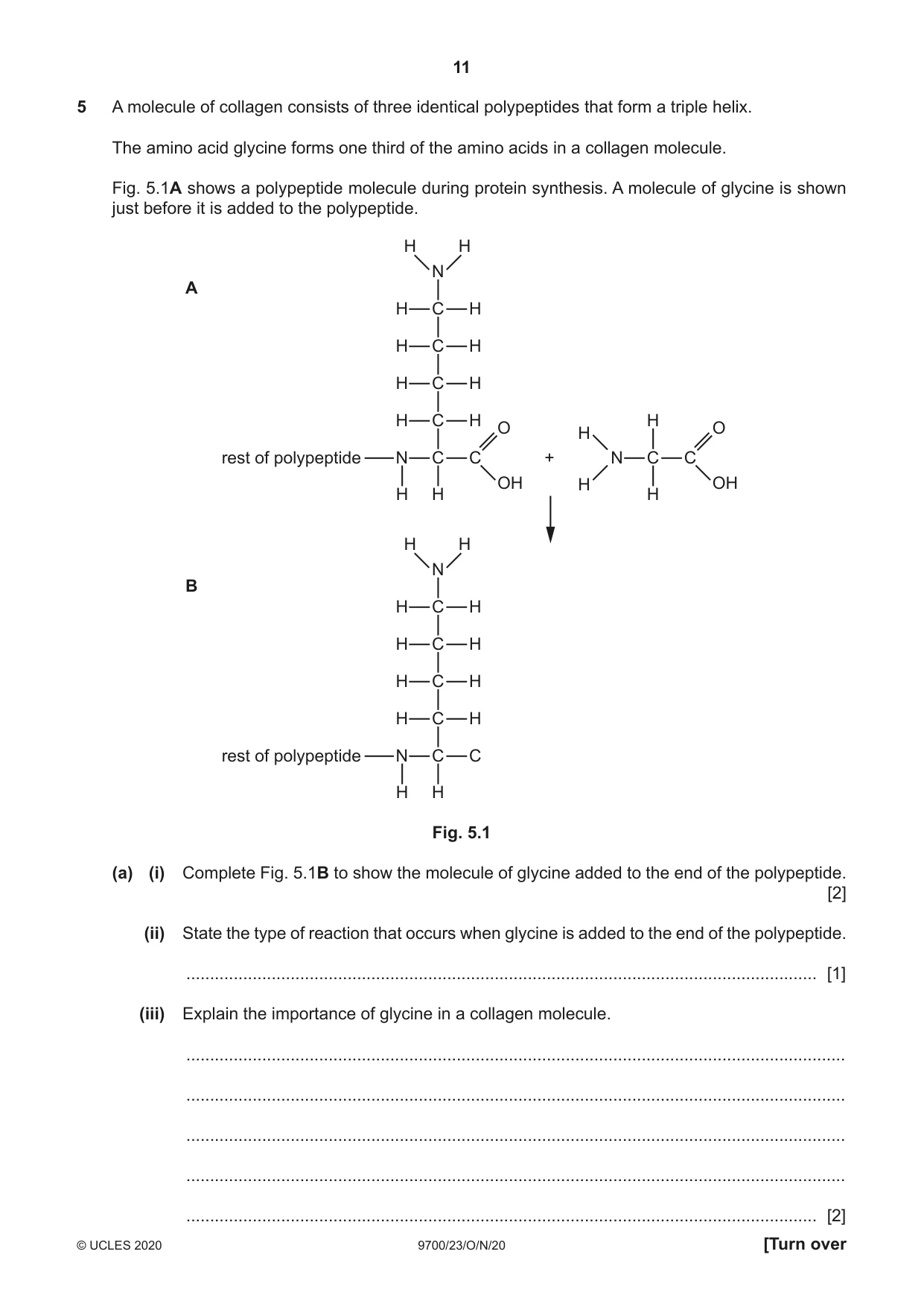
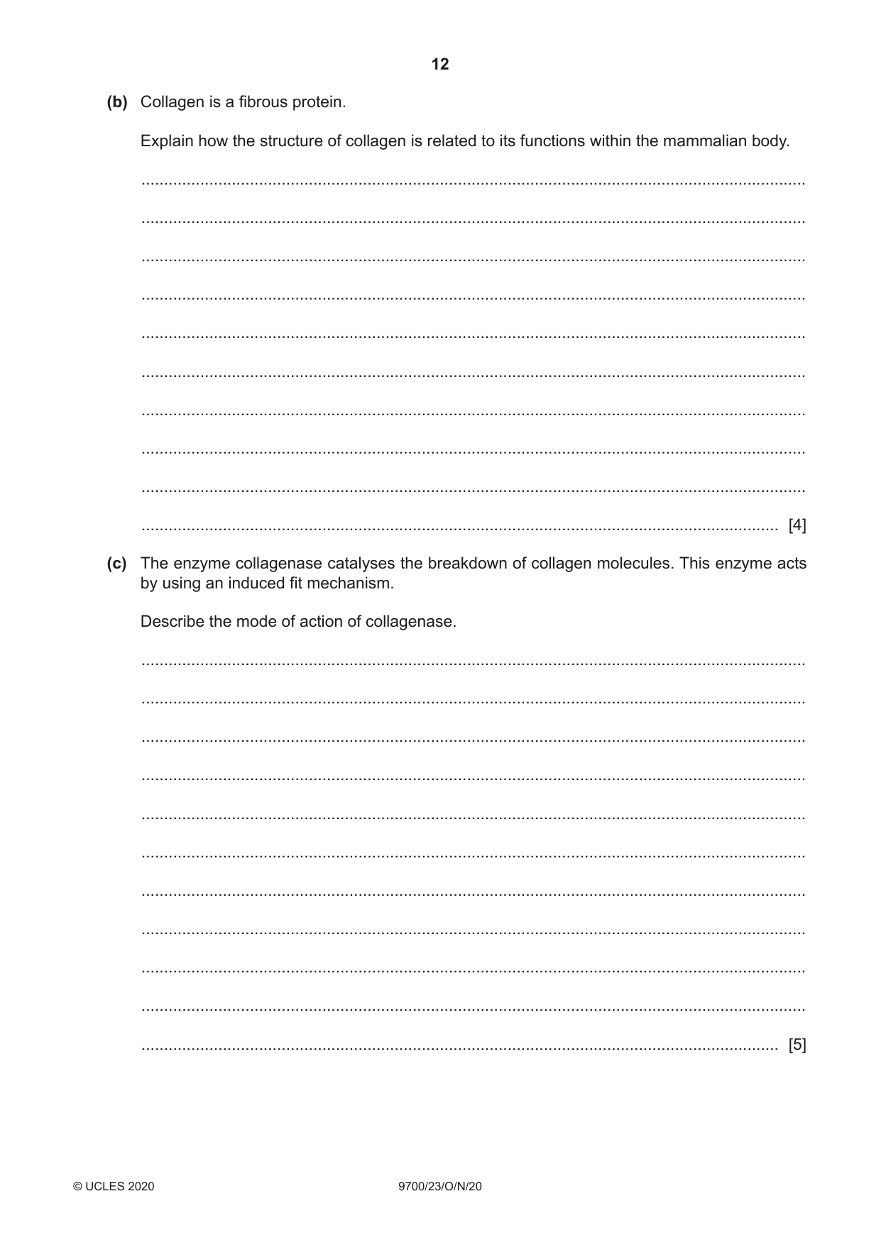
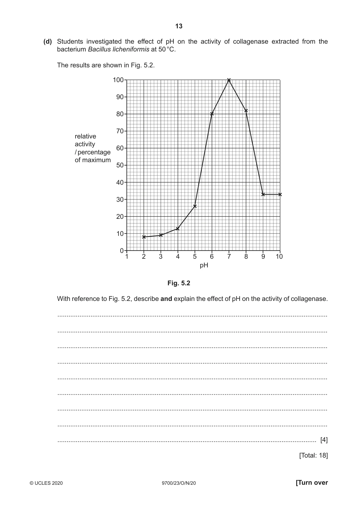
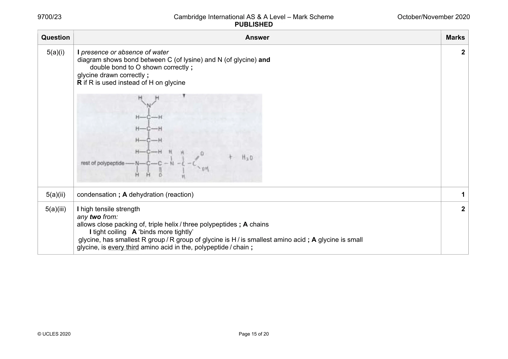
5 A molecule of collagen consists of three identical polypeptides that form a triple helix. The amino acid glycine forms one third of the amino acids in a collagen molecule. Fig. 5.1A shows a polypeptide molecule during protein synthesis. A molecule of glycine is shown just before it is added to the polypeptide. H H N A H C H H C H H C H H C H H O O H rest of polypeptide N C C + N C C OH H OH H H H H H N B H C H H C H H C H H C H rest of polypeptide N C C H H Fig. 5.1 (a) (i) Complete Fig. 5.1B to show the molecule of glycine added to the end of the polypeptide. [2] (ii) State the type of reaction that occurs when glycine is added to the end of the polypeptide. ..................................................................................................................................... [1] (iii) Explain the importance of glycine in a collagen molecule. ........................................................................................................................................... ........................................................................................................................................... ........................................................................................................................................... ........................................................................................................................................... ..................................................................................................................................... [2] (b) Collagen is a fibrous protein. Explain how the structure of collagen is related to its functions within the mammalian body. ................................................................................................................................................... ................................................................................................................................................... ................................................................................................................................................... ................................................................................................................................................... ................................................................................................................................................... ................................................................................................................................................... ................................................................................................................................................... ................................................................................................................................................... ................................................................................................................................................... ............................................................................................................................................. [4] (c) The enzyme collagenase catalyses the breakdown of collagen molecules. This enzyme acts by using an induced fit mechanism. Describe the mode of action of collagenase. ................................................................................................................................................... ................................................................................................................................................... ................................................................................................................................................... ................................................................................................................................................... ................................................................................................................................................... ................................................................................................................................................... ................................................................................................................................................... ................................................................................................................................................... ................................................................................................................................................... ................................................................................................................................................... ............................................................................................................................................. [5] (d) Students investigated the effect of pH on the activity of collagenase extracted from the bacterium Bacillus licheniformis at 50 °C. The results are shown in Fig. 5.2. 100 90 80 70 relative activity 60 / percentage of maximum 50 40 30 20 10 0 1 2 3 4 5 6 7 8 9 10 pH Fig. 5.2 With reference to Fig. 5.2, describe and explain the effect of pH on the activity of collagenase. ................................................................................................................................................... ................................................................................................................................................... ................................................................................................................................................... ................................................................................................................................................... ................................................................................................................................................... ................................................................................................................................................... ................................................................................................................................................... ................................................................................................................................................... ............................................................................................................................................. [4] [Total: 18]
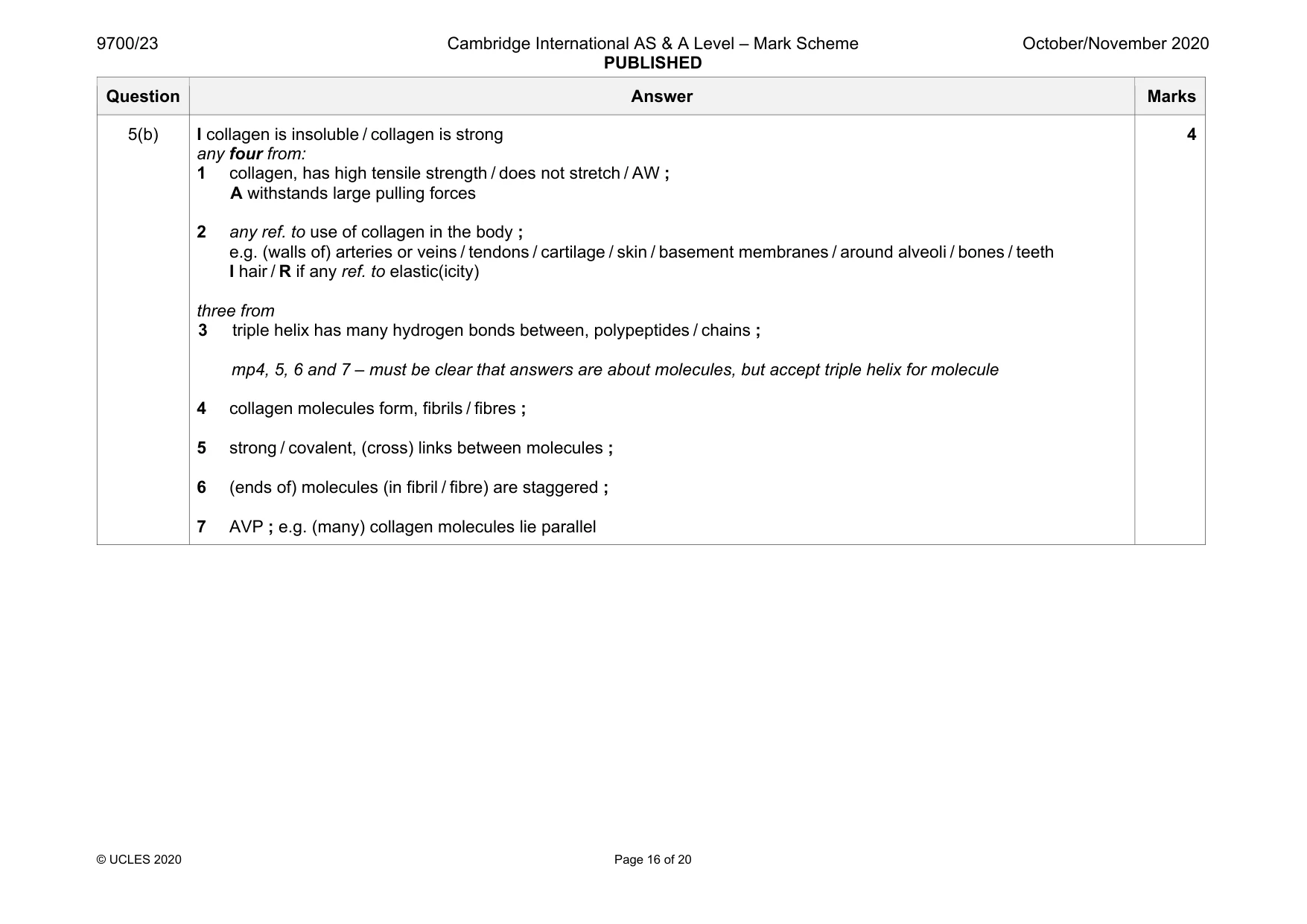
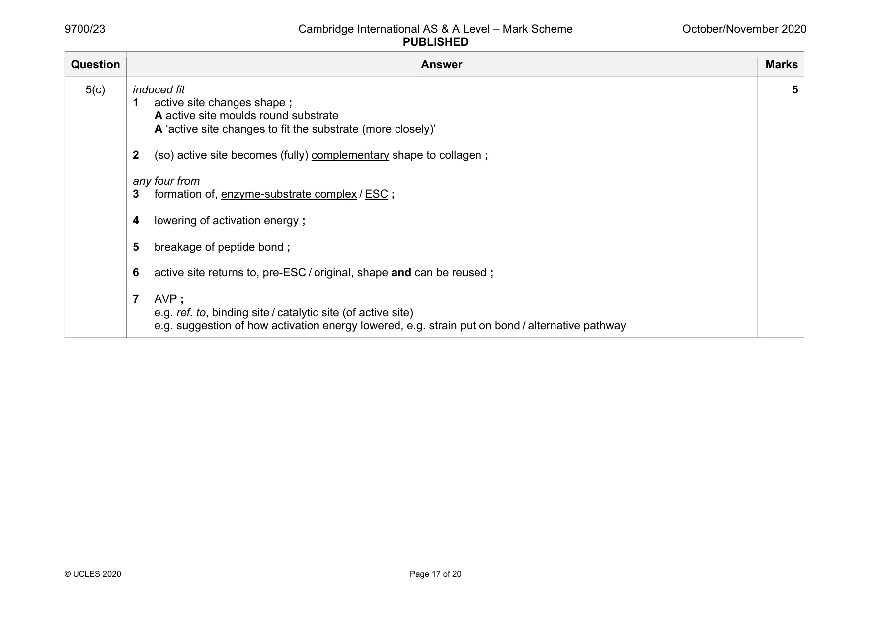
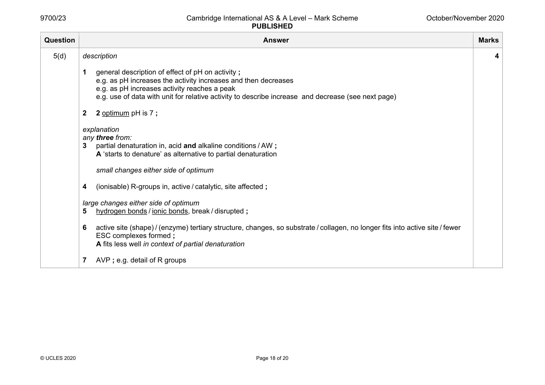
Mark scheme: 5(a)(i) I presence or absence of water diagram shows bond between C (of lysine) and N (of glycine) and double bond to O shown correctly ; glycine drawn correctly ; R if R is used instead of H on glycine 5(a)(ii) condensation ; A dehydration (reaction) 1 5(a)(iii) I high tensile strength any two from: allows close packing of, triple helix / three polypeptides ; A chains I tight coiling A ‘binds more tightly’ glycine, has smallest R group / R group of glycine is H / is smallest amino acid ; A glycine is small glycine, is every third amino acid in the, polypeptide / chain ; 2 Question Answer Marks 5(b) I collagen is insoluble / collagen is strong any four from: 1 collagen, has high tensile strength / does not stretch / AW ; A withstands large pulling forces 2 any ref. to use of collagen in the body ; e.g. (walls of) arteries or veins / tendons / cartilage / skin / basement membranes / around alveoli / bones / teeth I hair / R if any ref. to elastic(icity) three from 3 triple helix has many hydrogen bonds between, polypeptides / chains ; mp4, 5, 6 and 7 – must be clear that answers are about molecules, but accept triple helix for molecule 4 collagen molecules form, fibrils / fibres ; 5 strong / covalent, (cross) links between molecules ; 6 (ends of) molecules (in fibril / fibre) are staggered ; 7 AVP ; e.g. (many) collagen molecules lie parallel 4 Question Answer Marks 5(c) induced fit 1 active site changes shape ; A active site moulds round substrate A ‘active site changes to fit the substrate (more closely)’ 2 (so) active site becomes (fully) complementary shape to collagen ; any four from 3 formation of, enzyme-substrate complex / ESC ; 4 lowering of activation energy ; 5 breakage of peptide bond ; 6 active site returns to, pre-ESC / original, shape and can be reused ; 7 AVP ; e.g. ref. to, binding site / catalytic site (of active site) e.g. suggestion of how activation energy lowered, e.g. strain put on bond / alternative pathway 5 Question Answer Marks 5(d) description 1 general description of effect of pH on activity ; e.g. as pH increases the activity increases and then decreases e.g. as pH increases activity reaches a peak e.g. use of data with unit for relative activity to describe increase and decrease (see next page) 2 2 optimum pH is 7 ; explanation any three from: 3 partial denaturation in, acid and alkaline conditions / AW ; A ‘starts to denature’ as alternative to partial denaturation small changes either side of optimum 4 (ionisable) R-groups in, active / catalytic, site affected ; large changes either side of optimum 5 hydrogen bonds / ionic bonds, break / disrupted ; 6 active site (shape) / (enzyme) tertiary structure, changes, so substrate / collagen, no longer fits into active site / fewer ESC complexes formed ; A fits less well in context of partial denaturation 7 AVP ; e.g. detail of R groups 4
Q6 · Vorticella, which is a single-celled organism that lives in freshwater
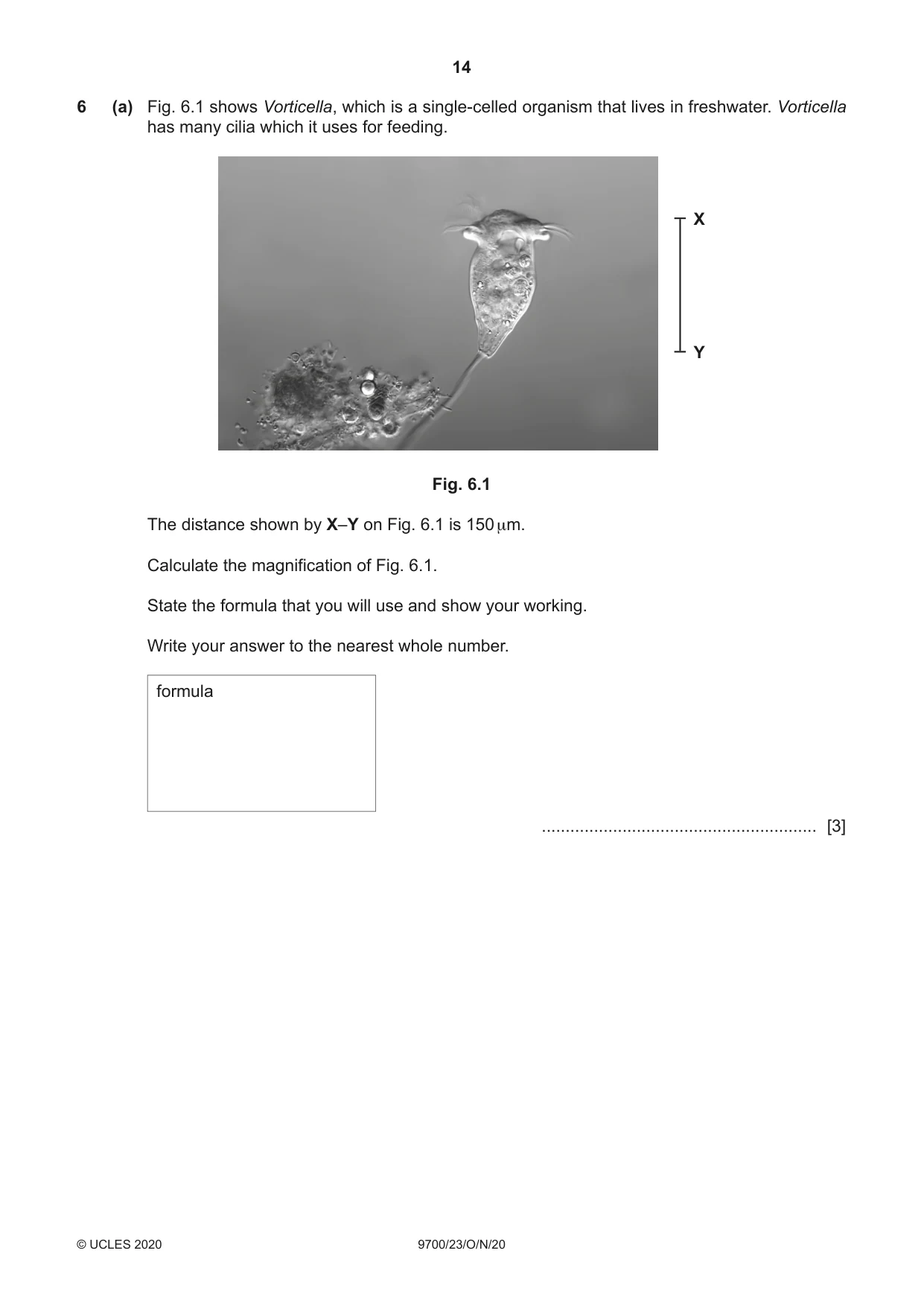
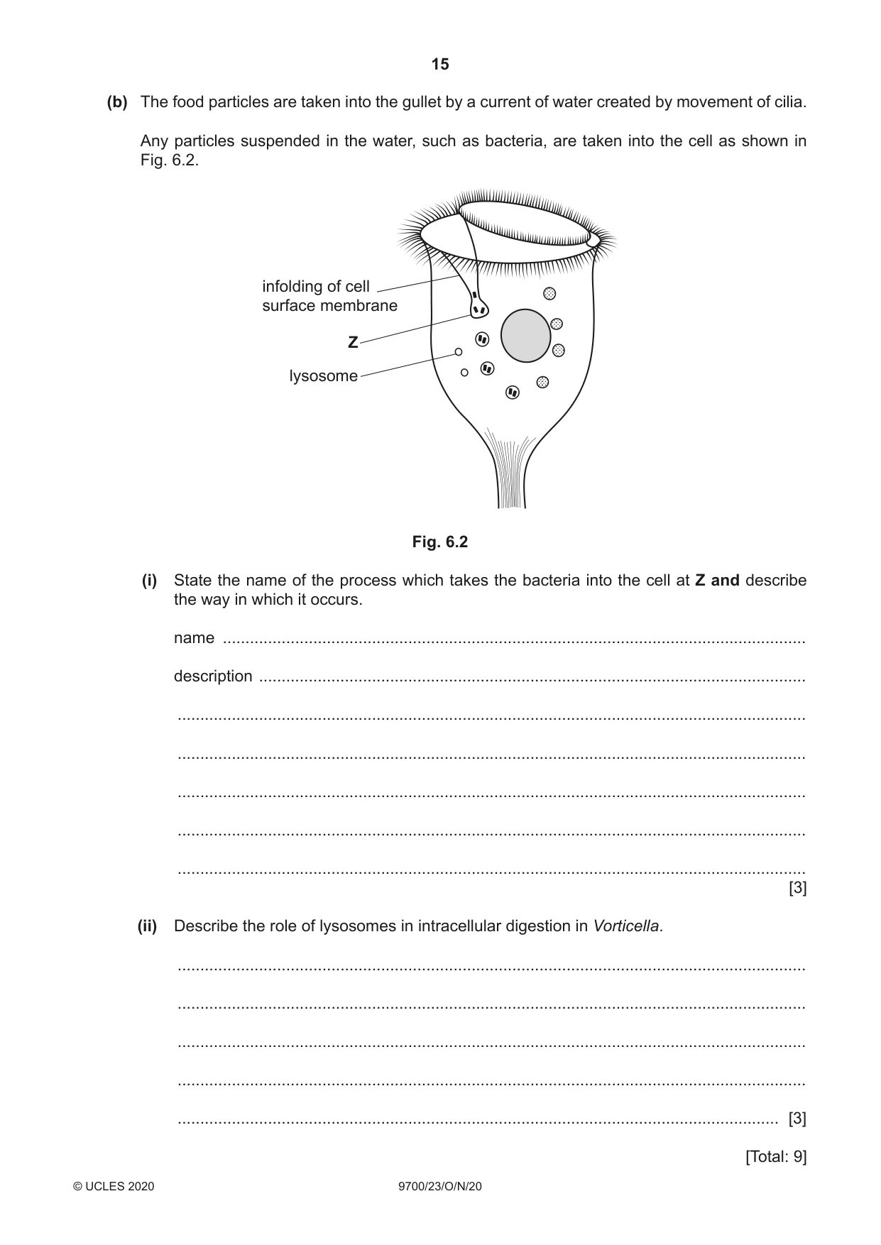
6 (a) Fig. 6.1 shows Vorticella, which is a single-celled organism that lives in freshwater. Vorticella has many cilia which it uses for feeding. X Y Fig. 6.1 The distance shown by X–Y on Fig. 6.1 is 150 μm. Calculate the magnification of Fig. 6.1. State the formula that you will use and show your working. Write your answer to the nearest whole number. formula .......................................................... [3] (b) The food particles are taken into the gullet by a current of water created by movement of cilia. Any particles suspended in the water, such as bacteria, are taken into the cell as shown in Fig. 6.2. infolding of cell surface membrane Z lysosome Fig. 6.2 (i) State the name of the process which takes the bacteria into the cell at Z and describe the way in which it occurs. name ................................................................................................................................. description ......................................................................................................................... ........................................................................................................................................... ........................................................................................................................................... ........................................................................................................................................... ........................................................................................................................................... ........................................................................................................................................... [3] (ii) Describe the role of lysosomes in intracellular digestion in Vorticella. ........................................................................................................................................... ........................................................................................................................................... ........................................................................................................................................... ........................................................................................................................................... ..................................................................................................................................... [3]
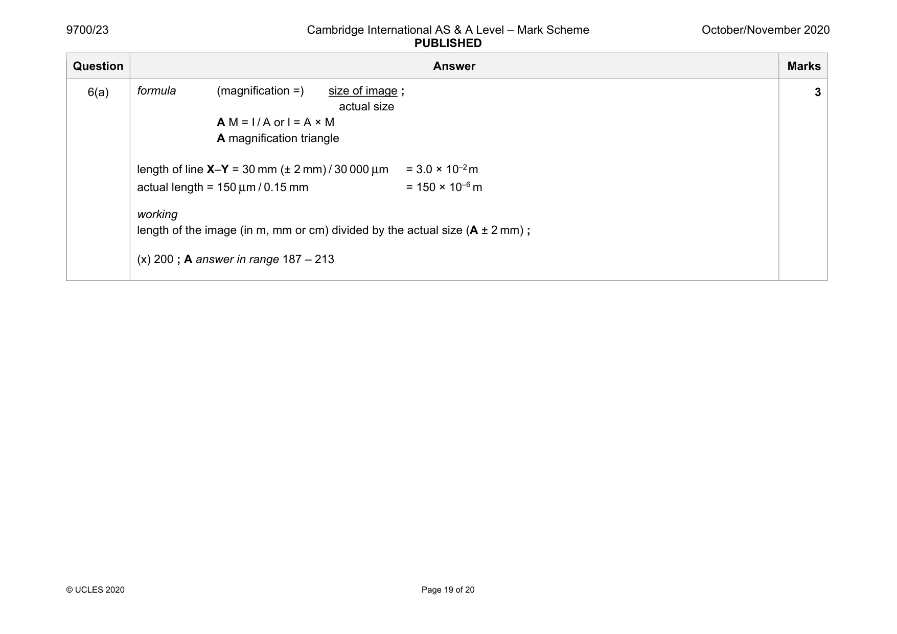
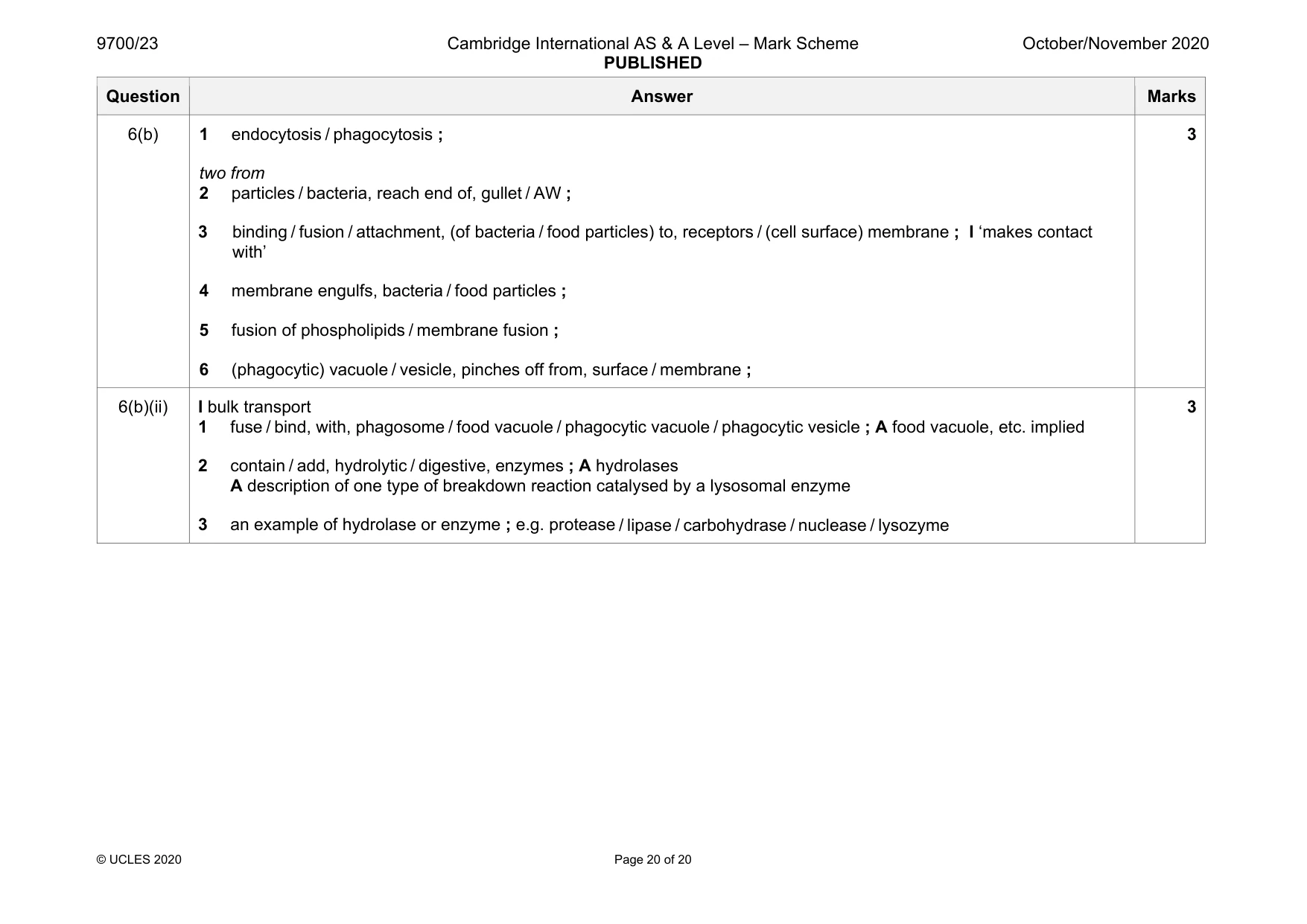
Mark scheme: 6(a) actual size A M = I / A or l = A × M A magnification triangle length of line X–Y = 30 mm (± 2 mm) / 30 000 μm = 3.0 × 10⁻² m actual length = 150 μm / 0.15 mm = 150 × 10–6 m working length of the image (in m, mm or cm) divided by the actual size (A ± 2 mm) ; (x) 200 ; A answer in range 187 – 213 3 Question Answer Marks 6(b) 1 endocytosis / phagocytosis ; two from 2 particles / bacteria, reach end of, gullet / AW ; 3 binding / fusion / attachment, (of bacteria / food particles) to, receptors / (cell surface) membrane ; I ‘makes contact with’ 4 membrane engulfs, bacteria / food particles ; 5 fusion of phospholipids / membrane fusion ; 6 (phagocytic) vacuole / vesicle, pinches off from, surface / membrane ; 3 6(b)(ii) I bulk transport 1 fuse / bind, with, phagosome / food vacuole / phagocytic vacuole / phagocytic vesicle ; A food vacuole, etc. implied 2 contain / add, hydrolytic / digestive, enzymes ; A hydrolases A description of one type of breakdown reaction catalysed by a lysosomal enzyme 3 an example of hydrolase or enzyme ; e.g. protease / lipase / carbohydrase / nuclease / lysozyme 3
What was in this paper
The subtopics covered by these 6 questions, and how many questions each got. Open one in a new tab to see every Cambridge question on it.
What you needed in this session
Cambridge’s own grade thresholds for 2020 Oct/Nov, Paper 2 · Variant 3. A higher threshold means an easier paper — the bar moves with how the cohort did.