Cambridge A Level Biology 9700 — 2020 Oct/Nov Paper 2 · Variant 1
9700/21/O/N/20 · 6 questions · 60 marks · ≈68 min
The question paper and its mark scheme, free to read here and free to download. This is Cambridge’s own paper, exactly as it was sat.
Question paper16 pages

Mark scheme13 pages
Answers below. Sit the paper first if you are practising.
Questions as text
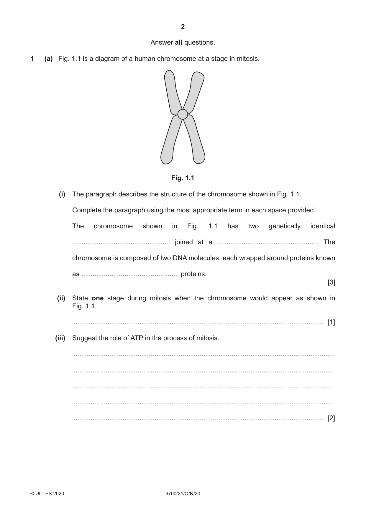
Q1 · A diagram of a human chromosome at a stage in mitosis
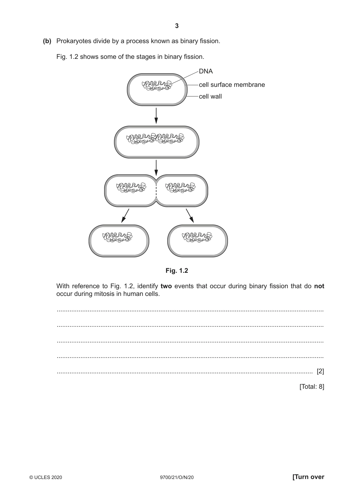
1 (a) Fig. 1.1 is a diagram of a human chromosome at a stage in mitosis. Fig. 1.1 (i) The paragraph describes the structure of the chromosome shown in Fig. 1.1. Complete the paragraph using the most appropriate term in each space provided. The chromosome shown in Fig. 1.1 has two genetically identical .................................................... joined at a .................................................... . The chromosome is composed of two DNA molecules, each wrapped around proteins known as .................................................... proteins. [3] (ii) State one stage during mitosis when the chromosome would appear as shown in Fig. 1.1. ..................................................................................................................................... [1] (iii) Suggest the role of ATP in the process of mitosis. ........................................................................................................................................... ........................................................................................................................................... ........................................................................................................................................... ........................................................................................................................................... ..................................................................................................................................... [2] (b) Prokaryotes divide by a process known as binary fission. Fig. 1.2 shows some of the stages in binary fission. DNA cell surface membrane cell wall Fig. 1.2 With reference to Fig. 1.2, identify two events that occur during binary fission that do not occur during mitosis in human cells. ................................................................................................................................................... ................................................................................................................................................... ................................................................................................................................................... ................................................................................................................................................... ............................................................................................................................................. [2] [Total: 8]
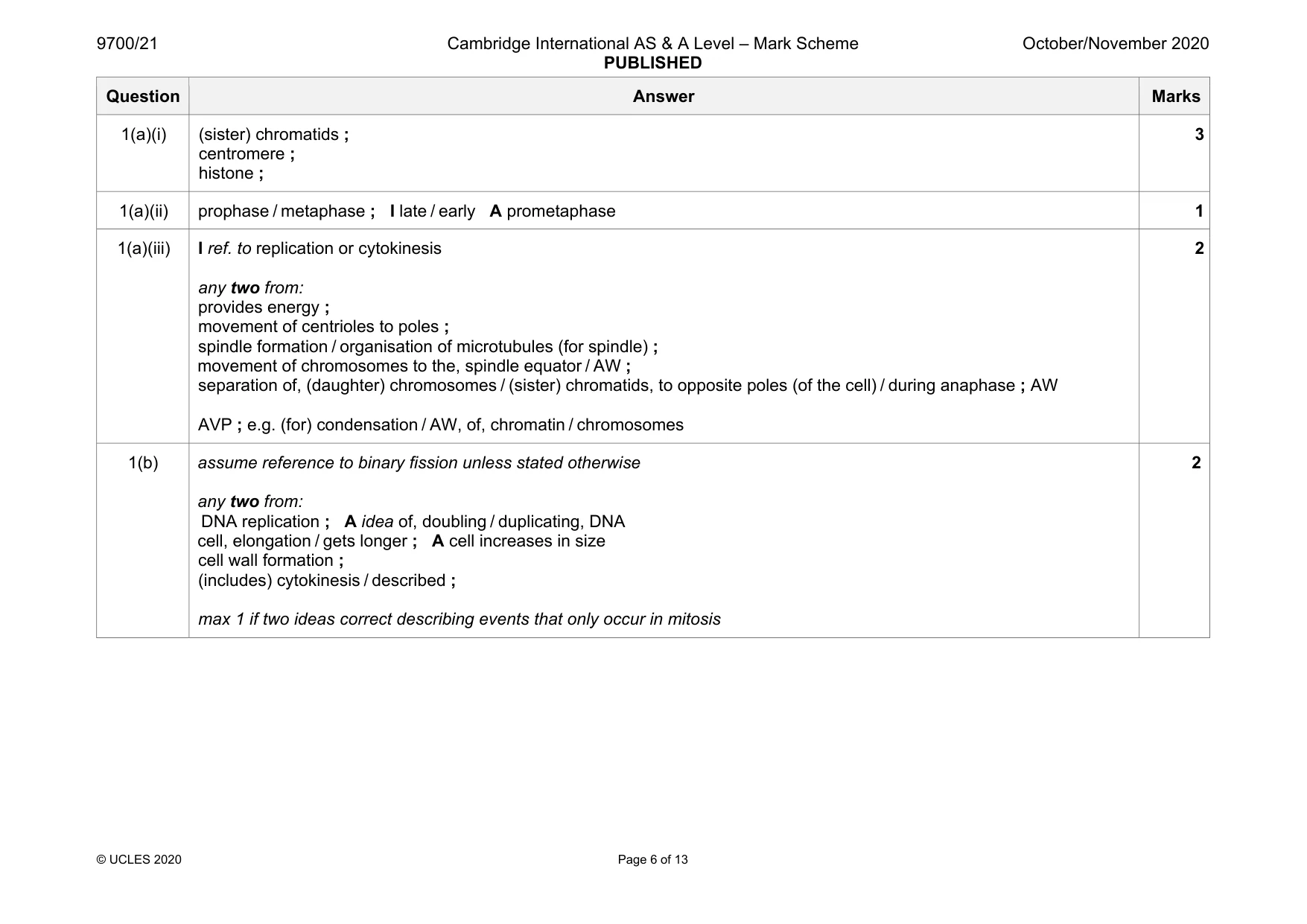
Mark scheme: 1(a)(i) (sister) chromatids ; centromere ; histone ; 3 1(a)(ii) prophase / metaphase ; I late / early A prometaphase 1 1(a)(iii) I ref. to replication or cytokinesis any two from: provides energy ; movement of centrioles to poles ; spindle formation / organisation of microtubules (for spindle) ; movement of chromosomes to the, spindle equator / AW ; separation of, (daughter) chromosomes / (sister) chromatids, to opposite poles (of the cell) / during anaphase ; AW AVP ; e.g. (for) condensation / AW, of, chromatin / chromosomes 2 1(b) assume reference to binary fission unless stated otherwise any two from: DNA replication ; A idea of, doubling / duplicating, DNA cell, elongation / gets longer ; A cell increases in size cell wall formation ; (includes) cytokinesis / described ; max 1 if two ideas correct describing events that only occur in mitosis 2
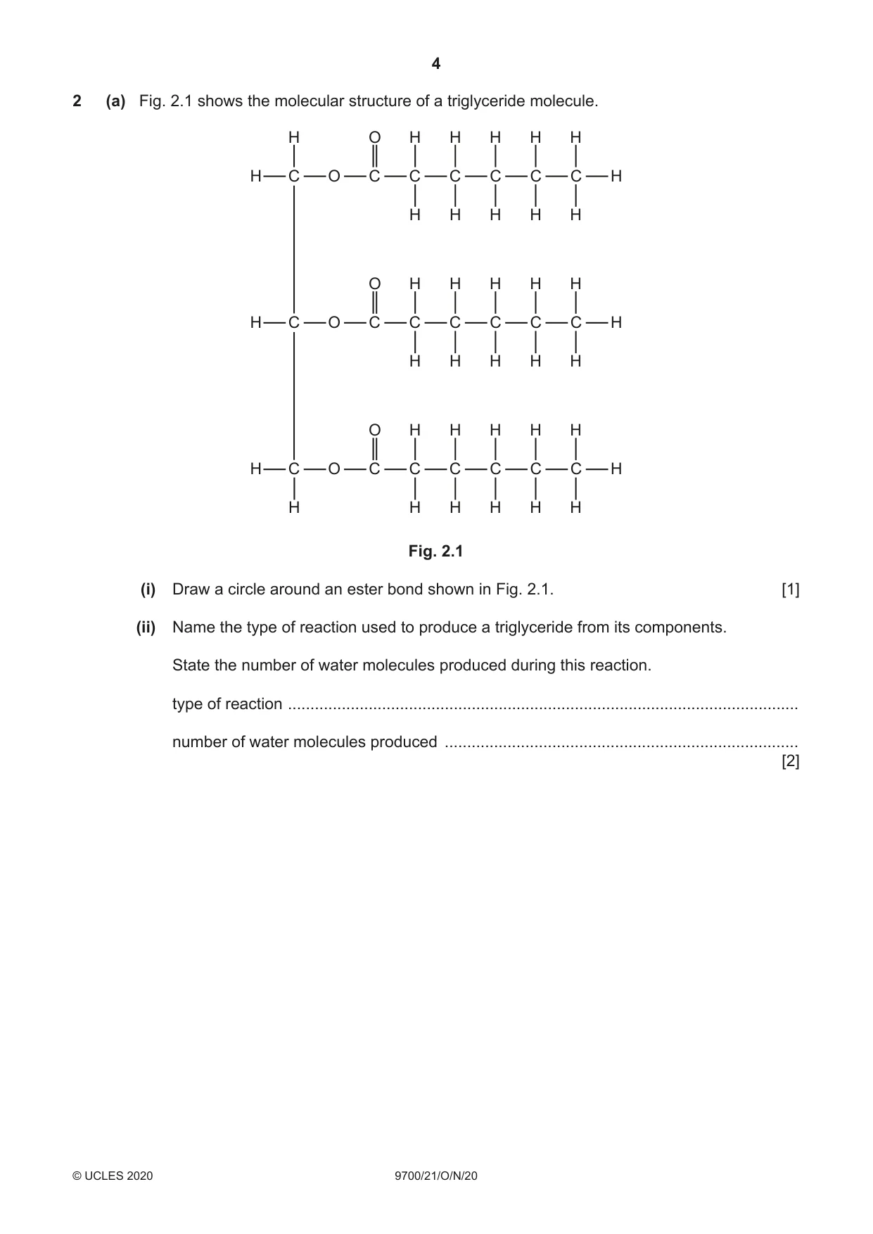
Q2 · The molecular structure of a triglyceride molecule
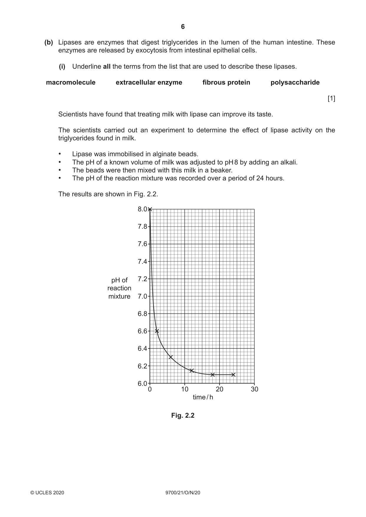
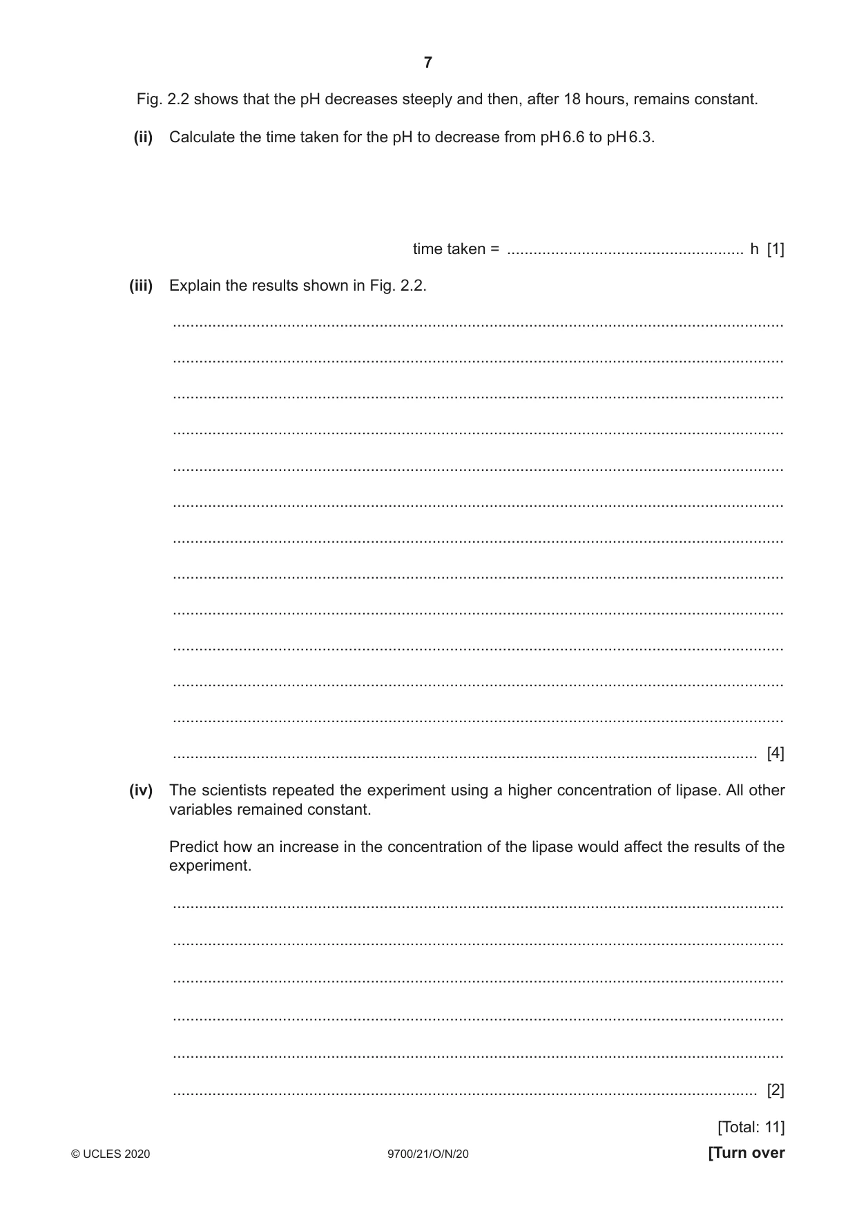
2 (a) Fig. 2.1 shows the molecular structure of a triglyceride molecule. H O H H H H H H C O C C C C C C H H H H H H O H H H H H H C O C C C C C C H H H H H H O H H H H H H C O C C C C C C H H H H H H H Fig. 2.1 (i) Draw a circle around an ester bond shown in Fig. 2.1. [1] (ii) Name the type of reaction used to produce a triglyceride from its components. State the number of water molecules produced during this reaction. type of reaction .................................................................................................................. number of water molecules produced ............................................................................... [2] (b) Lipases are enzymes that digest triglycerides in the lumen of the human intestine. These enzymes are released by exocytosis from intestinal epithelial cells. (i) Underline all the terms from the list that are used to describe these lipases. macromolecule extracellular enzyme fibrous protein polysaccharide [1] Scientists have found that treating milk with lipase can improve its taste. The scientists carried out an experiment to determine the effect of lipase activity on the triglycerides found in milk. • Lipase was immobilised in alginate beads. • The pH of a known volume of milk was adjusted to pH 8 by adding an alkali. • The beads were then mixed with this milk in a beaker. • The pH of the reaction mixture was recorded over a period of 24 hours. The results are shown in Fig. 2.2. 8.0 7.8 7.6 7.4 pH of 7.2 reaction mixture 7.0 6.8 6.6 6.4 6.2 6.0 0 10 20 30 time / h Fig. 2.2 Fig. 2.2 shows that the pH decreases steeply and then, after 18 hours, remains constant. (ii) Calculate the time taken for the pH to decrease from pH 6.6 to pH 6.3. time taken = ...................................................... h [1] (iii) Explain the results shown in Fig. 2.2. ........................................................................................................................................... ........................................................................................................................................... ........................................................................................................................................... ........................................................................................................................................... ........................................................................................................................................... ........................................................................................................................................... ........................................................................................................................................... ........................................................................................................................................... ........................................................................................................................................... ........................................................................................................................................... ........................................................................................................................................... ........................................................................................................................................... ..................................................................................................................................... [4] (iv) The scientists repeated the experiment using a higher concentration of lipase. All other variables remained constant. Predict how an increase in the concentration of the lipase would affect the results of the experiment. ........................................................................................................................................... ........................................................................................................................................... ........................................................................................................................................... ........................................................................................................................................... ........................................................................................................................................... ..................................................................................................................................... [2] [Total: 11]
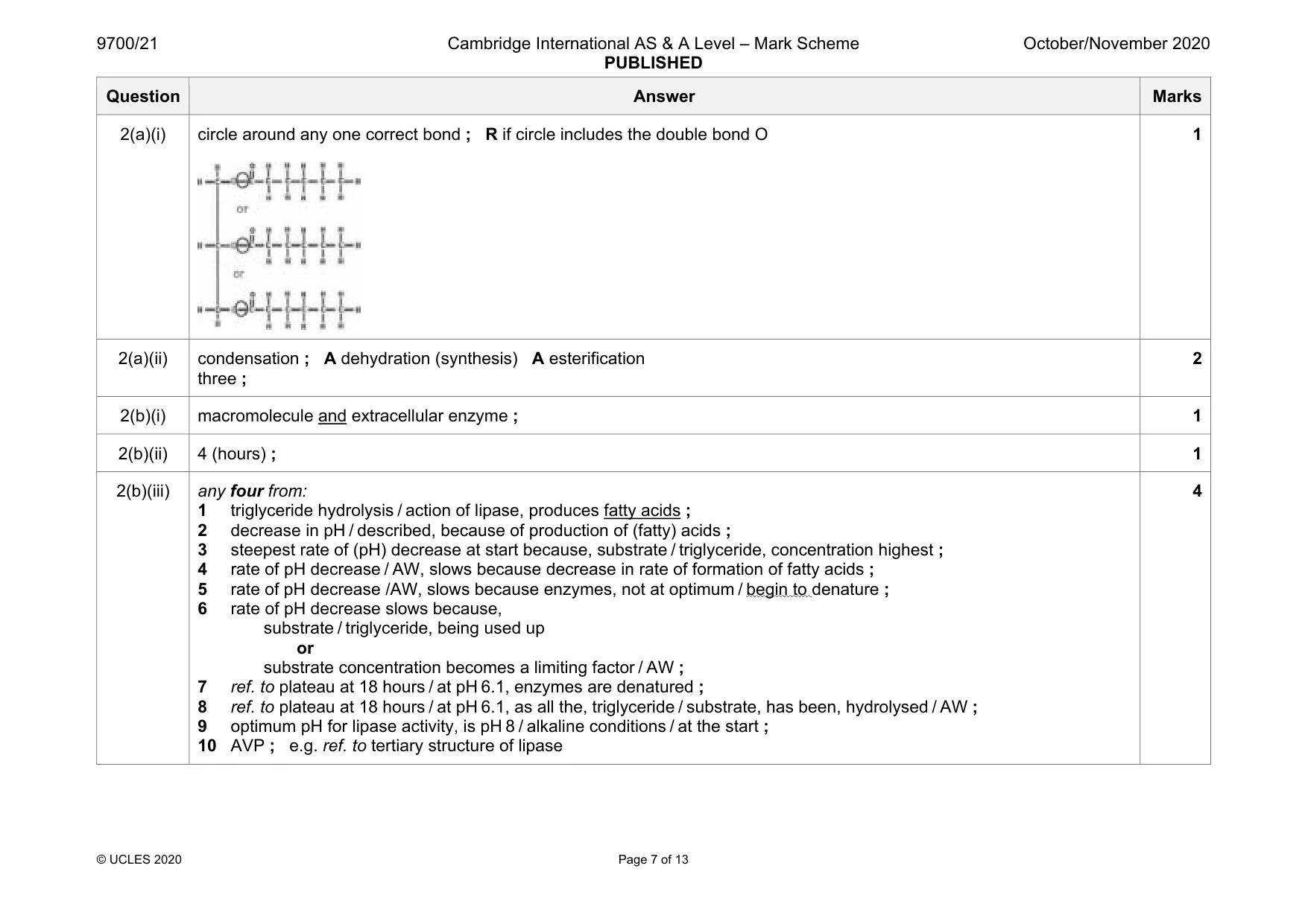
Mark scheme: 2(a)(i) circle around any one correct bond ; R if circle includes the double bond O 1 2(a)(ii) condensation ; A dehydration (synthesis) A esterification three ; 2 2(b)(i) macromolecule and extracellular enzyme ; 1 2(b)(ii) 4 (hours) ; 1 2(b)(iii) any four from: 1 triglyceride hydrolysis / action of lipase, produces fatty acids ; 2 decrease in pH / described, because of production of (fatty) acids ; 3 steepest rate of (pH) decrease at start because, substrate / triglyceride, concentration highest ; 4 rate of pH decrease / AW, slows because decrease in rate of formation of fatty acids ; 5 rate of pH decrease /AW, slows because enzymes, not at optimum / begin to denature ; 6 rate of pH decrease slows because, substrate / triglyceride, being used up or substrate concentration becomes a limiting factor / AW ; 7 ref. to plateau at 18 hours / at pH 6.1, enzymes are denatured ; 8 ref. to plateau at 18 hours / at pH 6.1, as all the, triglyceride / substrate, has been, hydrolysed / AW ; 9 optimum pH for lipase activity, is pH 8 / alkaline conditions / at the start ; 10 AVP ; e.g. ref. to tertiary structure of lipase 4 Question Answer Marks 2(b)(iv) any two from: (initial) decrease in pH would be more rapid ; plateau would be reached earlier ; AW plateau would be at the same pH (as the previous experiment) ; 2
Q3 · The circulatory system of mammals is a double circulation
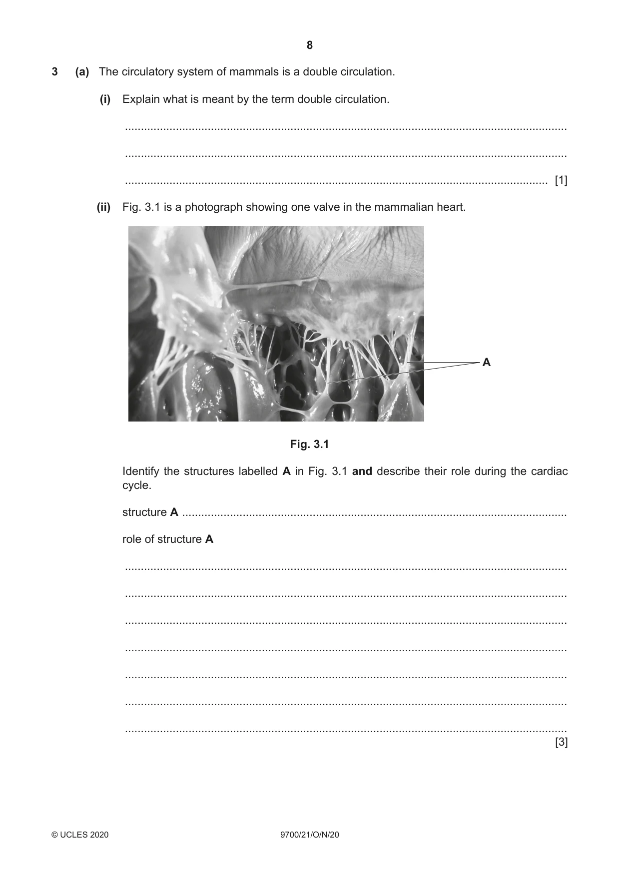
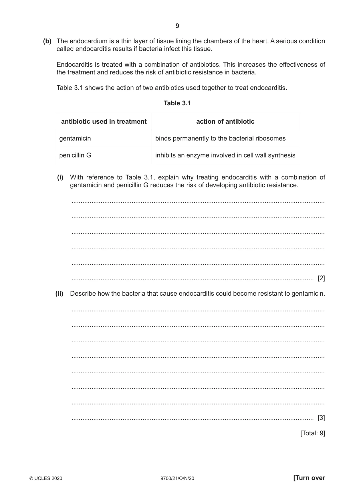
3 (a) The circulatory system of mammals is a double circulation. (i) Explain what is meant by the term double circulation. ........................................................................................................................................... ........................................................................................................................................... ..................................................................................................................................... [1] (ii) Fig. 3.1 is a photograph showing one valve in the mammalian heart. A Fig. 3.1 Identify the structures labelled A in Fig. 3.1 and describe their role during the cardiac cycle. structure A ......................................................................................................................... role of structure A ........................................................................................................................................... ........................................................................................................................................... ........................................................................................................................................... ........................................................................................................................................... ........................................................................................................................................... ........................................................................................................................................... ........................................................................................................................................... [3] (b) The endocardium is a thin layer of tissue lining the chambers of the heart. A serious condition called endocarditis results if bacteria infect this tissue. Endocarditis is treated with a combination of antibiotics. This increases the effectiveness of the treatment and reduces the risk of antibiotic resistance in bacteria. Table 3.1 shows the action of two antibiotics used together to treat endocarditis. Table 3.1 antibiotic used in treatment action of antibiotic gentamicin binds permanently to the bacterial ribosomes penicillin G inhibits an enzyme involved in cell wall synthesis (i) With reference to Table 3.1, explain why treating endocarditis with a combination of gentamicin and penicillin G reduces the risk of developing antibiotic resistance. ........................................................................................................................................... ........................................................................................................................................... ........................................................................................................................................... ........................................................................................................................................... ........................................................................................................................................... ..................................................................................................................................... [2] (ii) Describe how the bacteria that cause endocarditis could become resistant to gentamicin. ........................................................................................................................................... ........................................................................................................................................... ........................................................................................................................................... ........................................................................................................................................... ........................................................................................................................................... ........................................................................................................................................... ........................................................................................................................................... ..................................................................................................................................... [3] [Total: 9]
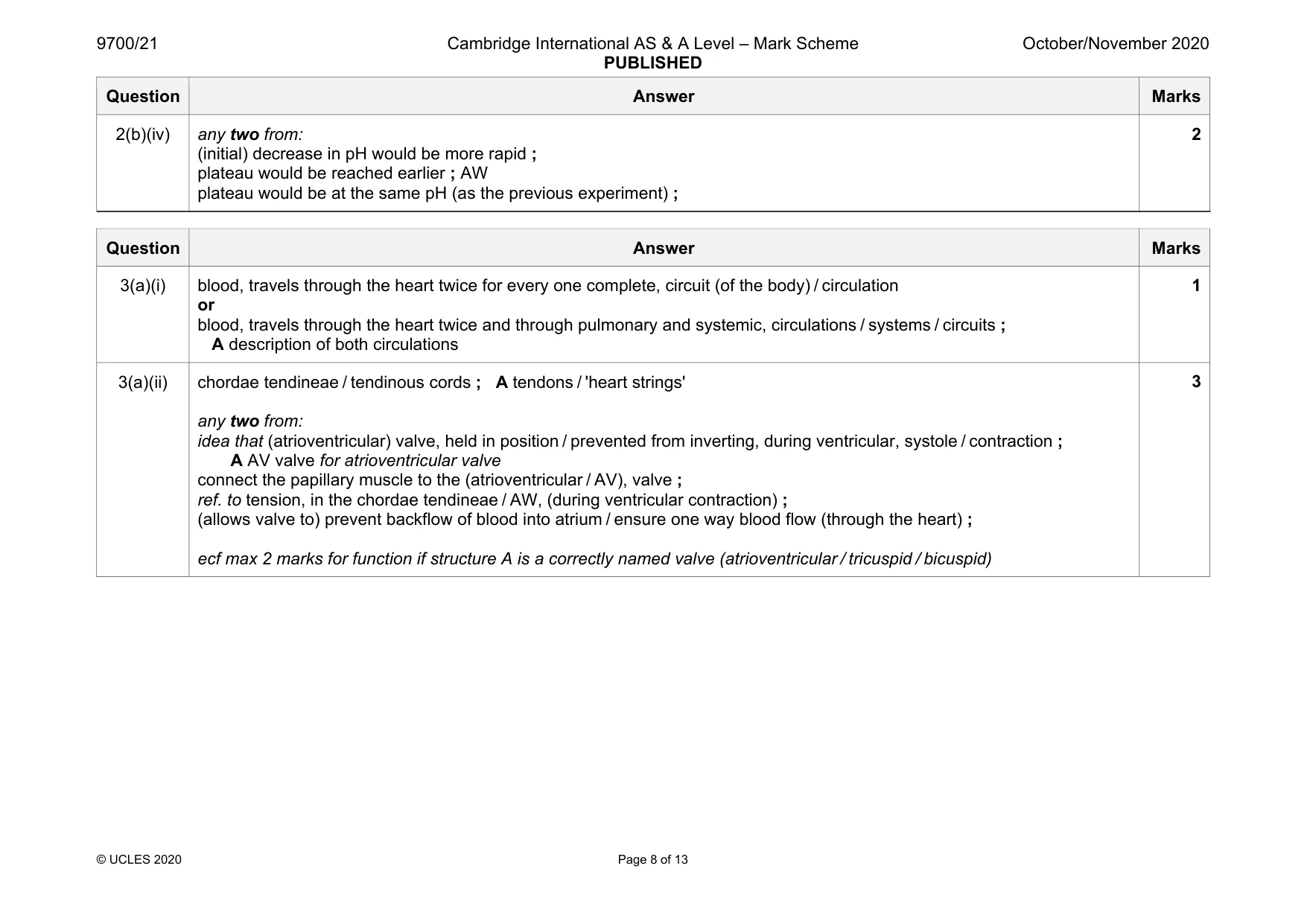
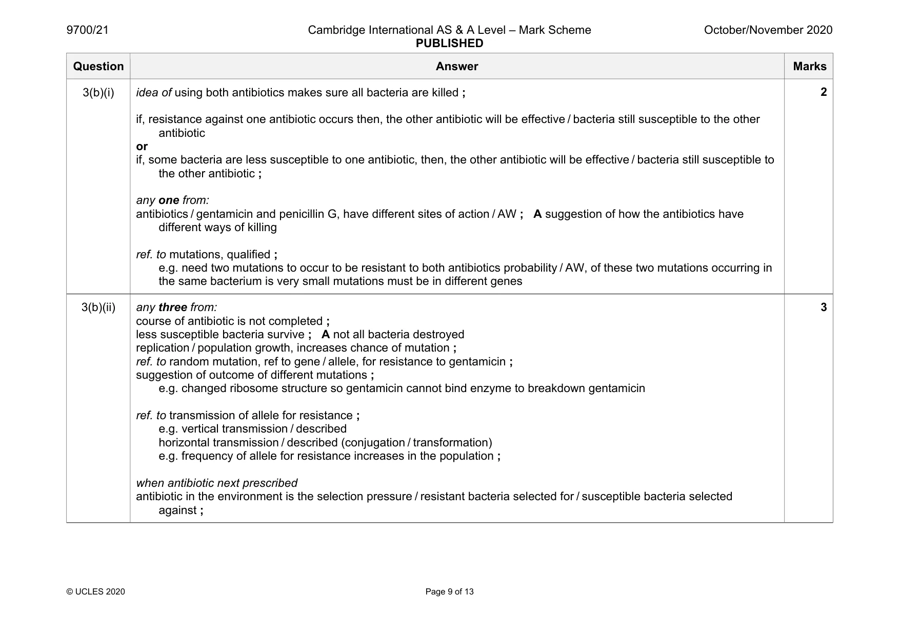
Mark scheme: 3(a)(i) blood, travels through the heart twice for every one complete, circuit (of the body) / circulation or blood, travels through the heart twice and through pulmonary and systemic, circulations / systems / circuits ; A description of both circulations 1 3(a)(ii) chordae tendineae / tendinous cords ; A tendons / 'heart strings' any two from: idea that (atrioventricular) valve, held in position / prevented from inverting, during ventricular, systole / contraction ; A AV valve for atrioventricular valve connect the papillary muscle to the (atrioventricular / AV), valve ; ref. to tension, in the chordae tendineae / AW, (during ventricular contraction) ; (allows valve to) prevent backflow of blood into atrium / ensure one way blood flow (through the heart) ; ecf max 2 marks for function if structure A is a correctly named valve (atrioventricular / tricuspid / bicuspid) 3 Question Answer Marks 3(b)(i) idea of using both antibiotics makes sure all bacteria are killed ; if, resistance against one antibiotic occurs then, the other antibiotic will be effective / bacteria still susceptible to the other antibiotic or if, some bacteria are less susceptible to one antibiotic, then, the other antibiotic will be effective / bacteria still susceptible to the other antibiotic ; any one from: antibiotics / gentamicin and penicillin G, have different sites of action / AW ; A suggestion of how the antibiotics have different ways of killing ref. to mutations, qualified ; e.g. need two mutations to occur to be resistant to both antibiotics probability / AW, of these two mutations occurring in the same bacterium is very small mutations must be in different genes 2 3(b)(ii) any three from: course of antibiotic is not completed ; less susceptible bacteria survive ; A not all bacteria destroyed replication / population growth, increases chance of mutation ; ref. to random mutation, ref to gene / allele, for resistance to gentamicin ; suggestion of outcome of different mutations ; e.g. changed ribosome structure so gentamicin cannot bind enzyme to breakdown gentamicin ref. to transmission of allele for resistance ; e.g. vertical transmission / described horizontal transmission / described (conjugation / transformation) e.g. frequency of allele for resistance increases in the population ; when antibiotic next prescribed antibiotic in the environment is the selection pressure / resistant bacteria selected for / susceptible bacteria selected against ; 3
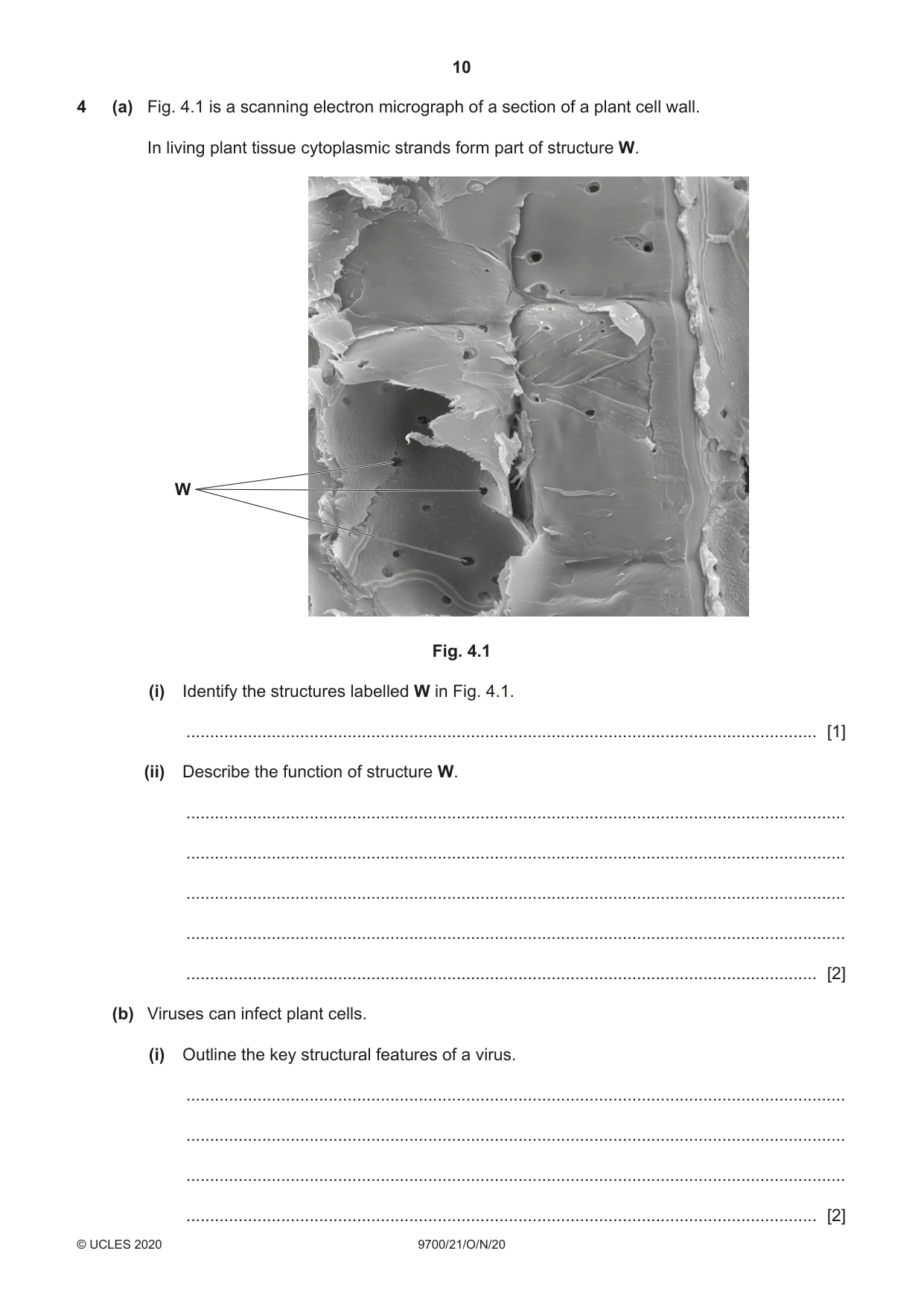
Q4 · A scanning electron micrograph of a section of a plant cell wall
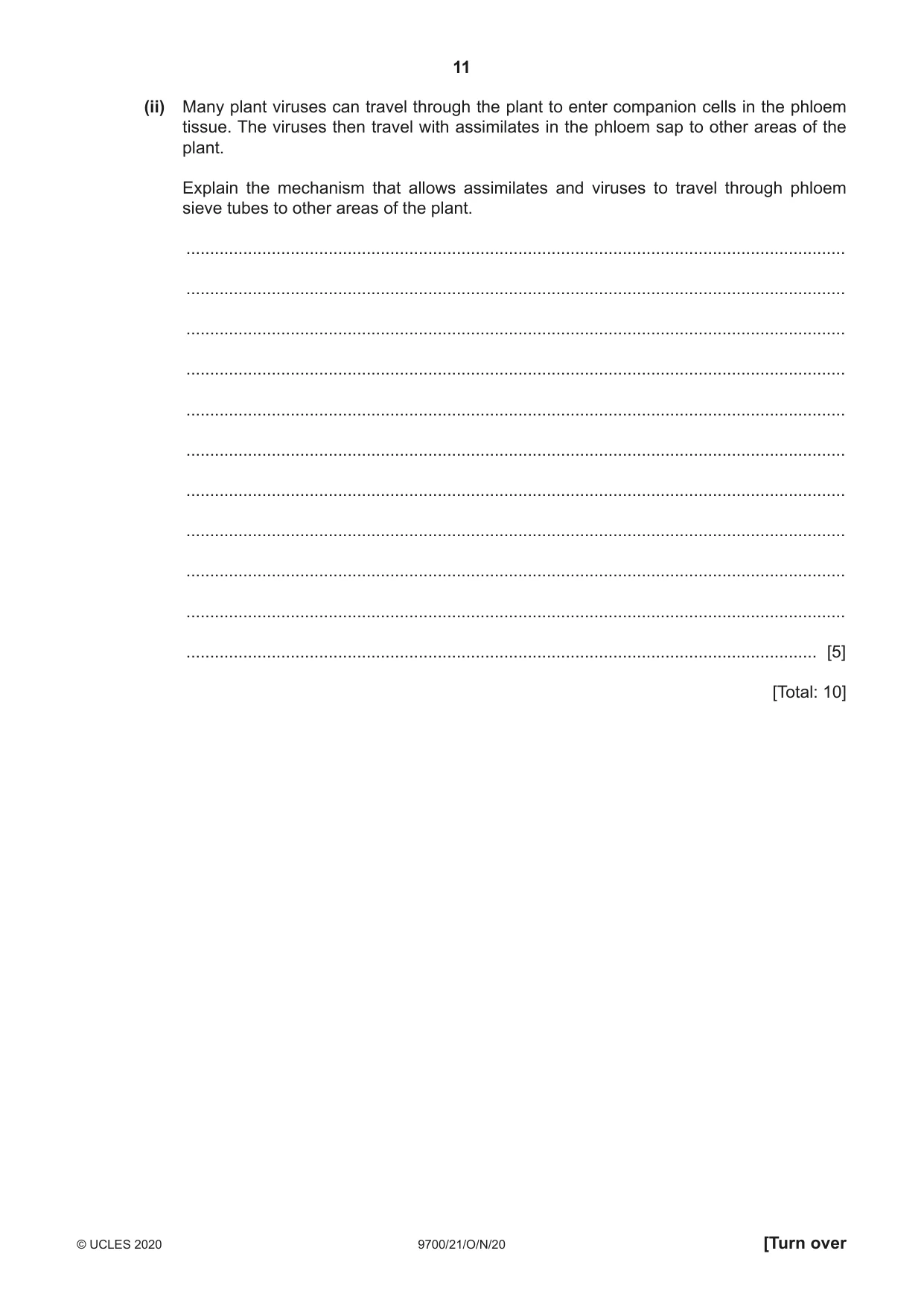
4 (a) Fig. 4.1 is a scanning electron micrograph of a section of a plant cell wall. In living plant tissue cytoplasmic strands form part of structure W. W Fig. 4.1 (i) Identify the structures labelled W in Fig. 4.1. ..................................................................................................................................... [1] (ii) Describe the function of structure W. ........................................................................................................................................... ........................................................................................................................................... ........................................................................................................................................... ........................................................................................................................................... ..................................................................................................................................... [2] (b) Viruses can infect plant cells. (i) Outline the key structural features of a virus. ........................................................................................................................................... ........................................................................................................................................... ........................................................................................................................................... ..................................................................................................................................... [2] (ii) Many plant viruses can travel through the plant to enter companion cells in the phloem tissue. The viruses then travel with assimilates in the phloem sap to other areas of the plant. Explain the mechanism that allows assimilates and viruses to travel through phloem sieve tubes to other areas of the plant. ........................................................................................................................................... ........................................................................................................................................... ........................................................................................................................................... ........................................................................................................................................... ........................................................................................................................................... ........................................................................................................................................... ........................................................................................................................................... ........................................................................................................................................... ........................................................................................................................................... ........................................................................................................................................... ..................................................................................................................................... [5] [Total: 10]
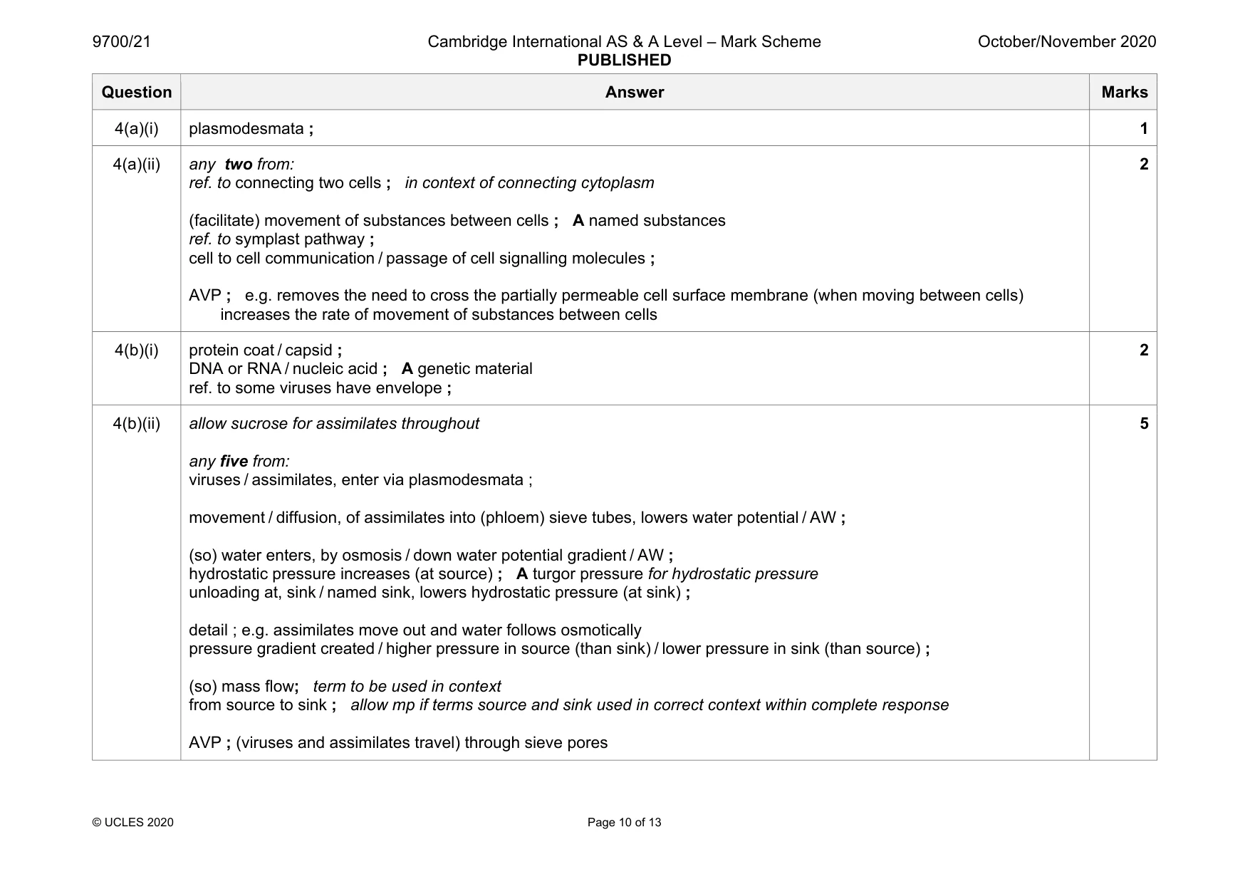
Mark scheme: 4(a)(i) plasmodesmata ; 1 4(a)(ii) any two from: ref. to connecting two cells ; in context of connecting cytoplasm (facilitate) movement of substances between cells ; A named substances ref. to symplast pathway ; cell to cell communication / passage of cell signalling molecules ; AVP ; e.g. removes the need to cross the partially permeable cell surface membrane (when moving between cells) increases the rate of movement of substances between cells 2 4(b)(i) protein coat / capsid ; DNA or RNA / nucleic acid ; A genetic material ref. to some viruses have envelope ; 2 4(b)(ii) allow sucrose for assimilates throughout any five from: viruses / assimilates, enter via plasmodesmata ; movement / diffusion, of assimilates into (phloem) sieve tubes, lowers water potential / AW ; (so) water enters, by osmosis / down water potential gradient / AW ; hydrostatic pressure increases (at source) ; A turgor pressure for hydrostatic pressure unloading at, sink / named sink, lowers hydrostatic pressure (at sink) ; detail ; e.g. assimilates move out and water follows osmotically pressure gradient created / higher pressure in source (than sink) / lower pressure in sink (than source) ; (so) mass flow; term to be used in context from source to sink ; allow mp if terms source and sink used in correct context within complete response AVP ; (viruses and assimilates travel) through sieve pores 5
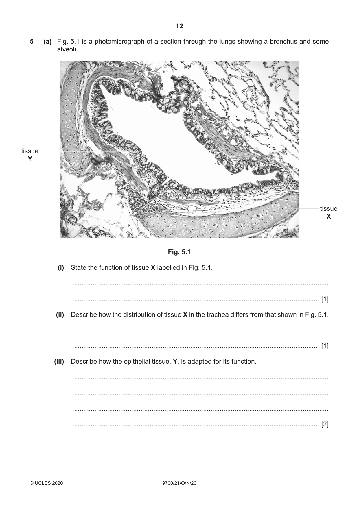
Q5 · A photomicrograph of a section through the lungs showing a bronchus and some alveoli
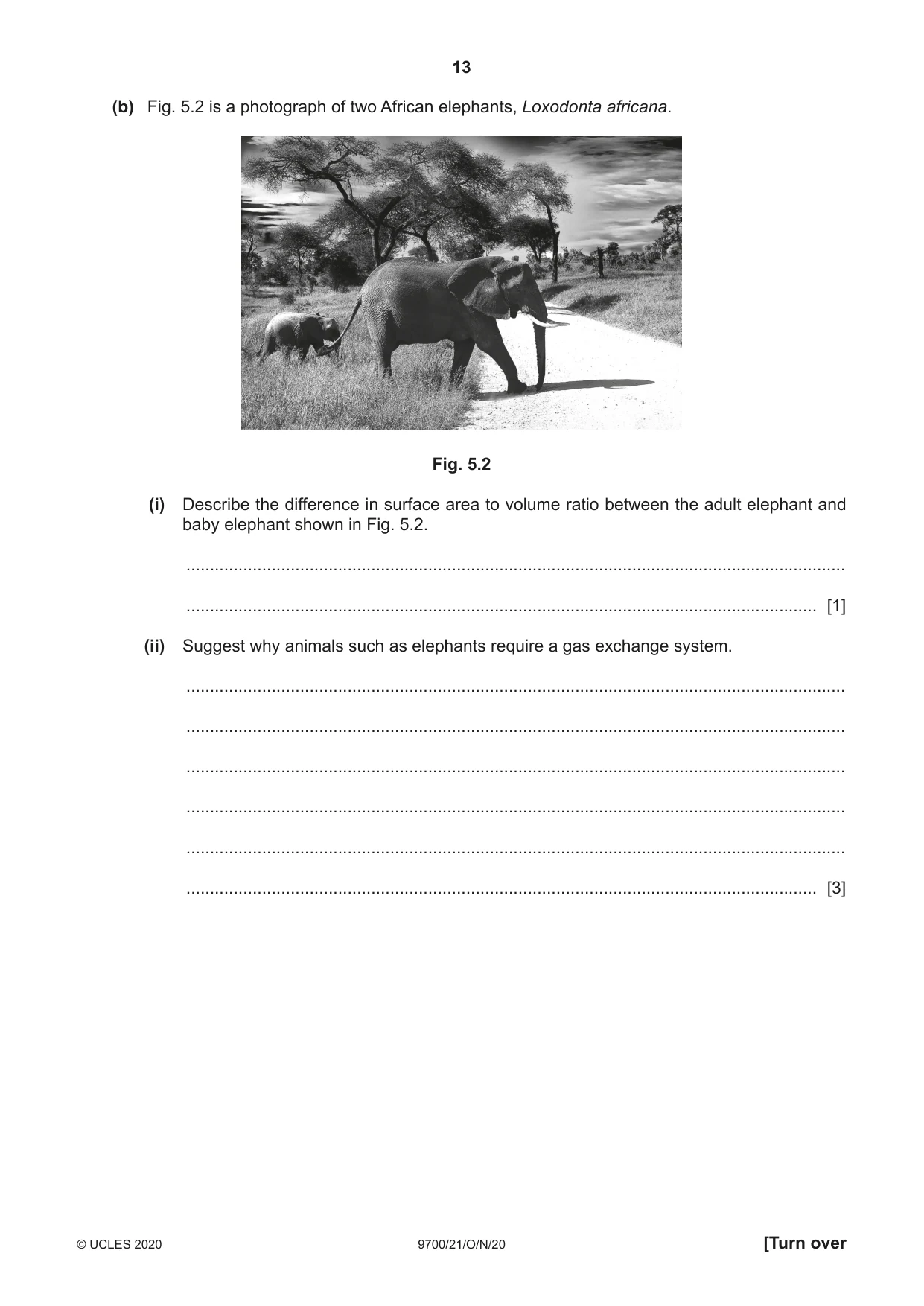
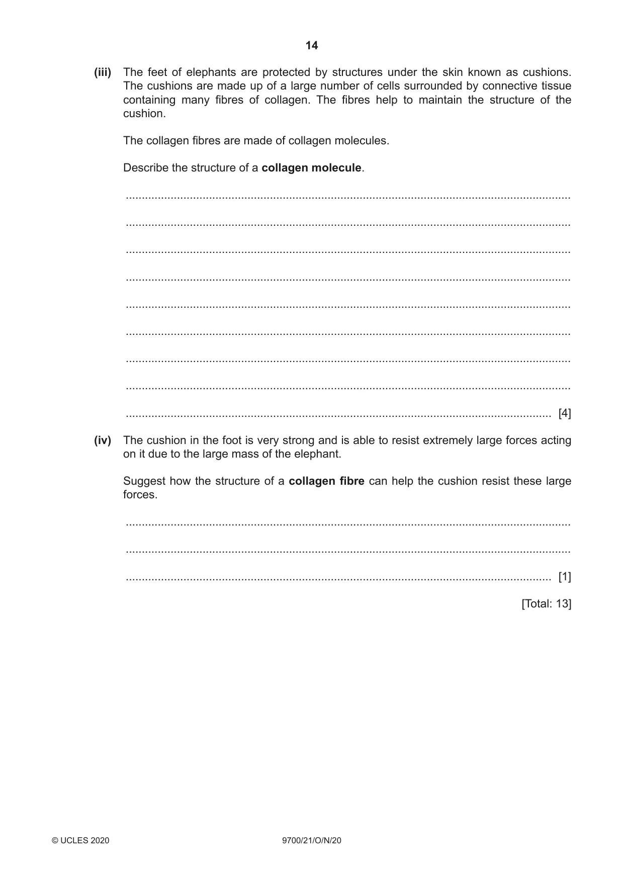
5 (a) Fig. 5.1 is a photomicrograph of a section through the lungs showing a bronchus and some alveoli. tissue Y tissue X Fig. 5.1 (i) State the function of tissue X labelled in Fig. 5.1. ........................................................................................................................................... ..................................................................................................................................... [1] (ii) Describe how the distribution of tissue X in the trachea differs from that shown in Fig. 5.1. ........................................................................................................................................... ..................................................................................................................................... [1] (iii) Describe how the epithelial tissue, Y, is adapted for its function. ........................................................................................................................................... ........................................................................................................................................... ........................................................................................................................................... ..................................................................................................................................... [2] (b) Fig. 5.2 is a photograph of two African elephants, Loxodonta africana. Fig. 5.2 (i) Describe the difference in surface area to volume ratio between the adult elephant and baby elephant shown in Fig. 5.2. ........................................................................................................................................... ..................................................................................................................................... [1] (ii) Suggest why animals such as elephants require a gas exchange system. ........................................................................................................................................... ........................................................................................................................................... ........................................................................................................................................... ........................................................................................................................................... ........................................................................................................................................... ..................................................................................................................................... [3] (iii) The feet of elephants are protected by structures under the skin known as cushions. The cushions are made up of a large number of cells surrounded by connective tissue containing many fibres of collagen. The fibres help to maintain the structure of the cushion. The collagen fibres are made of collagen molecules. Describe the structure of a collagen molecule. ........................................................................................................................................... ........................................................................................................................................... ........................................................................................................................................... ........................................................................................................................................... ........................................................................................................................................... ........................................................................................................................................... ........................................................................................................................................... ........................................................................................................................................... ..................................................................................................................................... [4] (iv) The cushion in the foot is very strong and is able to resist extremely large forces acting on it due to the large mass of the elephant. Suggest how the structure of a collagen fibre can help the cushion resist these large forces. ........................................................................................................................................... ........................................................................................................................................... ..................................................................................................................................... [1] [Total: 13]
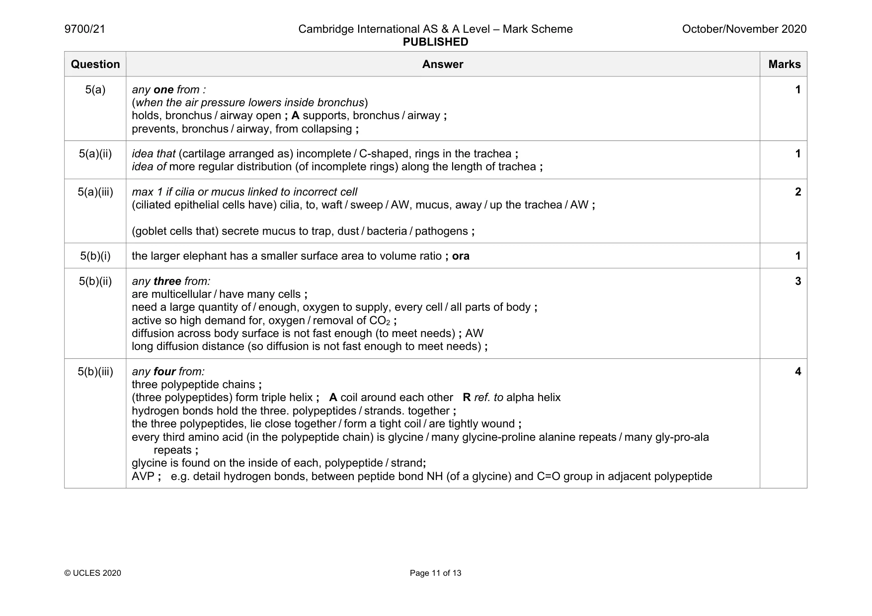
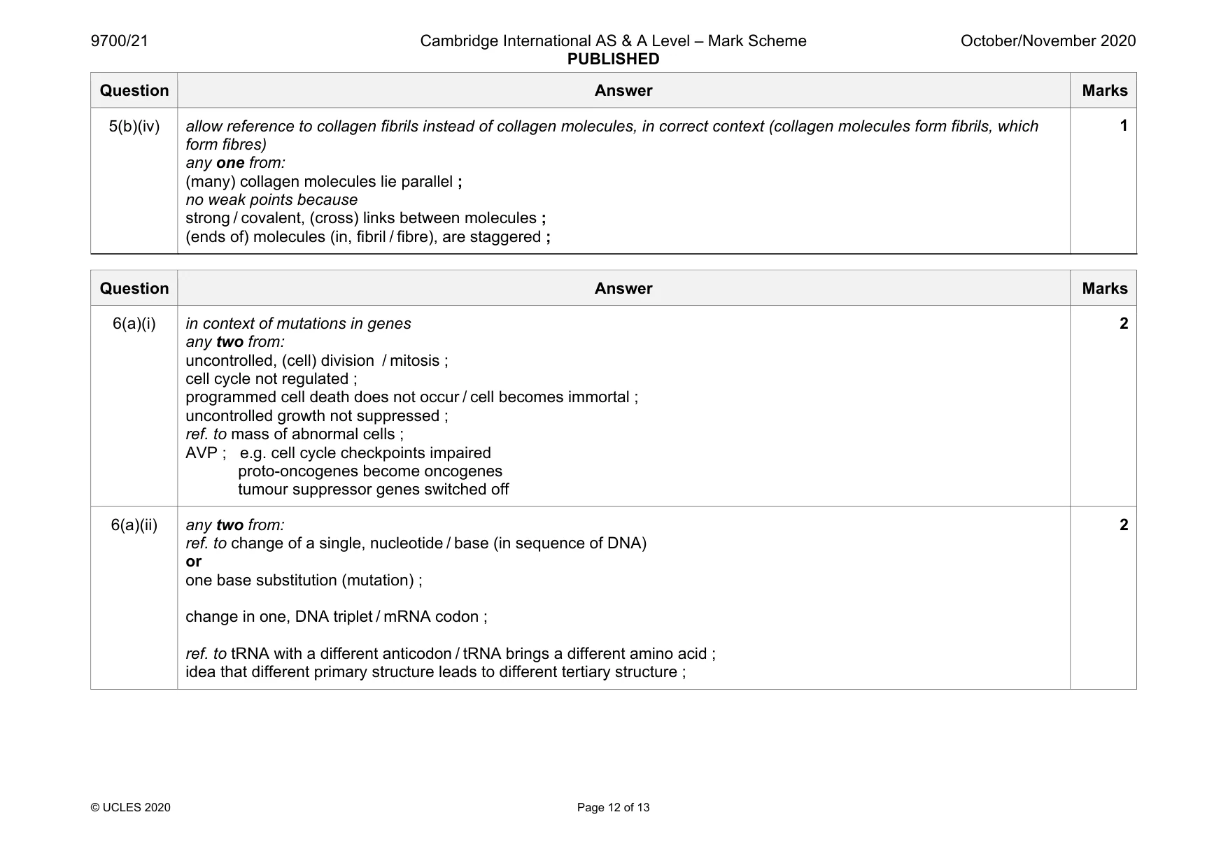
Mark scheme: 5(a) any one from : (when the air pressure lowers inside bronchus) holds, bronchus / airway open ; A supports, bronchus / airway ; prevents, bronchus / airway, from collapsing ; 1 5(a)(ii) idea that (cartilage arranged as) incomplete / C-shaped, rings in the trachea ; idea of more regular distribution (of incomplete rings) along the length of trachea ; 1 5(a)(iii) max 1 if cilia or mucus linked to incorrect cell (ciliated epithelial cells have) cilia, to, waft / sweep / AW, mucus, away / up the trachea / AW ; (goblet cells that) secrete mucus to trap, dust / bacteria / pathogens ; 2 5(b)(i) the larger elephant has a smaller surface area to volume ratio ; ora 1 5(b)(ii) any three from: are multicellular / have many cells ; need a large quantity of / enough, oxygen to supply, every cell / all parts of body ; active so high demand for, oxygen / removal of CO2 ; diffusion across body surface is not fast enough (to meet needs) ; AW long diffusion distance (so diffusion is not fast enough to meet needs) ; 3 5(b)(iii) any four from: three polypeptide chains ; (three polypeptides) form triple helix ; A coil around each other R ref. to alpha helix hydrogen bonds hold the three. polypeptides / strands. together ; the three polypeptides, lie close together / form a tight coil / are tightly wound ; every third amino acid (in the polypeptide chain) is glycine / many glycine-proline alanine repeats / many gly-pro-ala repeats ; glycine is found on the inside of each, polypeptide / strand; AVP ; e.g. detail hydrogen bonds, between peptide bond NH (of a glycine) and C=O group in adjacent polypeptide 4 Question Answer Marks 5(b)(iv) allow reference to collagen fibrils instead of collagen molecules, in correct context (collagen molecules form fibrils, which form fibres) any one from: (many) collagen molecules lie parallel ; no weak points because strong / covalent, (cross) links between molecules ; (ends of) molecules (in, fibril / fibre), are staggered ; 1
Q6 · Mutations in body cells can sometimes result in a tumour
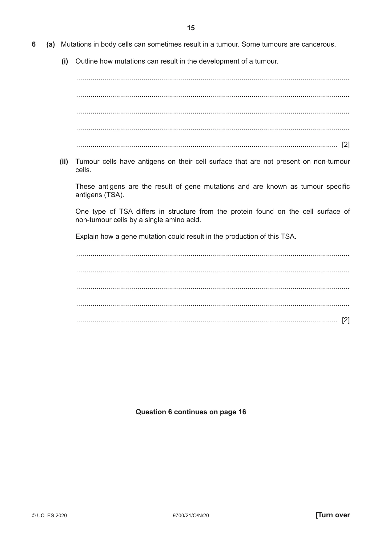
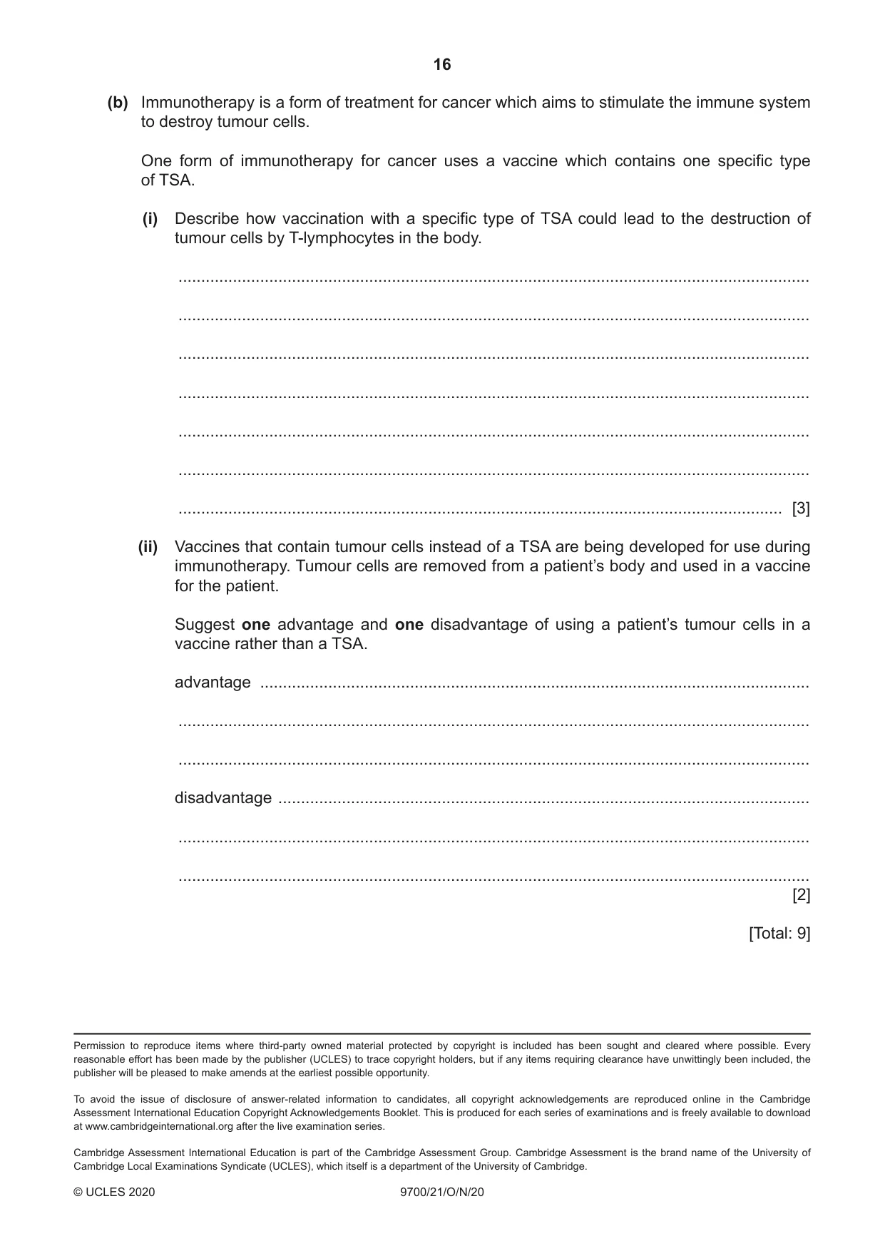
6 (a) Mutations in body cells can sometimes result in a tumour. Some tumours are cancerous. (i) Outline how mutations can result in the development of a tumour. ........................................................................................................................................... ........................................................................................................................................... ........................................................................................................................................... ........................................................................................................................................... ..................................................................................................................................... [2] (ii) Tumour cells have antigens on their cell surface that are not present on non-tumour cells. These antigens are the result of gene mutations and are known as tumour specific antigens (TSA). One type of TSA differs in structure from the protein found on the cell surface of non-tumour cells by a single amino acid. Explain how a gene mutation could result in the production of this TSA. ........................................................................................................................................... ........................................................................................................................................... ........................................................................................................................................... ........................................................................................................................................... ..................................................................................................................................... [2] Question 6 continues on page 16 (b) Immunotherapy is a form of treatment for cancer which aims to stimulate the immune system to destroy tumour cells. One form of immunotherapy for cancer uses a vaccine which contains one specific type of TSA. (i) Describe how vaccination with a specific type of TSA could lead to the destruction of tumour cells by T-lymphocytes in the body. ........................................................................................................................................... ........................................................................................................................................... ........................................................................................................................................... ........................................................................................................................................... ........................................................................................................................................... ........................................................................................................................................... ..................................................................................................................................... [3] (ii) Vaccines that contain tumour cells instead of a TSA are being developed for use during immunotherapy. Tumour cells are removed from a patient’s body and used in a vaccine for the patient. Suggest one advantage and one disadvantage of using a patient’s tumour cells in a vaccine rather than a TSA. advantage ......................................................................................................................... ........................................................................................................................................... ........................................................................................................................................... disadvantage ..................................................................................................................... ........................................................................................................................................... ........................................................................................................................................... [2] [Total: 9]
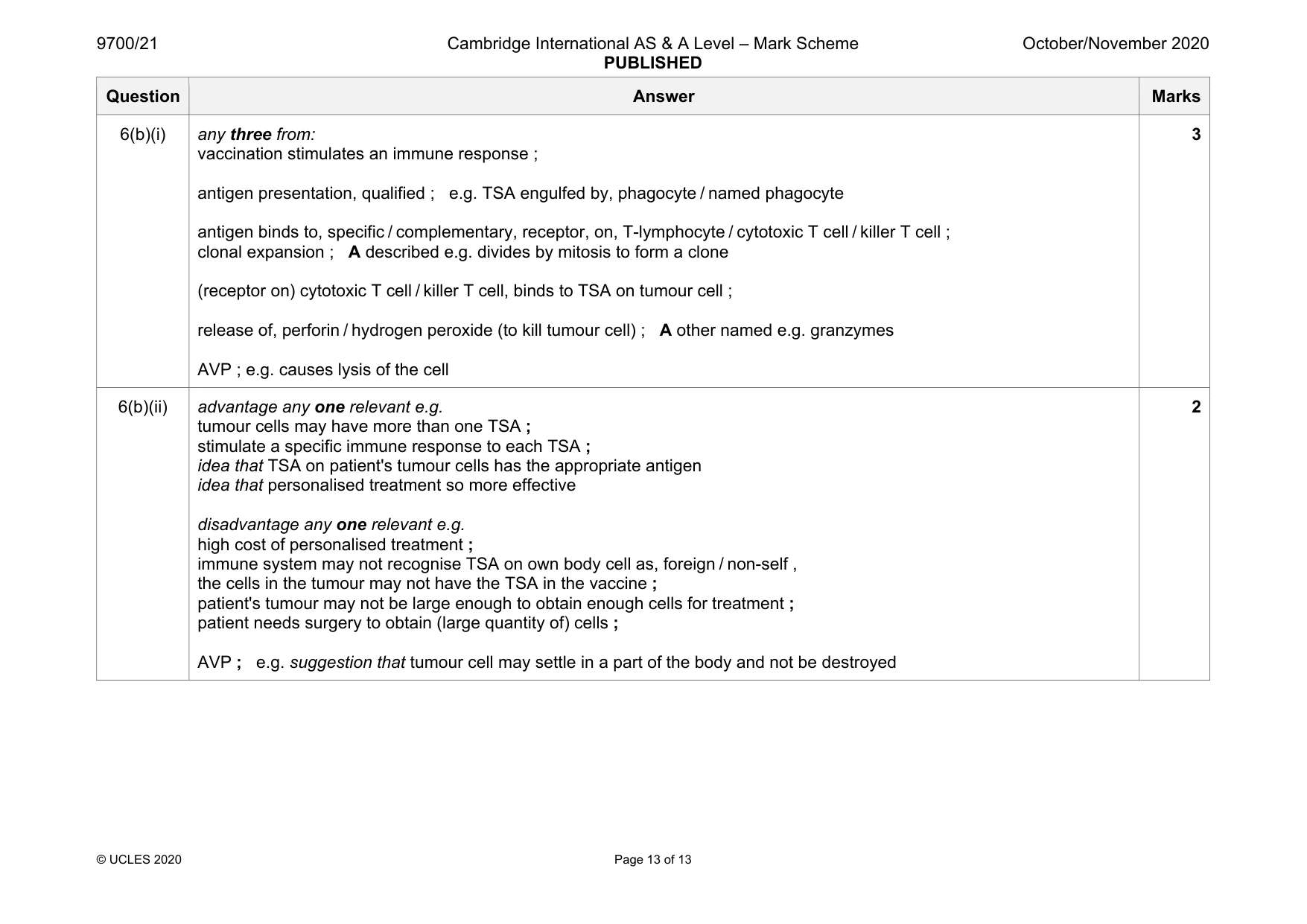
Mark scheme: 6(a)(i) in context of mutations in genes any two from: uncontrolled, (cell) division / mitosis ; cell cycle not regulated ; programmed cell death does not occur / cell becomes immortal ; uncontrolled growth not suppressed ; ref. to mass of abnormal cells ; AVP ; e.g. cell cycle checkpoints impaired proto-oncogenes become oncogenes tumour suppressor genes switched off 2 6(a)(ii) any two from: ref. to change of a single, nucleotide / base (in sequence of DNA) or one base substitution (mutation) ; change in one, DNA triplet / mRNA codon ; ref. to tRNA with a different anticodon / tRNA brings a different amino acid ; idea that different primary structure leads to different tertiary structure ; 2 Question Answer Marks 6(b)(i) any three from: vaccination stimulates an immune response ; antigen presentation, qualified ; e.g. TSA engulfed by, phagocyte / named phagocyte antigen binds to, specific / complementary, receptor, on, T-lymphocyte / cytotoxic T cell / killer T cell ; clonal expansion ; A described e.g. divides by mitosis to form a clone (receptor on) cytotoxic T cell / killer T cell, binds to TSA on tumour cell ; release of, perforin / hydrogen peroxide (to kill tumour cell) ; A other named e.g. granzymes AVP ; e.g. causes lysis of the cell 3 6(b)(ii) advantage any one relevant e.g. tumour cells may have more than one TSA ; stimulate a specific immune response to each TSA ; idea that TSA on patient's tumour cells has the appropriate antigen idea that personalised treatment so more effective disadvantage any one relevant e.g. high cost of personalised treatment ; immune system may not recognise TSA on own body cell as, foreign / non-self , the cells in the tumour may not have the TSA in the vaccine ; patient's tumour may not be large enough to obtain enough cells for treatment ; patient needs surgery to obtain (large quantity of) cells ; AVP ; e.g. suggestion that tumour cell may settle in a part of the body and not be destroyed 2
What was in this paper
The subtopics covered by these 6 questions, and how many questions each got. Open one in a new tab to see every Cambridge question on it.
What you needed in this session
Cambridge’s own grade thresholds for 2020 Oct/Nov, Paper 2 · Variant 1. A higher threshold means an easier paper — the bar moves with how the cohort did.