Cambridge A Level Biology 9700 — 2020 Oct/Nov Paper 2 · Variant 2
9700/22/O/N/20 · 6 questions · 60 marks · ≈68 min
The question paper and its mark scheme, free to read here and free to download. This is Cambridge’s own paper, exactly as it was sat.
Question paper20 pages
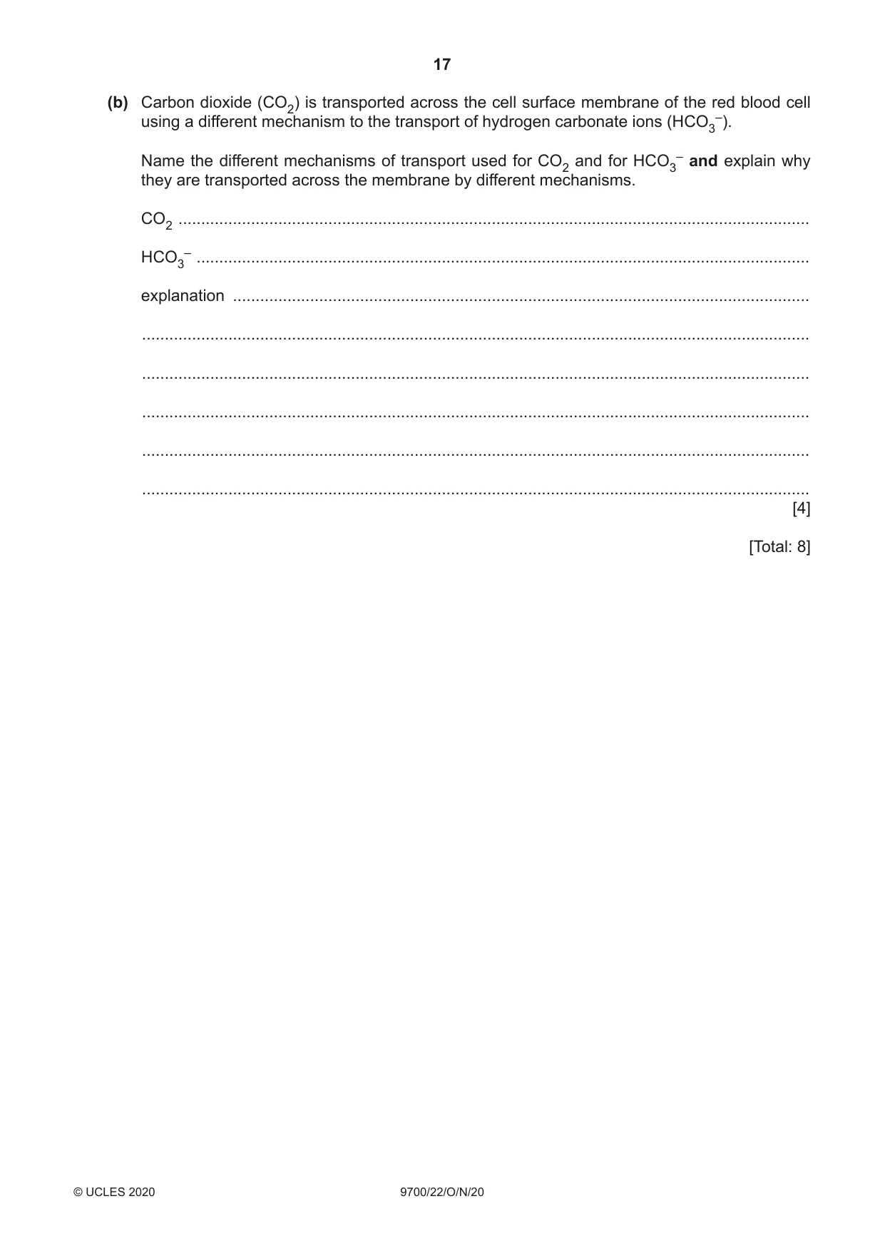


Mark scheme14 pages
Answers below. Sit the paper first if you are practising.

Questions as text
Q1 · A diagram drawn from a photomicrograph of a transverse section through part of a leaf
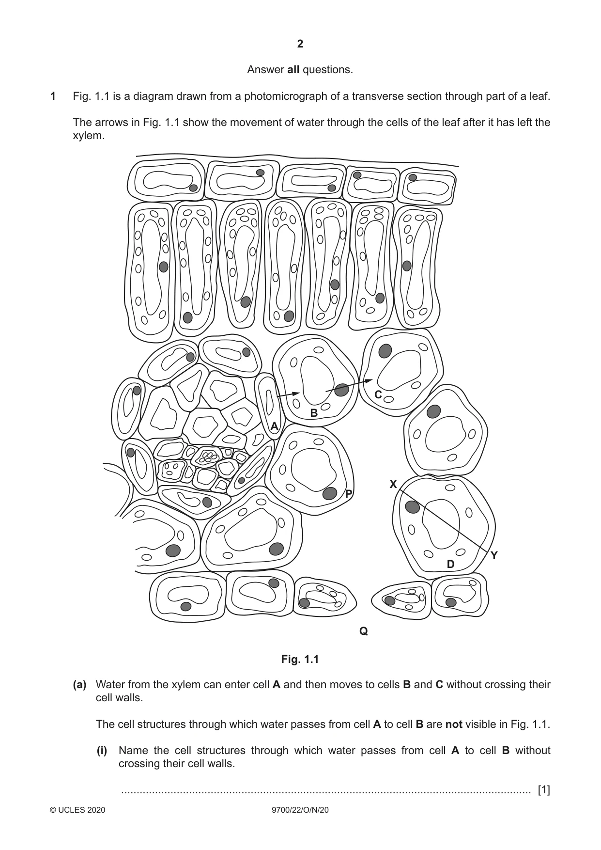
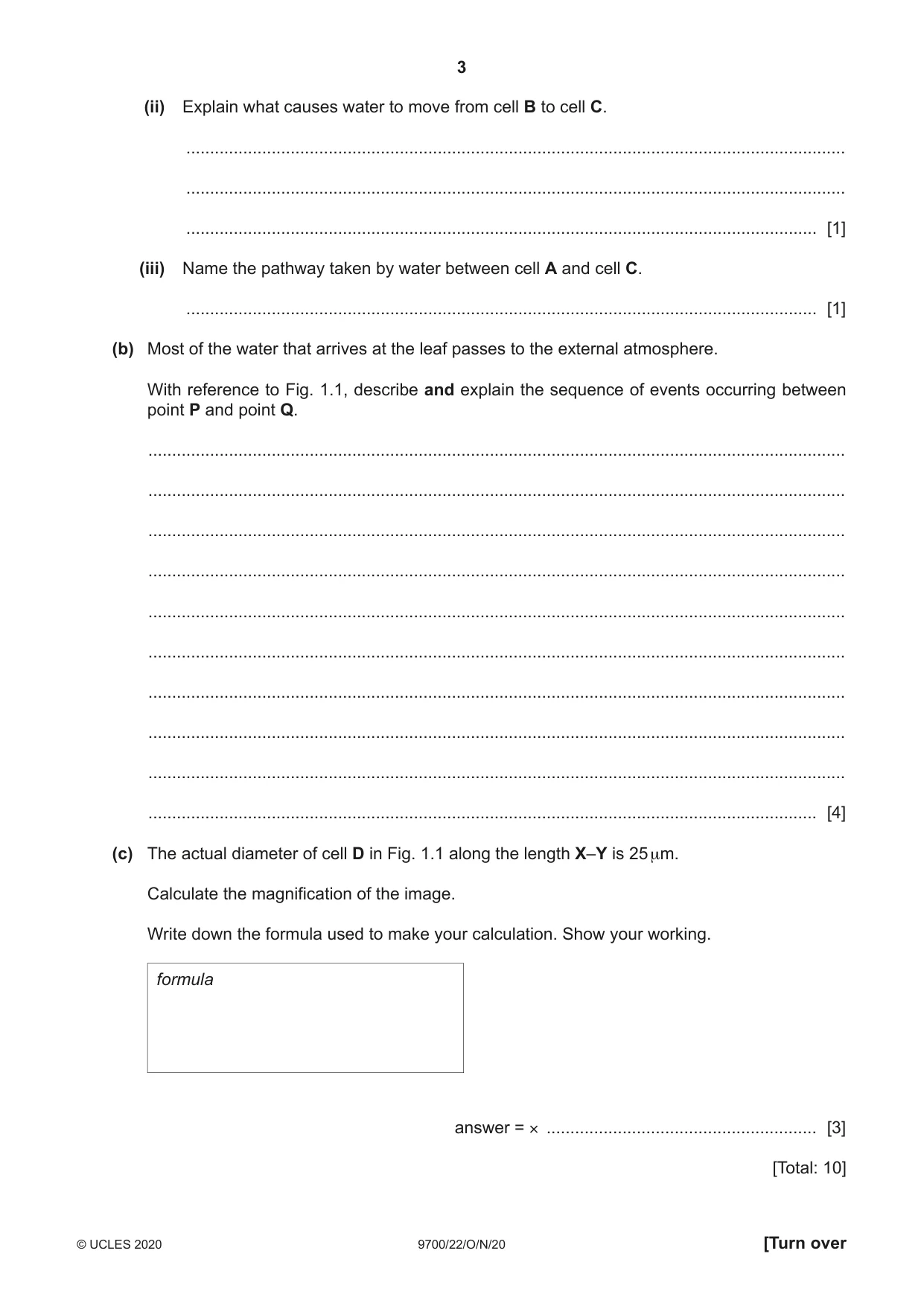
1 Fig. 1.1 is a diagram drawn from a photomicrograph of a transverse section through part of a leaf. The arrows in Fig. 1.1 show the movement of water through the cells of the leaf after it has left the xylem. C B A X P Y D Q Fig. 1.1 (a) Water from the xylem can enter cell A and then moves to cells B and C without crossing their cell walls. The cell structures through which water passes from cell A to cell B are not visible in Fig. 1.1. (i) Name the cell structures through which water passes from cell A to cell B without crossing their cell walls. ..................................................................................................................................... [1] (ii) Explain what causes water to move from cell B to cell C. ........................................................................................................................................... ........................................................................................................................................... ..................................................................................................................................... [1] (iii) Name the pathway taken by water between cell A and cell C. ..................................................................................................................................... [1] (b) Most of the water that arrives at the leaf passes to the external atmosphere. With reference to Fig. 1.1, describe and explain the sequence of events occurring between point P and point Q. ................................................................................................................................................... ................................................................................................................................................... ................................................................................................................................................... ................................................................................................................................................... ................................................................................................................................................... ................................................................................................................................................... ................................................................................................................................................... ................................................................................................................................................... ................................................................................................................................................... ............................................................................................................................................. [4] (c) The actual diameter of cell D in Fig. 1.1 along the length X–Y is 25 µm. Calculate the magnification of the image. Write down the formula used to make your calculation. Show your working. formula answer = × ......................................................... [3] [Total: 10]
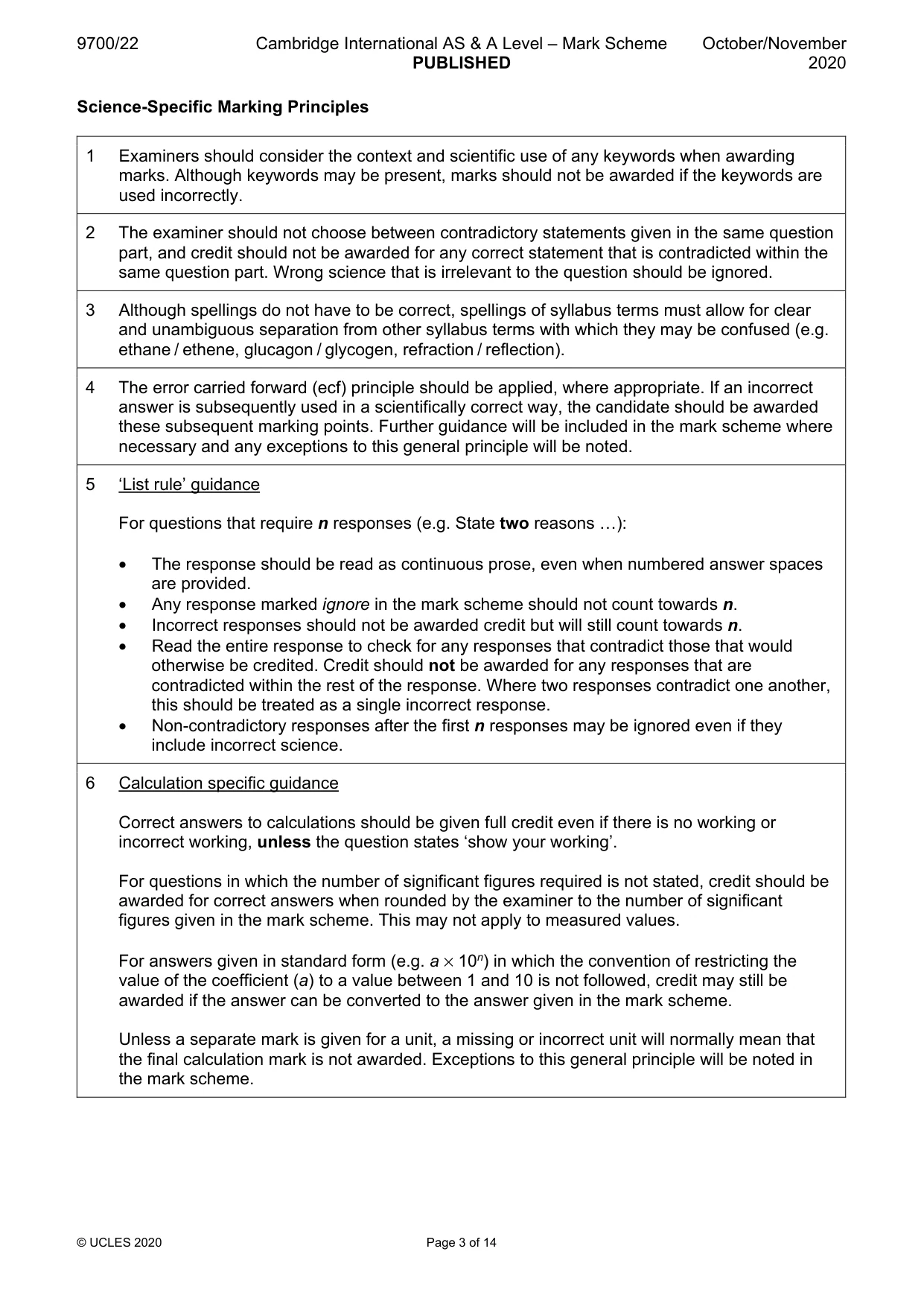
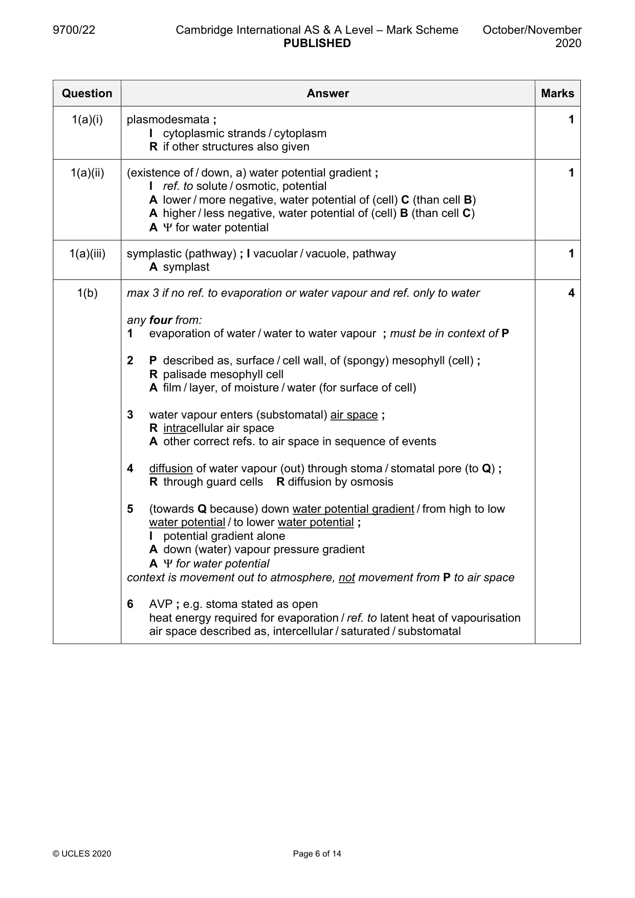
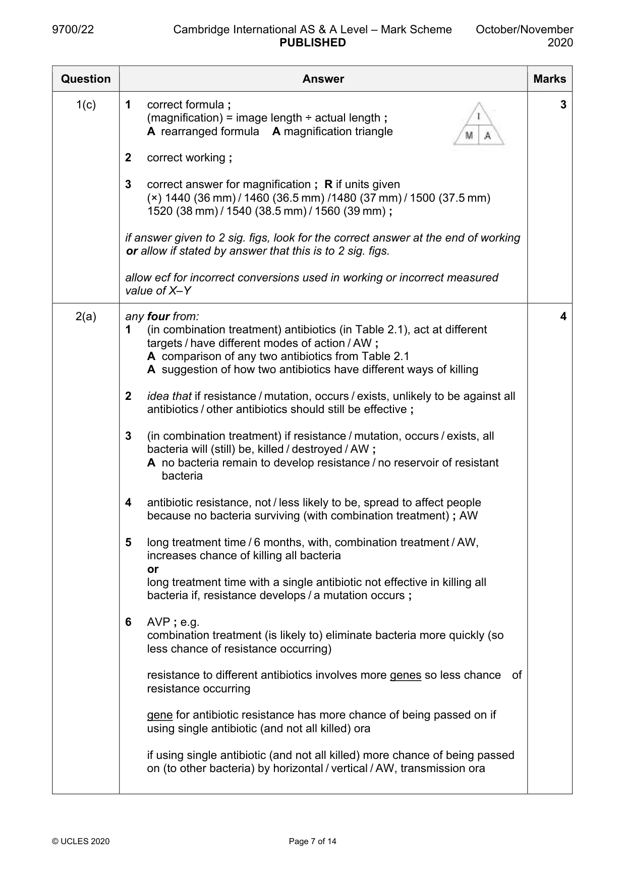
Mark scheme: Question Answer Marks 1(a)(i) plasmodesmata ; 1 I cytoplasmic strands / cytoplasm R if other structures also given 1(a)(ii) (existence of / down, a) water potential gradient ; 1 I ref. to solute / osmotic, potential A lower / more negative, water potential of (cell) C (than cell B) A higher / less negative, water potential of (cell) B (than cell C) A Ψ for water potential 1(a)(iii) symplastic (pathway) ; I vacuolar / vacuole, pathway 1 A symplast 1(b) max 3 if no ref. to evaporation or water vapour and ref. only to water 4 any four from: 1 evaporation of water / water to water vapour ; must be in context of P 2 P described as, surface / cell wall, of (spongy) mesophyll (cell) ; R palisade mesophyll cell A film / layer, of moisture / water (for surface of cell) 3 water vapour enters (substomatal) air space ; R intracellular air space A other correct refs. to air space in sequence of events 4 diffusion of water vapour (out) through stoma / stomatal pore (to Q) ; R through guard cells R diffusion by osmosis 5 (towards Q because) down water potential gradient / from high to low water potential / to lower water potential ; I potential gradient alone A down (water) vapour pressure gradient A Ψ for water potential context is movement out to atmosphere, not movement from P to air space 6 AVP ; e.g. stoma stated as open heat energy required for evaporation / ref. to latent heat of vapourisation air space described as, intercellular / saturated / substomatal 1(c) 1 correct formula ; 3 (magnification) = image length ÷ actual length ; A rearranged formula A magnification triangle 2 correct working ; 3 correct answer for magnification ; R if units given (×) 1440 (36 mm) / 1460 (36.5 mm) /1480 (37 mm) / 1500 (37.5 mm) 1520 (38 mm) / 1540 (38.5 mm) / 1560 (39 mm) ; if answer given to 2 sig. figs, look for the correct answer at the end of working or allow if stated by answer that this is to 2 sig. figs. allow ecf for incorrect conversions used in working or incorrect measured value of X–Y
Q2 · The treatment for people with active tuberculosis (TB) lasts six months and involves a…
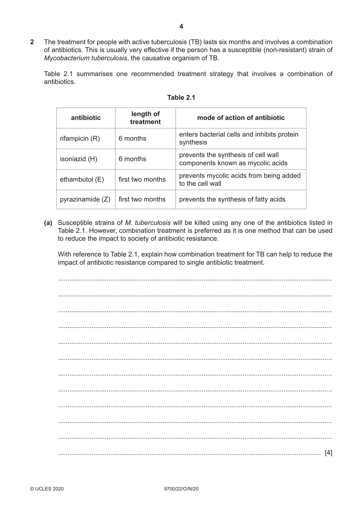
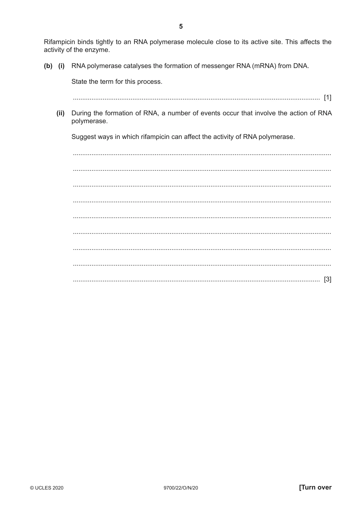
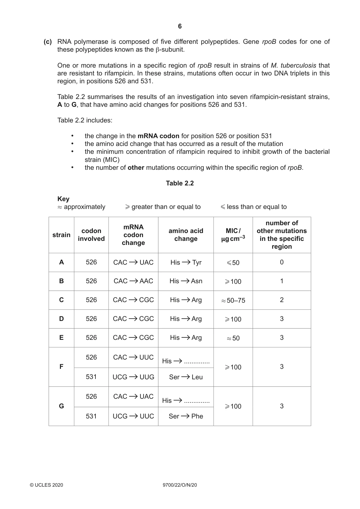
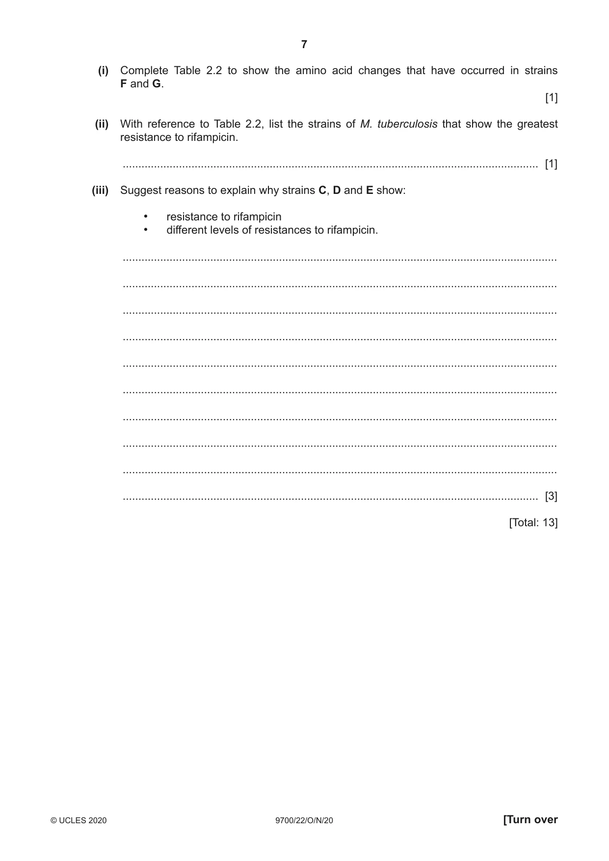
2 The treatment for people with active tuberculosis (TB) lasts six months and involves a combination of antibiotics. This is usually very effective if the person has a susceptible (non-resistant) strain of Mycobacterium tuberculosis, the causative organism of TB. Table 2.1 summarises one recommended treatment strategy that involves a combination of antibiotics. Table 2.1 length of antibiotic mode of action of antibiotic treatment enters bacterial cells and inhibits protein rifampicin (R) 6 months synthesis prevents the synthesis of cell wall isoniazid (H) 6 months components known as mycolic acids prevents mycolic acids from being added ethambutol (E) first two months to the cell wall pyrazinamide (Z) first two months prevents the synthesis of fatty acids (a) Susceptible strains of M. tuberculosis will be killed using any one of the antibiotics listed in Table 2.1. However, combination treatment is preferred as it is one method that can be used to reduce the impact to society of antibiotic resistance. With reference to Table 2.1, explain how combination treatment for TB can help to reduce the impact of antibiotic resistance compared to single antibiotic treatment. ................................................................................................................................................... ................................................................................................................................................... ................................................................................................................................................... ................................................................................................................................................... ................................................................................................................................................... ................................................................................................................................................... ................................................................................................................................................... ................................................................................................................................................... ................................................................................................................................................... ................................................................................................................................................... ................................................................................................................................................... ............................................................................................................................................. [4] Rifampicin binds tightly to an RNA polymerase molecule close to its active site. This affects the activity of the enzyme. (b) (i) RNA polymerase catalyses the formation of messenger RNA (mRNA) from DNA. State the term for this process. ..................................................................................................................................... [1] (ii) During the formation of RNA, a number of events occur that involve the action of RNA polymerase. Suggest ways in which rifampicin can affect the activity of RNA polymerase. ........................................................................................................................................... ........................................................................................................................................... ........................................................................................................................................... ........................................................................................................................................... ........................................................................................................................................... ........................................................................................................................................... ........................................................................................................................................... ........................................................................................................................................... ..................................................................................................................................... [3] (c) RNA polymerase is composed of five different polypeptides. Gene rpoB codes for one of these polypeptides known as the β-subunit. One or more mutations in a specific region of rpoB result in strains of M. tuberculosis that are resistant to rifampicin. In these strains, mutations often occur in two DNA triplets in this region, in positions 526 and 531. Table 2.2 summarises the results of an investigation into seven rifampicin-resistant strains, A to G, that have amino acid changes for positions 526 and 531. Table 2.2 includes: • the change in the mRNA codon for position 526 or position 531 • the amino acid change that has occurred as a result of the mutation • the minimum concentration of rifampicin required to inhibit growth of the bacterial strain (MIC) • the number of other mutations occurring within the specific region of rpoB. Table 2.2 Key . approximately H greater than or equal to G less than or equal to number of mRNA codon amino acid MIC / other mutations strain codon involved change µg cm–3 in the specific change region A 526 CAC UAC His Tyr G 50 0 B 526 CAC AAC His Asn H 100 1 C 526 CAC CGC His Arg . 50–75 2 D 526 CAC CGC His Arg H 100 3 E 526 CAC CGC His Arg . 50 3 526 CAC UUC His .............. F H 100 3 531 UCG UUG Ser Leu 526 CAC UAC His .............. G H 100 3 531 UCG UUC Ser Phe (i) Complete Table 2.2 to show the amino acid changes that have occurred in strains F and G. [1] (ii) With reference to Table 2.2, list the strains of M. tuberculosis that show the greatest resistance to rifampicin. ..................................................................................................................................... [1] (iii) Suggest reasons to explain why strains C, D and E show: • resistance to rifampicin • different levels of resistances to rifampicin. ........................................................................................................................................... ........................................................................................................................................... ........................................................................................................................................... ........................................................................................................................................... ........................................................................................................................................... ........................................................................................................................................... ........................................................................................................................................... ........................................................................................................................................... ........................................................................................................................................... ..................................................................................................................................... [3] [Total: 13]
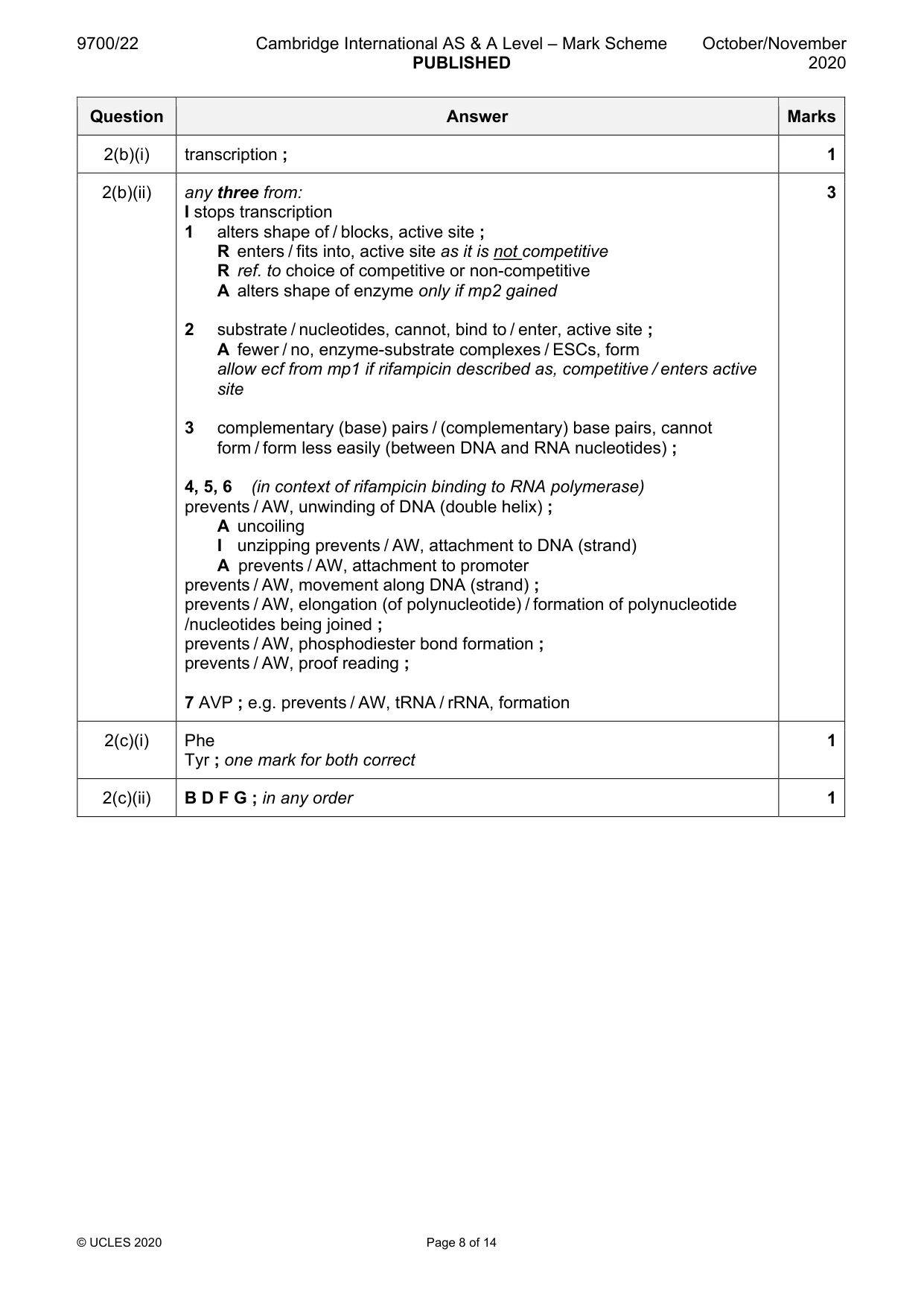
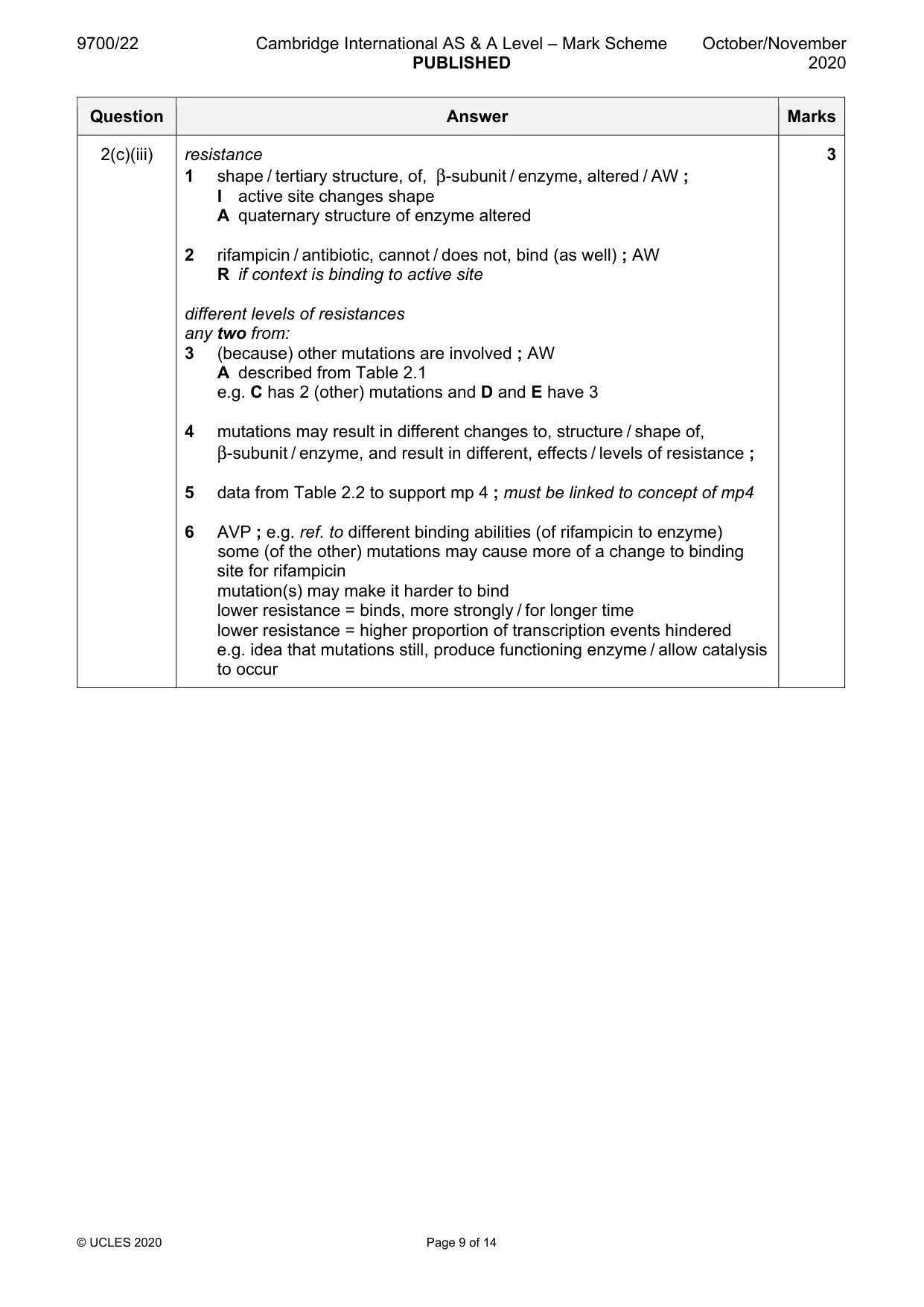
Mark scheme: 2(a) any four from: 4 1 (in combination treatment) antibiotics (in Table 2.1), act at different targets / have different modes of action / AW ; A comparison of any two antibiotics from Table 2.1 A suggestion of how two antibiotics have different ways of killing 2 idea that if resistance / mutation, occurs / exists, unlikely to be against all antibiotics / other antibiotics should still be effective ; 3 (in combination treatment) if resistance / mutation, occurs / exists, all bacteria will (still) be, killed / destroyed / AW ; A no bacteria remain to develop resistance / no reservoir of resistant bacteria 4 antibiotic resistance, not / less likely to be, spread to affect people because no bacteria surviving (with combination treatment) ; AW 5 long treatment time / 6 months, with, combination treatment / AW, increases chance of killing all bacteria or long treatment time with a single antibiotic not effective in killing all bacteria if, resistance develops / a mutation occurs ; 6 AVP ; e.g. combination treatment (is likely to) eliminate bacteria more quickly (so less chance of resistance occurring) resistance to different antibiotics involves more genes so less chance of resistance occurring gene for antibiotic resistance has more chance of being passed on if using single antibiotic (and not all killed) ora if using single antibiotic (and not all killed) more chance of being passed on (to other bacteria) by horizontal / vertical / AW, transmission ora 2(b)(i) transcription ; 1 2(b)(ii) any three from: 3 I stops transcription 1 alters shape of / blocks, active site ; R enters / fits into, active site as it is not competitive R ref. to choice of competitive or non-competitive A alters shape of enzyme only if mp2 gained 2 substrate / nucleotides, cannot, bind to / enter, active site ; A fewer / no, enzyme-substrate complexes / ESCs, form allow ecf from mp1 if rifampicin described as, competitive / enters active site 3 complementary (base) pairs / (complementary) base pairs, cannot form / form less easily (between DNA and RNA nucleotides) ; 4, 5, 6 (in context of rifampicin binding to RNA polymerase) prevents / AW, unwinding of DNA (double helix) ; A uncoiling I unzipping prevents / AW, attachment to DNA (strand) A prevents / AW, attachment to promoter prevents / AW, movement along DNA (strand) ; prevents / AW, elongation (of polynucleotide) / formation of polynucleotide /nucleotides being joined ; prevents / AW, phosphodiester bond formation ; prevents / AW, proof reading ; 7 AVP ; e.g. prevents / AW, tRNA / rRNA, formation 2(c)(i) Phe 1 Tyr ; one mark for both correct 2(c)(ii) B D F G ; in any order 1 2(c)(iii) resistance 3 1 shape / tertiary structure, of, β-subunit / enzyme, altered / AW ; I active site changes shape A quaternary structure of enzyme altered 2 rifampicin / antibiotic, cannot / does not, bind (as well) ; AW R if context is binding to active site different levels of resistances any two from: 3 (because) other mutations are involved ; AW A described from Table 2.1 e.g. C has 2 (other) mutations and D and E have 3 4 mutations may result in different changes to, structure / shape of, β-subunit / enzyme, and result in different, effects / levels of resistance ; 5 data from Table 2.2 to support mp 4 ; must be linked to concept of mp4 6 AVP ; e.g. ref. to different binding abilities (of rifampicin to enzyme) some (of the other) mutations may cause more of a change to binding site for rifampicin mutation(s) may make it harder to bind lower resistance = binds, more strongly / for longer time lower resistance = higher proportion of transcription events hindered e.g. idea that mutations still, produce functioning enzyme / allow catalysis to occur
Q3 · A photomicrograph of a section through lung tissue
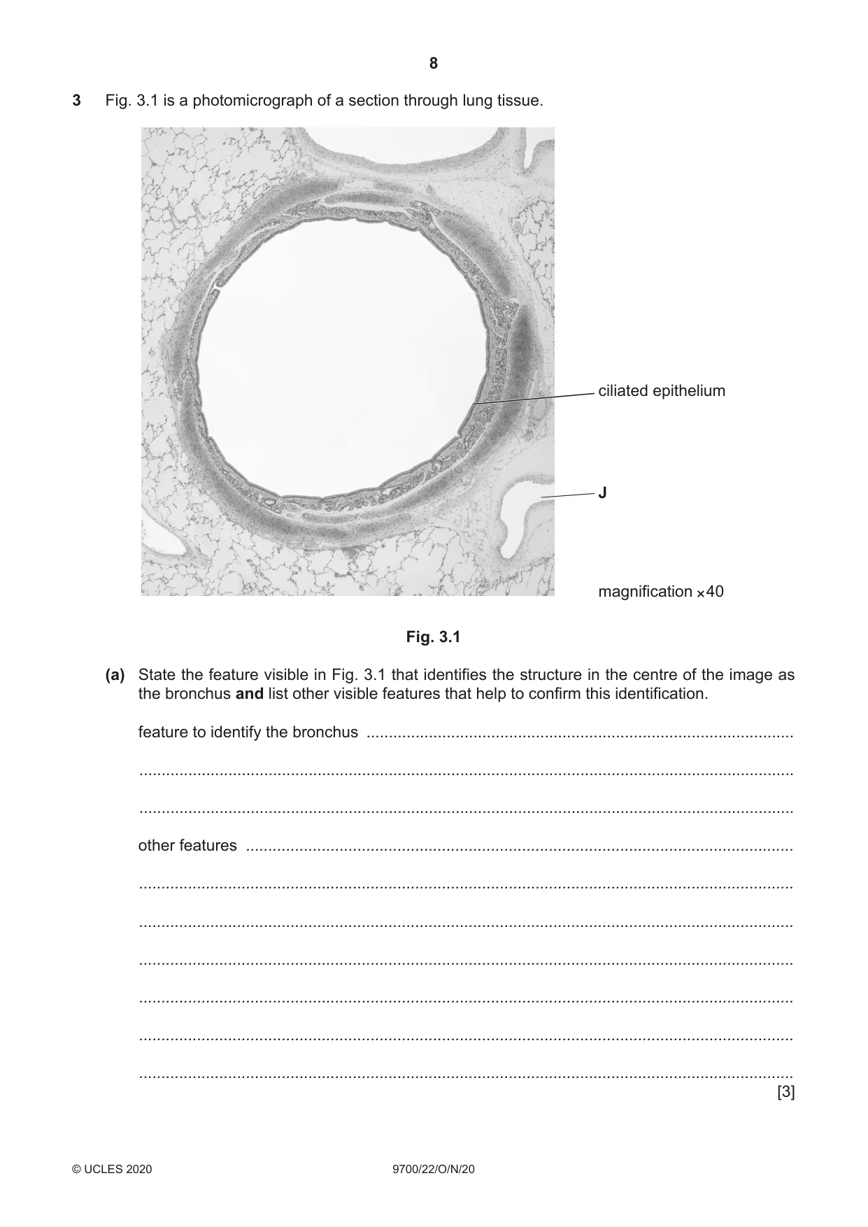
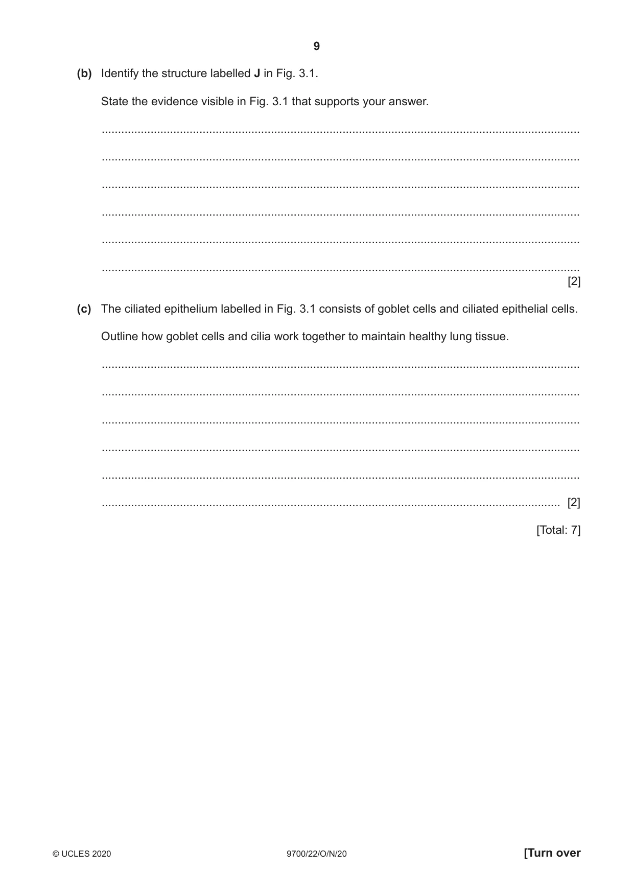
3 Fig. 3.1 is a photomicrograph of a section through lung tissue. ciliated epithelium J magnification × 40 Fig. 3.1 (a) State the feature visible in Fig. 3.1 that identifies the structure in the centre of the image as the bronchus and list other visible features that help to confirm this identification. feature to identify the bronchus ................................................................................................ ................................................................................................................................................... ................................................................................................................................................... other features ........................................................................................................................... ................................................................................................................................................... ................................................................................................................................................... ................................................................................................................................................... ................................................................................................................................................... ................................................................................................................................................... ................................................................................................................................................... [3] (b) Identify the structure labelled J in Fig. 3.1. State the evidence visible in Fig. 3.1 that supports your answer. ................................................................................................................................................... ................................................................................................................................................... ................................................................................................................................................... ................................................................................................................................................... ................................................................................................................................................... ................................................................................................................................................... [2] (c) The ciliated epithelium labelled in Fig. 3.1 consists of goblet cells and ciliated epithelial cells. Outline how goblet cells and cilia work together to maintain healthy lung tissue. ................................................................................................................................................... ................................................................................................................................................... ................................................................................................................................................... ................................................................................................................................................... ................................................................................................................................................... ............................................................................................................................................. [2] [Total: 7]
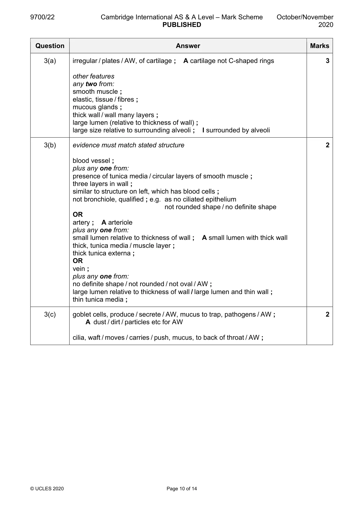
Mark scheme: 3(a) irregular / plates / AW, of cartilage ; A cartilage not C-shaped rings 3 other features any two from: smooth muscle ; elastic, tissue / fibres ; mucous glands ; thick wall / wall many layers ; large lumen (relative to thickness of wall) ; large size relative to surrounding alveoli ; I surrounded by alveoli 3(b) evidence must match stated structure 2 blood vessel ; plus any one from: presence of tunica media / circular layers of smooth muscle ; three layers in wall ; similar to structure on left, which has blood cells ; not bronchiole, qualified ; e.g. as no ciliated epithelium not rounded shape / no definite shape OR artery ; A arteriole plus any one from: small lumen relative to thickness of wall ; A small lumen with thick wall thick, tunica media / muscle layer ; thick tunica externa ; OR vein ; plus any one from: no definite shape / not rounded / not oval / AW ; large lumen relative to thickness of wall / large lumen and thin wall ; thin tunica media ; 3(c) goblet cells, produce / secrete / AW, mucus to trap, pathogens / AW ; 2 A dust / dirt / particles etc for AW cilia, waft / moves / carries / push, mucus, to back of throat / AW ;
Q4 · In the immune system, a plasma cell develops from an activated B-lymphocyte
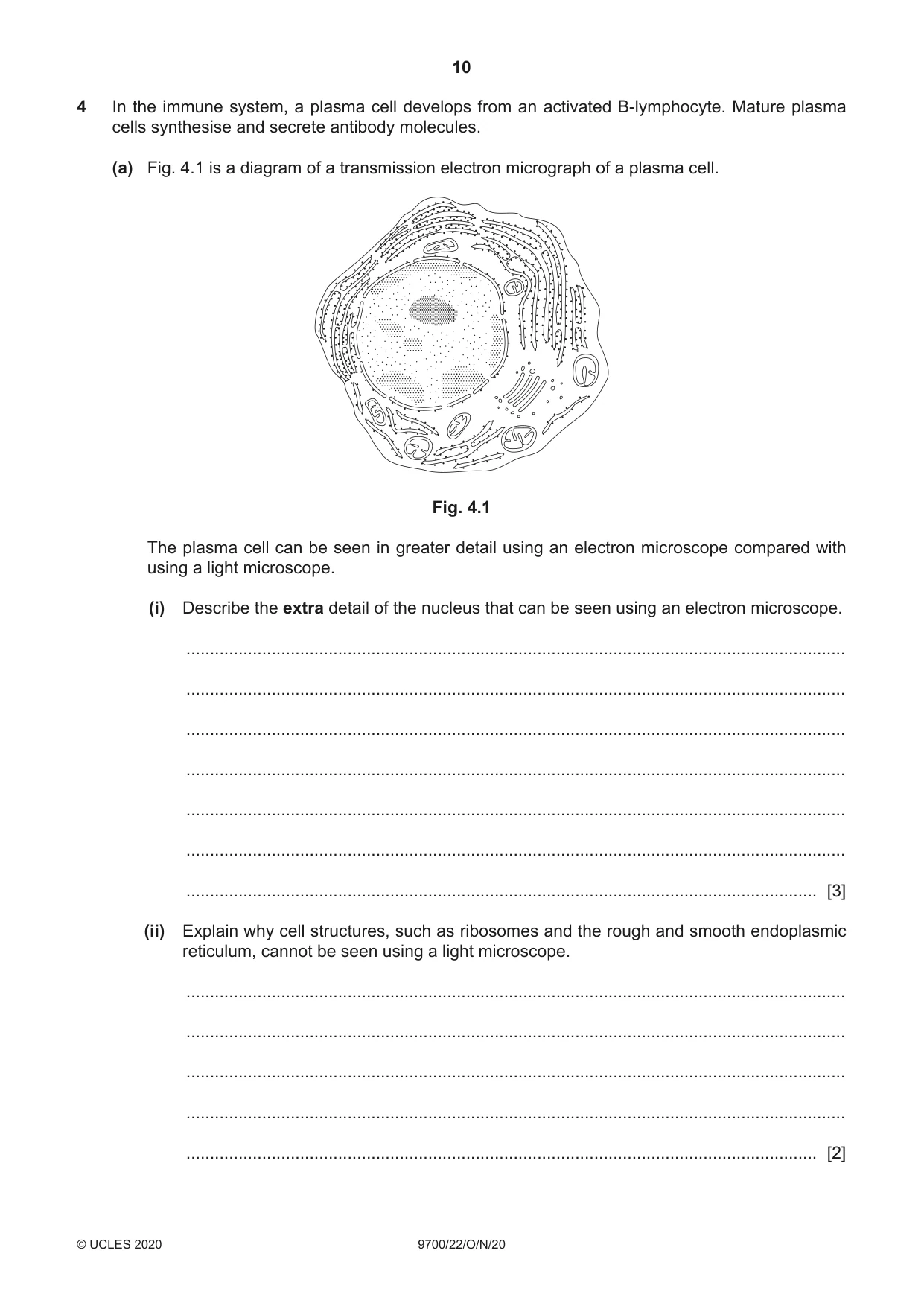
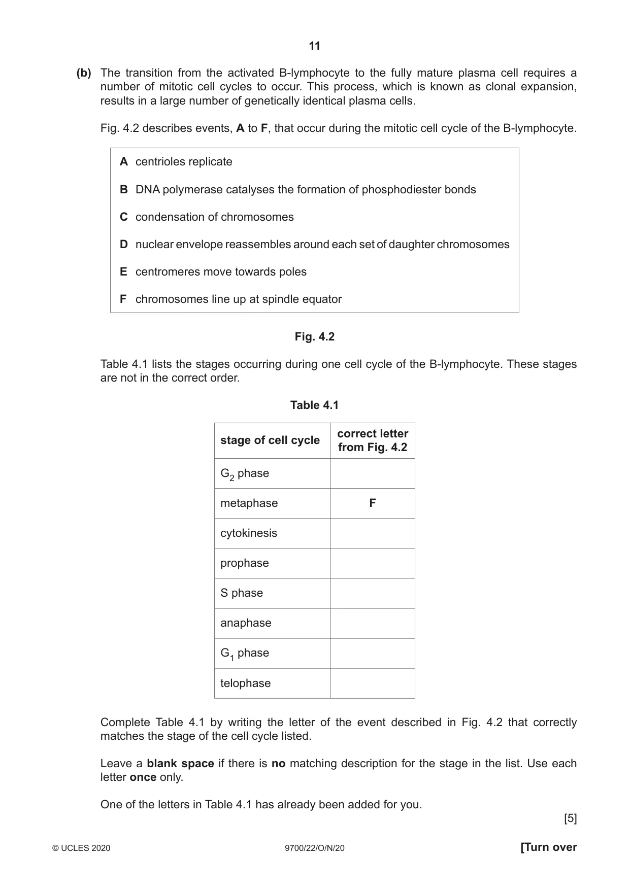
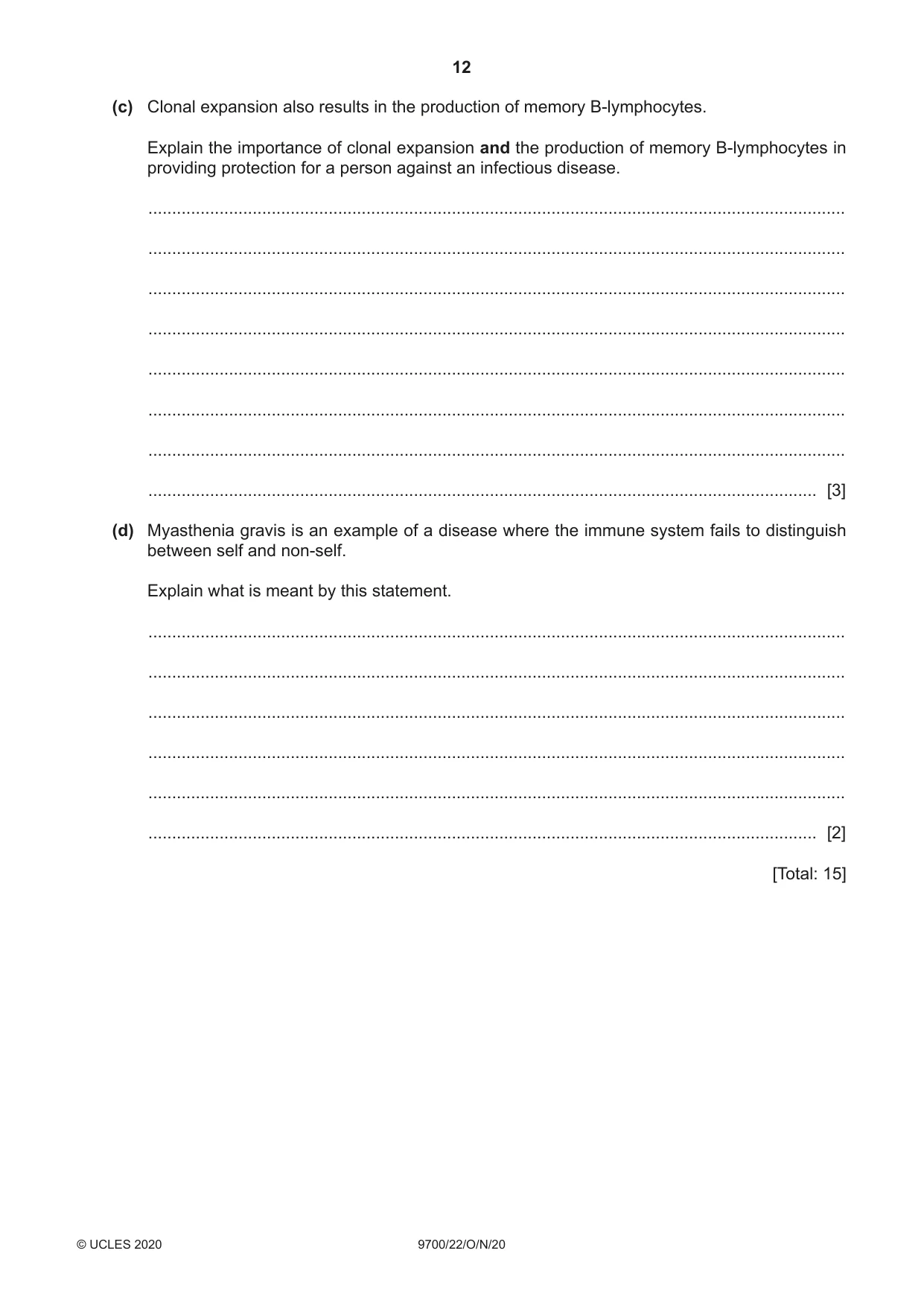
4 In the immune system, a plasma cell develops from an activated B-lymphocyte. Mature plasma cells synthesise and secrete antibody molecules. (a) Fig. 4.1 is a diagram of a transmission electron micrograph of a plasma cell. Fig. 4.1 The plasma cell can be seen in greater detail using an electron microscope compared with using a light microscope. (i) Describe the extra detail of the nucleus that can be seen using an electron microscope. ........................................................................................................................................... ........................................................................................................................................... ........................................................................................................................................... ........................................................................................................................................... ........................................................................................................................................... ........................................................................................................................................... ..................................................................................................................................... [3] (ii) Explain why cell structures, such as ribosomes and the rough and smooth endoplasmic reticulum, cannot be seen using a light microscope. ........................................................................................................................................... ........................................................................................................................................... ........................................................................................................................................... ........................................................................................................................................... ..................................................................................................................................... [2] (b) The transition from the activated B-lymphocyte to the fully mature plasma cell requires a number of mitotic cell cycles to occur. This process, which is known as clonal expansion, results in a large number of genetically identical plasma cells. Fig. 4.2 describes events, A to F, that occur during the mitotic cell cycle of the B-lymphocyte. A centrioles replicate B DNA polymerase catalyses the formation of phosphodiester bonds C condensation of chromosomes D nuclear envelope reassembles around each set of daughter chromosomes E centromeres move towards poles F chromosomes line up at spindle equator Fig. 4.2 Table 4.1 lists the stages occurring during one cell cycle of the B-lymphocyte. These stages are not in the correct order. Table 4.1 correct letter stage of cell cycle from Fig. 4.2 G2 phase metaphase F cytokinesis prophase S phase anaphase G1 phase telophase Complete Table 4.1 by writing the letter of the event described in Fig. 4.2 that correctly matches the stage of the cell cycle listed. Leave a blank space if there is no matching description for the stage in the list. Use each letter once only. One of the letters in Table 4.1 has already been added for you. [5] (c) Clonal expansion also results in the production of memory B-lymphocytes. Explain the importance of clonal expansion and the production of memory B-lymphocytes in providing protection for a person against an infectious disease. ................................................................................................................................................... ................................................................................................................................................... ................................................................................................................................................... ................................................................................................................................................... ................................................................................................................................................... ................................................................................................................................................... ................................................................................................................................................... ............................................................................................................................................. [3] (d) Myasthenia gravis is an example of a disease where the immune system fails to distinguish between self and non-self. Explain what is meant by this statement. ................................................................................................................................................... ................................................................................................................................................... ................................................................................................................................................... ................................................................................................................................................... ................................................................................................................................................... ............................................................................................................................................. [2] [Total: 15]
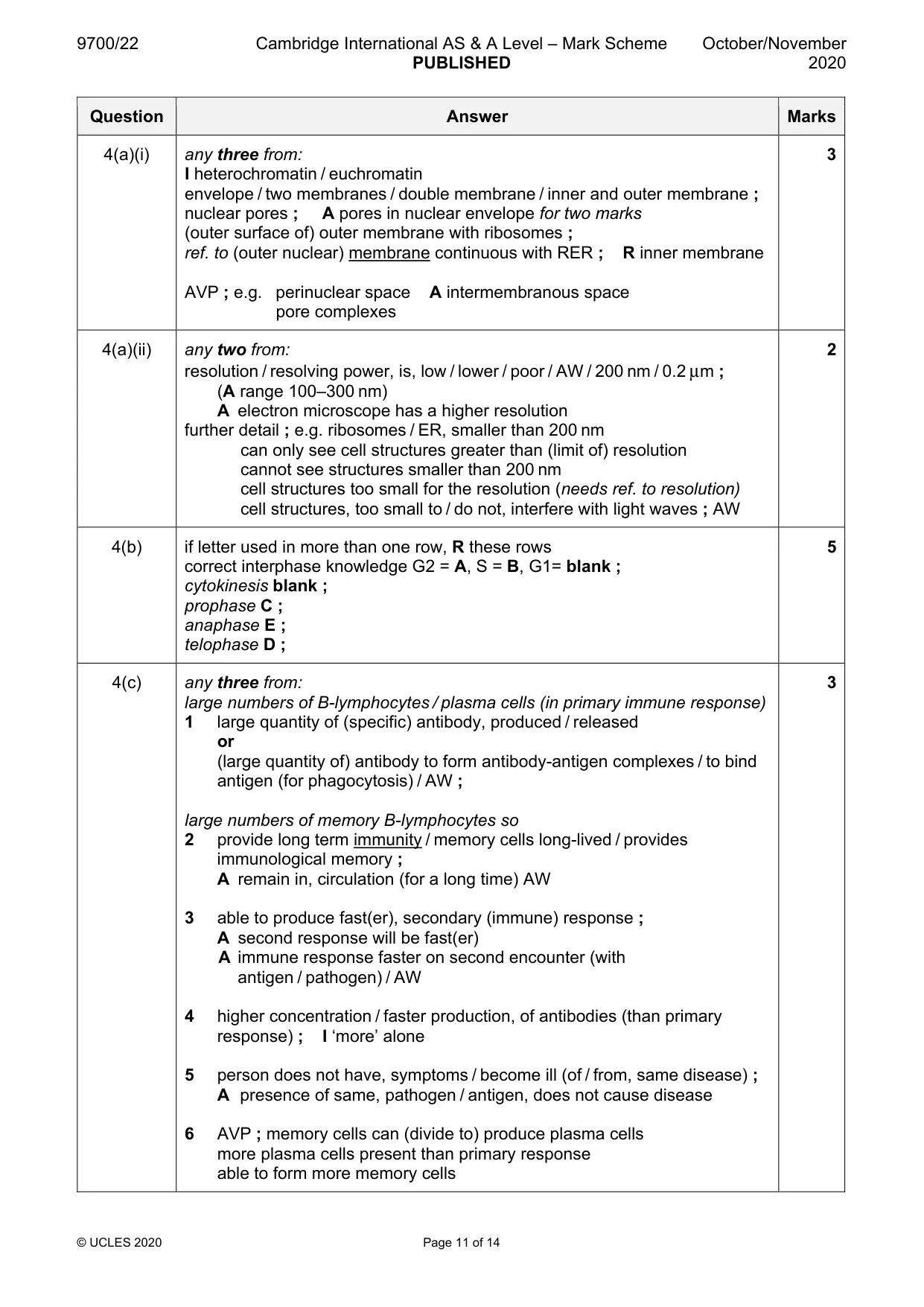
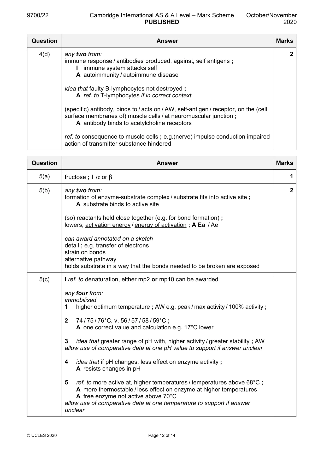
Mark scheme: 4(a)(i) any three from: 3 I heterochromatin / euchromatin envelope / two membranes / double membrane / inner and outer membrane ; nuclear pores ; A pores in nuclear envelope for two marks (outer surface of) outer membrane with ribosomes ; ref. to (outer nuclear) membrane continuous with RER ; R inner membrane AVP ; e.g. perinuclear space A intermembranous space pore complexes 4(a)(ii) any two from: 2 resolution / resolving power, is, low / lower / poor / AW / 200 nm / 0.2 μm ; (A range 100–300 nm) A electron microscope has a higher resolution further detail ; e.g. ribosomes / ER, smaller than 200 nm can only see cell structures greater than (limit of) resolution cannot see structures smaller than 200 nm cell structures too small for the resolution (needs ref. to resolution) cell structures, too small to / do not, interfere with light waves ; AW 4(b) if letter used in more than one row, R these rows 5 correct interphase knowledge G2 = A, S = B, G1= blank ; cytokinesis blank ; prophase C ; anaphase E ; telophase D ; 4(c) any three from: 3 large numbers of B-lymphocytes / plasma cells (in primary immune response) 1 large quantity of (specific) antibody, produced / released or (large quantity of) antibody to form antibody-antigen complexes / to bind antigen (for phagocytosis) / AW ; large numbers of memory B-lymphocytes so 2 provide long term immunity / memory cells long-lived / provides immunological memory ; A remain in, circulation (for a long time) AW 3 able to produce fast(er), secondary (immune) response ; A second response will be fast(er) A immune response faster on second encounter (with antigen / pathogen) / AW 4 higher concentration / faster production, of antibodies (than primary response) ; I ‘more’ alone 5 person does not have, symptoms / become ill (of / from, same disease) ; A presence of same, pathogen / antigen, does not cause disease 6 AVP ; memory cells can (divide to) produce plasma cells more plasma cells present than primary response able to form more memory cells 4(d) any two from: 2 immune response / antibodies produced, against, self antigens ; I immune system attacks self A autoimmunity / autoimmune disease idea that faulty B-lymphocytes not destroyed ; A ref. to T-lymphocytes if in correct context (specific) antibody, binds to / acts on / AW, self-antigen / receptor, on the (cell surface membranes of) muscle cells / at neuromuscular junction ; A antibody binds to acetylcholine receptors ref. to consequence to muscle cells ; e.g.(nerve) impulse conduction impaired action of transmitter substance hindered
Q5 · Sucrose phosphorylase is an enzyme found in some species of bacteria
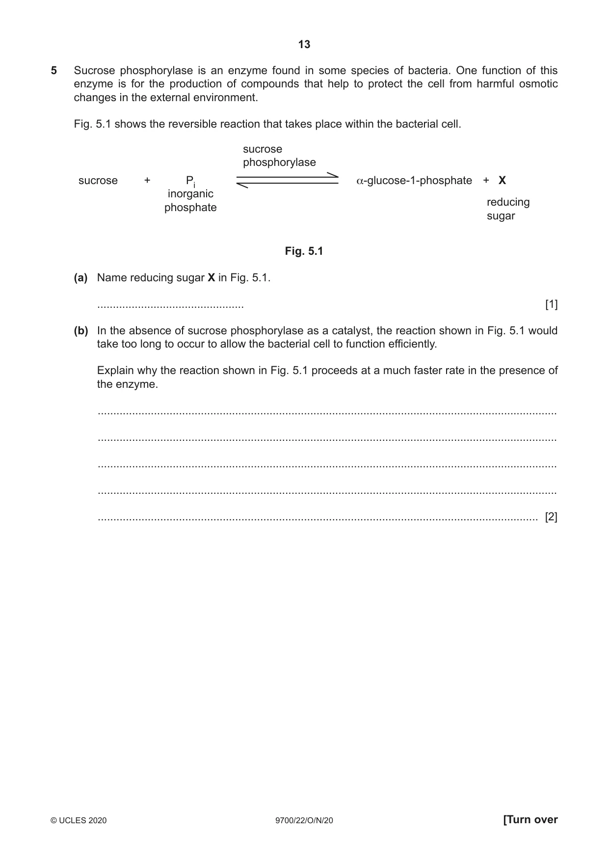
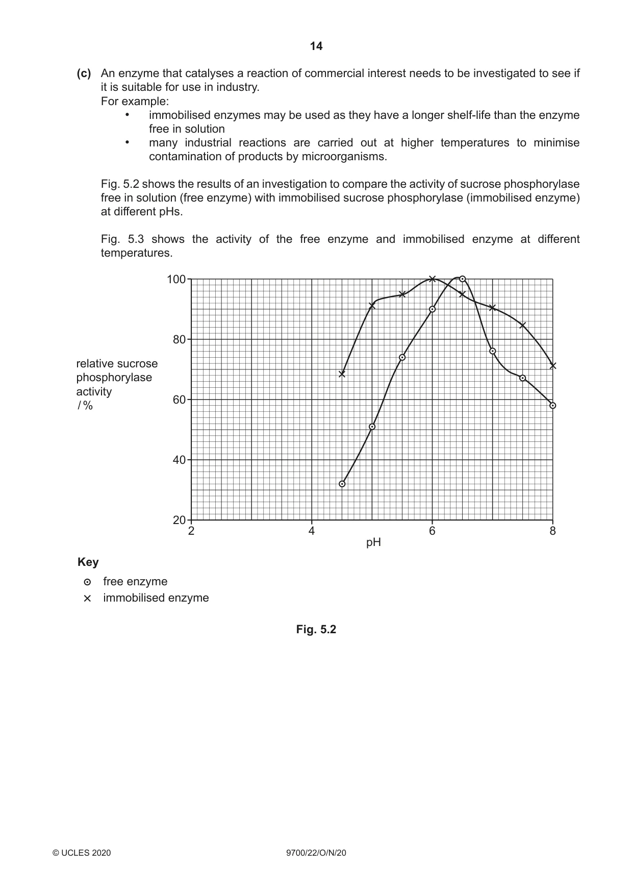
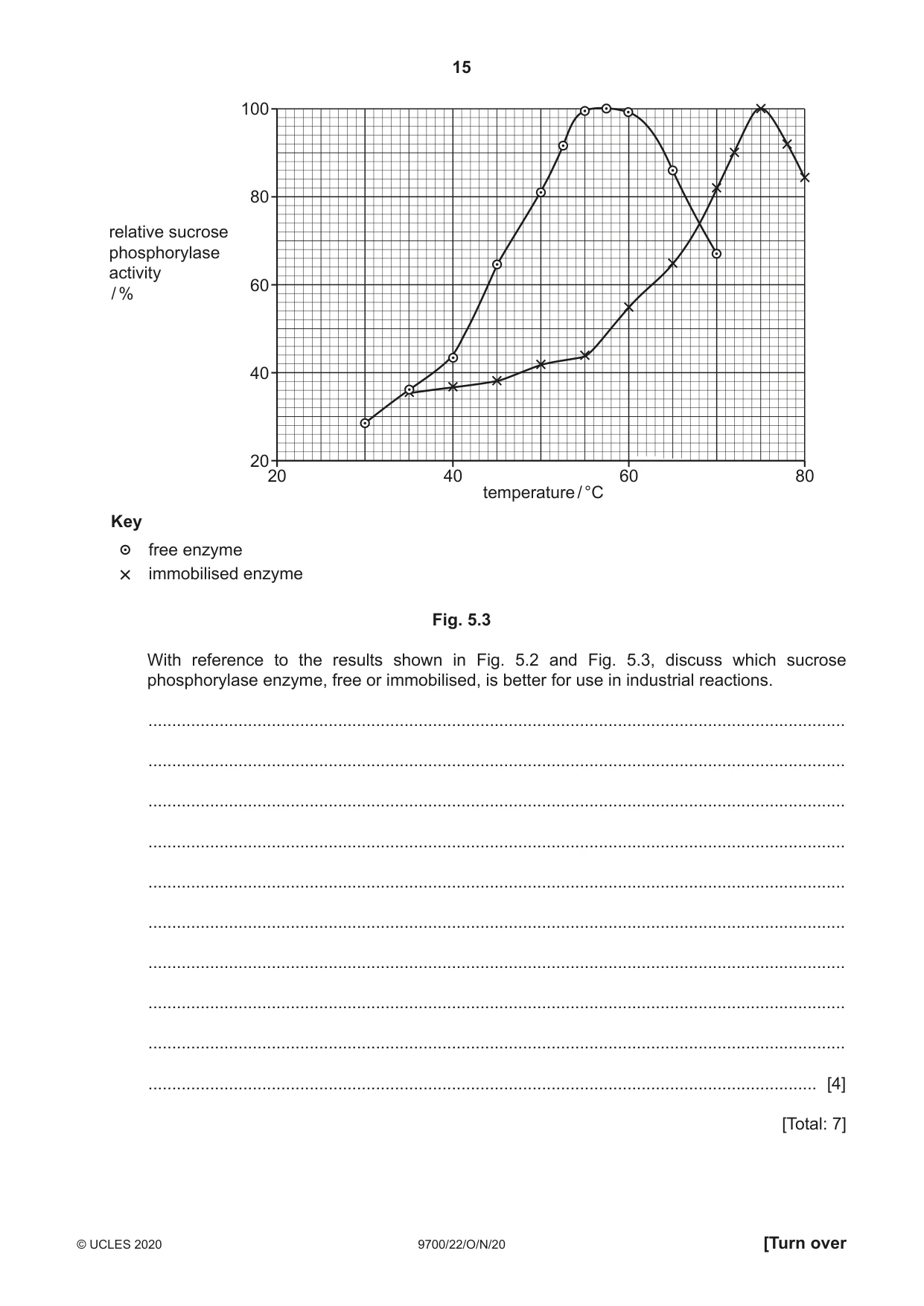
5 Sucrose phosphorylase is an enzyme found in some species of bacteria. One function of this enzyme is for the production of compounds that help to protect the cell from harmful osmotic changes in the external environment. Fig. 5.1 shows the reversible reaction that takes place within the bacterial cell. sucrose phosphorylase sucrose + Pi α-glucose-1-phosphate + X inorganic reducing phosphate sugar Fig. 5.1 (a) Name reducing sugar X in Fig. 5.1. ............................................... [1] (b) In the absence of sucrose phosphorylase as a catalyst, the reaction shown in Fig. 5.1 would take too long to occur to allow the bacterial cell to function efficiently. Explain why the reaction shown in Fig. 5.1 proceeds at a much faster rate in the presence of the enzyme. ................................................................................................................................................... ................................................................................................................................................... ................................................................................................................................................... ................................................................................................................................................... ............................................................................................................................................. [2] (c) An enzyme that catalyses a reaction of commercial interest needs to be investigated to see if it is suitable for use in industry. For example: • immobilised enzymes may be used as they have a longer shelf-life than the enzyme free in solution • many industrial reactions are carried out at higher temperatures to minimise contamination of products by microorganisms. Fig. 5.2 shows the results of an investigation to compare the activity of sucrose phosphorylase free in solution (free enzyme) with immobilised sucrose phosphorylase (immobilised enzyme) at different pHs. Fig. 5.3 shows the activity of the free enzyme and immobilised enzyme at different temperatures. 100 80 relative sucrose phosphorylase activity 60/ % 40 20 2 4 6 8 pH Key free enzyme immobilised enzyme Fig. 5.2 100 80 relative sucrose phosphorylase activity 60 / % 40 20 20 40 60 80 temperature / °C Key free enzyme immobilised enzyme Fig. 5.3 With reference to the results shown in Fig. 5.2 and Fig. 5.3, discuss which sucrose phosphorylase enzyme, free or immobilised, is better for use in industrial reactions. ................................................................................................................................................... ................................................................................................................................................... ................................................................................................................................................... ................................................................................................................................................... ................................................................................................................................................... ................................................................................................................................................... ................................................................................................................................................... ................................................................................................................................................... ................................................................................................................................................... ............................................................................................................................................. [4] [Total: 7]
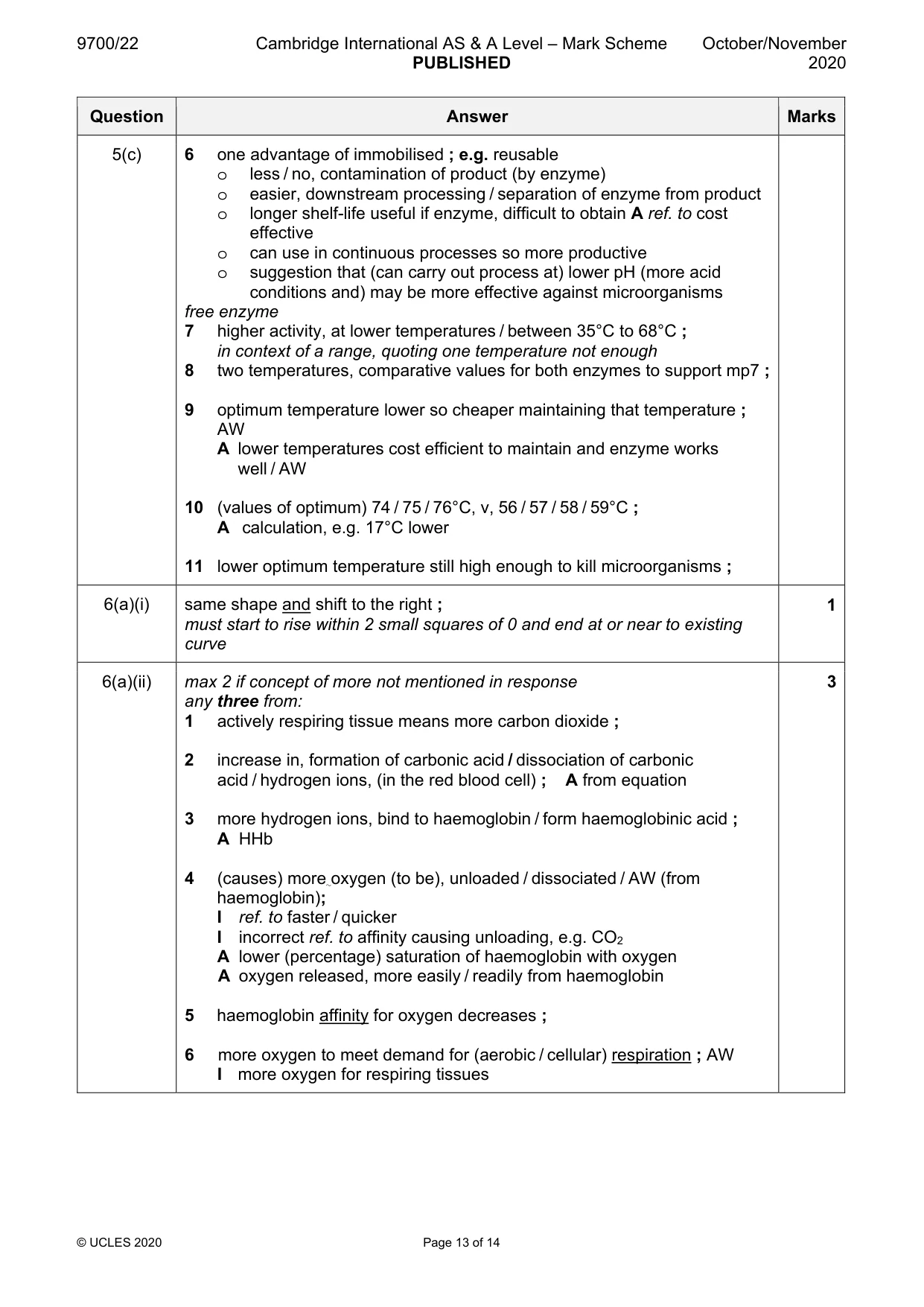
Mark scheme: 5(a) fructose ; I α or β 1 5(b) any two from: 2 formation of enzyme-substrate complex / substrate fits into active site ; A substrate binds to active site (so) reactants held close together (e.g. for bond formation) ; lowers, activation energy / energy of activation ; A Ea / Ae can award annotated on a sketch detail ; e.g. transfer of electrons strain on bonds alternative pathway holds substrate in a way that the bonds needed to be broken are exposed 5(c) I ref. to denaturation, either mp2 or mp10 can be awarded any four from: immobilised 1 higher optimum temperature ; AW e.g. peak / max activity / 100% activity ; 2 74 / 75 / 76°C, v, 56 / 57 / 58 / 59°C ; A one correct value and calculation e.g. 17°C lower 3 idea that greater range of pH with, higher activity / greater stability ; AW allow use of comparative data at one pH value to support if answer unclear 4 idea that if pH changes, less effect on enzyme activity ; A resists changes in pH 5 ref. to more active at, higher temperatures / temperatures above 68°C ; A more thermostable / less effect on enzyme at higher temperatures A free enzyme not active above 70°C allow use of comparative data at one temperature to support if answer unclear 5(c) 6 one advantage of immobilised ; e.g. reusable o less / no, contamination of product (by enzyme) o easier, downstream processing / separation of enzyme from product o longer shelf-life useful if enzyme, difficult to obtain A ref. to cost effective o can use in continuous processes so more productive o suggestion that (can carry out process at) lower pH (more acid conditions and) may be more effective against microorganisms free enzyme 7 higher activity, at lower temperatures / between 35°C to 68°C ; in context of a range, quoting one temperature not enough 8 two temperatures, comparative values for both enzymes to support mp7 ; 9 optimum temperature lower so cheaper maintaining that temperature ; AW A lower temperatures cost efficient to maintain and enzyme works well / AW 10 (values of optimum) 74 / 75 / 76°C, v, 56 / 57 / 58 / 59°C ; A calculation, e.g. 17°C lower 11 lower optimum temperature still high enough to kill microorganisms ;
Q6 · An oxygen dissociation curve for adult human haemoglobin
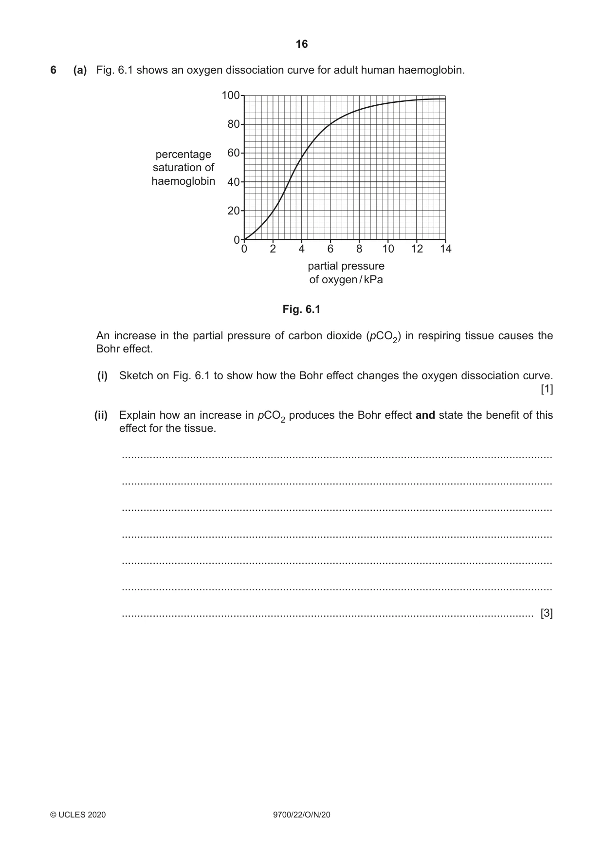
6 (a) Fig. 6.1 shows an oxygen dissociation curve for adult human haemoglobin. 100 80 percentage 60 saturation of haemoglobin 40 20 00 2 4 6 8 10 12 14 partial pressure of oxygen / kPa Fig. 6.1 An increase in the partial pressure of carbon dioxide (pCO2) in respiring tissue causes the Bohr effect. (i) Sketch on Fig. 6.1 to show how the Bohr effect changes the oxygen dissociation curve. [1] (ii) Explain how an increase in pCO2 produces the Bohr effect and state the benefit of this effect for the tissue. ........................................................................................................................................... ........................................................................................................................................... ........................................................................................................................................... ........................................................................................................................................... ........................................................................................................................................... ........................................................................................................................................... ..................................................................................................................................... [3] (b) Carbon dioxide (CO2) is transported across the cell surface membrane of the red blood cell using a different mechanism to the transport of hydrogen carbonate ions (HCO3–). Name the different mechanisms of transport used for CO2 and for HCO3– and explain why they are transported across the membrane by different mechanisms. CO2 ........................................................................................................................................... HCO3– ....................................................................................................................................... explanation ............................................................................................................................... ................................................................................................................................................... ................................................................................................................................................... ................................................................................................................................................... ................................................................................................................................................... ................................................................................................................................................... [4] [Total: 8]
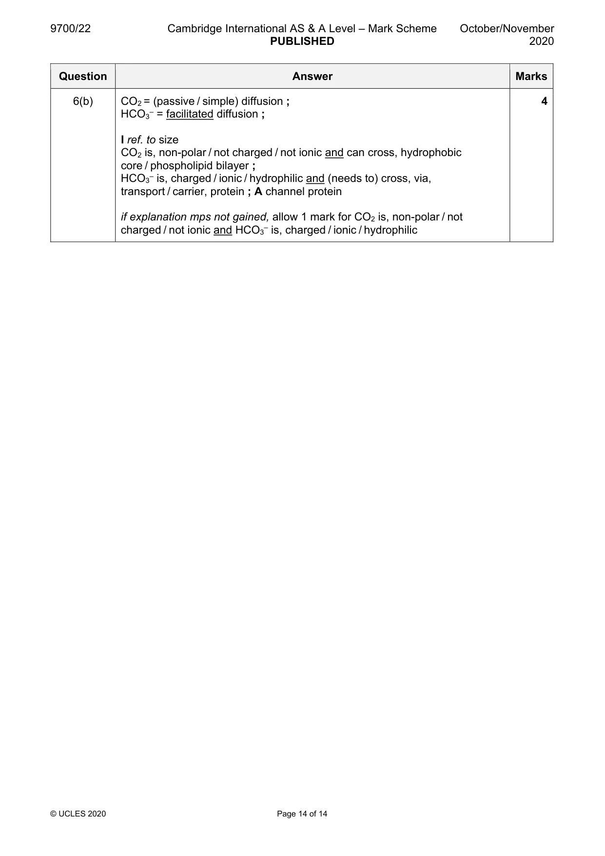
Mark scheme: 6(a)(i) same shape and shift to the right ; 1 must start to rise within 2 small squares of 0 and end at or near to existing curve 6(a)(ii) max 2 if concept of more not mentioned in response 3 any three from: 1 actively respiring tissue means more carbon dioxide ; 2 increase in, formation of carbonic acid / dissociation of carbonic acid / hydrogen ions, (in the red blood cell) ; A from equation 3 more hydrogen ions, bind to haemoglobin / form haemoglobinic acid ; A HHb 4 (causes) more oxygen (to be), unloaded / dissociated / AW (from haemoglobin); I ref. to faster / quicker I incorrect ref. to affinity causing unloading, e.g. CO2 A lower (percentage) saturation of haemoglobin with oxygen A oxygen released, more easily / readily from haemoglobin 5 haemoglobin affinity for oxygen decreases ; 6 more oxygen to meet demand for (aerobic / cellular) respiration ; AW I more oxygen for respiring tissues 6(b) CO2 = (passive / simple) diffusion ; 4 HCO3– = facilitated diffusion ; I ref. to size CO2 is, non-polar / not charged / not ionic and can cross, hydrophobic core / phospholipid bilayer ; HCO3– is, charged / ionic / hydrophilic and (needs to) cross, via, transport / carrier, protein ; A channel protein if explanation mps not gained, allow 1 mark for CO2 is, non-polar / not charged / not ionic and HCO3– is, charged / ionic / hydrophilic
What was in this paper
The subtopics covered by these 6 questions, and how many questions each got. Open one in a new tab to see every Cambridge question on it.
What you needed in this session
Cambridge’s own grade thresholds for 2020 Oct/Nov, Paper 2 · Variant 2. A higher threshold means an easier paper — the bar moves with how the cohort did.