Cambridge A Level Biology 9700 — 2020 May/June Paper 2 · Variant 2
9700/22/M/J/20 · 6 questions · 60 marks · ≈68 min
The question paper and its mark scheme, free to read here and free to download. This is Cambridge’s own paper, exactly as it was sat.
Question paper16 pages
Mark scheme13 pages
Answers below. Sit the paper first if you are practising.
Questions as text
Q1 · Picornaviruses are small viruses that are 30 nm in diameter
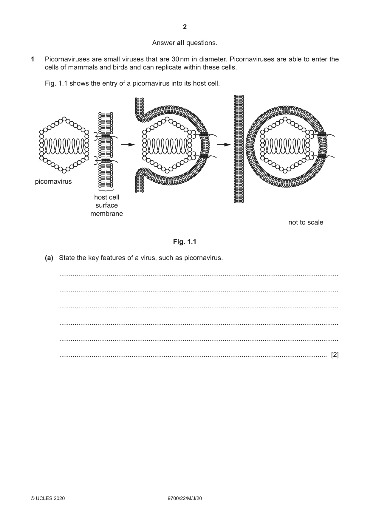
1 Picornaviruses are small viruses that are 30 nm in diameter. Picornaviruses are able to enter the cells of mammals and birds and can replicate within these cells. Fig. 1.1 shows the entry of a picornavirus into its host cell. picornavirus host cell surface membrane not to scale Fig. 1.1 (a) State the key features of a virus, such as picornavirus. ................................................................................................................................................... ................................................................................................................................................... ................................................................................................................................................... ................................................................................................................................................... ................................................................................................................................................... ............................................................................................................................................. [2] (b) State, with reasons, whether a picornavirus can be seen using the light microscope. ................................................................................................................................................... ................................................................................................................................................... ................................................................................................................................................... ................................................................................................................................................... ................................................................................................................................................... ................................................................................................................................................... ............................................................................................................................................. [3] (c) With reference to Fig. 1.1, describe how the picornavirus enters the host cell. ................................................................................................................................................... ................................................................................................................................................... ................................................................................................................................................... ................................................................................................................................................... ................................................................................................................................................... ................................................................................................................................................... ................................................................................................................................................... ............................................................................................................................................. [3] [Total: 8]
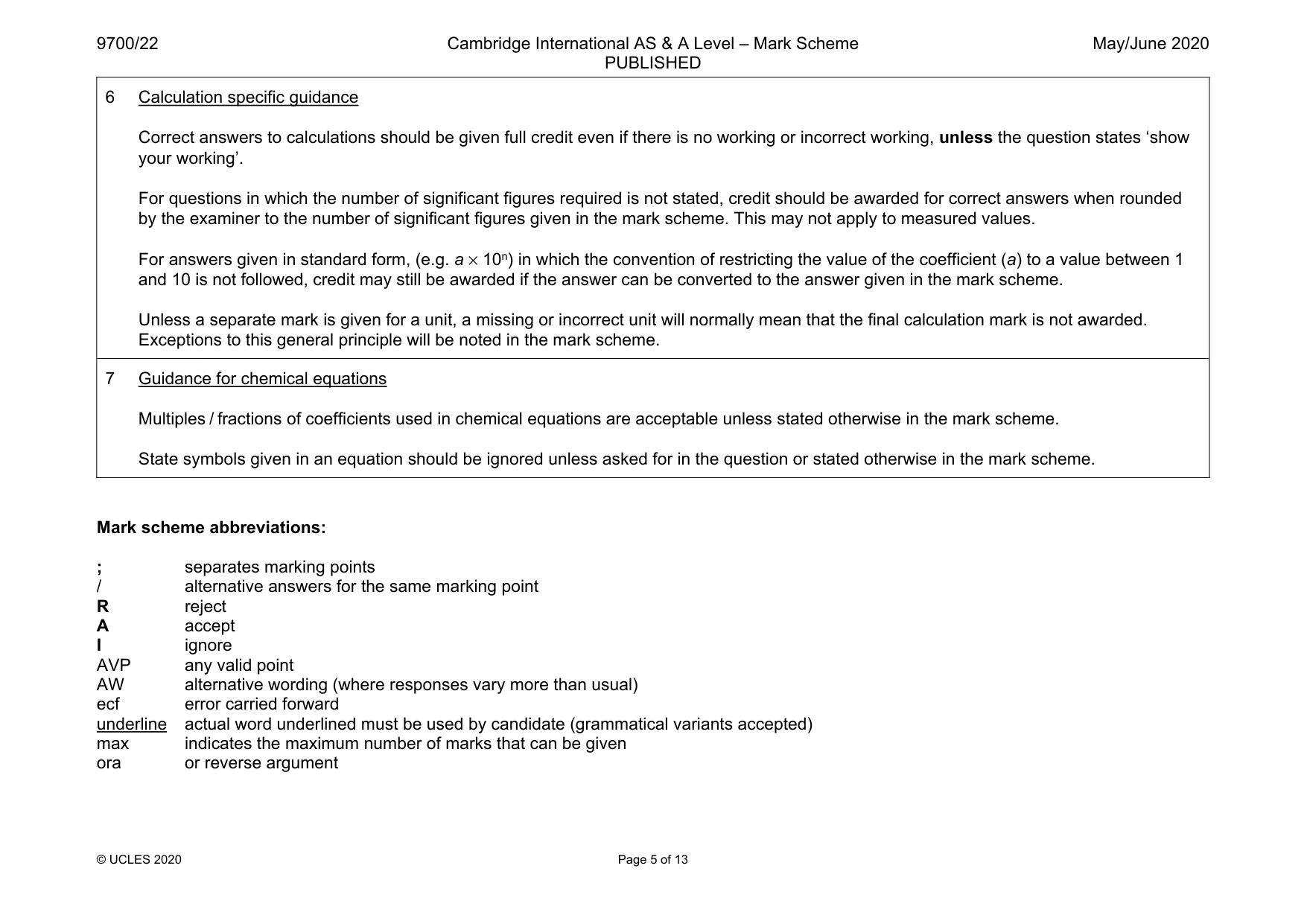
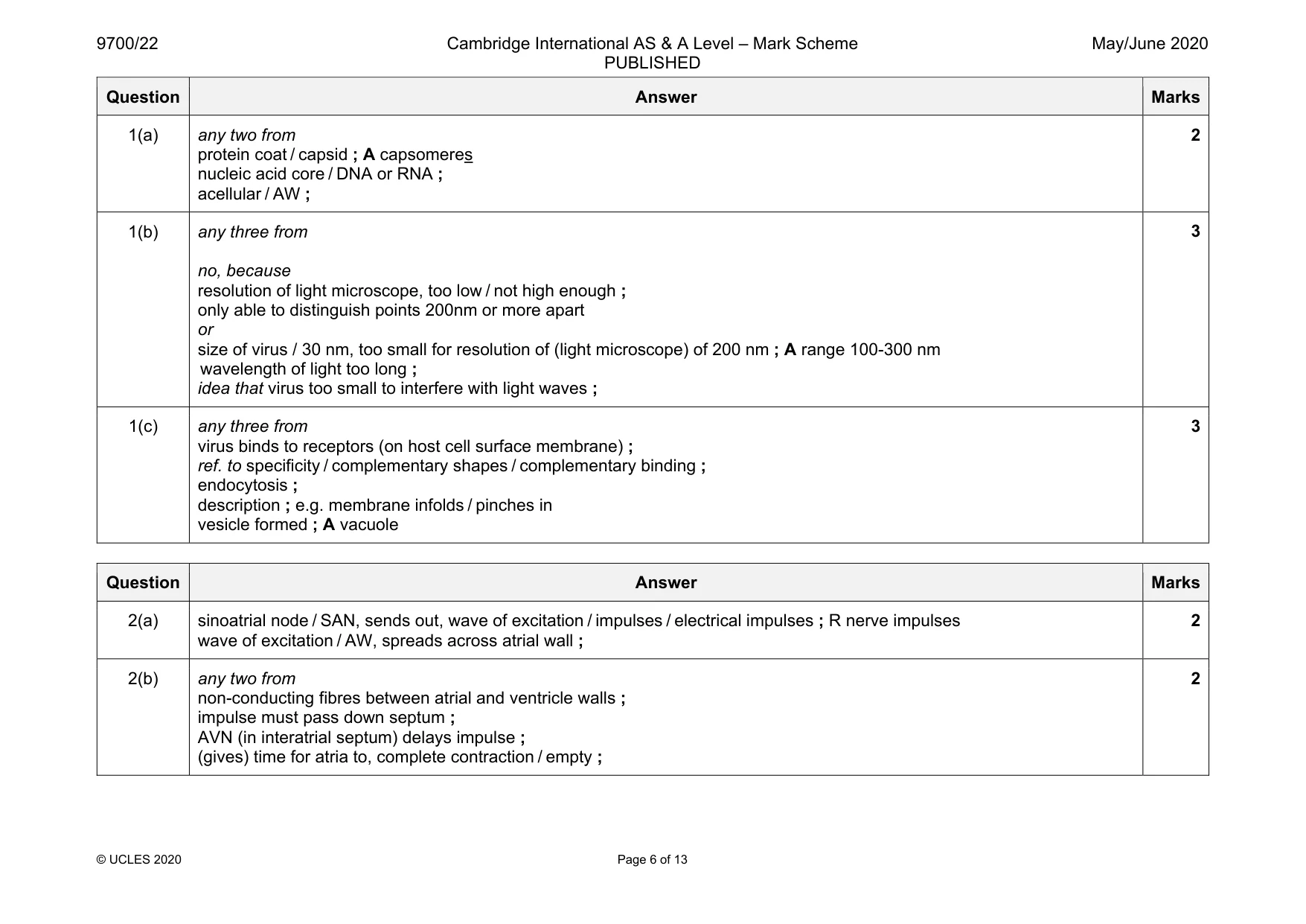
Mark scheme: 1(a) any two from protein coat / capsid ; A capsomeres nucleic acid core / DNA or RNA ; acellular / AW ; 2 1(b) any three from no, because resolution of light microscope, too low / not high enough ; only able to distinguish points 200nm or more apart or size of virus / 30 nm, too small for resolution of (light microscope) of 200 nm ; A range 100-300 nm wavelength of light too long ; idea that virus too small to interfere with light waves ; 3 1(c) any three from virus binds to receptors (on host cell surface membrane) ; ref. to specificity / complementary shapes / complementary binding ; endocytosis ; description ; e.g. membrane infolds / pinches in vesicle formed ; A vacuole 3
Q2 · In a healthy mammalian heart, contraction of the four chambers is coordinated by the…
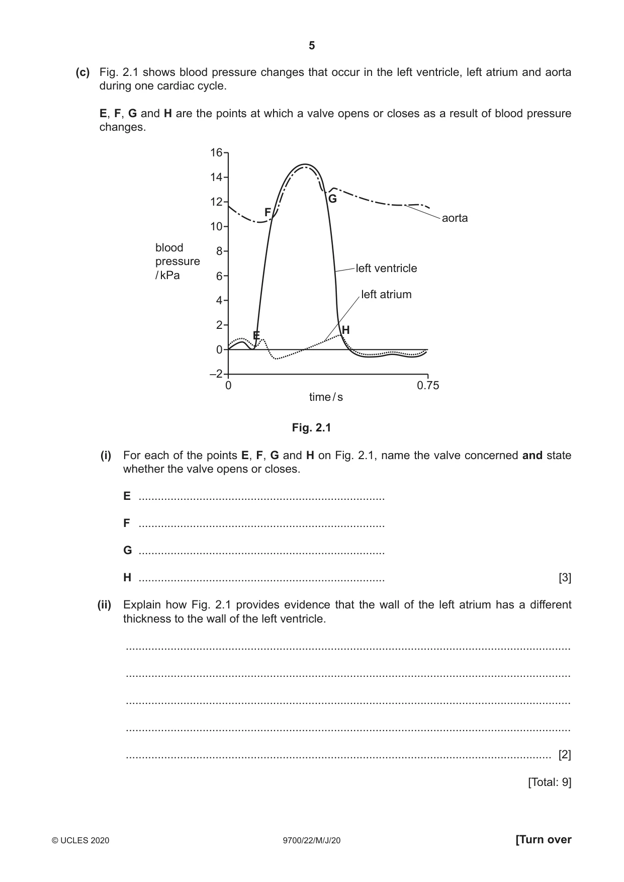
2 In a healthy mammalian heart, contraction of the four chambers is coordinated by the action of the sinoatrial node (SAN) and atrioventricular node (AVN). (a) After the atria fill with blood, atrial systole (contraction) occurs. State the events that occur to initiate and cause atrial systole. ................................................................................................................................................... ................................................................................................................................................... ................................................................................................................................................... ................................................................................................................................................... ................................................................................................................................................... ............................................................................................................................................. [2] (b) State and explain how the structure of the heart allows the atria to contract before the ventricles. ................................................................................................................................................... ................................................................................................................................................... ................................................................................................................................................... ................................................................................................................................................... ................................................................................................................................................... ................................................................................................................................................... ................................................................................................................................................... ............................................................................................................................................. [2] (c) Fig. 2.1 shows blood pressure changes that occur in the left ventricle, left atrium and aorta during one cardiac cycle. E, F, G and H are the points at which a valve opens or closes as a result of blood pressure changes. 16 14 12 G F aorta 10 blood 8 pressure left ventricle / kPa 6 left atrium 4 2 H E 0 –2 0 0.75 time / s Fig. 2.1 (i) For each of the points E, F, G and H on Fig. 2.1, name the valve concerned and state whether the valve opens or closes. E ............................................................................. F ............................................................................. G ............................................................................. H ............................................................................. [3] (ii) Explain how Fig. 2.1 provides evidence that the wall of the left atrium has a different thickness to the wall of the left ventricle. ........................................................................................................................................... ........................................................................................................................................... ........................................................................................................................................... ........................................................................................................................................... ..................................................................................................................................... [2] [Total: 9]
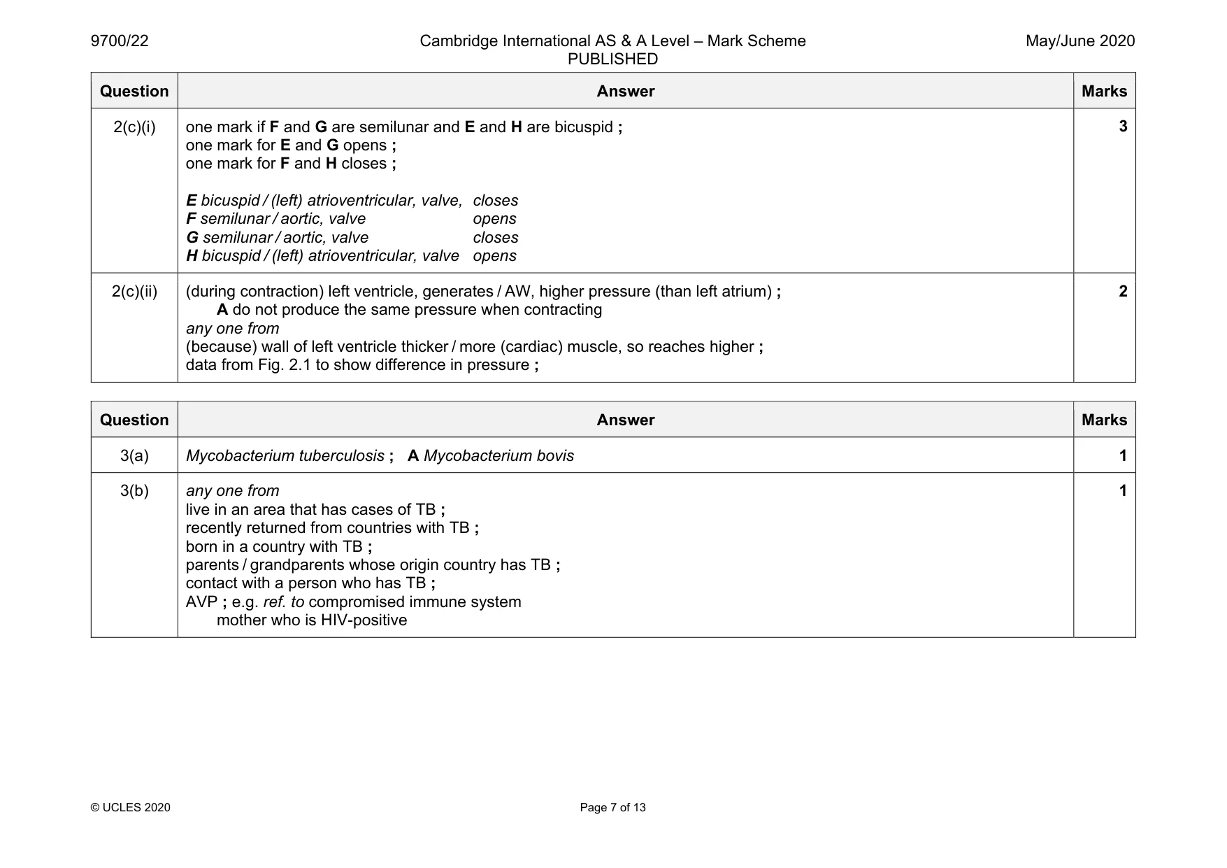
Mark scheme: 2(a) sinoatrial node / SAN, sends out, wave of excitation / impulses / electrical impulses ; R nerve impulses wave of excitation / AW, spreads across atrial wall ; 2 2(b) any two from non-conducting fibres between atrial and ventricle walls ; impulse must pass down septum ; AVN (in interatrial septum) delays impulse ; (gives) time for atria to, complete contraction / empty ; 2 Question Answer Marks 2(c)(i) one mark if F and G are semilunar and E and H are bicuspid ; one mark for E and G opens ; one mark for F and H closes ; E bicuspid / (left) atrioventricular, valve, closes F semilunar / aortic, valve opens G semilunar / aortic, valve closes H bicuspid / (left) atrioventricular, valve opens 3 2(c)(ii) (during contraction) left ventricle, generates / AW, higher pressure (than left atrium) ; A do not produce the same pressure when contracting any one from (because) wall of left ventricle thicker / more (cardiac) muscle, so reaches higher ; data from Fig. 2.1 to show difference in pressure ; 2
Q3 · The Bacillus Calmette-Guérin (BCG) vaccine is the only vaccine used to provide protection…
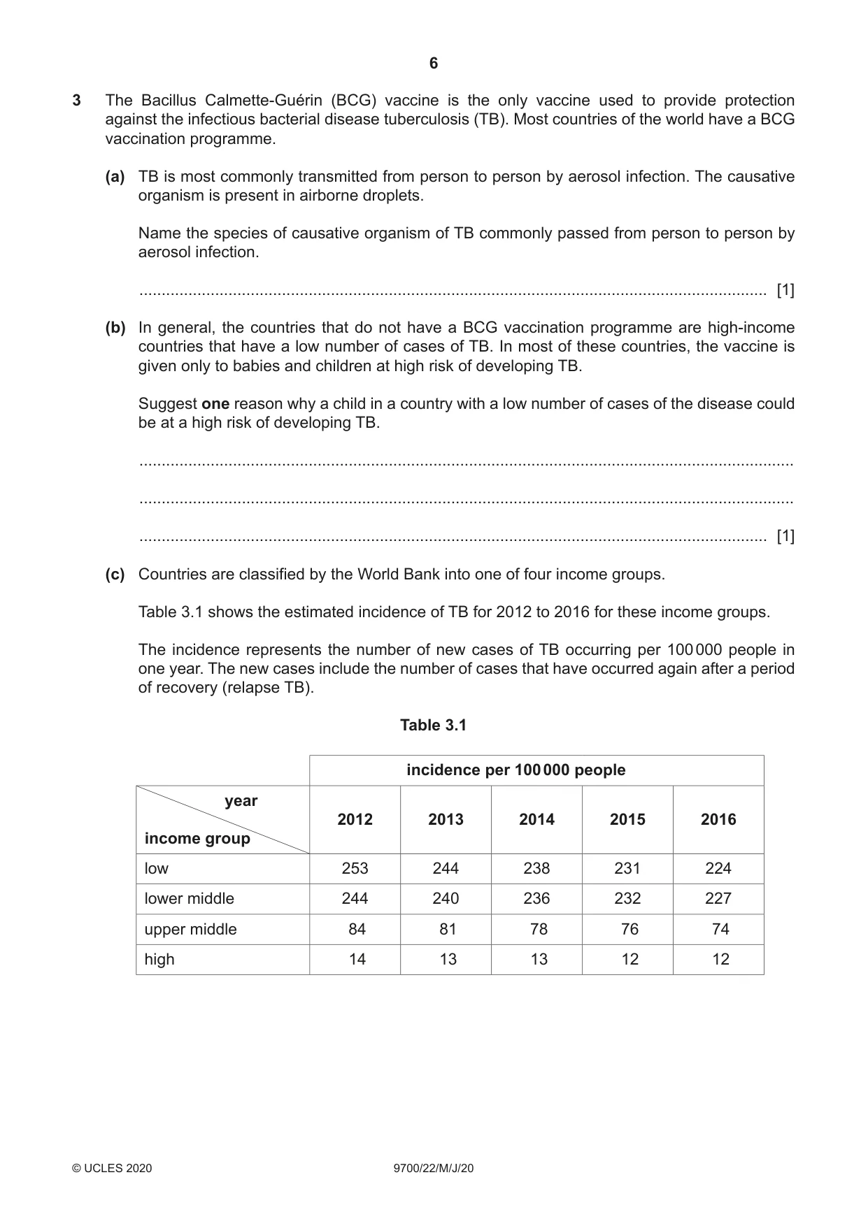
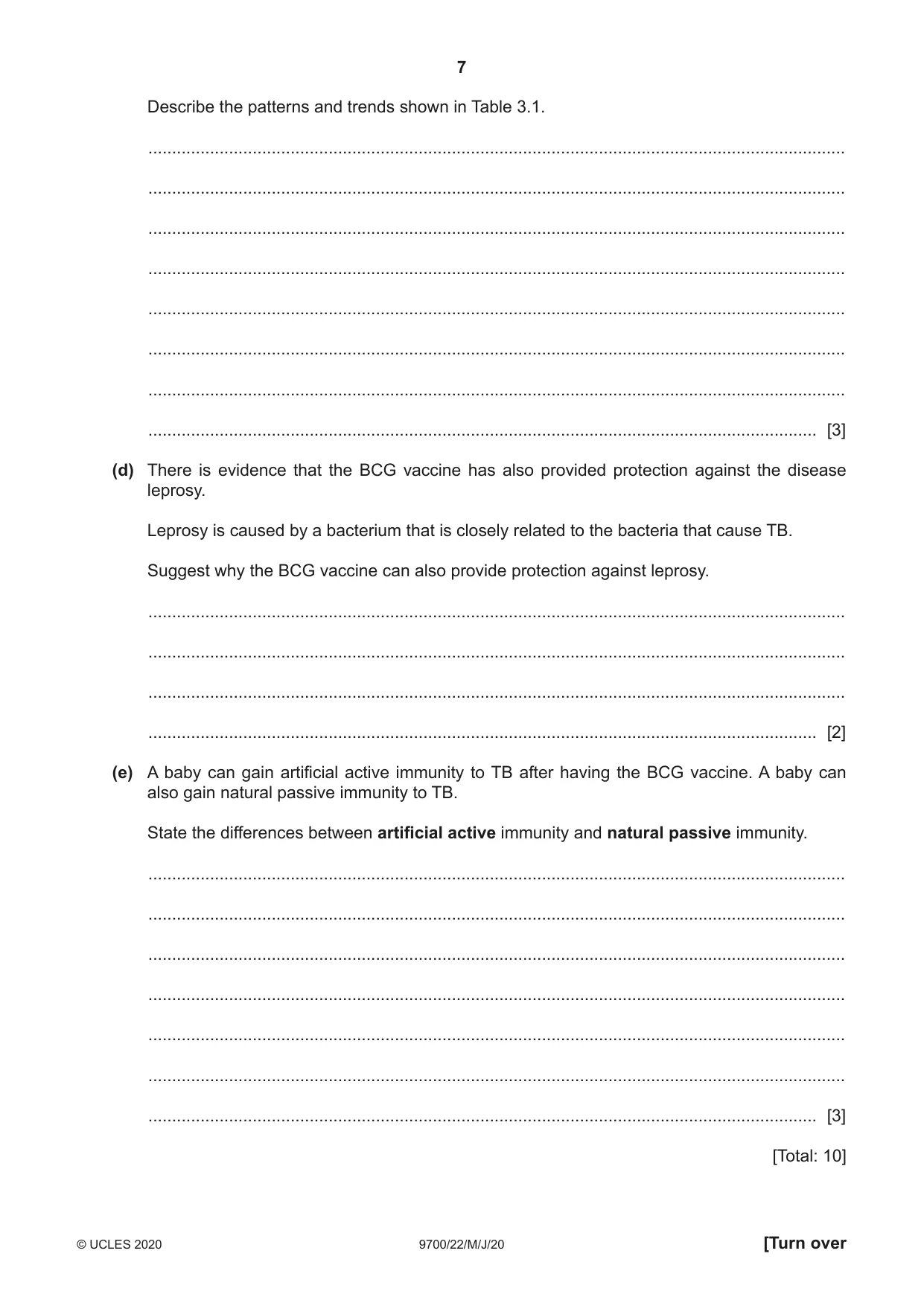
3 The Bacillus Calmette-Guérin (BCG) vaccine is the only vaccine used to provide protection against the infectious bacterial disease tuberculosis (TB). Most countries of the world have a BCG vaccination programme. (a) TB is most commonly transmitted from person to person by aerosol infection. The causative organism is present in airborne droplets. Name the species of causative organism of TB commonly passed from person to person by aerosol infection. ............................................................................................................................................. [1] (b) In general, the countries that do not have a BCG vaccination programme are high-income countries that have a low number of cases of TB. In most of these countries, the vaccine is given only to babies and children at high risk of developing TB. Suggest one reason why a child in a country with a low number of cases of the disease could be at a high risk of developing TB. ................................................................................................................................................... ................................................................................................................................................... ............................................................................................................................................. [1] (c) Countries are classified by the World Bank into one of four income groups. Table 3.1 shows the estimated incidence of TB for 2012 to 2016 for these income groups. The incidence represents the number of new cases of TB occurring per 100 000 people in one year. The new cases include the number of cases that have occurred again after a period of recovery (relapse TB). Table 3.1 incidence per 100 000 people year 2012 2013 2014 2015 2016 income group low 253 244 238 231 224 lower middle 244 240 236 232 227 upper middle 84 81 78 76 74 high 14 13 13 12 12 Describe the patterns and trends shown in Table 3.1. ................................................................................................................................................... ................................................................................................................................................... ................................................................................................................................................... ................................................................................................................................................... ................................................................................................................................................... ................................................................................................................................................... ................................................................................................................................................... ............................................................................................................................................. [3] (d) There is evidence that the BCG vaccine has also provided protection against the disease leprosy. Leprosy is caused by a bacterium that is closely related to the bacteria that cause TB. Suggest why the BCG vaccine can also provide protection against leprosy. ................................................................................................................................................... ................................................................................................................................................... ................................................................................................................................................... ............................................................................................................................................. [2] (e) A baby can gain artificial active immunity to TB after having the BCG vaccine. A baby can also gain natural passive immunity to TB. State the differences between artificial active immunity and natural passive immunity. ................................................................................................................................................... ................................................................................................................................................... ................................................................................................................................................... ................................................................................................................................................... ................................................................................................................................................... ................................................................................................................................................... ............................................................................................................................................. [3] [Total: 10]
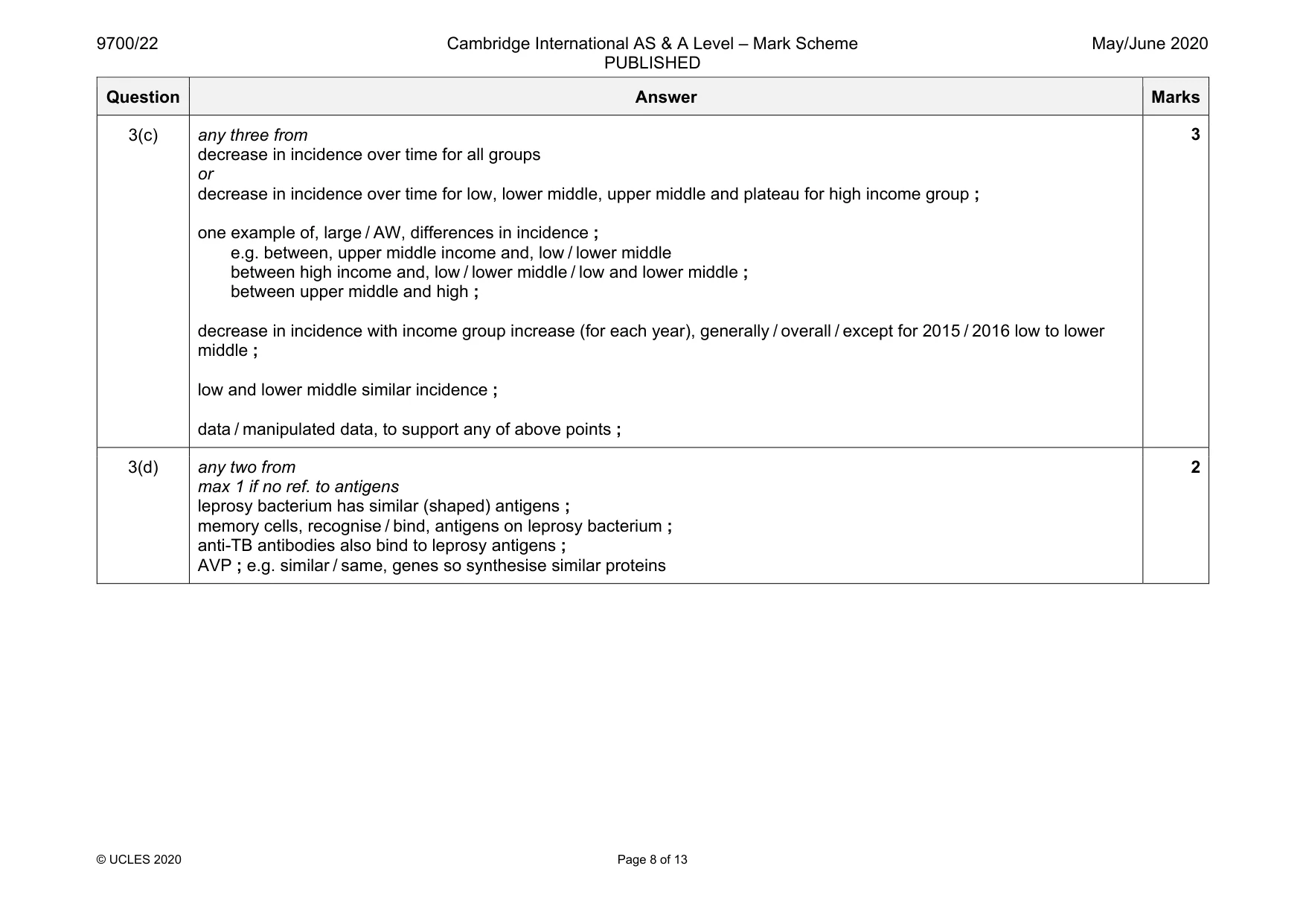
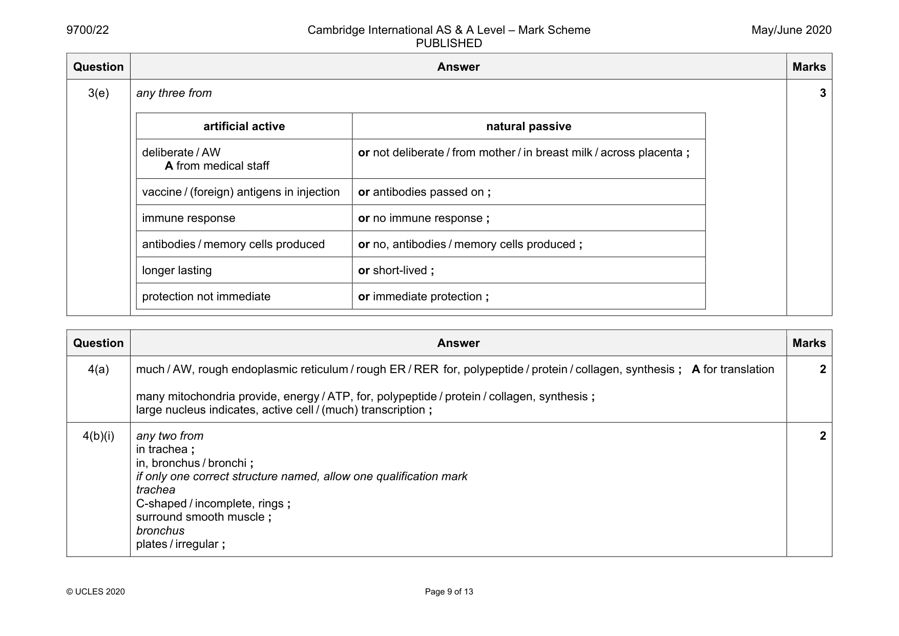
Mark scheme: 3(a) Mycobacterium tuberculosis ; A Mycobacterium bovis 1 3(b) any one from live in an area that has cases of TB ; recently returned from countries with TB ; born in a country with TB ; parents / grandparents whose origin country has TB ; contact with a person who has TB ; AVP ; e.g. ref. to compromised immune system mother who is HIV-positive 1 Question Answer Marks 3(c) any three from decrease in incidence over time for all groups or decrease in incidence over time for low, lower middle, upper middle and plateau for high income group ; one example of, large / AW, differences in incidence ; e.g. between, upper middle income and, low / lower middle between high income and, low / lower middle / low and lower middle ; between upper middle and high ; decrease in incidence with income group increase (for each year), generally / overall / except for 2015 / 2016 low to lower middle ; low and lower middle similar incidence ; data / manipulated data, to support any of above points ; 3 3(d) any two from max 1 if no ref. to antigens leprosy bacterium has similar (shaped) antigens ; memory cells, recognise / bind, antigens on leprosy bacterium ; anti-TB antibodies also bind to leprosy antigens ; AVP ; e.g. similar / same, genes so synthesise similar proteins 2 Question Answer Marks 3(e) any three from artificial active natural passive deliberate / AW A from medical staff or not deliberate / from mother / in breast milk / across placenta ; vaccine / (foreign) antigens in injection or antibodies passed on ; immune response or no immune response ; antibodies / memory cells produced or no, antibodies / memory cells produced ; longer lasting or short-lived ; protection not immediate or immediate protection ; 3
Q4 · Collagen is a major component of the cartilage found in some of the structures of the…
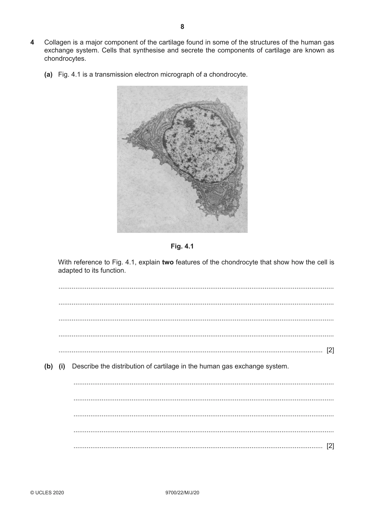
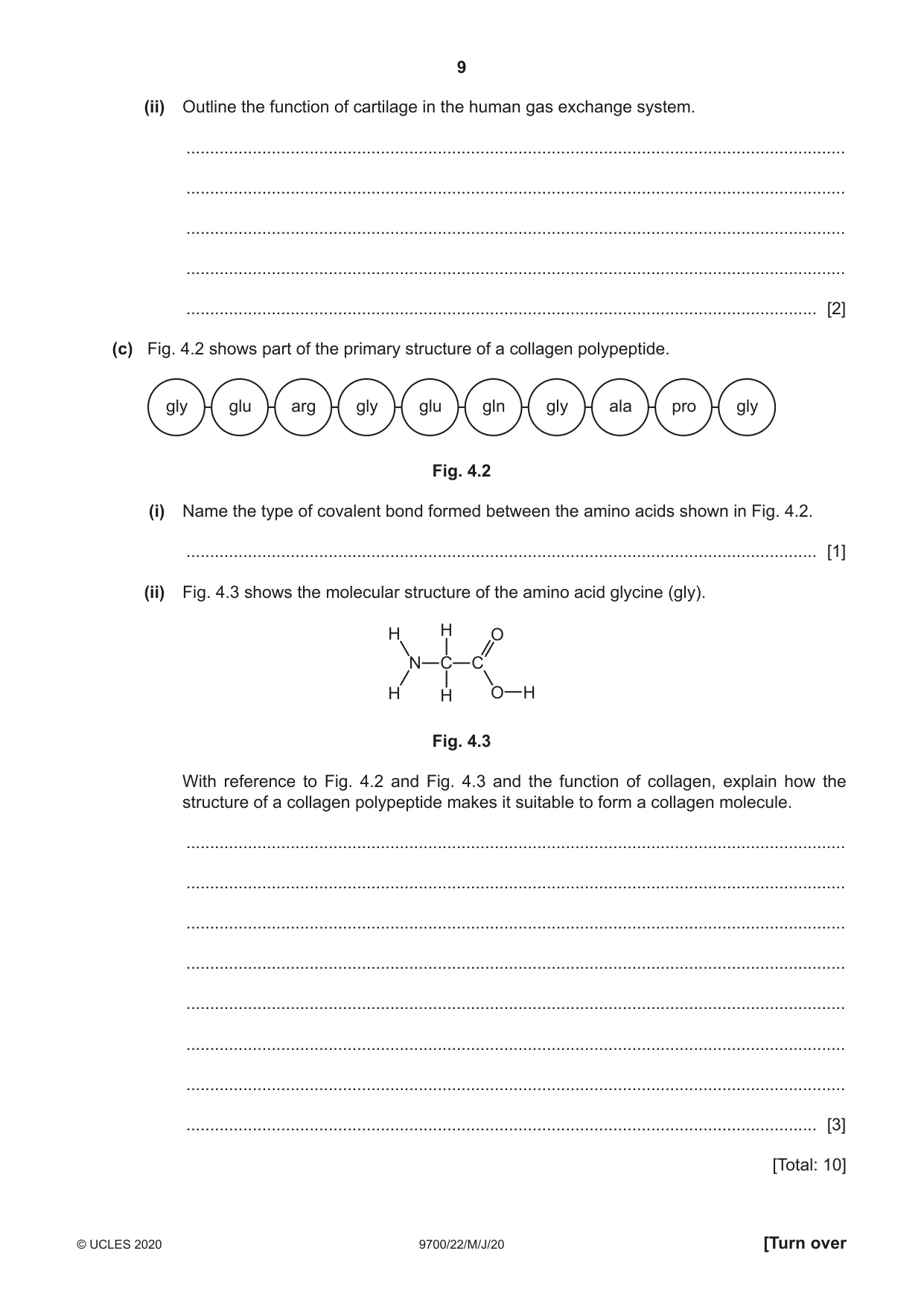
4 Collagen is a major component of the cartilage found in some of the structures of the human gas exchange system. Cells that synthesise and secrete the components of cartilage are known as chondrocytes. (a) Fig. 4.1 is a transmission electron micrograph of a chondrocyte. Fig. 4.1 With reference to Fig. 4.1, explain two features of the chondrocyte that show how the cell is adapted to its function. ................................................................................................................................................... ................................................................................................................................................... ................................................................................................................................................... ................................................................................................................................................... ............................................................................................................................................. [2] (b) (i) Describe the distribution of cartilage in the human gas exchange system. ........................................................................................................................................... ........................................................................................................................................... ........................................................................................................................................... ........................................................................................................................................... ..................................................................................................................................... [2] (ii) Outline the function of cartilage in the human gas exchange system. ........................................................................................................................................... ........................................................................................................................................... ........................................................................................................................................... ........................................................................................................................................... ..................................................................................................................................... [2] (c) Fig. 4.2 shows part of the primary structure of a collagen polypeptide. gly glu arg gly glu gln gly ala pro gly Fig. 4.2 (i) Name the type of covalent bond formed between the amino acids shown in Fig. 4.2. ..................................................................................................................................... [1] (ii) Fig. 4.3 shows the molecular structure of the amino acid glycine (gly). H H O N C C H H O H Fig. 4.3 With reference to Fig. 4.2 and Fig. 4.3 and the function of collagen, explain how the structure of a collagen polypeptide makes it suitable to form a collagen molecule. ........................................................................................................................................... ........................................................................................................................................... ........................................................................................................................................... ........................................................................................................................................... ........................................................................................................................................... ........................................................................................................................................... ........................................................................................................................................... ..................................................................................................................................... [3] [Total: 10]
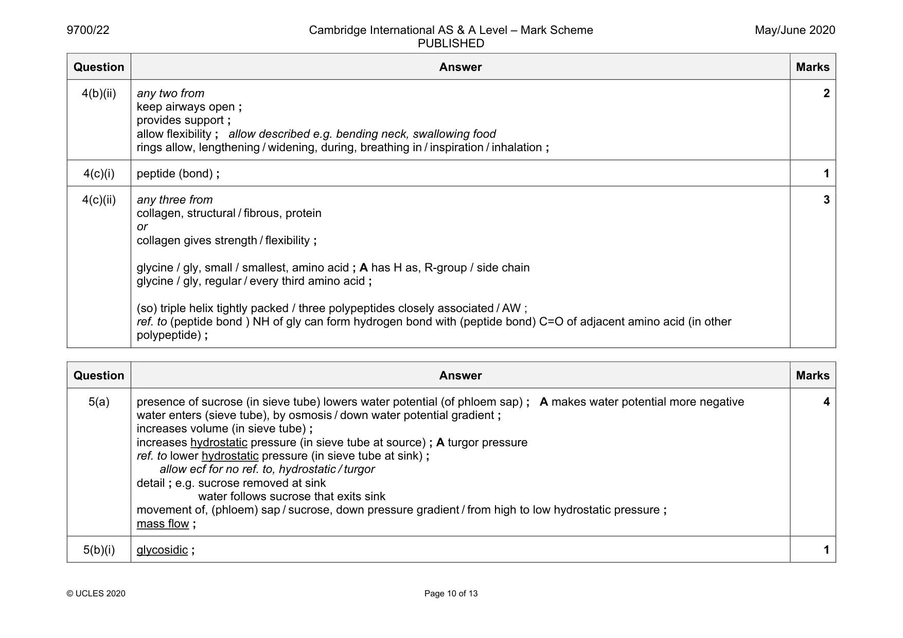
Mark scheme: 4(a) much / AW, rough endoplasmic reticulum / rough ER / RER for, polypeptide / protein / collagen, synthesis ; A for translation many mitochondria provide, energy / ATP, for, polypeptide / protein / collagen, synthesis ; large nucleus indicates, active cell / (much) transcription ; 4(b)(i) any two from in trachea ; in, bronchus / bronchi ; if only one correct structure named, allow one qualification mark trachea C-shaped / incomplete, rings ; surround smooth muscle ; bronchus plates / irregular ; 2 Question Answer Marks 4(b)(ii) any two from keep airways open ; provides support ; allow flexibility ; allow described e.g. bending neck, swallowing food rings allow, lengthening / widening, during, breathing in / inspiration / inhalation ; 2 4(c)(i) peptide (bond) ; 1 4(c)(ii) any three from collagen, structural / fibrous, protein or collagen gives strength / flexibility ; glycine / gly, small / smallest, amino acid ; A has H as, R-group / side chain glycine / gly, regular / every third amino acid ; (so) triple helix tightly packed / three polypeptides closely associated / AW ; ref. to (peptide bond ) NH of gly can form hydrogen bond with (peptide bond) C=O of adjacent amino acid (in other polypeptide) ; 3
Q5 · In most plants, sucrose is the main sugar that is transported from sources to sinks
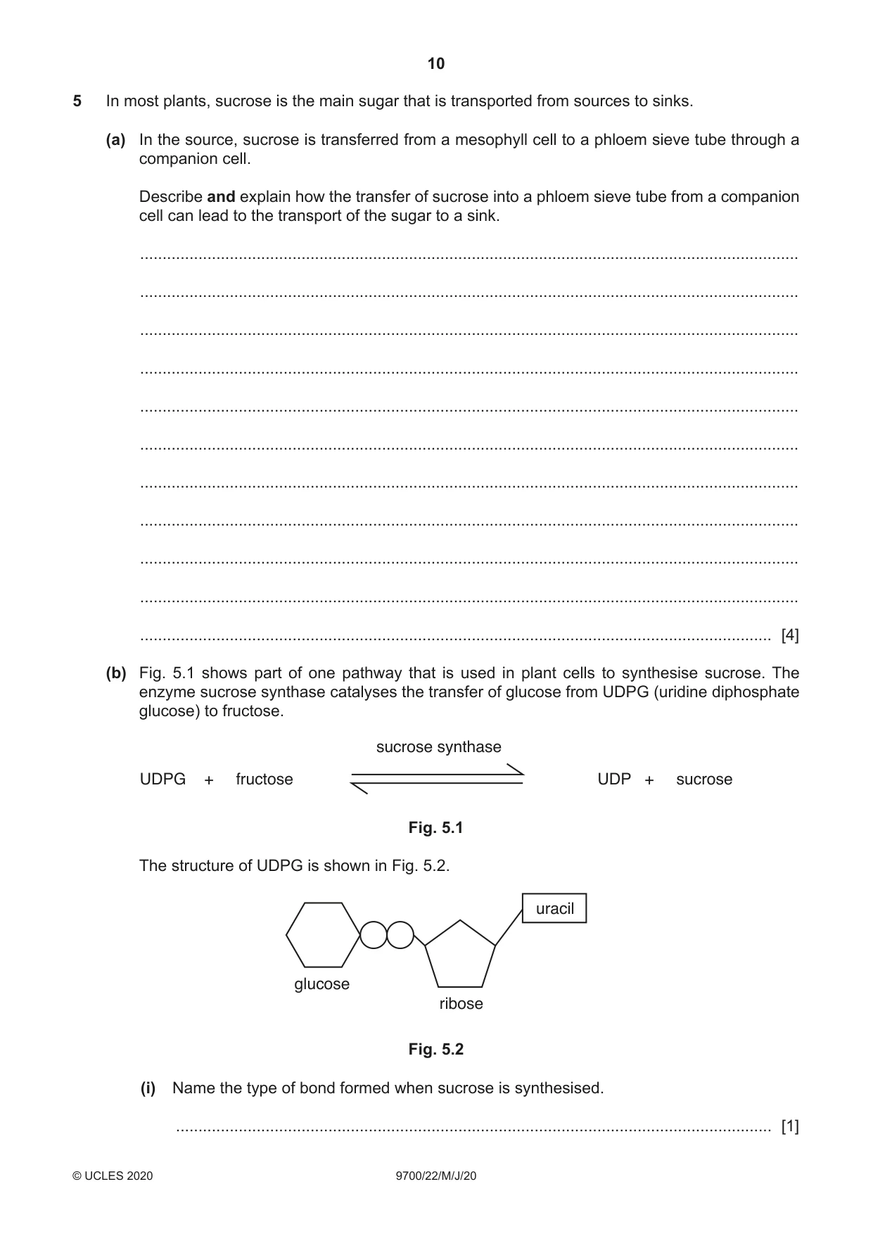
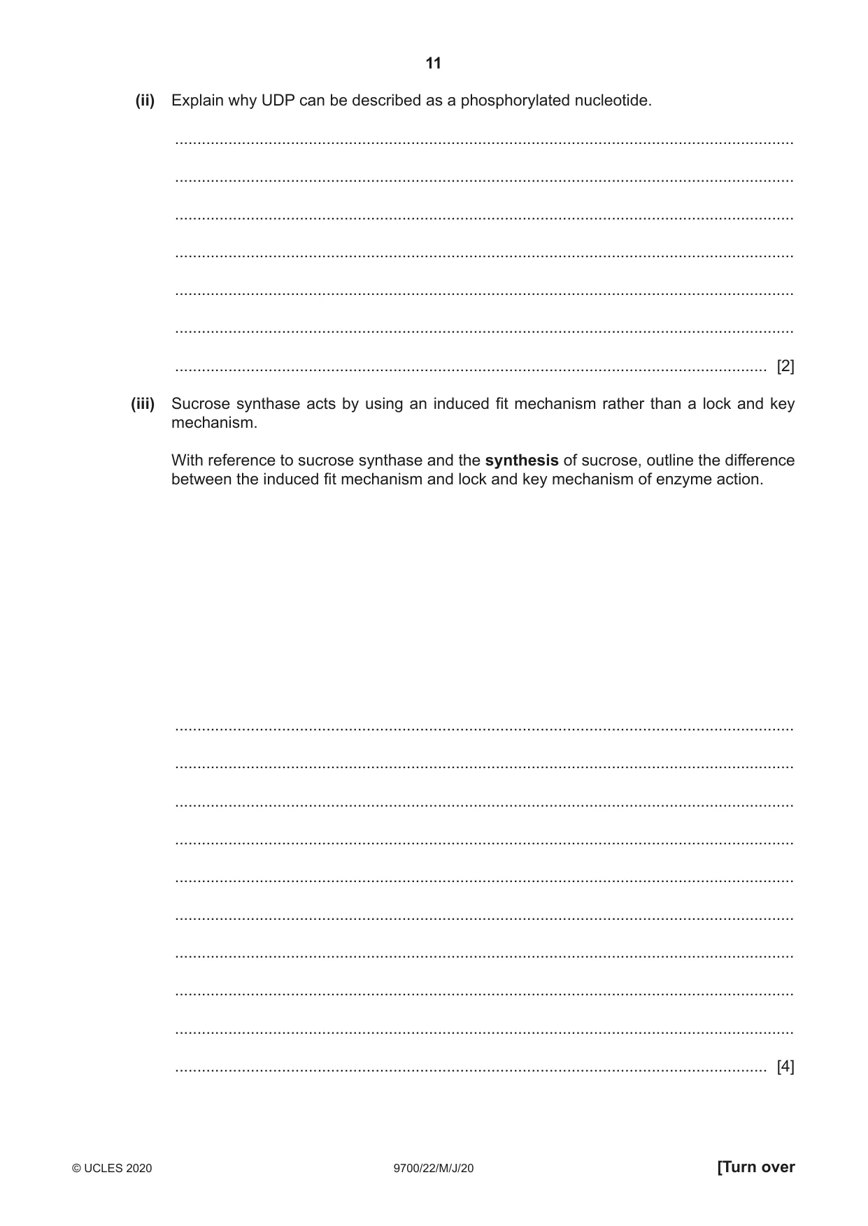
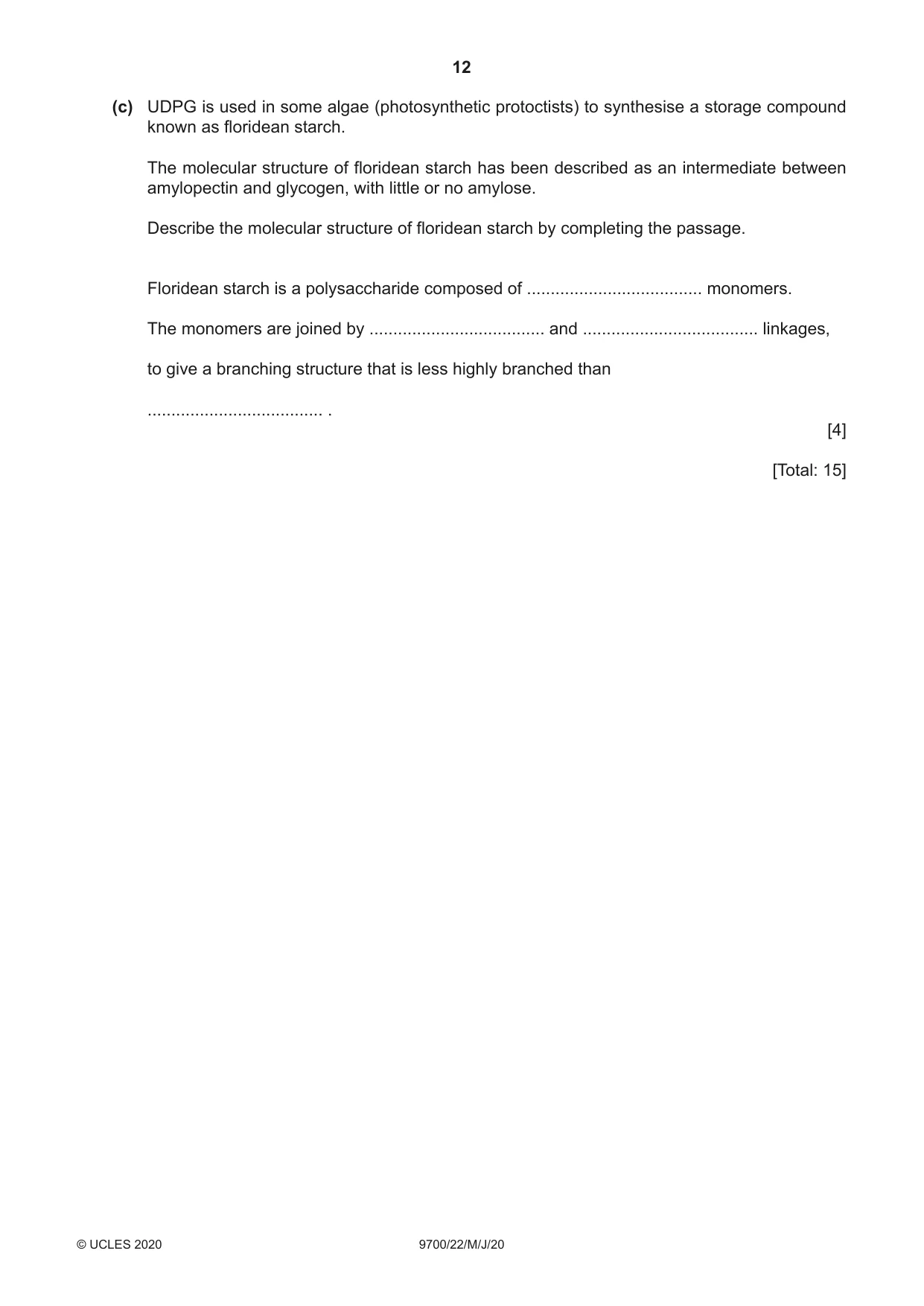
5 In most plants, sucrose is the main sugar that is transported from sources to sinks. (a) In the source, sucrose is transferred from a mesophyll cell to a phloem sieve tube through a companion cell. Describe and explain how the transfer of sucrose into a phloem sieve tube from a companion cell can lead to the transport of the sugar to a sink. ................................................................................................................................................... ................................................................................................................................................... ................................................................................................................................................... ................................................................................................................................................... ................................................................................................................................................... ................................................................................................................................................... ................................................................................................................................................... ................................................................................................................................................... ................................................................................................................................................... ................................................................................................................................................... ............................................................................................................................................. [4] (b) Fig. 5.1 shows part of one pathway that is used in plant cells to synthesise sucrose. The enzyme sucrose synthase catalyses the transfer of glucose from UDPG (uridine diphosphate glucose) to fructose. sucrose synthase UDPG + fructose UDP + sucrose Fig. 5.1 The structure of UDPG is shown in Fig. 5.2. uracil glucose ribose Fig. 5.2 (i) Name the type of bond formed when sucrose is synthesised. ..................................................................................................................................... [1] (ii) Explain why UDP can be described as a phosphorylated nucleotide. ........................................................................................................................................... ........................................................................................................................................... ........................................................................................................................................... ........................................................................................................................................... ........................................................................................................................................... ........................................................................................................................................... ..................................................................................................................................... [2] (iii) Sucrose synthase acts by using an induced fit mechanism rather than a lock and key mechanism. With reference to sucrose synthase and the synthesis of sucrose, outline the difference between the induced fit mechanism and lock and key mechanism of enzyme action. ........................................................................................................................................... ........................................................................................................................................... ........................................................................................................................................... ........................................................................................................................................... ........................................................................................................................................... ........................................................................................................................................... ........................................................................................................................................... ........................................................................................................................................... ........................................................................................................................................... ..................................................................................................................................... [4] (c) UDPG is used in some algae (photosynthetic protoctists) to synthesise a storage compound known as floridean starch. The molecular structure of floridean starch has been described as an intermediate between amylopectin and glycogen, with little or no amylose. Describe the molecular structure of floridean starch by completing the passage. Floridean starch is a polysaccharide composed of ..................................... monomers. The monomers are joined by ..................................... and ..................................... linkages, to give a branching structure that is less highly branched than ..................................... . [4] [Total: 15]
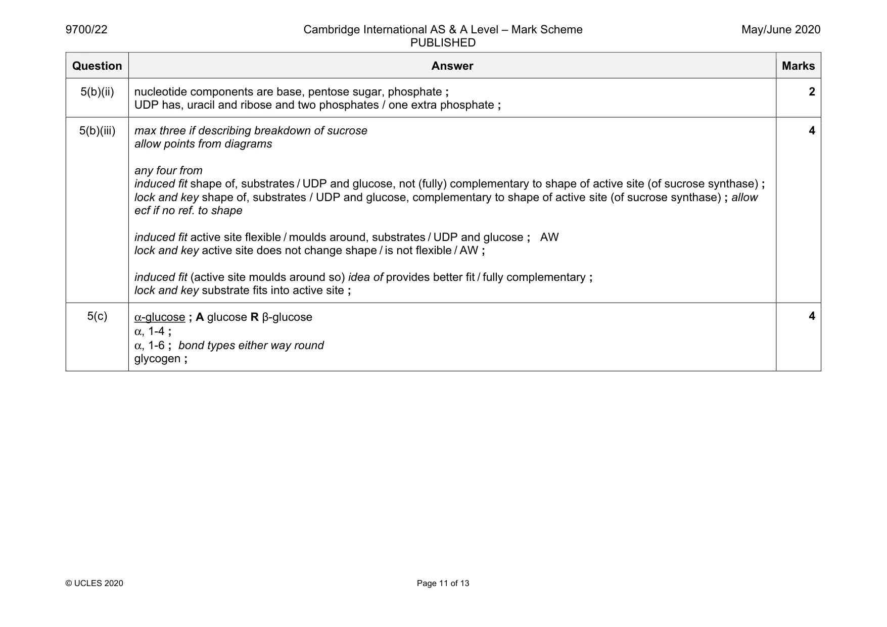
Mark scheme: 5(a) presence of sucrose (in sieve tube) lowers water potential (of phloem sap) ; A makes water potential more negative water enters (sieve tube), by osmosis / down water potential gradient ; increases volume (in sieve tube) ; increases hydrostatic pressure (in sieve tube at source) ; A turgor pressure ref. to lower hydrostatic pressure (in sieve tube at sink) ; allow ecf for no ref. to, hydrostatic / turgor detail ; e.g. sucrose removed at sink water follows sucrose that exits sink movement of, (phloem) sap / sucrose, down pressure gradient / from high to low hydrostatic pressure ; mass flow ; 4 5(b)(i) glycosidic ; 1 Question Answer Marks 5(b)(ii) nucleotide components are base, pentose sugar, phosphate ; UDP has, uracil and ribose and two phosphates / one extra phosphate ; 2 5(b)(iii) max three if describing breakdown of sucrose allow points from diagrams any four from induced fit shape of, substrates / UDP and glucose, not (fully) complementary to shape of active site (of sucrose synthase) ; lock and key shape of, substrates / UDP and glucose, complementary to shape of active site (of sucrose synthase) ; allow ecf if no ref. to shape induced fit active site flexible / moulds around, substrates / UDP and glucose ; AW lock and key active site does not change shape / is not flexible / AW ; induced fit (active site moulds around so) idea of provides better fit / fully complementary ; lock and key substrate fits into active site ; 4 5(c) α-glucose ; A glucose R β-glucose α, 1-4 ; α, 1-6 ; bond types either way round glycogen ; 4
Q6 · The mitotic cell cycle in dividing cells is very carefully controlled
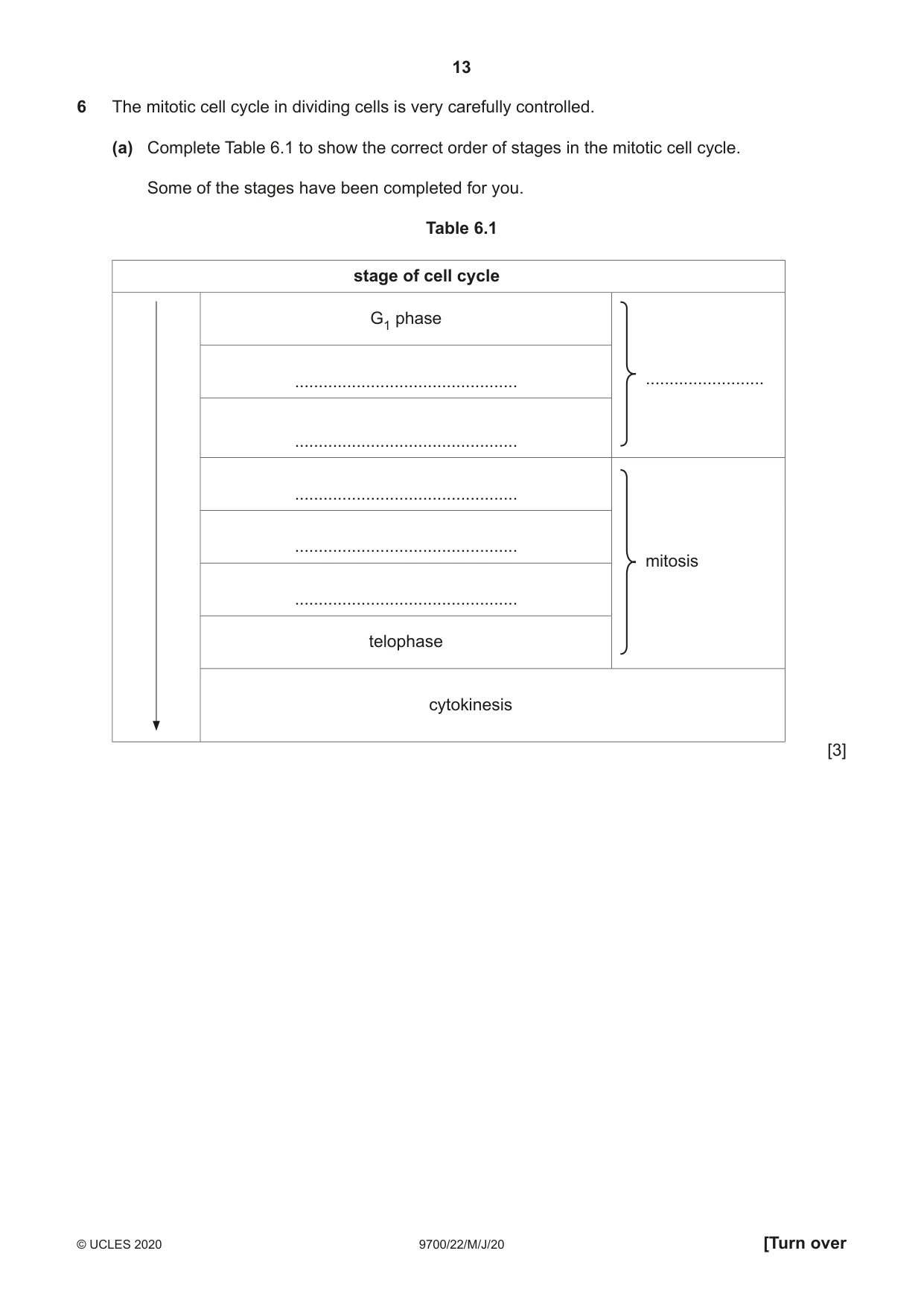
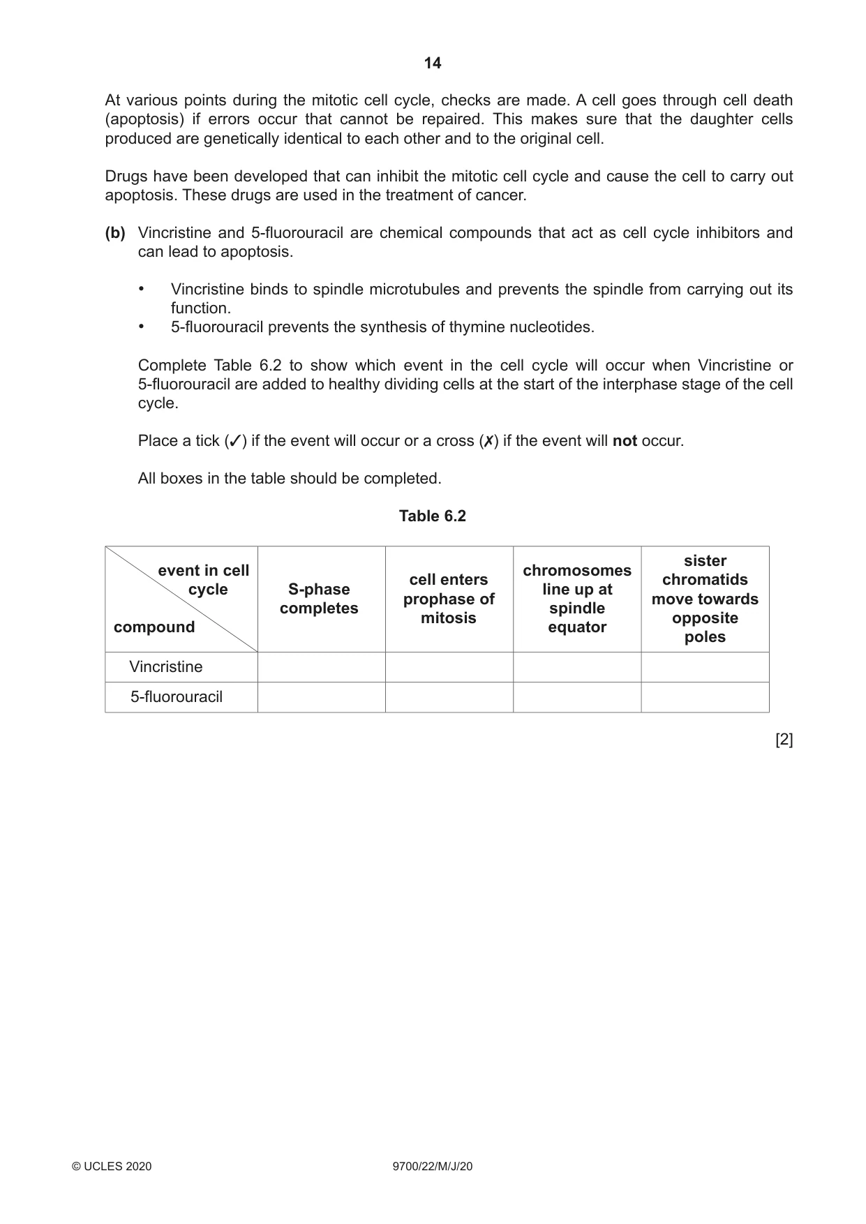
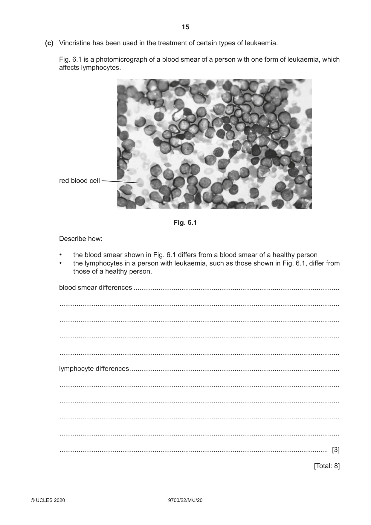
6 The mitotic cell cycle in dividing cells is very carefully controlled. (a) Complete Table 6.1 to show the correct order of stages in the mitotic cell cycle. Some of the stages have been completed for you. Table 6.1 stage of cell cycle G1 phase ............................................... ......................... ............................................... ............................................... ............................................... mitosis ............................................... telophase cytokinesis [3] At various points during the mitotic cell cycle, checks are made. A cell goes through cell death (apoptosis) if errors occur that cannot be repaired. This makes sure that the daughter cells produced are genetically identical to each other and to the original cell. Drugs have been developed that can inhibit the mitotic cell cycle and cause the cell to carry out apoptosis. These drugs are used in the treatment of cancer. (b) Vincristine and 5-fluorouracil are chemical compounds that act as cell cycle inhibitors and can lead to apoptosis. • Vincristine binds to spindle microtubules and prevents the spindle from carrying out its function. • 5-fluorouracil prevents the synthesis of thymine nucleotides. Complete Table 6.2 to show which event in the cell cycle will occur when Vincristine or 5-fluorouracil are added to healthy dividing cells at the start of the interphase stage of the cell cycle. Place a tick (3) if the event will occur or a cross (7) if the event will not occur. All boxes in the table should be completed. Table 6.2 sister event in cell chromosomes cell enters chromatids cycle S-phase line up at prophase of move towards completes spindle mitosis opposite compound equator poles Vincristine 5-fluorouracil [2] (c) Vincristine has been used in the treatment of certain types of leukaemia. Fig. 6.1 is a photomicrograph of a blood smear of a person with one form of leukaemia, which affects lymphocytes. red blood cell Fig. 6.1 Describe how: • the blood smear shown in Fig. 6.1 differs from a blood smear of a healthy person • the lymphocytes in a person with leukaemia, such as those shown in Fig. 6.1, differ from those of a healthy person. blood smear differences ............................................................................................................ ................................................................................................................................................... ................................................................................................................................................... ................................................................................................................................................... ................................................................................................................................................... lymphocyte differences .............................................................................................................. ................................................................................................................................................... ................................................................................................................................................... ................................................................................................................................................... ................................................................................................................................................... ............................................................................................................................................. [3] [Total: 8]
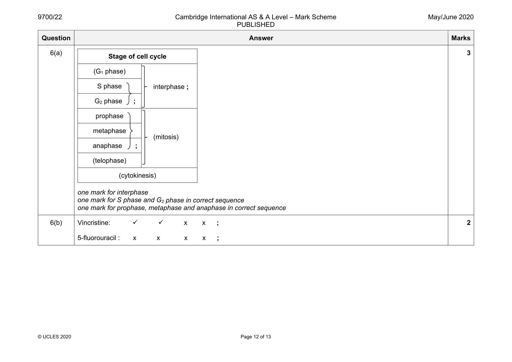
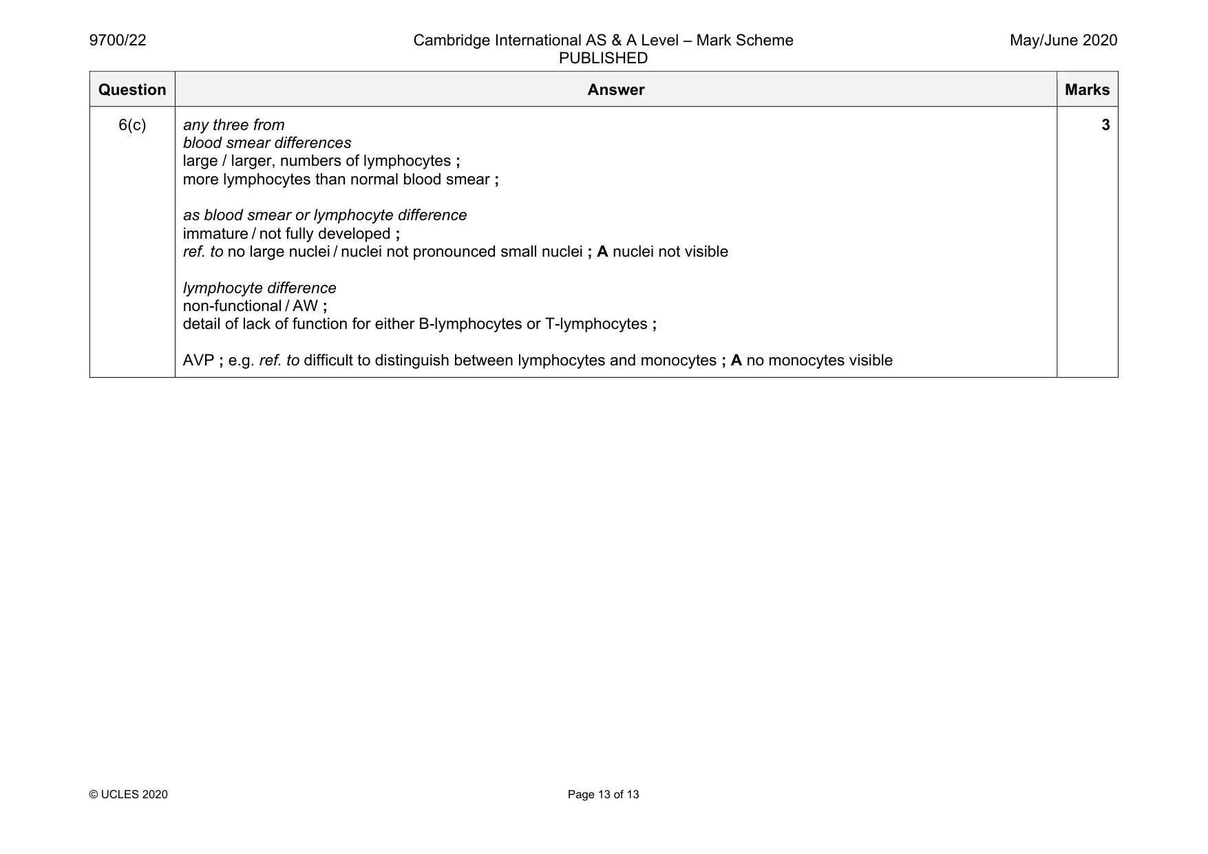
Mark scheme: 6(a) Stage of cell cycle (G1 phase) interphase ; S phase G2 phase ; prophase (mitosis) metaphase anaphase ; (telophase) (cytokinesis) one mark for interphase one mark for S phase and G2 phase in correct sequence one mark for prophase, metaphase and anaphase in correct sequence 3 6(b) Vincristine: x x ; 5-fluorouracil : x x x x ; 2 Question Answer Marks 6(c) any three from blood smear differences large / larger, numbers of lymphocytes ; more lymphocytes than normal blood smear ; as blood smear or lymphocyte difference immature / not fully developed ; ref. to no large nuclei / nuclei not pronounced small nuclei ; A nuclei not visible lymphocyte difference non-functional / AW ; detail of lack of function for either B-lymphocytes or T-lymphocytes ; AVP ; e.g. ref. to difficult to distinguish between lymphocytes and monocytes ; A no monocytes visible 3
More questions on Replication and division of nuclei and cells
What was in this paper
The subtopics covered by these 6 questions, and how many questions each got. Open one in a new tab to see every Cambridge question on it.