Cambridge A Level Biology 9700 — 2019 Oct/Nov Paper 2 · Variant 3
9700/23/O/N/19 · 6 questions · 60 marks · ≈68 min
The question paper and its mark scheme, free to read here and free to download. This is Cambridge’s own paper, exactly as it was sat.
Question paper16 pages
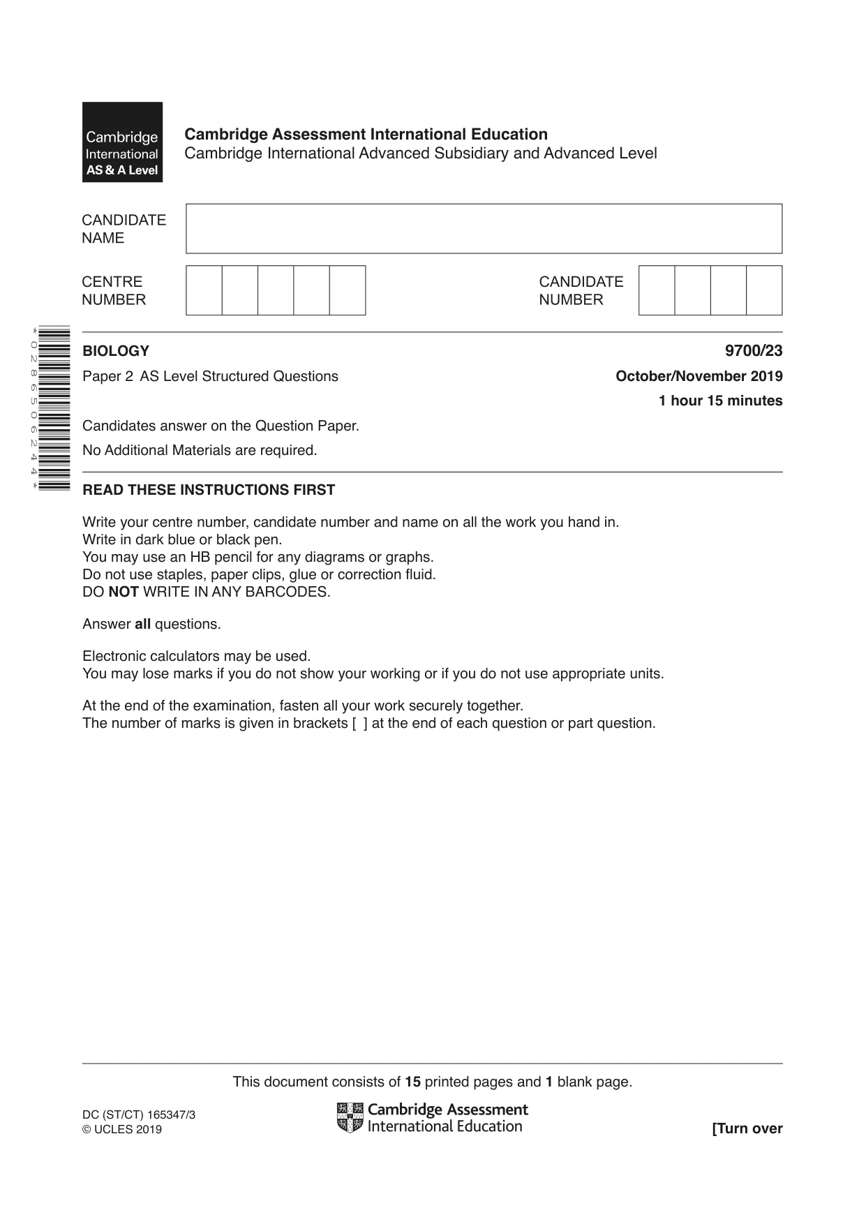
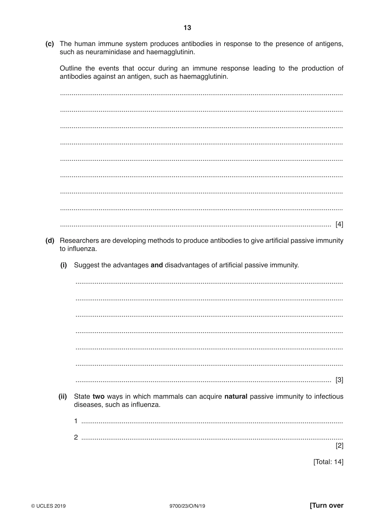
Mark scheme18 pages
Answers below. Sit the paper first if you are practising.
Questions as text
Q1 · A diagram of a molecule of haemoglobin
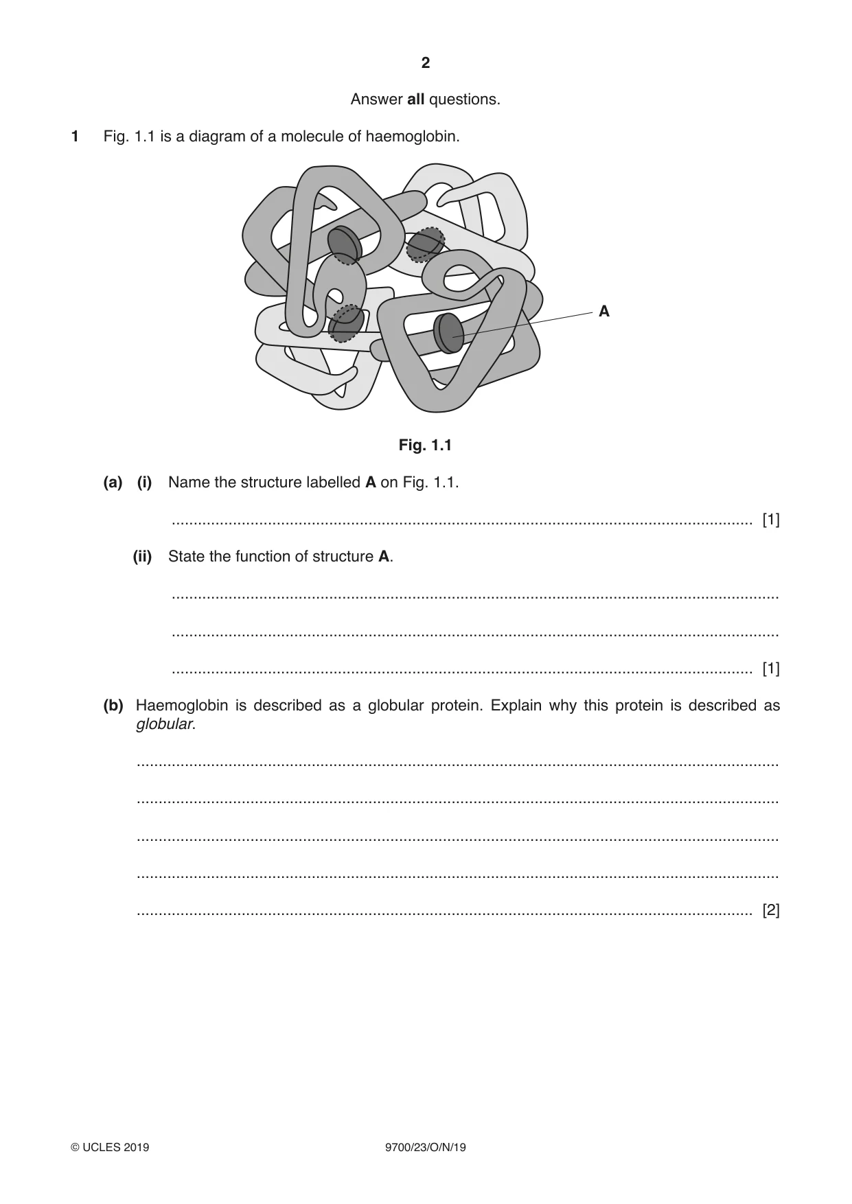
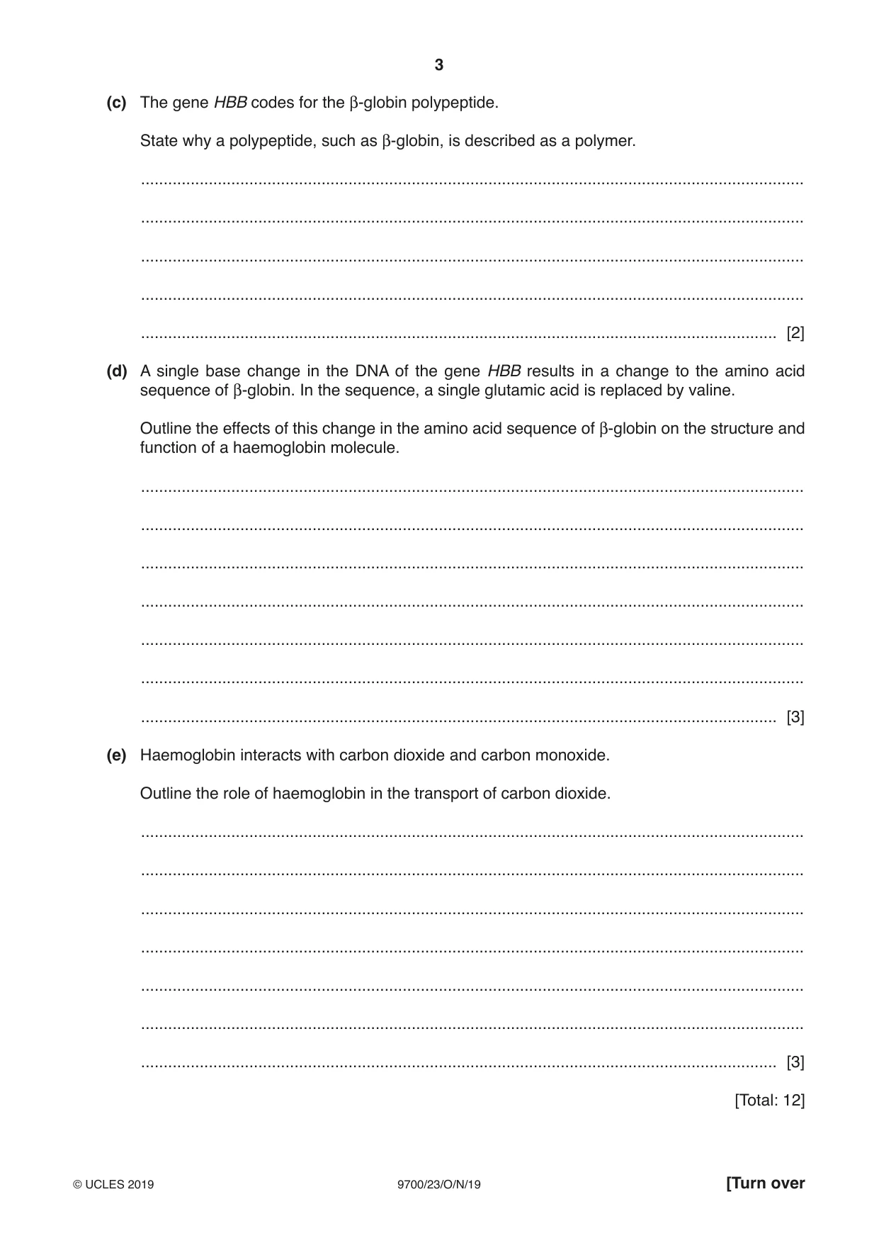
1 Fig. 1.1 is a diagram of a molecule of haemoglobin. A Fig. 1.1 (a) (i) Name the structure labelled A on Fig. 1.1. ..................................................................................................................................... [1] (ii) State the function of structure A. ........................................................................................................................................... ........................................................................................................................................... ..................................................................................................................................... [1] (b) Haemoglobin is described as a globular protein. Explain why this protein is described as globular. ................................................................................................................................................... ................................................................................................................................................... ................................................................................................................................................... ................................................................................................................................................... ............................................................................................................................................. [2] (c) The gene HBB codes for the β-globin polypeptide. State why a polypeptide, such as β-globin, is described as a polymer. ................................................................................................................................................... ................................................................................................................................................... ................................................................................................................................................... ................................................................................................................................................... ............................................................................................................................................. [2] (d) A single base change in the DNA of the gene HBB results in a change to the amino acid sequence of β-globin. In the sequence, a single glutamic acid is replaced by valine. Outline the effects of this change in the amino acid sequence of β-globin on the structure and function of a haemoglobin molecule. ................................................................................................................................................... ................................................................................................................................................... ................................................................................................................................................... ................................................................................................................................................... ................................................................................................................................................... ................................................................................................................................................... ............................................................................................................................................. [3] (e) Haemoglobin interacts with carbon dioxide and carbon monoxide. Outline the role of haemoglobin in the transport of carbon dioxide. ................................................................................................................................................... ................................................................................................................................................... ................................................................................................................................................... ................................................................................................................................................... ................................................................................................................................................... ................................................................................................................................................... ............................................................................................................................................. [3] [Total: 12]
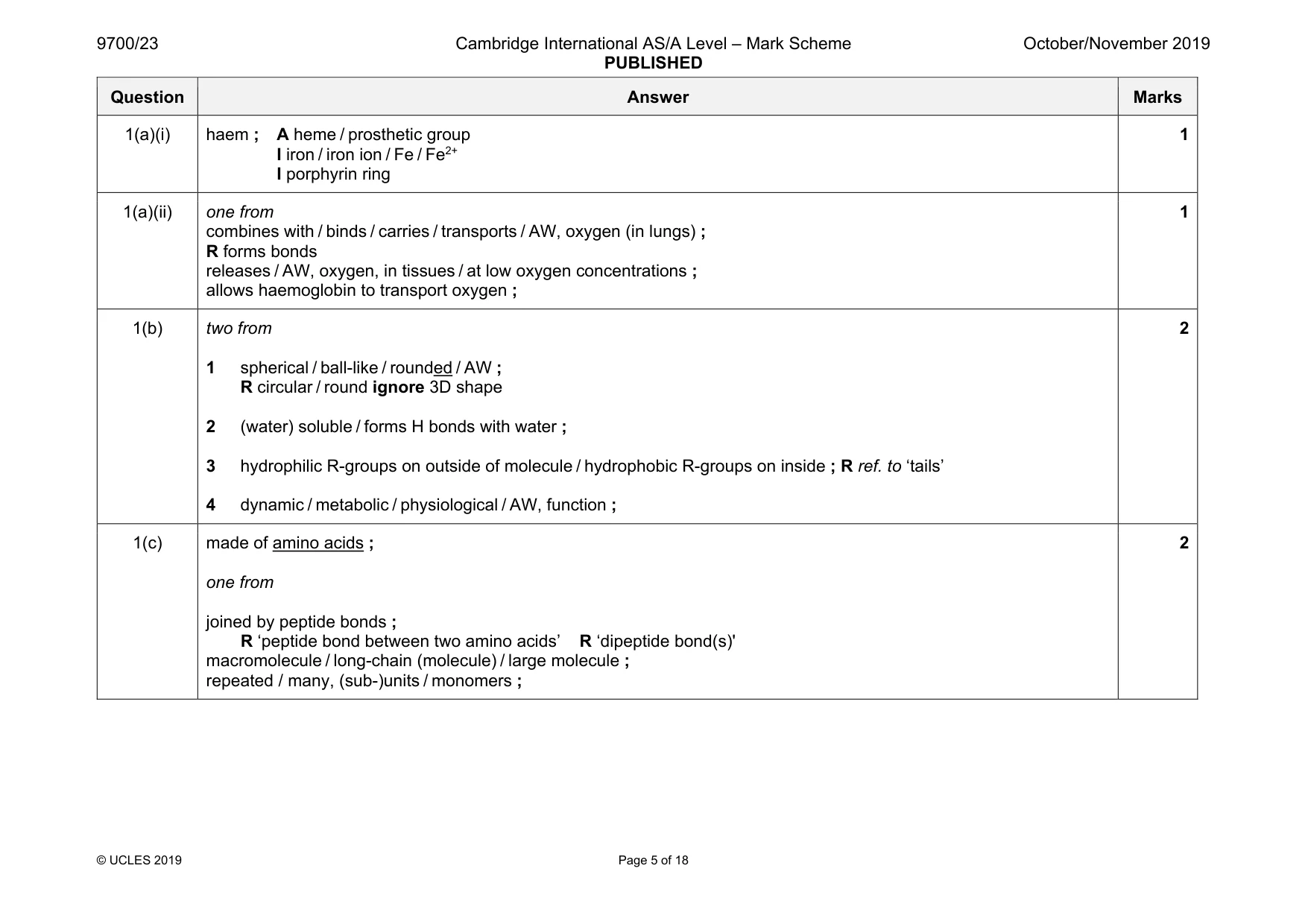
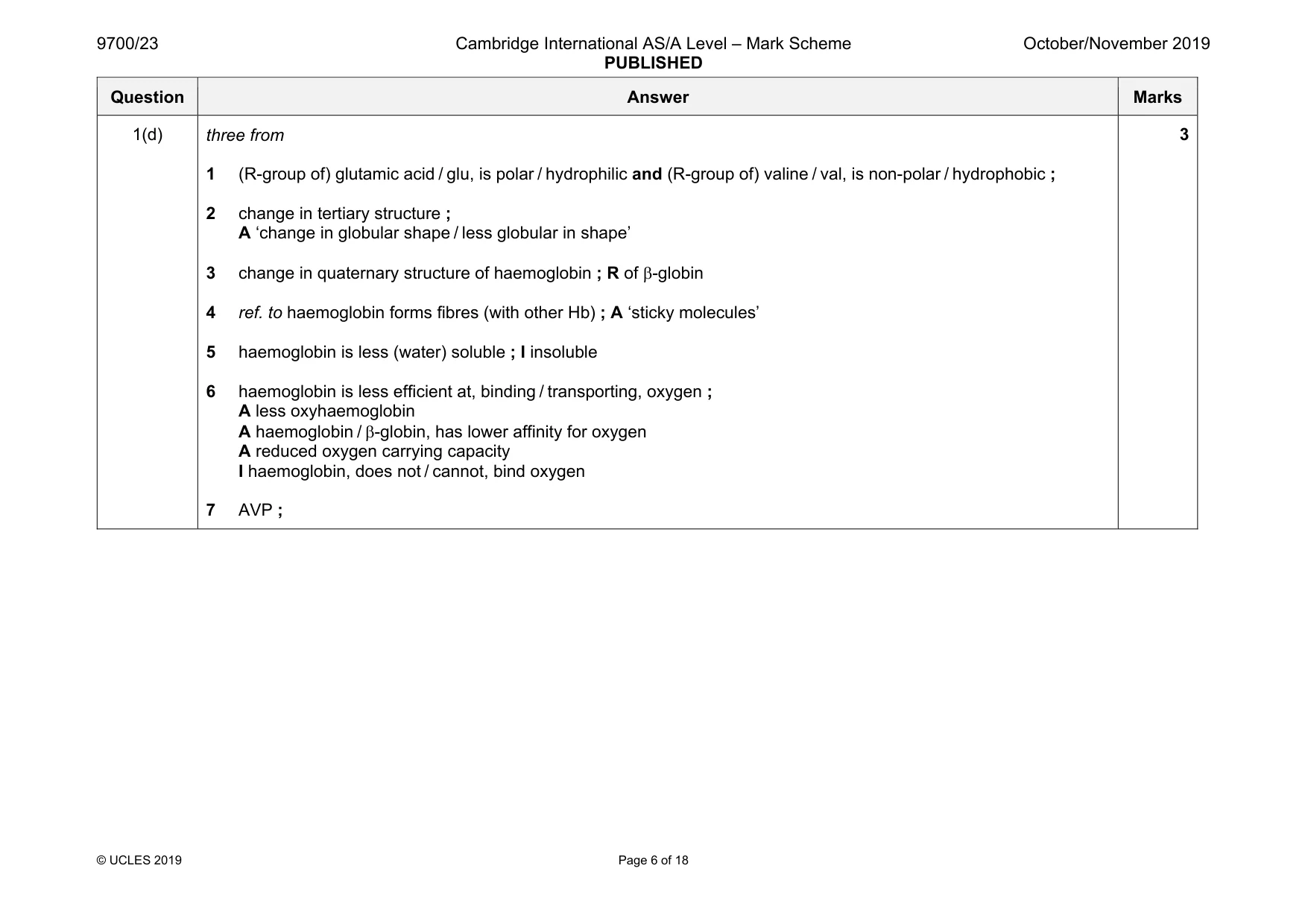
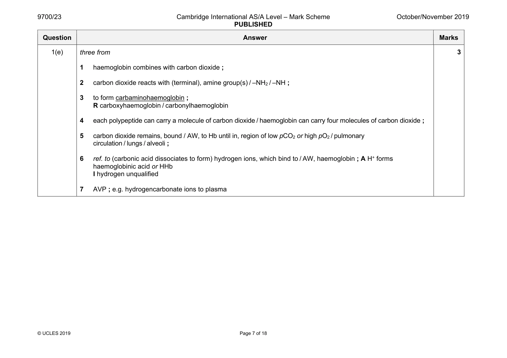
Mark scheme: 1(a)(i) haem ; A heme / prosthetic group I iron / iron ion / Fe / Fe2+ I porphyrin ring 1 1(a)(ii) one from combines with / binds / carries / transports / AW, oxygen (in lungs) ; R forms bonds releases / AW, oxygen, in tissues / at low oxygen concentrations ; allows haemoglobin to transport oxygen ; 1 1(b) two from 1 spherical / ball-like / rounded / AW ; R circular / round ignore 3D shape 2 (water) soluble / forms H bonds with water ; 3 hydrophilic R-groups on outside of molecule / hydrophobic R-groups on inside ; R ref. to ‘tails’ 4 dynamic / metabolic / physiological / AW, function ; 2 1(c) made of amino acids ; one from joined by peptide bonds ; R ‘peptide bond between two amino acids’ R ‘dipeptide bond(s)' macromolecule / long-chain (molecule) / large molecule ; repeated / many, (sub-)units / monomers ; 2 Question Answer Marks 1(d) three from 1 (R-group of) glutamic acid / glu, is polar / hydrophilic and (R-group of) valine / val, is non-polar / hydrophobic ; 2 change in tertiary structure ; A ‘change in globular shape / less globular in shape’ 3 change in quaternary structure of haemoglobin ; R of β-globin 4 ref. to haemoglobin forms fibres (with other Hb) ; A ‘sticky molecules’ 5 haemoglobin is less (water) soluble ; I insoluble 6 haemoglobin is less efficient at, binding / transporting, oxygen ; A less oxyhaemoglobin A haemoglobin / β-globin, has lower affinity for oxygen A reduced oxygen carrying capacity I haemoglobin, does not / cannot, bind oxygen 7 AVP ; 3 Question Answer Marks 1(e) three from 1 haemoglobin combines with carbon dioxide ; 2 carbon dioxide reacts with (terminal), amine group(s) / –NH2 / –NH ; 3 to form carbaminohaemoglobin ; R carboxyhaemoglobin / carbonylhaemoglobin 4 each polypeptide can carry a molecule of carbon dioxide / haemoglobin can carry four molecules of carbon dioxide ; 5 carbon dioxide remains, bound / AW, to Hb until in, region of low pCO2 or high pO2 / pulmonary circulation / lungs / alveoli ; 6 ref. to (carbonic acid dissociates to form) hydrogen ions, which bind to / AW, haemoglobin ; A H+ forms haemoglobinic acid or HHb I hydrogen unqualified 7 AVP ; e.g. hydrogencarbonate ions to plasma 3
Q2 · Meristematic tissue is found in the growing region of plants, such as root tips
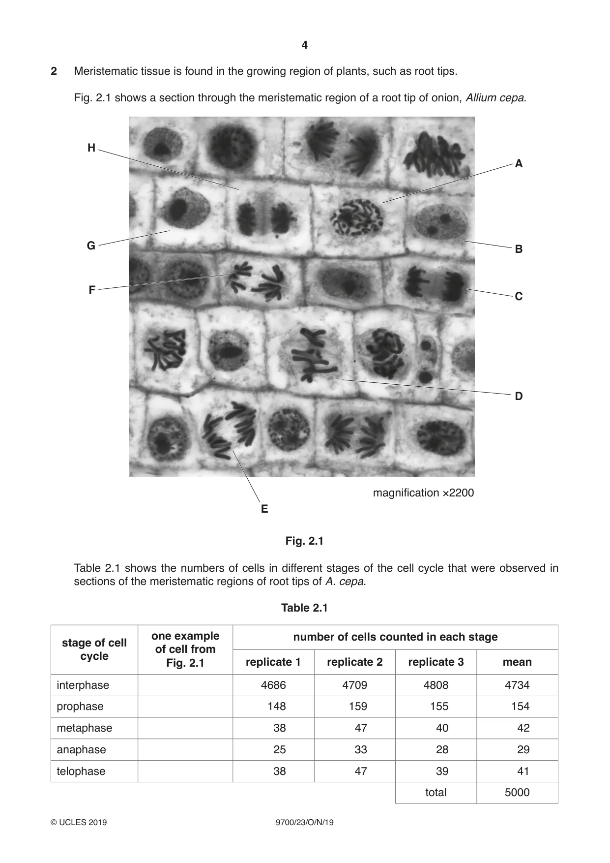
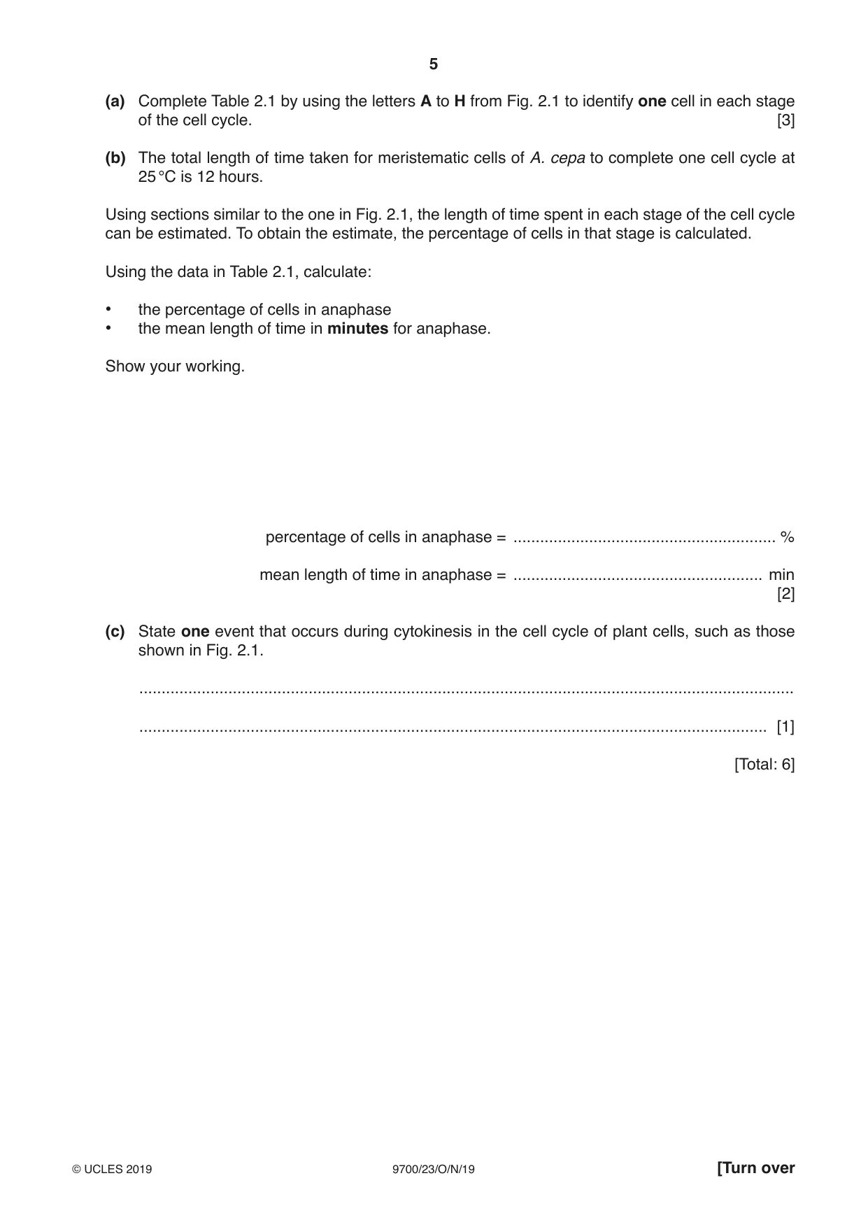
2 Meristematic tissue is found in the growing region of plants, such as root tips. Fig. 2.1 shows a section through the meristematic region of a root tip of onion, Allium cepa. H A G B F C D magnification ×2200 E Fig. 2.1 Table 2.1 shows the numbers of cells in different stages of the cell cycle that were observed in sections of the meristematic regions of root tips of A. cepa. Table 2.1 one example number of cells counted in each stage stage of cell of cell from cycle Fig. 2.1 replicate 1 replicate 2 replicate 3 mean interphase 4686 4709 4808 4734 prophase 148 159 155 154 metaphase 38 47 40 42 anaphase 25 33 28 29 telophase 38 47 39 41 total 5000 (a) Complete Table 2.1 by using the letters A to H from Fig. 2.1 to identify one cell in each stage of the cell cycle. [3] (b) The total length of time taken for meristematic cells of A. cepa to complete one cell cycle at 25 °C is 12 hours. Using sections similar to the one in Fig. 2.1, the length of time spent in each stage of the cell cycle can be estimated. To obtain the estimate, the percentage of cells in that stage is calculated. Using the data in Table 2.1, calculate: • the percentage of cells in anaphase • the mean length of time in minutes for anaphase. Show your working. percentage of cells in anaphase = ........................................................... % mean length of time in anaphase = ........................................................ min [2] (c) State one event that occurs during cytokinesis in the cell cycle of plant cells, such as those shown in Fig. 2.1. ................................................................................................................................................... ............................................................................................................................................. [1] [Total: 6]
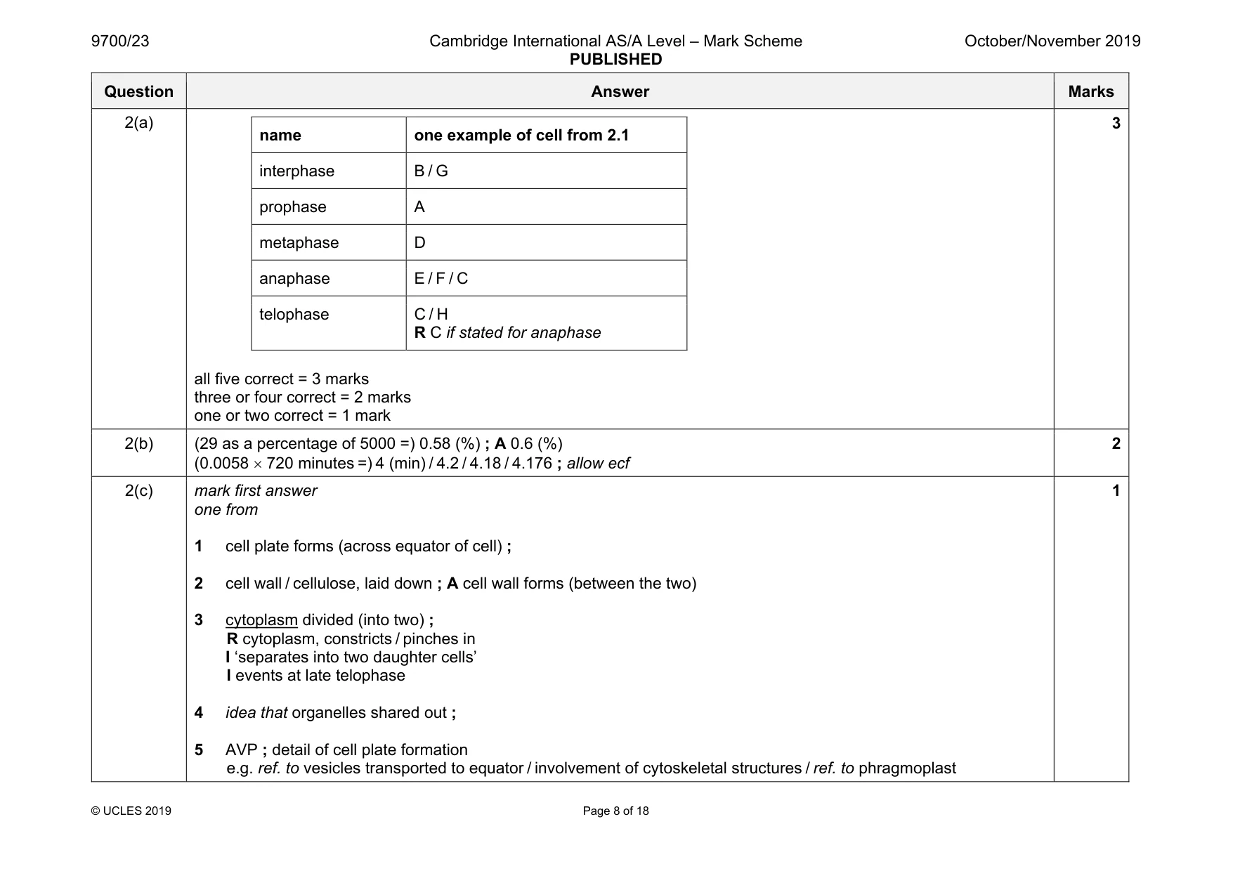
Mark scheme: 2(a) name one example of cell from 2.1 interphase B / G prophase A metaphase D anaphase E / F / C telophase C / H R C if stated for anaphase all five correct = 3 marks three or four correct = 2 marks one or two correct = 1 mark 3 2(b) (29 as a percentage of 5000 =) 0.58 (%) ; A 0.6 (%) (0.0058 × 720 minutes =) 4 (min) / 4.2 / 4.18 / 4.176 ; allow ecf 2 2(c) mark first answer one from 1 cell plate forms (across equator of cell) ; 2 cell wall / cellulose, laid down ; A cell wall forms (between the two) 3 cytoplasm divided (into two) ; R cytoplasm, constricts / pinches in I ‘separates into two daughter cells’ I events at late telophase 4 idea that organelles shared out ; 5 AVP ; detail of cell plate formation e.g. ref. to vesicles transported to equator / involvement of cytoskeletal structures / ref. to phragmoplast 1
Q3 · The tomato plant, Solanum lycopersicum, does not tolerate periods of drought (water…
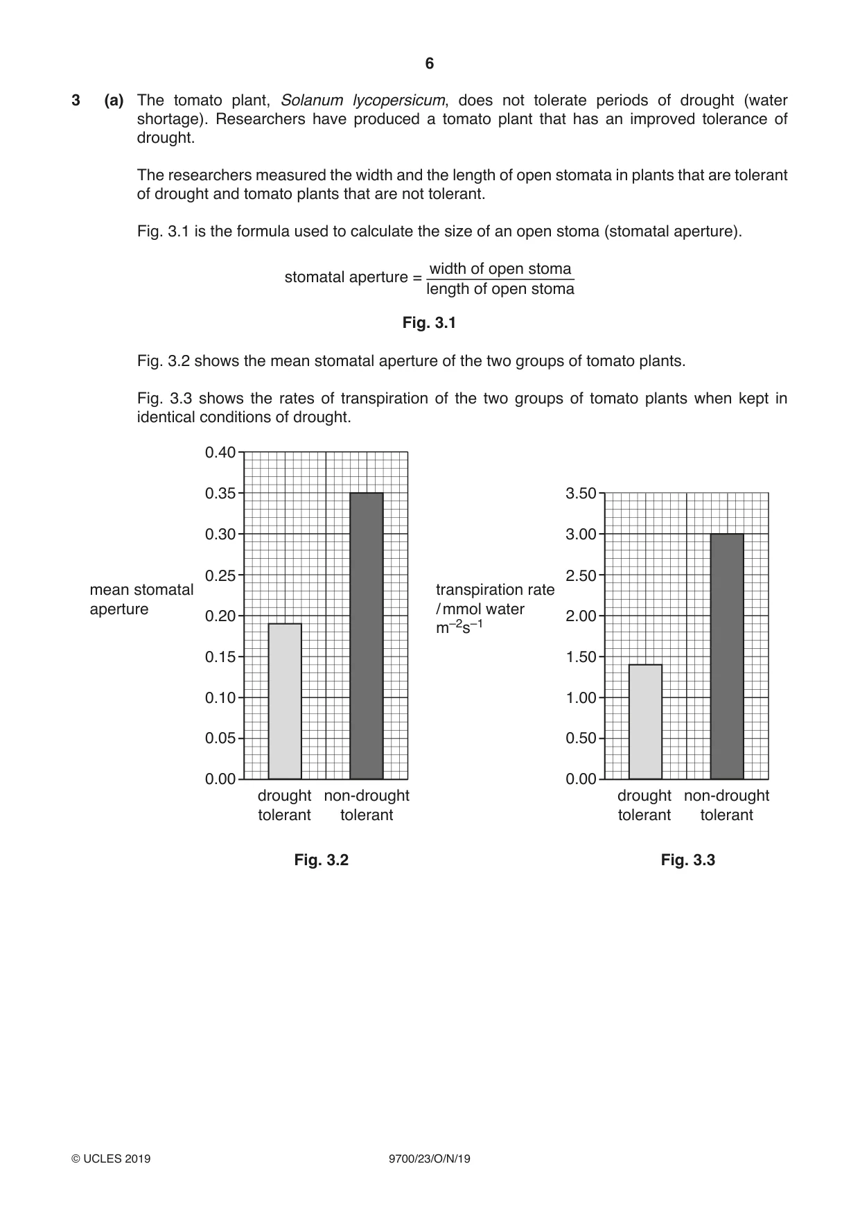
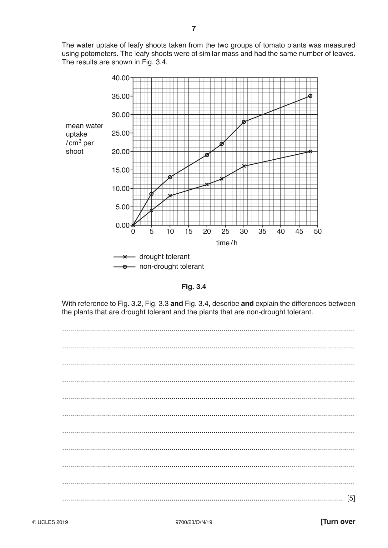
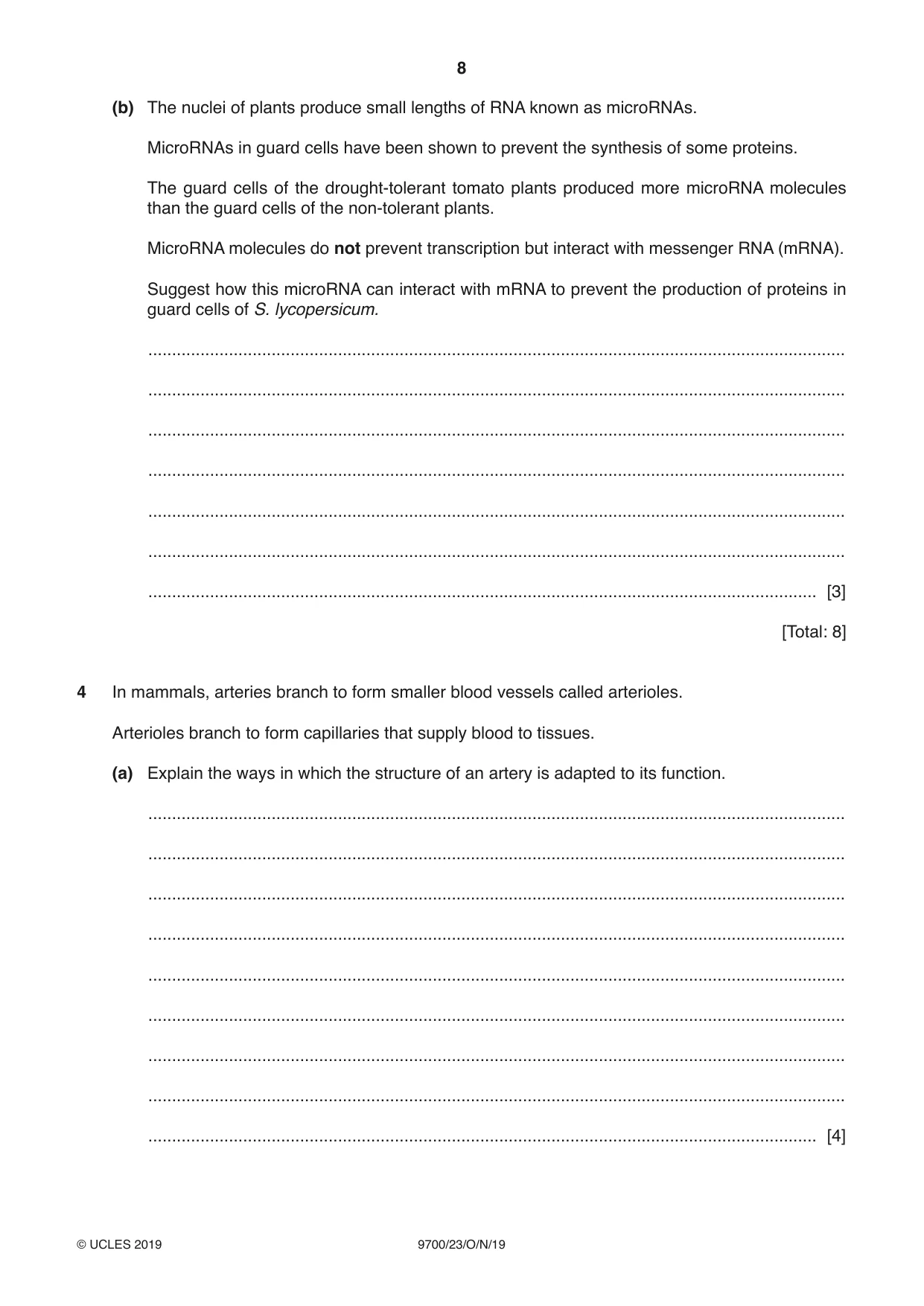
3 (a) The tomato plant, Solanum lycopersicum, does not tolerate periods of drought (water shortage). Researchers have produced a tomato plant that has an improved tolerance of drought. The researchers measured the width and the length of open stomata in plants that are tolerant of drought and tomato plants that are not tolerant. Fig. 3.1 is the formula used to calculate the size of an open stoma (stomatal aperture). width of open stoma stomatal aperture = length of open stoma Fig. 3.1 Fig. 3.2 shows the mean stomatal aperture of the two groups of tomato plants. Fig. 3.3 shows the rates of transpiration of the two groups of tomato plants when kept in identical conditions of drought. 0.40 0.35 3.50 0.30 3.00 0.25 2.50 mean stomatal transpiration rate aperture / mmol water 0.20 2.00 m–2s–1 0.15 1.50 0.10 1.00 0.05 0.50 0.00 0.00 drought non-drought drought non-drought tolerant tolerant tolerant tolerant Fig. 3.2 Fig. 3.3 The water uptake of leafy shoots taken from the two groups of tomato plants was measured using potometers. The leafy shoots were of similar mass and had the same number of leaves. The results are shown in Fig. 3.4. 40.00 35.00 30.00 mean water uptake 25.00 / cm3 per shoot 20.00 15.00 10.00 5.00 0.00 0 5 10 15 20 25 30 35 40 45 50 time / h drought tolerant non-drought tolerant Fig. 3.4 With reference to Fig. 3.2, Fig. 3.3 and Fig. 3.4, describe and explain the differences between the plants that are drought tolerant and the plants that are non-drought tolerant. ................................................................................................................................................... ................................................................................................................................................... ................................................................................................................................................... ................................................................................................................................................... ................................................................................................................................................... ................................................................................................................................................... ................................................................................................................................................... ................................................................................................................................................... ................................................................................................................................................... ................................................................................................................................................... ............................................................................................................................................. [5] (b) The nuclei of plants produce small lengths of RNA known as microRNAs. MicroRNAs in guard cells have been shown to prevent the synthesis of some proteins. The guard cells of the drought-tolerant tomato plants produced more microRNA molecules than the guard cells of the non-tolerant plants. MicroRNA molecules do not prevent transcription but interact with messenger RNA (mRNA). Suggest how this microRNA can interact with mRNA to prevent the production of proteins in guard cells of S. lycopersicum. ................................................................................................................................................... ................................................................................................................................................... ................................................................................................................................................... ................................................................................................................................................... ................................................................................................................................................... ................................................................................................................................................... ............................................................................................................................................. [3] [Total: 8]
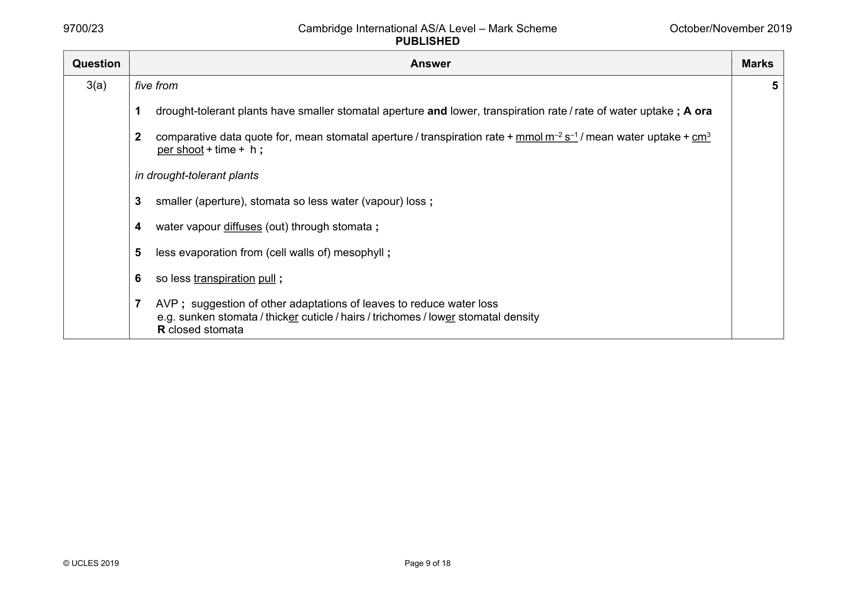
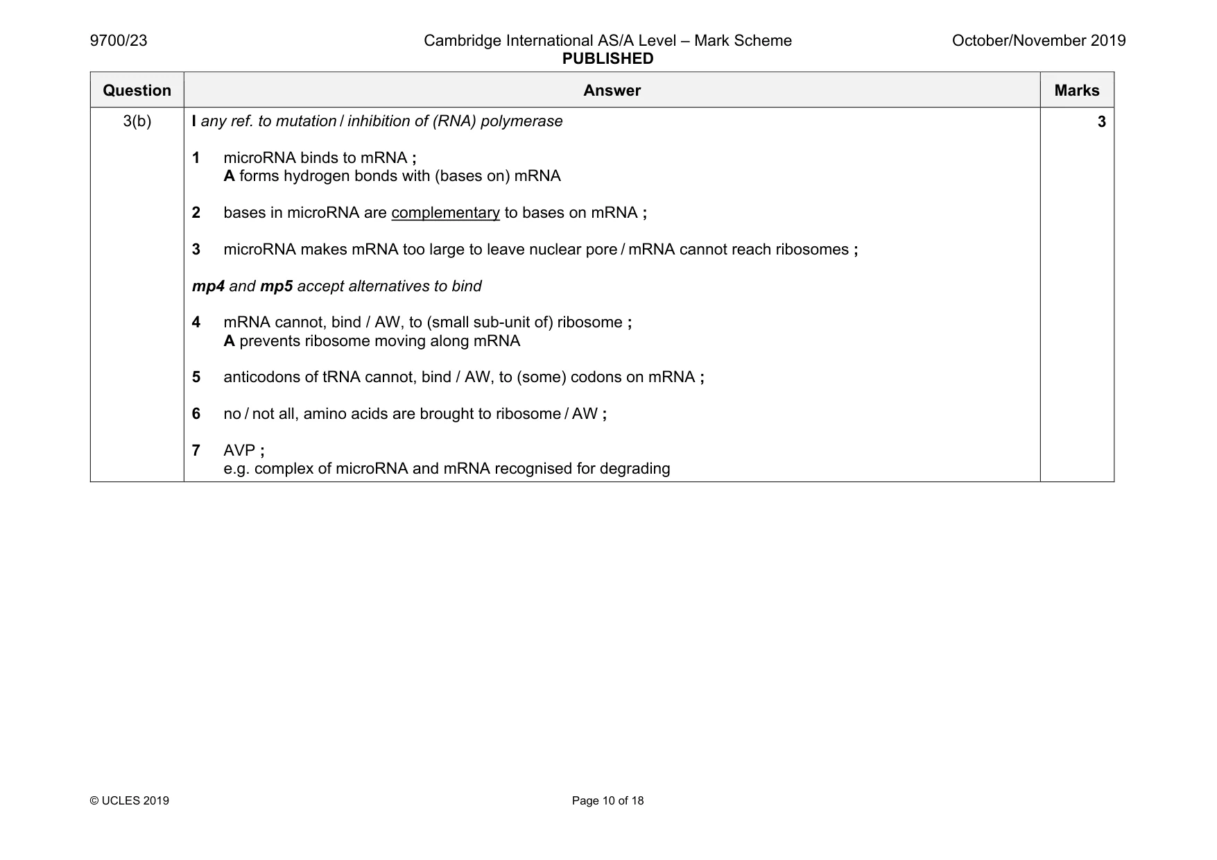
Mark scheme: 3(a) five from 1 drought-tolerant plants have smaller stomatal aperture and lower, transpiration rate / rate of water uptake ; A ora 2 comparative data quote for, mean stomatal aperture / transpiration rate + mmol m–2 s–1 / mean water uptake + cm3 per shoot + time + h ; in drought-tolerant plants 3 smaller (aperture), stomata so less water (vapour) loss ; 4 water vapour diffuses (out) through stomata ; 5 less evaporation from (cell walls of) mesophyll ; 6 so less transpiration pull ; 7 AVP ; suggestion of other adaptations of leaves to reduce water loss e.g. sunken stomata / thicker cuticle / hairs / trichomes / lower stomatal density R closed stomata 5 Question Answer Marks 3(b) I any ref. to mutation / inhibition of (RNA) polymerase 1 microRNA binds to mRNA ; A forms hydrogen bonds with (bases on) mRNA 2 bases in microRNA are complementary to bases on mRNA ; 3 microRNA makes mRNA too large to leave nuclear pore / mRNA cannot reach ribosomes ; mp4 and mp5 accept alternatives to bind 4 mRNA cannot, bind / AW, to (small sub-unit of) ribosome ; A prevents ribosome moving along mRNA 5 anticodons of tRNA cannot, bind / AW, to (some) codons on mRNA ; 6 no / not all, amino acids are brought to ribosome / AW ; 7 AVP ; e.g. complex of microRNA and mRNA recognised for degrading 3
Q4 · In mammals, arteries branch to form smaller blood vessels called arterioles
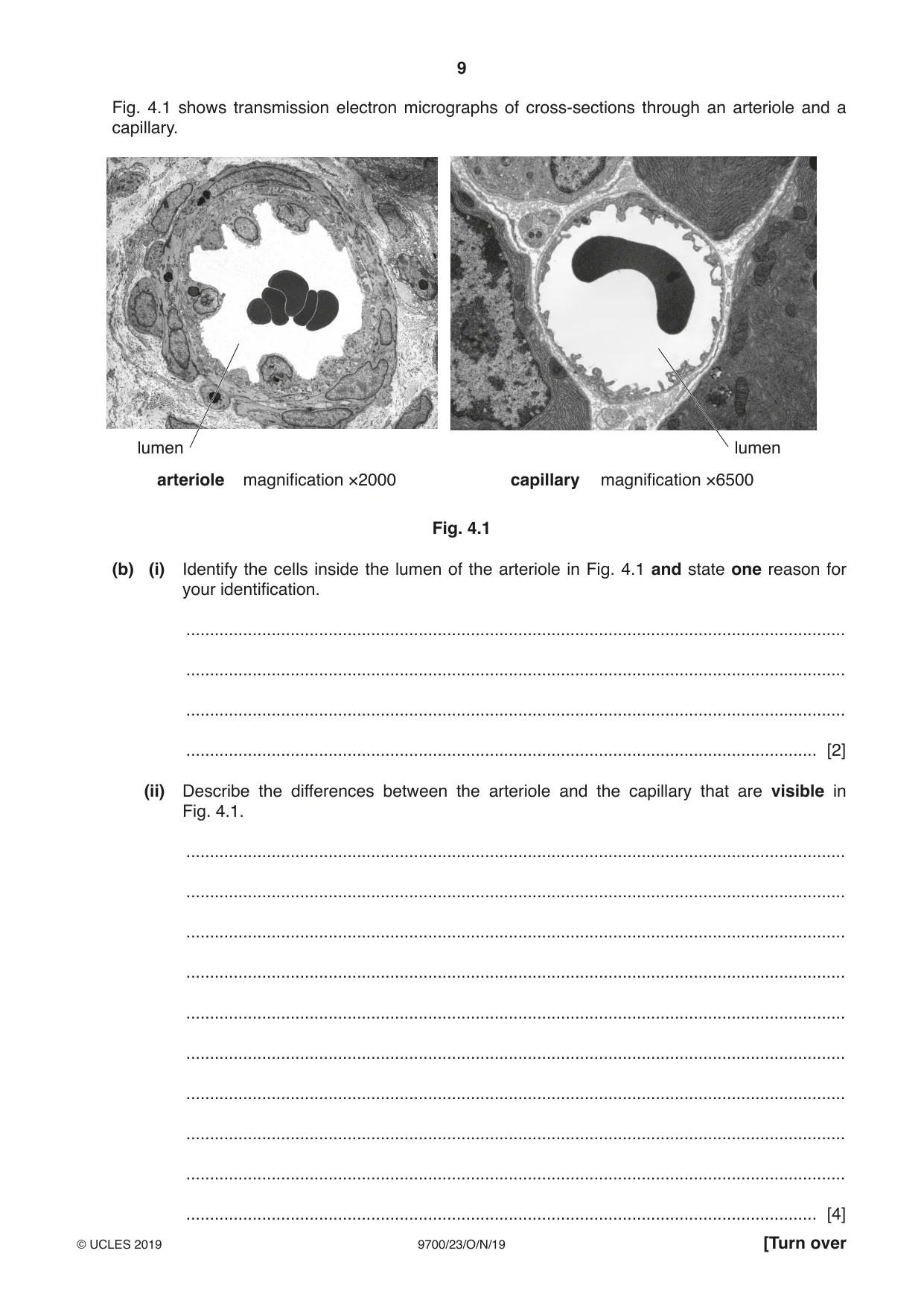
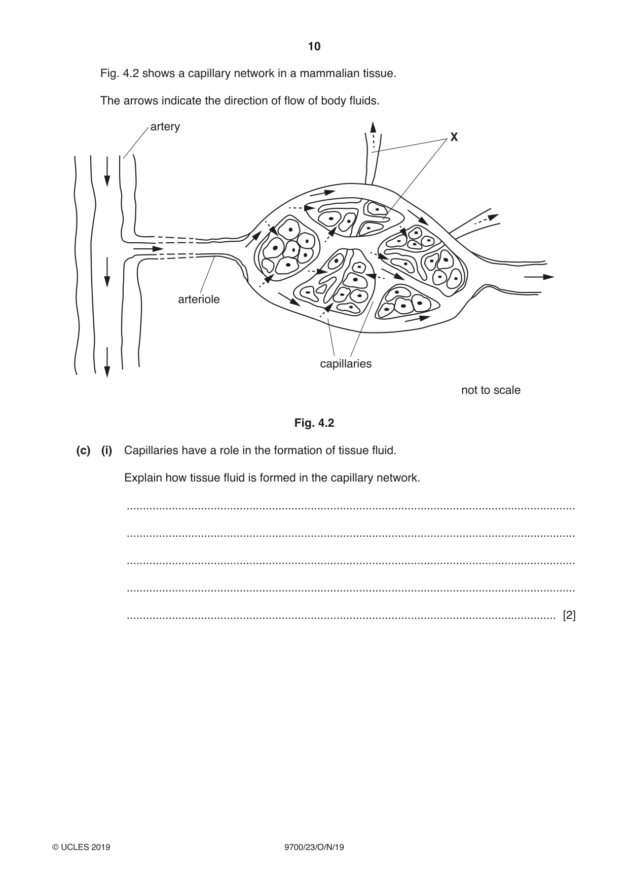
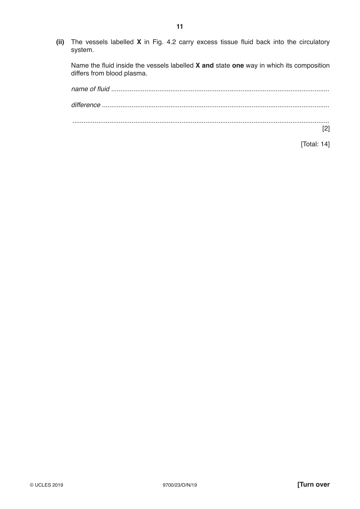
4 In mammals, arteries branch to form smaller blood vessels called arterioles. Arterioles branch to form capillaries that supply blood to tissues. (a) Explain the ways in which the structure of an artery is adapted to its function. ................................................................................................................................................... ................................................................................................................................................... ................................................................................................................................................... ................................................................................................................................................... ................................................................................................................................................... ................................................................................................................................................... ................................................................................................................................................... ................................................................................................................................................... ............................................................................................................................................. [4] Fig. 4.1 shows transmission electron micrographs of cross-sections through an arteriole and a capillary. lumen lumen arteriole magnification ×2000 capillary magnification ×6500 Fig. 4.1 (b) (i) Identify the cells inside the lumen of the arteriole in Fig. 4.1 and state one reason for your identification. ........................................................................................................................................... ........................................................................................................................................... ........................................................................................................................................... ..................................................................................................................................... [2] (ii) Describe the differences between the arteriole and the capillary that are visible in Fig. 4.1. ........................................................................................................................................... ........................................................................................................................................... ........................................................................................................................................... ........................................................................................................................................... ........................................................................................................................................... ........................................................................................................................................... ........................................................................................................................................... ........................................................................................................................................... ........................................................................................................................................... ..................................................................................................................................... [4] Fig. 4.2 shows a capillary network in a mammalian tissue. The arrows indicate the direction of flow of body fluids. artery X arteriole capillaries not to scale Fig. 4.2 (c) (i) Capillaries have a role in the formation of tissue fluid. Explain how tissue fluid is formed in the capillary network. ........................................................................................................................................... ........................................................................................................................................... ........................................................................................................................................... ........................................................................................................................................... ..................................................................................................................................... [2] (ii) The vessels labelled X in Fig. 4.2 carry excess tissue fluid back into the circulatory system. Name the fluid inside the vessels labelled X and state one way in which its composition differs from blood plasma. name of fluid ...................................................................................................................... difference ........................................................................................................................... ........................................................................................................................................... [2] [Total: 14]
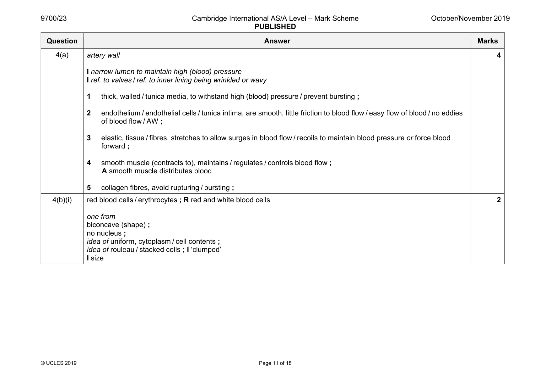
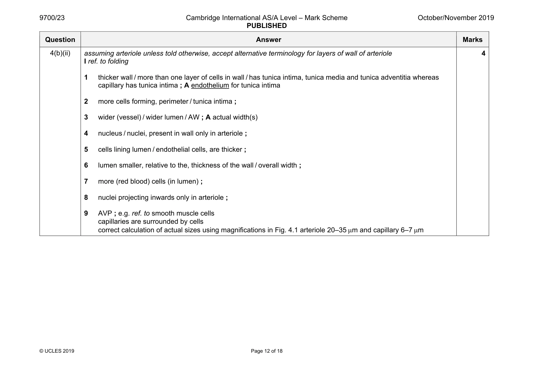
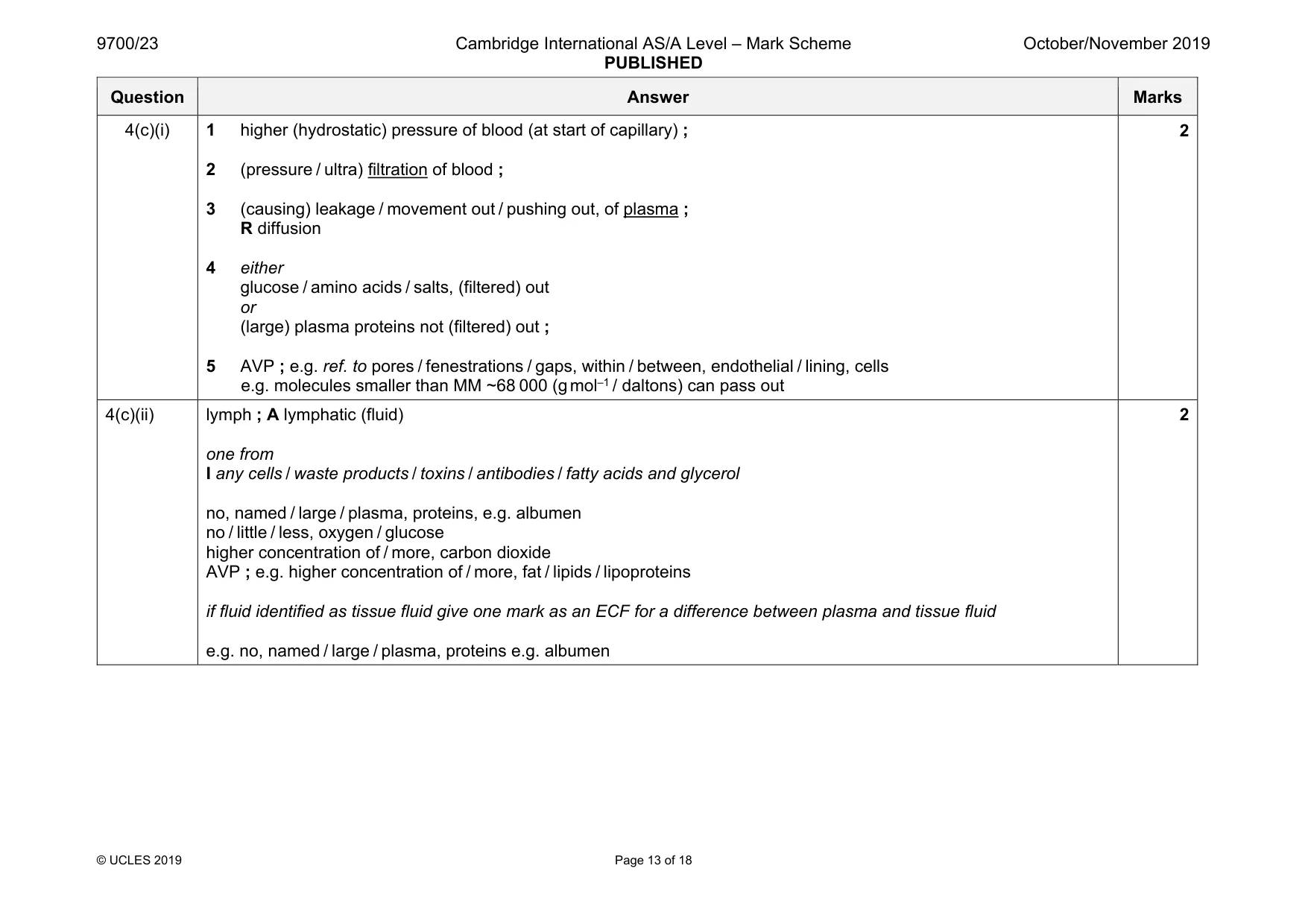
Mark scheme: 4(a) artery wall I narrow lumen to maintain high (blood) pressure I ref. to valves / ref. to inner lining being wrinkled or wavy 1 thick, walled / tunica media, to withstand high (blood) pressure / prevent bursting ; 2 endothelium / endothelial cells / tunica intima, are smooth, little friction to blood flow / easy flow of blood / no eddies of blood flow / AW ; 3 elastic, tissue / fibres, stretches to allow surges in blood flow / recoils to maintain blood pressure or force blood forward ; 4 smooth muscle (contracts to), maintains / regulates / controls blood flow ; A smooth muscle distributes blood 5 collagen fibres, avoid rupturing / bursting ; 4 4(b)(i) red blood cells / erythrocytes ; R red and white blood cells one from biconcave (shape) ; no nucleus ; idea of uniform, cytoplasm / cell contents ; idea of rouleau / stacked cells ; I ‘clumped’ I size 2 Question Answer Marks 4(b)(ii) assuming arteriole unless told otherwise, accept alternative terminology for layers of wall of arteriole I ref. to folding 1 thicker wall / more than one layer of cells in wall / has tunica intima, tunica media and tunica adventitia whereas capillary has tunica intima ; A endothelium for tunica intima 2 more cells forming, perimeter / tunica intima ; 3 wider (vessel) / wider lumen / AW ; A actual width(s) 4 nucleus / nuclei, present in wall only in arteriole ; 5 cells lining lumen / endothelial cells, are thicker ; 6 lumen smaller, relative to the, thickness of the wall / overall width ; 7 more (red blood) cells (in lumen) ; 8 nuclei projecting inwards only in arteriole ; 9 AVP ; e.g. ref. to smooth muscle cells capillaries are surrounded by cells correct calculation of actual sizes using magnifications in Fig. 4.1 arteriole 20–35 µm and capillary 6–7 µm 4 Question Answer Marks 4(c)(i) 1 higher (hydrostatic) pressure of blood (at start of capillary) ; 2 (pressure / ultra) filtration of blood ; 3 (causing) leakage / movement out / pushing out, of plasma ; R diffusion 4 either glucose / amino acids / salts, (filtered) out or (large) plasma proteins not (filtered) out ; 5 AVP ; e.g. ref. to pores / fenestrations / gaps, within / between, endothelial / lining, cells e.g. molecules smaller than MM ~68 000 (g mol–1 / daltons) can pass out 2 4(c)(ii) lymph ; A lymphatic (fluid) one from I any cells / waste products / toxins / antibodies / fatty acids and glycerol no, named / large / plasma, proteins, e.g. albumen no / little / less, oxygen / glucose higher concentration of / more, carbon dioxide AVP ; e.g. higher concentration of / more, fat / lipids / lipoproteins if fluid identified as tissue fluid give one mark as an ECF for a difference between plasma and tissue fluid e.g. no, named / large / plasma, proteins e.g. albumen 2
Q5 · Influenza is an infectious disease caused by the influenza A virus
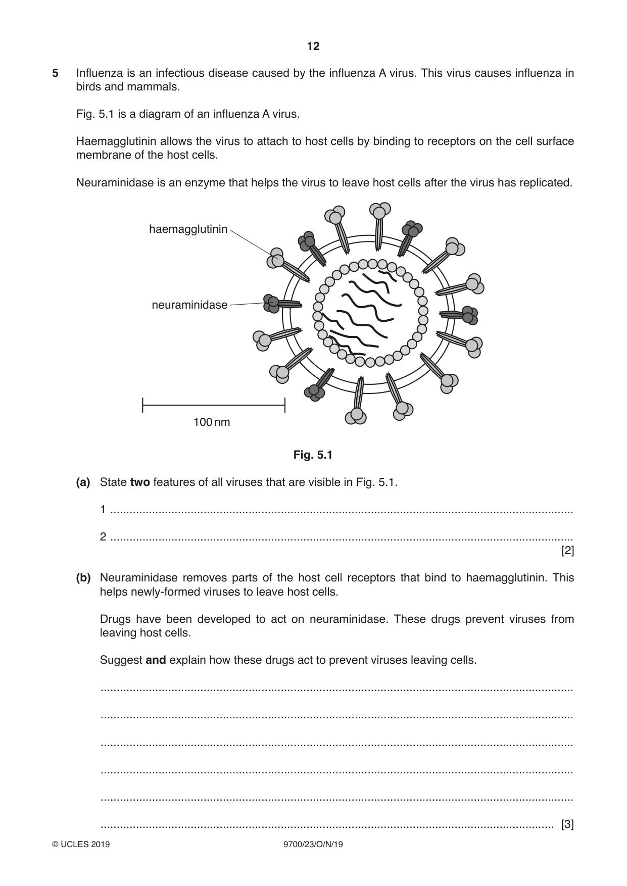
5 Influenza is an infectious disease caused by the influenza A virus. This virus causes influenza in birds and mammals. Fig. 5.1 is a diagram of an influenza A virus. Haemagglutinin allows the virus to attach to host cells by binding to receptors on the cell surface membrane of the host cells. Neuraminidase is an enzyme that helps the virus to leave host cells after the virus has replicated. haemagglutinin neuraminidase 100 nm Fig. 5.1 (a) State two features of all viruses that are visible in Fig. 5.1. 1 ................................................................................................................................................ 2 ................................................................................................................................................ [2] (b) Neuraminidase removes parts of the host cell receptors that bind to haemagglutinin. This helps newly-formed viruses to leave host cells. Drugs have been developed to act on neuraminidase. These drugs prevent viruses from leaving host cells. Suggest and explain how these drugs act to prevent viruses leaving cells. ................................................................................................................................................... ................................................................................................................................................... ................................................................................................................................................... ................................................................................................................................................... ................................................................................................................................................... ............................................................................................................................................. [3] (c) The human immune system produces antibodies in response to the presence of antigens, such as neuraminidase and haemagglutinin. Outline the events that occur during an immune response leading to the production of antibodies against an antigen, such as haemagglutinin. ................................................................................................................................................... ................................................................................................................................................... ................................................................................................................................................... ................................................................................................................................................... ................................................................................................................................................... ................................................................................................................................................... ................................................................................................................................................... ................................................................................................................................................... ............................................................................................................................................. [4] (d) Researchers are developing methods to produce antibodies to give artificial passive immunity to influenza. (i) Suggest the advantages and disadvantages of artificial passive immunity. ........................................................................................................................................... ........................................................................................................................................... ........................................................................................................................................... ........................................................................................................................................... ........................................................................................................................................... ........................................................................................................................................... ..................................................................................................................................... [3] (ii) State two ways in which mammals can acquire natural passive immunity to infectious diseases, such as influenza. 1 ........................................................................................................................................ 2 ........................................................................................................................................ [2] [Total: 14]
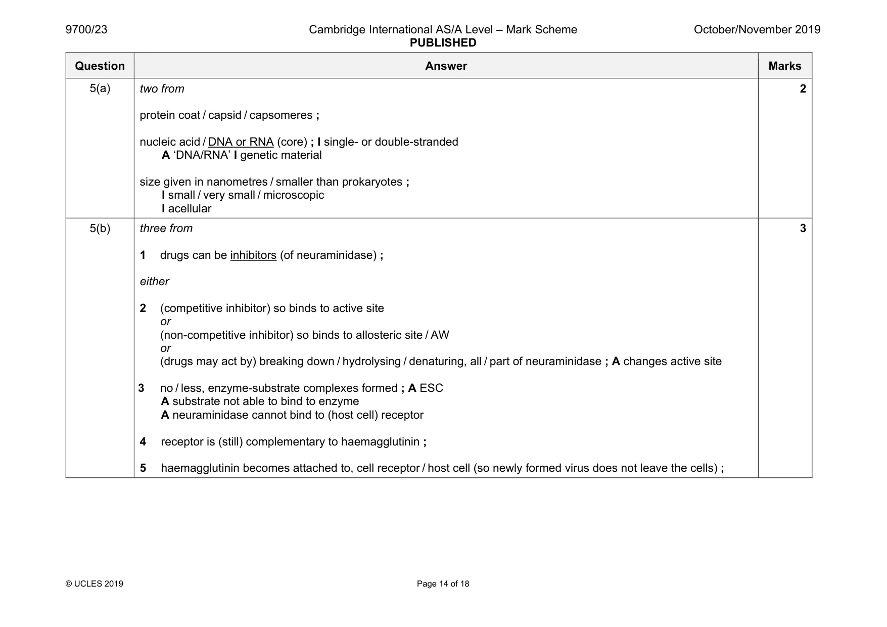
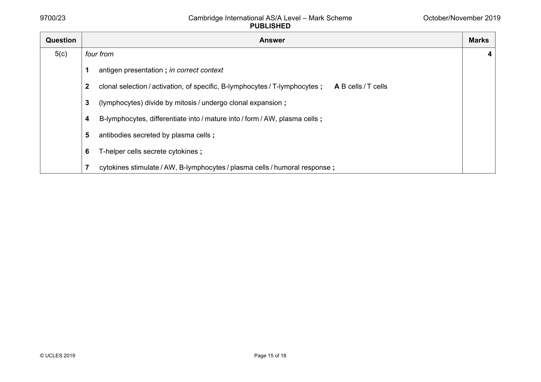
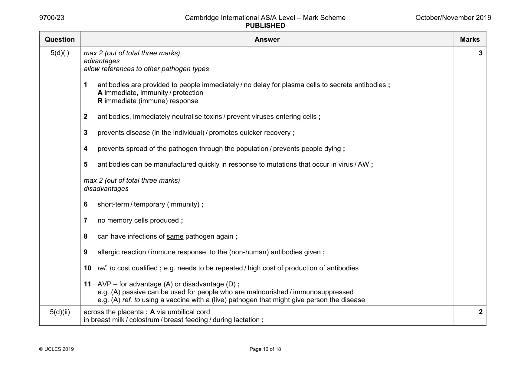
Mark scheme: 5(a) two from protein coat / capsid / capsomeres ; nucleic acid / DNA or RNA (core) ; I single- or double-stranded A ‘DNA/RNA’ I genetic material size given in nanometres / smaller than prokaryotes ; I small / very small / microscopic I acellular 2 5(b) three from 1 drugs can be inhibitors (of neuraminidase) ; either 2 (competitive inhibitor) so binds to active site or (non-competitive inhibitor) so binds to allosteric site / AW or (drugs may act by) breaking down / hydrolysing / denaturing, all / part of neuraminidase ; A changes active site 3 no / less, enzyme-substrate complexes formed ; A ESC A substrate not able to bind to enzyme A neuraminidase cannot bind to (host cell) receptor 4 receptor is (still) complementary to haemagglutinin ; 5 haemagglutinin becomes attached to, cell receptor / host cell (so newly formed virus does not leave the cells) ; 3 Question Answer Marks 5(c) four from 1 antigen presentation ; in correct context 2 clonal selection / activation, of specific, B-lymphocytes / T-lymphocytes ; A B cells / T cells 3 (lymphocytes) divide by mitosis / undergo clonal expansion ; 4 B-lymphocytes, differentiate into / mature into / form / AW, plasma cells ; 5 antibodies secreted by plasma cells ; 6 T-helper cells secrete cytokines ; 7 cytokines stimulate / AW, B-lymphocytes / plasma cells / humoral response ; 4 Question Answer Marks 5(d)(i) max 2 (out of total three marks) advantages allow references to other pathogen types 1 antibodies are provided to people immediately / no delay for plasma cells to secrete antibodies ; A immediate, immunity / protection R immediate (immune) response 2 antibodies, immediately neutralise toxins / prevent viruses entering cells ; 3 prevents disease (in the individual) / promotes quicker recovery ; 4 prevents spread of the pathogen through the population / prevents people dying ; 5 antibodies can be manufactured quickly in response to mutations that occur in virus / AW ; max 2 (out of total three marks) disadvantages 6 short-term / temporary (immunity) ; 7 no memory cells produced ; 8 can have infections of same pathogen again ; 9 allergic reaction / immune response, to the (non-human) antibodies given ; 10 ref. to cost qualified ; e.g. needs to be repeated / high cost of production of antibodies 11 AVP – for advantage (A) or disadvantage (D) ; e.g. (A) passive can be used for people who are malnourished / immunosuppressed e.g. (A) ref. to using a vaccine with a (live) pathogen that might give person the disease 3 5(d)(ii) across the placenta ; A via umbilical cord in breast milk / colostrum / breast feeding / during lactation ; 2
Q6 · A diagram of the cell surface membrane of a squamous epithelial cell lining an alveolus
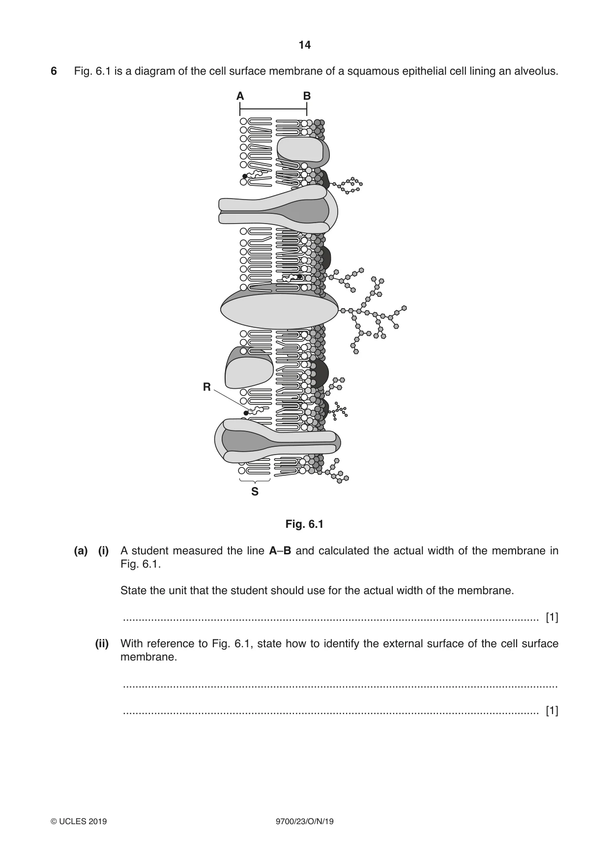
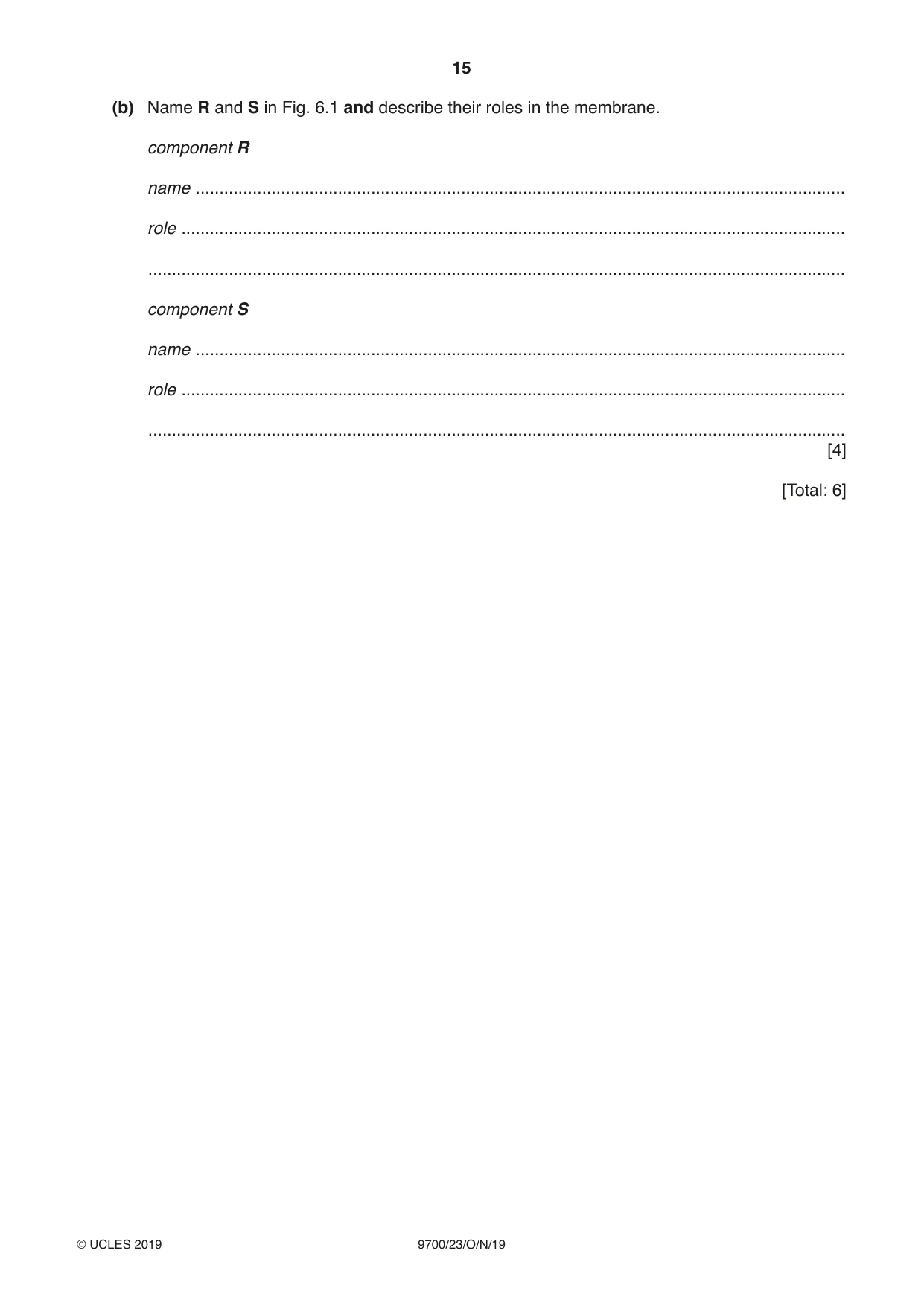
6 Fig. 6.1 is a diagram of the cell surface membrane of a squamous epithelial cell lining an alveolus. A B R S Fig. 6.1 (a) (i) A student measured the line A–B and calculated the actual width of the membrane in Fig. 6.1. State the unit that the student should use for the actual width of the membrane. ..................................................................................................................................... [1] (ii) With reference to Fig. 6.1, state how to identify the external surface of the cell surface membrane. ........................................................................................................................................... ..................................................................................................................................... [1] (b) Name R and S in Fig. 6.1 and describe their roles in the membrane. component R name ......................................................................................................................................... role ............................................................................................................................................ ................................................................................................................................................... component S name ......................................................................................................................................... role ............................................................................................................................................ ................................................................................................................................................... [4] [Total: 6]
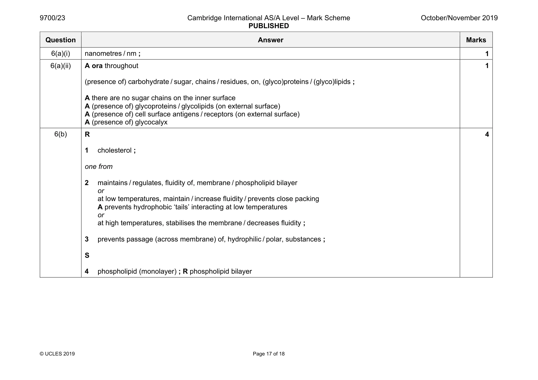
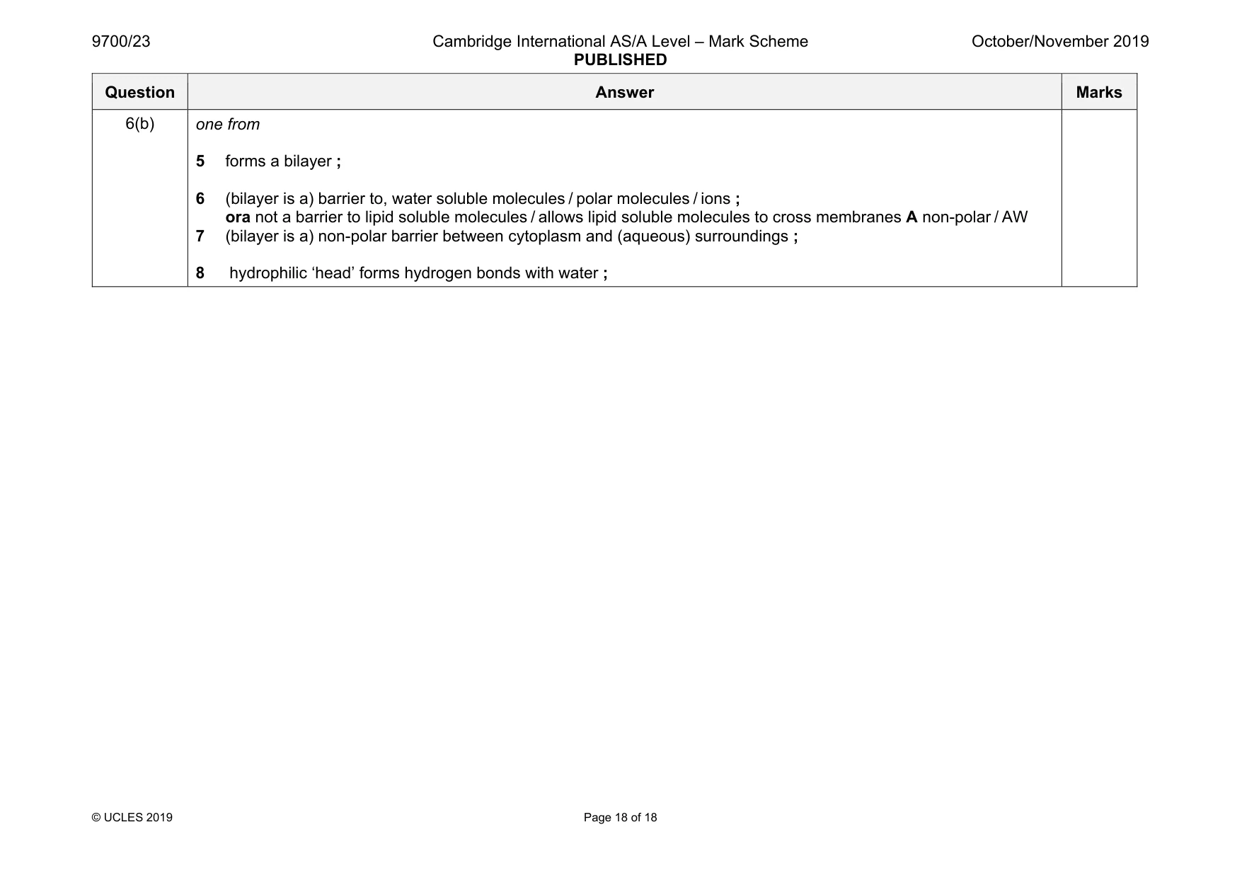
Mark scheme: 6(a)(i) nanometres / nm ; 1 6(a)(ii) A ora throughout (presence of) carbohydrate / sugar, chains / residues, on, (glyco)proteins / (glyco)lipids ; A there are no sugar chains on the inner surface A (presence of) glycoproteins / glycolipids (on external surface) A (presence of) cell surface antigens / receptors (on external surface) A (presence of) glycocalyx 1 6(b) R 1 cholesterol ; one from 2 maintains / regulates, fluidity of, membrane / phospholipid bilayer or at low temperatures, maintain / increase fluidity / prevents close packing A prevents hydrophobic ‘tails’ interacting at low temperatures or at high temperatures, stabilises the membrane / decreases fluidity ; 3 prevents passage (across membrane) of, hydrophilic / polar, substances ; S 4 phospholipid (monolayer) ; R phospholipid bilayer 4 Question Answer Marks 6(b) one from 5 forms a bilayer ; 6 (bilayer is a) barrier to, water soluble molecules / polar molecules / ions ; ora not a barrier to lipid soluble molecules / allows lipid soluble molecules to cross membranes A non-polar / AW 7 (bilayer is a) non-polar barrier between cytoplasm and (aqueous) surroundings ; 8 hydrophilic ‘head’ forms hydrogen bonds with water ;
What was in this paper
The subtopics covered by these 6 questions, and how many questions each got. Open one in a new tab to see every Cambridge question on it.
What you needed in this session
Cambridge’s own grade thresholds for 2019 Oct/Nov, Paper 2 · Variant 3. A higher threshold means an easier paper — the bar moves with how the cohort did.