Cambridge A Level Biology 9700 — 2019 Oct/Nov Paper 2 · Variant 2
9700/22/O/N/19 · 6 questions · 60 marks · ≈68 min
The question paper and its mark scheme, free to read here and free to download. This is Cambridge’s own paper, exactly as it was sat.
Question paper16 pages
Mark scheme14 pages
Answers below. Sit the paper first if you are practising.
Questions as text
Q1 · A photomicrograph of a transverse section through the stem of creeping buttercup…
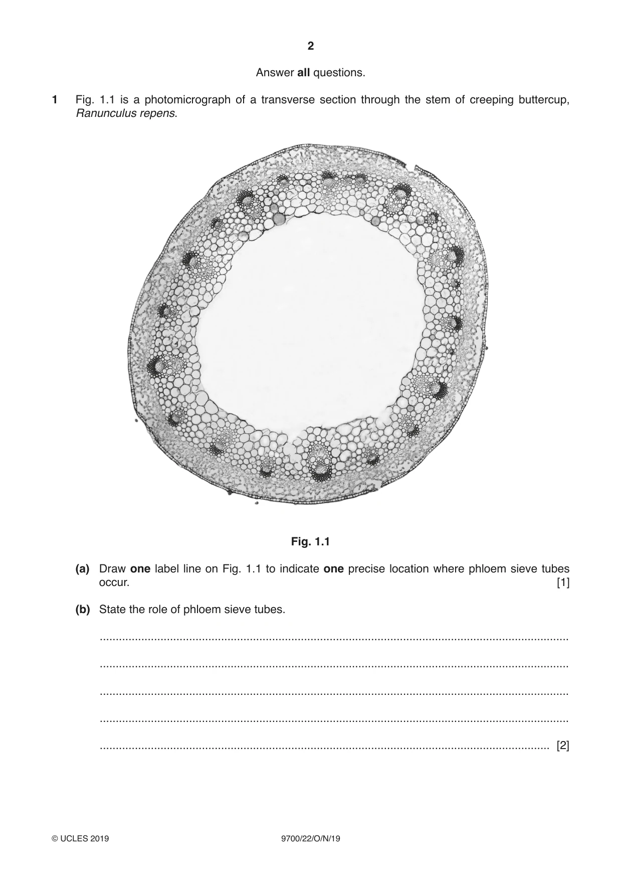
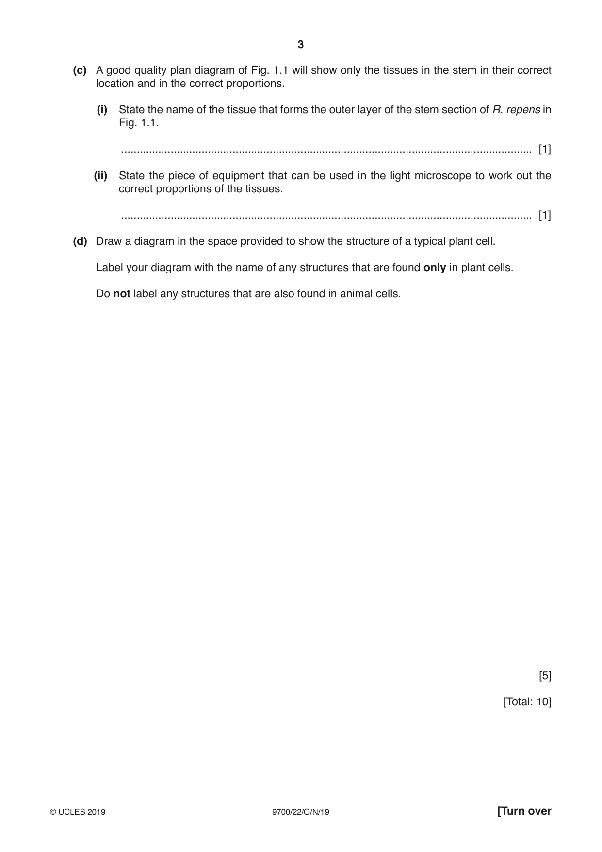
1 Fig. 1.1 is a photomicrograph of a transverse section through the stem of creeping buttercup, Ranunculus repens. Fig. 1.1 (a) Draw one label line on Fig. 1.1 to indicate one precise location where phloem sieve tubes occur. [1] (b) State the role of phloem sieve tubes. ................................................................................................................................................... ................................................................................................................................................... ................................................................................................................................................... ................................................................................................................................................... ............................................................................................................................................. [2] (c) A good quality plan diagram of Fig. 1.1 will show only the tissues in the stem in their correct location and in the correct proportions. (i) State the name of the tissue that forms the outer layer of the stem section of R. repens in Fig. 1.1. ..................................................................................................................................... [1] (ii) State the piece of equipment that can be used in the light microscope to work out the correct proportions of the tissues. ..................................................................................................................................... [1] (d) Draw a diagram in the space provided to show the structure of a typical plant cell. Label your diagram with the name of any structures that are found only in plant cells. Do not label any structures that are also found in animal cells. [5] [Total: 10]
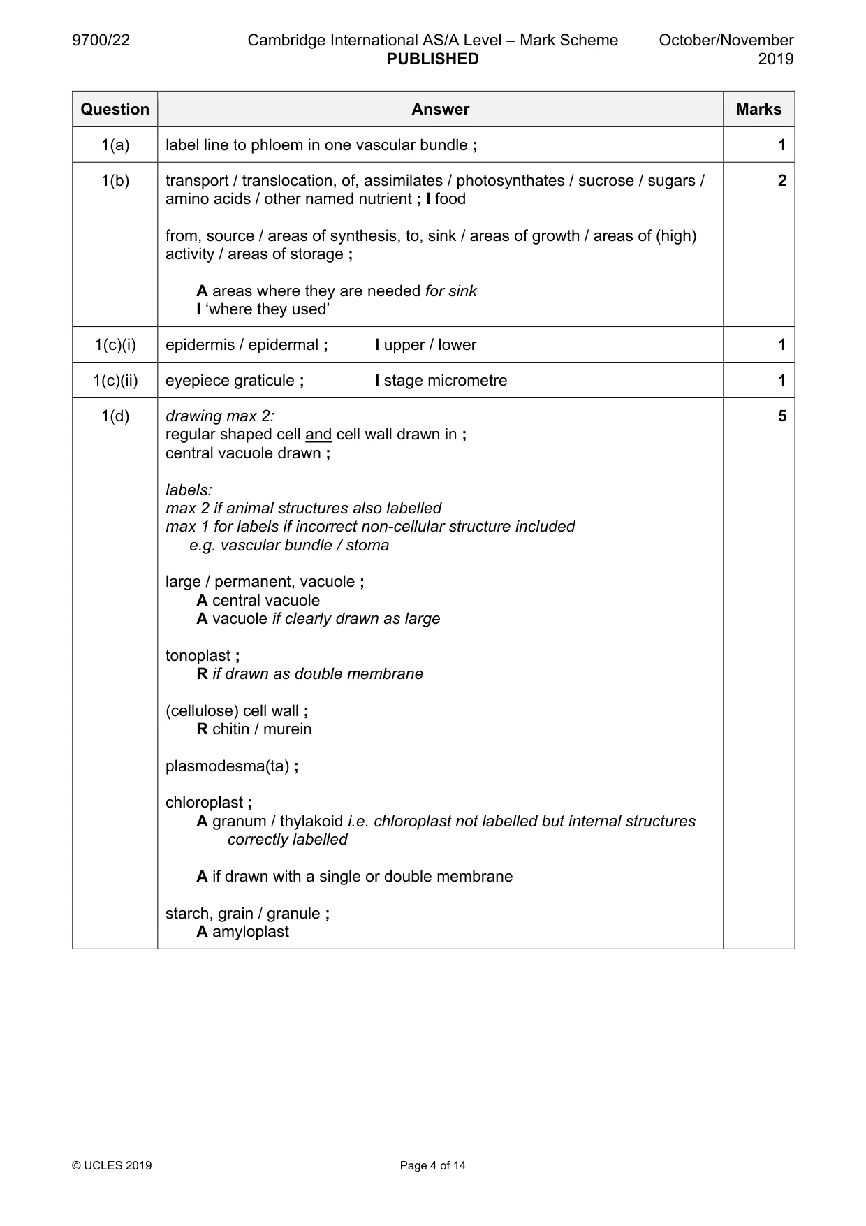
Mark scheme: Question Answer Marks 1(a) label line to phloem in one vascular bundle ; 1 1(b) transport / translocation, of, assimilates / photosynthates / sucrose / sugars / 2 amino acids / other named nutrient ; I food from, source / areas of synthesis, to, sink / areas of growth / areas of (high) activity / areas of storage ; A areas where they are needed for sink I ‘where they used’ 1(c)(i) epidermis / epidermal ; I upper / lower 1 1(c)(ii) eyepiece graticule ; I stage micrometre 1 1(d) drawing max 2: 5 regular shaped cell and cell wall drawn in ; central vacuole drawn ; labels: max 2 if animal structures also labelled max 1 for labels if incorrect non-cellular structure included e.g. vascular bundle / stoma large / permanent, vacuole ; A central vacuole A vacuole if clearly drawn as large tonoplast ; R if drawn as double membrane (cellulose) cell wall ; R chitin / murein plasmodesma(ta) ; chloroplast ; A granum / thylakoid i.e. chloroplast not labelled but internal structures correctly labelled A if drawn with a single or double membrane starch, grain / granule ; A amyloplast
More questions on Cells as the basic units of living organisms
Q2 · People who smoke tobacco are at an increased risk of developing non-infectious diseases…
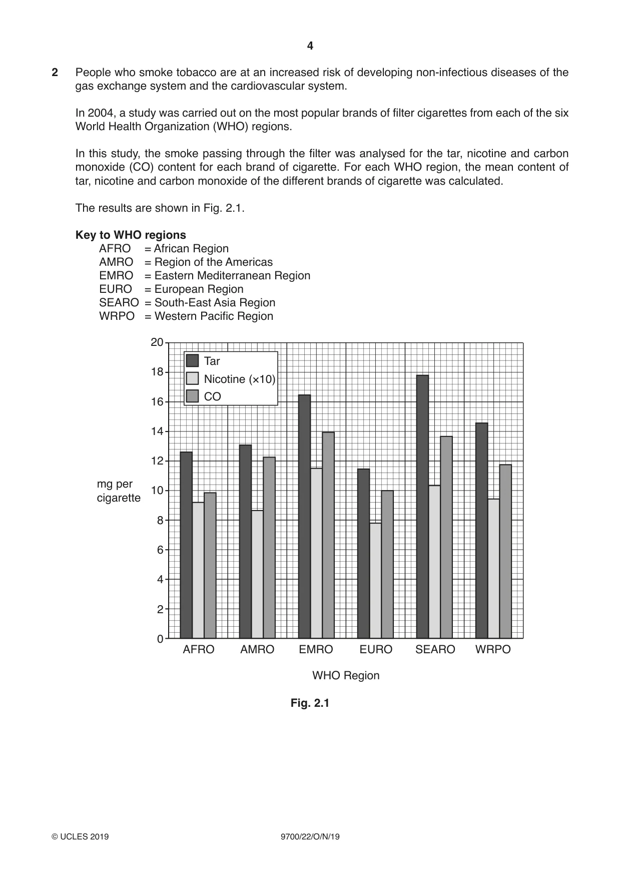
2 People who smoke tobacco are at an increased risk of developing non-infectious diseases of the gas exchange system and the cardiovascular system. In 2004, a study was carried out on the most popular brands of filter cigarettes from each of the six World Health Organization (WHO) regions. In this study, the smoke passing through the filter was analysed for the tar, nicotine and carbon monoxide (CO) content for each brand of cigarette. For each WHO region, the mean content of tar, nicotine and carbon monoxide of the different brands of cigarette was calculated. The results are shown in Fig. 2.1. Key to WHO regions AFRO = African Region AMRO = Region of the Americas EMRO = Eastern Mediterranean Region EURO = European Region SEARO = South-East Asia Region WRPO = Western Pacific Region 20 Tar 18 Nicotine (×10) CO 16 14 12 mg per 10 cigarette 8 6 4 2 0 AFRO AMRO EMRO EURO SEARO WRPO WHO Region Fig. 2.1 (a) With reference to Fig. 2.1, list the two WHO regions where smokers are at the highest risk of developing diseases of the gas exchange or cardiovascular systems. ................................................................................................................................................... ................................................................................................................................................... ............................................................................................................................................. [1] (b) With reference to Fig. 2.1, explain why smoking a popular brand of cigarette from the AFRO region is more likely to increase the risk of blood clots forming than smoking a popular brand of cigarette from the EURO region. ................................................................................................................................................... ................................................................................................................................................... ................................................................................................................................................... ................................................................................................................................................... ............................................................................................................................................. [2] (c) One short-term effect of cigarette smoking is a decrease in the supply of oxygen to body tissues. Describe and explain why cigarette smoking leads to a decrease in the supply of oxygen to body tissues. ................................................................................................................................................... ................................................................................................................................................... ................................................................................................................................................... ................................................................................................................................................... ................................................................................................................................................... ................................................................................................................................................... ................................................................................................................................................... ................................................................................................................................................... ................................................................................................................................................... ................................................................................................................................................... ............................................................................................................................................. [4] [Total: 7]
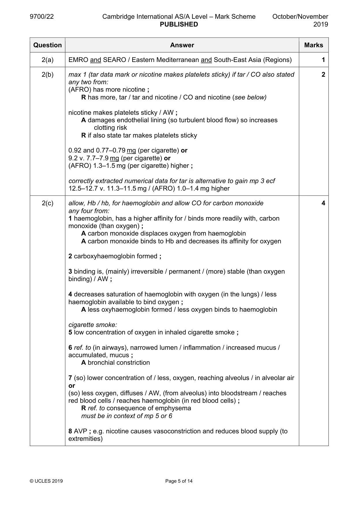
Mark scheme: 2(a) EMRO and SEARO / Eastern Mediterranean and South-East Asia (Regions) 1 2(b) max 1 (tar data mark or nicotine makes platelets sticky) if tar / CO also stated 2 any two from: (AFRO) has more nicotine ; R has more, tar / tar and nicotine / CO and nicotine (see below) nicotine makes platelets sticky / AW ; A damages endothelial lining (so turbulent blood flow) so increases clotting risk R if also state tar makes platelets sticky 0.92 and 0.77–0.79 mg (per cigarette) or 9.2 v. 7.7–7.9 mg (per cigarette) or (AFRO) 1.3–1.5 mg (per cigarette) higher ; correctly extracted numerical data for tar is alternative to gain mp 3 ecf 12.5–12.7 v. 11.3–11.5 mg / (AFRO) 1.0–1.4 mg higher 2(c) allow, Hb / hb, for haemoglobin and allow CO for carbon monoxide 4 any four from: 1 haemoglobin, has a higher affinity for / binds more readily with, carbon monoxide (than oxygen) ; A carbon monoxide displaces oxygen from haemoglobin A carbon monoxide binds to Hb and decreases its affinity for oxygen 2 carboxyhaemoglobin formed ; 3 binding is, (mainly) irreversible / permanent / (more) stable (than oxygen binding) / AW ; 4 decreases saturation of haemoglobin with oxygen (in the lungs) / less haemoglobin available to bind oxygen ; A less oxyhaemoglobin formed / less oxygen binds to haemoglobin cigarette smoke: 5 low concentration of oxygen in inhaled cigarette smoke ; 6 ref. to (in airways), narrowed lumen / inflammation / increased mucus / accumulated, mucus ; A bronchial constriction 7 (so) lower concentration of / less, oxygen, reaching alveolus / in alveolar air or (so) less oxygen, diffuses / AW, (from alveolus) into bloodstream / reaches red blood cells / reaches haemoglobin (in red blood cells) ; R ref. to consequence of emphysema must be in context of mp 5 or 6 8 AVP ; e.g. nicotine causes vasoconstriction and reduces blood supply (to extremities)
Q3 · A red blood cell goes through a number of stages in the bone marrow before it is released…
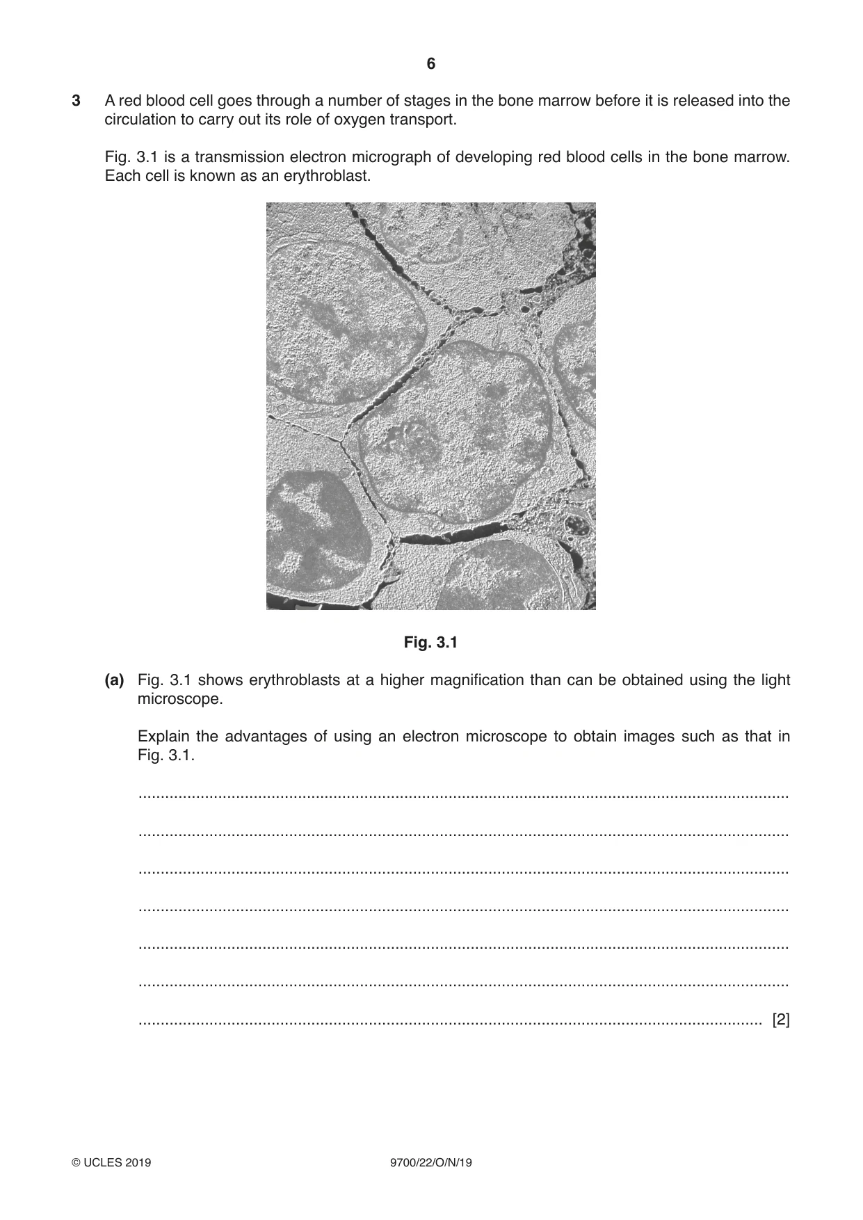
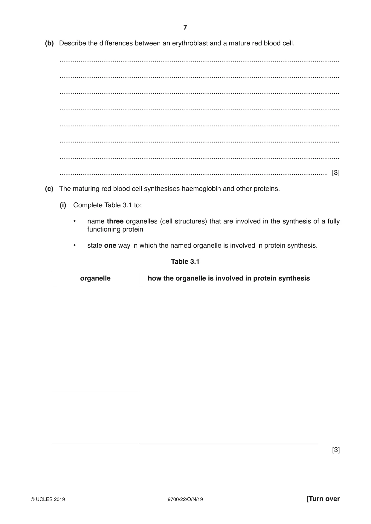
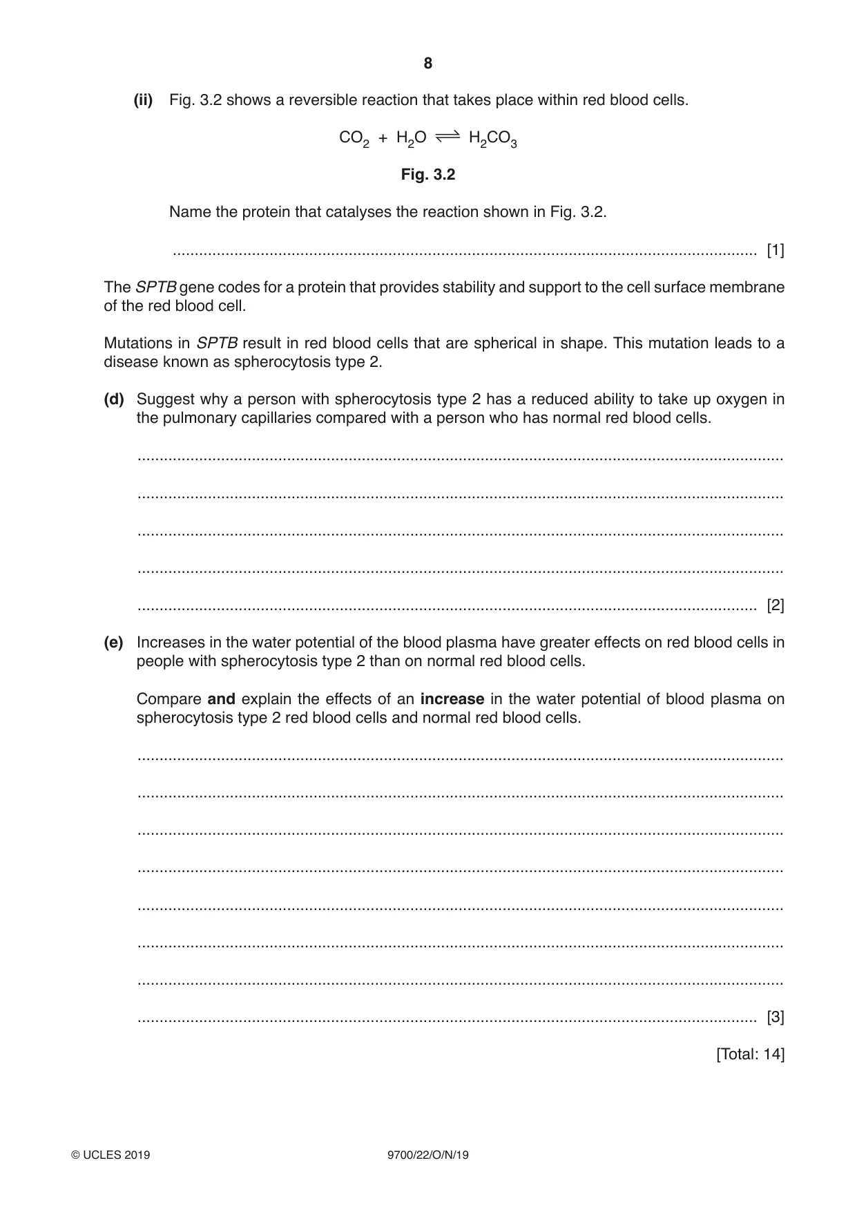
3 A red blood cell goes through a number of stages in the bone marrow before it is released into the circulation to carry out its role of oxygen transport. Fig. 3.1 is a transmission electron micrograph of developing red blood cells in the bone marrow. Each cell is known as an erythroblast. Fig. 3.1 (a) Fig. 3.1 shows erythroblasts at a higher magnification than can be obtained using the light microscope. Explain the advantages of using an electron microscope to obtain images such as that in Fig. 3.1. ................................................................................................................................................... ................................................................................................................................................... ................................................................................................................................................... ................................................................................................................................................... ................................................................................................................................................... ................................................................................................................................................... ............................................................................................................................................. [2] (b) Describe the differences between an erythroblast and a mature red blood cell. ................................................................................................................................................... ................................................................................................................................................... ................................................................................................................................................... ................................................................................................................................................... ................................................................................................................................................... ................................................................................................................................................... ................................................................................................................................................... ............................................................................................................................................. [3] (c) The maturing red blood cell synthesises haemoglobin and other proteins. (i) Complete Table 3.1 to: • name three organelles (cell structures) that are involved in the synthesis of a fully functioning protein • state one way in which the named organelle is involved in protein synthesis. Table 3.1 organelle how the organelle is involved in protein synthesis [3] (ii) Fig. 3.2 shows a reversible reaction that takes place within red blood cells. CO2 + H2O H2CO3 Fig. 3.2 Name the protein that catalyses the reaction shown in Fig. 3.2. ..................................................................................................................................... [1] The SPTB gene codes for a protein that provides stability and support to the cell surface membrane of the red blood cell. Mutations in SPTB result in red blood cells that are spherical in shape. This mutation leads to a disease known as spherocytosis type 2. (d) Suggest why a person with spherocytosis type 2 has a reduced ability to take up oxygen in the pulmonary capillaries compared with a person who has normal red blood cells. ................................................................................................................................................... ................................................................................................................................................... ................................................................................................................................................... ................................................................................................................................................... ............................................................................................................................................. [2] (e) Increases in the water potential of the blood plasma have greater effects on red blood cells in people with spherocytosis type 2 than on normal red blood cells. Compare and explain the effects of an increase in the water potential of blood plasma on spherocytosis type 2 red blood cells and normal red blood cells. ................................................................................................................................................... ................................................................................................................................................... ................................................................................................................................................... ................................................................................................................................................... ................................................................................................................................................... ................................................................................................................................................... ................................................................................................................................................... ............................................................................................................................................. [3] [Total: 14]
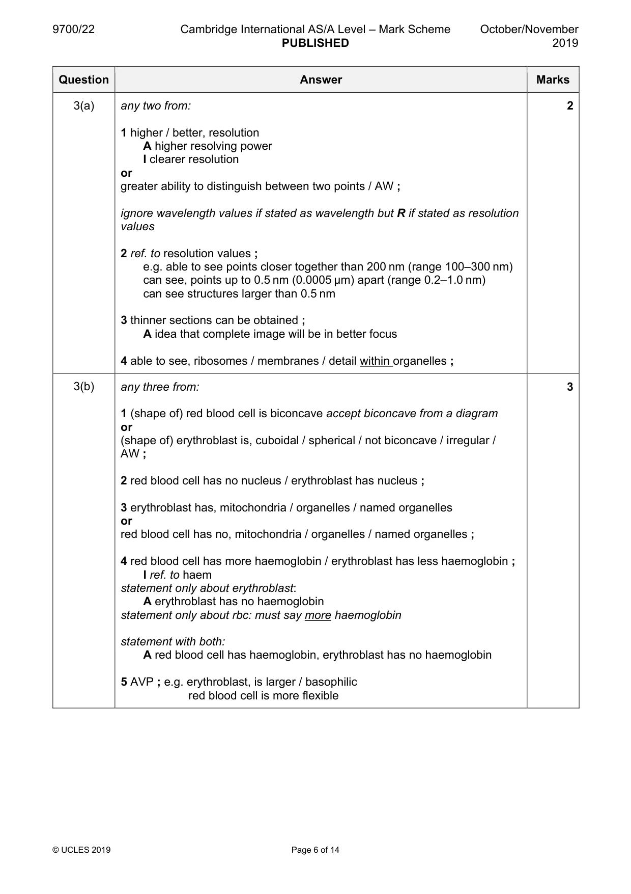
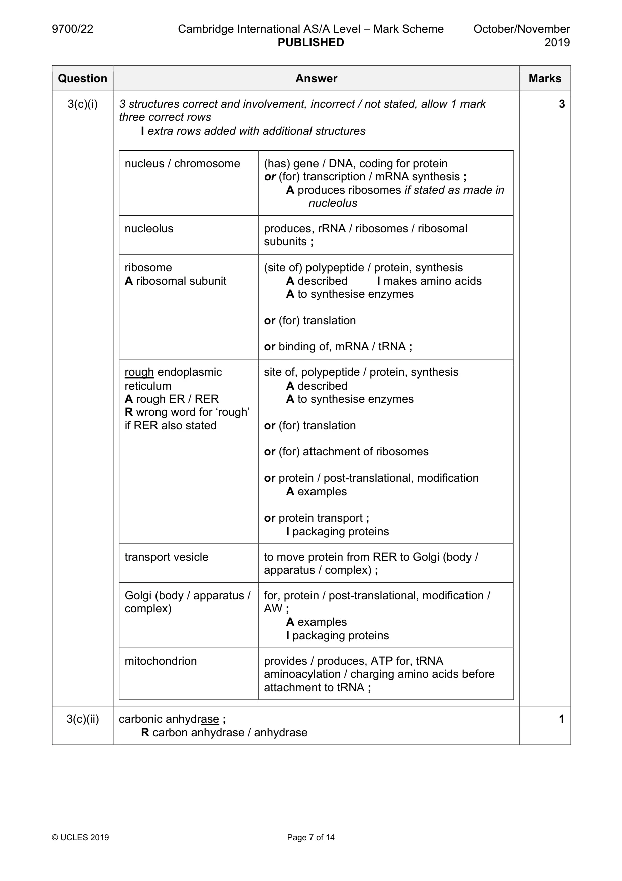
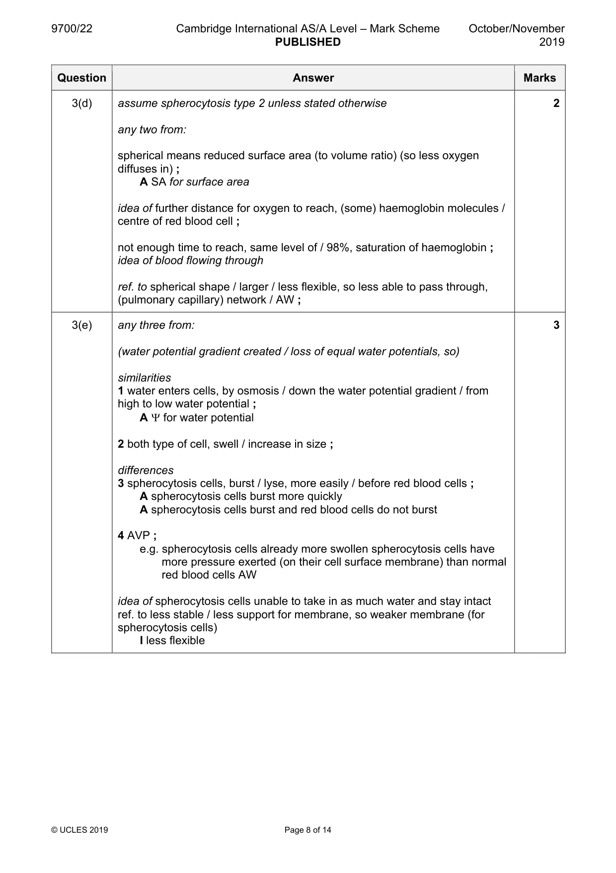
Mark scheme: 3(a) any two from: 2 1 higher / better, resolution A higher resolving power I clearer resolution or greater ability to distinguish between two points / AW ; ignore wavelength values if stated as wavelength but R if stated as resolution values 2 ref. to resolution values ; e.g. able to see points closer together than 200 nm (range 100–300 nm) can see, points up to 0.5 nm (0.0005 µm) apart (range 0.2–1.0 nm) can see structures larger than 0.5 nm 3 thinner sections can be obtained ; A idea that complete image will be in better focus 4 able to see, ribosomes / membranes / detail within organelles ; 3(b) any three from: 3 1 (shape of) red blood cell is biconcave accept biconcave from a diagram or (shape of) erythroblast is, cuboidal / spherical / not biconcave / irregular / AW ; 2 red blood cell has no nucleus / erythroblast has nucleus ; 3 erythroblast has, mitochondria / organelles / named organelles or red blood cell has no, mitochondria / organelles / named organelles ; 4 red blood cell has more haemoglobin / erythroblast has less haemoglobin ; I ref. to haem statement only about erythroblast: A erythroblast has no haemoglobin statement only about rbc: must say more haemoglobin statement with both: A red blood cell has haemoglobin, erythroblast has no haemoglobin 5 AVP ; e.g. erythroblast, is larger / basophilic red blood cell is more flexible 3(c)(i) 3 structures correct and involvement, incorrect / not stated, allow 1 mark 3 three correct rows I extra rows added with additional structures nucleus / chromosome (has) gene / DNA, coding for protein or (for) transcription / mRNA synthesis ; A produces ribosomes if stated as made in nucleolus nucleolus produces, rRNA / ribosomes / ribosomal subunits ; ribosome (site of) polypeptide / protein, synthesis A ribosomal subunit A described I makes amino acids A to synthesise enzymes or (for) translation or binding of, mRNA / tRNA ; rough endoplasmic site of, polypeptide / protein, synthesis reticulum A described A rough ER / RER A to synthesise enzymes R wrong word for ‘rough’ if RER also stated or (for) translation or (for) attachment of ribosomes or protein / post-translational, modification A examples or protein transport ; I packaging proteins transport vesicle to move protein from RER to Golgi (body / apparatus / complex) ; Golgi (body / apparatus / for, protein / post-translational, modification / complex) AW ; A examples I packaging proteins mitochondrion provides / produces, ATP for, tRNA aminoacylation / charging amino acids before attachment to tRNA ; 3(c)(ii) carbonic anhydrase ; 1 R carbon anhydrase / anhydrase 3(d) assume spherocytosis type 2 unless stated otherwise 2 any two from: spherical means reduced surface area (to volume ratio) (so less oxygen diffuses in) ; A SA for surface area idea of further distance for oxygen to reach, (some) haemoglobin molecules / centre of red blood cell ; not enough time to reach, same level of / 98%, saturation of haemoglobin ; idea of blood flowing through ref. to spherical shape / larger / less flexible, so less able to pass through, (pulmonary capillary) network / AW ; 3(e) any three from: 3 (water potential gradient created / loss of equal water potentials, so) similarities 1 water enters cells, by osmosis / down the water potential gradient / from high to low water potential ; A Ψ for water potential 2 both type of cell, swell / increase in size ; differences 3 spherocytosis cells, burst / lyse, more easily / before red blood cells ; A spherocytosis cells burst more quickly A spherocytosis cells burst and red blood cells do not burst 4 AVP ; e.g. spherocytosis cells already more swollen spherocytosis cells have more pressure exerted (on their cell surface membrane) than normal red blood cells AW idea of spherocytosis cells unable to take in as much water and stay intact ref. to less stable / less support for membrane, so weaker membrane (for spherocytosis cells) I less flexible
Q4 · Carbohydrates, proteins and nucleic acids are types of biological molecule
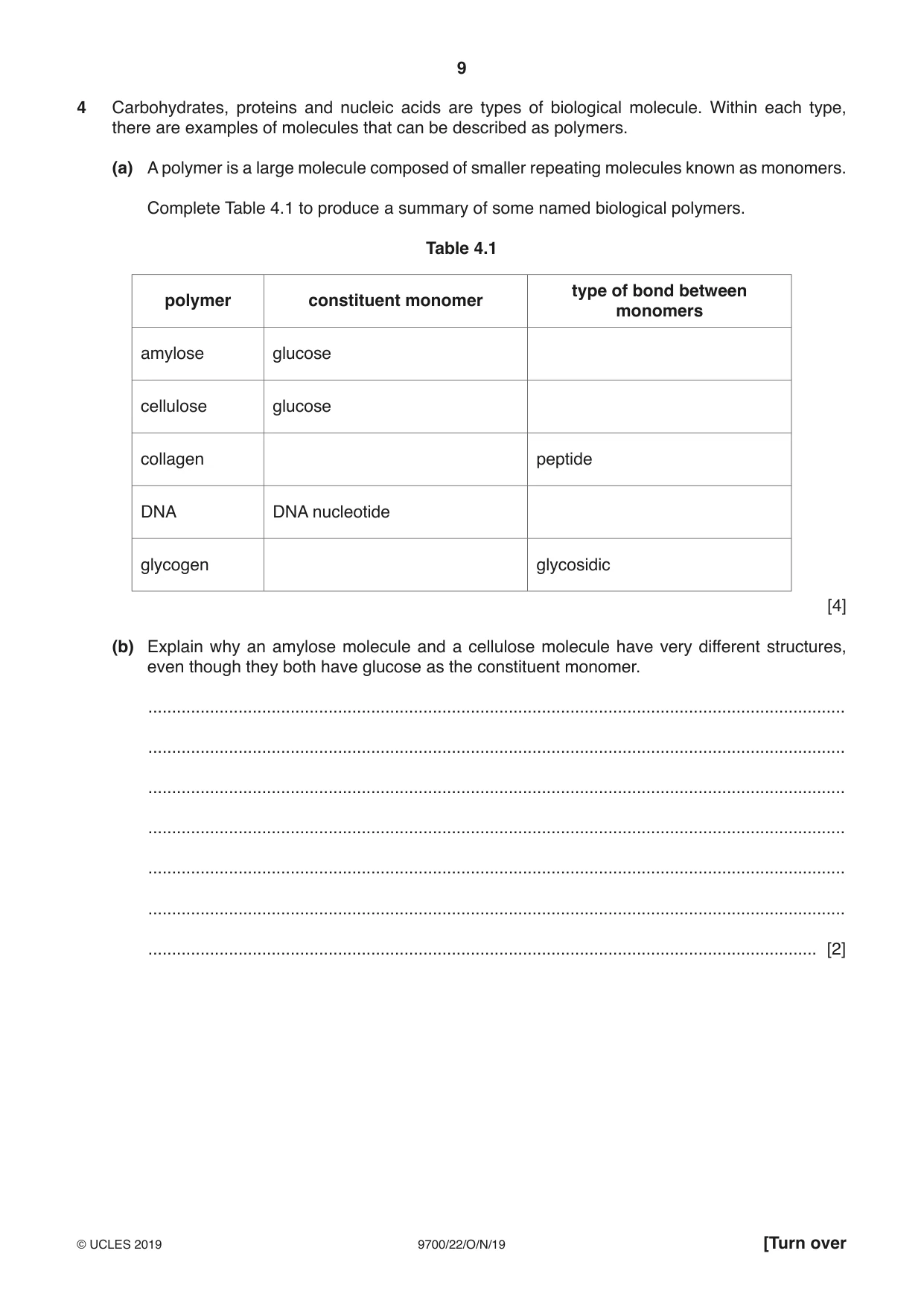
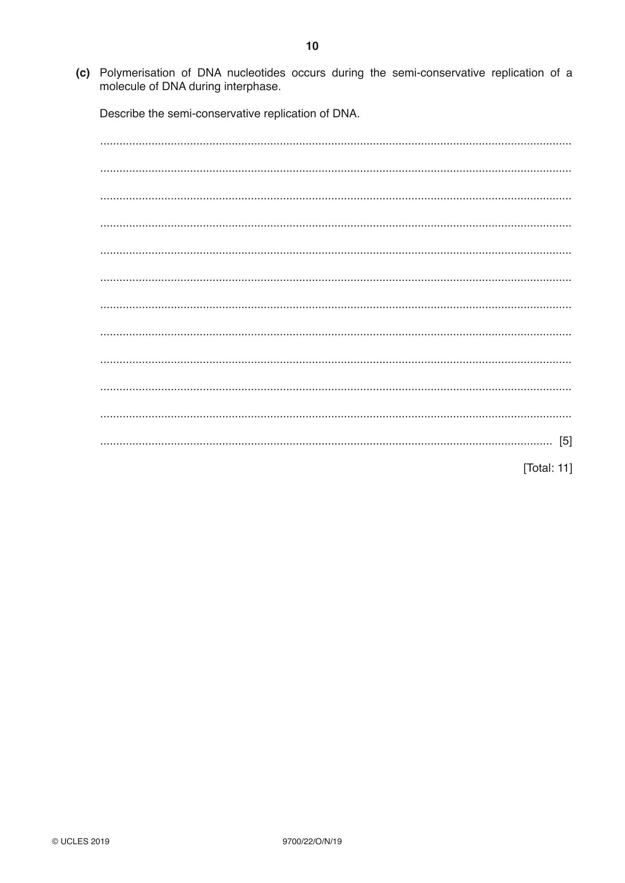
4 Carbohydrates, proteins and nucleic acids are types of biological molecule. Within each type, there are examples of molecules that can be described as polymers. (a) A polymer is a large molecule composed of smaller repeating molecules known as monomers. Complete Table 4.1 to produce a summary of some named biological polymers. Table 4.1 type of bond between polymer constituent monomer monomers amylose glucose cellulose glucose collagen peptide DNA DNA nucleotide glycogen glycosidic [4] (b) Explain why an amylose molecule and a cellulose molecule have very different structures, even though they both have glucose as the constituent monomer. ................................................................................................................................................... ................................................................................................................................................... ................................................................................................................................................... ................................................................................................................................................... ................................................................................................................................................... ................................................................................................................................................... ............................................................................................................................................. [2] (c) Polymerisation of DNA nucleotides occurs during the semi-conservative replication of a molecule of DNA during interphase. Describe the semi-conservative replication of DNA. ................................................................................................................................................... ................................................................................................................................................... ................................................................................................................................................... ................................................................................................................................................... ................................................................................................................................................... ................................................................................................................................................... ................................................................................................................................................... ................................................................................................................................................... ................................................................................................................................................... ................................................................................................................................................... ................................................................................................................................................... ............................................................................................................................................. [5] [Total: 11]
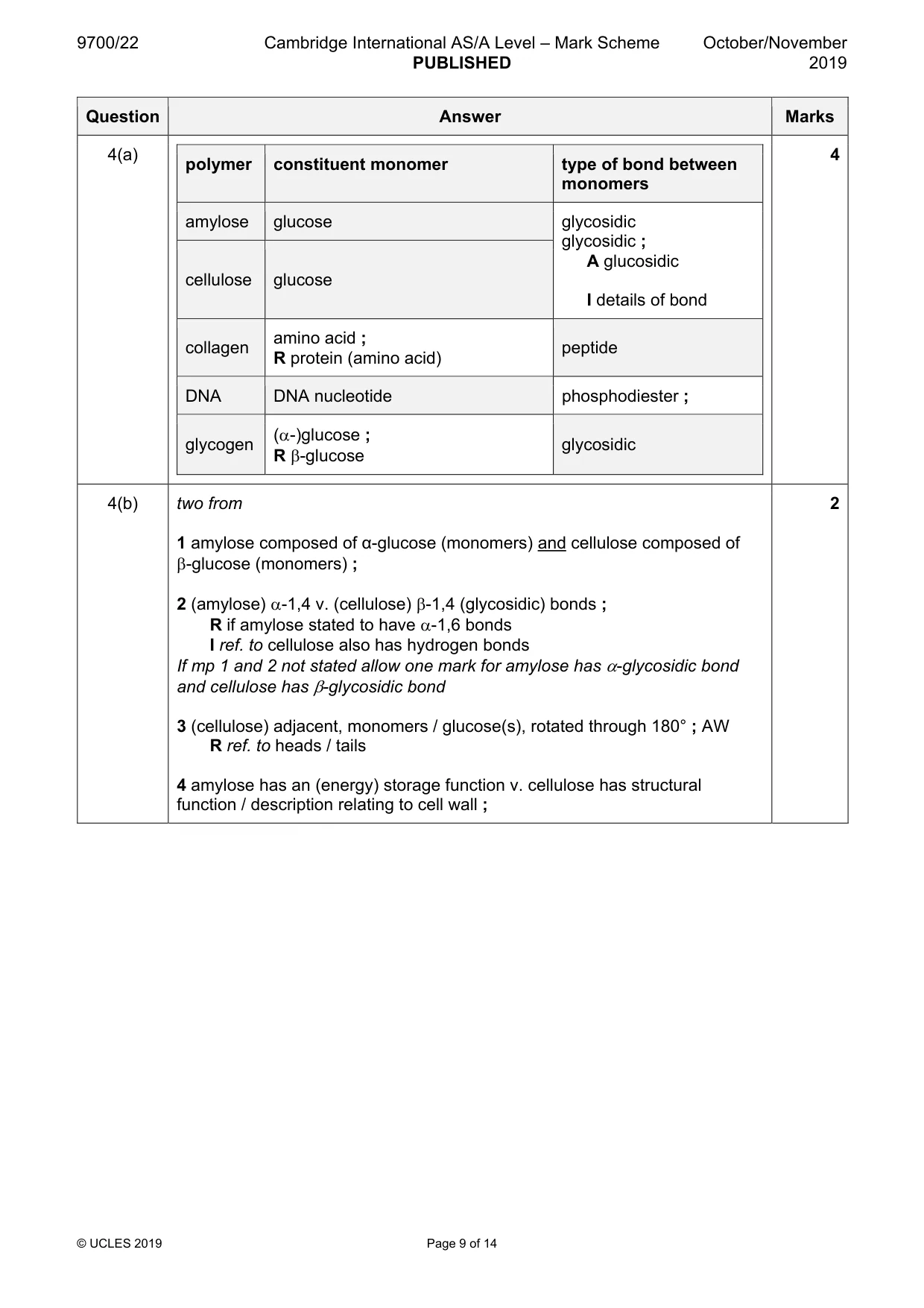
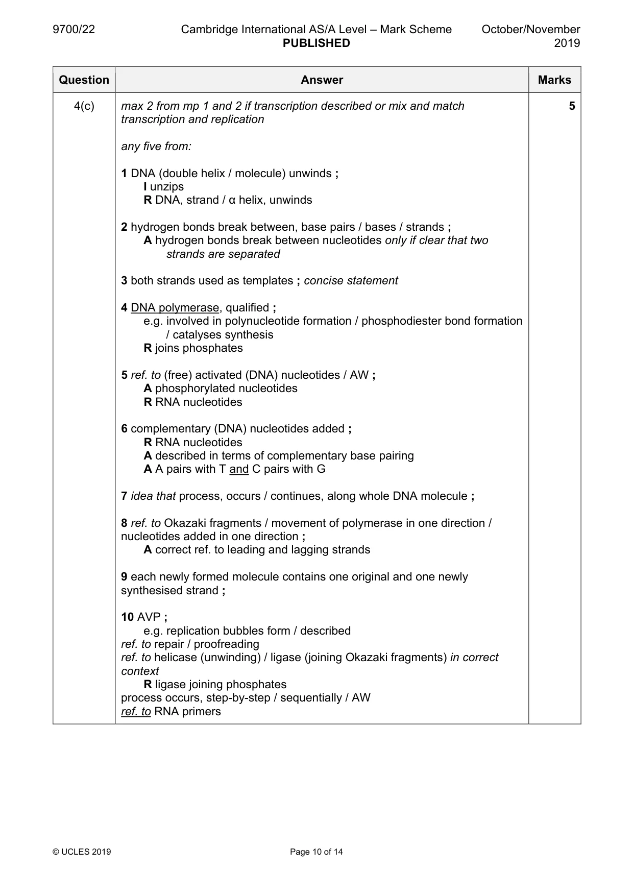
Mark scheme: 4(a) 4 polymer constituent monomer type of bond between monomers amylose glucose glycosidic glycosidic ; A glucosidic cellulose glucose I details of bond amino acid ; collagen peptide R protein (amino acid) DNA DNA nucleotide phosphodiester ; (α-)glucose ; glycogen glycosidic R β-glucose 4(b) two from 2 1 amylose composed of α-glucose (monomers) and cellulose composed of β-glucose (monomers) ; 2 (amylose) α-1,4 v. (cellulose) β-1,4 (glycosidic) bonds ; R if amylose stated to have α-1,6 bonds I ref. to cellulose also has hydrogen bonds If mp 1 and 2 not stated allow one mark for amylose has α-glycosidic bond and cellulose has β-glycosidic bond 3 (cellulose) adjacent, monomers / glucose(s), rotated through 180° ; AW R ref. to heads / tails 4 amylose has an (energy) storage function v. cellulose has structural function / description relating to cell wall ; 4(c) max 2 from mp 1 and 2 if transcription described or mix and match 5 transcription and replication any five from: 1 DNA (double helix / molecule) unwinds ; I unzips R DNA, strand / α helix, unwinds 2 hydrogen bonds break between, base pairs / bases / strands ; A hydrogen bonds break between nucleotides only if clear that two strands are separated 3 both strands used as templates ; concise statement 4 DNA polymerase, qualified ; e.g. involved in polynucleotide formation / phosphodiester bond formation / catalyses synthesis R joins phosphates 5 ref. to (free) activated (DNA) nucleotides / AW ; A phosphorylated nucleotides R RNA nucleotides 6 complementary (DNA) nucleotides added ; R RNA nucleotides A described in terms of complementary base pairing A A pairs with T and C pairs with G 7 idea that process, occurs / continues, along whole DNA molecule ; 8 ref. to Okazaki fragments / movement of polymerase in one direction / nucleotides added in one direction ; A correct ref. to leading and lagging strands 9 each newly formed molecule contains one original and one newly synthesised strand ; 10 AVP ; e.g. replication bubbles form / described ref. to repair / proofreading ref. to helicase (unwinding) / ligase (joining Okazaki fragments) in correct context R ligase joining phosphates process occurs, step-by-step / sequentially / AW ref. to RNA primers
Q5 · Countries that have a high number of cases of malaria also have problems with diseases…
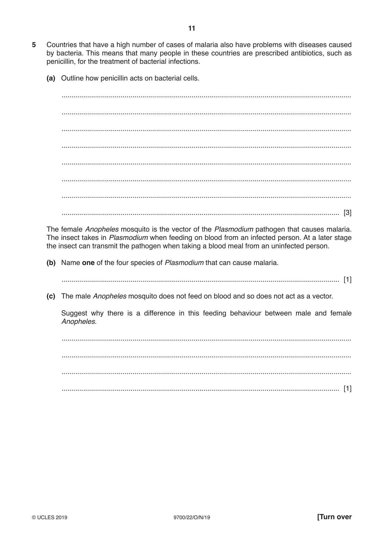
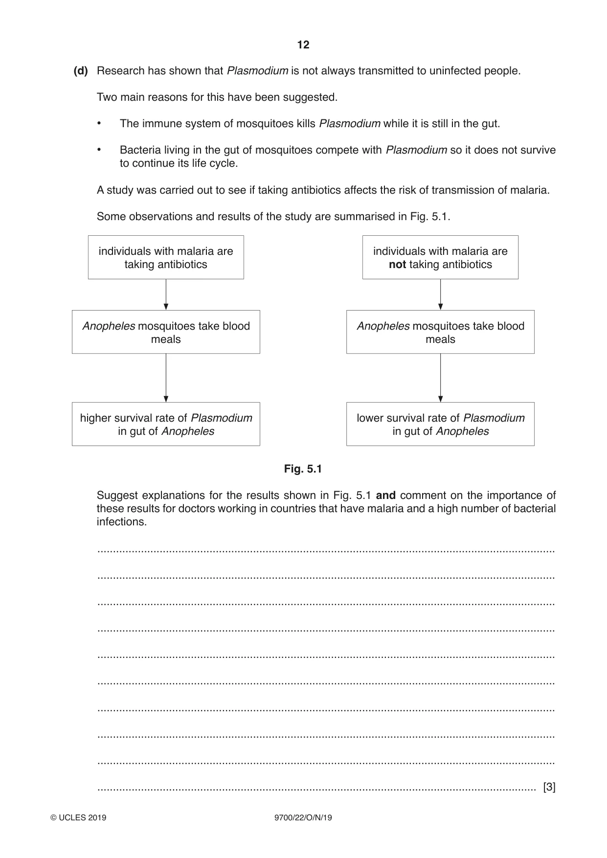
5 Countries that have a high number of cases of malaria also have problems with diseases caused by bacteria. This means that many people in these countries are prescribed antibiotics, such as penicillin, for the treatment of bacterial infections. (a) Outline how penicillin acts on bacterial cells. ................................................................................................................................................... ................................................................................................................................................... ................................................................................................................................................... ................................................................................................................................................... ................................................................................................................................................... ................................................................................................................................................... ................................................................................................................................................... ............................................................................................................................................. [3] The female Anopheles mosquito is the vector of the Plasmodium pathogen that causes malaria. The insect takes in Plasmodium when feeding on blood from an infected person. At a later stage the insect can transmit the pathogen when taking a blood meal from an uninfected person. (b) Name one of the four species of Plasmodium that can cause malaria. ............................................................................................................................................. [1] (c) The male Anopheles mosquito does not feed on blood and so does not act as a vector. Suggest why there is a difference in this feeding behaviour between male and female Anopheles. ................................................................................................................................................... ................................................................................................................................................... ................................................................................................................................................... ............................................................................................................................................. [1] (d) Research has shown that Plasmodium is not always transmitted to uninfected people. Two main reasons for this have been suggested. • The immune system of mosquitoes kills Plasmodium while it is still in the gut. • Bacteria living in the gut of mosquitoes compete with Plasmodium so it does not survive to continue its life cycle. A study was carried out to see if taking antibiotics affects the risk of transmission of malaria. Some observations and results of the study are summarised in Fig. 5.1. individuals with malaria are individuals with malaria are taking antibiotics not taking antibiotics Anopheles mosquitoes take blood Anopheles mosquitoes take blood meals meals higher survival rate of Plasmodium lower survival rate of Plasmodium in gut of Anopheles in gut of Anopheles Fig. 5.1 Suggest explanations for the results shown in Fig. 5.1 and comment on the importance of these results for doctors working in countries that have malaria and a high number of bacterial infections. ................................................................................................................................................... ................................................................................................................................................... ................................................................................................................................................... ................................................................................................................................................... ................................................................................................................................................... ................................................................................................................................................... ................................................................................................................................................... ................................................................................................................................................... ................................................................................................................................................... ............................................................................................................................................. [3] (e) After many years of intense research and development, WHO reported in 2016 that a pilot vaccination programme would be trialled between 2017 and 2020. The programme uses a vaccine acting against the most widespread species of Plasmodium. Explain the difficulties faced by researchers in developing a malaria vaccine. ................................................................................................................................................... ................................................................................................................................................... ................................................................................................................................................... ................................................................................................................................................... ................................................................................................................................................... ................................................................................................................................................... ................................................................................................................................................... ................................................................................................................................................... ................................................................................................................................................... ............................................................................................................................................. [4] [Total: 12]
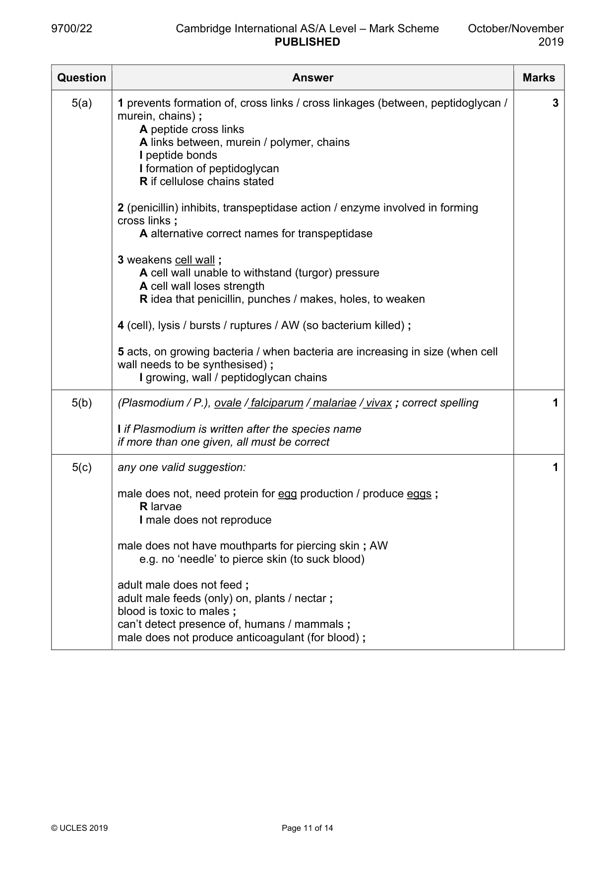
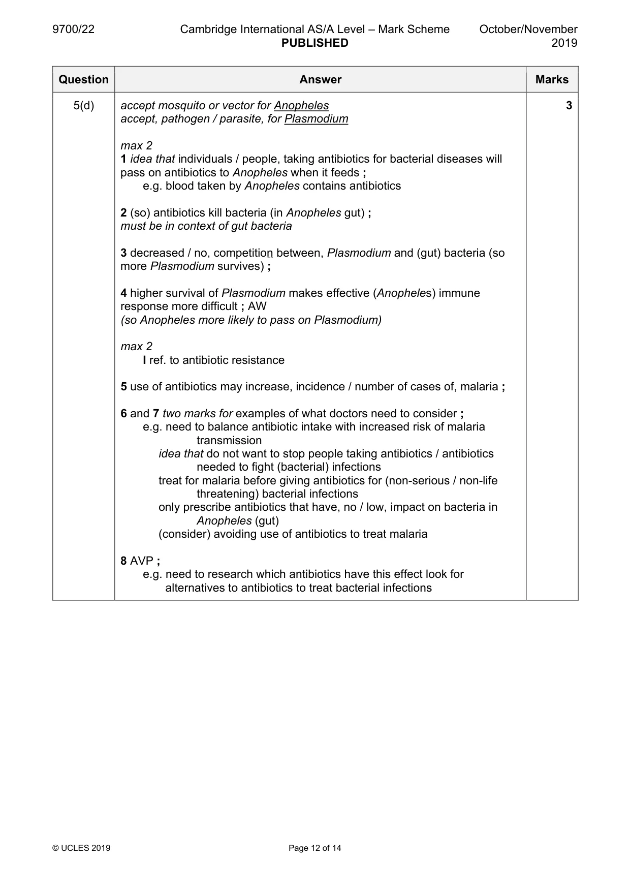
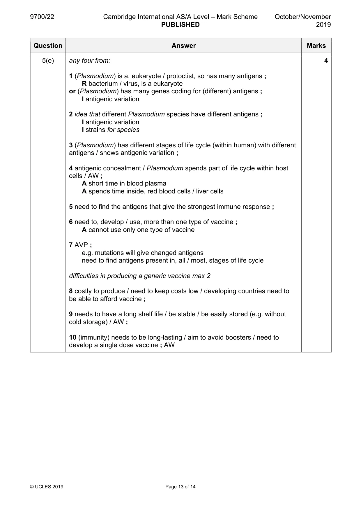
Mark scheme: 5(a) 1 prevents formation of, cross links / cross linkages (between, peptidoglycan / 3 murein, chains) ; A peptide cross links A links between, murein / polymer, chains I peptide bonds I formation of peptidoglycan R if cellulose chains stated 2 (penicillin) inhibits, transpeptidase action / enzyme involved in forming cross links ; A alternative correct names for transpeptidase 3 weakens cell wall ; A cell wall unable to withstand (turgor) pressure A cell wall loses strength R idea that penicillin, punches / makes, holes, to weaken 4 (cell), lysis / bursts / ruptures / AW (so bacterium killed) ; 5 acts, on growing bacteria / when bacteria are increasing in size (when cell wall needs to be synthesised) ; I growing, wall / peptidoglycan chains 5(b) (Plasmodium / P.), ovale / falciparum / malariae / vivax ; correct spelling 1 I if Plasmodium is written after the species name if more than one given, all must be correct 5(c) any one valid suggestion: 1 male does not, need protein for egg production / produce eggs ; R larvae I male does not reproduce male does not have mouthparts for piercing skin ; AW e.g. no ‘needle’ to pierce skin (to suck blood) adult male does not feed ; adult male feeds (only) on, plants / nectar ; blood is toxic to males ; can’t detect presence of, humans / mammals ; male does not produce anticoagulant (for blood) ; 5(d) accept mosquito or vector for Anopheles 3 accept, pathogen / parasite, for Plasmodium max 2 1 idea that individuals / people, taking antibiotics for bacterial diseases will pass on antibiotics to Anopheles when it feeds ; e.g. blood taken by Anopheles contains antibiotics 2 (so) antibiotics kill bacteria (in Anopheles gut) ; must be in context of gut bacteria 3 decreased / no, competition between, Plasmodium and (gut) bacteria (so more Plasmodium survives) ; 4 higher survival of Plasmodium makes effective (Anopheles) immune response more difficult ; AW (so Anopheles more likely to pass on Plasmodium) max 2 I ref. to antibiotic resistance 5 use of antibiotics may increase, incidence / number of cases of, malaria ; 6 and 7 two marks for examples of what doctors need to consider ; e.g. need to balance antibiotic intake with increased risk of malaria transmission idea that do not want to stop people taking antibiotics / antibiotics needed to fight (bacterial) infections treat for malaria before giving antibiotics for (non-serious / non-life threatening) bacterial infections only prescribe antibiotics that have, no / low, impact on bacteria in Anopheles (gut) (consider) avoiding use of antibiotics to treat malaria 8 AVP ; e.g. need to research which antibiotics have this effect look for alternatives to antibiotics to treat bacterial infections 5(e) any four from: 4 1 (Plasmodium) is a, eukaryote / protoctist, so has many antigens ; R bacterium / virus, is a eukaryote or (Plasmodium) has many genes coding for (different) antigens ; I antigenic variation 2 idea that different Plasmodium species have different antigens ; I antigenic variation I strains for species 3 (Plasmodium) has different stages of life cycle (within human) with different antigens / shows antigenic variation ; 4 antigenic concealment / Plasmodium spends part of life cycle within host cells / AW ; A short time in blood plasma A spends time inside, red blood cells / liver cells 5 need to find the antigens that give the strongest immune response ; 6 need to, develop / use, more than one type of vaccine ; A cannot use only one type of vaccine 7 AVP ; e.g. mutations will give changed antigens need to find antigens present in, all / most, stages of life cycle difficulties in producing a generic vaccine max 2 8 costly to produce / need to keep costs low / developing countries need to be able to afford vaccine ; 9 needs to have a long shelf life / be stable / be easily stored (e.g. without cold storage) / AW ; 10 (immunity) needs to be long-lasting / aim to avoid boosters / need to develop a single dose vaccine ; AW
Q6 · Telomerase is an enzyme that is important in maintaining the telomeres that are located…
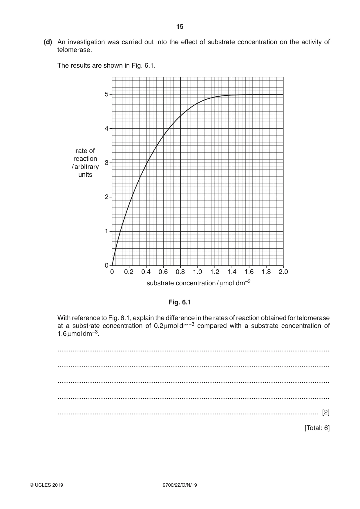
6 Telomerase is an enzyme that is important in maintaining the telomeres that are located at the ends of chromosomes. (a) Explain the importance of telomeres. ................................................................................................................................................... ................................................................................................................................................... ................................................................................................................................................... ................................................................................................................................................... ............................................................................................................................................. [2] (b) Suggest why telomerase is present in much higher quantities in stem cells and cancer cells than in other cell types. ................................................................................................................................................... ................................................................................................................................................... ............................................................................................................................................. [1] (c) Studies suggest that telomerase is a flexible molecule when binding its substrate. The active site changes its shape slightly to allow the formation of the enzyme-substrate complex. Name the theory that describes this mode of enzyme action. ............................................................................................................................................. [1] (d) An investigation was carried out into the effect of substrate concentration on the activity of telomerase. The results are shown in Fig. 6.1. 5 4 rate of reaction 3 / arbitrary units 2 1 0 0 0.2 0.4 0.6 0.8 1.0 1.2 1.4 1.6 1.8 2.0 substrate concentration / μmol dm–3 Fig. 6.1 With reference to Fig. 6.1, explain the difference in the rates of reaction obtained for telomerase at a substrate concentration of 0.2 μmol dm−3 compared with a substrate concentration of 1.6 μmol dm−3. ................................................................................................................................................... ................................................................................................................................................... ................................................................................................................................................... ................................................................................................................................................... ............................................................................................................................................. [2] [Total: 6]
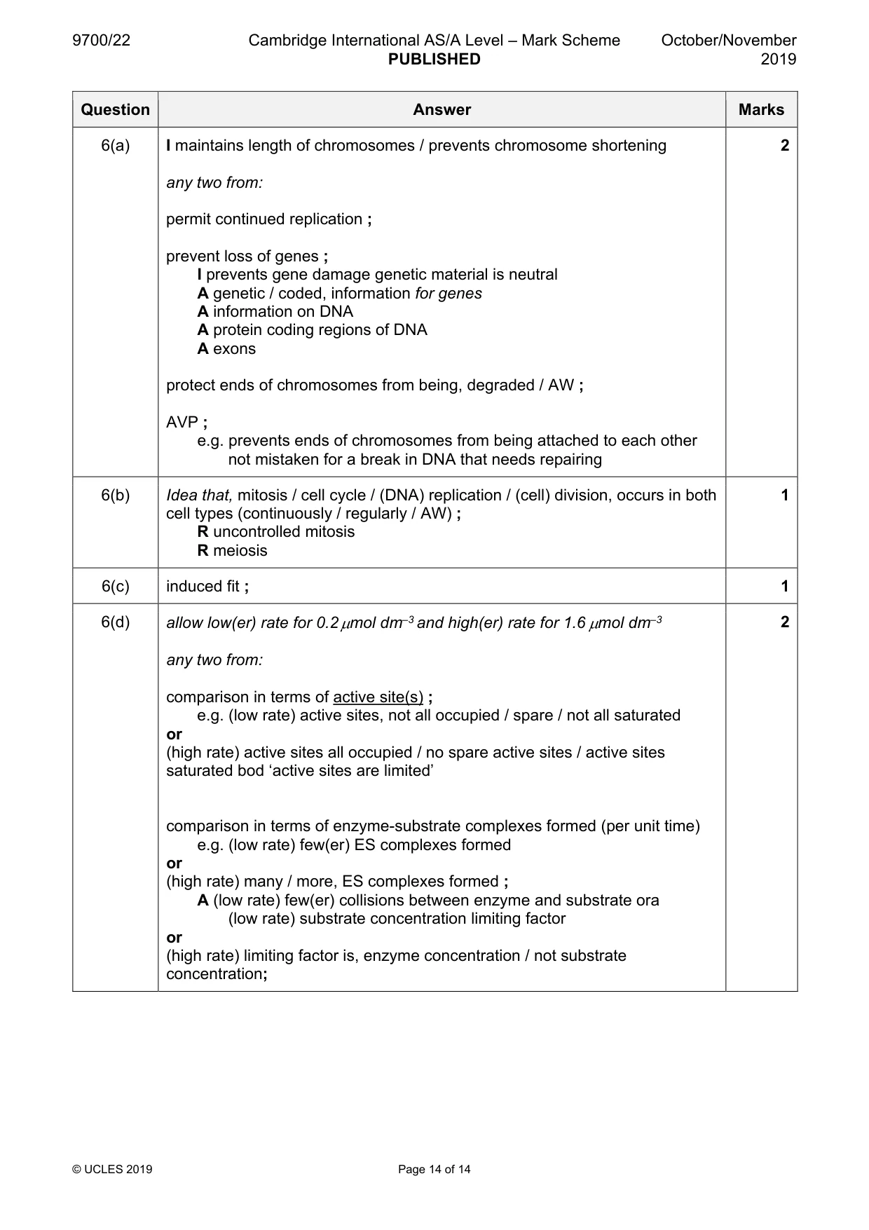
Mark scheme: 6(a) I maintains length of chromosomes / prevents chromosome shortening 2 any two from: permit continued replication ; prevent loss of genes ; I prevents gene damage genetic material is neutral A genetic / coded, information for genes A information on DNA A protein coding regions of DNA A exons protect ends of chromosomes from being, degraded / AW ; AVP ; e.g. prevents ends of chromosomes from being attached to each other not mistaken for a break in DNA that needs repairing 6(b) Idea that, mitosis / cell cycle / (DNA) replication / (cell) division, occurs in both 1 cell types (continuously / regularly / AW) ; R uncontrolled mitosis R meiosis 6(c) induced fit ; 1 6(d) allow low(er) rate for 0.2 µmol dm–3 and high(er) rate for 1.6 µmol dm–3 2 any two from: comparison in terms of active site(s) ; e.g. (low rate) active sites, not all occupied / spare / not all saturated or (high rate) active sites all occupied / no spare active sites / active sites saturated bod ‘active sites are limited’ comparison in terms of enzyme-substrate complexes formed (per unit time) e.g. (low rate) few(er) ES complexes formed or (high rate) many / more, ES complexes formed ; A (low rate) few(er) collisions between enzyme and substrate ora (low rate) substrate concentration limiting factor or (high rate) limiting factor is, enzyme concentration / not substrate concentration;
What was in this paper
The subtopics covered by these 6 questions, and how many questions each got. Open one in a new tab to see every Cambridge question on it.
What you needed in this session
Cambridge’s own grade thresholds for 2019 Oct/Nov, Paper 2 · Variant 2. A higher threshold means an easier paper — the bar moves with how the cohort did.