Cambridge A Level Biology 9700 — 2018 May/June Paper 2 · Variant 3
9700/23/M/J/18 · 6 questions · 60 marks · ≈68 min
The question paper and its mark scheme, free to read here and free to download. This is Cambridge’s own paper, exactly as it was sat.
Question paper20 pages



Mark scheme15 pages
Answers below. Sit the paper first if you are practising.
Questions as text
Q1 · A photomicrograph of root tip meristem
1 Fig. 1.1 is a photomicrograph of root tip meristem. Different stages of the cell cycle are visible. Some cells are in the same stage of the cell cycle and some are in the same stage of mitosis. G A F B C E D Fig. 1.1 (a) Cell D and cell E are in the same stage of the cell cycle. State the difference between the nucleus of cell D and cell E. ................................................................................................................................................... ...............................................................................................................................................[1] (b) Name the stage of mitosis occurring in each of cells A, B and C. cell A ......................................................................................................................................... cell B ......................................................................................................................................... cell C .....................................................................................................................................[3] (c) Cells F and G are newly formed cells. Cytokinesis has occurred with the formation of a cell plate. Describe the events that have occurred in the stage of mitosis immediately before cytokinesis. ................................................................................................................................................... ................................................................................................................................................... ................................................................................................................................................... ...............................................................................................................................................[2] [Total: 6]
Mark scheme: 1(a) (nucleus of cell) D has two nucleoli v E has one nucleolus ; 1 1(b) A = (early / mid) prophase ; R late B = (mid / late) prophase ; R early C = anaphase ; 3 1(c) I name of stage two from: ref. to (daughter) chromosomes at poles ; A (sister) chromatids at poles R chromatids move to poles A two separate groups of (daughter) chromosomes A two nuclei form (enclosing chromosomes) I there are two nuclei in context of at each pole nuclear envelope reassembles (around chromosomes) ; A nuclear membranes reassemble nucleolus reappears / nucleoli reappear ; (daughter) chromosomes, become diffuse / become long and thin / decondense / uncoil ; A become chromatin I disappears spindle, disassembles / AW ; 2
Q2 · Adipose tissue, which is composed of cells known as adipocytes, stores large quantities…
2 Adipose tissue, which is composed of cells known as adipocytes, stores large quantities of triglycerides and functions as an energy storage tissue. Fig. 2.1 is a photomicrograph of adipose tissue. Fig. 2.1 (a) Adipocytes can be very large in size compared to other body cells. This is due to a large lipid droplet within the cell. The largest adipocyte in Fig. 2.1 has a mean diameter of 35 µm. A person with good eyesight can see cells of 0.05 mm or greater diameter without a magnifying glass or any other optical aid. State whether the person can see this adipocyte without any optical aid. Show your working to justify your answer. ...................................................................................................................................... ................................................................................................................................................... ...............................................................................................................................................[1] (b) Only some of the organelles within the adipocyte can be seen using a high quality light microscope set at the highest magnification. Organelles such as rough endoplasmic reticulum, smooth endoplasmic reticulum and ribosomes are only visible using an electron microscope. Explain why these organelles are not visible using a light microscope. ................................................................................................................................................... ................................................................................................................................................... ................................................................................................................................................... ................................................................................................................................................... ...............................................................................................................................................[2] (c) Adipocytes synthesise triglyceride lipase (ATGL), an enzyme that catalyses the formation or breakdown of triglycerides, as shown in Fig. 2.1. lipase triglyceride fatty acids + glycerol Fig. 2.1 The balance between triglyceride formation and breakdown is controlled by hormones. Fig. 2.2 is a summary of events occurring in an adipocyte when glycogen energy stores have been used up. tissue hormone fluid cell surface membrane ATP second messenger molecule cytoplasm inactive enzyme active enzyme inactive ATGL active ATGL triglyceride fatty acids + glycerol Fig. 2.2 (i) Name the type of bond broken by active ATGL to produce fatty acids and glycerol. .......................................................................................................................................[1] (ii) Name and outline the process by which the fatty acids shown in Fig. 2.2 exit the cell. ........................................................................................................................................... ........................................................................................................................................... ........................................................................................................................................... ........................................................................................................................................... ........................................................................................................................................... .......................................................................................................................................[3] (iii) Fig. 2.2 is an example of cell signalling within the body. With reference to Fig. 2.2, outline the process of cell signalling. ........................................................................................................................................... ........................................................................................................................................... ........................................................................................................................................... ........................................................................................................................................... ........................................................................................................................................... ........................................................................................................................................... ........................................................................................................................................... ........................................................................................................................................... ........................................................................................................................................... ........................................................................................................................................... ........................................................................................................................................... .......................................................................................................................................[4] (d) The fatty acids released from adipocytes are transported in blood plasma and are taken up by cells. Although most cell types can metabolise fatty acids to synthesise ATP in the presence of oxygen, red blood cells cannot do this. Suggest why red blood cells cannot metabolise fatty acids to synthesise ATP. ................................................................................................................................................... ...............................................................................................................................................[1] [Total: 12]
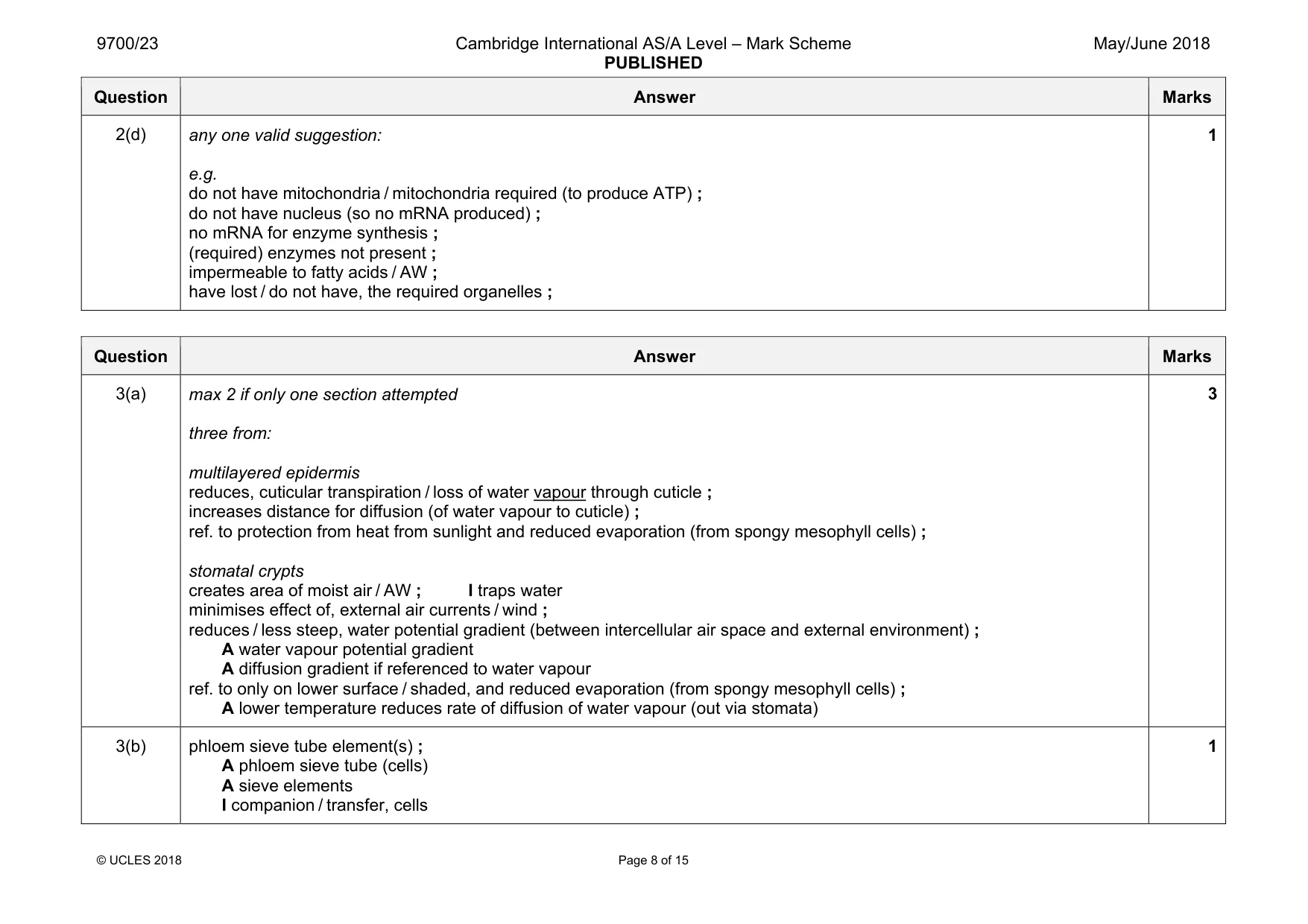
Mark scheme: 2(a) too small to see 35 µm is 0.035 mm (smaller than 0.05 mm) or 0.05 mm is 50 µm (larger than 35 µm) ; 1 2(b) in context of light microscope two from: 1 resolution / resolving power, poor / not high enough / AW ; 2 ability to distinguish between two points not high enough / AW ; 3 ref. to limit of resolution ; e.g. organelles smaller than, 0.2 µm / 200 nm, not visible A organelles smaller than 200 nm (accept range 100–300 nm) 4 organelle too small to interfere with light waves ; A wavelength of light too long I organelles are small 2 2(c)(i) ester ; 1 Question Answer Marks 2(c)(ii) facilitated diffusion ; plus two from: (diffusion) through (membrane), transport / channel / carrier, protein ; A pore I transmembrane / integral, protein high to low concentration or down, concentration / diffusion, gradient ; passive ; A no, ATP / (metabolic) energy, required if active transport incorrectly stated allow ecf through (membrane), transport / carrier, protein ; R channel protein low to high concentration / against a concentration gradient / requires ATP / requires (metabolic) energy ; 3 2(c)(iii) four from: 1 ref. to hormones, released into / arrive at adipocyte in, blood stream ; A released by another, cell / tissue (elsewhere in the body) 2 hormone is, (chemical) signal / signalling molecule / messenger molecule ; 3 (adipocyte is) target cell ; A (adipose is) target tissue 4 (hormone) binds to / AW, receptor ; in context of on cell surface membrane of adipocyte 5 ref.to receptor (shape) complementary to hormone ; A specific receptor for hormone 6 (binding) triggers / stimulates, reactions within the cell ; A cascade of reactions I binding causes a reaction to occur 7 cell signalling results in a response, qualified ; e.g. triglyceride breakdown fatty acids (and glycerol) produced fatty acids leave, the cell / via transport protein 4 Question Answer Marks 2(d) any one valid suggestion: e.g. do not have mitochondria / mitochondria required (to produce ATP) ; do not have nucleus (so no mRNA produced) ; no mRNA for enzyme synthesis ; (required) enzymes not present ; impermeable to fatty acids / AW ; have lost / do not have, the required organelles ; 1
Q3 · Nerium oleander is a xerophytic plant
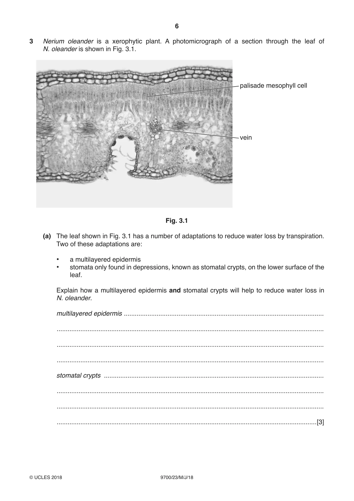
3 Nerium oleander is a xerophytic plant. A photomicrograph of a section through the leaf of N. oleander is shown in Fig. 3.1. palisade mesophyll cell vein Fig. 3.1 (a) The leaf shown in Fig. 3.1 has a number of adaptations to reduce water loss by transpiration. Two of these adaptations are: • a multilayered epidermis • stomata only found in depressions, known as stomatal crypts, on the lower surface of the leaf. Explain how a multilayered epidermis and stomatal crypts will help to reduce water loss in N. oleander. multilayered epidermis .............................................................................................................. ................................................................................................................................................... ................................................................................................................................................... ................................................................................................................................................... stomatal crypts ......................................................................................................................... ................................................................................................................................................... ................................................................................................................................................... ...............................................................................................................................................[3] Sucrose, amino acids and other assimilates synthesised in palisade mesophyll cells of N. oleander pass to the vein, where they can be transported within specialised cells from the source to the sink. (b) Name the cells specialised for the transport of assimilates in N. oleander. ...............................................................................................................................................[1] (c) Explain the difference between a source and a sink. ................................................................................................................................................... ................................................................................................................................................... ................................................................................................................................................... ................................................................................................................................................... ...............................................................................................................................................[2] One of the enzymes involved in the synthesis of sucrose in the cytoplasm of palisade mesophyll cells is known as cyFBPase. The gene coding for this enzyme is cyFBP. The importance of cyFBPase in plant growth can be investigated using plants with a mutation in gene cyFBP. These plants cannot synthesise cyFBPase. (d) (i) State what is meant by a gene mutation. ........................................................................................................................................... .......................................................................................................................................[1] (ii) Suggest one way in which the mutation of cyFBP prevents the synthesis of cyFBPase. ........................................................................................................................................... ........................................................................................................................................... .......................................................................................................................................[1] (e) Monoclonal antibody can be produced that is specific to cyFBPase. This antibody is used by investigators to check that the plants with the cyFBP mutation do not synthesise this enzyme. (i) In monoclonal antibody production, a small mammal is inoculated with cyFBPase and several weeks later cells are removed from the spleen. Some of these cells are required for the production process. Describe the events occurring within the body of the small mammal that lead to the formation of the cells needed for monoclonal antibody production. ........................................................................................................................................... ........................................................................................................................................... ........................................................................................................................................... ........................................................................................................................................... ........................................................................................................................................... ........................................................................................................................................... ........................................................................................................................................... ........................................................................................................................................... ........................................................................................................................................... ........................................................................................................................................... .......................................................................................................................................[4] (ii) Anti-cyFBPase monoclonal antibody is added to extracts taken from the leaves of the plants with the cyFBP mutation. State the expected results following addition of the monoclonal antibody that would confirm the absence of cyFBPase in the leaf extracts. ........................................................................................................................................... .......................................................................................................................................[1] (f) Investigations have shown that plants with the cyFBP mutation grow to a much smaller height and have proportionately far less starch stored in their roots than normal plants. Suggest why plants with the cyFBP mutation will store less starch in their roots. ................................................................................................................................................... ................................................................................................................................................... ................................................................................................................................................... ................................................................................................................................................... ...............................................................................................................................................[2] [Total: 15]
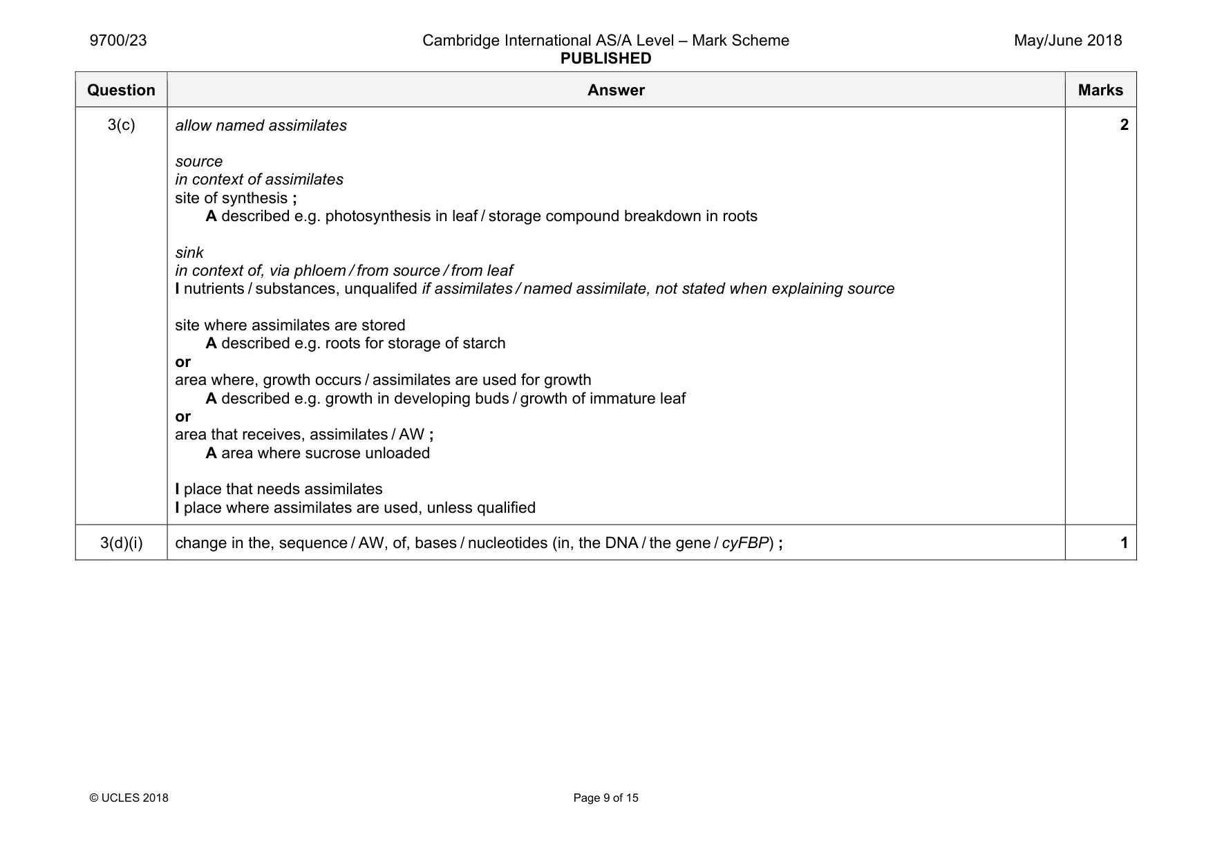
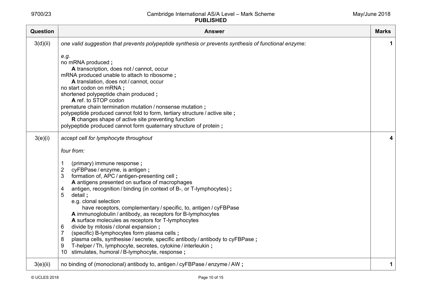
Mark scheme: 3(a) max 2 if only one section attempted three from: multilayered epidermis reduces, cuticular transpiration / loss of water vapour through cuticle ; increases distance for diffusion (of water vapour to cuticle) ; ref. to protection from heat from sunlight and reduced evaporation (from spongy mesophyll cells) ; stomatal crypts creates area of moist air / AW ; I traps water minimises effect of, external air currents / wind ; reduces / less steep, water potential gradient (between intercellular air space and external environment) ; A water vapour potential gradient A diffusion gradient if referenced to water vapour ref. to only on lower surface / shaded, and reduced evaporation (from spongy mesophyll cells) ; A lower temperature reduces rate of diffusion of water vapour (out via stomata) 3 3(b) phloem sieve tube element(s) ; A phloem sieve tube (cells) A sieve elements I companion / transfer, cells 1 Question Answer Marks 3(c) allow named assimilates source in context of assimilates site of synthesis ; A described e.g. photosynthesis in leaf / storage compound breakdown in roots sink in context of, via phloem / from source / from leaf I nutrients / substances, unqualifed if assimilates / named assimilate, not stated when explaining source site where assimilates are stored A described e.g. roots for storage of starch or area where, growth occurs / assimilates are used for growth A described e.g. growth in developing buds / growth of immature leaf or area that receives, assimilates / AW ; A area where sucrose unloaded I place that needs assimilates I place where assimilates are used, unless qualified 2 3(d)(i) change in the, sequence / AW, of, bases / nucleotides (in, the DNA / the gene / cyFBP) ; 1 Question Answer Marks 3(d)(ii) one valid suggestion that prevents polypeptide synthesis or prevents synthesis of functional enzyme: e.g. no mRNA produced ; A transcription, does not / cannot, occur mRNA produced unable to attach to ribosome ; A translation, does not / cannot, occur no start codon on mRNA ; shortened polypeptide chain produced ; A ref. to STOP codon premature chain termination mutation / nonsense mutation ; polypeptide produced cannot fold to form, tertiary structure / active site ; R changes shape of active site preventing function polypeptide produced cannot form quaternary structure of protein ; 1 3(e)(i) accept cell for lymphocyte throughout four from: 1 (primary) immune response ; 2 cyFBPase / enzyme, is antigen ; 3 formation of, APC / antigen-presenting cell ; A antigens presented on surface of macrophages 4 antigen, recognition / binding (in context of B-, or T-lymphocytes) ; 5 detail ; e.g. clonal selection have receptors, complementary / specific, to, antigen / cyFBPase A immunoglobulin / antibody, as receptors for B-lymphocytes A surface molecules as receptors for T-lymphocytes 6 divide by mitosis / clonal expansion ; 7 (specific) B-lymphocytes form plasma cells ; 8 plasma cells, synthesise / secrete, specific antibody / antibody to cyFBPase ; 9 T-helper / Th, lymphocyte, secretes, cytokine / interleukin ; 10 stimulates, humoral / B-lymphocyte, response ; 4 3(e)(ii) no binding of (monoclonal) antibody to, antigen / cyFBPase / enzyme / AW ; 1 Question Answer Marks 3(f) two from: (less cyFBPase / enzyme, so) less sucrose synthesised ; A no enzyme to synthesise sucrose less sucrose transported (source to sink) ; A less sucrose unloaded at sink ; ref. to less glucose for synthesis of starch / AW (on context of sucrose hydrolysis) ; 2
Q4 · Oxygen enters the blood stream from the alveoli in the lungs and carbon dioxide leaves…
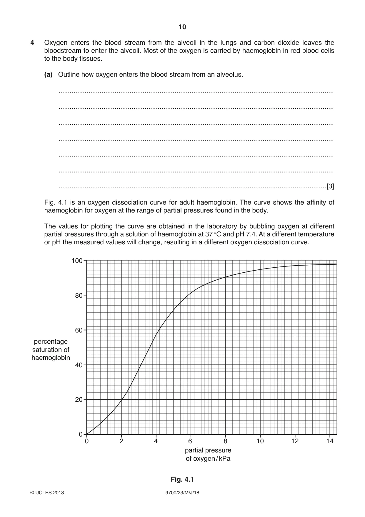
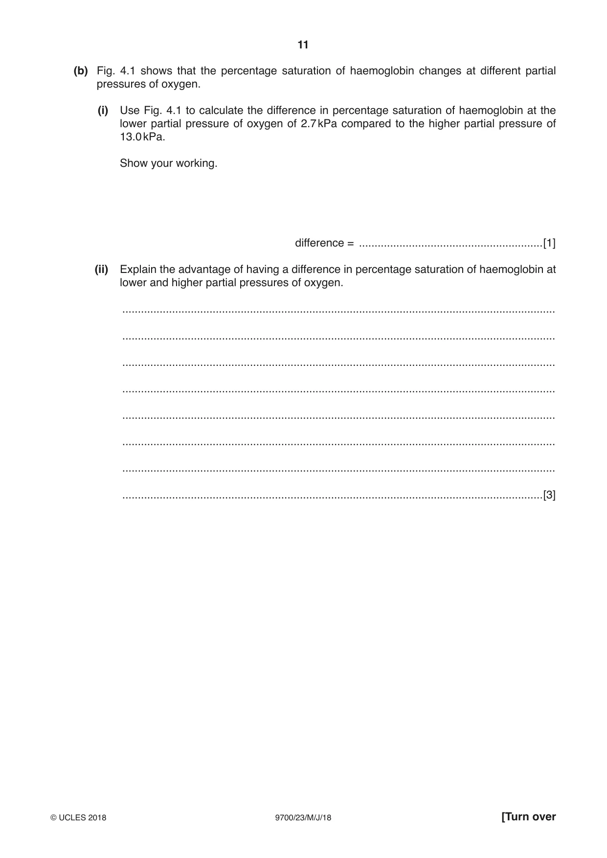
4 Oxygen enters the blood stream from the alveoli in the lungs and carbon dioxide leaves the bloodstream to enter the alveoli. Most of the oxygen is carried by haemoglobin in red blood cells to the body tissues. (a) Outline how oxygen enters the blood stream from an alveolus. ................................................................................................................................................... ................................................................................................................................................... ................................................................................................................................................... ................................................................................................................................................... ................................................................................................................................................... ................................................................................................................................................... ...............................................................................................................................................[3] Fig. 4.1 is an oxygen dissociation curve for adult haemoglobin. The curve shows the affinity of haemoglobin for oxygen at the range of partial pressures found in the body. The values for plotting the curve are obtained in the laboratory by bubbling oxygen at different partial pressures through a solution of haemoglobin at 37 °C and pH 7.4. At a different temperature or pH the measured values will change, resulting in a different oxygen dissociation curve. 100 80 60 percentage saturation of haemoglobin 40 20 0 0 2 4 6 8 10 12 14 partial pressure of oxygen / kPa Fig. 4.1 (b) Fig. 4.1 shows that the percentage saturation of haemoglobin changes at different partial pressures of oxygen. (i) Use Fig. 4.1 to calculate the difference in percentage saturation of haemoglobin at the lower partial pressure of oxygen of 2.7 kPa compared to the higher partial pressure of 13.0 kPa. Show your working. difference = ...........................................................[1] (ii) Explain the advantage of having a difference in percentage saturation of haemoglobin at lower and higher partial pressures of oxygen. ........................................................................................................................................... ........................................................................................................................................... ........................................................................................................................................... ........................................................................................................................................... ........................................................................................................................................... ........................................................................................................................................... ........................................................................................................................................... .......................................................................................................................................[3] In a person with sickle cell anaemia, the ability of haemoglobin to transport oxygen and carbon dioxide is severely affected. The cause of this disease is a mutation in the gene coding for the β-globin polypeptide of haemoglobin. (c) Define the term disease. ................................................................................................................................................... ................................................................................................................................................... ................................................................................................................................................... ................................................................................................................................................... ...............................................................................................................................................[2] (d) Outline the differences between the HbA (normal) and HbS (sickle cell) alleles of the gene coding for the β-globin polypeptide and explain how these differences lead to a change in the haemoglobin molecule formed. ................................................................................................................................................... ................................................................................................................................................... ................................................................................................................................................... ................................................................................................................................................... ................................................................................................................................................... ................................................................................................................................................... ................................................................................................................................................... ................................................................................................................................................... ................................................................................................................................................... ................................................................................................................................................... ................................................................................................................................................... ...............................................................................................................................................[5] [Total: 14] Question 5 starts on page 14
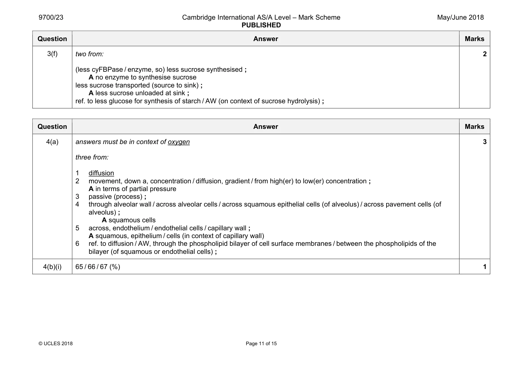
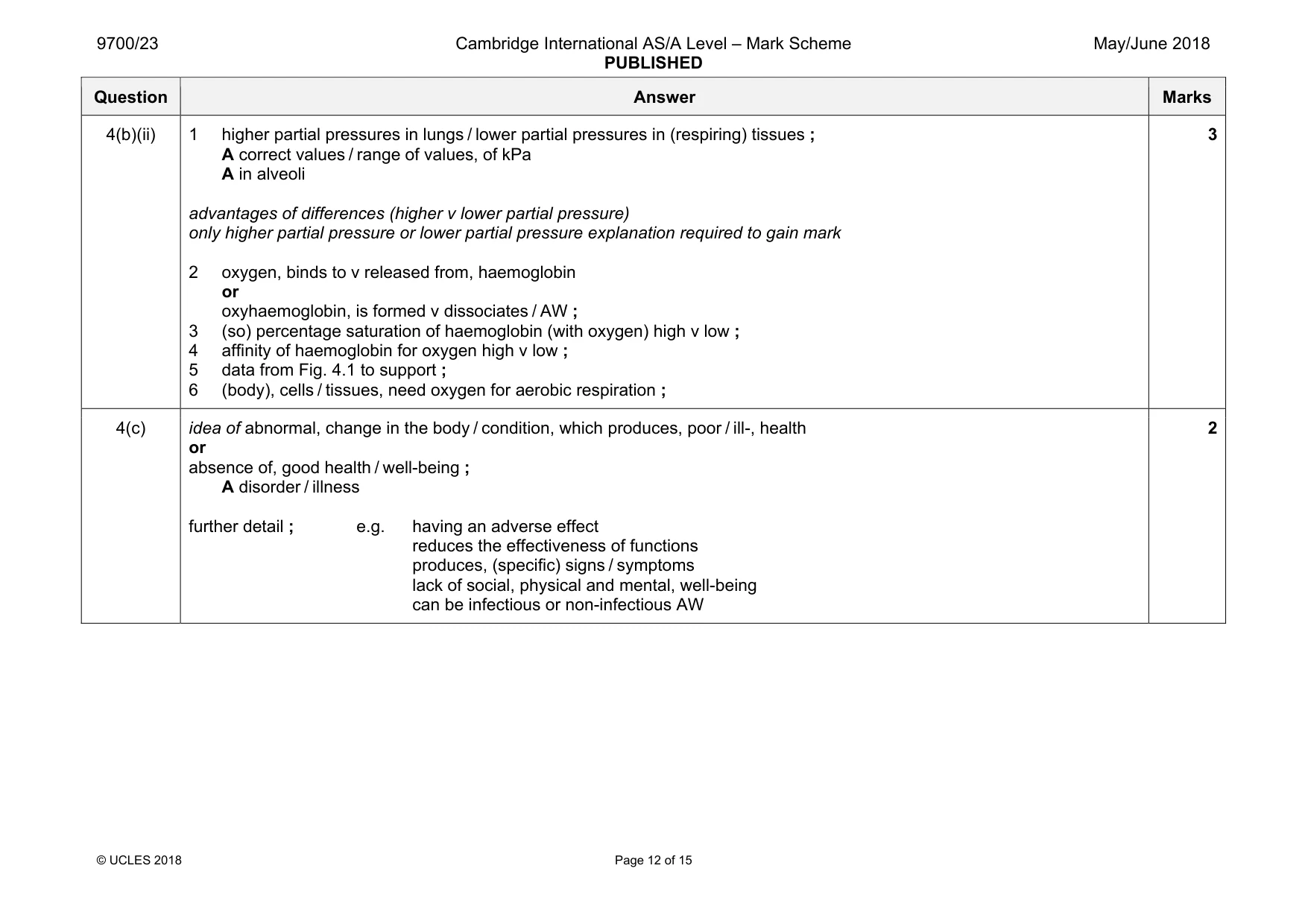
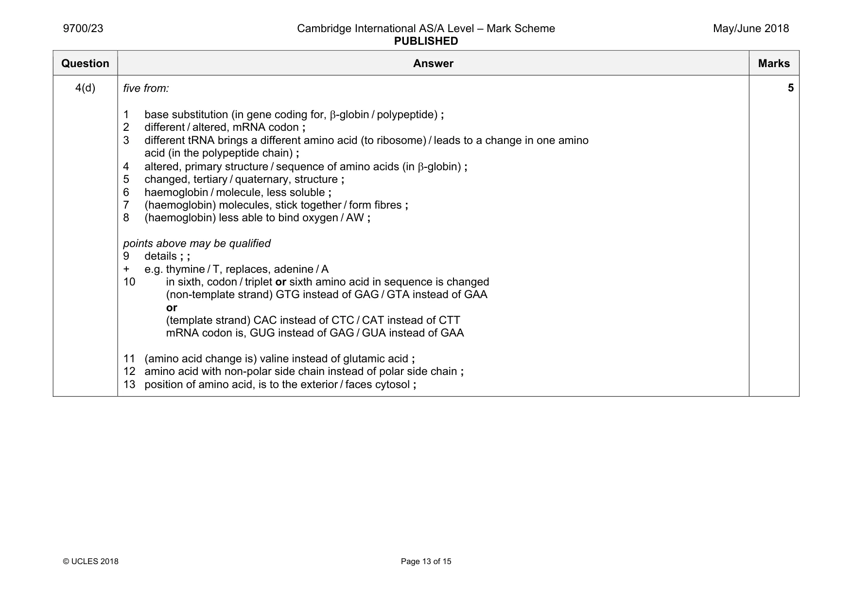
Mark scheme: 4(a) answers must be in context of oxygen three from: 1 diffusion 2 movement, down a, concentration / diffusion, gradient / from high(er) to low(er) concentration ; A in terms of partial pressure 3 passive (process) ; 4 through alveolar wall / across alveolar cells / across squamous epithelial cells (of alveolus) / across pavement cells (of alveolus) ; A squamous cells 5 across, endothelium / endothelial cells / capillary wall ; A squamous, epithelium / cells (in context of capillary wall) 6 ref. to diffusion / AW, through the phospholipid bilayer of cell surface membranes / between the phospholipids of the bilayer (of squamous or endothelial cells) ; 3 4(b)(i) 65 / 66 / 67 (%) 1 Question Answer Marks 4(b)(ii) 1 higher partial pressures in lungs / lower partial pressures in (respiring) tissues ; A correct values / range of values, of kPa A in alveoli advantages of differences (higher v lower partial pressure) only higher partial pressure or lower partial pressure explanation required to gain mark 2 oxygen, binds to v released from, haemoglobin or oxyhaemoglobin, is formed v dissociates / AW ; 3 (so) percentage saturation of haemoglobin (with oxygen) high v low ; 4 affinity of haemoglobin for oxygen high v low ; 5 data from Fig. 4.1 to support ; 6 (body), cells / tissues, need oxygen for aerobic respiration ; 3 4(c) idea of abnormal, change in the body / condition, which produces, poor / ill-, health or absence of, good health / well-being ; A disorder / illness further detail ; e.g. having an adverse effect reduces the effectiveness of functions produces, (specific) signs / symptoms lack of social, physical and mental, well-being can be infectious or non-infectious AW 2 Question Answer Marks 4(d) five from: 1 base substitution (in gene coding for, β-globin / polypeptide) ; 2 different / altered, mRNA codon ; 3 different tRNA brings a different amino acid (to ribosome) / leads to a change in one amino acid (in the polypeptide chain) ; 4 altered, primary structure / sequence of amino acids (in β-globin) ; 5 changed, tertiary / quaternary, structure ; 6 haemoglobin / molecule, less soluble ; 7 (haemoglobin) molecules, stick together / form fibres ; 8 (haemoglobin) less able to bind oxygen / AW ; points above may be qualified 9 details ; ; + e.g. thymine / T, replaces, adenine / A 10 in sixth, codon / triplet or sixth amino acid in sequence is changed (non-template strand) GTG instead of GAG / GTA instead of GAA or (template strand) CAC instead of CTC / CAT instead of CTT mRNA codon is, GUG instead of GAG / GUA instead of GAA 11 (amino acid change is) valine instead of glutamic acid ; 12 amino acid with non-polar side chain instead of polar side chain ; 13 position of amino acid, is to the exterior / faces cytosol ; 5
Q5 · The fig tree, Ficus carica, and the papaya tree, Carica papaya, produce a milky-looking…
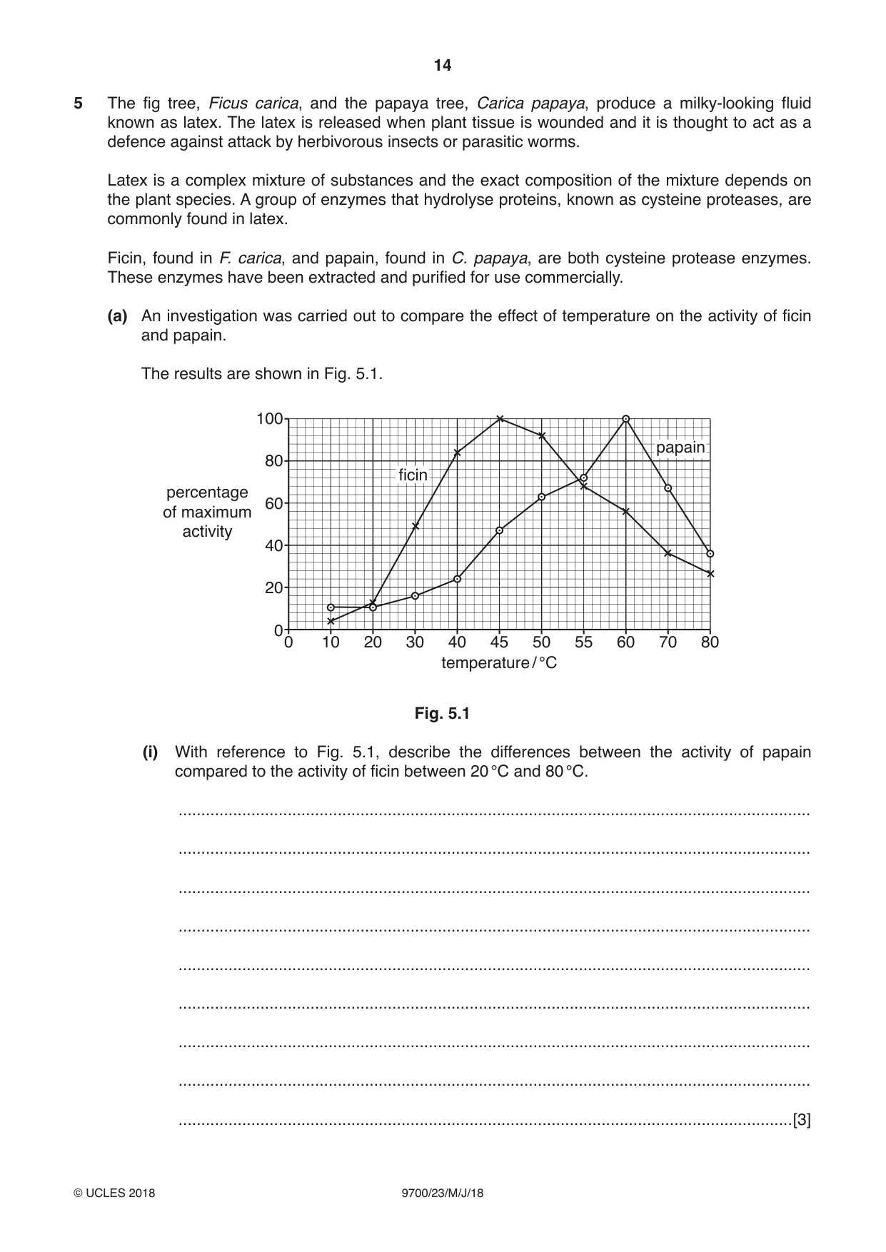
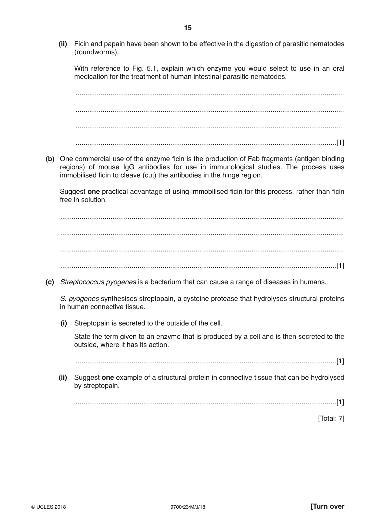
5 The fig tree, Ficus carica, and the papaya tree, Carica papaya, produce a milky-looking fluid known as latex. The latex is released when plant tissue is wounded and it is thought to act as a defence against attack by herbivorous insects or parasitic worms. Latex is a complex mixture of substances and the exact composition of the mixture depends on the plant species. A group of enzymes that hydrolyse proteins, known as cysteine proteases, are commonly found in latex. Ficin, found in F. carica, and papain, found in C. papaya, are both cysteine protease enzymes. These enzymes have been extracted and purified for use commercially. (a) An investigation was carried out to compare the effect of temperature on the activity of ficin and papain. The results are shown in Fig. 5.1. 100 papain 80 ficin percentage 60 of maximum activity 40 20 0 0 10 20 30 40 45 50 55 60 70 80 temperature / °C Fig. 5.1 (i) With reference to Fig. 5.1, describe the differences between the activity of papain compared to the activity of ficin between 20 °C and 80 °C. ........................................................................................................................................... ........................................................................................................................................... ........................................................................................................................................... ........................................................................................................................................... ........................................................................................................................................... ........................................................................................................................................... ........................................................................................................................................... ........................................................................................................................................... .......................................................................................................................................[3] (ii) Ficin and papain have been shown to be effective in the digestion of parasitic nematodes (roundworms). With reference to Fig. 5.1, explain which enzyme you would select to use in an oral medication for the treatment of human intestinal parasitic nematodes. ........................................................................................................................................... ........................................................................................................................................... ........................................................................................................................................... .......................................................................................................................................[1] (b) One commercial use of the enzyme ficin is the production of Fab fragments (antigen binding regions) of mouse IgG antibodies for use in immunological studies. The process uses immobilised ficin to cleave (cut) the antibodies in the hinge region. Suggest one practical advantage of using immobilised ficin for this process, rather than ficin free in solution. ................................................................................................................................................... ................................................................................................................................................... ................................................................................................................................................... ...............................................................................................................................................[1] (c) Streptococcus pyogenes is a bacterium that can cause a range of diseases in humans. S. pyogenes synthesises streptopain, a cysteine protease that hydrolyses structural proteins in human connective tissue. (i) Streptopain is secreted to the outside of the cell. State the term given to an enzyme that is produced by a cell and is then secreted to the outside, where it has its action. .......................................................................................................................................[1] (ii) Suggest one example of a structural protein in connective tissue that can be hydrolysed by streptopain. .......................................................................................................................................[1] [Total: 7]
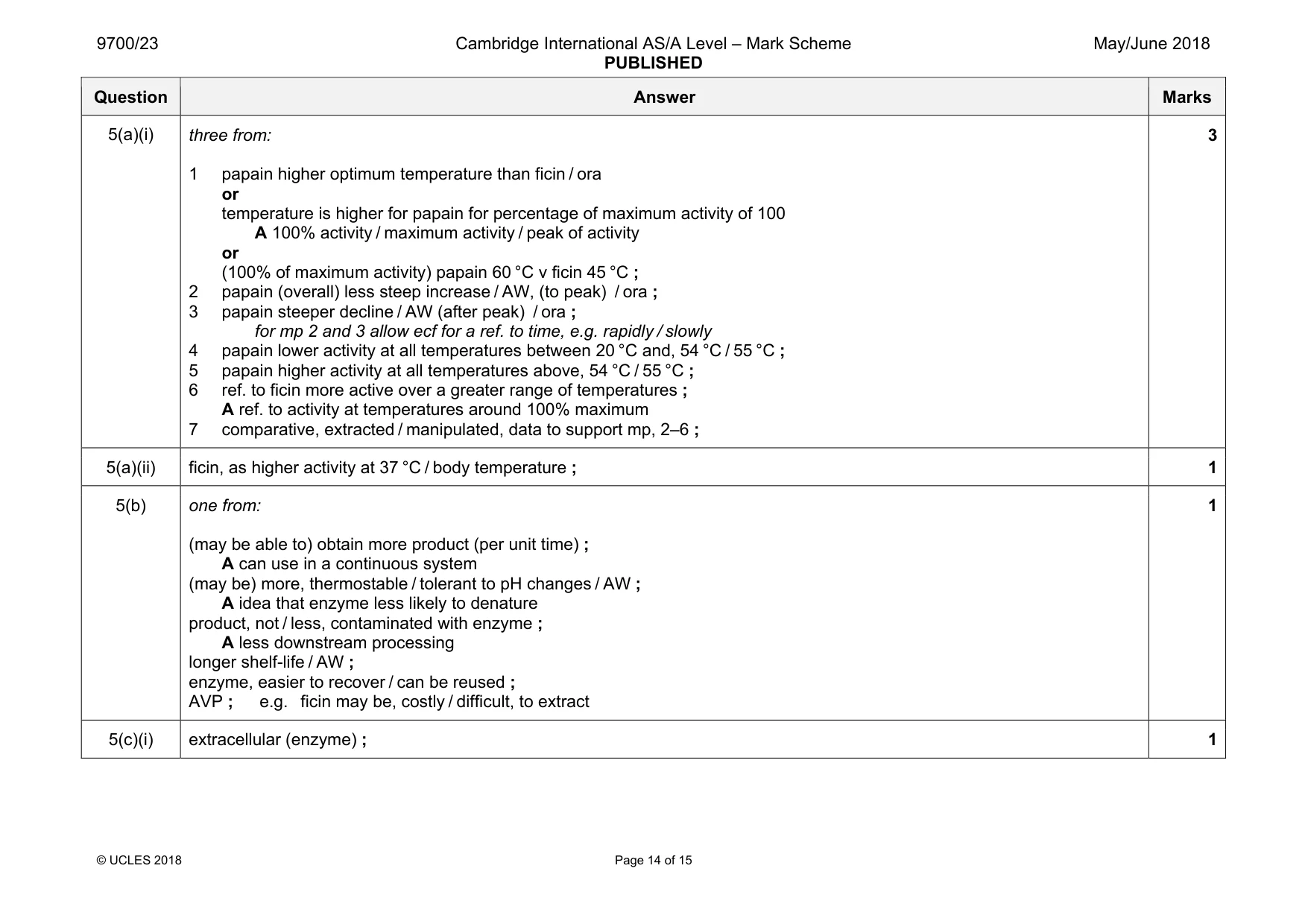
Mark scheme: 5(a)(i) three from: 1 papain higher optimum temperature than ficin / ora or temperature is higher for papain for percentage of maximum activity of 100 A 100% activity / maximum activity / peak of activity or (100% of maximum activity) papain 60 °C v ficin 45 °C ; 2 papain (overall) less steep increase / AW, (to peak) / ora ; 3 papain steeper decline / AW (after peak) / ora ; for mp 2 and 3 allow ecf for a ref. to time, e.g. rapidly / slowly 4 papain lower activity at all temperatures between 20 °C and, 54 °C / 55 °C ; 5 papain higher activity at all temperatures above, 54 °C / 55 °C ; 6 ref. to ficin more active over a greater range of temperatures ; A ref. to activity at temperatures around 100% maximum 7 comparative, extracted / manipulated, data to support mp, 2–6 ; 3 5(a)(ii) ficin, as higher activity at 37 °C / body temperature ; 1 5(b) one from: (may be able to) obtain more product (per unit time) ; A can use in a continuous system (may be) more, thermostable / tolerant to pH changes / AW ; A idea that enzyme less likely to denature product, not / less, contaminated with enzyme ; A less downstream processing longer shelf-life / AW ; enzyme, easier to recover / can be reused ; AVP ; e.g. ficin may be, costly / difficult, to extract 1 5(c)(i) extracellular (enzyme) ; 1 Question Answer Marks 5(c)(ii) any one valid example: e.g. collagen / elastin ; 1
Q6 · A list of infectious diseases
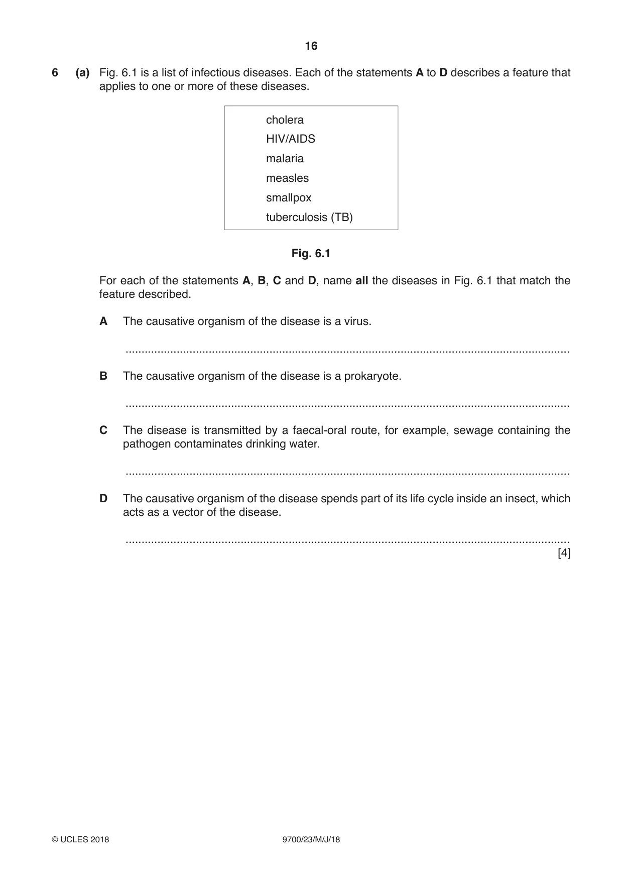
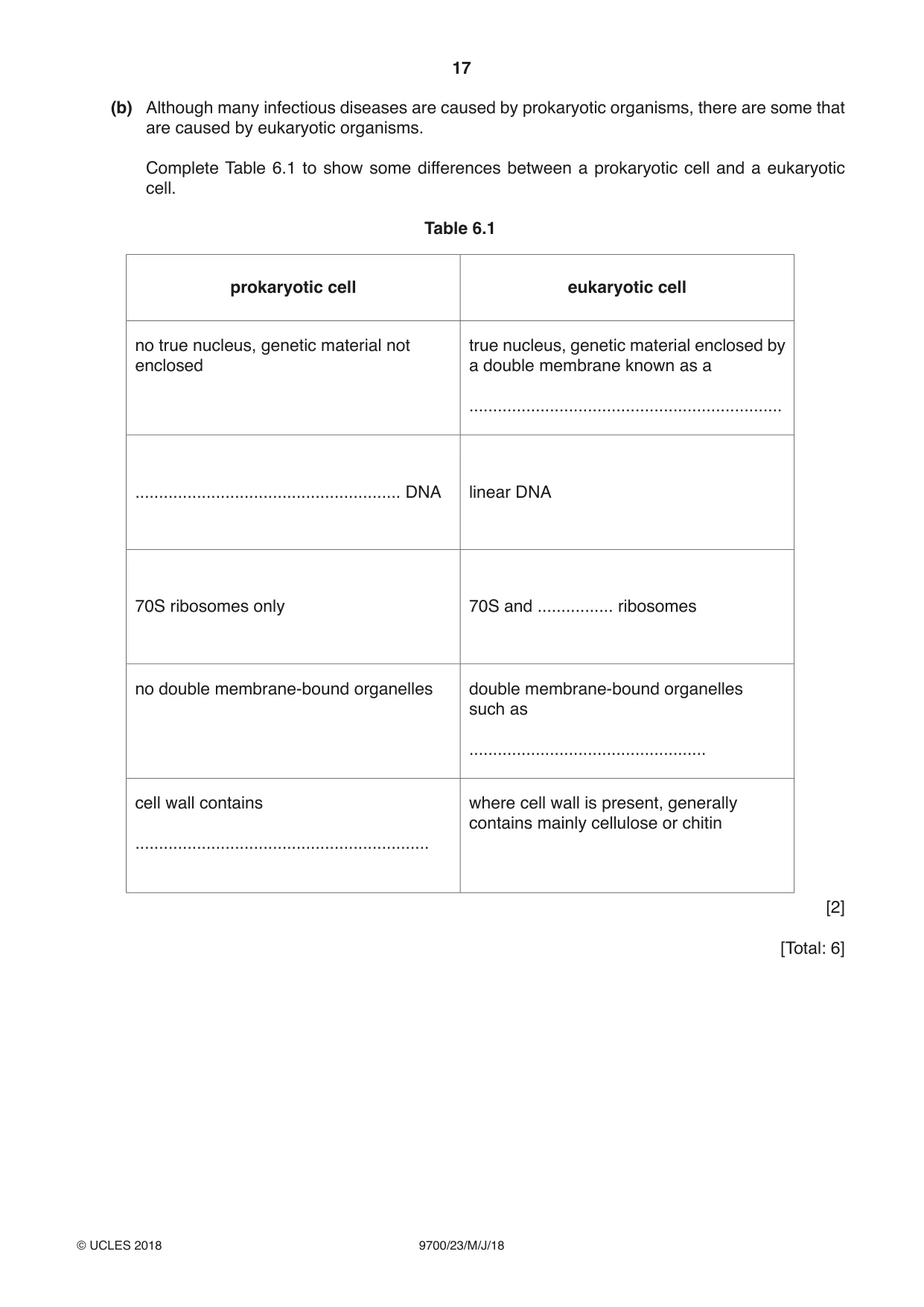
6 (a) Fig. 6.1 is a list of infectious diseases. Each of the statements A to D describes a feature that applies to one or more of these diseases. cholera HIV/AIDS malaria measles smallpox tuberculosis (TB) Fig. 6.1 For each of the statements A, B, C and D, name all the diseases in Fig. 6.1 that match the feature described. A The causative organism of the disease is a virus. ........................................................................................................................................... B The causative organism of the disease is a prokaryote. ........................................................................................................................................... C The disease is transmitted by a faecal-oral route, for example, sewage containing the pathogen contaminates drinking water. ........................................................................................................................................... D The causative organism of the disease spends part of its life cycle inside an insect, which acts as a vector of the disease. ........................................................................................................................................... [4] (b) Although many infectious diseases are caused by prokaryotic organisms, there are some that are caused by eukaryotic organisms. Complete Table 6.1 to show some differences between a prokaryotic cell and a eukaryotic cell. Table 6.1 prokaryotic cell eukaryotic cell no true nucleus, genetic material not true nucleus, genetic material enclosed by enclosed a double membrane known as a .................................................................. ........................................................ DNA linear DNA 70S ribosomes only 70S and ................ ribosomes no double membrane-bound organelles double membrane-bound organelles such as .................................................. cell wall contains where cell wall is present, generally contains mainly cellulose or chitin .............................................................. [2] [Total: 6]
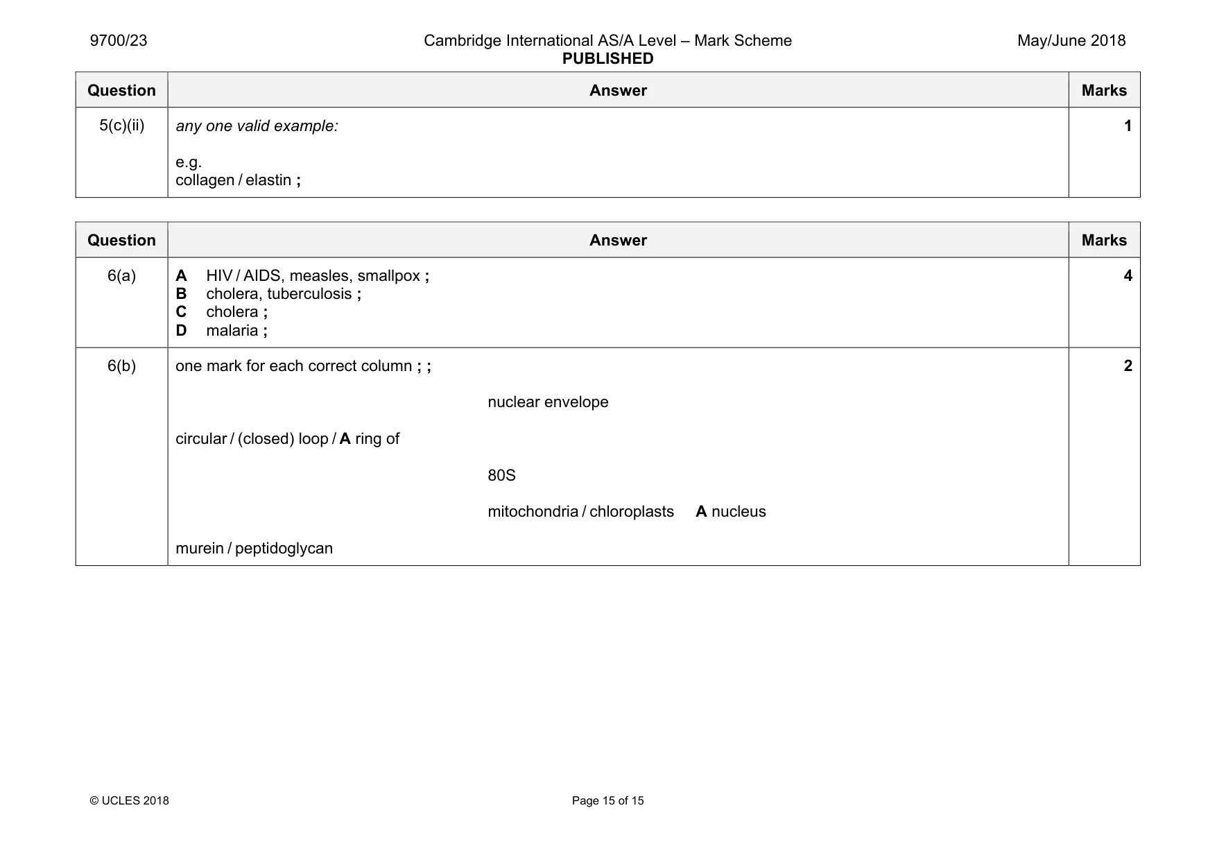
Mark scheme: 6(a) A HIV / AIDS, measles, smallpox ; B cholera, tuberculosis ; C cholera ; D malaria ; 4 6(b) one mark for each correct column ; ; nuclear envelope circular / (closed) loop / A ring of 80S mitochondria / chloroplasts A nucleus murein / peptidoglycan 2
What was in this paper
The subtopics covered by these 6 questions, and how many questions each got. Open one in a new tab to see every Cambridge question on it.
What you needed in this session
Cambridge’s own grade thresholds for 2018 May/June, Paper 2 · Variant 3. A higher threshold means an easier paper — the bar moves with how the cohort did.