Cambridge A Level Biology 9700 — 2018 May/June Paper 2 · Variant 1
9700/21/M/J/18 · 6 questions · 60 marks · ≈68 min
The question paper and its mark scheme, free to read here and free to download. This is Cambridge’s own paper, exactly as it was sat.
Question paper16 pages
Mark scheme14 pages
Answers below. Sit the paper first if you are practising.
Questions as text
Q1 · A transmission electron micrograph of a cell from the root of thale cress, Arabidopsis…
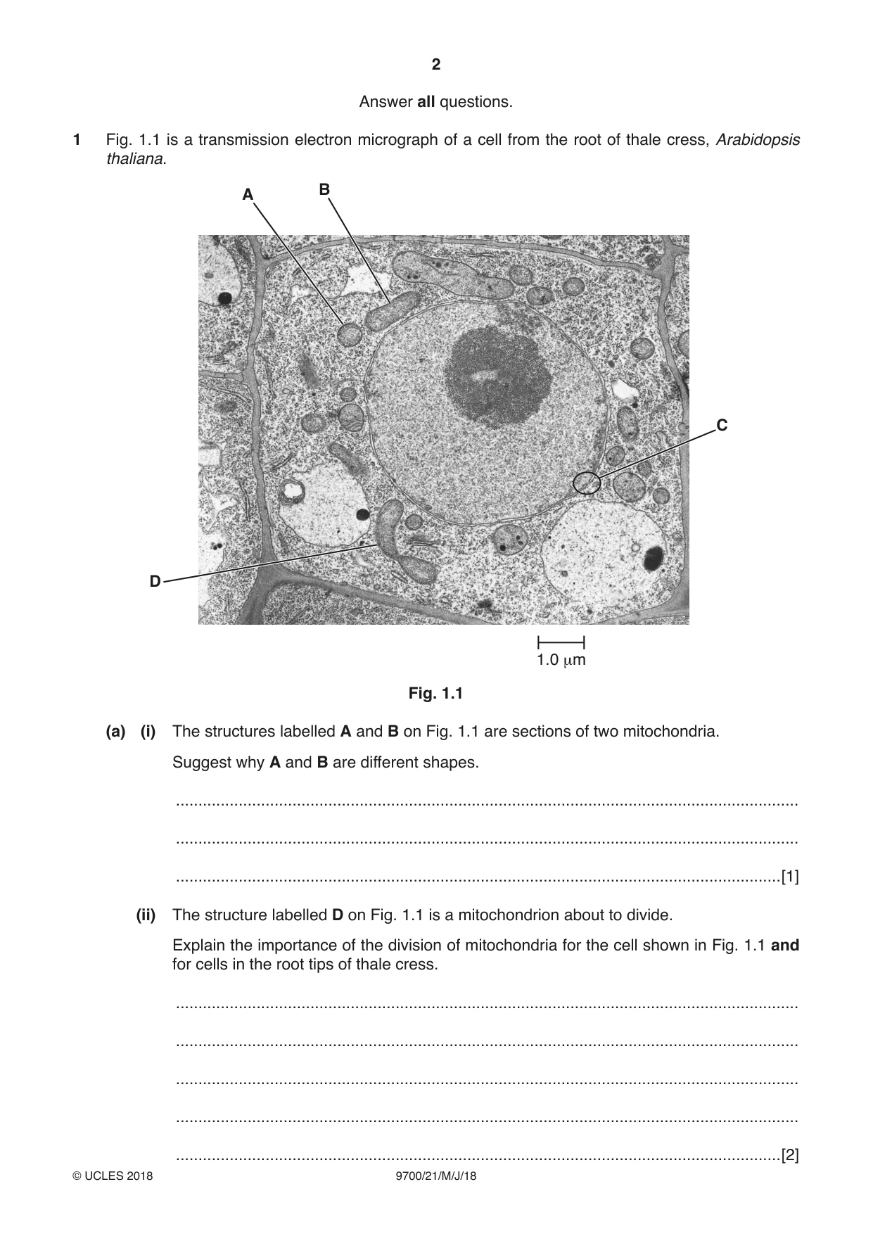
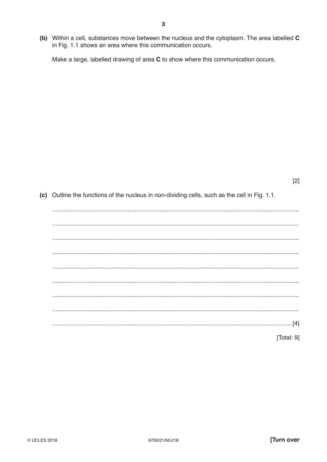
1 Fig. 1.1 is a transmission electron micrograph of a cell from the root of thale cress, Arabidopsis thaliana. B A C D 1.0 μm Fig. 1.1 (a) (i) The structures labelled A and B on Fig. 1.1 are sections of two mitochondria. Suggest why A and B are different shapes. ........................................................................................................................................... ........................................................................................................................................... .......................................................................................................................................[1] (ii) The structure labelled D on Fig. 1.1 is a mitochondrion about to divide. Explain the importance of the division of mitochondria for the cell shown in Fig. 1.1 and for cells in the root tips of thale cress. ........................................................................................................................................... ........................................................................................................................................... ........................................................................................................................................... ........................................................................................................................................... .......................................................................................................................................[2] (b) Within a cell, substances move between the nucleus and the cytoplasm. The area labelled C in Fig. 1.1 shows an area where this communication occurs. Make a large, labelled drawing of area C to show where this communication occurs. [2] (c) Outline the functions of the nucleus in non-dividing cells, such as the cell in Fig. 1.1. ................................................................................................................................................... ................................................................................................................................................... ................................................................................................................................................... ................................................................................................................................................... ................................................................................................................................................... ................................................................................................................................................... ................................................................................................................................................... ................................................................................................................................................... ...............................................................................................................................................[4] [Total: 9]
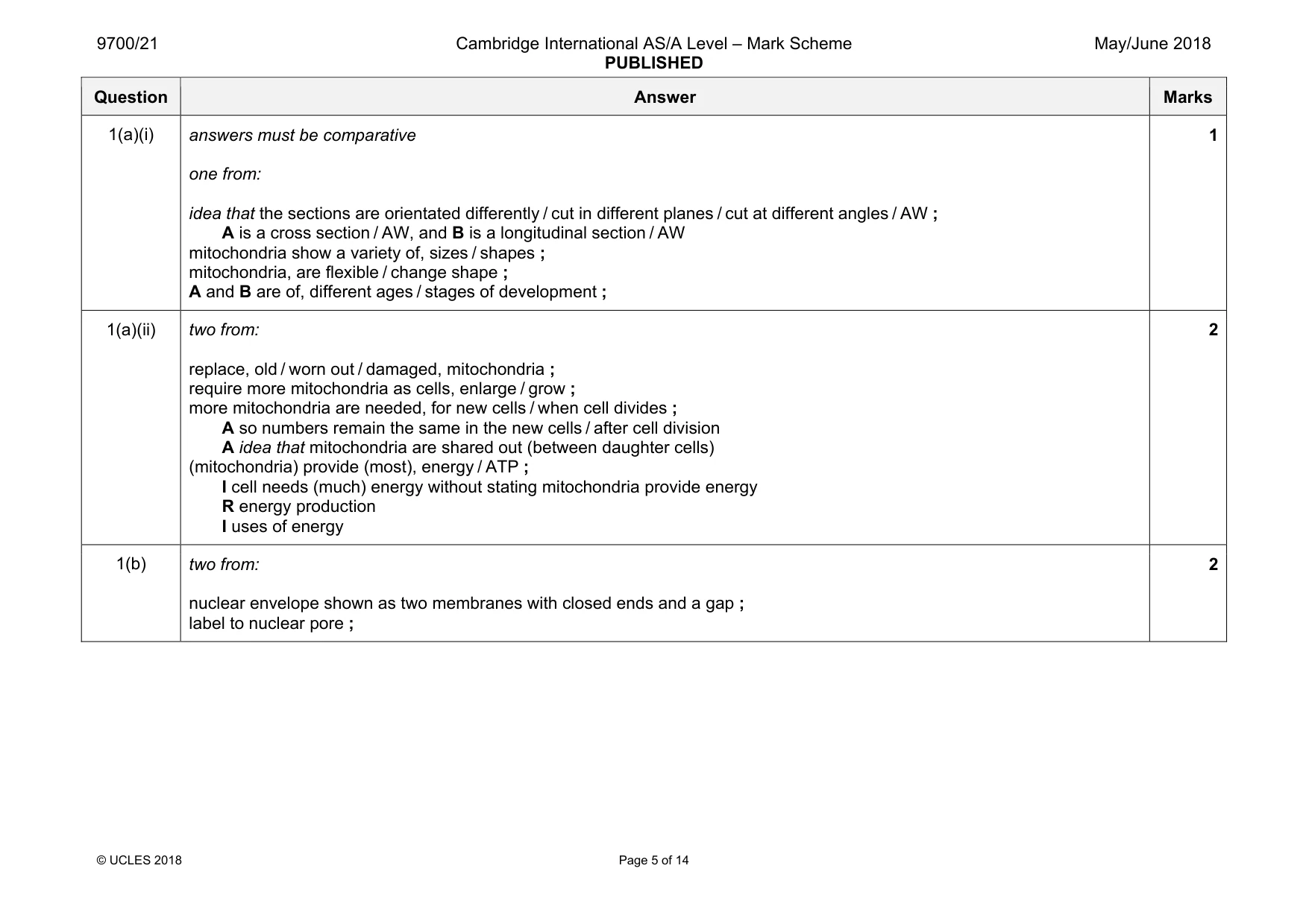
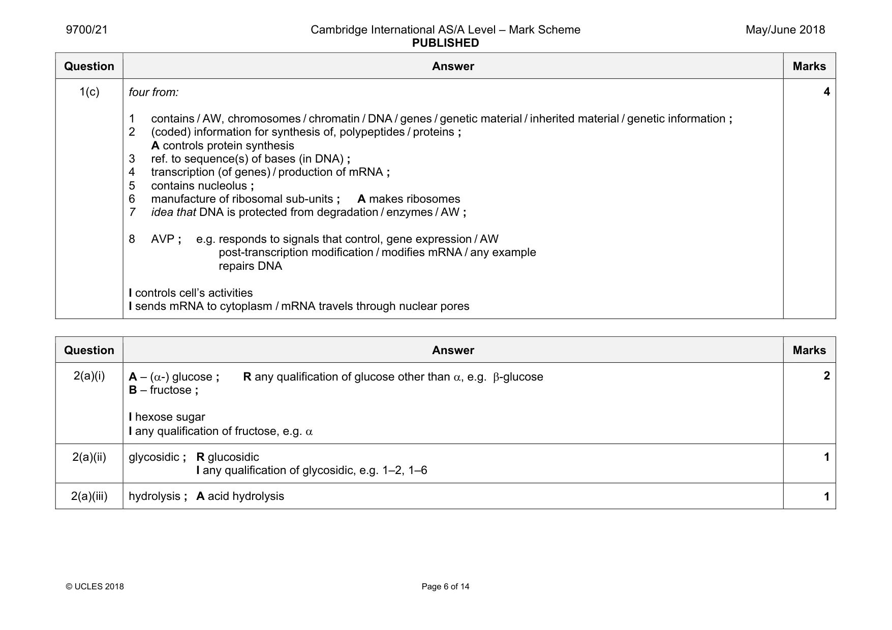
Mark scheme: 1(a)(i) answers must be comparative one from: idea that the sections are orientated differently / cut in different planes / cut at different angles / AW ; A is a cross section / AW, and B is a longitudinal section / AW mitochondria show a variety of, sizes / shapes ; mitochondria, are flexible / change shape ; A and B are of, different ages / stages of development ; 1 1(a)(ii) two from: replace, old / worn out / damaged, mitochondria ; require more mitochondria as cells, enlarge / grow ; more mitochondria are needed, for new cells / when cell divides ; A so numbers remain the same in the new cells / after cell division A idea that mitochondria are shared out (between daughter cells) (mitochondria) provide (most), energy / ATP ; I cell needs (much) energy without stating mitochondria provide energy R energy production I uses of energy 2 1(b) two from: nuclear envelope shown as two membranes with closed ends and a gap ; label to nuclear pore ; 2 Question Answer Marks 1(c) four from: 1 contains / AW, chromosomes / chromatin / DNA / genes / genetic material / inherited material / genetic information ; 2 (coded) information for synthesis of, polypeptides / proteins ; A controls protein synthesis 3 ref. to sequence(s) of bases (in DNA) ; 4 transcription (of genes) / production of mRNA ; 5 contains nucleolus ; 6 manufacture of ribosomal sub-units ; A makes ribosomes 7 idea that DNA is protected from degradation / enzymes / AW ; 8 AVP ; e.g. responds to signals that control, gene expression / AW post-transcription modification / modifies mRNA / any example repairs DNA I controls cell’s activities I sends mRNA to cytoplasm / mRNA travels through nuclear pores 4
More questions on Cells as the basic units of living organisms
Q2 · Sucrose is a disaccharide
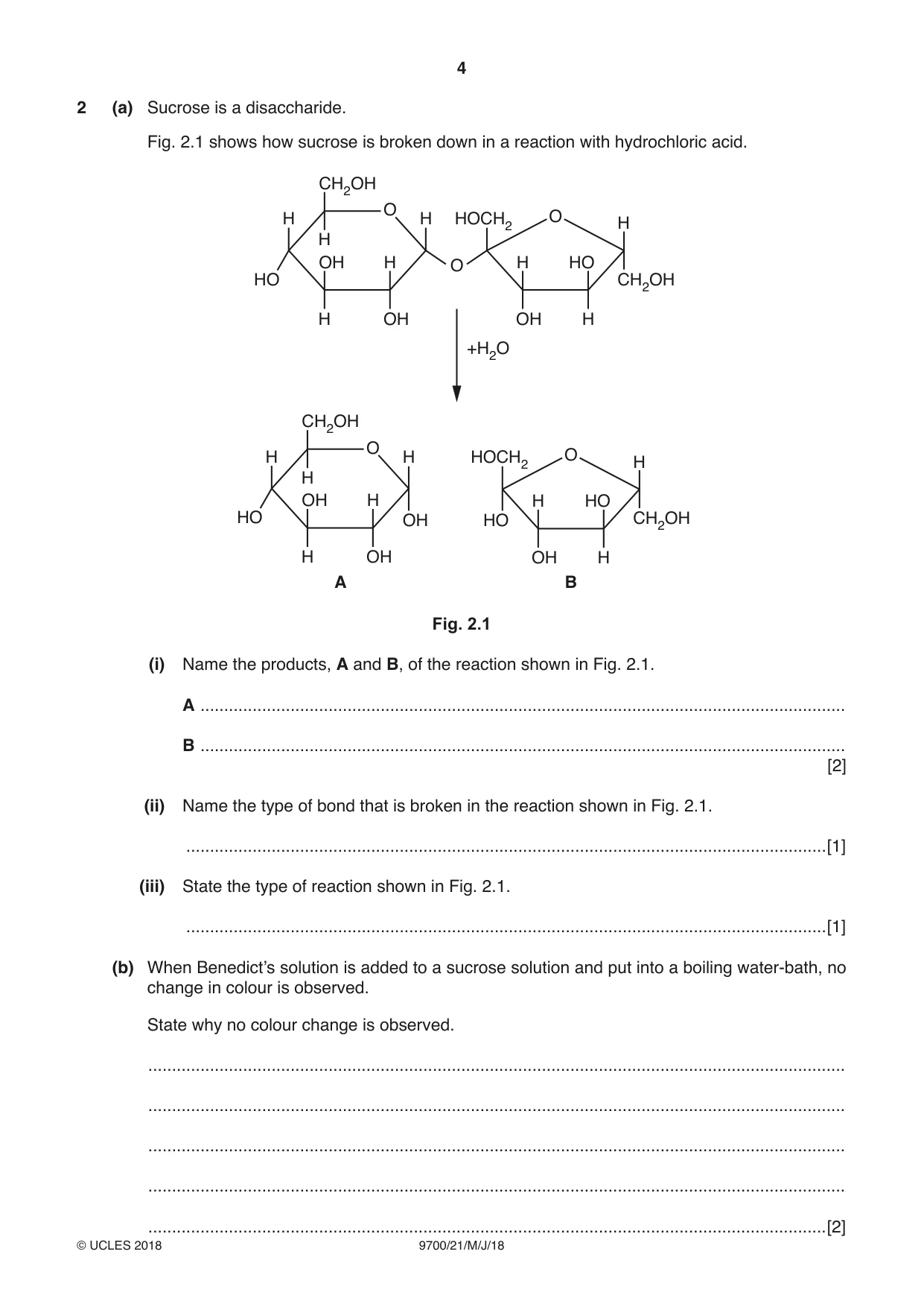
2 (a) Sucrose is a disaccharide. Fig. 2.1 shows how sucrose is broken down in a reaction with hydrochloric acid. CH2OH O H H HOCH2 O H H OH H O H HO HO CH2OH H OH OH H +H2O CH2OH O H H HOCH2 O H H OH H H HO HO OH HO CH2OH H OH OH H A B Fig. 2.1 (i) Name the products, A and B, of the reaction shown in Fig. 2.1. A ........................................................................................................................................ B ........................................................................................................................................ [2] (ii) Name the type of bond that is broken in the reaction shown in Fig. 2.1. .......................................................................................................................................[1] (iii) State the type of reaction shown in Fig. 2.1. .......................................................................................................................................[1] (b) When Benedict’s solution is added to a sucrose solution and put into a boiling water-bath, no change in colour is observed. State why no colour change is observed. ................................................................................................................................................... ................................................................................................................................................... ................................................................................................................................................... ................................................................................................................................................... (c) Sugar beet plants, Beta vulgaris, store sucrose in their roots. Describe and explain how sucrose is transported from the phloem in the leaves of sugar beet to storage tissues in the root. ................................................................................................................................................... ................................................................................................................................................... ................................................................................................................................................... ................................................................................................................................................... ................................................................................................................................................... ................................................................................................................................................... ................................................................................................................................................... ................................................................................................................................................... ................................................................................................................................................... ...............................................................................................................................................[4] [Total: 10]
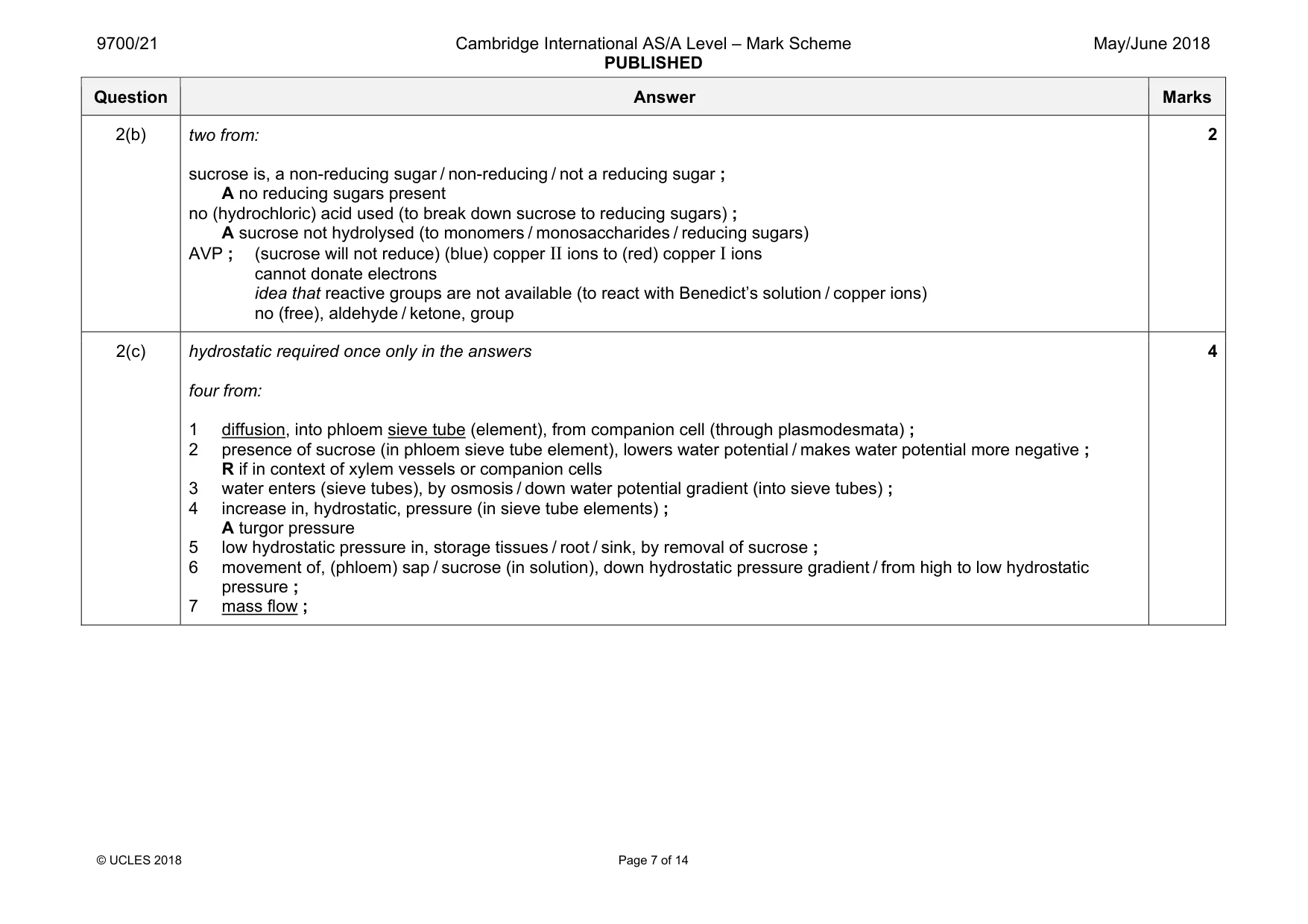
Mark scheme: 2(a)(i) B – fructose ; I hexose sugar I any qualification of fructose, e.g. α 2 2(a)(ii) glycosidic ; R glucosidic I any qualification of glycosidic, e.g. 1–2, 1–6 1 2(a)(iii) hydrolysis ; A acid hydrolysis 1 Question Answer Marks 2(b) two from: sucrose is, a non-reducing sugar / non-reducing / not a reducing sugar ; A no reducing sugars present no (hydrochloric) acid used (to break down sucrose to reducing sugars) ; A sucrose not hydrolysed (to monomers / monosaccharides / reducing sugars) AVP ; (sucrose will not reduce) (blue) copper II ions to (red) copper I ions cannot donate electrons idea that reactive groups are not available (to react with Benedict’s solution / copper ions) no (free), aldehyde / ketone, group 2 2(c) hydrostatic required once only in the answers four from: 1 diffusion, into phloem sieve tube (element), from companion cell (through plasmodesmata) ; 2 presence of sucrose (in phloem sieve tube element), lowers water potential / makes water potential more negative ; R if in context of xylem vessels or companion cells 3 water enters (sieve tubes), by osmosis / down water potential gradient (into sieve tubes) ; 4 increase in, hydrostatic, pressure (in sieve tube elements) ; A turgor pressure 5 low hydrostatic pressure in, storage tissues / root / sink, by removal of sucrose ; 6 movement of, (phloem) sap / sucrose (in solution), down hydrostatic pressure gradient / from high to low hydrostatic pressure ; 7 mass flow ; 4
Q3 · Researchers isolated a sucrase enzyme from the bacterium Bacillus subtilis
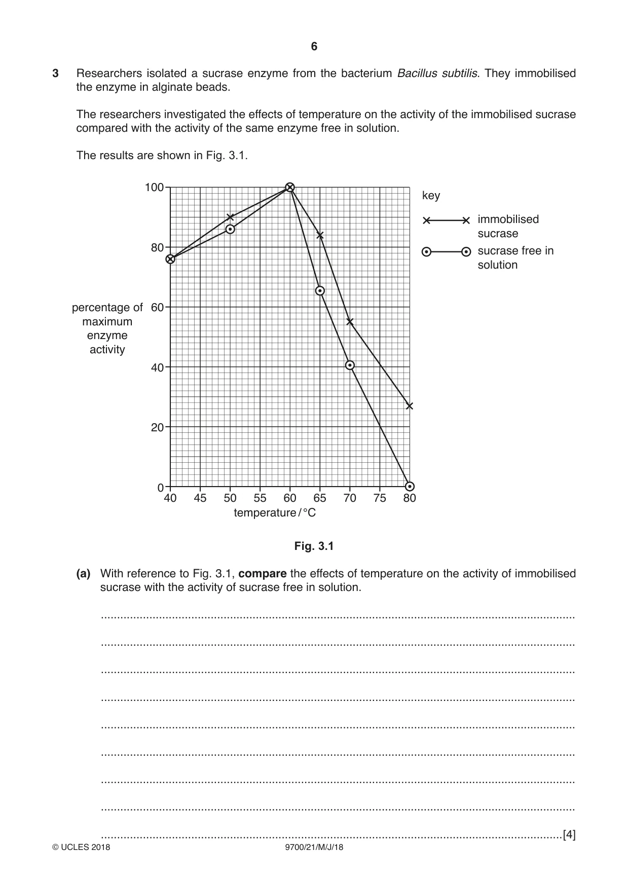
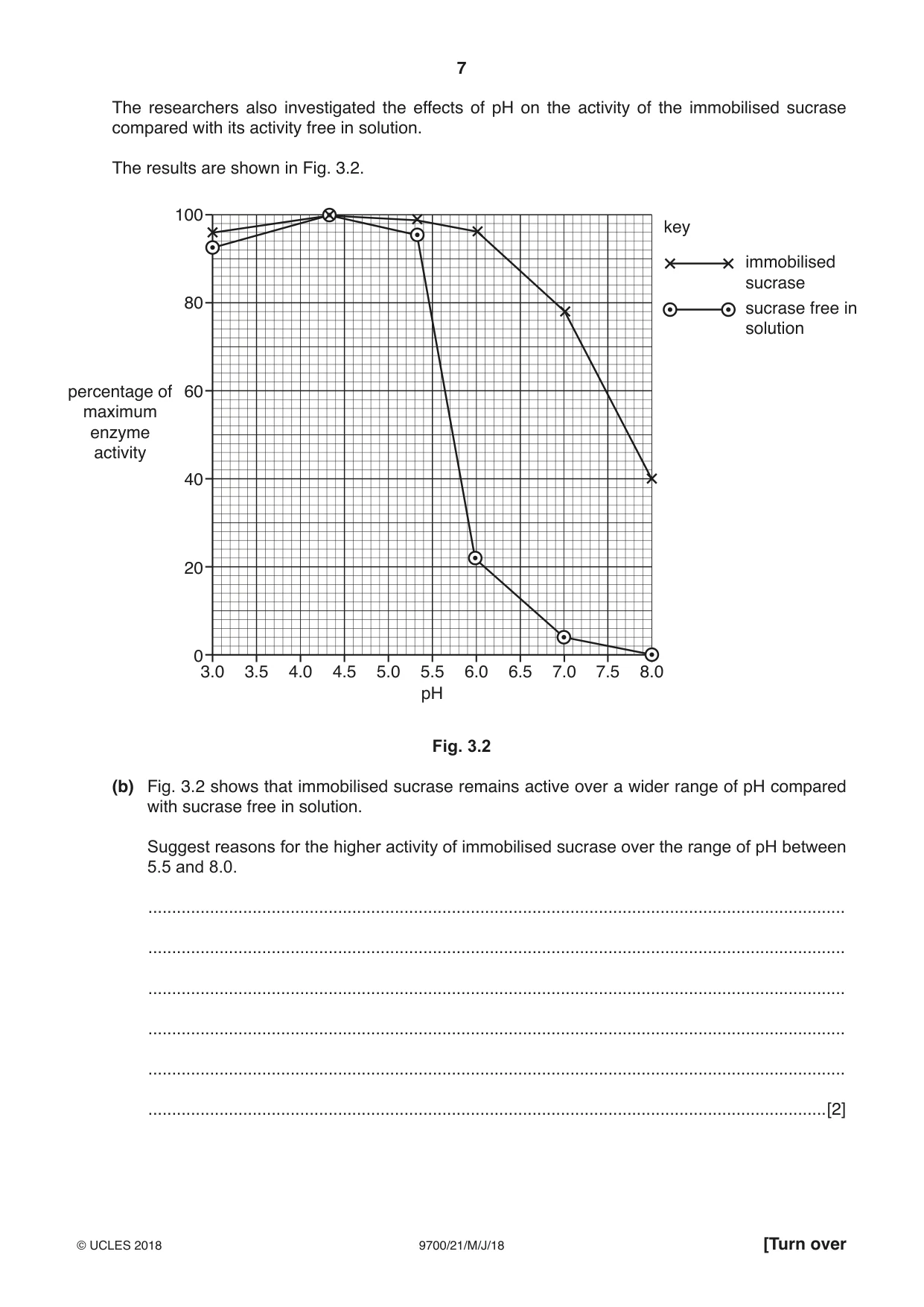
3 Researchers isolated a sucrase enzyme from the bacterium Bacillus subtilis. They immobilised the enzyme in alginate beads. The researchers investigated the effects of temperature on the activity of the immobilised sucrase compared with the activity of the same enzyme free in solution. The results are shown in Fig. 3.1. 100 key immobilised sucrase 80 sucrase free in solution percentage of 60 maximum enzyme activity 40 20 0 40 45 50 55 60 65 70 75 80 temperature / °C Fig. 3.1 (a) With reference to Fig. 3.1, compare the effects of temperature on the activity of immobilised sucrase with the activity of sucrase free in solution. ................................................................................................................................................... ................................................................................................................................................... ................................................................................................................................................... ................................................................................................................................................... ................................................................................................................................................... ................................................................................................................................................... ................................................................................................................................................... ................................................................................................................................................... The researchers also investigated the effects of pH on the activity of the immobilised sucrase compared with its activity free in solution. The results are shown in Fig. 3.2. 100 key immobilised sucrase 80 sucrase free in solution percentage of 60 maximum enzyme activity 40 20 0 3.0 3.5 4.0 4.5 5.0 5.5 6.0 6.5 7.0 7.5 8.0 pH Fig. 3.2 (b) Fig. 3.2 shows that immobilised sucrase remains active over a wider range of pH compared with sucrase free in solution. Suggest reasons for the higher activity of immobilised sucrase over the range of pH between 5.5 and 8.0. ................................................................................................................................................... ................................................................................................................................................... ................................................................................................................................................... ................................................................................................................................................... ................................................................................................................................................... ...............................................................................................................................................[2] (c) State one variable that the researchers should keep constant in both investigations and explain your answer in terms of enzyme action. variable ..................................................................................................................................... ................................................................................................................................................... explanation ............................................................................................................................... ................................................................................................................................................... ................................................................................................................................................... ...............................................................................................................................................[2] (d) There are many advantages of using immobilised enzymes in industry. Suggest two advantages of using immobilised enzymes in industry other than remaining active over a greater range of pH. ................................................................................................................................................... ................................................................................................................................................... ................................................................................................................................................... ................................................................................................................................................... ...............................................................................................................................................[2] [Total: 10]
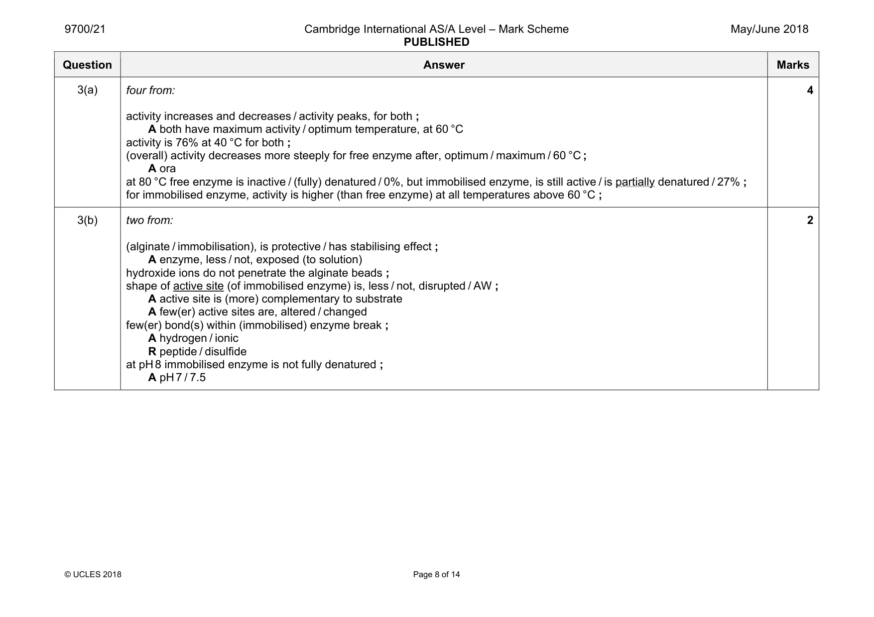
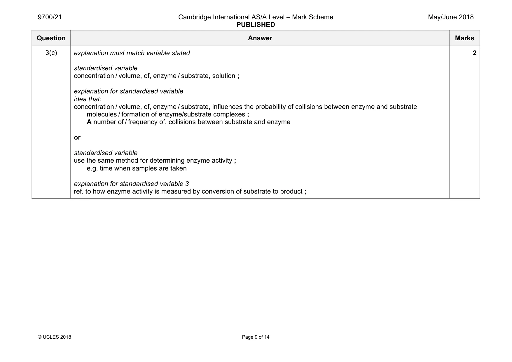
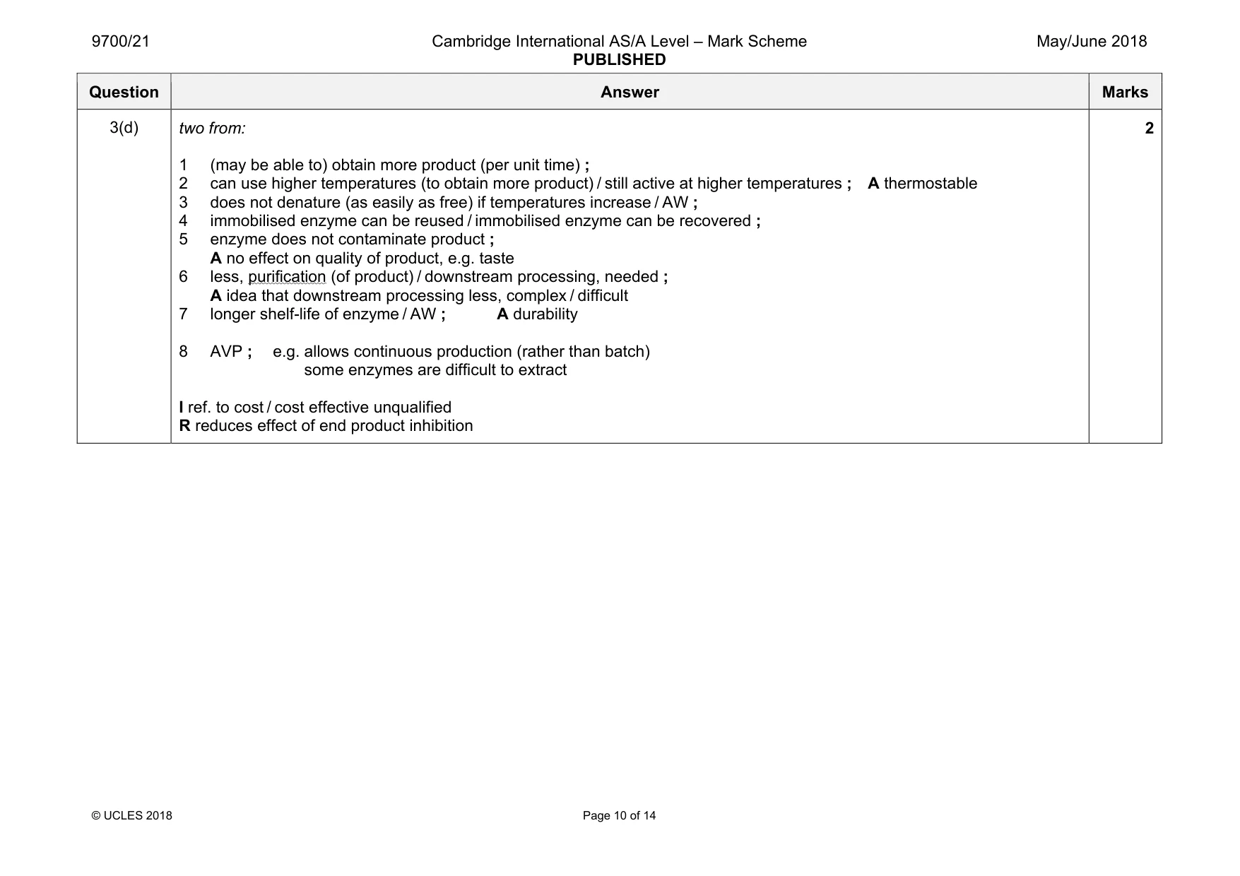
Mark scheme: 3(a) four from: activity increases and decreases / activity peaks, for both ; A both have maximum activity / optimum temperature, at 60 °C activity is 76% at 40 °C for both ; (overall) activity decreases more steeply for free enzyme after, optimum / maximum / 60 °C ; A ora at 80 °C free enzyme is inactive / (fully) denatured / 0%, but immobilised enzyme, is still active / is partially denatured / 27% ; for immobilised enzyme, activity is higher (than free enzyme) at all temperatures above 60 °C ; 4 3(b) two from: (alginate / immobilisation), is protective / has stabilising effect ; A enzyme, less / not, exposed (to solution) hydroxide ions do not penetrate the alginate beads ; shape of active site (of immobilised enzyme) is, less / not, disrupted / AW ; A active site is (more) complementary to substrate A few(er) active sites are, altered / changed few(er) bond(s) within (immobilised) enzyme break ; A hydrogen / ionic R peptide / disulfide at pH 8 immobilised enzyme is not fully denatured ; A pH 7 / 7.5 2 Question Answer Marks 3(c) explanation must match variable stated standardised variable concentration / volume, of, enzyme / substrate, solution ; explanation for standardised variable idea that: concentration / volume, of, enzyme / substrate, influences the probability of collisions between enzyme and substrate molecules / formation of enzyme/substrate complexes ; A number of / frequency of, collisions between substrate and enzyme or standardised variable use the same method for determining enzyme activity ; e.g. time when samples are taken explanation for standardised variable 3 ref. to how enzyme activity is measured by conversion of substrate to product ; 2 Question Answer Marks 3(d) two from: 1 (may be able to) obtain more product (per unit time) ; 2 can use higher temperatures (to obtain more product) / still active at higher temperatures ; A thermostable 3 does not denature (as easily as free) if temperatures increase / AW ; 4 immobilised enzyme can be reused / immobilised enzyme can be recovered ; 5 enzyme does not contaminate product ; A no effect on quality of product, e.g. taste 6 less, purification (of product) / downstream processing, needed ; A idea that downstream processing less, complex / difficult 7 longer shelf-life of enzyme / AW ; A durability 8 AVP ; e.g. allows continuous production (rather than batch) some enzymes are difficult to extract I ref. to cost / cost effective unqualified R reduces effect of end product inhibition 2
Q4 · A ribbon model of a molecule of haemoglobin
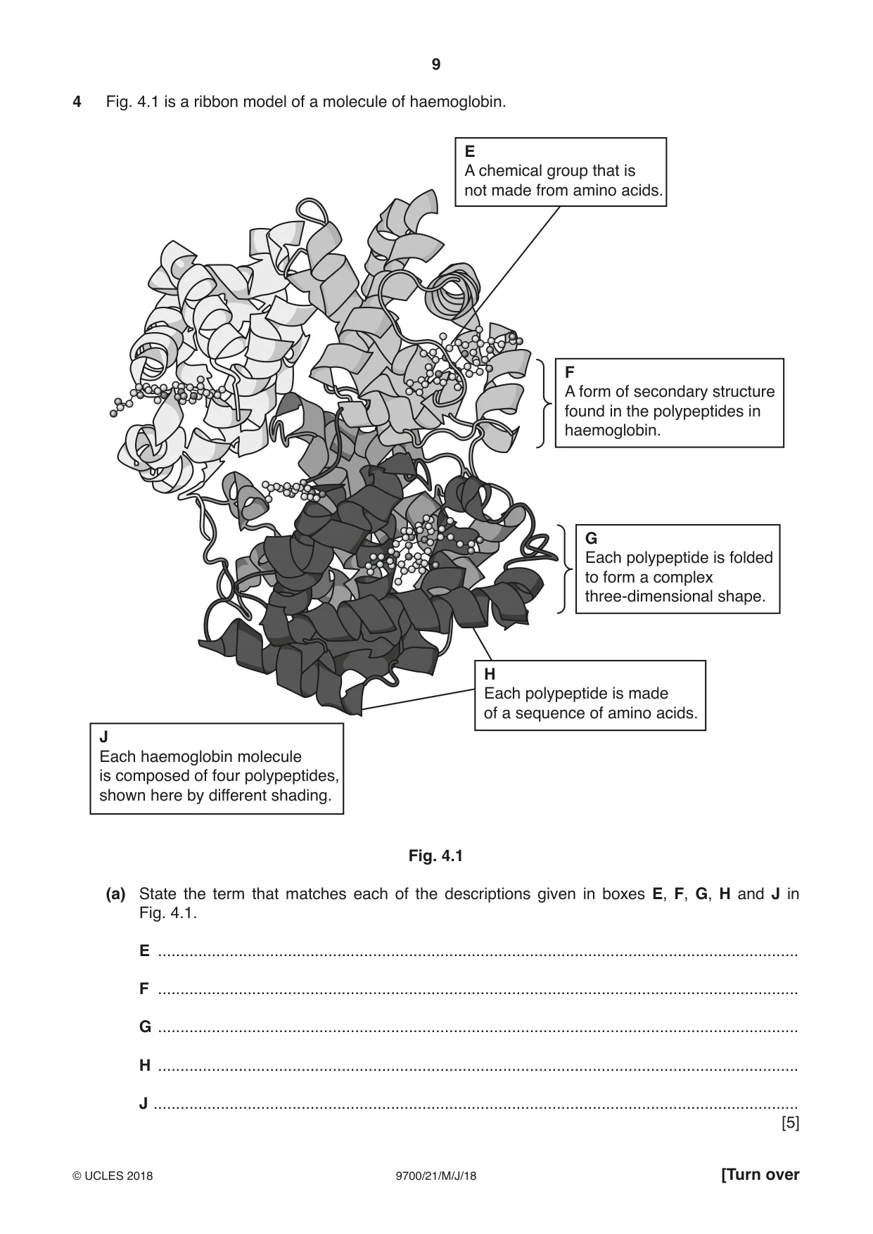
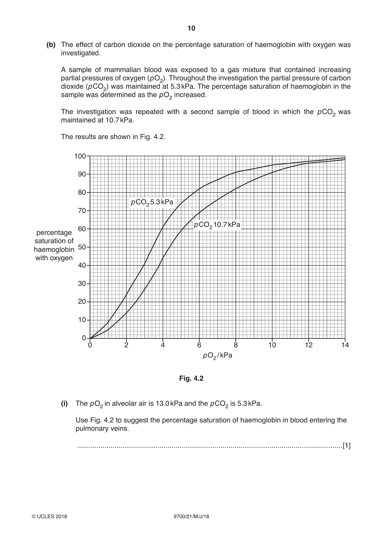
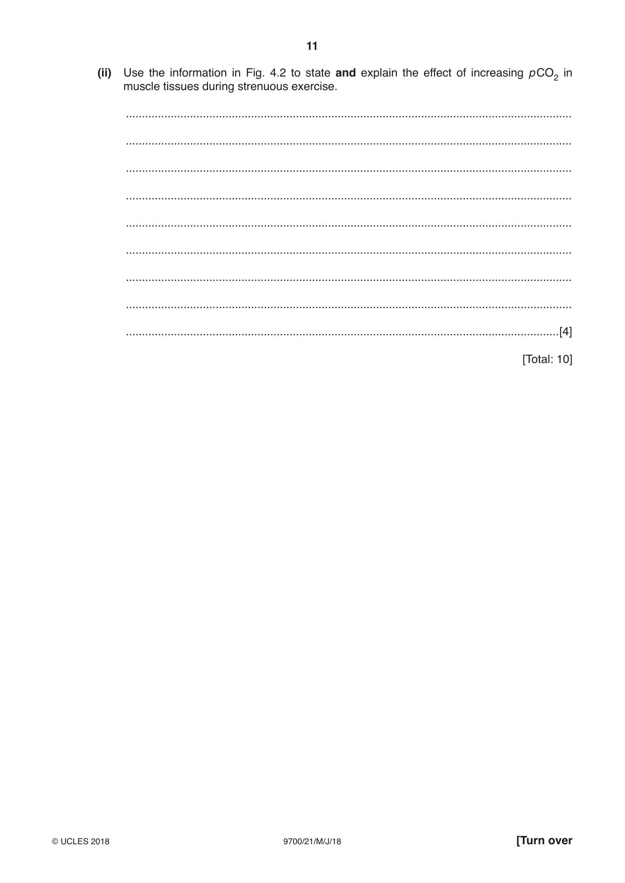
4 Fig. 4.1 is a ribbon model of a molecule of haemoglobin. E A chemical group that is not made from amino acids. F A form of secondary structure found in the polypeptides in haemoglobin. G Each polypeptide is folded to form a complex three-dimensional shape. H Each polypeptide is made of a sequence of amino acids. J Each haemoglobin molecule is composed of four polypeptides, shown here by different shading. Fig. 4.1 (a) State the term that matches each of the descriptions given in boxes E, F, G, H and J in Fig. 4.1. E ............................................................................................................................................... F ............................................................................................................................................... G ............................................................................................................................................... H ............................................................................................................................................... J ................................................................................................................................................ [5] (b) The effect of carbon dioxide on the percentage saturation of haemoglobin with oxygen was investigated. A sample of mammalian blood was exposed to a gas mixture that contained increasing partial pressures of oxygen (p O2). Throughout the investigation the partial pressure of carbon dioxide (p CO2) was maintained at 5.3 kPa. The percentage saturation of haemoglobin in the sample was determined as the p O2 increased. The investigation was repeated with a second sample of blood in which the p CO2 was maintained at 10.7 kPa. The results are shown in Fig. 4.2. 100 90 80 p CO2 5.3 kPa 70 p CO2 10.7 kPa 60 percentage saturation of 50haemoglobin with oxygen 40 30 20 10 0 0 2 4 6 8 10 12 14 p O2 / kPa Fig. 4.2 (i) The p O2 in alveolar air is 13.0 kPa and the p CO2 is 5.3 kPa. Use Fig. 4.2 to suggest the percentage saturation of haemoglobin in blood entering the pulmonary veins. .......................................................................................................................................[1] (ii) Use the information in Fig. 4.2 to state and explain the effect of increasing p CO2 in muscle tissues during strenuous exercise. ........................................................................................................................................... ........................................................................................................................................... ........................................................................................................................................... ........................................................................................................................................... ........................................................................................................................................... ........................................................................................................................................... ........................................................................................................................................... ........................................................................................................................................... .......................................................................................................................................[4] [Total: 10]
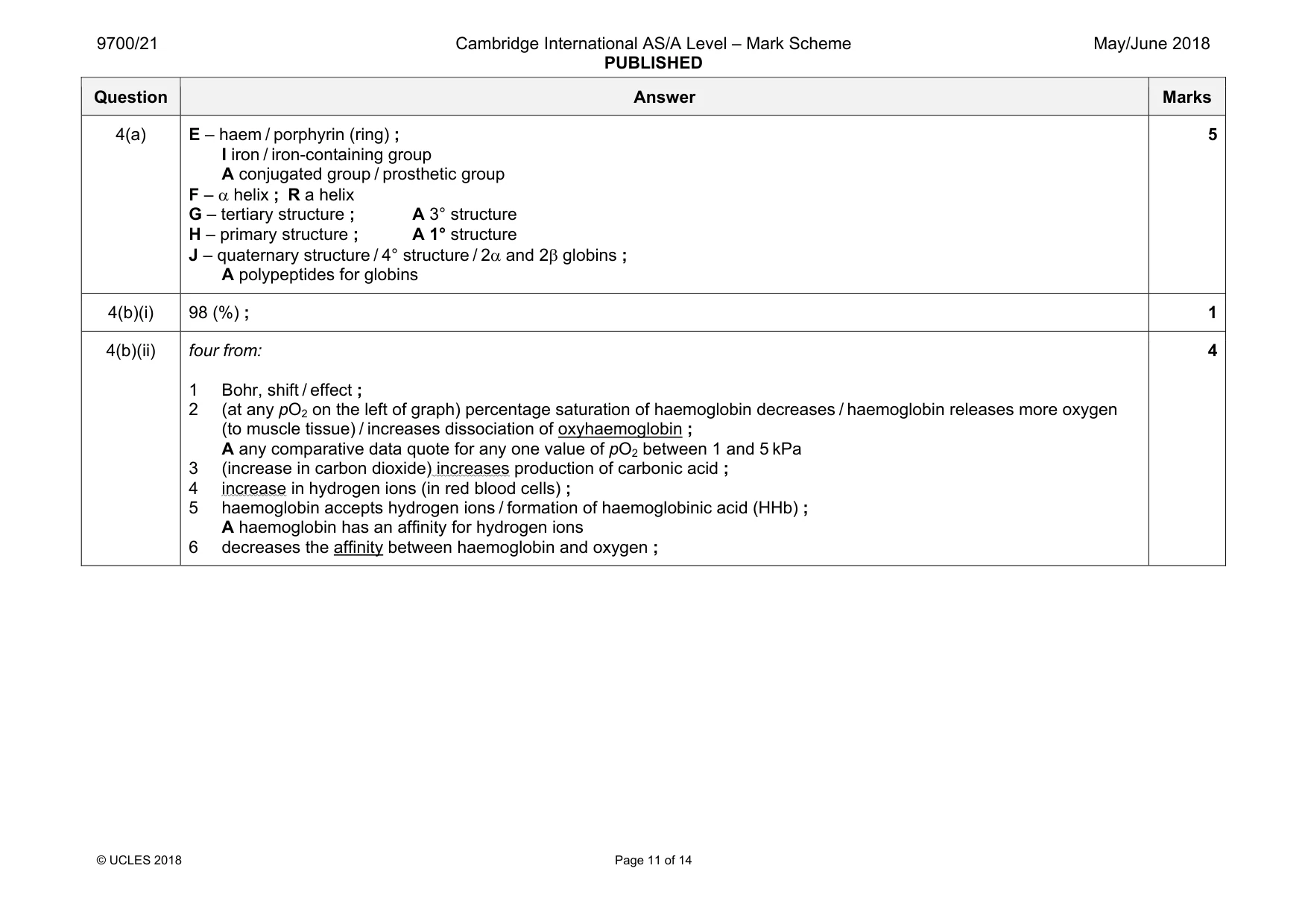
Mark scheme: 4(a) E – haem / porphyrin (ring) ; I iron / iron-containing group A conjugated group / prosthetic group F – α helix ; R a helix G – tertiary structure ; A 3° structure H – primary structure ; A 1° structure J – quaternary structure / 4° structure / 2α and 2β globins ; A polypeptides for globins 5 4(b)(i) 98 (%) ; 1 4(b)(ii) four from: 1 Bohr, shift / effect ; 2 (at any pO2 on the left of graph) percentage saturation of haemoglobin decreases / haemoglobin releases more oxygen (to muscle tissue) / increases dissociation of oxyhaemoglobin ; A any comparative data quote for any one value of pO2 between 1 and 5 kPa 3 (increase in carbon dioxide) increases production of carbonic acid ; 4 increase in hydrogen ions (in red blood cells) ; 5 haemoglobin accepts hydrogen ions / formation of haemoglobinic acid (HHb) ; A haemoglobin has an affinity for hydrogen ions 6 decreases the affinity between haemoglobin and oxygen ; 4
Q5 · The toxins released by some pathogenic bacteria can be altered chemically so that they…
5 (a) The toxins released by some pathogenic bacteria can be altered chemically so that they are harmless. These harmless toxins are called toxoids. Toxoids are used in vaccines to provide protection against some infectious diseases. Describe the response of the immune system to the injection of a toxoid. ................................................................................................................................................... ................................................................................................................................................... ................................................................................................................................................... ................................................................................................................................................... ................................................................................................................................................... ................................................................................................................................................... ................................................................................................................................................... ................................................................................................................................................... ................................................................................................................................................... ................................................................................................................................................... ................................................................................................................................................... ...............................................................................................................................................[5] (b) Myasthenia gravis (MG) is described as an autoimmune disease. It is a long-term condition that results from a failure of the immune system. (i) Explain why MG is known as an autoimmune disease. .......................................................................................................................................................................... ........................................................................................................................................... ........................................................................................................................................... ........................................................................................................................................... ........................................................................................................................................... ........................................................................................................................................... .......................................................................................................................................[3] (ii) Suggest why MG is a long-term condition. ........................................................................................................................................... ........................................................................................................................................... ........................................................................................................................................... ........................................................................................................................................... ........................................................................................................................................... .......................................................................................................................................[2] [Total: 10] Question 6 starts on page 14
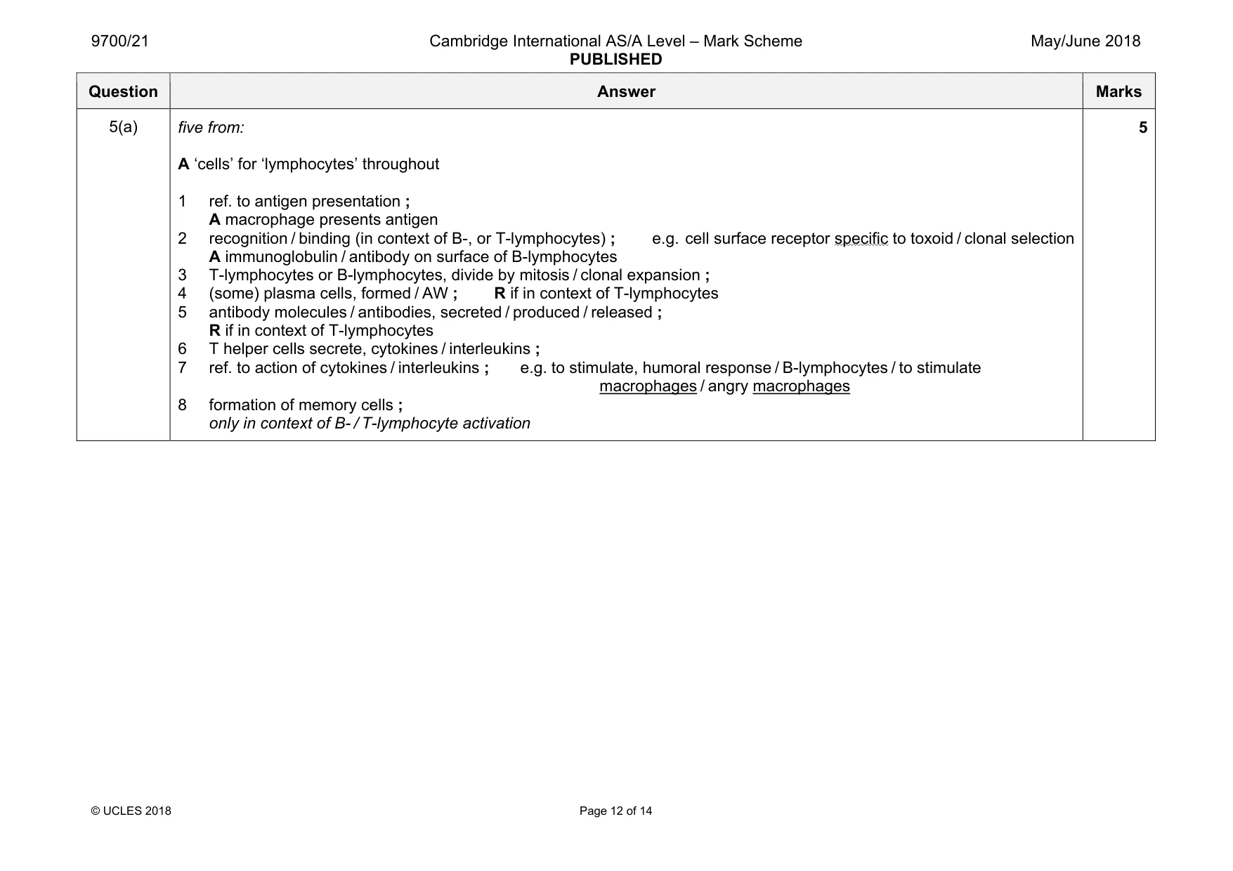
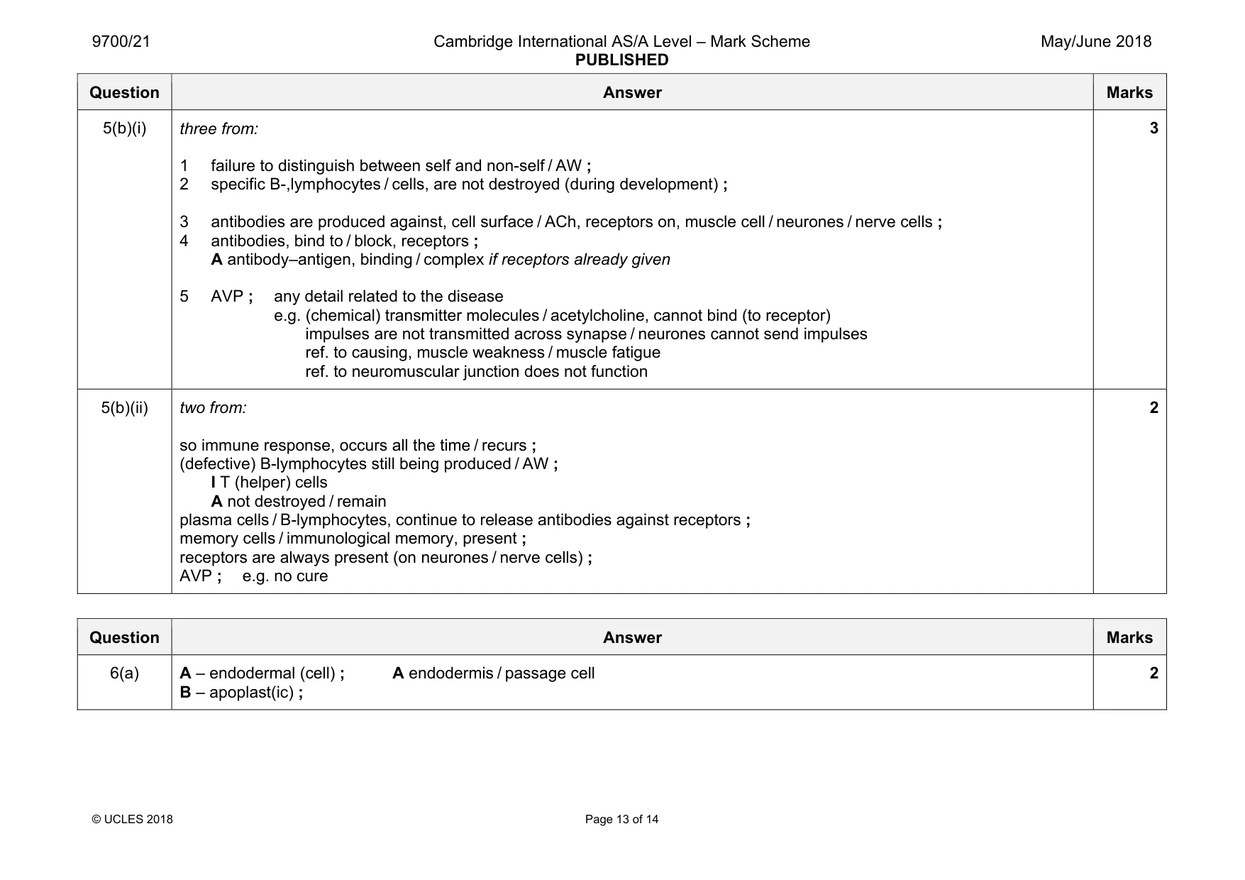
Mark scheme: 5(a) five from: A ‘cells’ for ‘lymphocytes’ throughout 1 ref. to antigen presentation ; A macrophage presents antigen 2 recognition / binding (in context of B-, or T-lymphocytes) ; e.g. cell surface receptor specific to toxoid / clonal selection A immunoglobulin / antibody on surface of B-lymphocytes 3 T-lymphocytes or B-lymphocytes, divide by mitosis / clonal expansion ; 4 (some) plasma cells, formed / AW ; R if in context of T-lymphocytes 5 antibody molecules / antibodies, secreted / produced / released ; R if in context of T-lymphocytes 6 T helper cells secrete, cytokines / interleukins ; 7 ref. to action of cytokines / interleukins ; e.g. to stimulate, humoral response / B-lymphocytes / to stimulate macrophages / angry macrophages 8 formation of memory cells ; only in context of B- / T-lymphocyte activation 5 Question Answer Marks 5(b)(i) three from: 1 failure to distinguish between self and non-self / AW ; 2 specific B-,lymphocytes / cells, are not destroyed (during development) ; 3 antibodies are produced against, cell surface / ACh, receptors on, muscle cell / neurones / nerve cells ; 4 antibodies, bind to / block, receptors ; A antibody–antigen, binding / complex if receptors already given 5 AVP ; any detail related to the disease e.g. (chemical) transmitter molecules / acetylcholine, cannot bind (to receptor) impulses are not transmitted across synapse / neurones cannot send impulses ref. to causing, muscle weakness / muscle fatigue ref. to neuromuscular junction does not function 3 5(b)(ii) two from: so immune response, occurs all the time / recurs ; (defective) B-lymphocytes still being produced / AW ; I T (helper) cells A not destroyed / remain plasma cells / B-lymphocytes, continue to release antibodies against receptors ; memory cells / immunological memory, present ; receptors are always present (on neurones / nerve cells) ; AVP ; e.g. no cure 2
Q6 · Water absorbed by plant roots travels by different pathways from root hairs to the xylem
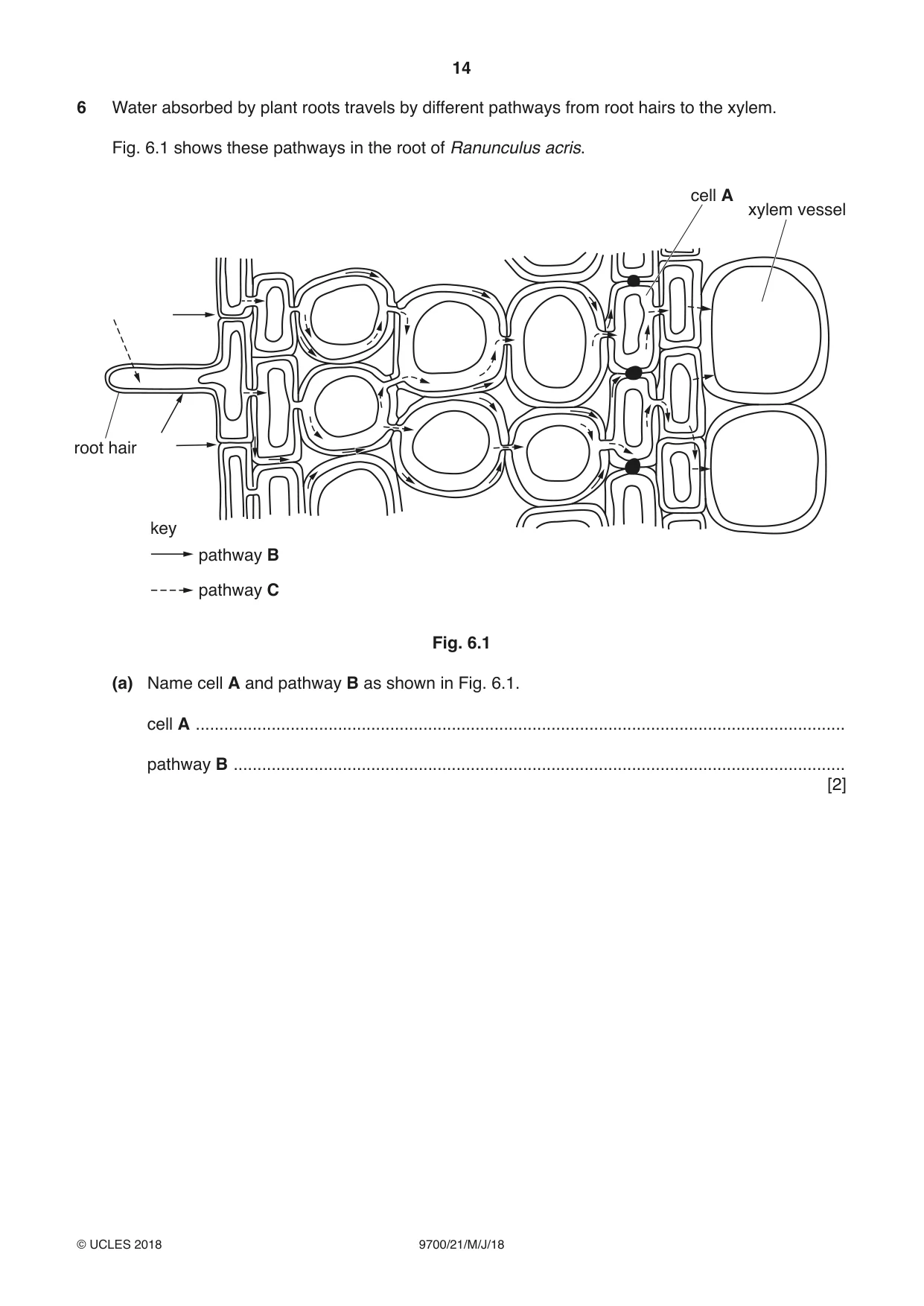
6 Water absorbed by plant roots travels by different pathways from root hairs to the xylem. Fig. 6.1 shows these pathways in the root of Ranunculus acris. cell A xylem vessel root hair key pathway B pathway C Fig. 6.1 (a) Name cell A and pathway B as shown in Fig. 6.1. cell A ......................................................................................................................................... pathway B ................................................................................................................................. [2] (b) Transpiration occurring at the leaves is mainly responsible for movement of water across the root of R. acris. Explain how transpiration is responsible for the movement of water across the root as shown in Fig. 6.1. ................................................................................................................................................... ................................................................................................................................................... ................................................................................................................................................... ................................................................................................................................................... ................................................................................................................................................... ................................................................................................................................................... ................................................................................................................................................... ................................................................................................................................................... ................................................................................................................................................... ................................................................................................................................................... ................................................................................................................................................... ...............................................................................................................................................[5] (c) Explain why the movement of water in pathway C is slower than in pathway B. ................................................................................................................................................... ................................................................................................................................................... ................................................................................................................................................... ................................................................................................................................................... ................................................................................................................................................... ...............................................................................................................................................[2] Question 6 continues on page 16 (d) Ions are taken up by root hair cells using active transport and facilitated diffusion. Describe two ways in which facilitated diffusion differs from active transport. 1 ................................................................................................................................................ ................................................................................................................................................... ................................................................................................................................................... 2 ................................................................................................................................................ ................................................................................................................................................... ................................................................................................................................................... [2] [Total: 11]
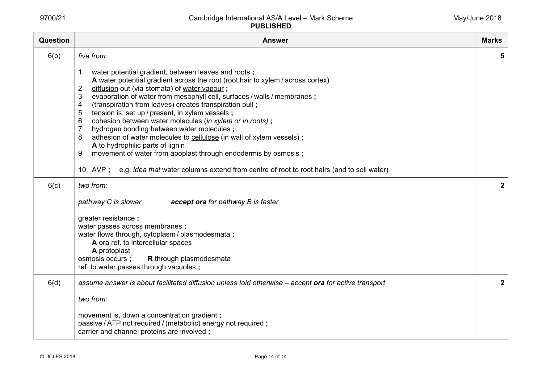
Mark scheme: 6(a) A – endodermal (cell) ; A endodermis / passage cell B – apoplast(ic) ; 2 Question Answer Marks 6(b) five from: 1 water potential gradient, between leaves and roots ; A water potential gradient across the root (root hair to xylem / across cortex) 2 diffusion out (via stomata) of water vapour ; 3 evaporation of water from mesophyll cell, surfaces / walls / membranes ; 4 (transpiration from leaves) creates transpiration pull ; 5 tension is, set up / present, in xylem vessels ; 6 cohesion between water molecules (in xylem or in roots) ; 7 hydrogen bonding between water molecules ; 8 adhesion of water molecules to cellulose (in wall of xylem vessels) ; A to hydrophilic parts of lignin 9 movement of water from apoplast through endodermis by osmosis ; 10 AVP ; e.g. idea that water columns extend from centre of root to root hairs (and to soil water) 5 6(c) two from: pathway C is slower accept ora for pathway B is faster greater resistance ; water passes across membranes ; water flows through, cytoplasm / plasmodesmata ; A ora ref. to intercellular spaces A protoplast osmosis occurs ; R through plasmodesmata ref. to water passes through vacuoles ; 2 6(d) assume answer is about facilitated diffusion unless told otherwise – accept ora for active transport two from: movement is, down a concentration gradient ; passive / ATP not required / (metabolic) energy not required ; carrier and channel proteins are involved ; 2
What was in this paper
The subtopics covered by these 6 questions, and how many questions each got. Open one in a new tab to see every Cambridge question on it.
What you needed in this session
Cambridge’s own grade thresholds for 2018 May/June, Paper 2 · Variant 1. A higher threshold means an easier paper — the bar moves with how the cohort did.