Cambridge A Level Biology 9700 — 2018 May/June Paper 2 · Variant 2
9700/22/M/J/18 · 6 questions · 60 marks · ≈68 min
The question paper and its mark scheme, free to read here and free to download. This is Cambridge’s own paper, exactly as it was sat.
Question paper16 pages

Mark scheme18 pages
Answers below. Sit the paper first if you are practising.
Questions as text
Q1 · A drawing of a photomicrograph of a spongy mesophyll cell from a leaf
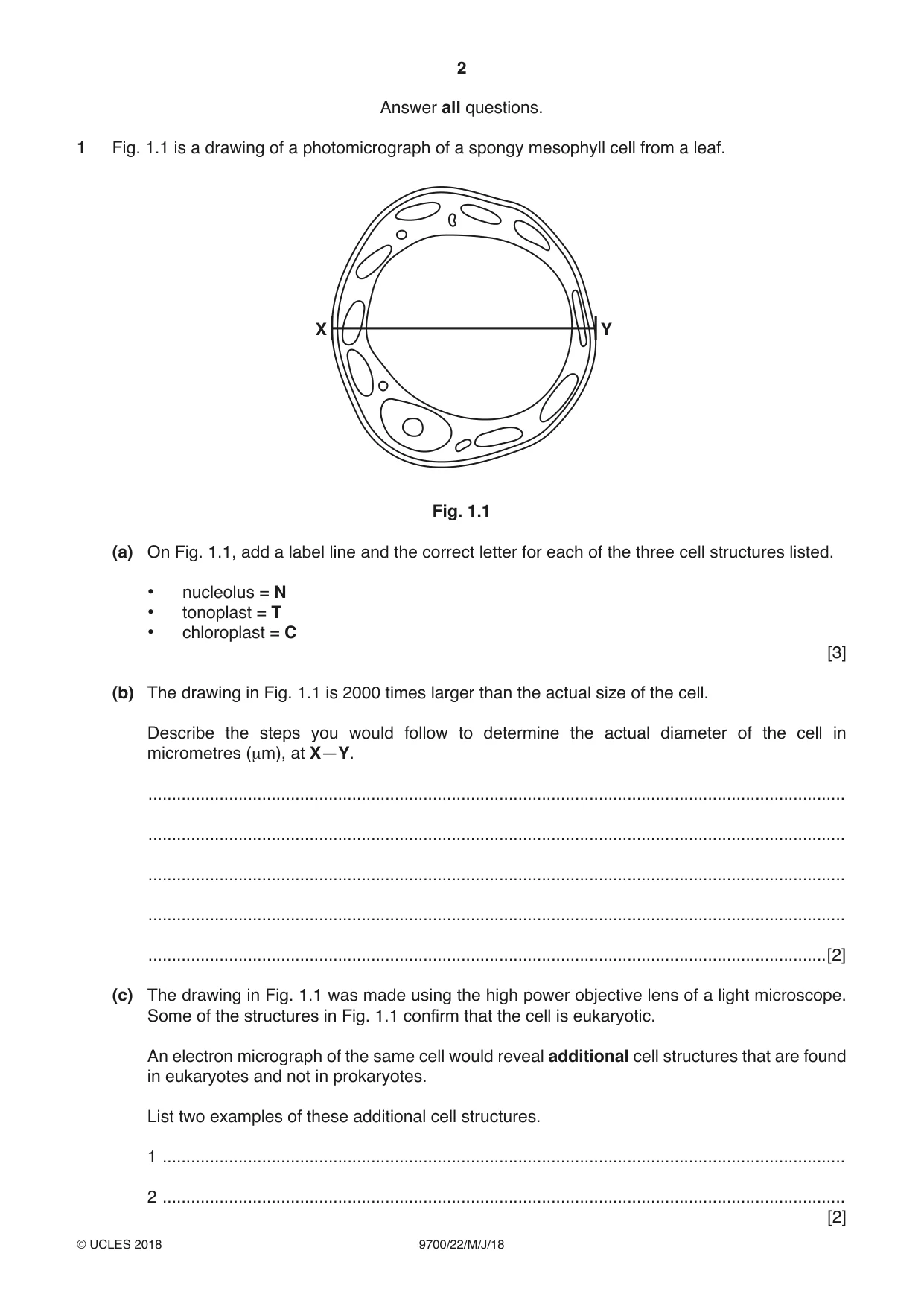
1 Fig. 1.1 is a drawing of a photomicrograph of a spongy mesophyll cell from a leaf. X Y Fig. 1.1 (a) On Fig. 1.1, add a label line and the correct letter for each of the three cell structures listed. • nucleolus = N • tonoplast = T • chloroplast = C [3] (b) The drawing in Fig. 1.1 is 2000 times larger than the actual size of the cell. Describe the steps you would follow to determine the actual diameter of the cell in micrometres (µm), at X—Y. ................................................................................................................................................... ................................................................................................................................................... ................................................................................................................................................... ................................................................................................................................................... ...............................................................................................................................................[2] (c) The drawing in Fig. 1.1 was made using the high power objective lens of a light microscope. Some of the structures in Fig. 1.1 confirm that the cell is eukaryotic. An electron micrograph of the same cell would reveal additional cell structures that are found in eukaryotes and not in prokaryotes. List two examples of these additional cell structures. 1 ................................................................................................................................................ 2 ................................................................................................................................................ [2] (d) Some of the water that moves out of the xylem within the leaves takes an apoplastic pathway to the spongy mesophyll cells. There is a film of water on the external surfaces of these cells. This allows the intercellular air spaces to become saturated with water vapour. (i) State, in terms of water movement from the xylem to the spongy mesophyll cells in the leaf, what is meant by the apoplastic pathway. ........................................................................................................................................... ........................................................................................................................................... ........................................................................................................................................... ........................................................................................................................................... .......................................................................................................................................[2] (ii) Outline the properties of water that contribute to the apoplastic movement of water to the spongy mesophyll cells and to the movement of water into the intercellular air spaces. ........................................................................................................................................... ........................................................................................................................................... ........................................................................................................................................... ........................................................................................................................................... ........................................................................................................................................... ........................................................................................................................................... ........................................................................................................................................... ........................................................................................................................................... ........................................................................................................................................... .......................................................................................................................................[4] (iii) Describe what happens to the water vapour in the intercellular air spaces during the day and explain why this happens. ........................................................................................................................................... ........................................................................................................................................... ........................................................................................................................................... ........................................................................................................................................... .......................................................................................................................................[2] [Total: 15]
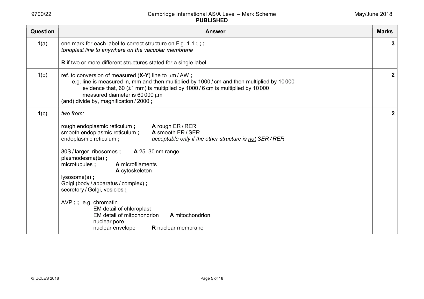
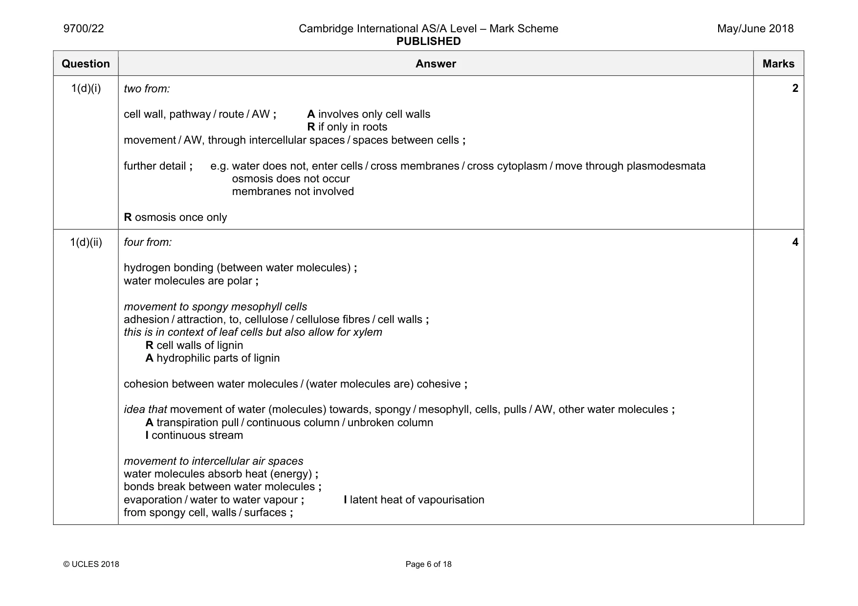
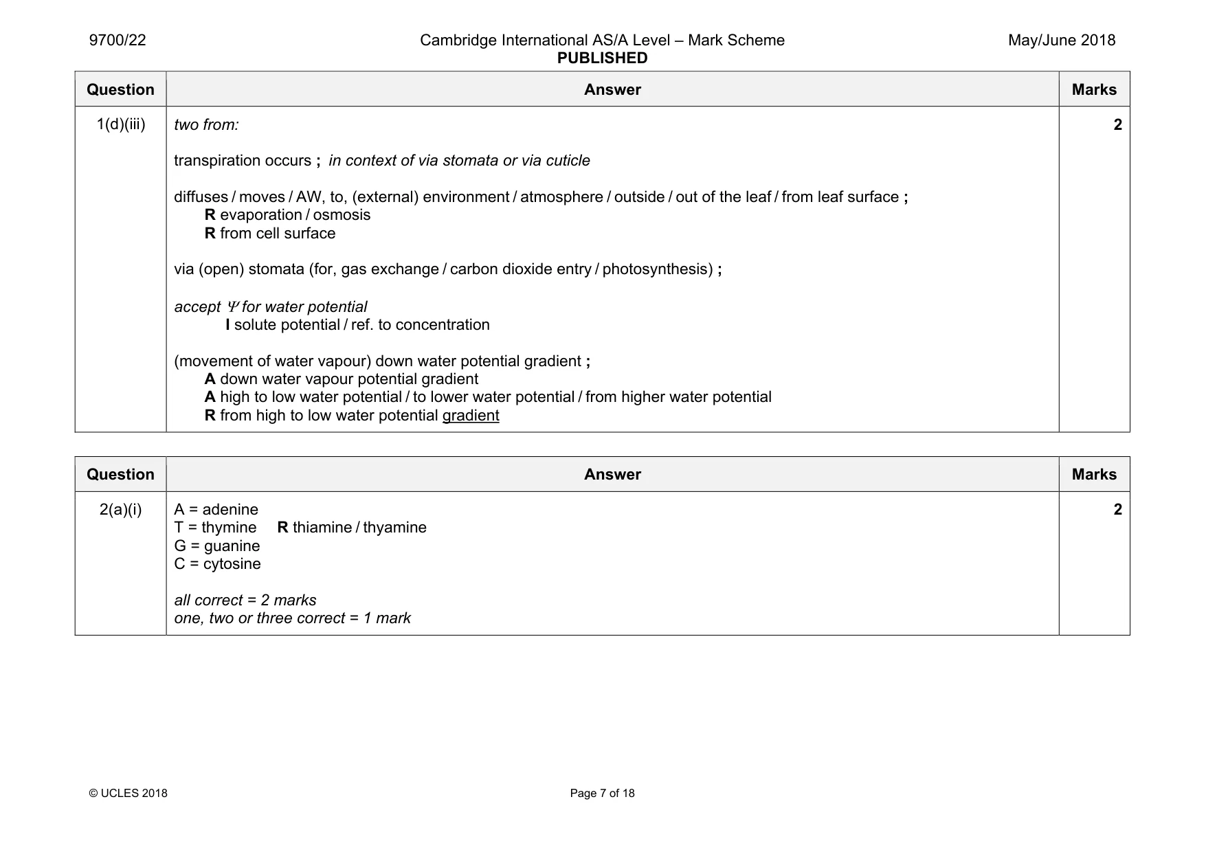
Mark scheme: 1(a) one mark for each label to correct structure on Fig. 1.1 ; ; ; tonoplast line to anywhere on the vacuolar membrane R if two or more different structures stated for a single label 3 1(b) ref. to conversion of measured (X-Y) line to µm / AW ; e.g. line is measured in, mm and then multiplied by 1000 / cm and then multiplied by 10 000 evidence that, 60 (±1 mm) is multiplied by 1000 / 6 cm is multiplied by 10 000 measured diameter is 60 000 µm (and) divide by, magnification / 2000 ; 2 1(c) two from: rough endoplasmic reticulum ; A rough ER / RER smooth endoplasmic reticulum ; A smooth ER / SER endoplasmic reticulum ; acceptable only if the other structure is not SER / RER 80S / larger, ribosomes ; A 25–30 nm range plasmodesma(ta) ; microtubules ; A microfilaments A cytoskeleton lysosome(s) ; Golgi (body / apparatus / complex) ; secretory / Golgi, vesicles ; AVP ; ; e.g. chromatin EM detail of chloroplast EM detail of mitochondrion A mitochondrion nuclear pore nuclear envelope R nuclear membrane 2 Question Answer Marks 1(d)(i) two from: cell wall, pathway / route / AW ; A involves only cell walls R if only in roots movement / AW, through intercellular spaces / spaces between cells ; further detail ; e.g. water does not, enter cells / cross membranes / cross cytoplasm / move through plasmodesmata osmosis does not occur membranes not involved R osmosis once only 2 1(d)(ii) four from: hydrogen bonding (between water molecules) ; water molecules are polar ; movement to spongy mesophyll cells adhesion / attraction, to, cellulose / cellulose fibres / cell walls ; this is in context of leaf cells but also allow for xylem R cell walls of lignin A hydrophilic parts of lignin cohesion between water molecules / (water molecules are) cohesive ; idea that movement of water (molecules) towards, spongy / mesophyll, cells, pulls / AW, other water molecules ; A transpiration pull / continuous column / unbroken column I continuous stream movement to intercellular air spaces water molecules absorb heat (energy) ; bonds break between water molecules ; evaporation / water to water vapour ; I latent heat of vapourisation from spongy cell, walls / surfaces ; 4 Question Answer Marks 1(d)(iii) two from: transpiration occurs ; in context of via stomata or via cuticle diffuses / moves / AW, to, (external) environment / atmosphere / outside / out of the leaf / from leaf surface ; R evaporation / osmosis R from cell surface via (open) stomata (for, gas exchange / carbon dioxide entry / photosynthesis) ; accept Ψ for water potential I solute potential / ref. to concentration (movement of water vapour) down water potential gradient ; A down water vapour potential gradient A high to low water potential / to lower water potential / from higher water potential R from high to low water potential gradient 2
Q2 · In 1953, James Watson and Francis Crick published details about the structure of DNA
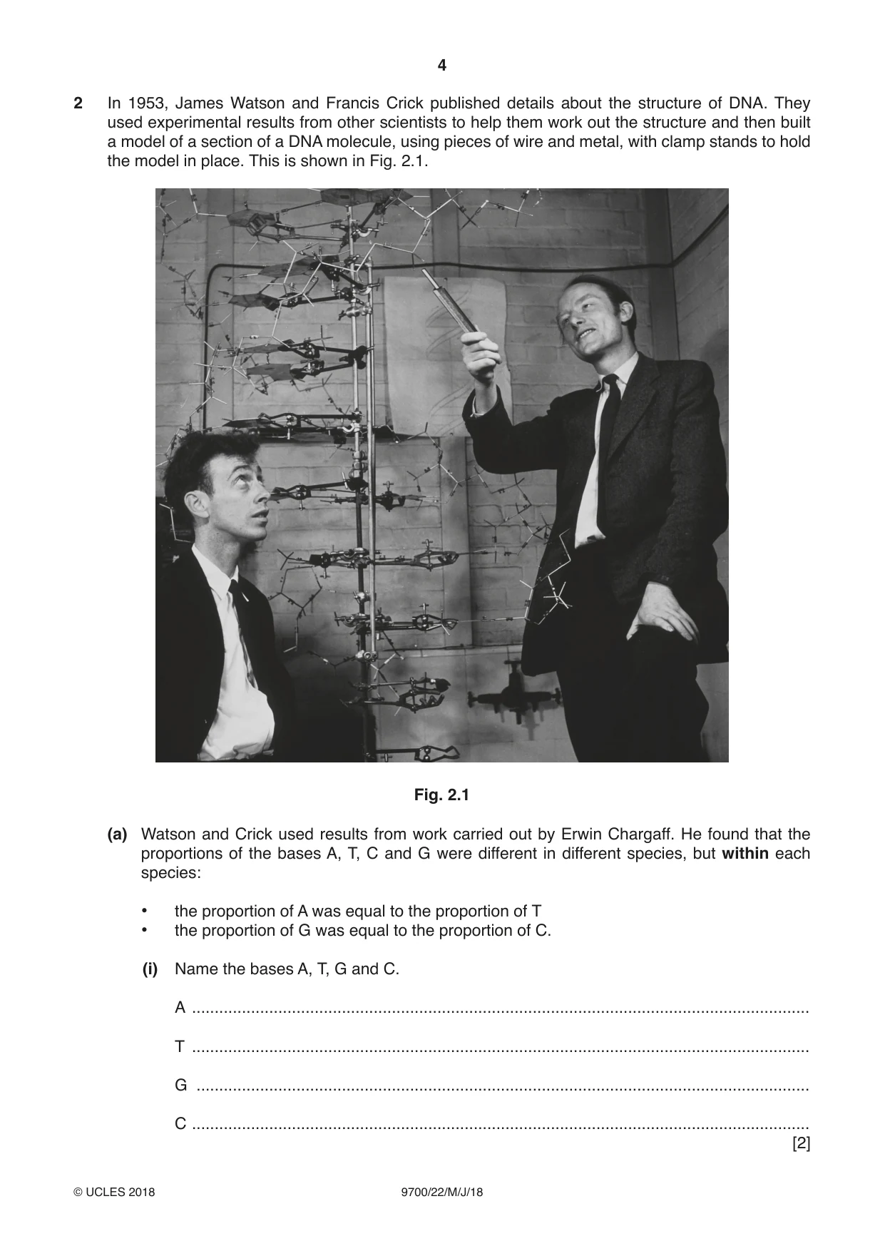
2 In 1953, James Watson and Francis Crick published details about the structure of DNA. They used experimental results from other scientists to help them work out the structure and then built a model of a section of a DNA molecule, using pieces of wire and metal, with clamp stands to hold the model in place. This is shown in Fig. 2.1. Fig. 2.1 (a) Watson and Crick used results from work carried out by Erwin Chargaff. He found that the proportions of the bases A, T, C and G were different in different species, but within each species: • the proportion of A was equal to the proportion of T • the proportion of G was equal to the proportion of C. (i) Name the bases A, T, G and C. A ........................................................................................................................................ T ........................................................................................................................................ G ....................................................................................................................................... C ........................................................................................................................................ [2] (ii) Suggest and explain how Chargaff’s findings helped Watson and Crick work out the structure of DNA. ........................................................................................................................................... ........................................................................................................................................... ........................................................................................................................................... ........................................................................................................................................... ........................................................................................................................................... ........................................................................................................................................... ........................................................................................................................................... .......................................................................................................................................[3] (b) Phoebus Levene isolated the nucleotides of DNA and identified the carbohydrate component of each nucleotide. State the name of this carbohydrate component. ...............................................................................................................................................[1] (c) Before the discovery of the structure of DNA as the molecule of inheritance, scientists thought that proteins were most likely to be the molecules that carried information. Suggest how the structure of proteins made scientists think that these were the molecules that carried information. ................................................................................................................................................... ................................................................................................................................................... ................................................................................................................................................... ................................................................................................................................................... ...............................................................................................................................................[2] [Total: 8]
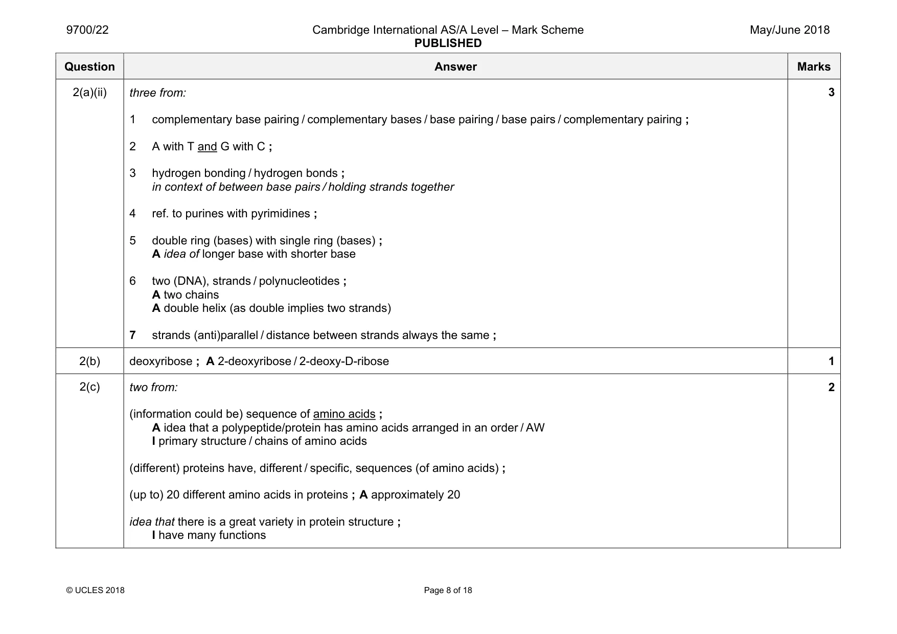
Mark scheme: 2(a)(i) A = adenine T = thymine R thiamine / thyamine G = guanine C = cytosine all correct = 2 marks one, two or three correct = 1 mark 2 Question Answer Marks 2(a)(ii) three from: 1 complementary base pairing / complementary bases / base pairing / base pairs / complementary pairing ; 2 A with T and G with C ; 3 hydrogen bonding / hydrogen bonds ; in context of between base pairs / holding strands together 4 ref. to purines with pyrimidines ; 5 double ring (bases) with single ring (bases) ; A idea of longer base with shorter base 6 two (DNA), strands / polynucleotides ; A two chains A double helix (as double implies two strands) 7 strands (anti)parallel / distance between strands always the same ; 3 2(b) deoxyribose ; A 2-deoxyribose / 2-deoxy-D-ribose 1 2(c) two from: (information could be) sequence of amino acids ; A idea that a polypeptide/protein has amino acids arranged in an order / AW I primary structure / chains of amino acids (different) proteins have, different / specific, sequences (of amino acids) ; (up to) 20 different amino acids in proteins ; A approximately 20 idea that there is a great variety in protein structure ; I have many functions 2
More questions on Structure of nucleic acids and replication of DNA
Q3 · Bacteria may be classified according to differences in cell wall structure
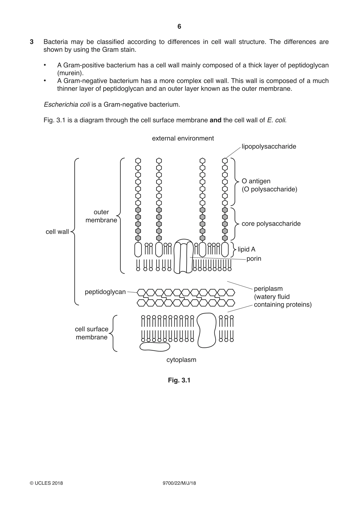
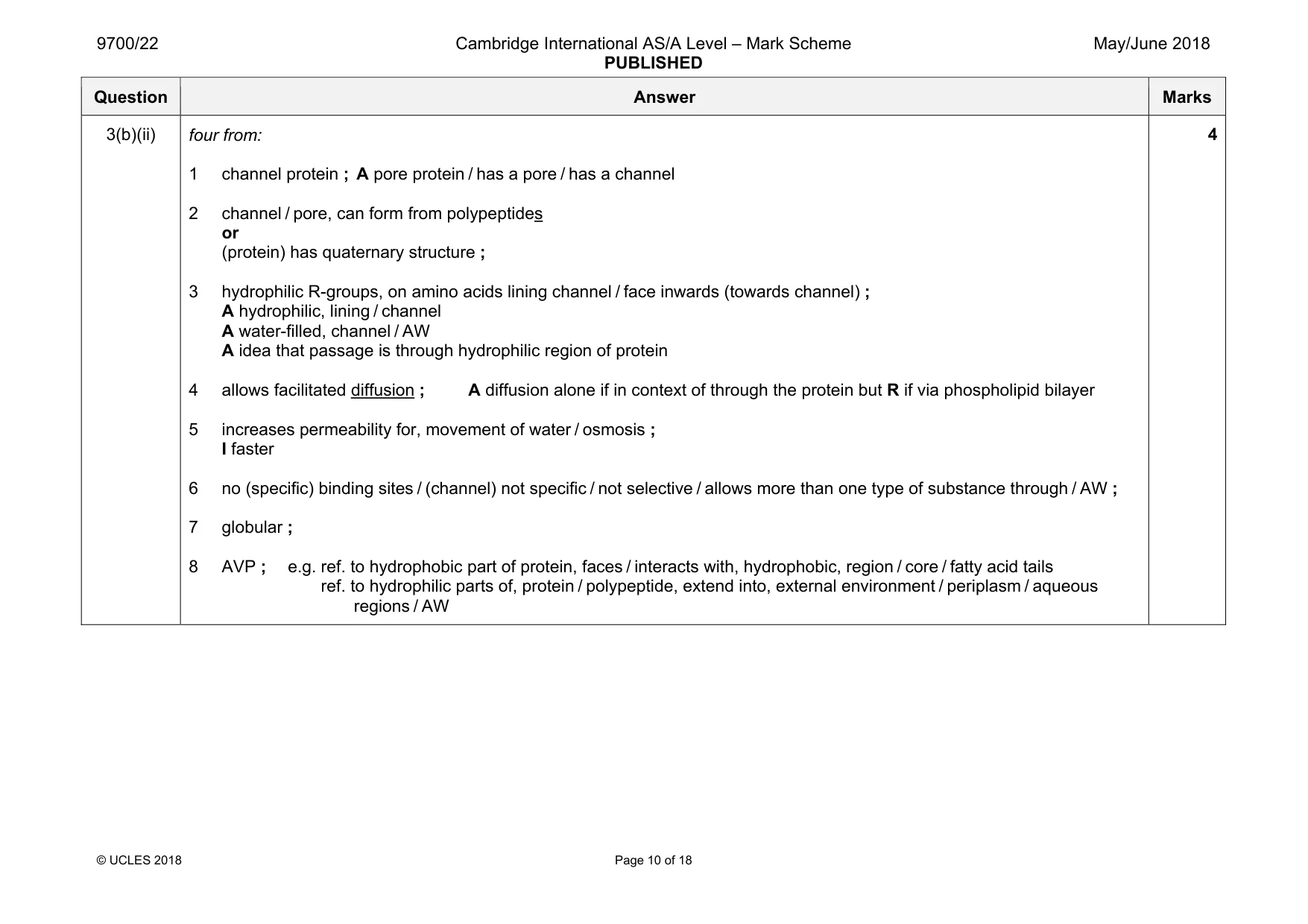
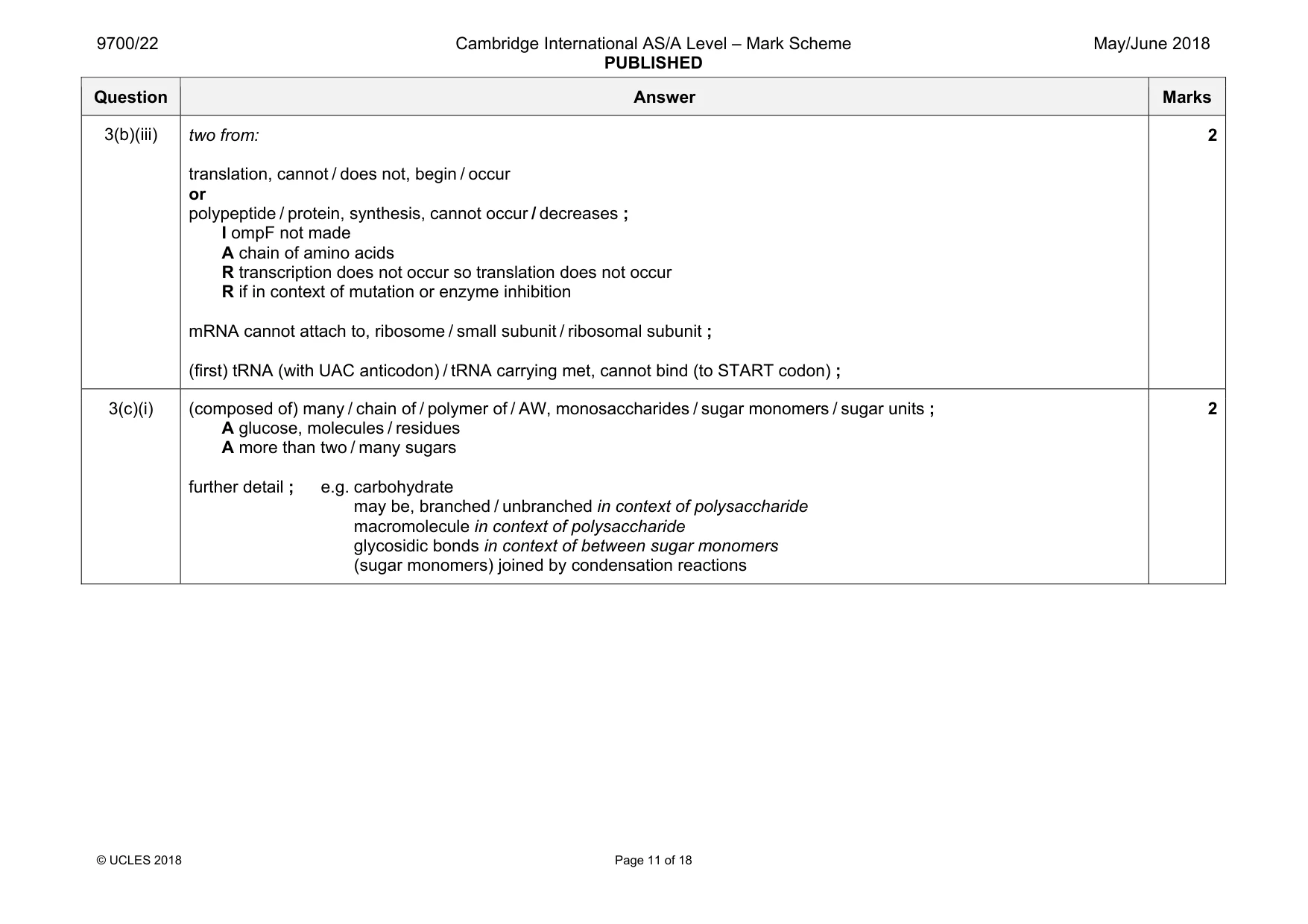
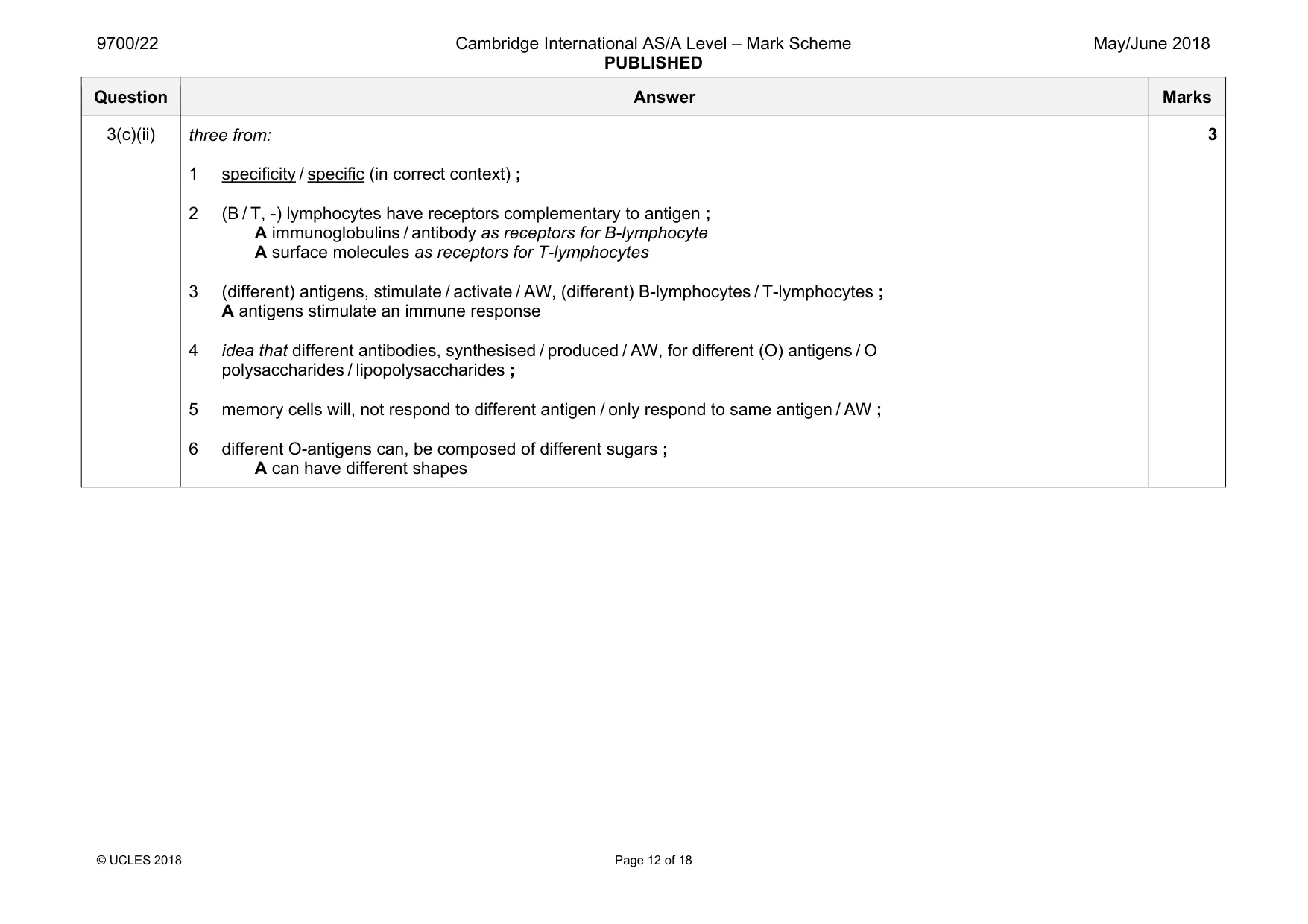
3 Bacteria may be classified according to differences in cell wall structure. The differences are shown by using the Gram stain. • A Gram-positive bacterium has a cell wall mainly composed of a thick layer of peptidoglycan (murein). • A Gram-negative bacterium has a more complex cell wall. This wall is composed of a much thinner layer of peptidoglycan and an outer layer known as the outer membrane. Escherichia coli is a Gram-negative bacterium. Fig. 3.1 is a diagram through the cell surface membrane and the cell wall of E. coli. external environment lipopolysaccharide O antigen (O polysaccharide) outer membrane core polysaccharide cell wall lipid A porin periplasm peptidoglycan (watery fluid containing proteins) cell surface membrane cytoplasm Fig. 3.1 (a) The antibiotic penicillin kills bacteria by causing them to lyse (burst). It is more effective in treating diseases caused by Gram-positive bacteria than diseases caused by Gram-negative bacteria. Outline how penicillin acts on bacteria and use Fig. 3.1 to suggest why penicillin has little or no effect at treating diseases caused by Gram-negative bacteria, such as some strains of E. coli. ................................................................................................................................................... ................................................................................................................................................... ................................................................................................................................................... ................................................................................................................................................... ................................................................................................................................................... ................................................................................................................................................... ................................................................................................................................................... ...............................................................................................................................................[3] (b) The outer membrane contains transport proteins called OmpF porins. These porins allow the passive movement of water, ions and small, polar molecules across the outer membrane. Each OmpF porin is formed from three identical polypeptides. (i) Explain what is meant by the term passive. .......................................................................................................................................[1] (ii) Suggest and explain the features of an OmpF porin as a membrane transport protein. ........................................................................................................................................... ........................................................................................................................................... ........................................................................................................................................... ........................................................................................................................................... ........................................................................................................................................... ........................................................................................................................................... ........................................................................................................................................... ........................................................................................................................................... ........................................................................................................................................... .......................................................................................................................................[4] (iii) E. coli can regulate the number of OmpF porins in the outer membrane to adapt to changing conditions. One control mechanism used by E. coli involves the production of a small mRNA molecule known as micF. MicF binds to the part of the mRNA molecule containing the START codon for the OmpF polypeptide. Suggest and explain how the presence of micF prevents production of OmpF porins. ........................................................................................................................................... ........................................................................................................................................... ........................................................................................................................................... ........................................................................................................................................... ........................................................................................................................................... .......................................................................................................................................[2] (c) Fig. 3.1 shows that the outer membrane of the cell wall of E. coli contains lipopolysaccharides. These are not present in the cell surface membrane. Each lipopolysaccharide (LPS) consists of a lipid and a polysaccharide portion. The O antigen is the outer part of the polysaccharide portion of the LPS. It faces the aqueous external environment. (i) Define the term polysaccharide. ........................................................................................................................................... ........................................................................................................................................... ........................................................................................................................................... ........................................................................................................................................... .......................................................................................................................................[2] (ii) Some strains of E. coli are pathogenic. Different pathogenic strains have different O antigens. Suggest and explain why infection with one pathogenic strain of E. coli does not provide immunity to a different pathogenic strain. ........................................................................................................................................... ........................................................................................................................................... ........................................................................................................................................... ........................................................................................................................................... ........................................................................................................................................... ........................................................................................................................................... ........................................................................................................................................... .......................................................................................................................................[3] [Total: 15]
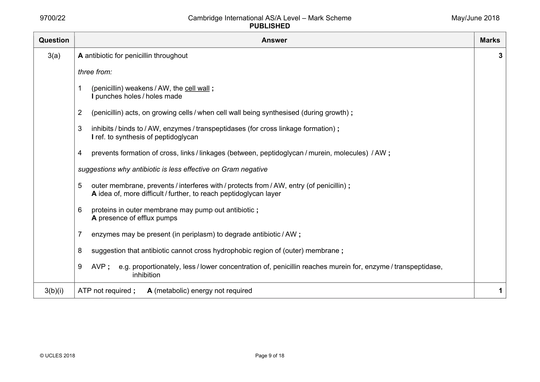
Mark scheme: 3(a) A antibiotic for penicillin throughout three from: 1 (penicillin) weakens / AW, the cell wall ; I punches holes / holes made 2 (penicillin) acts, on growing cells / when cell wall being synthesised (during growth) ; 3 inhibits / binds to / AW, enzymes / transpeptidases (for cross linkage formation) ; I ref. to synthesis of peptidoglycan 4 prevents formation of cross, links / linkages (between, peptidoglycan / murein, molecules) / AW ; suggestions why antibiotic is less effective on Gram negative 5 outer membrane, prevents / interferes with / protects from / AW, entry (of penicillin) ; A idea of, more difficult / further, to reach peptidoglycan layer 6 proteins in outer membrane may pump out antibiotic ; A presence of efflux pumps 7 enzymes may be present (in periplasm) to degrade antibiotic / AW ; 8 suggestion that antibiotic cannot cross hydrophobic region of (outer) membrane ; 9 AVP ; e.g. proportionately, less / lower concentration of, penicillin reaches murein for, enzyme / transpeptidase, inhibition 3 3(b)(i) ATP not required ; A (metabolic) energy not required 1 Question Answer Marks 3(b)(ii) four from: 1 channel protein ; A pore protein / has a pore / has a channel 2 channel / pore, can form from polypeptides or (protein) has quaternary structure ; 3 hydrophilic R-groups, on amino acids lining channel / face inwards (towards channel) ; A hydrophilic, lining / channel A water-filled, channel / AW A idea that passage is through hydrophilic region of protein 4 allows facilitated diffusion ; A diffusion alone if in context of through the protein but R if via phospholipid bilayer 5 increases permeability for, movement of water / osmosis ; I faster 6 no (specific) binding sites / (channel) not specific / not selective / allows more than one type of substance through / AW ; 7 globular ; 8 AVP ; e.g. ref. to hydrophobic part of protein, faces / interacts with, hydrophobic, region / core / fatty acid tails ref. to hydrophilic parts of, protein / polypeptide, extend into, external environment / periplasm / aqueous regions / AW 4 Question Answer Marks 3(b)(iii) two from: translation, cannot / does not, begin / occur or polypeptide / protein, synthesis, cannot occur / decreases ; I ompF not made A chain of amino acids R transcription does not occur so translation does not occur R if in context of mutation or enzyme inhibition mRNA cannot attach to, ribosome / small subunit / ribosomal subunit ; (first) tRNA (with UAC anticodon) / tRNA carrying met, cannot bind (to START codon) ; 2 3(c)(i) (composed of) many / chain of / polymer of / AW, monosaccharides / sugar monomers / sugar units ; A glucose, molecules / residues A more than two / many sugars further detail ; e.g. carbohydrate may be, branched / unbranched in context of polysaccharide macromolecule in context of polysaccharide glycosidic bonds in context of between sugar monomers (sugar monomers) joined by condensation reactions 2 Question Answer Marks 3(c)(ii) three from: 1 specificity / specific (in correct context) ; 2 (B / T, -) lymphocytes have receptors complementary to antigen ; A immunoglobulins / antibody as receptors for B-lymphocyte A surface molecules as receptors for T-lymphocytes 3 (different) antigens, stimulate / activate / AW, (different) B-lymphocytes / T-lymphocytes ; A antigens stimulate an immune response 4 idea that different antibodies, synthesised / produced / AW, for different (O) antigens / O polysaccharides / lipopolysaccharides ; 5 memory cells will, not respond to different antigen / only respond to same antigen / AW ; 6 different O-antigens can, be composed of different sugars ; A can have different shapes 3
Q4 · A photomicrograph showing some cells in interphase and some cells in stages of mitosis
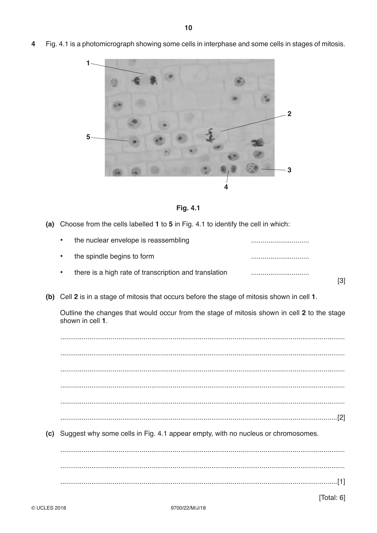
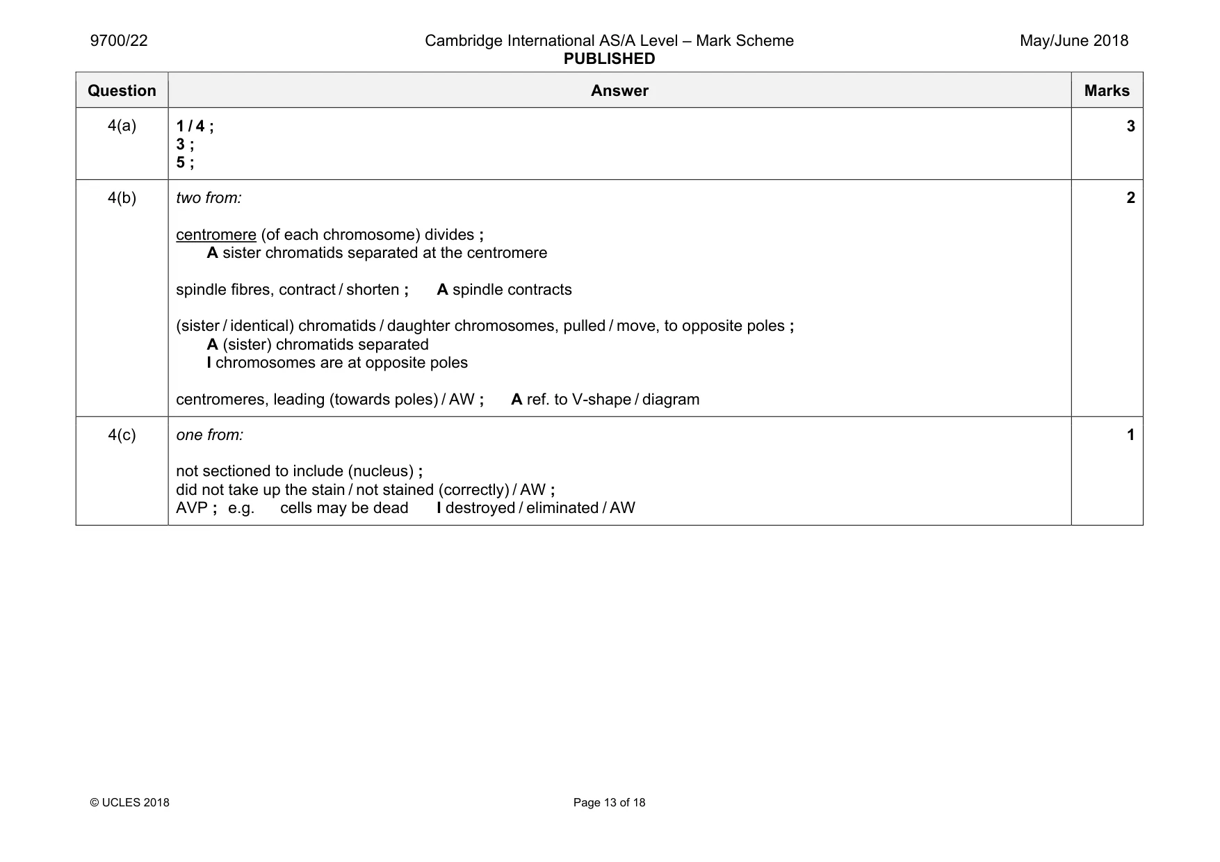
4 Fig. 4.1 is a photomicrograph showing some cells in interphase and some cells in stages of mitosis. 1 2 5 3 4 Fig. 4.1 (a) Choose from the cells labelled 1 to 5 in Fig. 4.1 to identify the cell in which: • the nuclear envelope is reassembling .............................. • the spindle begins to form .............................. • there is a high rate of transcription and translation .............................. [3] (b) Cell 2 is in a stage of mitosis that occurs before the stage of mitosis shown in cell 1. Outline the changes that would occur from the stage of mitosis shown in cell 2 to the stage shown in cell 1. ................................................................................................................................................... ................................................................................................................................................... ................................................................................................................................................... ................................................................................................................................................... ................................................................................................................................................... ...............................................................................................................................................[2] (c) Suggest why some cells in Fig. 4.1 appear empty, with no nucleus or chromosomes. ................................................................................................................................................... ................................................................................................................................................... ...............................................................................................................................................[1] [Total: 6]
Mark scheme: 4(a) 1 / 4 ; 3 ; 5 ; 3 4(b) two from: centromere (of each chromosome) divides ; A sister chromatids separated at the centromere spindle fibres, contract / shorten ; A spindle contracts (sister / identical) chromatids / daughter chromosomes, pulled / move, to opposite poles ; A (sister) chromatids separated I chromosomes are at opposite poles centromeres, leading (towards poles) / AW ; A ref. to V-shape / diagram 2 4(c) one from: not sectioned to include (nucleus) ; did not take up the stain / not stained (correctly) / AW ; AVP ; e.g. cells may be dead I destroyed / eliminated / AW 1
Q5 · When tobacco smoke is inhaled, chemicals such as nicotine and carbon monoxide enter the…
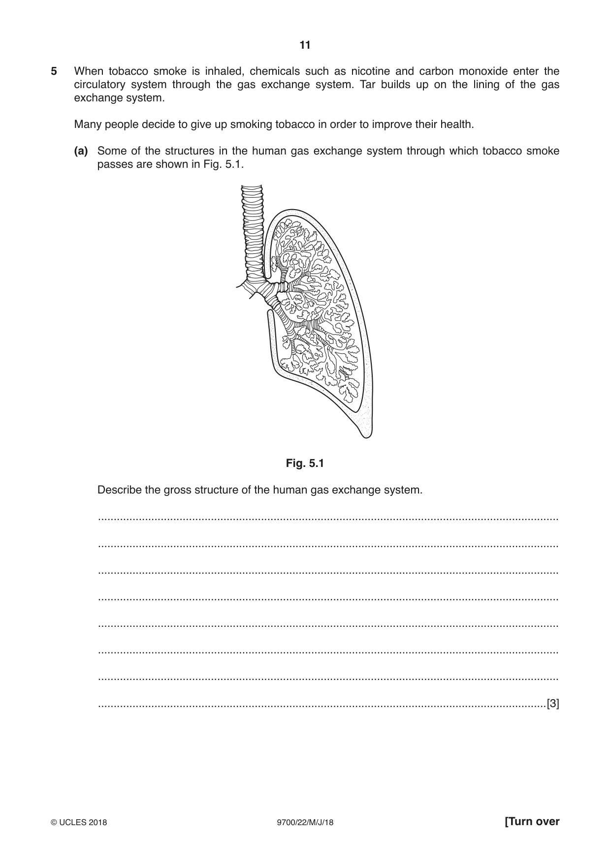
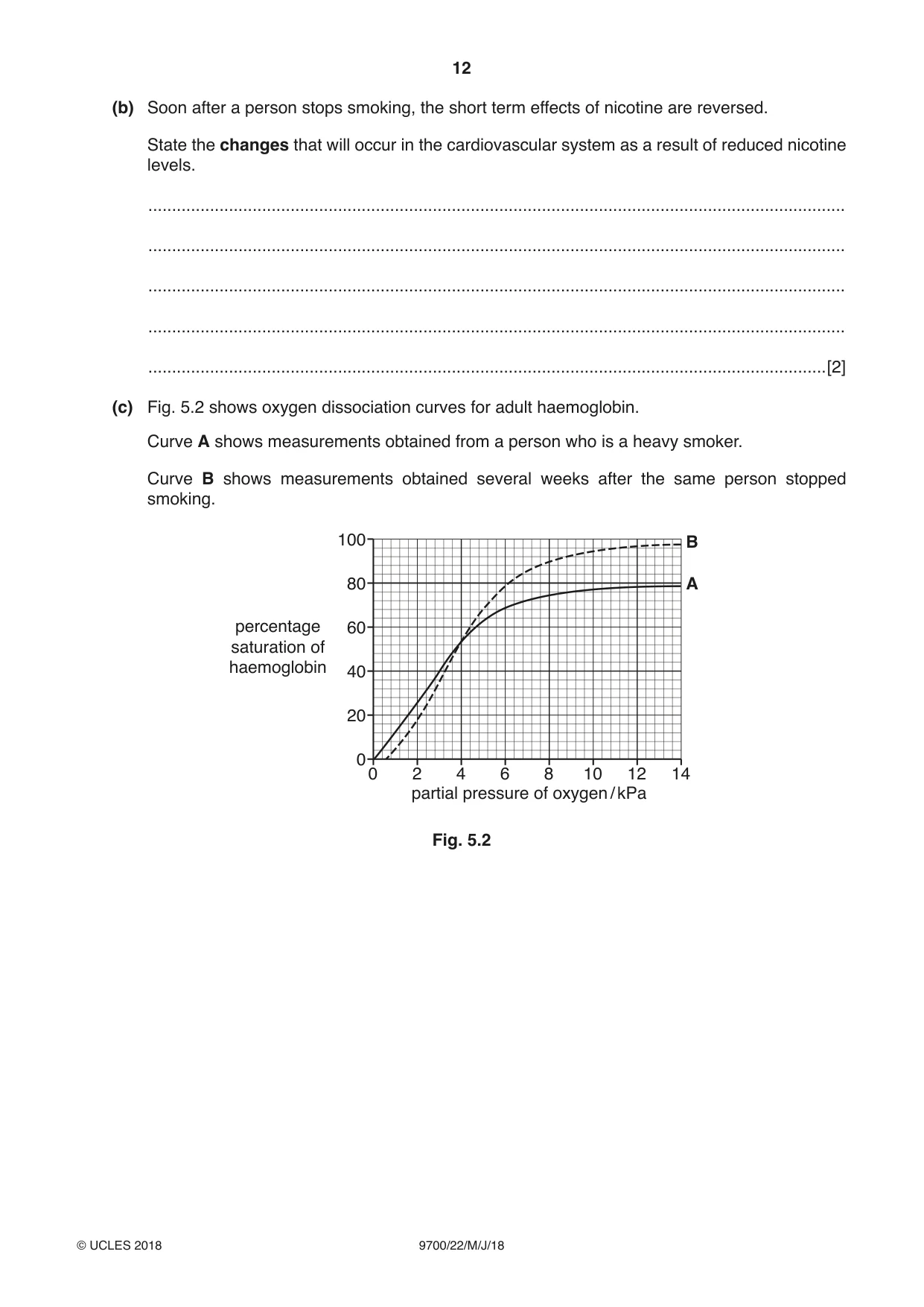
5 When tobacco smoke is inhaled, chemicals such as nicotine and carbon monoxide enter the circulatory system through the gas exchange system. Tar builds up on the lining of the gas exchange system. Many people decide to give up smoking tobacco in order to improve their health. (a) Some of the structures in the human gas exchange system through which tobacco smoke passes are shown in Fig. 5.1. Fig. 5.1 Describe the gross structure of the human gas exchange system. ................................................................................................................................................... ................................................................................................................................................... ................................................................................................................................................... ................................................................................................................................................... ................................................................................................................................................... ................................................................................................................................................... ................................................................................................................................................... ...............................................................................................................................................[3] (b) Soon after a person stops smoking, the short term effects of nicotine are reversed. State the changes that will occur in the cardiovascular system as a result of reduced nicotine levels. ................................................................................................................................................... ................................................................................................................................................... ................................................................................................................................................... ................................................................................................................................................... ...............................................................................................................................................[2] (c) Fig. 5.2 shows oxygen dissociation curves for adult haemoglobin. Curve A shows measurements obtained from a person who is a heavy smoker. Curve B shows measurements obtained several weeks after the same person stopped smoking. 100 B 80 A percentage 60 saturation of haemoglobin 40 20 0 0 2 4 6 8 10 12 14 partial pressure of oxygen / kPa Fig. 5.2 With reference to Fig. 5.2, describe and explain how the results show some of the health benefits of stopping smoking. ................................................................................................................................................... ................................................................................................................................................... ................................................................................................................................................... ................................................................................................................................................... ................................................................................................................................................... ................................................................................................................................................... ................................................................................................................................................... ................................................................................................................................................... ................................................................................................................................................... ................................................................................................................................................... ................................................................................................................................................... ...............................................................................................................................................[4] (d) A person who gives up smoking decreases their risk of developing lung cancer, a non-infectious disease. Explain why lung cancer is described as a non-infectious disease. ................................................................................................................................................... ................................................................................................................................................... ................................................................................................................................................... ................................................................................................................................................... ................................................................................................................................................... ................................................................................................................................................... ...............................................................................................................................................[2] [Total: 11]
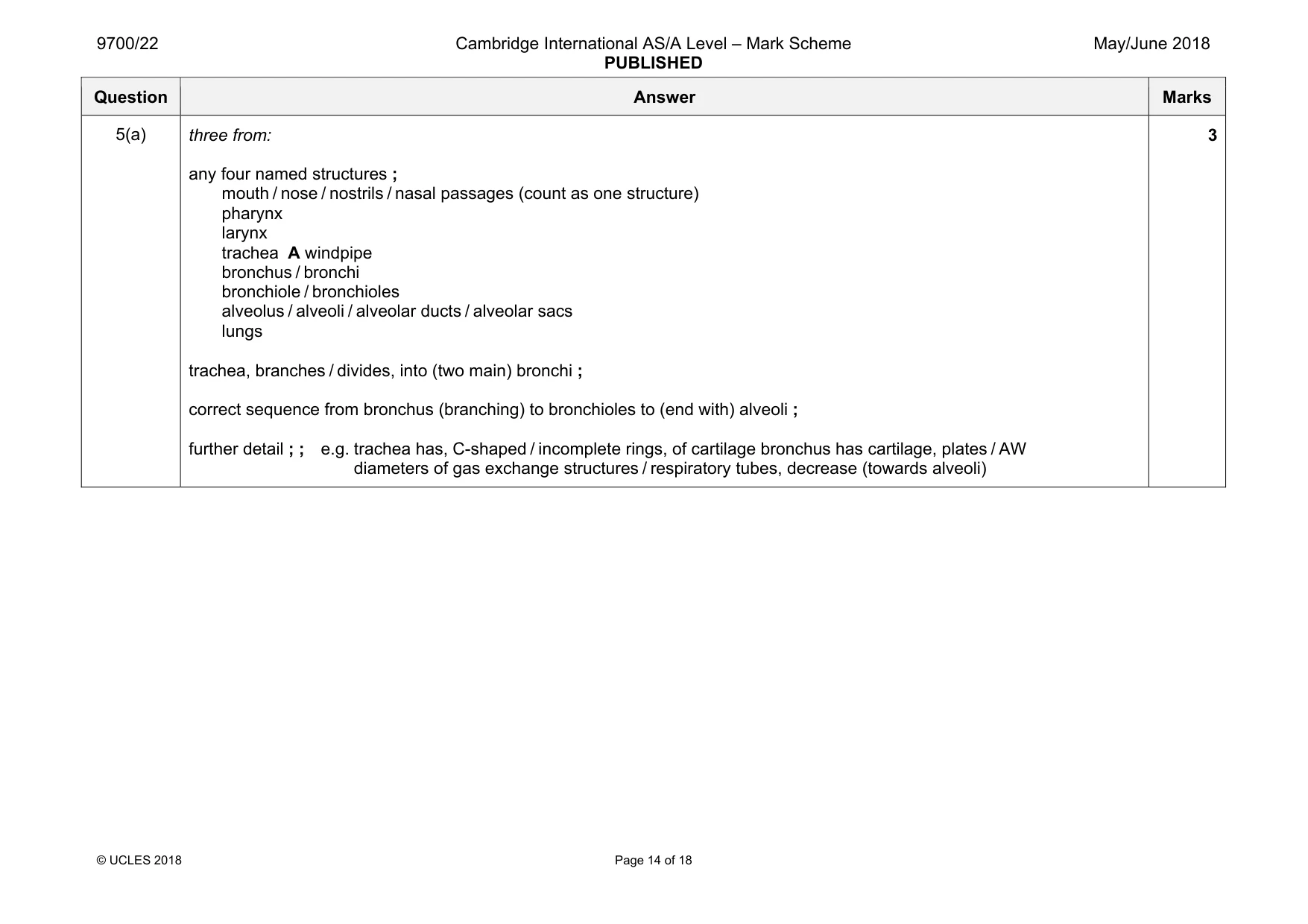
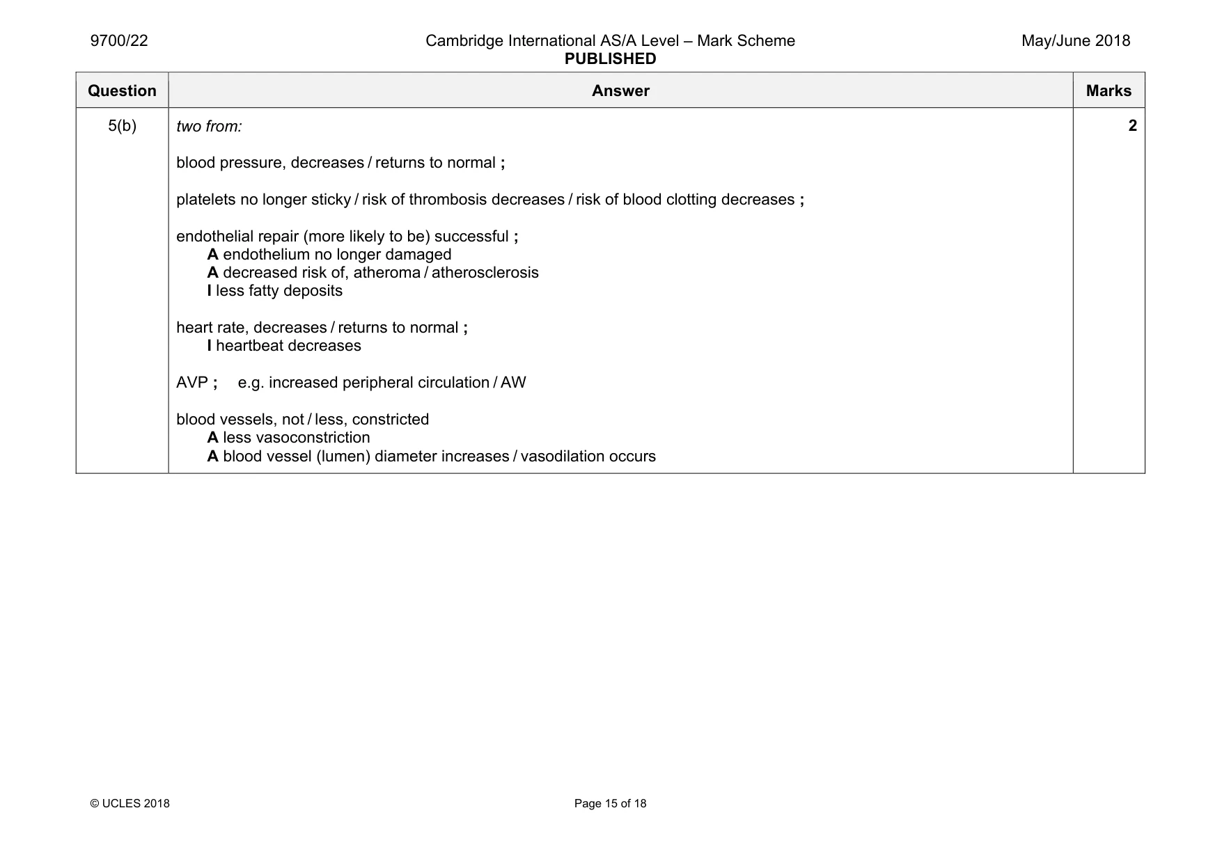
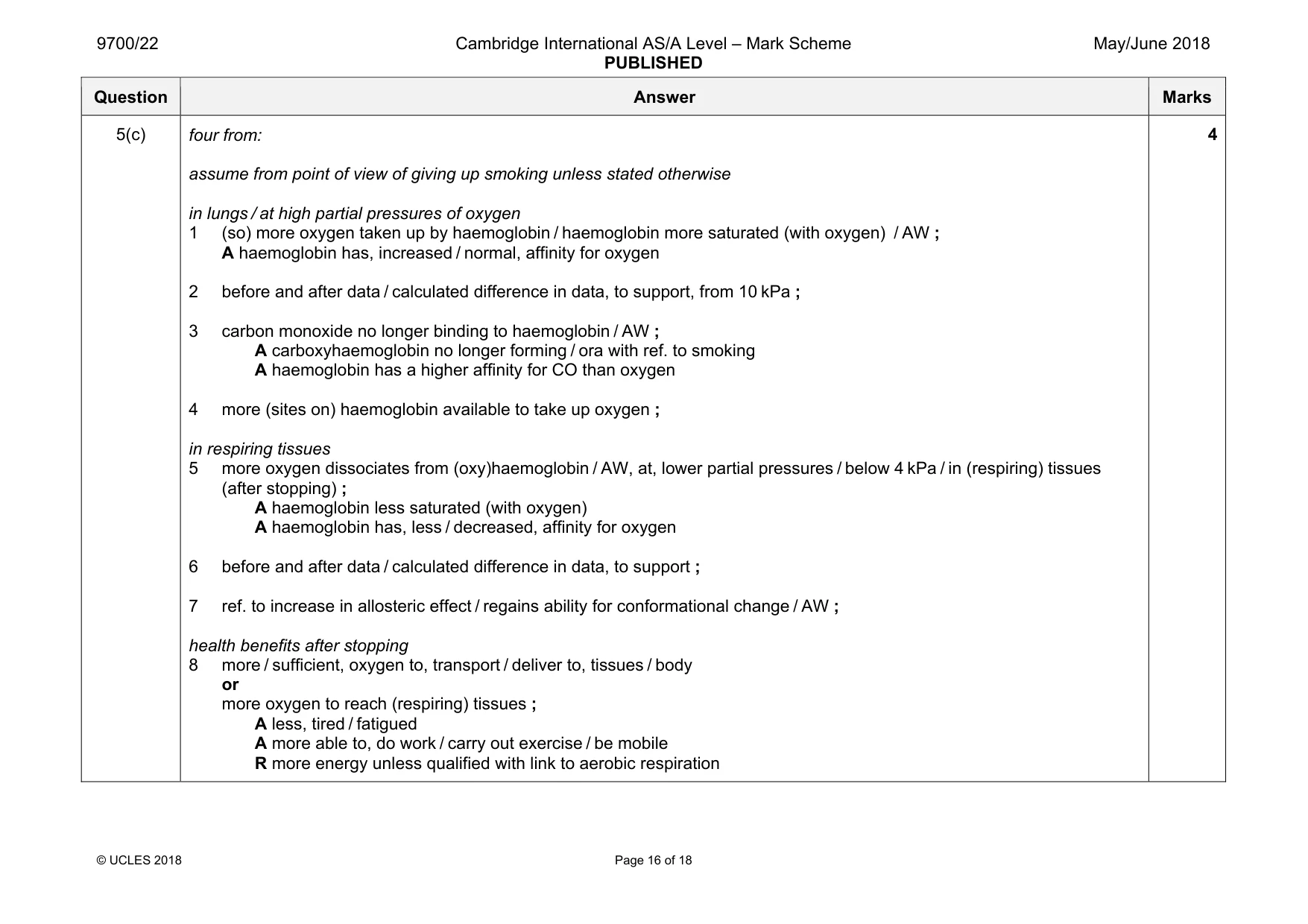
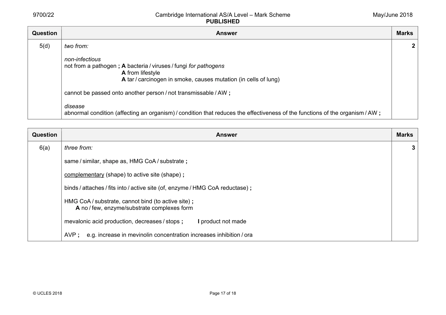
Mark scheme: 5(a) three from: any four named structures ; mouth / nose / nostrils / nasal passages (count as one structure) pharynx larynx trachea A windpipe bronchus / bronchi bronchiole / bronchioles alveolus / alveoli / alveolar ducts / alveolar sacs lungs trachea, branches / divides, into (two main) bronchi ; correct sequence from bronchus (branching) to bronchioles to (end with) alveoli ; further detail ; ; e.g. trachea has, C-shaped / incomplete rings, of cartilage bronchus has cartilage, plates / AW diameters of gas exchange structures / respiratory tubes, decrease (towards alveoli) 3 Question Answer Marks 5(b) two from: blood pressure, decreases / returns to normal ; platelets no longer sticky / risk of thrombosis decreases / risk of blood clotting decreases ; endothelial repair (more likely to be) successful ; A endothelium no longer damaged A decreased risk of, atheroma / atherosclerosis I less fatty deposits heart rate, decreases / returns to normal ; I heartbeat decreases AVP ; e.g. increased peripheral circulation / AW blood vessels, not / less, constricted A less vasoconstriction A blood vessel (lumen) diameter increases / vasodilation occurs 2 Question Answer Marks 5(c) four from: assume from point of view of giving up smoking unless stated otherwise in lungs / at high partial pressures of oxygen 1 (so) more oxygen taken up by haemoglobin / haemoglobin more saturated (with oxygen) / AW ; A haemoglobin has, increased / normal, affinity for oxygen 2 before and after data / calculated difference in data, to support, from 10 kPa ; 3 carbon monoxide no longer binding to haemoglobin / AW ; A carboxyhaemoglobin no longer forming / ora with ref. to smoking A haemoglobin has a higher affinity for CO than oxygen 4 more (sites on) haemoglobin available to take up oxygen ; in respiring tissues 5 more oxygen dissociates from (oxy)haemoglobin / AW, at, lower partial pressures / below 4 kPa / in (respiring) tissues (after stopping) ; A haemoglobin less saturated (with oxygen) A haemoglobin has, less / decreased, affinity for oxygen 6 before and after data / calculated difference in data, to support ; 7 ref. to increase in allosteric effect / regains ability for conformational change / AW ; health benefits after stopping 8 more / sufficient, oxygen to, transport / deliver to, tissues / body or more oxygen to reach (respiring) tissues ; A less, tired / fatigued A more able to, do work / carry out exercise / be mobile R more energy unless qualified with link to aerobic respiration 4 Question Answer Marks 5(d) two from: non-infectious not from a pathogen ; A bacteria / viruses / fungi for pathogens A from lifestyle A tar / carcinogen in smoke, causes mutation (in cells of lung) cannot be passed onto another person / not transmissable / AW ; disease abnormal condition (affecting an organism) / condition that reduces the effectiveness of the functions of the organism / AW ; 2
Q6 · Enzyme inhibitors and monoclonal antibodies can be used in the treatment of disease
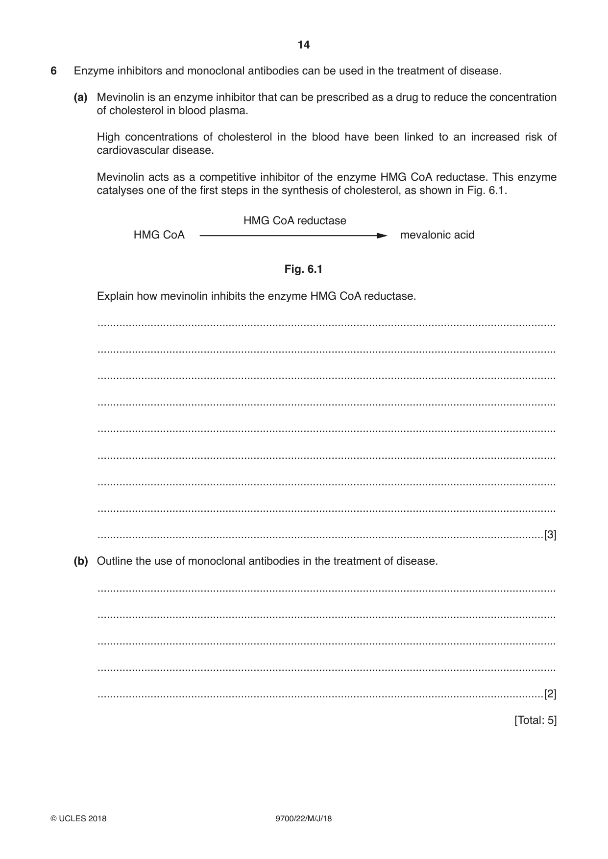
6 Enzyme inhibitors and monoclonal antibodies can be used in the treatment of disease. (a) Mevinolin is an enzyme inhibitor that can be prescribed as a drug to reduce the concentration of cholesterol in blood plasma. High concentrations of cholesterol in the blood have been linked to an increased risk of cardiovascular disease. Mevinolin acts as a competitive inhibitor of the enzyme HMG CoA reductase. This enzyme catalyses one of the first steps in the synthesis of cholesterol, as shown in Fig. 6.1. HMG CoA reductase HMG CoA mevalonic acid Fig. 6.1 Explain how mevinolin inhibits the enzyme HMG CoA reductase. ................................................................................................................................................... ................................................................................................................................................... ................................................................................................................................................... ................................................................................................................................................... ................................................................................................................................................... ................................................................................................................................................... ................................................................................................................................................... ................................................................................................................................................... ...............................................................................................................................................[3] (b) Outline the use of monoclonal antibodies in the treatment of disease. ................................................................................................................................................... ................................................................................................................................................... ................................................................................................................................................... ................................................................................................................................................... ...............................................................................................................................................[2] [Total: 5]
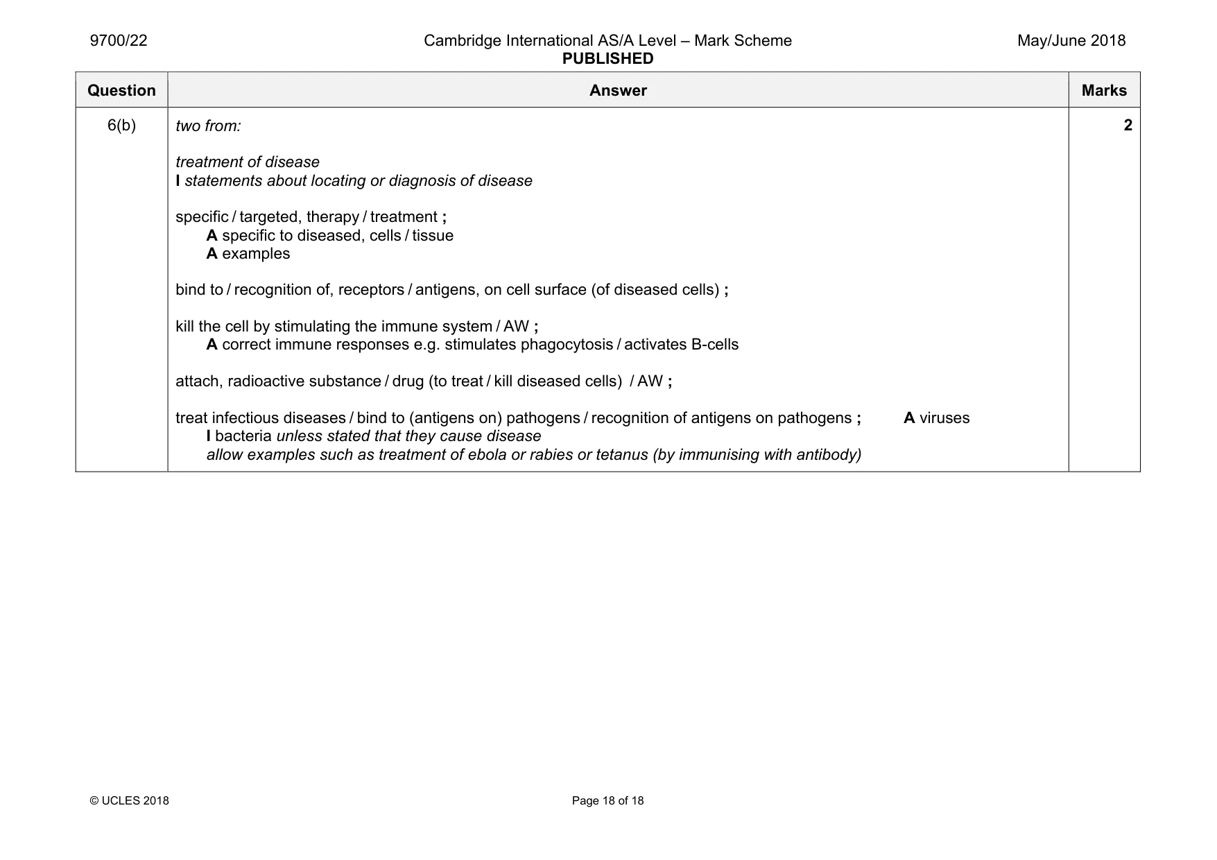
Mark scheme: 6(a) three from: same / similar, shape as, HMG CoA / substrate ; complementary (shape) to active site (shape) ; binds / attaches / fits into / active site (of, enzyme / HMG CoA reductase) ; HMG CoA / substrate, cannot bind (to active site) ; A no / few, enzyme/substrate complexes form mevalonic acid production, decreases / stops ; I product not made AVP ; e.g. increase in mevinolin concentration increases inhibition / ora 3 Question Answer Marks 6(b) two from: treatment of disease I statements about locating or diagnosis of disease specific / targeted, therapy / treatment ; A specific to diseased, cells / tissue A examples bind to / recognition of, receptors / antigens, on cell surface (of diseased cells) ; kill the cell by stimulating the immune system / AW ; A correct immune responses e.g. stimulates phagocytosis / activates B-cells attach, radioactive substance / drug (to treat / kill diseased cells) / AW ; treat infectious diseases / bind to (antigens on) pathogens / recognition of antigens on pathogens ; A viruses I bacteria unless stated that they cause disease allow examples such as treatment of ebola or rabies or tetanus (by immunising with antibody) 2
What was in this paper
The subtopics covered by these 6 questions, and how many questions each got. Open one in a new tab to see every Cambridge question on it.
What you needed in this session
Cambridge’s own grade thresholds for 2018 May/June, Paper 2 · Variant 2. A higher threshold means an easier paper — the bar moves with how the cohort did.