Cambridge A Level Biology 9700 — 2017 May/June Paper 2 · Variant 2
9700/22/M/J/17 · 6 questions · 60 marks · ≈68 min
The question paper and its mark scheme, free to read here and free to download. This is Cambridge’s own paper, exactly as it was sat.
Question paper16 pages

Mark scheme15 pages
Answers below. Sit the paper first if you are practising.
Questions as text
Q1 · In multicellular organisms, the structure of different cell types is adapted to their…
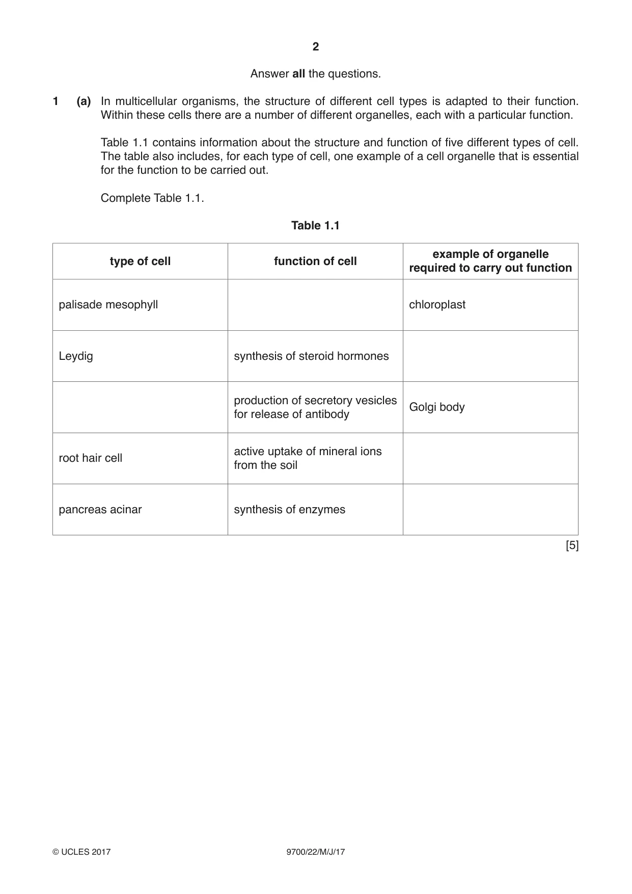
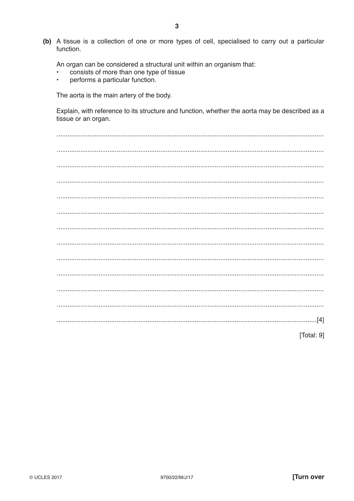
1 (a) In multicellular organisms, the structure of different cell types is adapted to their function. Within these cells there are a number of different organelles, each with a particular function. Table 1.1 contains information about the structure and function of five different types of cell. The table also includes, for each type of cell, one example of a cell organelle that is essential for the function to be carried out. Complete Table 1.1. Table 1.1 example of organelle type of cell function of cell required to carry out function palisade mesophyll chloroplast Leydig synthesis of steroid hormones production of secretory vesicles Golgi body for release of antibody active uptake of mineral ions root hair cell from the soil pancreas acinar synthesis of enzymes [5] (b) A tissue is a collection of one or more types of cell, specialised to carry out a particular function. An organ can be considered a structural unit within an organism that: • consists of more than one type of tissue • performs a particular function. The aorta is the main artery of the body. Explain, with reference to its structure and function, whether the aorta may be described as a tissue or an organ. ................................................................................................................................................... ................................................................................................................................................... ................................................................................................................................................... ................................................................................................................................................... ................................................................................................................................................... ................................................................................................................................................... ................................................................................................................................................... ................................................................................................................................................... ................................................................................................................................................... ................................................................................................................................................... ................................................................................................................................................... ................................................................................................................................................... ...............................................................................................................................................[4] [Total: 9]
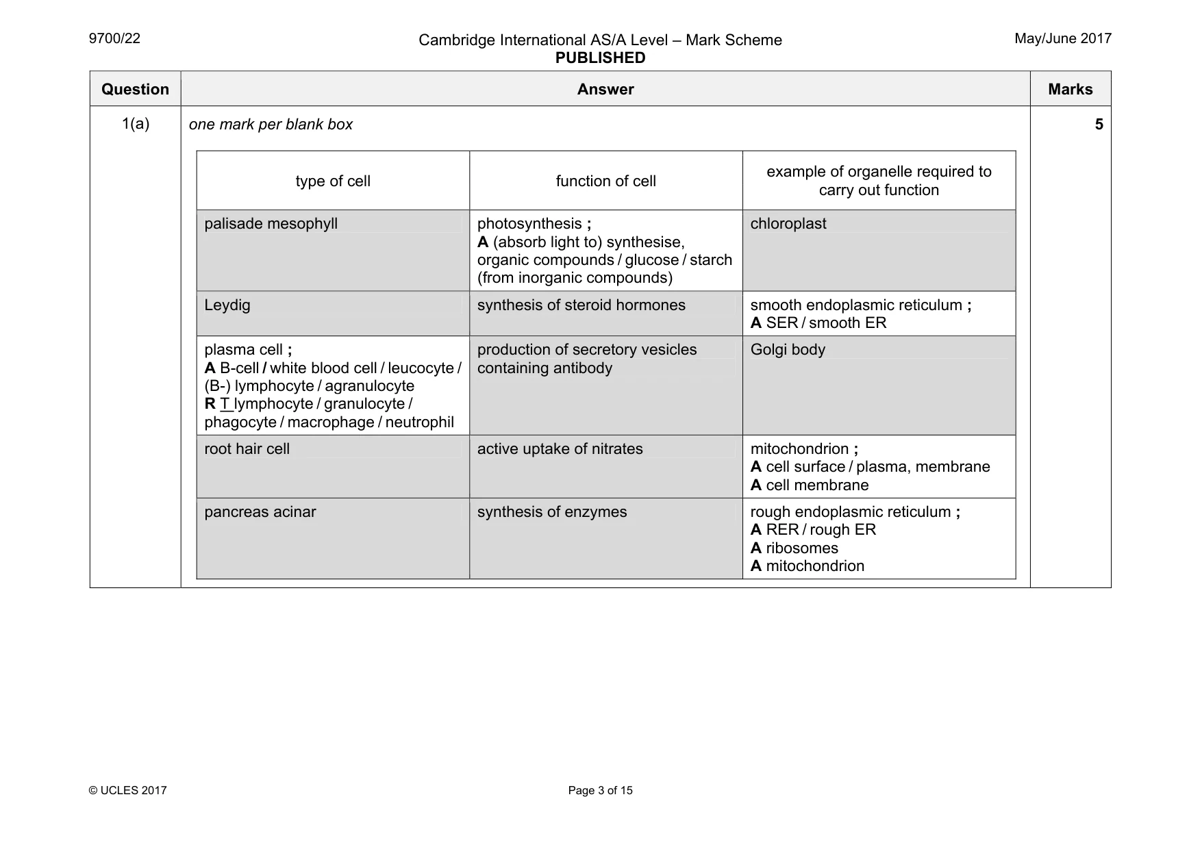
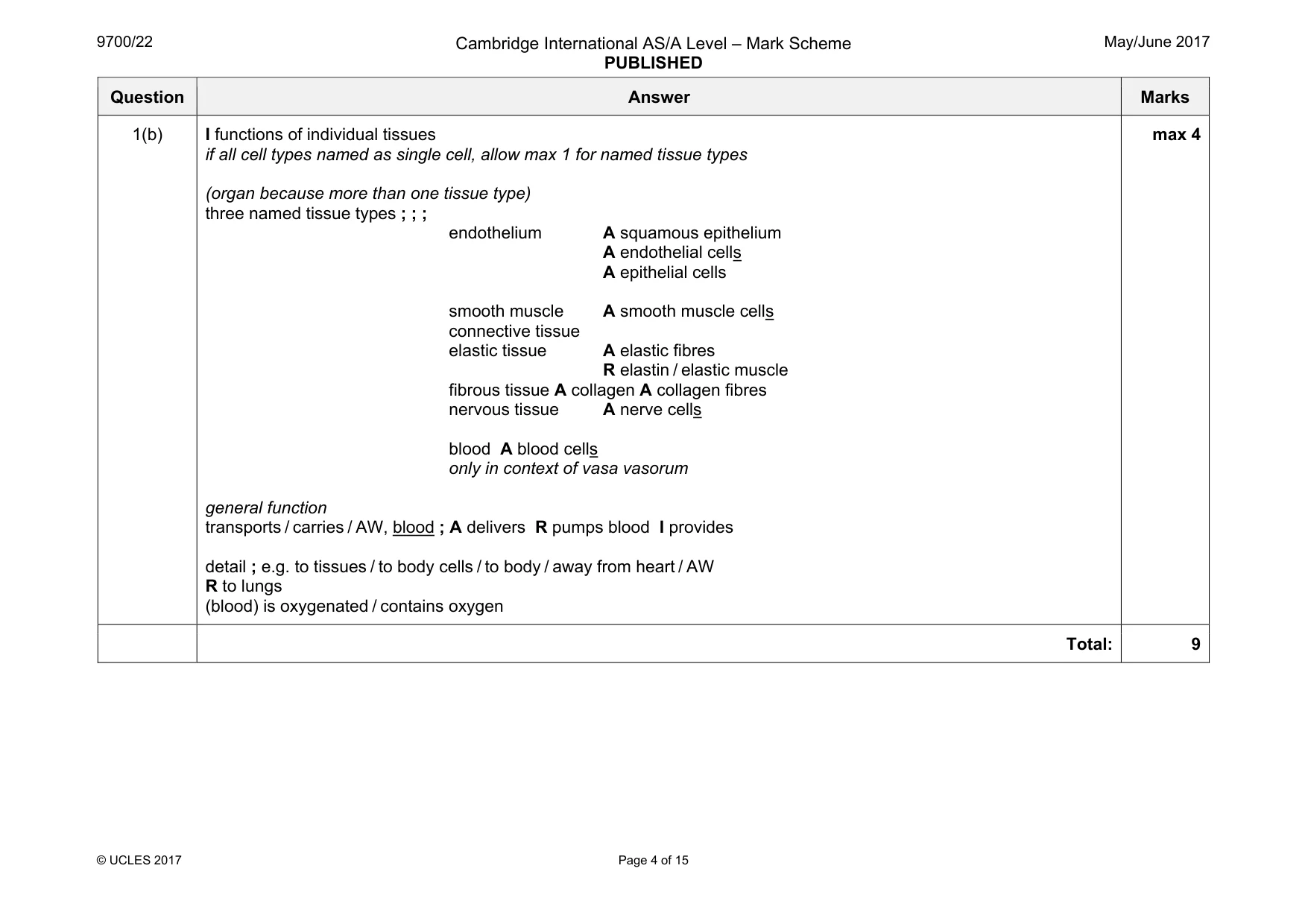
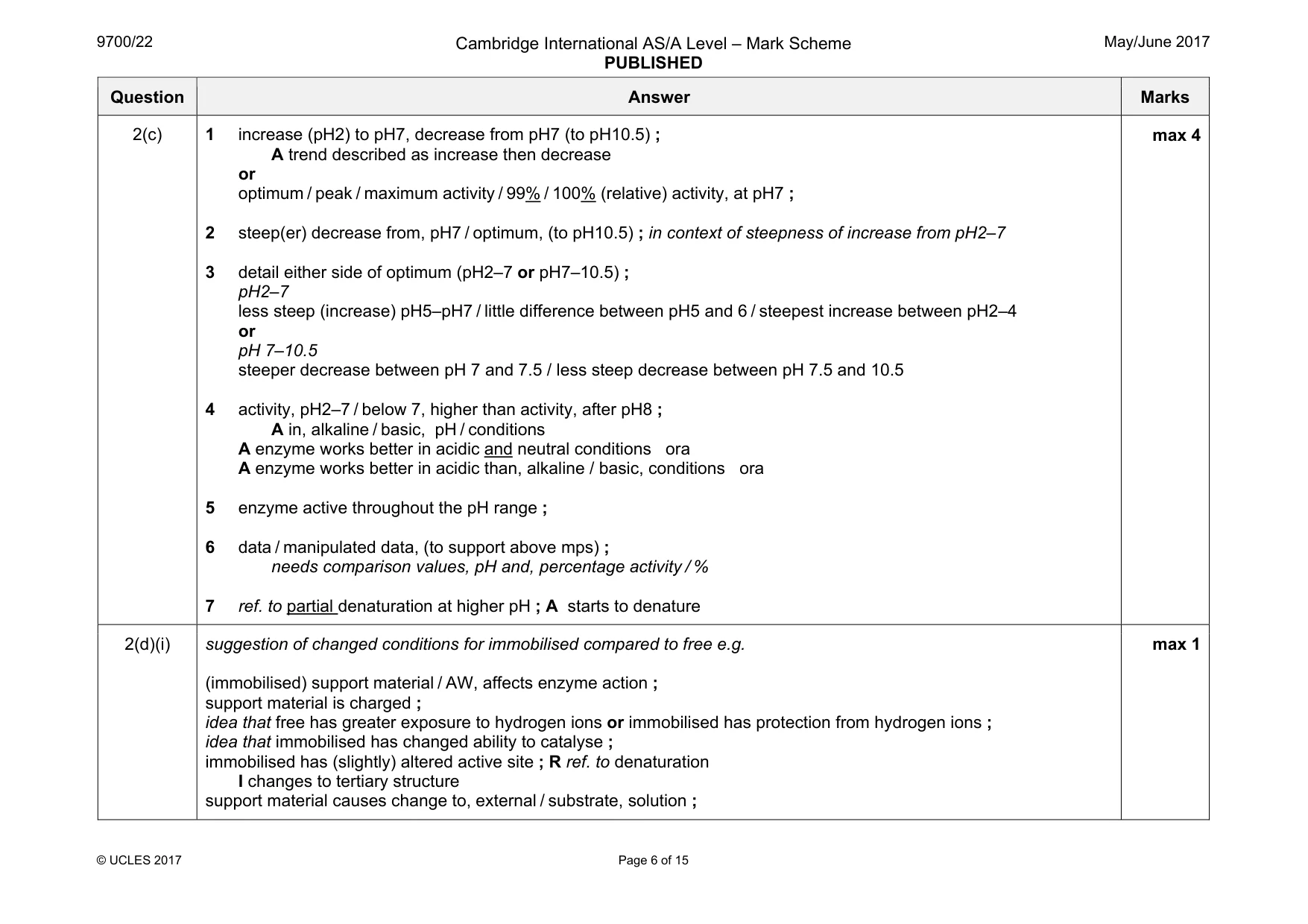
Mark scheme: 1(a) one mark per blank box type of cell function of cell example of organelle required to carry out function palisade mesophyll photosynthesis ; A (absorb light to) synthesise, organic compounds / glucose / starch (from inorganic compounds) chloroplast Leydig synthesis of steroid hormones smooth endoplasmic reticulum ; A SER / smooth ER plasma cell ; A B-cell / white blood cell / leucocyte / (B-) lymphocyte / agranulocyte R T lymphocyte / granulocyte / phagocyte / macrophage / neutrophil production of secretory vesicles containing antibody Golgi body root hair cell active uptake of nitrates mitochondrion ; A cell surface / plasma, membrane A cell membrane pancreas acinar synthesis of enzymes rough endoplasmic reticulum ; A RER / rough ER A ribosomes A mitochondrion 5 Question Answer Marks 1(b) I functions of individual tissues if all cell types named as single cell, allow max 1 for named tissue types (organ because more than one tissue type) three named tissue types ; ; ; endothelium A squamous epithelium A endothelial cells A epithelial cells smooth muscle A smooth muscle cells connective tissue elastic tissue A elastic fibres R elastin / elastic muscle fibrous tissue A collagen A collagen fibres nervous tissue A nerve cells blood A blood cells only in context of vasa vasorum general function transports / carries / AW, blood ; A delivers R pumps blood I provides detail ; e.g. to tissues / to body cells / to body / away from heart / AW R to lungs (blood) is oxygenated / contains oxygen max 4 Total: 9
More questions on Cells as the basic units of living organisms
Q2 · Lipase is an enzyme with many commercial uses
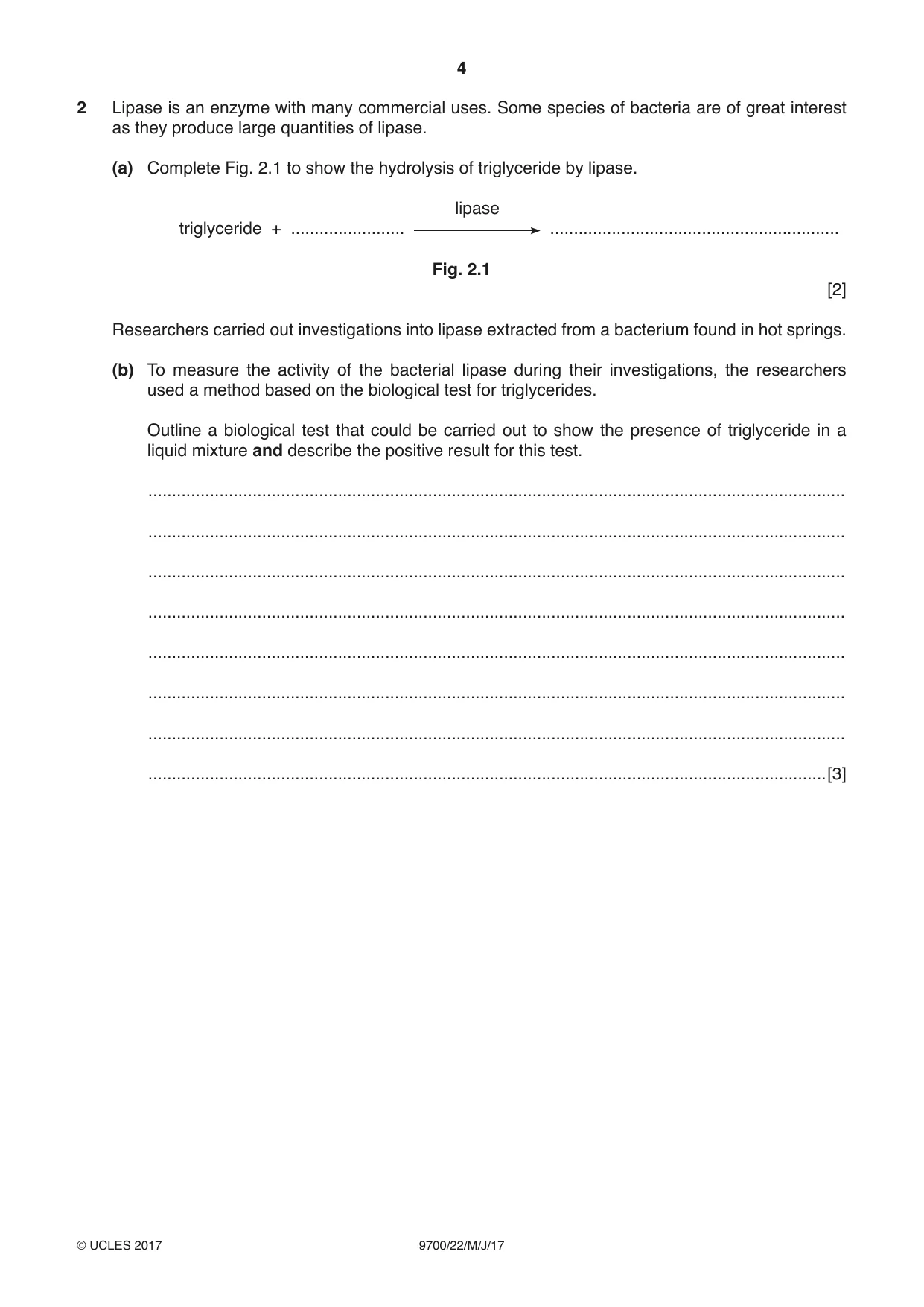
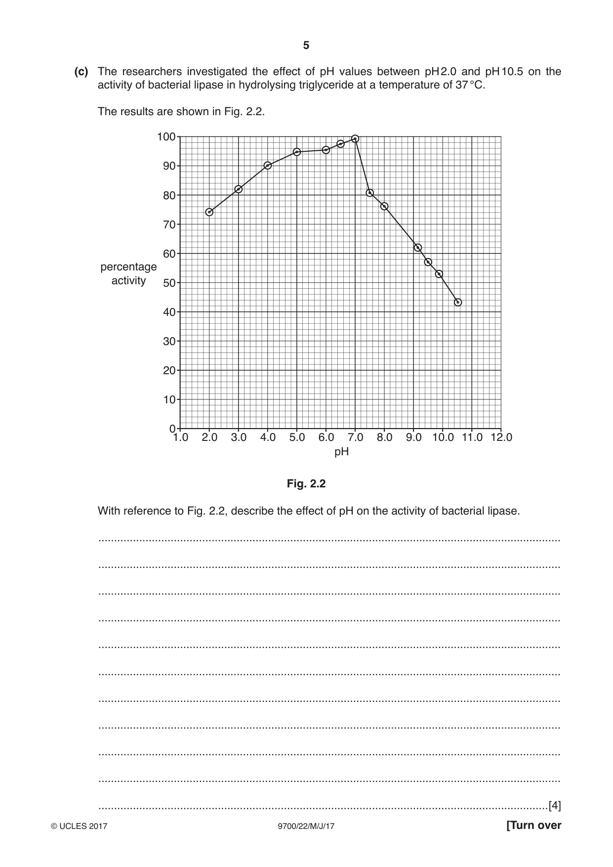
2 Lipase is an enzyme with many commercial uses. Some species of bacteria are of great interest as they produce large quantities of lipase. (a) Complete Fig. 2.1 to show the hydrolysis of triglyceride by lipase. lipase triglyceride + ........................ ............................................................. Fig. 2.1 [2] Researchers carried out investigations into lipase extracted from a bacterium found in hot springs. (b) To measure the activity of the bacterial lipase during their investigations, the researchers used a method based on the biological test for triglycerides. Outline a biological test that could be carried out to show the presence of triglyceride in a liquid mixture and describe the positive result for this test. ................................................................................................................................................... ................................................................................................................................................... ................................................................................................................................................... ................................................................................................................................................... ................................................................................................................................................... ................................................................................................................................................... ................................................................................................................................................... ...............................................................................................................................................[3] (c) The researchers investigated the effect of pH values between pH 2.0 and pH 10.5 on the activity of bacterial lipase in hydrolysing triglyceride at a temperature of 37 °C. The results are shown in Fig. 2.2. 100 90 80 70 60 percentage activity 50 40 30 20 10 0 1.0 2.0 3.0 4.0 5.0 6.0 7.0 8.0 9.0 10.0 11.0 12.0 pH Fig. 2.2 With reference to Fig. 2.2, describe the effect of pH on the activity of bacterial lipase. ................................................................................................................................................... ................................................................................................................................................... ................................................................................................................................................... ................................................................................................................................................... ................................................................................................................................................... ................................................................................................................................................... ................................................................................................................................................... ................................................................................................................................................... ................................................................................................................................................... ................................................................................................................................................... ...............................................................................................................................................[4] (d) A separate investigation into the effect of pH on the same bacterial lipase compared the enzyme free in solution with the enzyme immobilised by physical attachment to a stable polymer. At a temperature of 37 °C, the optimum pH of the enzyme free in solution was the same as that shown in Fig. 2.2. The optimum pH of the immobilised enzyme was measured as pH 4. (i) Suggest one reason to explain why the enzyme free in solution has a different optimum pH compared to the immobilised enzyme. ........................................................................................................................................... ........................................................................................................................................... .......................................................................................................................................[1] (ii) Suggest one advantage of immobilising the extracted lipase for commercial use. ........................................................................................................................................... ........................................................................................................................................... .......................................................................................................................................[1] [Total: 11]
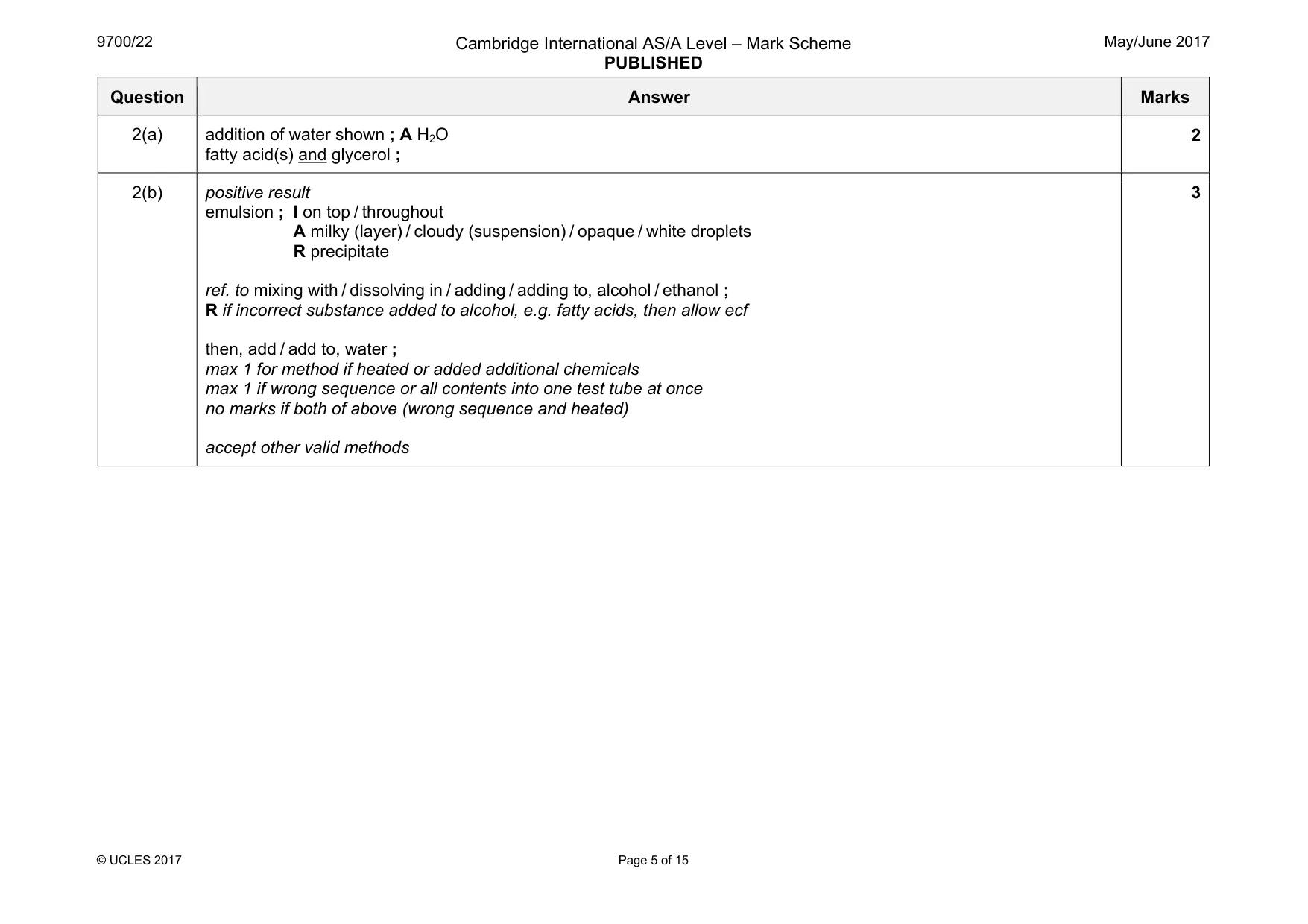
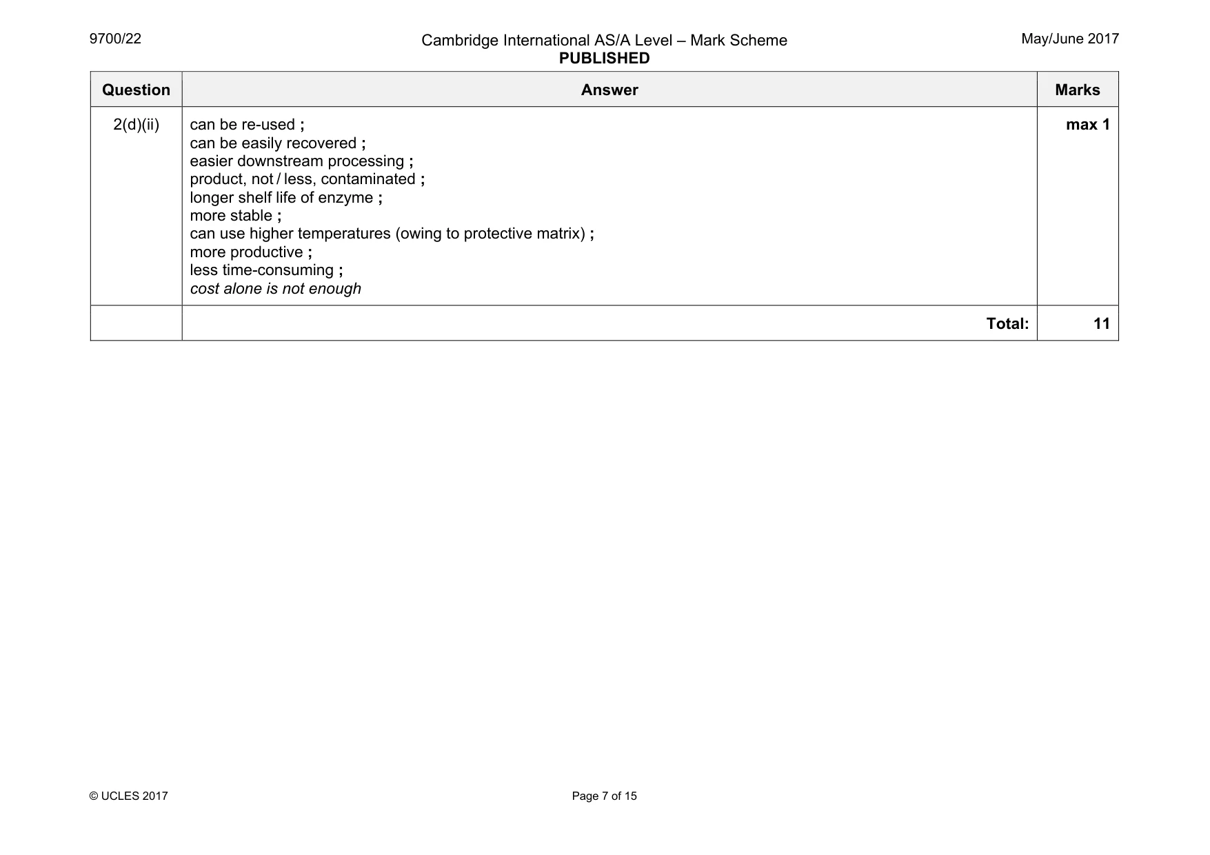
Mark scheme: 2(a) addition of water shown ; A H2O fatty acid(s) and glycerol ; 2 2(b) positive result emulsion ; I on top / throughout A milky (layer) / cloudy (suspension) / opaque / white droplets R precipitate ref. to mixing with / dissolving in / adding / adding to, alcohol / ethanol ; R if incorrect substance added to alcohol, e.g. fatty acids, then allow ecf then, add / add to, water ; max 1 for method if heated or added additional chemicals max 1 if wrong sequence or all contents into one test tube at once no marks if both of above (wrong sequence and heated) accept other valid methods 3 Question Answer Marks 2(c) 1 increase (pH2) to pH7, decrease from pH7 (to pH10.5) ; A trend described as increase then decrease or optimum / peak / maximum activity / 99% / 100% (relative) activity, at pH7 ; 2 steep(er) decrease from, pH7 / optimum, (to pH10.5) ; in context of steepness of increase from pH2–7 3 detail either side of optimum (pH2–7 or pH7–10.5) ; pH2–7 less steep (increase) pH5–pH7 / little difference between pH5 and 6 / steepest increase between pH2–4 or pH 7–10.5 steeper decrease between pH 7 and 7.5 / less steep decrease between pH 7.5 and 10.5 4 activity, pH2–7 / below 7, higher than activity, after pH8 ; A in, alkaline / basic, pH / conditions A enzyme works better in acidic and neutral conditions ora A enzyme works better in acidic than, alkaline / basic, conditions ora 5 enzyme active throughout the pH range ; 6 data / manipulated data, (to support above mps) ; needs comparison values, pH and, percentage activity / % 7 ref. to partial denaturation at higher pH ; A starts to denature max 4 2(d)(i) suggestion of changed conditions for immobilised compared to free e.g. (immobilised) support material / AW, affects enzyme action ; support material is charged ; idea that free has greater exposure to hydrogen ions or immobilised has protection from hydrogen ions ; idea that immobilised has changed ability to catalyse ; immobilised has (slightly) altered active site ; R ref. to denaturation I changes to tertiary structure support material causes change to, external / substrate, solution ; max 1 Question Answer Marks 2(d)(ii) can be re-used ; can be easily recovered ; easier downstream processing ; product, not / less, contaminated ; longer shelf life of enzyme ; more stable ; can use higher temperatures (owing to protective matrix) ; more productive ; less time-consuming ; cost alone is not enough max 1 Total: 11
Q3 · Malaria is a disease caused by the protoctist, Plasmodium
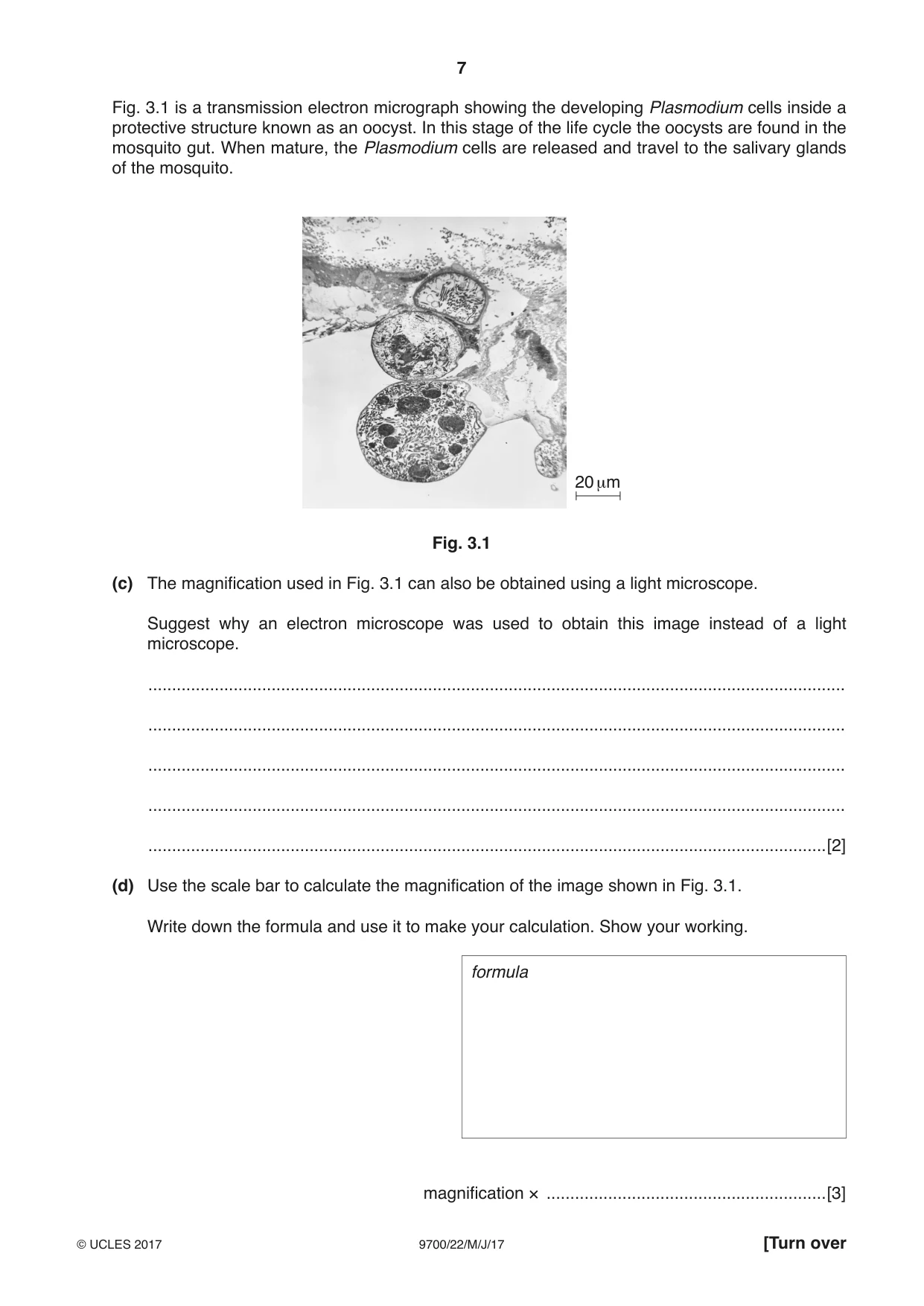
3 Malaria is a disease caused by the protoctist, Plasmodium. The organism has a very complex life cycle as it has two hosts, a human and a mosquito. (a) Name one of the four species of Plasmodium that infects humans. ...............................................................................................................................................[1] (b) State the name of the mosquito that is host to Plasmodium. ...............................................................................................................................................[1] Fig. 3.1 is a transmission electron micrograph showing the developing Plasmodium cells inside a protective structure known as an oocyst. In this stage of the life cycle the oocysts are found in the mosquito gut. When mature, the Plasmodium cells are released and travel to the salivary glands of the mosquito. 20 μm Fig. 3.1 (c) The magnification used in Fig. 3.1 can also be obtained using a light microscope. Suggest why an electron microscope was used to obtain this image instead of a light microscope. ................................................................................................................................................... ................................................................................................................................................... ................................................................................................................................................... ................................................................................................................................................... ...............................................................................................................................................[2] (d) Use the scale bar to calculate the magnification of the image shown in Fig. 3.1. Write down the formula and use it to make your calculation. Show your working. formula magnification × ...........................................................[3] (e) Outline the role of the mosquito in the transmission of malaria. ................................................................................................................................................... ................................................................................................................................................... ................................................................................................................................................... ................................................................................................................................................... ...............................................................................................................................................[2] [Total: 9]
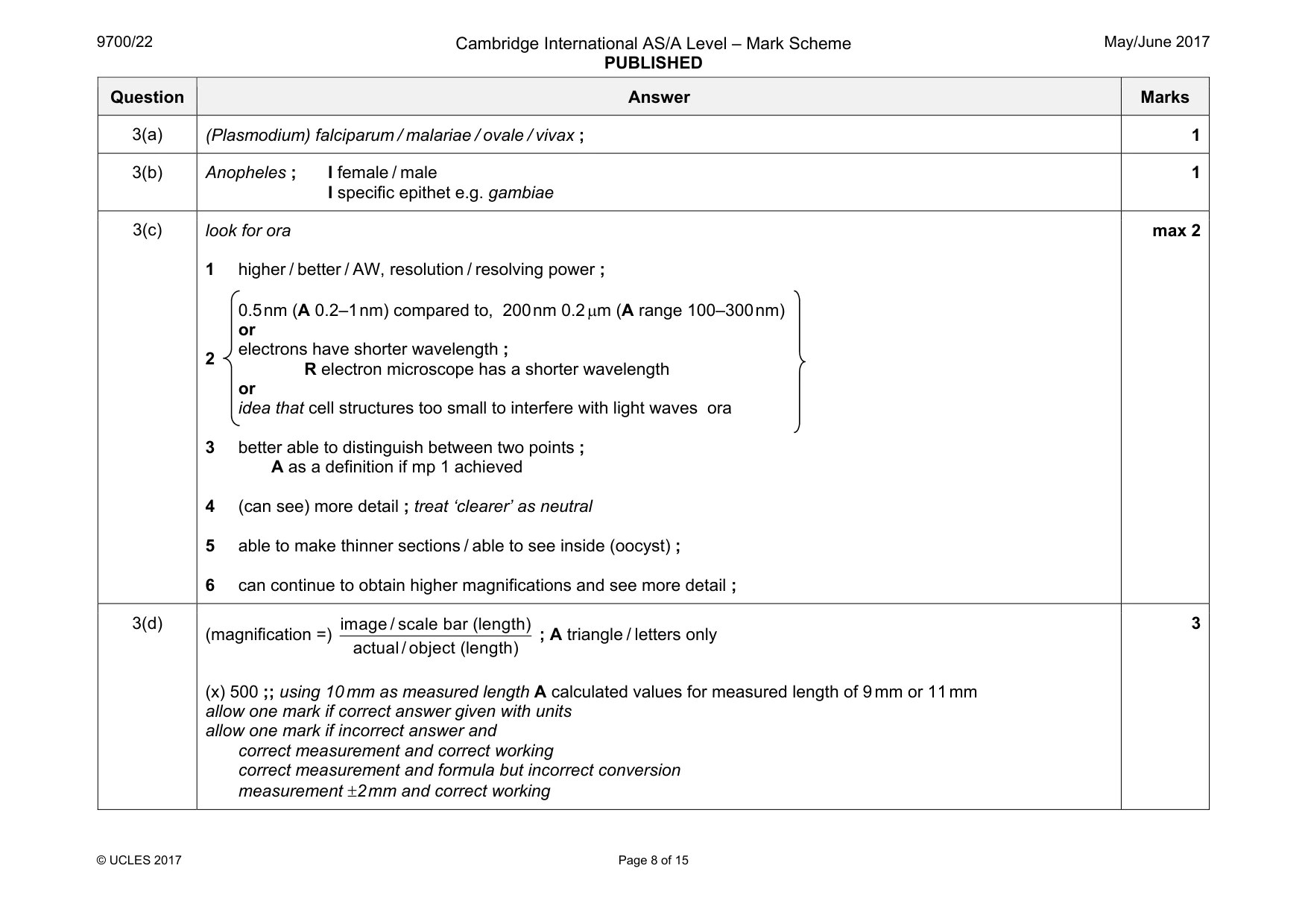
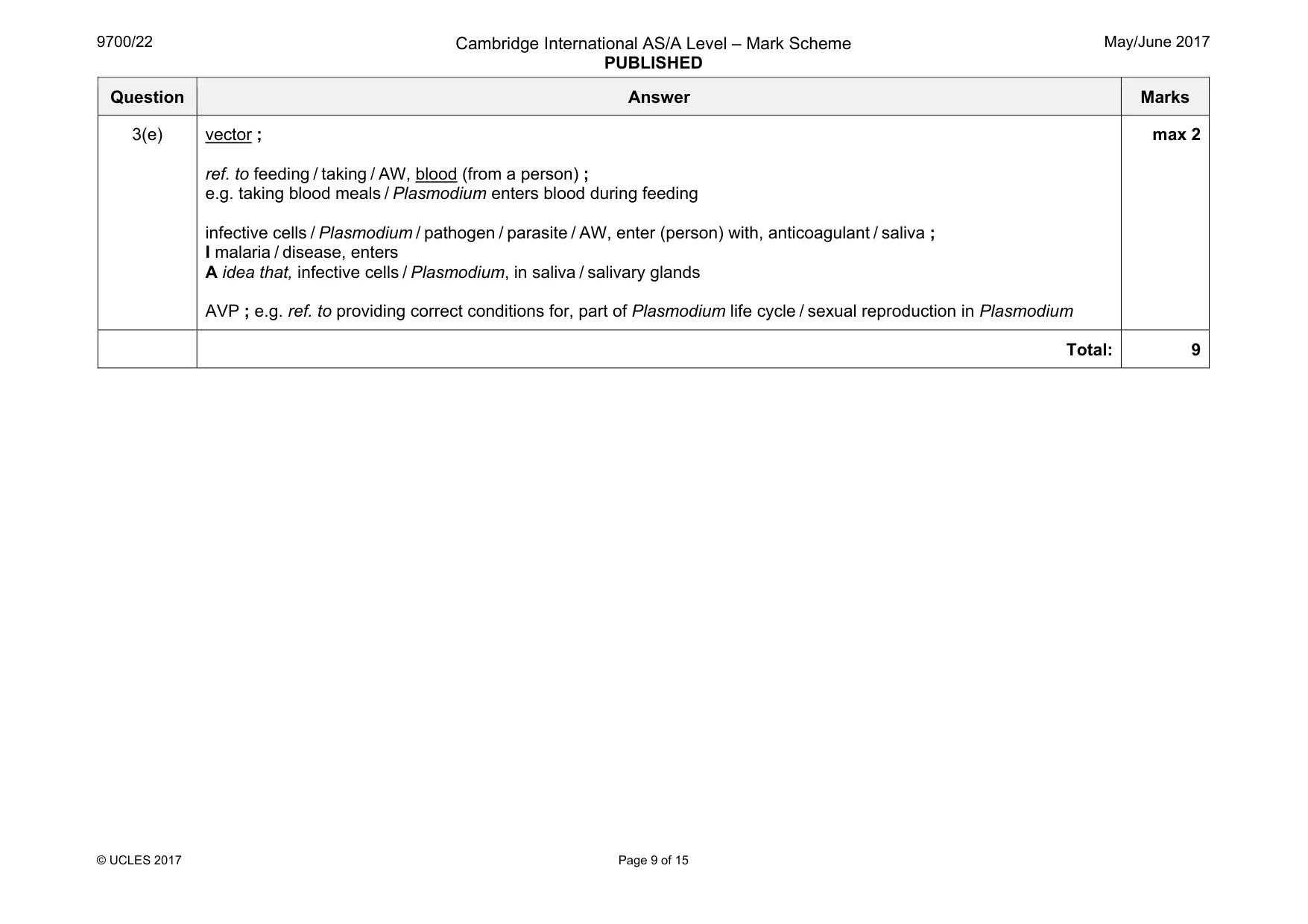
Mark scheme: 3(a) (Plasmodium) falciparum / malariae / ovale / vivax ; 1 3(b) Anopheles ; I female / male I specific epithet e.g. gambiae 1 3(c) look for ora 1 higher / better / AW, resolution / resolving power ; 2 0.5 nm (A 0.2–1 nm) compared to, 200 nm 0.2 µm (A range 100–300 nm) or electrons have shorter wavelength ; R electron microscope has a shorter wavelength or idea that cell structures too small to interfere with light waves ora 3 better able to distinguish between two points ; A as a definition if mp 1 achieved 4 (can see) more detail ; treat ‘clearer’ as neutral 5 able to make thinner sections / able to see inside (oocyst) ; 6 can continue to obtain higher magnifications and see more detail ; max 2 3(d) (magnification =) image / scale bar (length) actual/ object (length) ; A triangle / letters only (x) 500 ;; using 10 mm as measured length A calculated values for measured length of 9 mm or 11 mm allow one mark if correct answer given with units allow one mark if incorrect answer and correct measurement and correct working correct measurement and formula but incorrect conversion measurement ±2 mm and correct working 3 Question Answer Marks 3(e) vector ; ref. to feeding / taking / AW, blood (from a person) ; e.g. taking blood meals / Plasmodium enters blood during feeding infective cells / Plasmodium / pathogen / parasite / AW, enter (person) with, anticoagulant / saliva ; I malaria / disease, enters A idea that, infective cells / Plasmodium, in saliva / salivary glands AVP ; e.g. ref. to providing correct conditions for, part of Plasmodium life cycle / sexual reproduction in Plasmodium max 2 Total: 9
Q4 · Meristematic tissue is found in the growing regions of plants, such as root tips
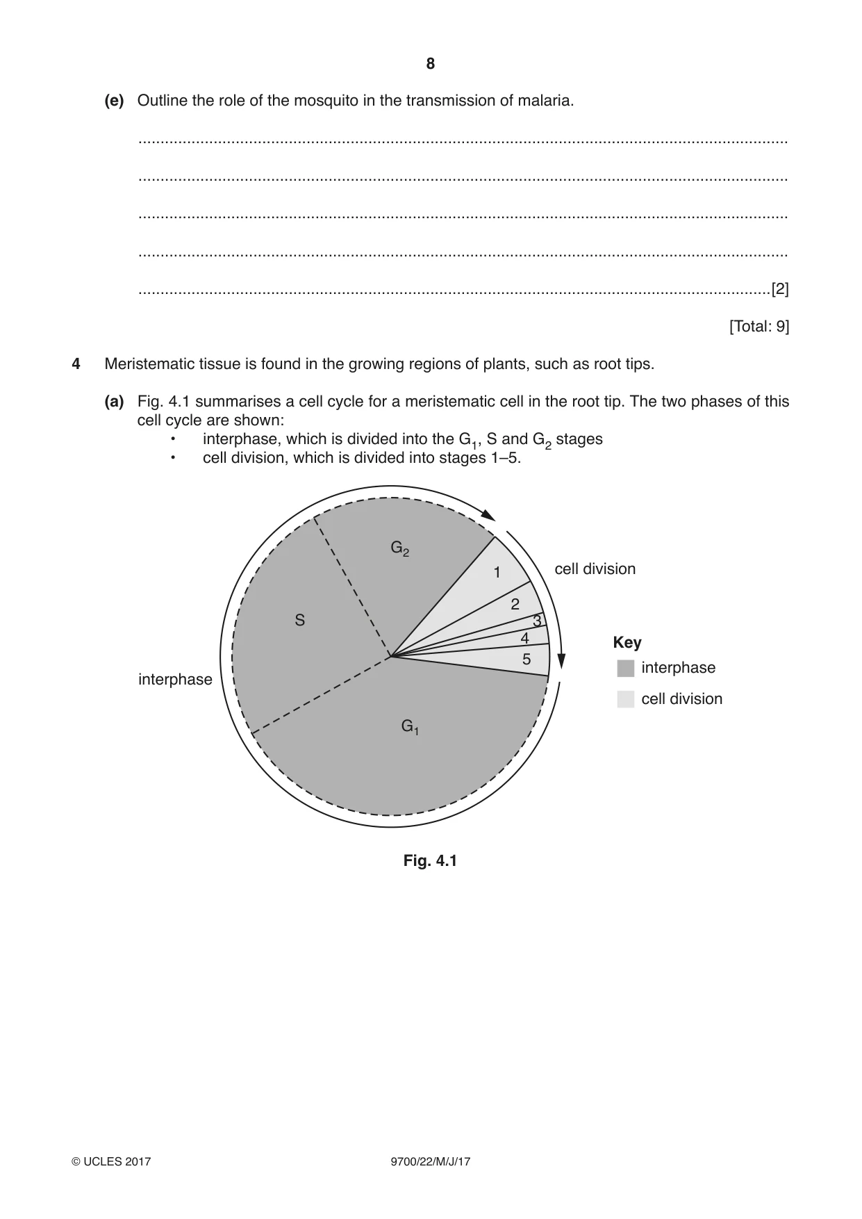
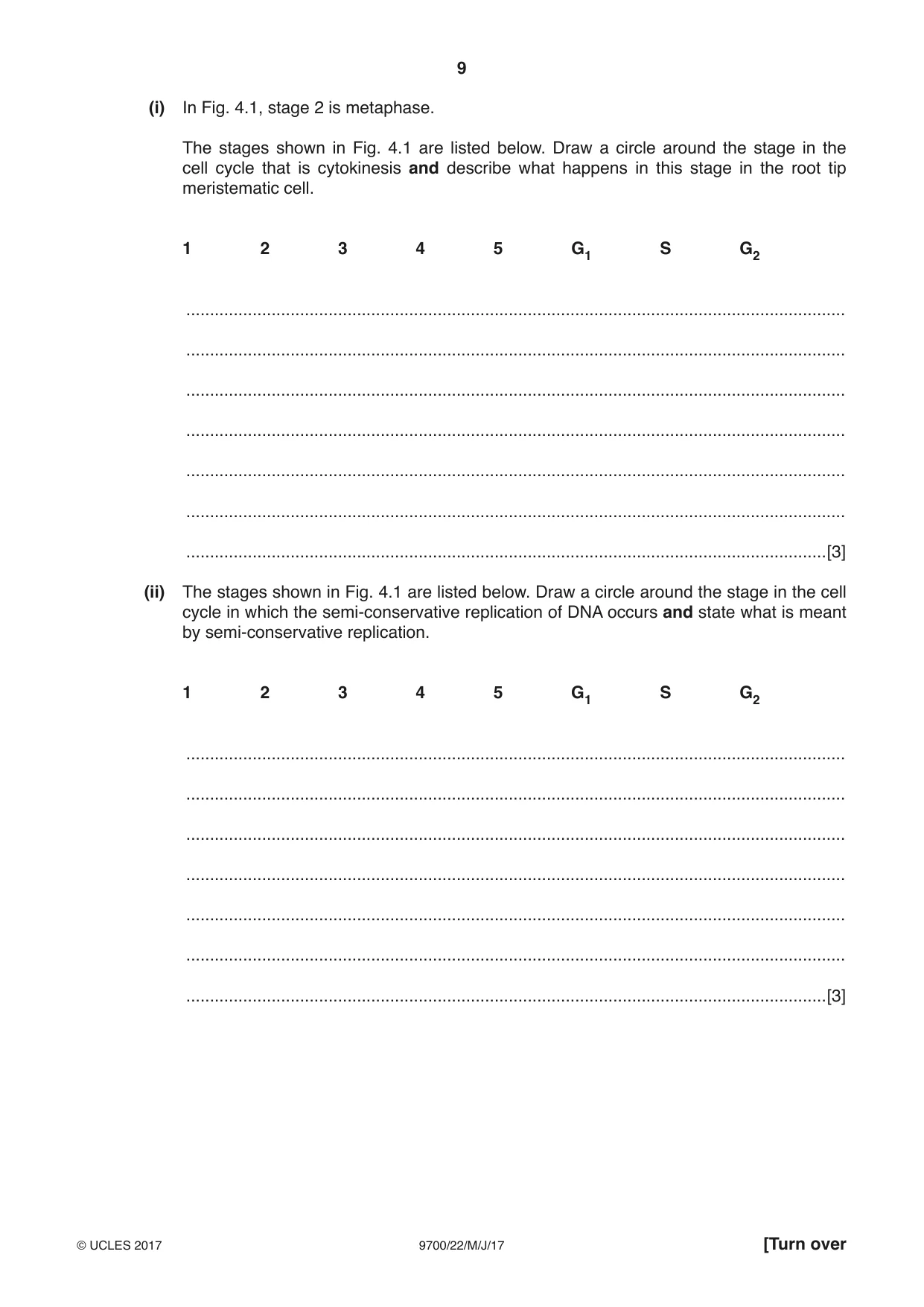
4 Meristematic tissue is found in the growing regions of plants, such as root tips. (a) Fig. 4.1 summarises a cell cycle for a meristematic cell in the root tip. The two phases of this cell cycle are shown: • interphase, which is divided into the G1, S and G2 stages • cell division, which is divided into stages 1–5. G2 1 cell division 2 S 3 4 Key 5 interphase interphase cell division G1 Fig. 4.1 (i) In Fig. 4.1, stage 2 is metaphase. The stages shown in Fig. 4.1 are listed below. Draw a circle around the stage in the cell cycle that is cytokinesis and describe what happens in this stage in the root tip meristematic cell. 1 2 3 4 5 G1 S G2 ........................................................................................................................................... ........................................................................................................................................... ........................................................................................................................................... ........................................................................................................................................... ........................................................................................................................................... ........................................................................................................................................... .......................................................................................................................................[3] (ii) The stages shown in Fig. 4.1 are listed below. Draw a circle around the stage in the cell cycle in which the semi-conservative replication of DNA occurs and state what is meant by semi-conservative replication. 1 2 3 4 5 G1 S G2 ........................................................................................................................................... ........................................................................................................................................... ........................................................................................................................................... ........................................................................................................................................... ........................................................................................................................................... ........................................................................................................................................... .......................................................................................................................................[3] (b) Meristematic cells have a similar role to stem cells found in animals. Suggest the role of a meristematic cell and explain the features that help it to carry out its role. ................................................................................................................................................... ................................................................................................................................................... ................................................................................................................................................... ................................................................................................................................................... ................................................................................................................................................... ................................................................................................................................................... ................................................................................................................................................... ...............................................................................................................................................[3] (c) Meristematic regions in the plant can sometimes be described as strong sinks. (i) State what is meant by a sink. ........................................................................................................................................... ........................................................................................................................................... .......................................................................................................................................[1] (ii) Suggest what is meant by a strong sink. ........................................................................................................................................... ........................................................................................................................................... .......................................................................................................................................[1] (d) Describe and explain how sucrose is loaded into phloem sieve tubes. ................................................................................................................................................... ................................................................................................................................................... ................................................................................................................................................... ................................................................................................................................................... ................................................................................................................................................... ................................................................................................................................................... ................................................................................................................................................... ................................................................................................................................................... ................................................................................................................................................... ................................................................................................................................................... ................................................................................................................................................... ................................................................................................................................................... ................................................................................................................................................... ................................................................................................................................................... ................................................................................................................................................... ...............................................................................................................................................[4] [Total: 15]
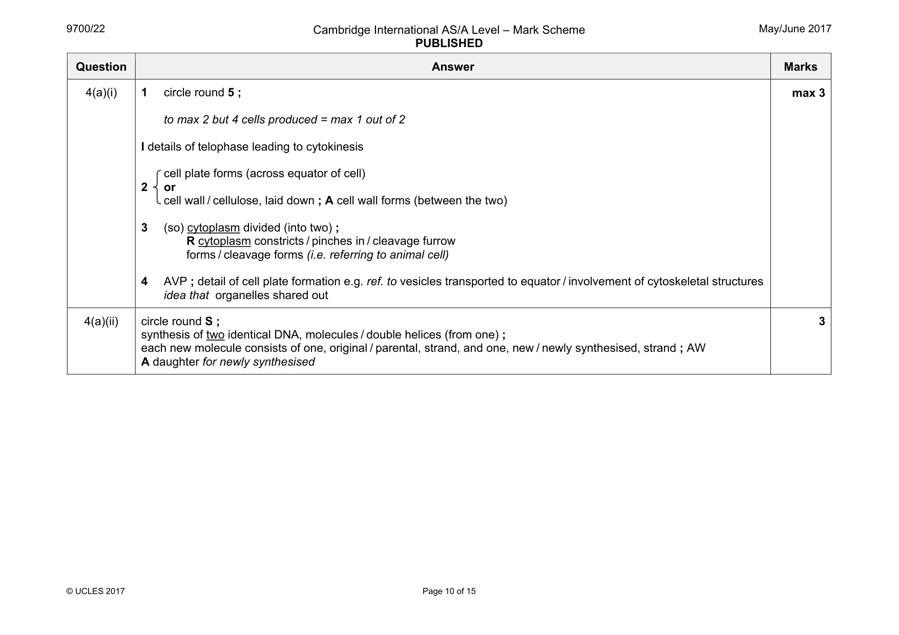
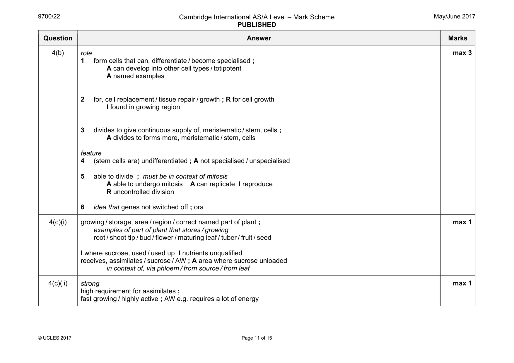
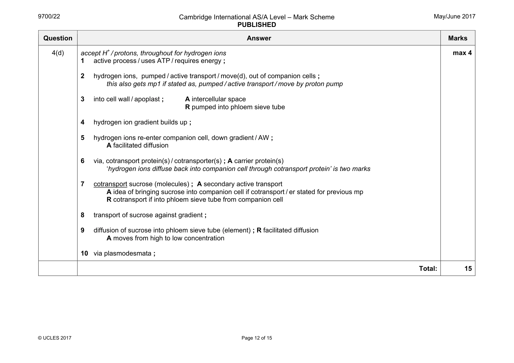
Mark scheme: 4(a)(i) 1 circle round 5 ; to max 2 but 4 cells produced = max 1 out of 2 I details of telophase leading to cytokinesis cell plate forms (across equator of cell) 2 or cell wall / cellulose, laid down ; A cell wall forms (between the two) 3 (so) cytoplasm divided (into two) ; R cytoplasm constricts / pinches in / cleavage furrow forms / cleavage forms (i.e. referring to animal cell) 4 AVP ; detail of cell plate formation e.g. ref. to vesicles transported to equator / involvement of cytoskeletal structures idea that organelles shared out max 3 4(a)(ii) circle round S ; synthesis of two identical DNA, molecules / double helices (from one) ; each new molecule consists of one, original / parental, strand, and one, new / newly synthesised, strand ; AW A daughter for newly synthesised 3 Question Answer Marks 4(b) role 1 form cells that can, differentiate / become specialised ; A can develop into other cell types / totipotent A named examples 2 for, cell replacement / tissue repair / growth ; R for cell growth I found in growing region 3 divides to give continuous supply of, meristematic / stem, cells ; A divides to forms more, meristematic / stem, cells feature 4 (stem cells are) undifferentiated ; A not specialised / unspecialised 5 able to divide ; must be in context of mitosis A able to undergo mitosis A can replicate I reproduce R uncontrolled division 6 idea that genes not switched off ; ora max 3 4(c)(i) growing / storage, area / region / correct named part of plant ; examples of part of plant that stores / growing root / shoot tip / bud / flower / maturing leaf / tuber / fruit / seed I where sucrose, used / used up I nutrients unqualified receives, assimilates / sucrose / AW ; A area where sucrose unloaded in context of, via phloem / from source / from leaf max 1 4(c)(ii) strong high requirement for assimilates ; fast growing / highly active ; AW e.g. requires a lot of energy max 1 Question Answer Marks 4(d) accept H+ / protons, throughout for hydrogen ions 1 active process / uses ATP / requires energy ; 2 hydrogen ions, pumped / active transport / move(d), out of companion cells ; this also gets mp1 if stated as, pumped / active transport / move by proton pump 3 into cell wall / apoplast ; A intercellular space R pumped into phloem sieve tube 4 hydrogen ion gradient builds up ; 5 hydrogen ions re-enter companion cell, down gradient / AW ; A facilitated diffusion 6 via, cotransport protein(s) / cotransporter(s) ; A carrier protein(s) ‘hydrogen ions diffuse back into companion cell through cotransport protein’ is two marks 7 cotransport sucrose (molecules) ; A secondary active transport A idea of bringing sucrose into companion cell if cotransport / er stated for previous mp R cotransport if into phloem sieve tube from companion cell 8 transport of sucrose against gradient ; 9 diffusion of sucrose into phloem sieve tube (element) ; R facilitated diffusion A moves from high to low concentration 10 via plasmodesmata ; max 4 Total: 15
More questions on Replication and division of nuclei and cells
Q5 · Some pathogens can enter the human body through the gas exchange system
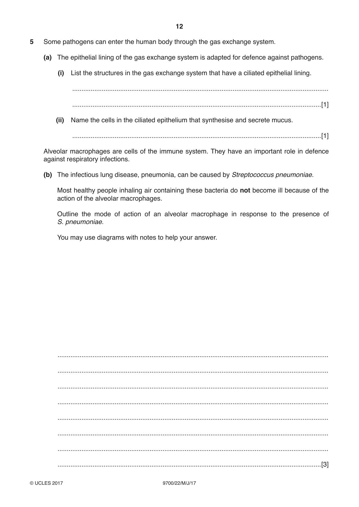
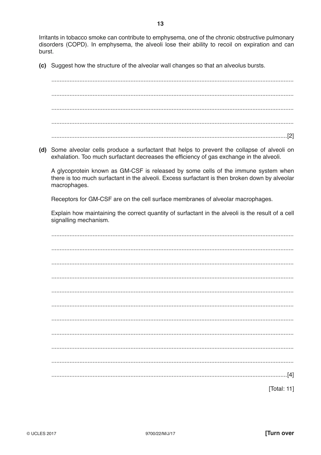
5 Some pathogens can enter the human body through the gas exchange system. (a) The epithelial lining of the gas exchange system is adapted for defence against pathogens. (i) List the structures in the gas exchange system that have a ciliated epithelial lining. ........................................................................................................................................... .......................................................................................................................................[1] (ii) Name the cells in the ciliated epithelium that synthesise and secrete mucus. .......................................................................................................................................[1] Alveolar macrophages are cells of the immune system. They have an important role in defence against respiratory infections. (b) The infectious lung disease, pneumonia, can be caused by Streptococcus pneumoniae. Most healthy people inhaling air containing these bacteria do not become ill because of the action of the alveolar macrophages. Outline the mode of action of an alveolar macrophage in response to the presence of S. pneumoniae. You may use diagrams with notes to help your answer. ................................................................................................................................................... ................................................................................................................................................... ................................................................................................................................................... ................................................................................................................................................... ................................................................................................................................................... ................................................................................................................................................... ................................................................................................................................................... ...............................................................................................................................................[3] Irritants in tobacco smoke can contribute to emphysema, one of the chronic obstructive pulmonary disorders (COPD). In emphysema, the alveoli lose their ability to recoil on expiration and can burst. (c) Suggest how the structure of the alveolar wall changes so that an alveolus bursts. ................................................................................................................................................... ................................................................................................................................................... ................................................................................................................................................... ................................................................................................................................................... ...............................................................................................................................................[2] (d) Some alveolar cells produce a surfactant that helps to prevent the collapse of alveoli on exhalation. Too much surfactant decreases the efficiency of gas exchange in the alveoli. A glycoprotein known as GM-CSF is released by some cells of the immune system when there is too much surfactant in the alveoli. Excess surfactant is then broken down by alveolar macrophages. Receptors for GM-CSF are on the cell surface membranes of alveolar macrophages. Explain how maintaining the correct quantity of surfactant in the alveoli is the result of a cell signalling mechanism. ................................................................................................................................................... ................................................................................................................................................... ................................................................................................................................................... ................................................................................................................................................... ................................................................................................................................................... ................................................................................................................................................... ................................................................................................................................................... ................................................................................................................................................... ................................................................................................................................................... ................................................................................................................................................... ...............................................................................................................................................[4] [Total: 11]
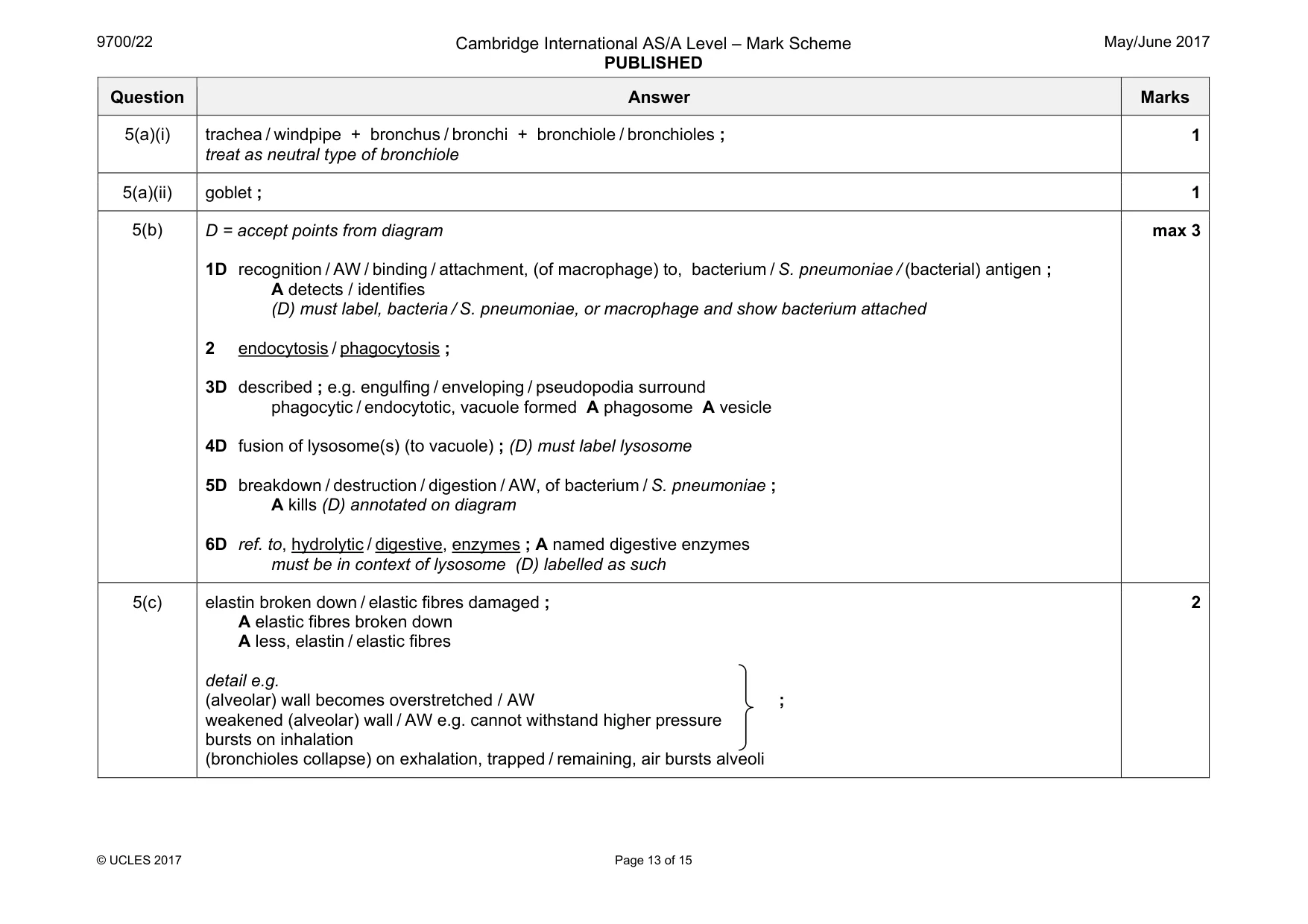
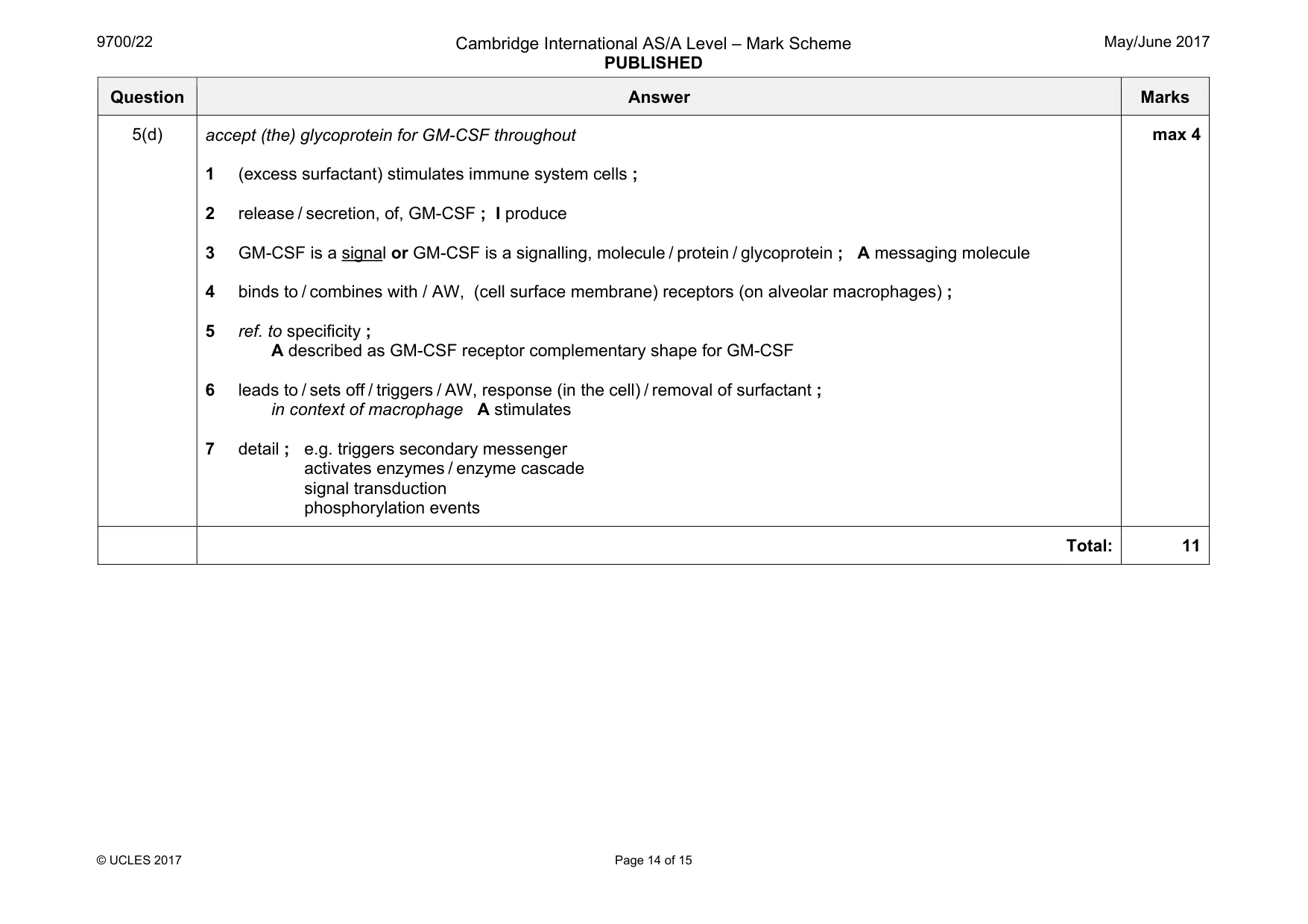
Mark scheme: 5(a)(i) trachea / windpipe + bronchus / bronchi + bronchiole / bronchioles ; treat as neutral type of bronchiole 1 5(a)(ii) goblet ; 1 5(b) D = accept points from diagram 1D recognition / AW / binding / attachment, (of macrophage) to, bacterium / S. pneumoniae / (bacterial) antigen ; A detects / identifies (D) must label, bacteria / S. pneumoniae, or macrophage and show bacterium attached 2 endocytosis / phagocytosis ; 3D described ; e.g. engulfing / enveloping / pseudopodia surround phagocytic / endocytotic, vacuole formed A phagosome A vesicle 4D fusion of lysosome(s) (to vacuole) ; (D) must label lysosome 5D breakdown / destruction / digestion / AW, of bacterium / S. pneumoniae ; A kills (D) annotated on diagram 6D ref. to, hydrolytic / digestive, enzymes ; A named digestive enzymes must be in context of lysosome (D) labelled as such max 3 5(c) elastin broken down / elastic fibres damaged ; A elastic fibres broken down A less, elastin / elastic fibres detail e.g. (alveolar) wall becomes overstretched / AW ; weakened (alveolar) wall / AW e.g. cannot withstand higher pressure bursts on inhalation (bronchioles collapse) on exhalation, trapped / remaining, air bursts alveoli 2 Question Answer Marks 5(d) accept (the) glycoprotein for GM-CSF throughout 1 (excess surfactant) stimulates immune system cells ; 2 release / secretion, of, GM-CSF ; I produce 3 GM-CSF is a signal or GM-CSF is a signalling, molecule / protein / glycoprotein ; A messaging molecule 4 binds to / combines with / AW, (cell surface membrane) receptors (on alveolar macrophages) ; 5 ref. to specificity ; A described as GM-CSF receptor complementary shape for GM-CSF 6 leads to / sets off / triggers / AW, response (in the cell) / removal of surfactant ; in context of macrophage A stimulates 7 detail ; e.g. triggers secondary messenger activates enzymes / enzyme cascade signal transduction phosphorylation events max 4 Total: 11
Q6 · As a result of transcription and translation, a polypeptide chain is produced
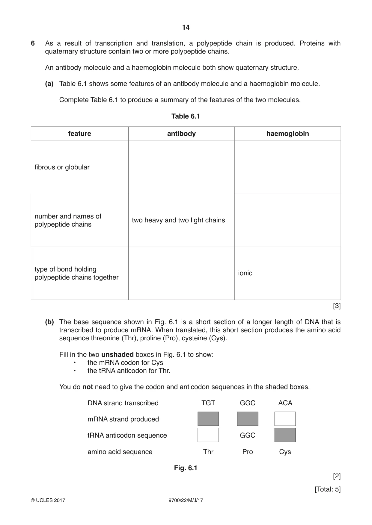
6 As a result of transcription and translation, a polypeptide chain is produced. Proteins with quaternary structure contain two or more polypeptide chains. An antibody molecule and a haemoglobin molecule both show quaternary structure. (a) Table 6.1 shows some features of an antibody molecule and a haemoglobin molecule. Complete Table 6.1 to produce a summary of the features of the two molecules. Table 6.1 feature antibody haemoglobin fibrous or globular number and names of two heavy and two light chains polypeptide chains type of bond holding ionic polypeptide chains together [3] (b) The base sequence shown in Fig. 6.1 is a short section of a longer length of DNA that is transcribed to produce mRNA. When translated, this short section produces the amino acid sequence threonine (Thr), proline (Pro), cysteine (Cys). Fill in the two unshaded boxes in Fig. 6.1 to show: • the mRNA codon for Cys • the tRNA anticodon for Thr. You do not need to give the codon and anticodon sequences in the shaded boxes. DNA strand transcribed TGT GGC ACA mRNA strand produced tRNA anticodon sequence GGC amino acid sequence Thr Pro Cys Fig. 6.1 [2]
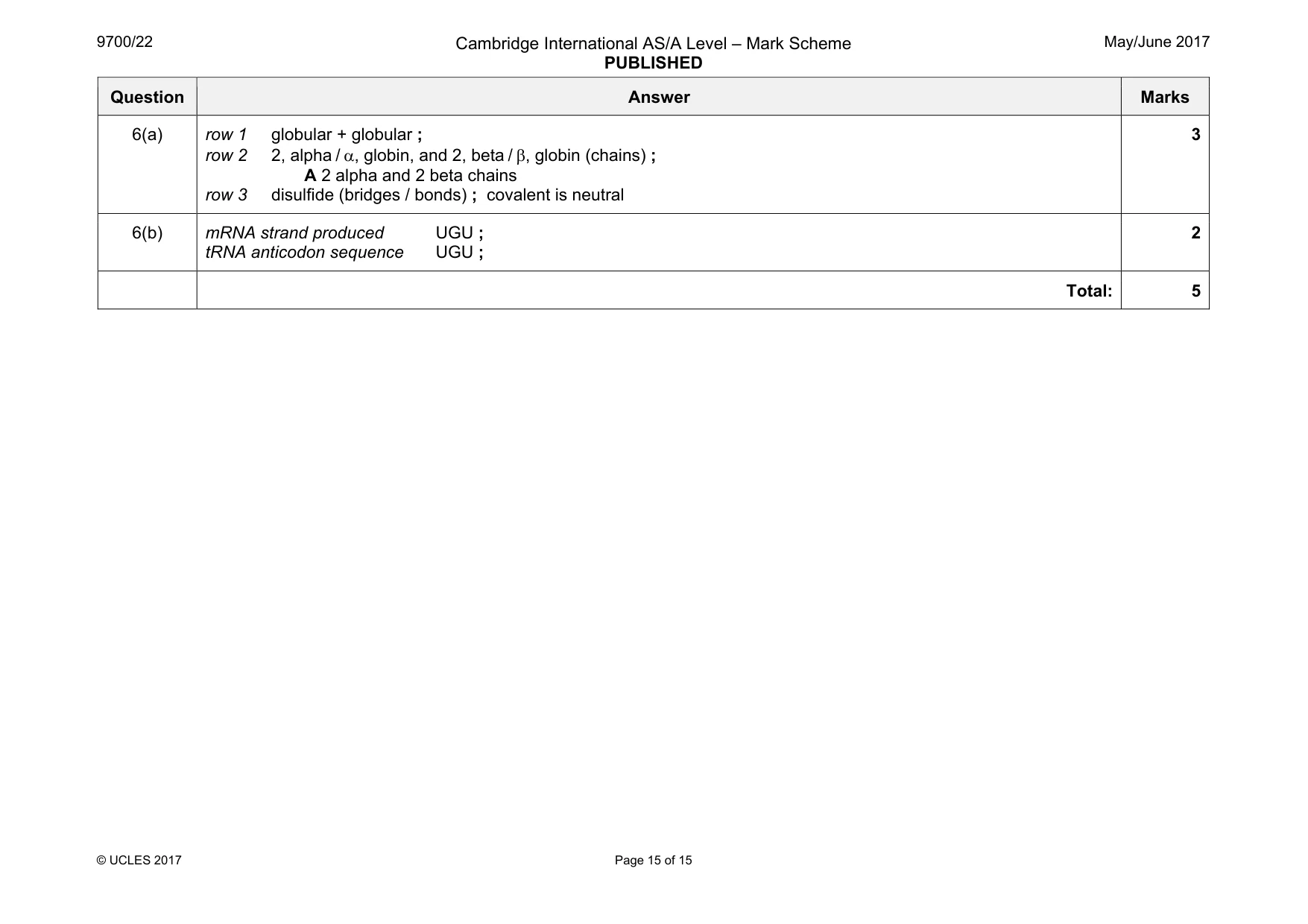
Mark scheme: 6(a) row 1 globular + globular ; row 2 2, alpha / α, globin, and 2, beta / β, globin (chains) ; A 2 alpha and 2 beta chains row 3 disulfide (bridges / bonds) ; covalent is neutral 3 6(b) mRNA strand produced UGU ; tRNA anticodon sequence UGU ; 2 Total: 5
What was in this paper
The subtopics covered by these 6 questions, and how many questions each got. Open one in a new tab to see every Cambridge question on it.
What you needed in this session
Cambridge’s own grade thresholds for 2017 May/June, Paper 2 · Variant 2. A higher threshold means an easier paper — the bar moves with how the cohort did.