Cambridge A Level Biology 9700 — 2017 May/June Paper 2 · Variant 1
9700/21/M/J/17 · 6 questions · 60 marks · ≈68 min
The question paper and its mark scheme, free to read here and free to download. This is Cambridge’s own paper, exactly as it was sat.
Question paper16 pages
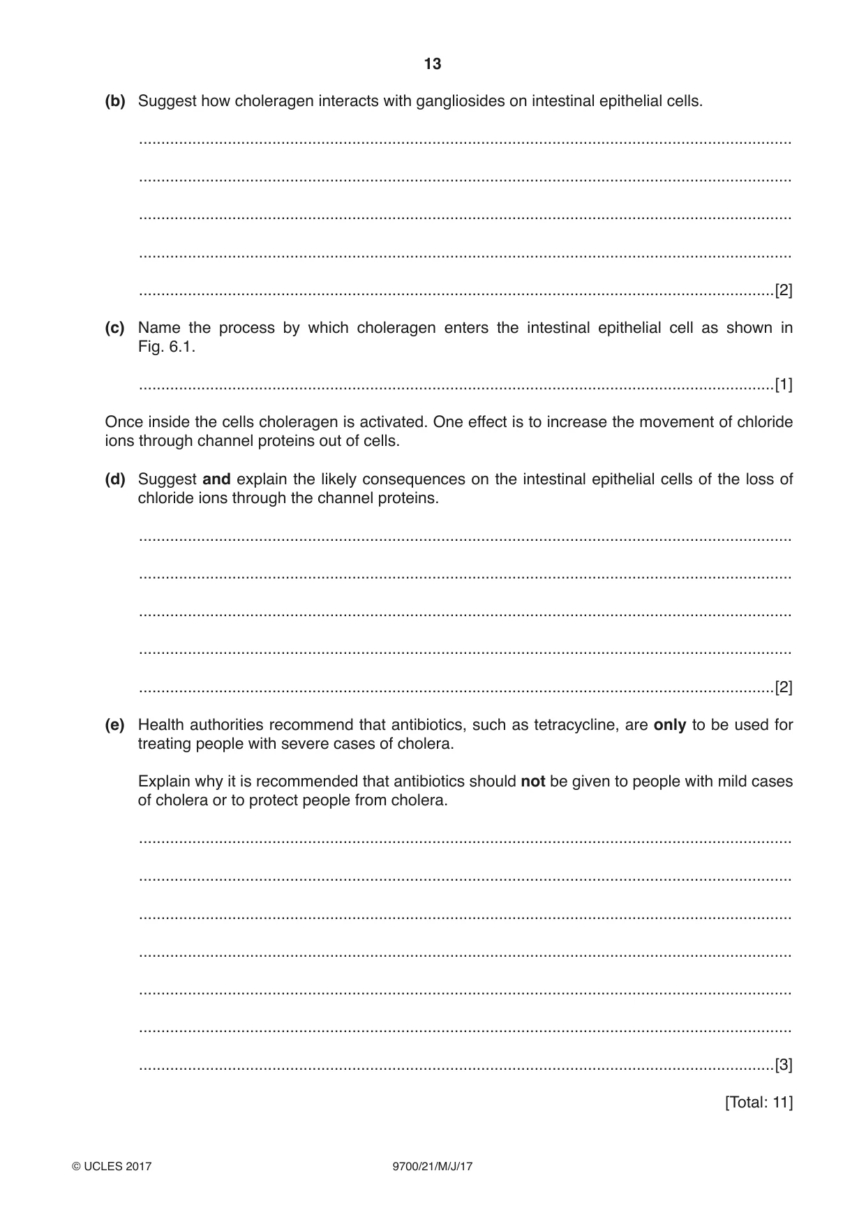


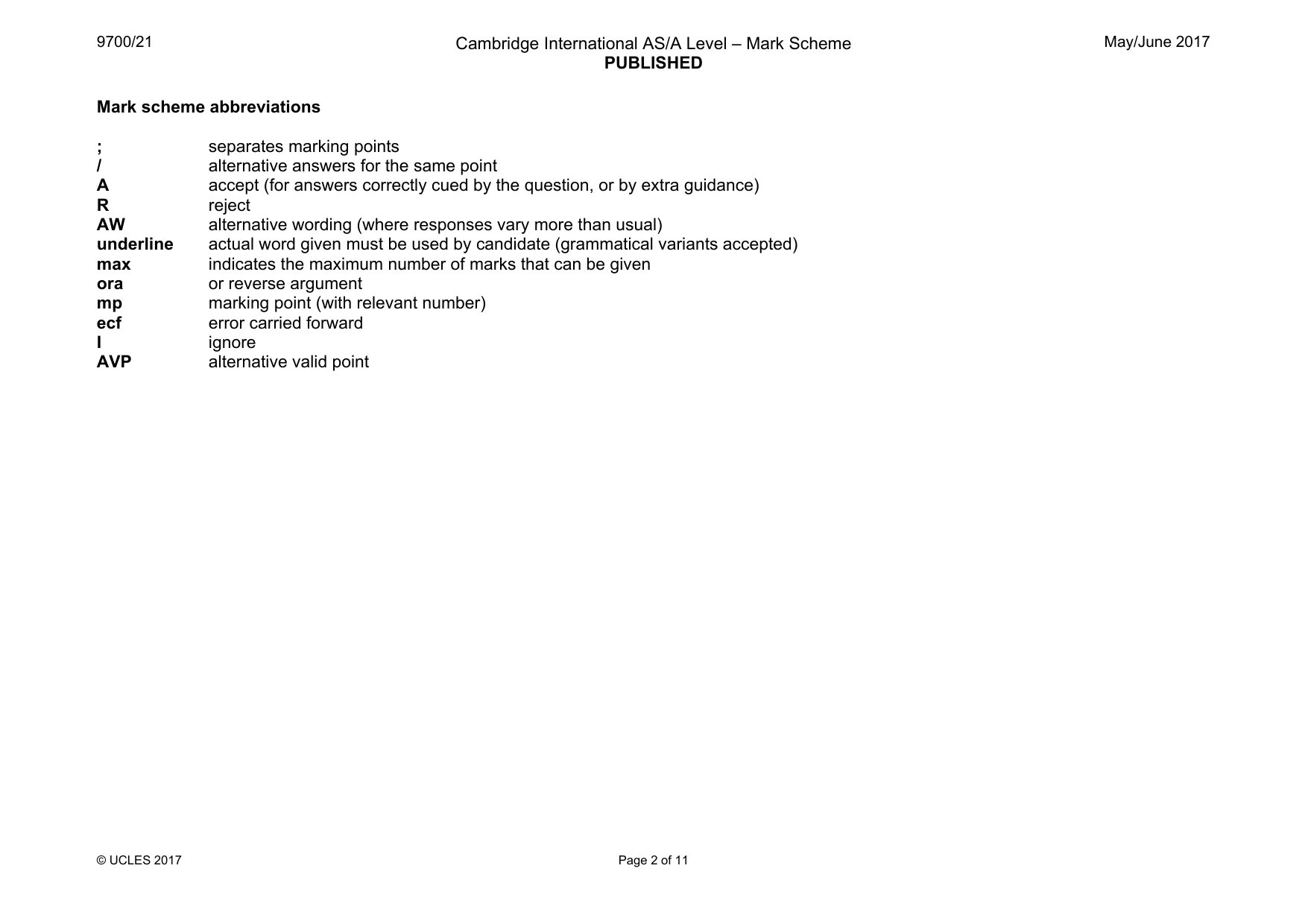
Mark scheme11 pages
Answers below. Sit the paper first if you are practising.
Questions as text
Q1 · Each of the statements A to D describes a structure found in eukaryotic cells
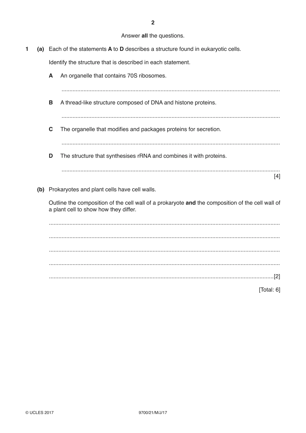
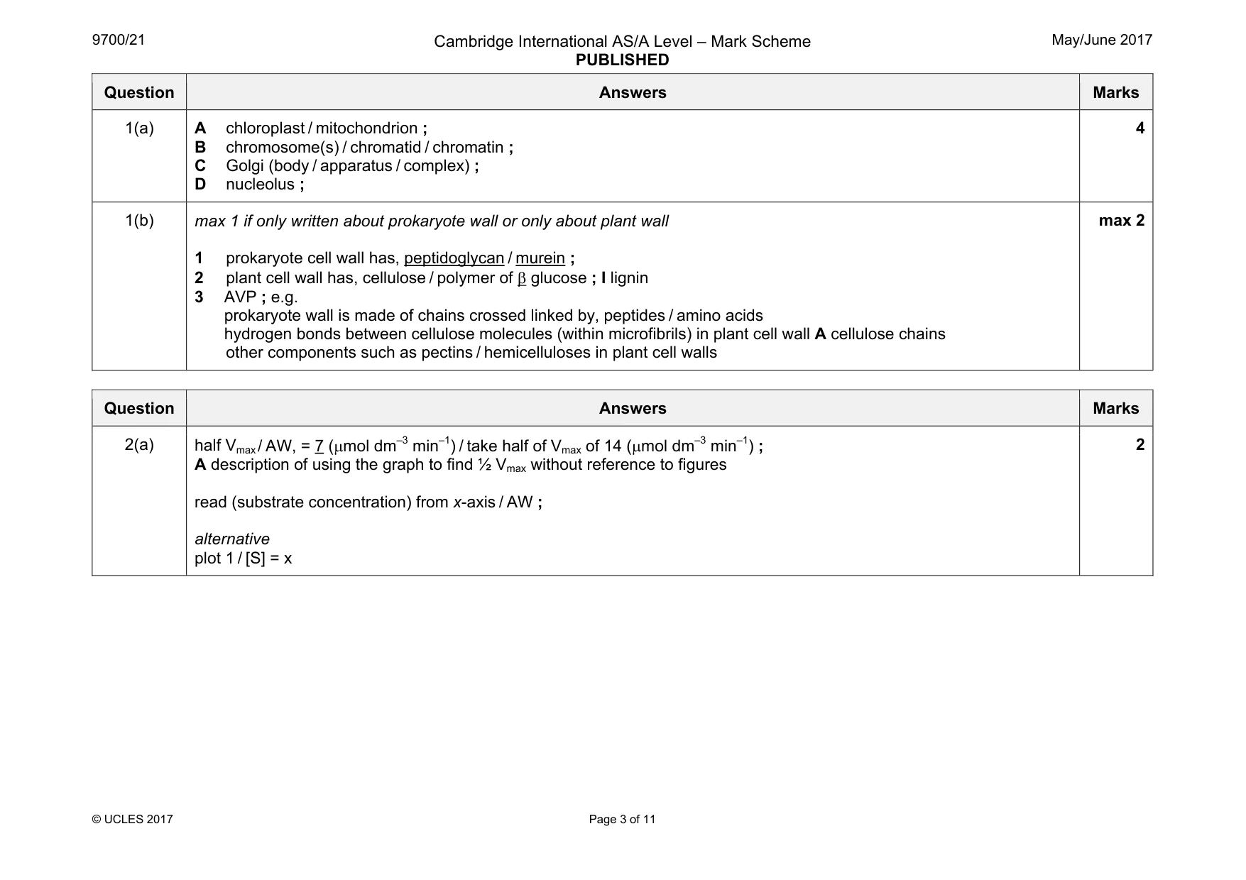
1 (a) Each of the statements A to D describes a structure found in eukaryotic cells. Identify the structure that is described in each statement. A An organelle that contains 70S ribosomes. ........................................................................................................................................... B A thread-like structure composed of DNA and histone proteins. ........................................................................................................................................... C The organelle that modifies and packages proteins for secretion. ........................................................................................................................................... D The structure that synthesises rRNA and combines it with proteins. ........................................................................................................................................... [4] (b) Prokaryotes and plant cells have cell walls. Outline the composition of the cell wall of a prokaryote and the composition of the cell wall of a plant cell to show how they differ. ................................................................................................................................................... ................................................................................................................................................... ................................................................................................................................................... ................................................................................................................................................... ...............................................................................................................................................[2] [Total: 6]
Mark scheme: 1(a) A chloroplast / mitochondrion ; B chromosome(s) / chromatid / chromatin ; C Golgi (body / apparatus / complex) ; D nucleolus ; 4 1(b) max 1 if only written about prokaryote wall or only about plant wall 1 prokaryote cell wall has, peptidoglycan / murein ; 2 plant cell wall has, cellulose / polymer of β glucose ; I lignin 3 AVP ; e.g. prokaryote wall is made of chains crossed linked by, peptides / amino acids hydrogen bonds between cellulose molecules (within microfibrils) in plant cell wall A cellulose chains other components such as pectins / hemicelluloses in plant cell walls max 2
More questions on Cells as the basic units of living organisms
Q2 · Phosphatases are enzymes that catalyse the removal of phosphate groups from organic…
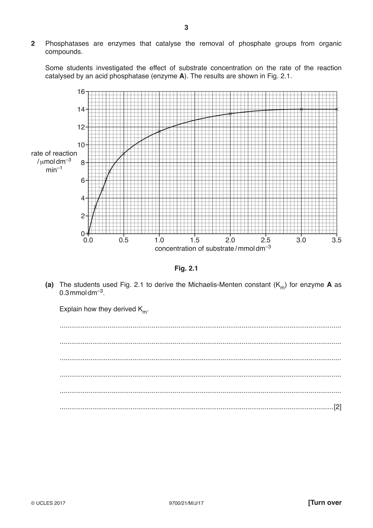
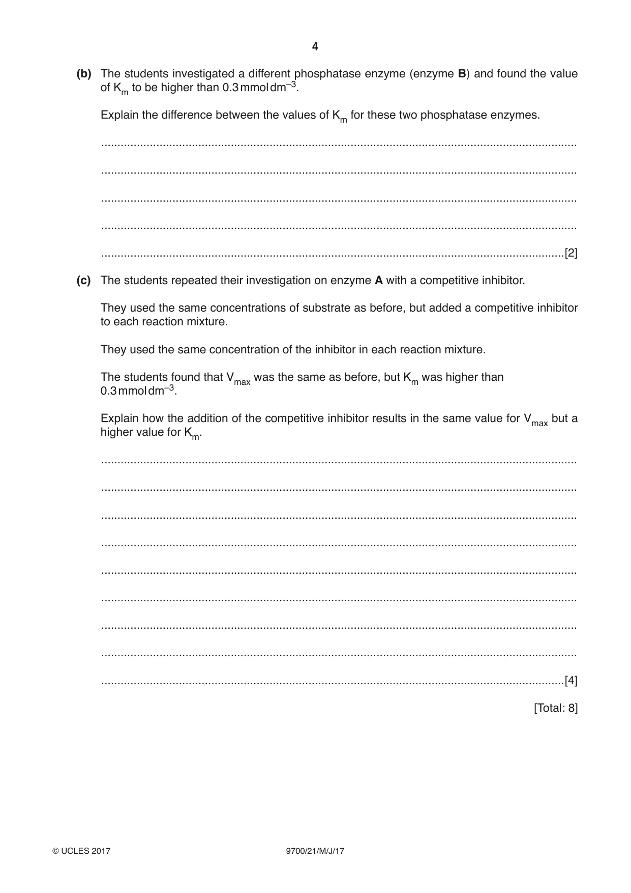
2 Phosphatases are enzymes that catalyse the removal of phosphate groups from organic compounds. Some students investigated the effect of substrate concentration on the rate of the reaction catalysed by an acid phosphatase (enzyme A). The results are shown in Fig. 2.1. 16 14 12 10 rate of reaction / μmol dm–3 8 min–1 6 4 2 0 0.0 0.5 1.0 1.5 2.0 2.5 3.0 3.5 concentration of substrate / mmol dm–3 Fig. 2.1 (a) The students used Fig. 2.1 to derive the Michaelis-Menten constant (Km) for enzyme A as 0.3 mmol dm–3. Explain how they derived Km. ................................................................................................................................................... ................................................................................................................................................... ................................................................................................................................................... ................................................................................................................................................... ................................................................................................................................................... ...............................................................................................................................................[2] (b) The students investigated a different phosphatase enzyme (enzyme B) and found the value of Km to be higher than 0.3 mmol dm–3. Explain the difference between the values of Km for these two phosphatase enzymes. ................................................................................................................................................... ................................................................................................................................................... ................................................................................................................................................... ................................................................................................................................................... ...............................................................................................................................................[2] (c) The students repeated their investigation on enzyme A with a competitive inhibitor. They used the same concentrations of substrate as before, but added a competitive inhibitor to each reaction mixture. They used the same concentration of the inhibitor in each reaction mixture. The students found that Vmax was the same as before, but Km was higher than 0.3 mmol dm–3. Explain how the addition of the competitive inhibitor results in the same value for Vmax but a higher value for Km. ................................................................................................................................................... ................................................................................................................................................... ................................................................................................................................................... ................................................................................................................................................... ................................................................................................................................................... ................................................................................................................................................... ................................................................................................................................................... ................................................................................................................................................... ...............................................................................................................................................[4] [Total: 8]
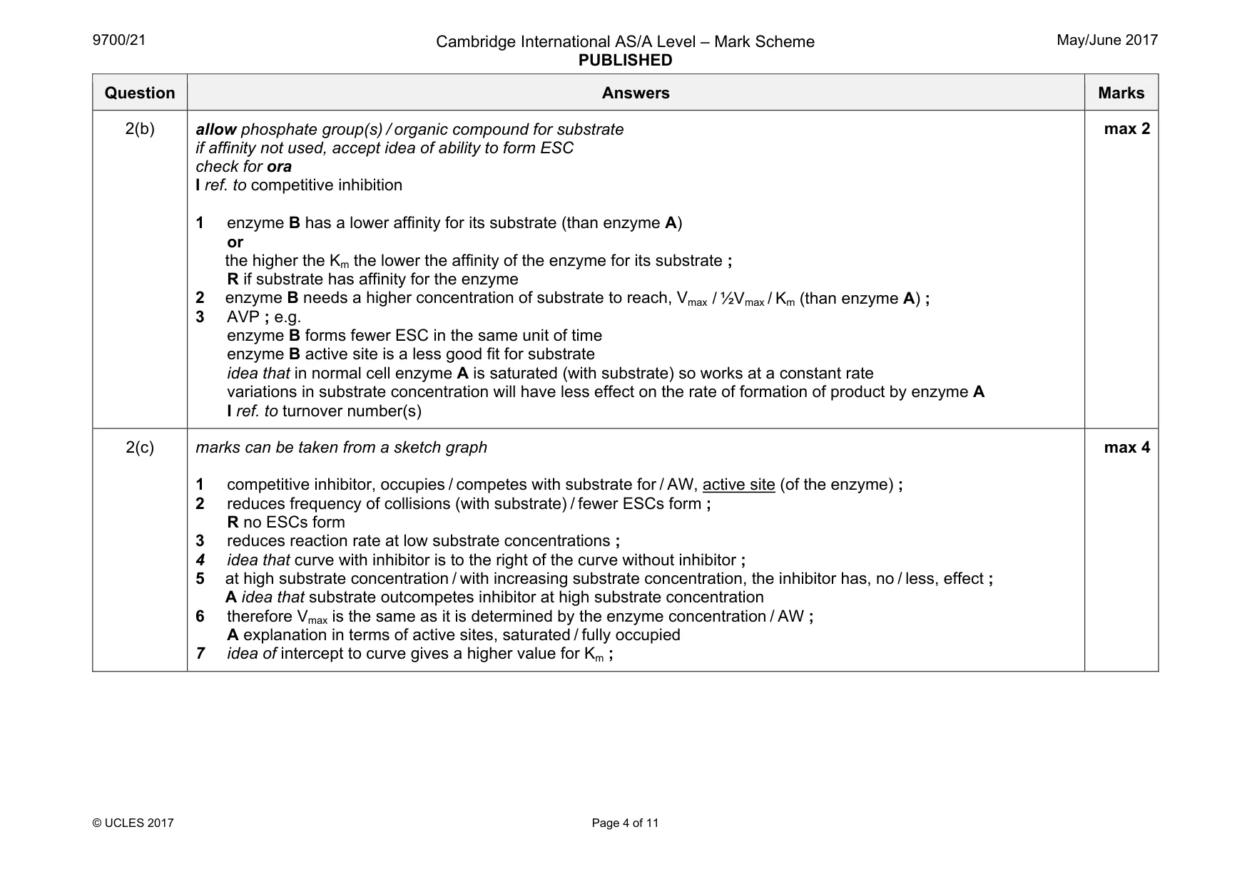
Mark scheme: 2(a) A description of using the graph to find ½ Vmax without reference to figures read (substrate concentration) from x-axis / AW ; alternative plot 1 / [S] = x 2 Question Answers Marks 2(b) allow phosphate group(s) / organic compound for substrate if affinity not used, accept idea of ability to form ESC check for ora I ref. to competitive inhibition 1 enzyme B has a lower affinity for its substrate (than enzyme A) or the higher the Km the lower the affinity of the enzyme for its substrate ; R if substrate has affinity for the enzyme 2 enzyme B needs a higher concentration of substrate to reach, Vmax / ½Vmax / Km (than enzyme A) ; 3 AVP ; e.g. enzyme B forms fewer ESC in the same unit of time enzyme B active site is a less good fit for substrate idea that in normal cell enzyme A is saturated (with substrate) so works at a constant rate variations in substrate concentration will have less effect on the rate of formation of product by enzyme A I ref. to turnover number(s) max 2 2(c) marks can be taken from a sketch graph 1 competitive inhibitor, occupies / competes with substrate for / AW, active site (of the enzyme) ; 2 reduces frequency of collisions (with substrate) / fewer ESCs form ; R no ESCs form 3 reduces reaction rate at low substrate concentrations ; 4 idea that curve with inhibitor is to the right of the curve without inhibitor ; 5 at high substrate concentration / with increasing substrate concentration, the inhibitor has, no / less, effect ; A idea that substrate outcompetes inhibitor at high substrate concentration 6 therefore Vmax is the same as it is determined by the enzyme concentration / AW ; A explanation in terms of active sites, saturated / fully occupied 7 idea of intercept to curve gives a higher value for Km ; max 4
Q3 · A diagram that shows the structure of an antibody molecule
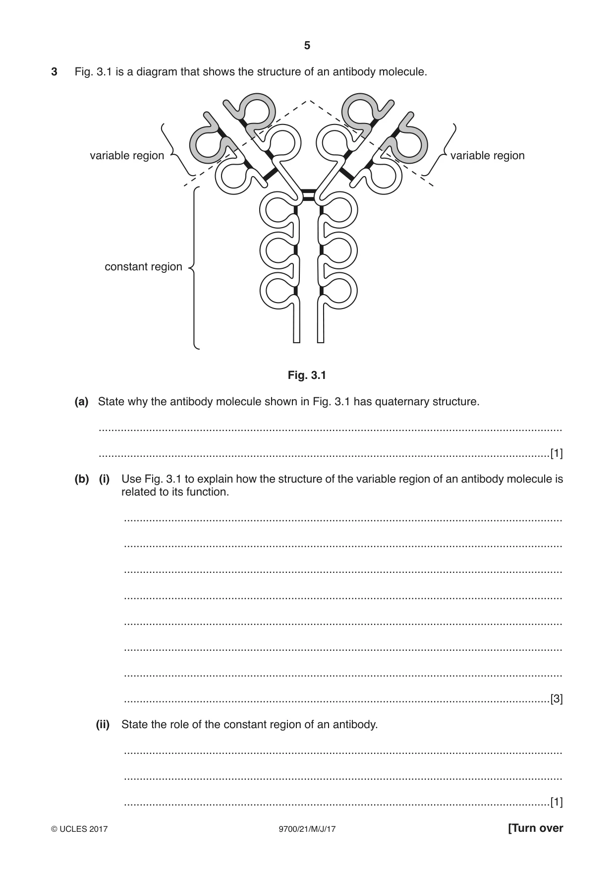
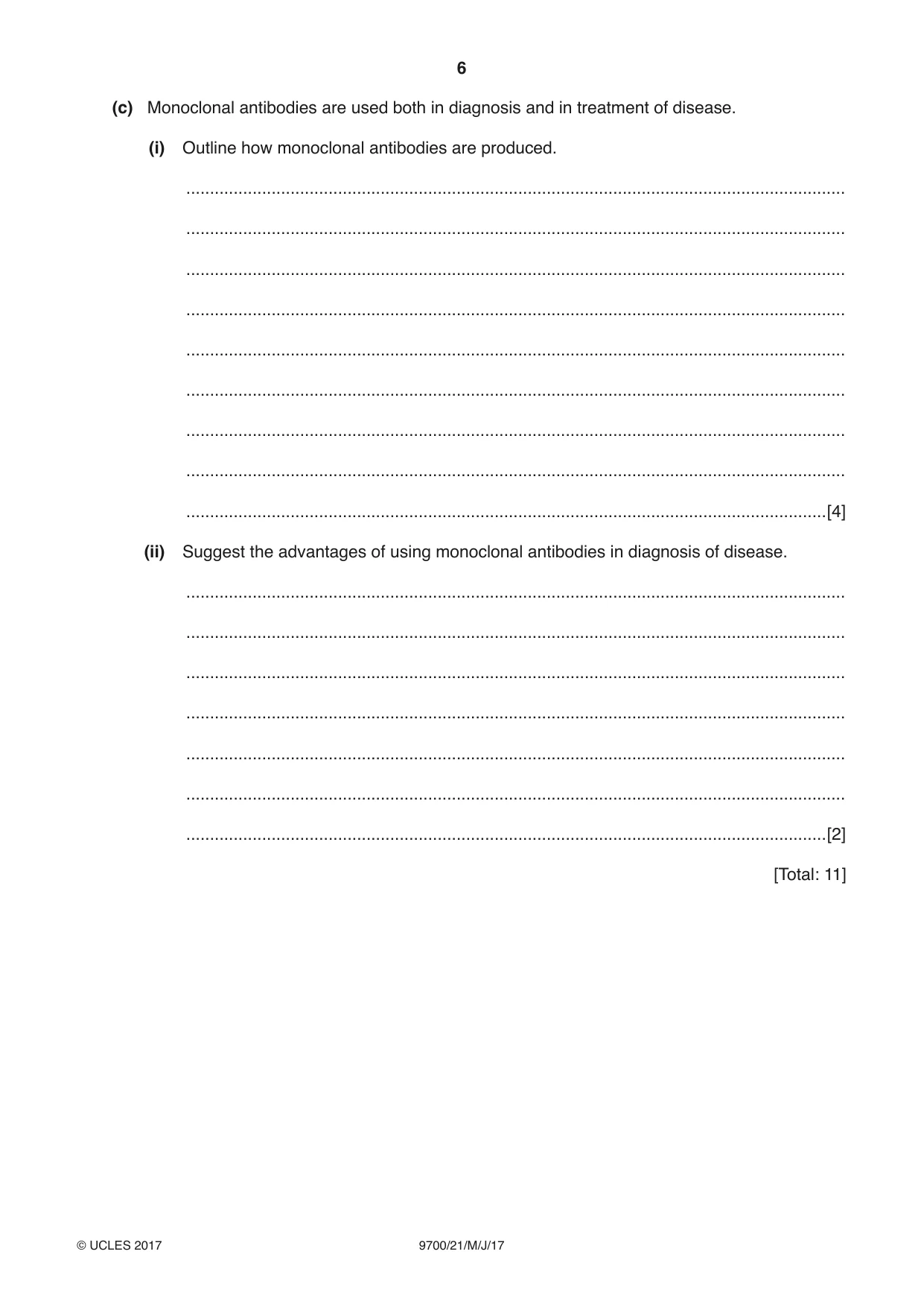
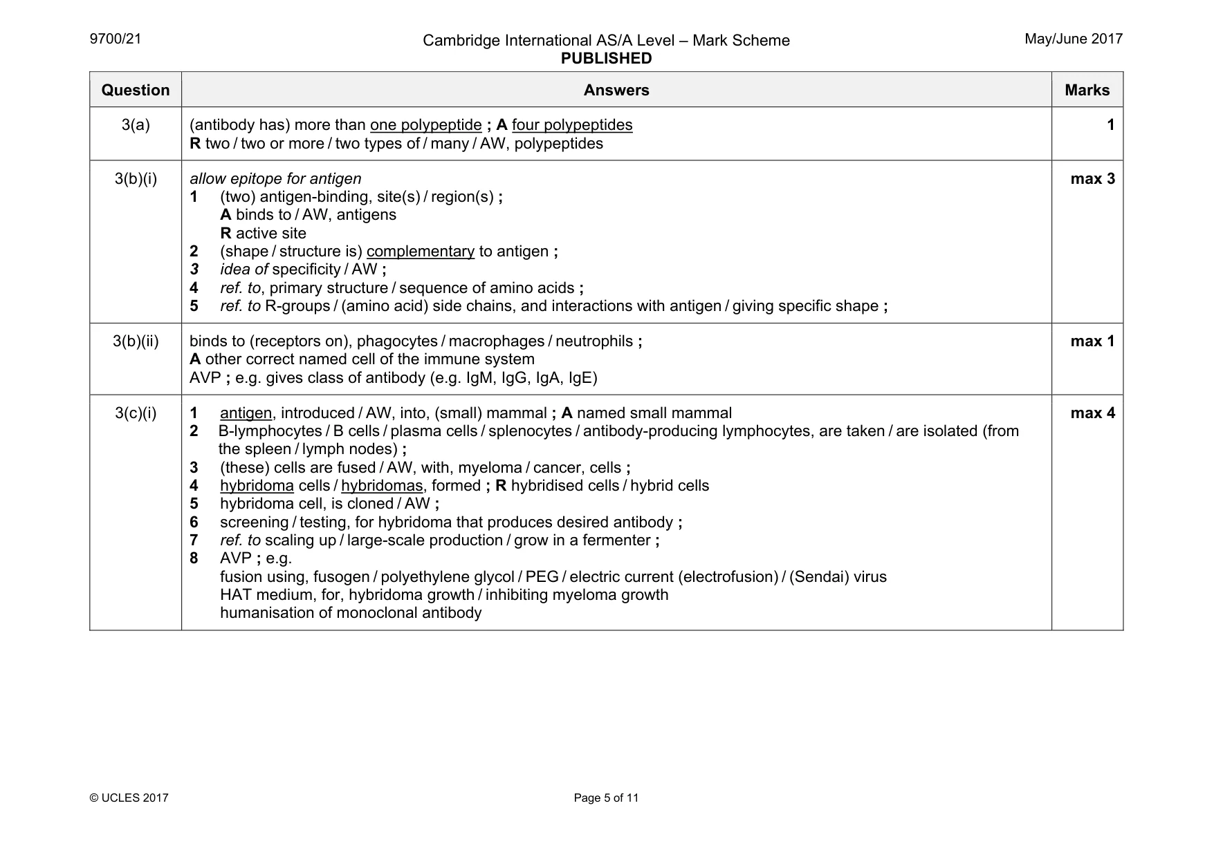
3 Fig. 3.1 is a diagram that shows the structure of an antibody molecule. variable region variable region constant region Fig. 3.1 (a) State why the antibody molecule shown in Fig. 3.1 has quaternary structure. ................................................................................................................................................... ...............................................................................................................................................[1] (b) (i) Use Fig. 3.1 to explain how the structure of the variable region of an antibody molecule is related to its function. ........................................................................................................................................... ........................................................................................................................................... ........................................................................................................................................... ........................................................................................................................................... ........................................................................................................................................... ........................................................................................................................................... ........................................................................................................................................... .......................................................................................................................................[3] (ii) State the role of the constant region of an antibody. ........................................................................................................................................... ........................................................................................................................................... .......................................................................................................................................[1] (c) Monoclonal antibodies are used both in diagnosis and in treatment of disease. (i) Outline how monoclonal antibodies are produced. ........................................................................................................................................... ........................................................................................................................................... ........................................................................................................................................... ........................................................................................................................................... ........................................................................................................................................... ........................................................................................................................................... ........................................................................................................................................... ........................................................................................................................................... .......................................................................................................................................[4] (ii) Suggest the advantages of using monoclonal antibodies in diagnosis of disease. ........................................................................................................................................... ........................................................................................................................................... ........................................................................................................................................... ........................................................................................................................................... ........................................................................................................................................... ........................................................................................................................................... .......................................................................................................................................[2] [Total: 11]
Mark scheme: 3(a) (antibody has) more than one polypeptide ; A four polypeptides R two / two or more / two types of / many / AW, polypeptides 1 3(b)(i) allow epitope for antigen 1 (two) antigen-binding, site(s) / region(s) ; A binds to / AW, antigens R active site 2 (shape / structure is) complementary to antigen ; 3 idea of specificity / AW ; 4 ref. to, primary structure / sequence of amino acids ; 5 ref. to R-groups / (amino acid) side chains, and interactions with antigen / giving specific shape ; max 3 3(b)(ii) binds to (receptors on), phagocytes / macrophages / neutrophils ; A other correct named cell of the immune system AVP ; e.g. gives class of antibody (e.g. IgM, IgG, IgA, IgE) max 1 3(c)(i) 1 antigen, introduced / AW, into, (small) mammal ; A named small mammal 2 B-lymphocytes / B cells / plasma cells / splenocytes / antibody-producing lymphocytes, are taken / are isolated (from the spleen / lymph nodes) ; 3 (these) cells are fused / AW, with, myeloma / cancer, cells ; 4 hybridoma cells / hybridomas, formed ; R hybridised cells / hybrid cells 5 hybridoma cell, is cloned / AW ; 6 screening / testing, for hybridoma that produces desired antibody ; 7 ref. to scaling up / large-scale production / grow in a fermenter ; 8 AVP ; e.g. fusion using, fusogen / polyethylene glycol / PEG / electric current (electrofusion) / (Sendai) virus HAT medium, for, hybridoma growth / inhibiting myeloma growth humanisation of monoclonal antibody max 4 Question Answers Marks 3(c)(ii) I suggestions for treatment 1 monoclonal antibodies used all have the same specificity ; R ‘are specific’ unqualified 2 detect only one, antigen / epitope ; 3 can distinguish between different, pathogens / strains of, pathogens ; A types of cancer cells 4 can be, labelled / tagged / marked / AW ; e.g. with fluorescent label 5 monoclonal antibodies can detect location of, tissues expressing antigen / cancer cells / blood clots ; A idea of locating areas of infection 6 fast(er) (diagnosis) ; 7 can detect antibody levels (e.g. HIV) ; 8 AVP ; e.g. some pathogens cannot be cultured I ref. to cost max 2
Q4 · Part of a DNA molecule
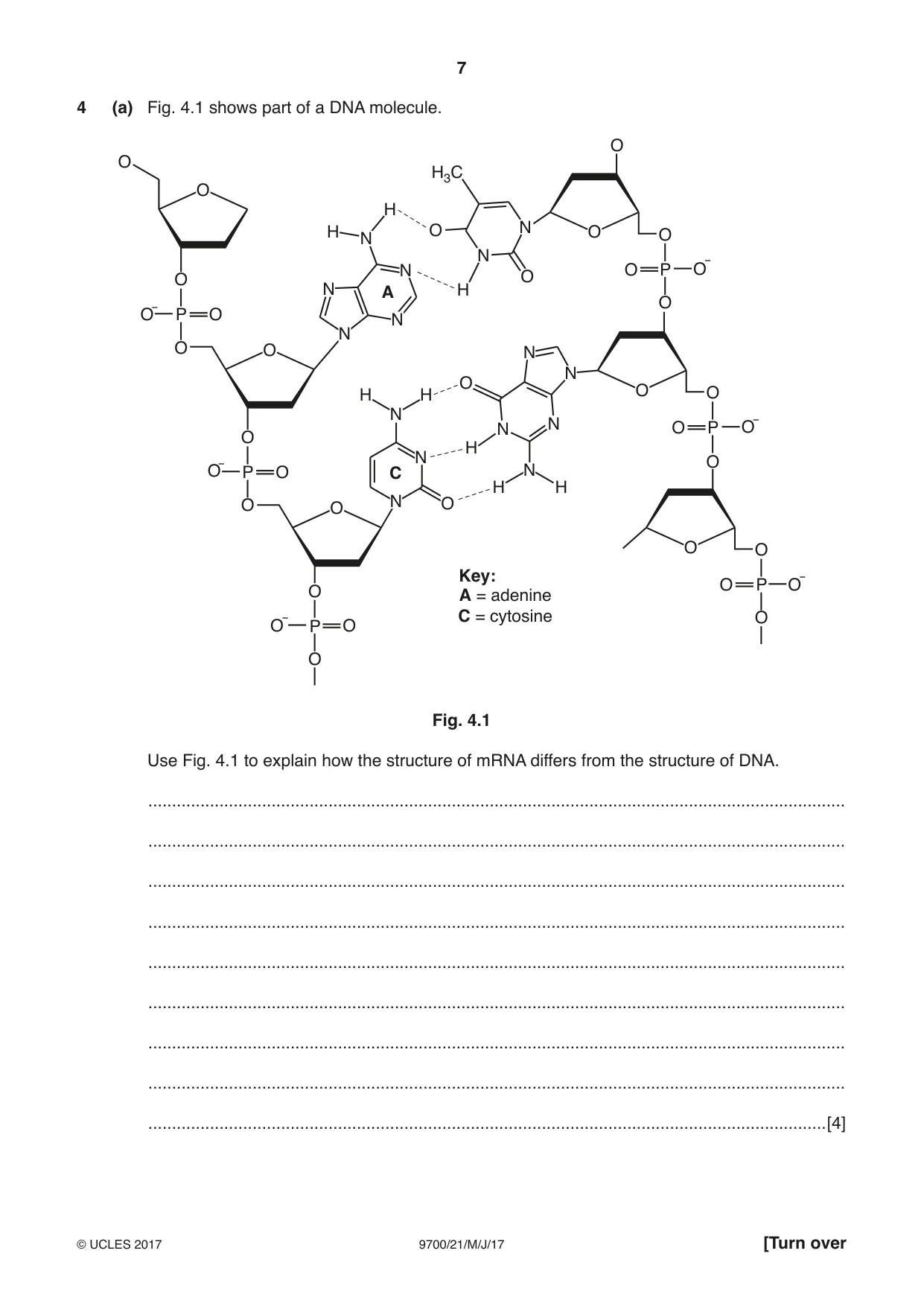
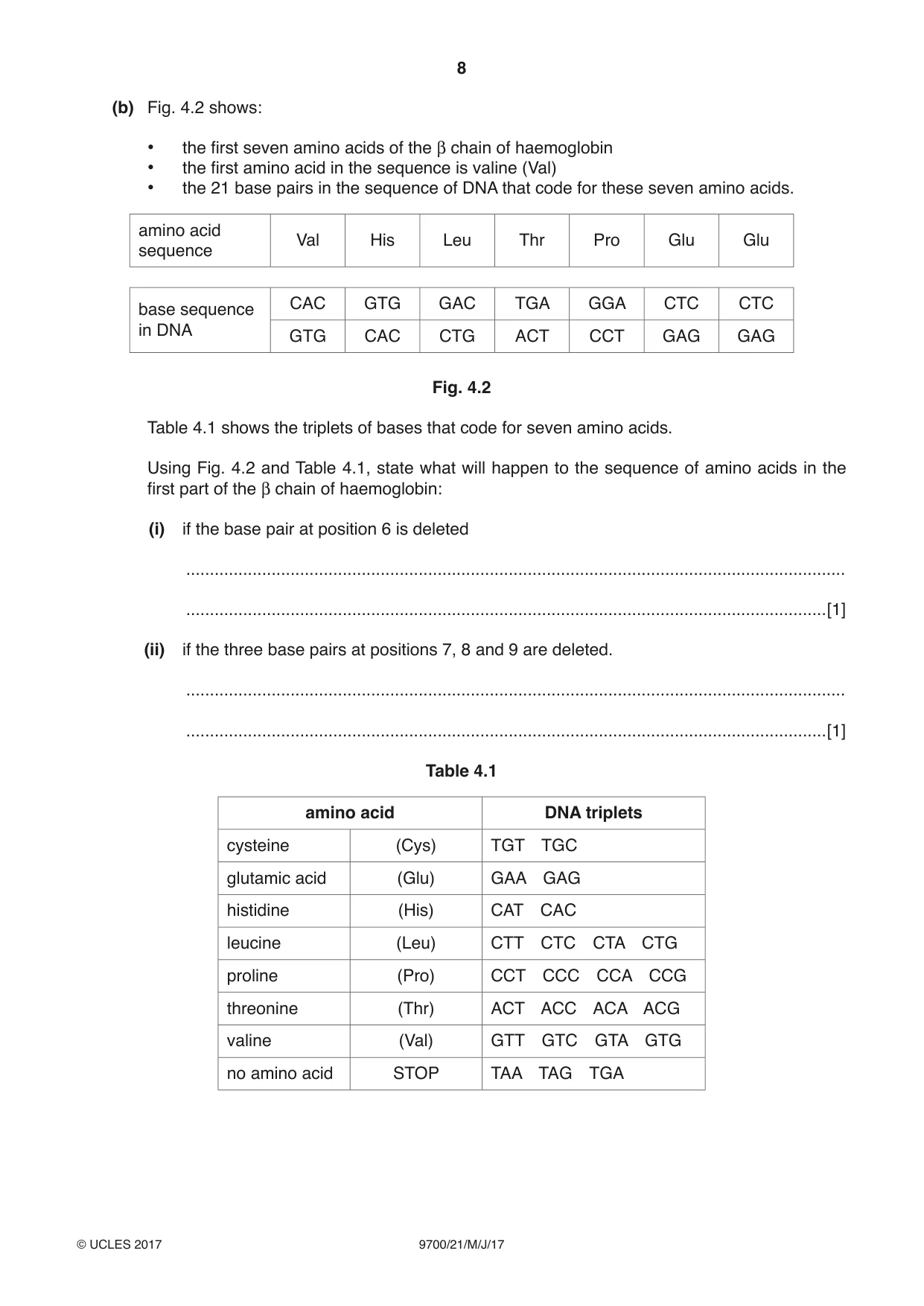
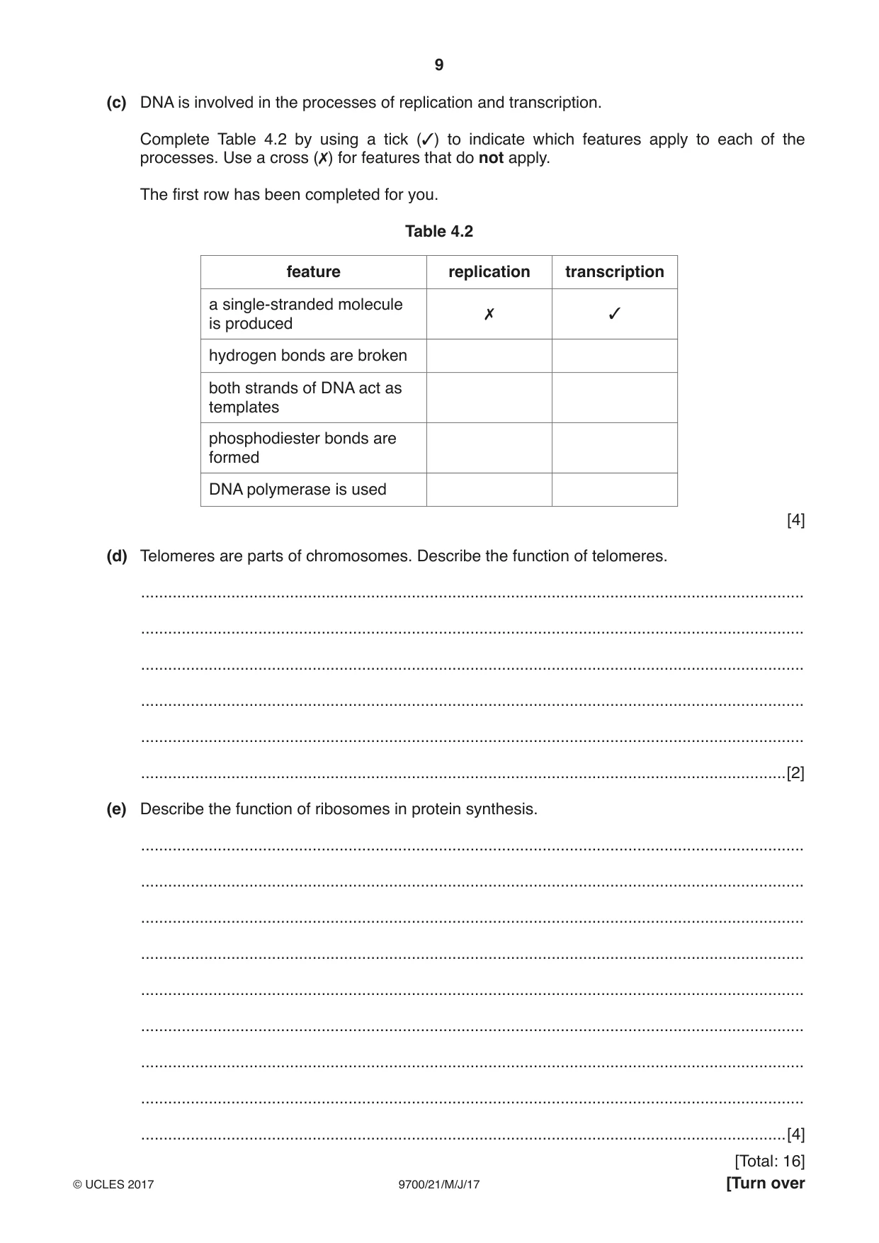
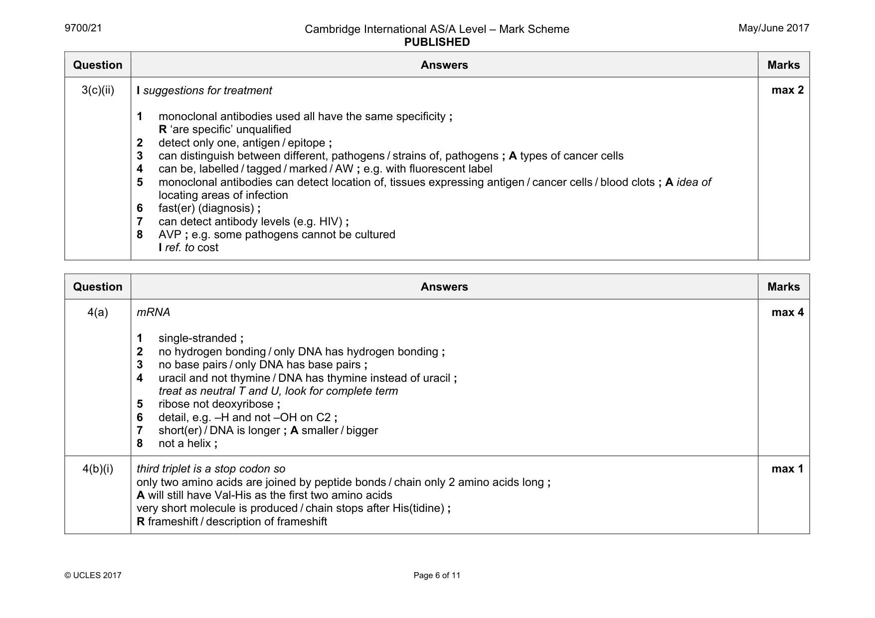
4 (a) Fig. 4.1 shows part of a DNA molecule. O O H3C O H H O N O O N N N O P O¯ O O N A H O O¯ P O N N O O N N O H H O O N N O P O¯ N O H N O O¯ P O C N H H O O N O O O Key: O P O¯ O A = adenine C = cytosine O O¯ P O O Fig. 4.1 Use Fig. 4.1 to explain how the structure of mRNA differs from the structure of DNA. ................................................................................................................................................... ................................................................................................................................................... ................................................................................................................................................... ................................................................................................................................................... ................................................................................................................................................... ................................................................................................................................................... ................................................................................................................................................... ................................................................................................................................................... ...............................................................................................................................................[4] (b) Fig. 4.2 shows: • the first seven amino acids of the β chain of haemoglobin • the first amino acid in the sequence is valine (Val) • the 21 base pairs in the sequence of DNA that code for these seven amino acids. amino acid Val His Leu Thr Pro Glu Glu sequence CAC GTG GAC TGA GGA CTC CTC base sequence in DNA GTG CAC CTG ACT CCT GAG GAG Fig. 4.2 Table 4.1 shows the triplets of bases that code for seven amino acids. Using Fig. 4.2 and Table 4.1, state what will happen to the sequence of amino acids in the first part of the β chain of haemoglobin: (i) if the base pair at position 6 is deleted ........................................................................................................................................... .......................................................................................................................................[1] (ii) if the three base pairs at positions 7, 8 and 9 are deleted. ........................................................................................................................................... .......................................................................................................................................[1] Table 4.1 amino acid DNA triplets cysteine (Cys) TGT TGC glutamic acid (Glu) GAA GAG histidine (His) CAT CAC leucine (Leu) CTT CTC CTA CTG proline (Pro) CCT CCC CCA CCG threonine (Thr) ACT ACC ACA ACG valine (Val) GTT GTC GTA GTG no amino acid STOP TAA TAG TGA (c) DNA is involved in the processes of replication and transcription. Complete Table 4.2 by using a tick (3) to indicate which features apply to each of the processes. Use a cross (7) for features that do not apply. The first row has been completed for you. Table 4.2 feature replication transcription a single-stranded molecule 7 3 is produced hydrogen bonds are broken both strands of DNA act as templates phosphodiester bonds are formed DNA polymerase is used [4] (d) Telomeres are parts of chromosomes. Describe the function of telomeres. ................................................................................................................................................... ................................................................................................................................................... ................................................................................................................................................... ................................................................................................................................................... ................................................................................................................................................... ...............................................................................................................................................[2] (e) Describe the function of ribosomes in protein synthesis. ................................................................................................................................................... ................................................................................................................................................... ................................................................................................................................................... ................................................................................................................................................... ................................................................................................................................................... ................................................................................................................................................... ................................................................................................................................................... ................................................................................................................................................... ...............................................................................................................................................[4]
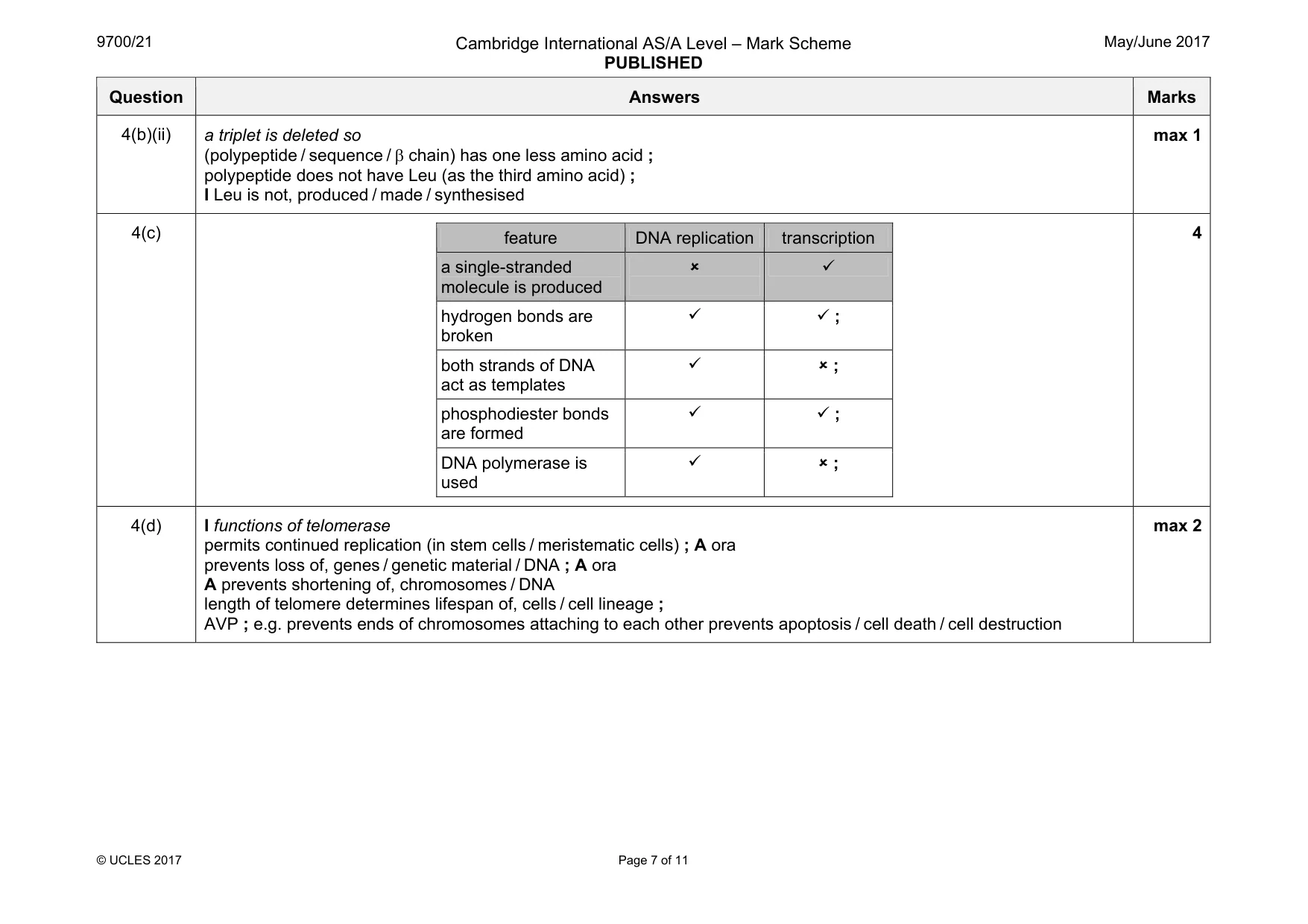
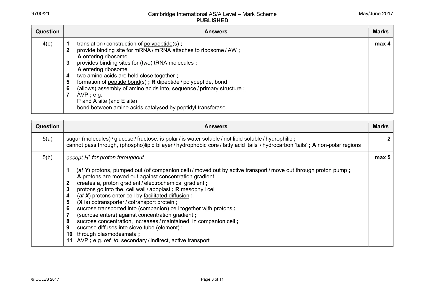
Mark scheme: 4(a) mRNA 1 single-stranded ; 2 no hydrogen bonding / only DNA has hydrogen bonding ; 3 no base pairs / only DNA has base pairs ; 4 uracil and not thymine / DNA has thymine instead of uracil ; treat as neutral T and U, look for complete term 5 ribose not deoxyribose ; 6 detail, e.g. –H and not –OH on C2 ; 7 short(er) / DNA is longer ; A smaller / bigger 8 not a helix ; max 4 4(b)(i) third triplet is a stop codon so only two amino acids are joined by peptide bonds / chain only 2 amino acids long ; A will still have Val-His as the first two amino acids very short molecule is produced / chain stops after His(tidine) ; R frameshift / description of frameshift max 1 Question Answers Marks 4(b)(ii) a triplet is deleted so (polypeptide / sequence / β chain) has one less amino acid ; polypeptide does not have Leu (as the third amino acid) ; I Leu is not, produced / made / synthesised max 1 4(c) feature DNA replication transcription a single-stranded molecule is produced 8 9 hydrogen bonds are broken 9 9 ; both strands of DNA act as templates 9 8 ; phosphodiester bonds are formed 9 9 ; DNA polymerase is used 9 8 ; 4 4(d) I functions of telomerase permits continued replication (in stem cells / meristematic cells) ; A ora prevents loss of, genes / genetic material / DNA ; A ora A prevents shortening of, chromosomes / DNA length of telomere determines lifespan of, cells / cell lineage ; AVP ; e.g. prevents ends of chromosomes attaching to each other prevents apoptosis / cell death / cell destruction max 2 Question Answers Marks 4(e) 1 translation / construction of polypeptide(s) ; 2 provide binding site for mRNA / mRNA attaches to ribosome / AW ; A entering ribosome 3 provides binding sites for (two) tRNA molecules ; A entering ribosome 4 two amino acids are held close together ; 5 formation of peptide bond(s) ; R dipeptide / polypeptide, bond 6 (allows) assembly of amino acids into, sequence / primary structure ; 7 AVP ; e.g. P and A site (and E site) bond between amino acids catalysed by peptidyl transferase max 4
Q5 · Sugar molecules enter cells through transport proteins
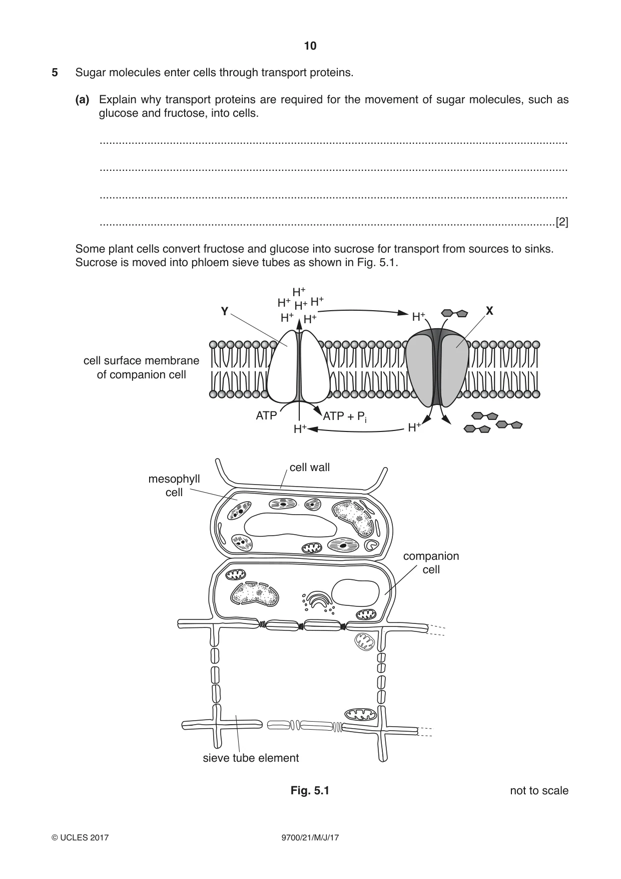
5 Sugar molecules enter cells through transport proteins. (a) Explain why transport proteins are required for the movement of sugar molecules, such as glucose and fructose, into cells. ................................................................................................................................................... ................................................................................................................................................... ................................................................................................................................................... ...............................................................................................................................................[2] Some plant cells convert fructose and glucose into sucrose for transport from sources to sinks. Sucrose is moved into phloem sieve tubes as shown in Fig. 5.1. H+ H+ H+ Y X H+H+H+ H+ cell surface membrane of companion cell ATP ATP + Pi H+ H+ cell wall mesophyll cell companion cell sieve tube element Fig. 5.1 not to scale (b) Use the information in Fig. 5.1 to explain how sucrose: • moves into the companion cell • moves from the companion cell into the sieve tube element. ................................................................................................................................................... ................................................................................................................................................... ................................................................................................................................................... ................................................................................................................................................... ................................................................................................................................................... ................................................................................................................................................... ................................................................................................................................................... ................................................................................................................................................... ................................................................................................................................................... ................................................................................................................................................... ................................................................................................................................................... ...............................................................................................................................................[5] (c) Sucrose travels in phloem sieve tubes to sinks. State two examples of sinks. 1 ................................................................................................................................................ 2 ............................................................................................................................................[1] [Total: 8]
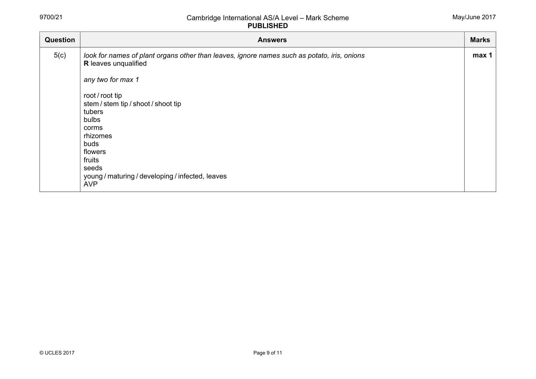
Mark scheme: 5(a) sugar (molecules) / glucose / fructose, is polar / is water soluble / not lipid soluble / hydrophilic ; cannot pass through, (phospho)lipid bilayer / hydrophobic core / fatty acid ‘tails’ / hydrocarbon ‘tails’ ; A non-polar regions 2 5(b) accept H+ for proton throughout 1 (at Y) protons, pumped out (of companion cell) / moved out by active transport / move out through proton pump ; A protons are moved out against concentration gradient 2 creates a, proton gradient / electrochemical gradient ; 3 protons go into the, cell wall / apoplast ; R mesophyll cell 4 (at X) protons enter cell by facilitated diffusion ; 5 (X is) cotransporter / cotransport protein ; 6 sucrose transported into (companion) cell together with protons ; 7 (sucrose enters) against concentration gradient ; 8 sucrose concentration, increases / maintained, in companion cell ; 9 sucrose diffuses into sieve tube (element) ; 10 through plasmodesmata ; 11 AVP ; e.g. ref. to, secondary / indirect, active transport max 5 Question Answers Marks 5(c) look for names of plant organs other than leaves, ignore names such as potato, iris, onions R leaves unqualified any two for max 1 root / root tip stem / stem tip / shoot / shoot tip tubers bulbs corms rhizomes buds flowers fruits seeds young / maturing / developing / infected, leaves AVP max 1
Q6 · Cholera bacteria release the toxin, choleragen, when they are in the intestine
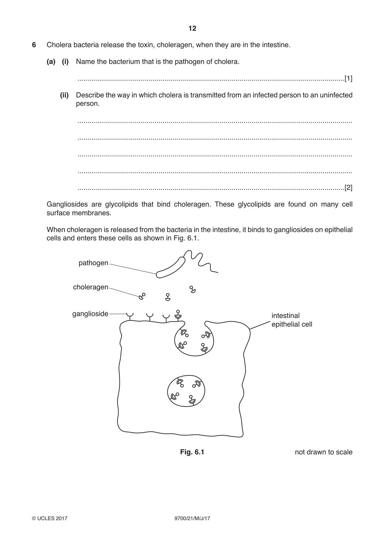
6 Cholera bacteria release the toxin, choleragen, when they are in the intestine. (a) (i) Name the bacterium that is the pathogen of cholera. .......................................................................................................................................[1] (ii) Describe the way in which cholera is transmitted from an infected person to an uninfected person. ........................................................................................................................................... ........................................................................................................................................... ........................................................................................................................................... ........................................................................................................................................... .......................................................................................................................................[2] Gangliosides are glycolipids that bind choleragen. These glycolipids are found on many cell surface membranes. When choleragen is released from the bacteria in the intestine, it binds to gangliosides on epithelial cells and enters these cells as shown in Fig. 6.1. pathogen choleragen ganglioside intestinal epithelial cell Fig. 6.1 not drawn to scale (b) Suggest how choleragen interacts with gangliosides on intestinal epithelial cells. ................................................................................................................................................... ................................................................................................................................................... ................................................................................................................................................... ................................................................................................................................................... ...............................................................................................................................................[2] (c) Name the process by which choleragen enters the intestinal epithelial cell as shown in Fig. 6.1. ...............................................................................................................................................[1] Once inside the cells choleragen is activated. One effect is to increase the movement of chloride ions through channel proteins out of cells. (d) Suggest and explain the likely consequences on the intestinal epithelial cells of the loss of chloride ions through the channel proteins. ................................................................................................................................................... ................................................................................................................................................... ................................................................................................................................................... ................................................................................................................................................... ...............................................................................................................................................[2] (e) Health authorities recommend that antibiotics, such as tetracycline, are only to be used for treating people with severe cases of cholera. Explain why it is recommended that antibiotics should not be given to people with mild cases of cholera or to protect people from cholera. ................................................................................................................................................... ................................................................................................................................................... ................................................................................................................................................... ................................................................................................................................................... ................................................................................................................................................... ................................................................................................................................................... ...............................................................................................................................................[3] [Total: 11]
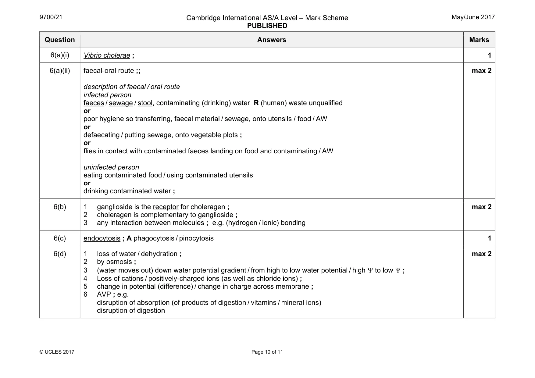
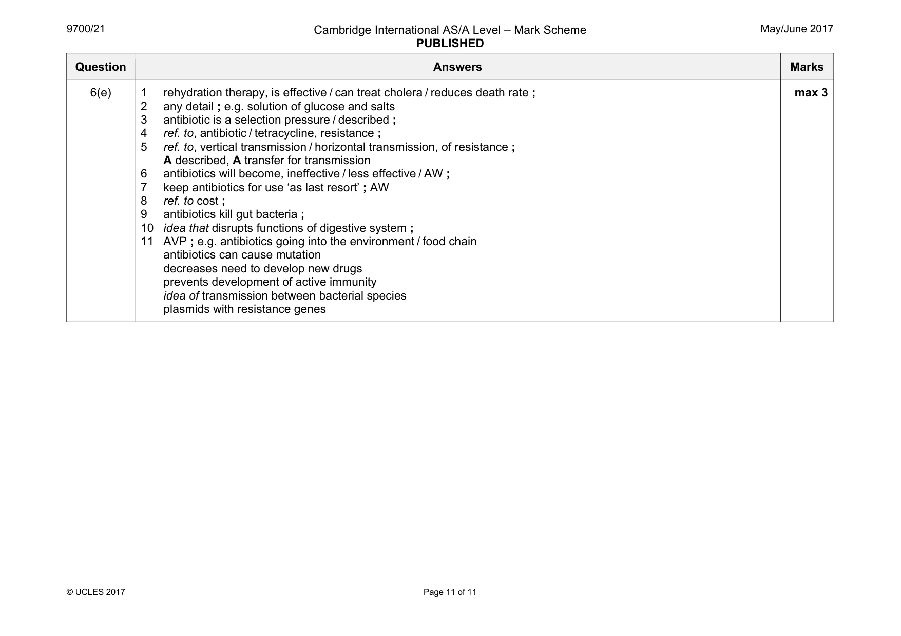
Mark scheme: 6(a)(i) Vibrio cholerae ; 1 6(a)(ii) faecal-oral route ;; description of faecal / oral route infected person faeces / sewage / stool, contaminating (drinking) water R (human) waste unqualified or poor hygiene so transferring, faecal material / sewage, onto utensils / food / AW or defaecating / putting sewage, onto vegetable plots ; or flies in contact with contaminated faeces landing on food and contaminating / AW uninfected person eating contaminated food / using contaminated utensils or drinking contaminated water ; max 2 6(b) 1 ganglioside is the receptor for choleragen ; 2 choleragen is complementary to ganglioside ; 3 any interaction between molecules ; e.g. (hydrogen / ionic) bonding max 2 6(c) endocytosis ; A phagocytosis / pinocytosis 1 6(d) 1 loss of water / dehydration ; 2 by osmosis ; 3 (water moves out) down water potential gradient / from high to low water potential / high Ψ to low Ψ ; 4 Loss of cations / positively-charged ions (as well as chloride ions) ; 5 change in potential (difference) / change in charge across membrane ; 6 AVP ; e.g. disruption of absorption (of products of digestion / vitamins / mineral ions) disruption of digestion max 2 Question Answers Marks 6(e) 1 rehydration therapy, is effective / can treat cholera / reduces death rate ; 2 any detail ; e.g. solution of glucose and salts 3 antibiotic is a selection pressure / described ; 4 ref. to, antibiotic / tetracycline, resistance ; 5 ref. to, vertical transmission / horizontal transmission, of resistance ; A described, A transfer for transmission 6 antibiotics will become, ineffective / less effective / AW ; 7 keep antibiotics for use ‘as last resort’ ; AW 8 ref. to cost ; 9 antibiotics kill gut bacteria ; 10 idea that disrupts functions of digestive system ; 11 AVP ; e.g. antibiotics going into the environment / food chain antibiotics can cause mutation decreases need to develop new drugs prevents development of active immunity idea of transmission between bacterial species plasmids with resistance genes max 3
What was in this paper
The subtopics covered by these 6 questions, and how many questions each got. Open one in a new tab to see every Cambridge question on it.
What you needed in this session
Cambridge’s own grade thresholds for 2017 May/June, Paper 2 · Variant 1. A higher threshold means an easier paper — the bar moves with how the cohort did.