Cambridge A Level Biology 9700 — 2021 May/June Paper 2 · Variant 3
9700/23/M/J/21 · 6 questions · 60 marks · ≈68 min
The question paper and its mark scheme, free to read here and free to download. This is Cambridge’s own paper, exactly as it was sat.
Question paper16 pages

Mark scheme19 pages
Answers below. Sit the paper first if you are practising.
Questions as text
Q1 · The Golgi body, rough endoplasmic reticulum (RER) and smooth endoplasmic reticulum (SER)…
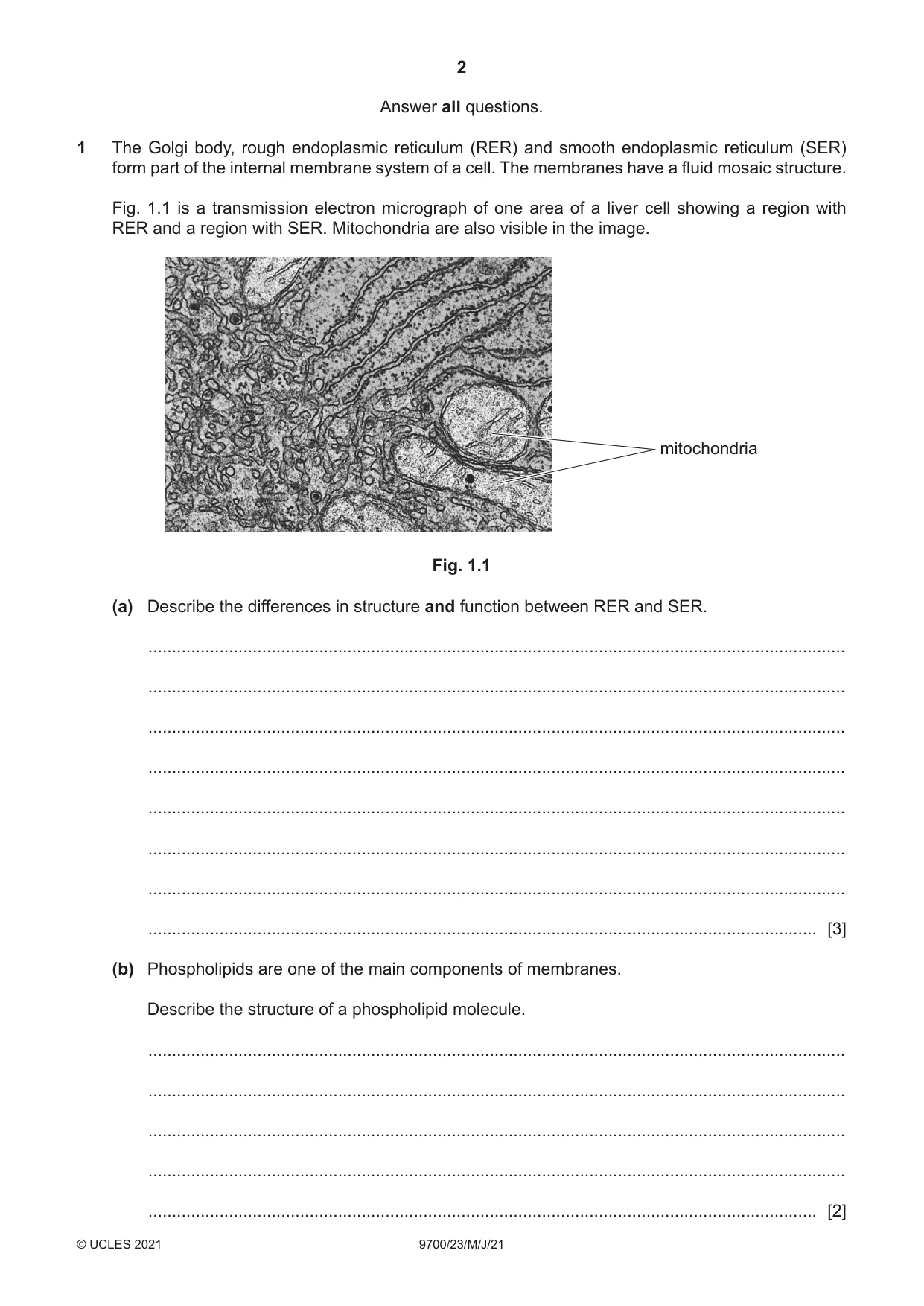
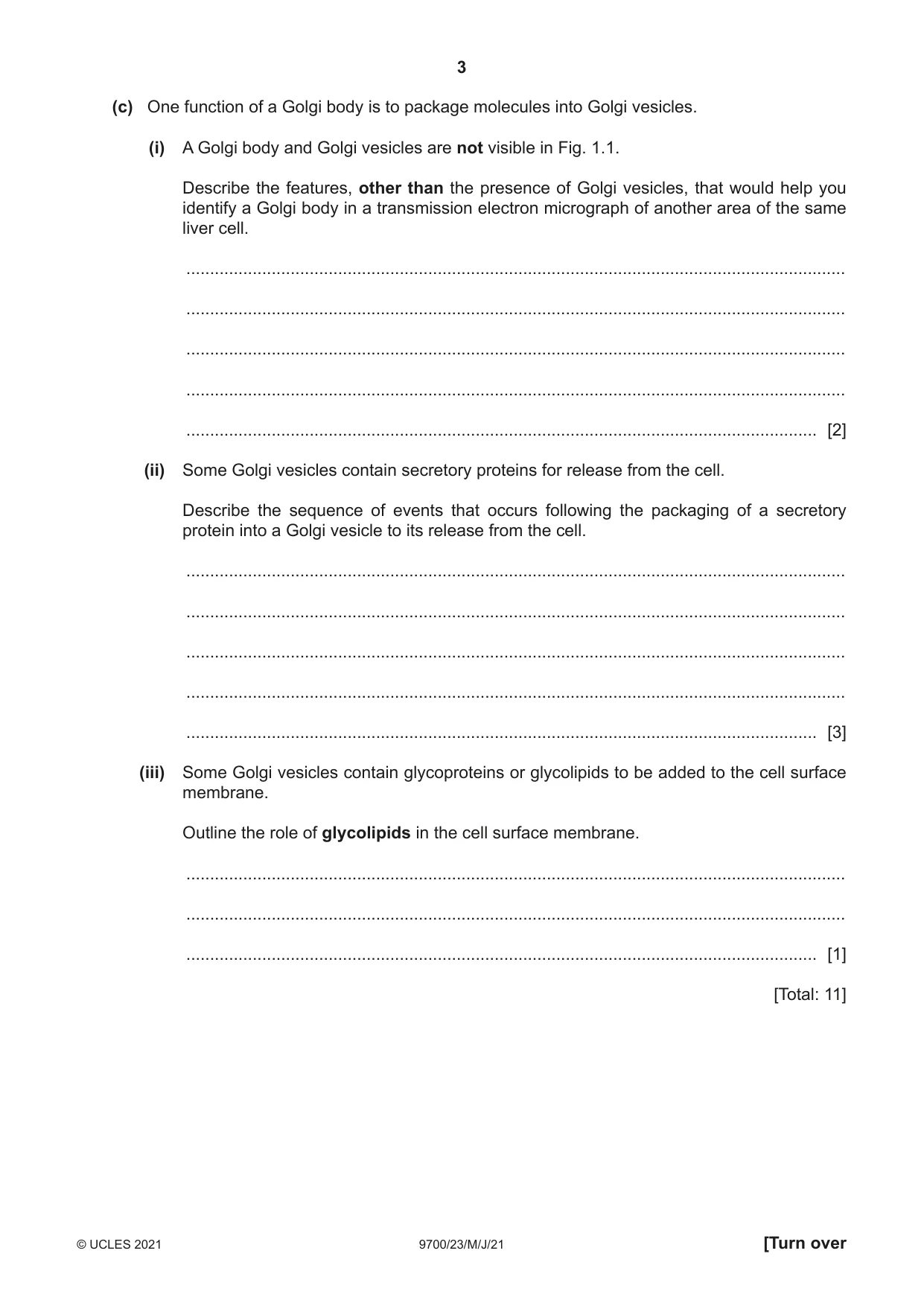
1 The Golgi body, rough endoplasmic reticulum (RER) and smooth endoplasmic reticulum (SER) form part of the internal membrane system of a cell. The membranes have a fluid mosaic structure. Fig. 1.1 is a transmission electron micrograph of one area of a liver cell showing a region with RER and a region with SER. Mitochondria are also visible in the image. mitochondria Fig. 1.1 (a) Describe the differences in structure and function between RER and SER. ................................................................................................................................................... ................................................................................................................................................... ................................................................................................................................................... ................................................................................................................................................... ................................................................................................................................................... ................................................................................................................................................... ................................................................................................................................................... ............................................................................................................................................. [3] (b) Phospholipids are one of the main components of membranes. Describe the structure of a phospholipid molecule. ................................................................................................................................................... ................................................................................................................................................... ................................................................................................................................................... ................................................................................................................................................... ............................................................................................................................................. [2] (c) One function of a Golgi body is to package molecules into Golgi vesicles. (i) A Golgi body and Golgi vesicles are not visible in Fig. 1.1. Describe the features, other than the presence of Golgi vesicles, that would help you identify a Golgi body in a transmission electron micrograph of another area of the same liver cell. ........................................................................................................................................... ........................................................................................................................................... ........................................................................................................................................... ........................................................................................................................................... ..................................................................................................................................... [2] (ii) Some Golgi vesicles contain secretory proteins for release from the cell. Describe the sequence of events that occurs following the packaging of a secretory protein into a Golgi vesicle to its release from the cell. ........................................................................................................................................... ........................................................................................................................................... ........................................................................................................................................... ........................................................................................................................................... ..................................................................................................................................... [3] (iii) Some Golgi vesicles contain glycoproteins or glycolipids to be added to the cell surface membrane. Outline the role of glycolipids in the cell surface membrane. ........................................................................................................................................... ........................................................................................................................................... ..................................................................................................................................... [1] [Total: 11]
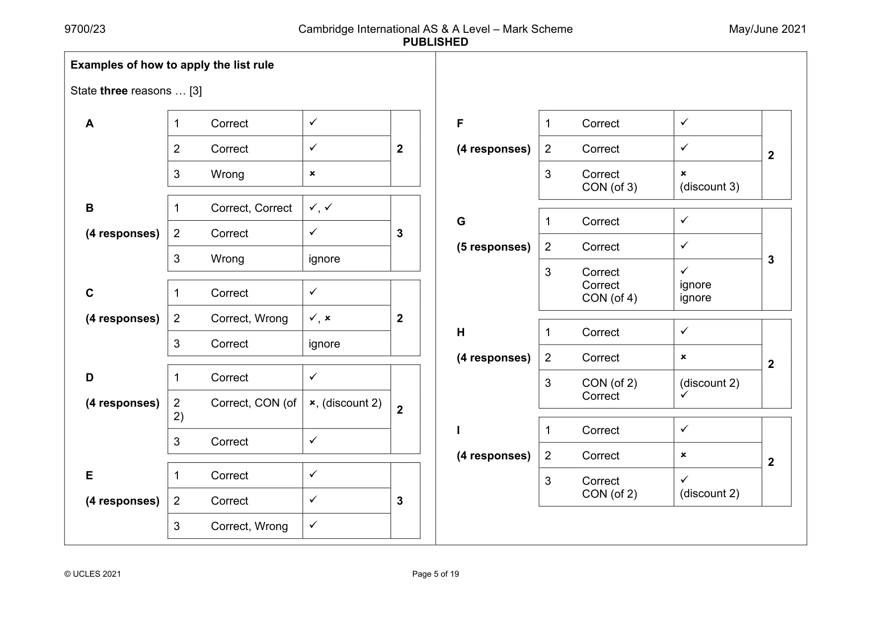
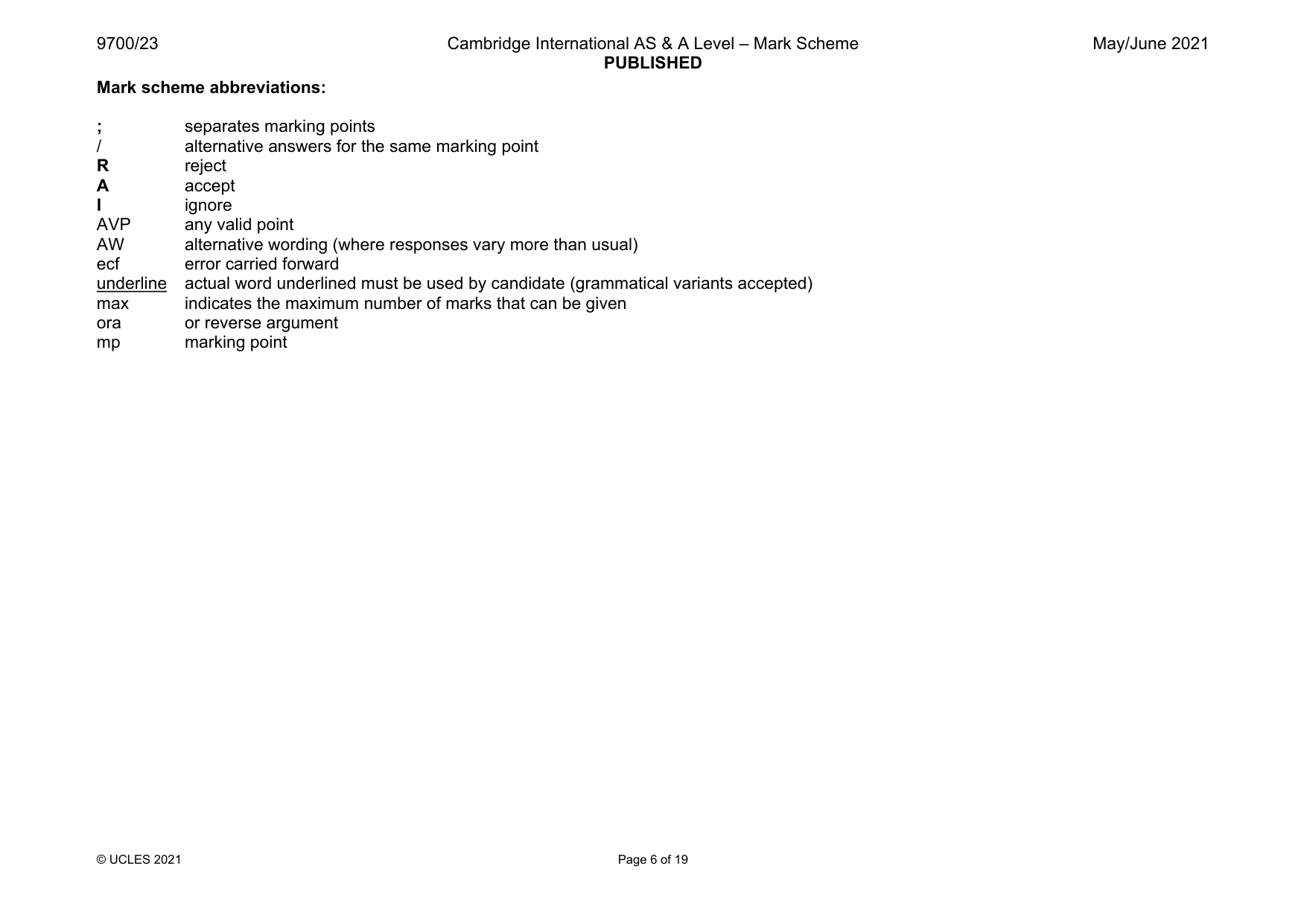
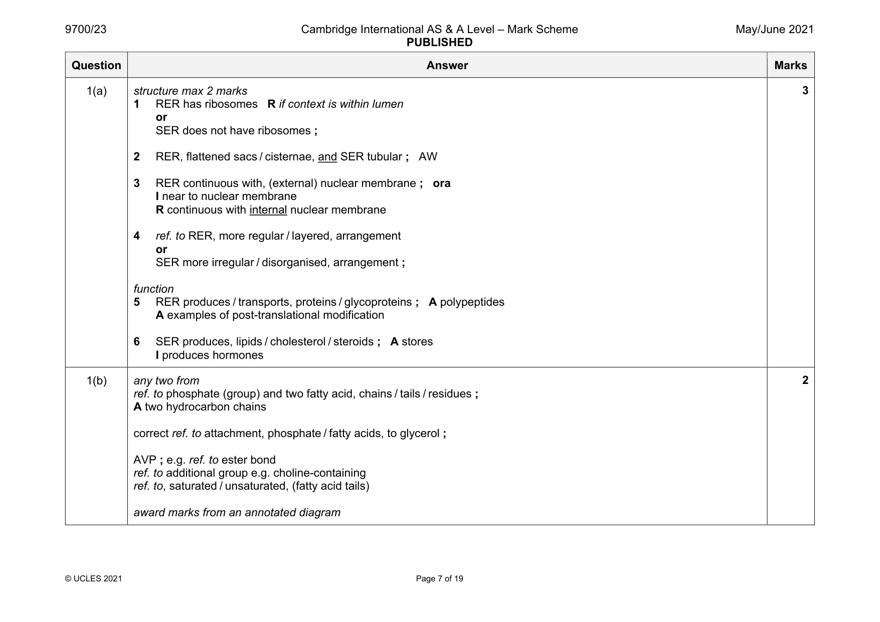
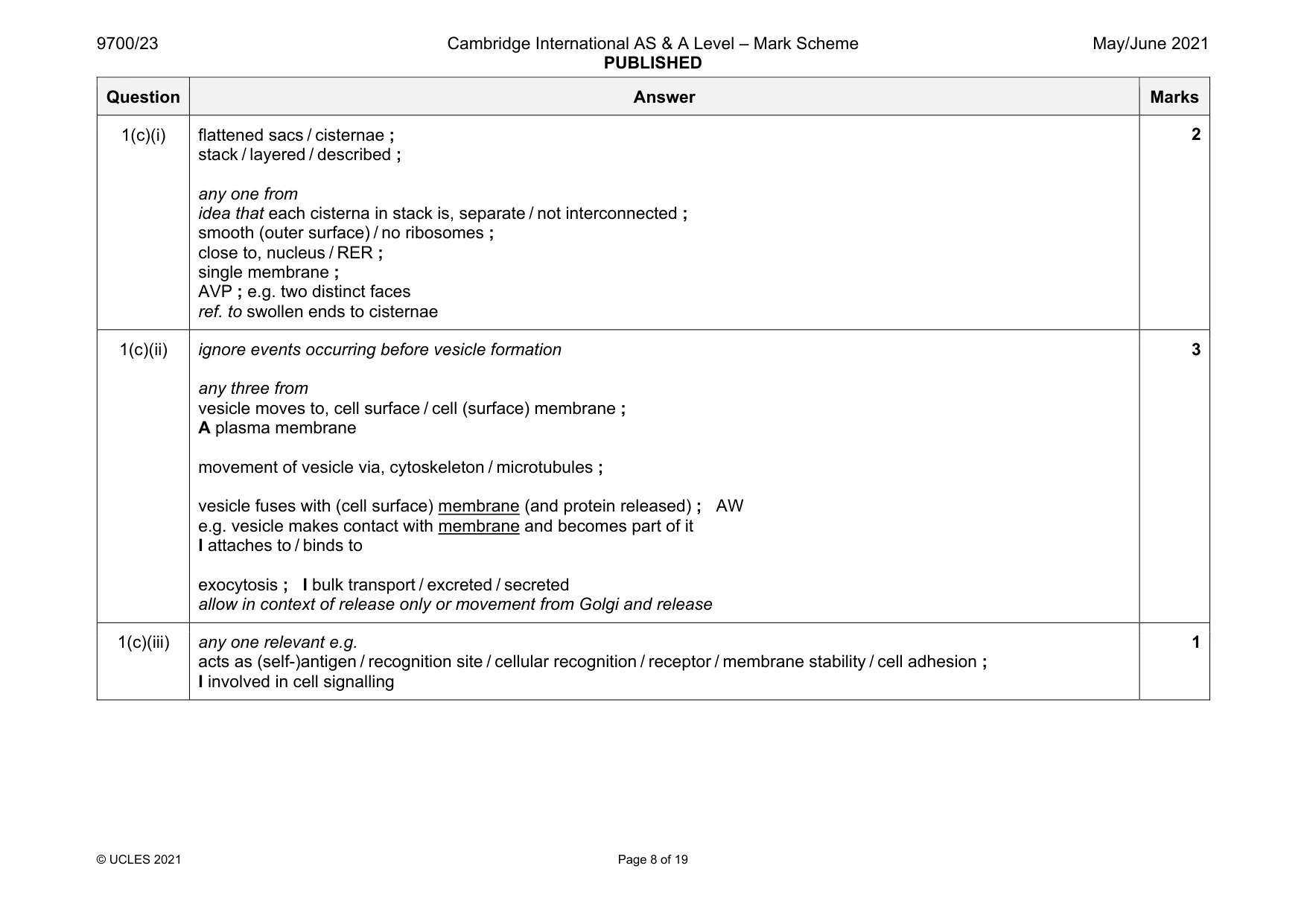
Mark scheme: 1(a) structure max 2 marks 1 RER has ribosomes R if context is within lumen or SER does not have ribosomes ; 2 RER, flattened sacs / cisternae, and SER tubular ; AW 3 RER continuous with, (external) nuclear membrane ; ora I near to nuclear membrane R continuous with internal nuclear membrane 4 ref. to RER, more regular / layered, arrangement or SER more irregular / disorganised, arrangement ; function 5 RER produces / transports, proteins / glycoproteins ; A polypeptides A examples of post-translational modification 6 SER produces, lipids / cholesterol / steroids ; A stores I produces hormones 1(b) any two from ref. to phosphate (group) and two fatty acid, chains / tails / residues ; A two hydrocarbon chains correct ref. to attachment, phosphate / fatty acids, to glycerol ; AVP ; e.g. ref. to ester bond ref. to additional group e.g. choline-containing ref. to, saturated / unsaturated, (fatty acid tails) award marks from an annotated diagram 2 Question Answer Marks 1(c)(i) flattened sacs / cisternae ; stack / layered / described ; any one from idea that each cisterna in stack is, separate / not interconnected ; smooth (outer surface) / no ribosomes ; close to, nucleus / RER ; single membrane ; AVP ; e.g. two distinct faces ref. to swollen ends to cisternae 2 1(c)(ii) ignore events occurring before vesicle formation any three from vesicle moves to, cell surface / cell (surface) membrane ; A plasma membrane movement of vesicle via, cytoskeleton / microtubules ; vesicle fuses with (cell surface) membrane (and protein released) ; AW e.g. vesicle makes contact with membrane and becomes part of it I attaches to / binds to exocytosis ; I bulk transport / excreted / secreted allow in context of release only or movement from Golgi and release 3 1(c)(iii) any one relevant e.g. acts as (self-)antigen / recognition site / cellular recognition / receptor / membrane stability / cell adhesion ; I involved in cell signalling 1
More questions on Cells as the basic units of living organisms
Q2 · Smallpox, measles and HIV/AIDS are infectious diseases caused by different viruses
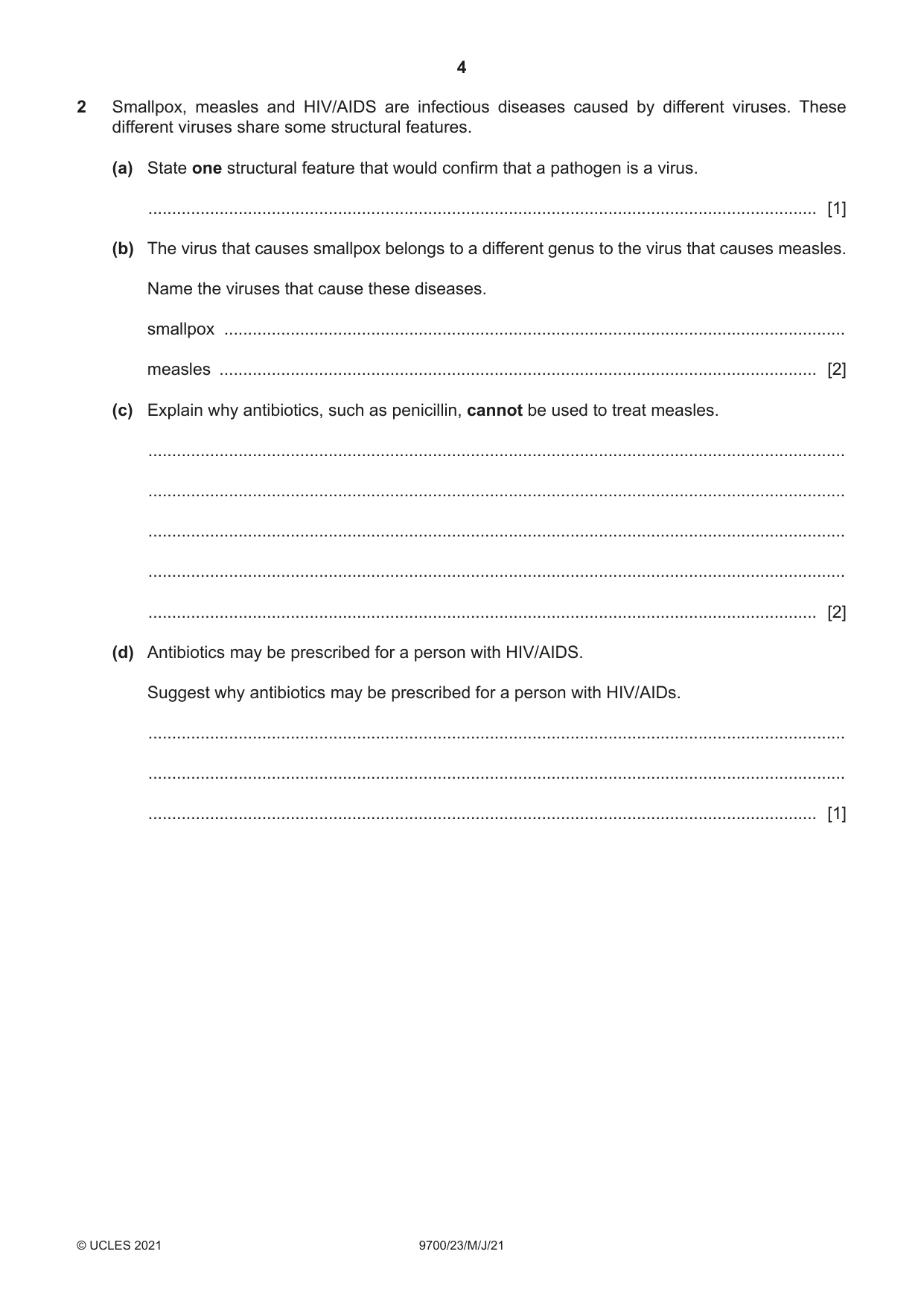
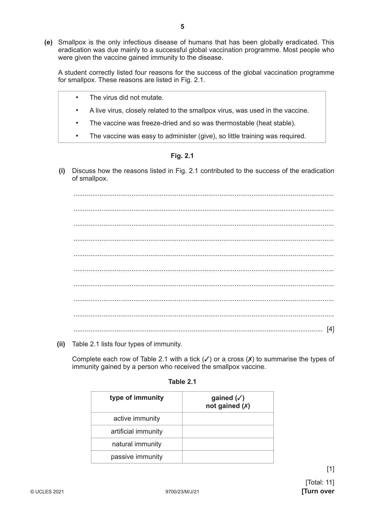
2 Smallpox, measles and HIV/AIDS are infectious diseases caused by different viruses. These different viruses share some structural features. (a) State one structural feature that would confirm that a pathogen is a virus. ............................................................................................................................................. [1] (b) The virus that causes smallpox belongs to a different genus to the virus that causes measles. Name the viruses that cause these diseases. smallpox ................................................................................................................................... measles .............................................................................................................................. [2] (c) Explain why antibiotics, such as penicillin, cannot be used to treat measles. ................................................................................................................................................... ................................................................................................................................................... ................................................................................................................................................... ................................................................................................................................................... ............................................................................................................................................. [2] (d) Antibiotics may be prescribed for a person with HIV/AIDS. Suggest why antibiotics may be prescribed for a person with HIV/AIDs. ................................................................................................................................................... ................................................................................................................................................... ............................................................................................................................................. [1] (e) Smallpox is the only infectious disease of humans that has been globally eradicated. This eradication was due mainly to a successful global vaccination programme. Most people who were given the vaccine gained immunity to the disease. A student correctly listed four reasons for the success of the global vaccination programme for smallpox. These reasons are listed in Fig. 2.1. • The virus did not mutate. • A live virus, closely related to the smallpox virus, was used in the vaccine. • The vaccine was freeze-dried and so was thermostable (heat stable). • The vaccine was easy to administer (give), so little training was required. Fig. 2.1 (i) Discuss how the reasons listed in Fig. 2.1 contributed to the success of the eradication of smallpox. ........................................................................................................................................... ........................................................................................................................................... ........................................................................................................................................... ........................................................................................................................................... ........................................................................................................................................... ........................................................................................................................................... ........................................................................................................................................... ........................................................................................................................................... ........................................................................................................................................... ..................................................................................................................................... [4] (ii) Table 2.1 lists four types of immunity. Complete each row of Table 2.1 with a tick (3) or a cross (✗) to summarise the types of immunity gained by a person who received the smallpox vaccine. Table 2.1 type of immunity gained (3) not gained (✗) active immunity artificial immunity natural immunity passive immunity [1] [Total: 11]
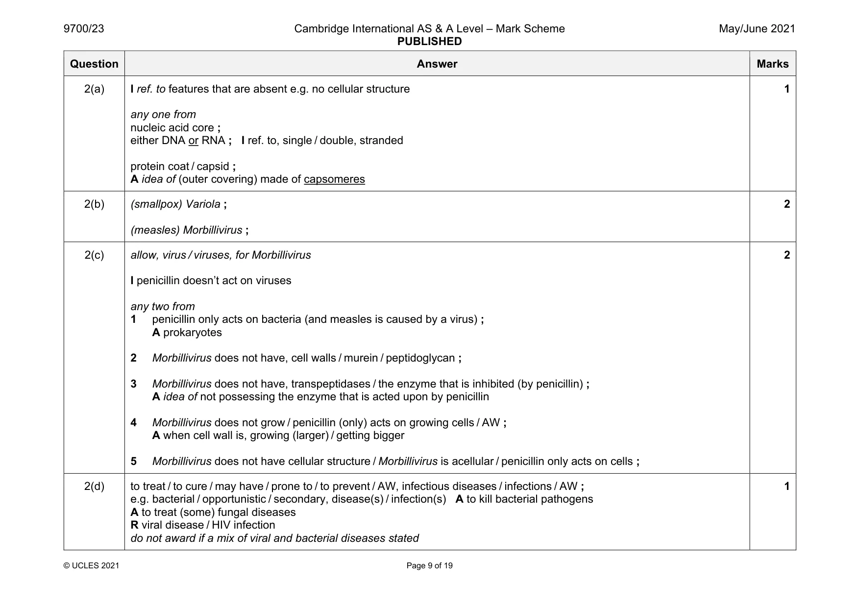
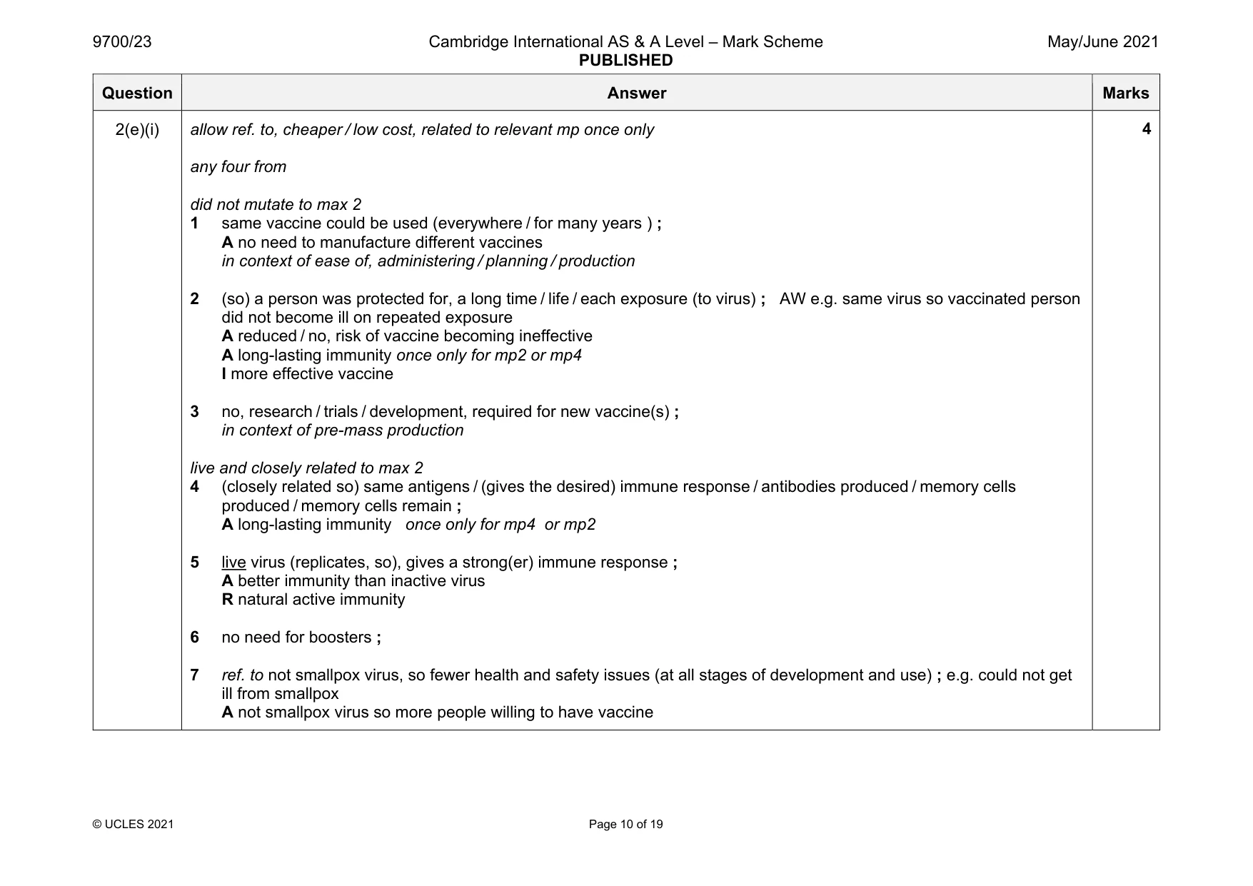
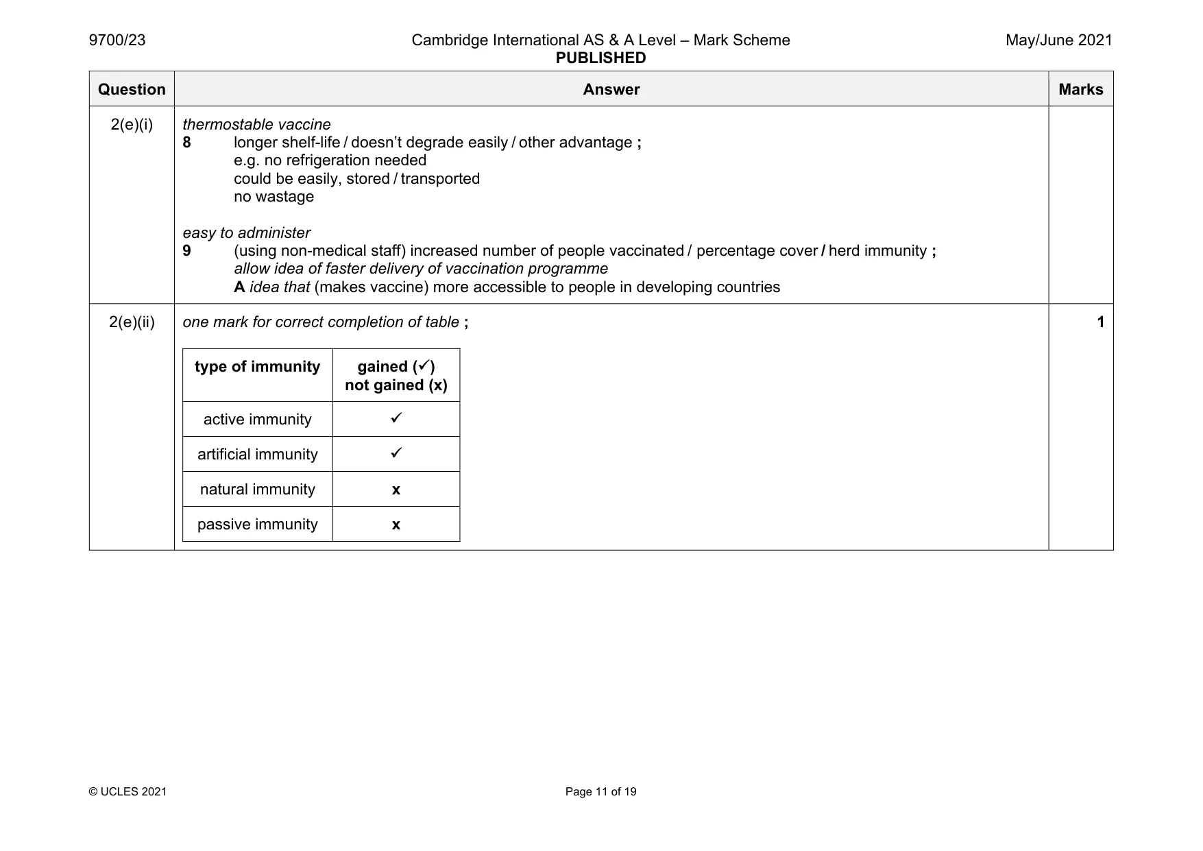
Mark scheme: 2(a) I ref. to features that are absent e.g. no cellular structure any one from nucleic acid core ; either DNA or RNA ; I ref. to, single / double, stranded protein coat / capsid ; A idea of (outer covering) made of capsomeres 1 2(b) (smallpox) Variola ; (measles) Morbillivirus ; 2 2(c) allow, virus / viruses, for Morbillivirus I penicillin doesn’t act on viruses any two from 1 penicillin only acts on bacteria (and measles is caused by a virus) ; A prokaryotes 2 Morbillivirus does not have, cell walls / murein / peptidoglycan ; 3 Morbillivirus does not have, transpeptidases / the enzyme that is inhibited (by penicillin) ; A idea of not possessing the enzyme that is acted upon by penicillin 4 Morbillivirus does not grow / penicillin (only) acts on growing cells / AW ; A when cell wall is, growing (larger) / getting bigger 5 Morbillivirus does not have cellular structure / Morbillivirus is acellular / penicillin only acts on cells ; 2 2(d) to treat / to cure / may have / prone to / to prevent / AW, infectious diseases / infections / AW ; e.g. bacterial / opportunistic / secondary, disease(s) / infection(s) A to kill bacterial pathogens A to treat (some) fungal diseases R viral disease / HIV infection do not award if a mix of viral and bacterial diseases stated 1 Question Answer Marks 2(e)(i) allow ref. to, cheaper / low cost, related to relevant mp once only any four from did not mutate to max 2 1 same vaccine could be used (everywhere / for many years ) ; A no need to manufacture different vaccines in context of ease of, administering / planning / production 2 (so) a person was protected for, a long time / life / each exposure (to virus) ; AW e.g. same virus so vaccinated person did not become ill on repeated exposure A reduced / no, risk of vaccine becoming ineffective A long-lasting immunity once only for mp2 or mp4 I more effective vaccine 3 no, research / trials / development, required for new vaccine(s) ; in context of pre-mass production live and closely related to max 2 4 (closely related so) same antigens / (gives the desired) immune response / antibodies produced / memory cells produced / memory cells remain ; A long-lasting immunity once only for mp4 or mp2 5 live virus (replicates, so), gives a strong(er) immune response ; A better immunity than inactive virus R natural active immunity 6 no need for boosters ; 7 ref. to not smallpox virus, so fewer health and safety issues (at all stages of development and use) ; e.g. could not get ill from smallpox A not smallpox virus so more people willing to have vaccine 4 Question Answer Marks 2(e)(i) thermostable vaccine 8 longer shelf-life / doesn’t degrade easily / other advantage ; e.g. no refrigeration needed could be easily, stored / transported no wastage easy to administer 9 (using non-medical staff) increased number of people vaccinated / percentage cover / herd immunity ; allow idea of faster delivery of vaccination programme A idea that (makes vaccine) more accessible to people in developing countries 2(e)(ii) one mark for correct completion of table ; type of immunity gained () not gained (x) active immunity artificial immunity natural immunity x passive immunity x 1
Q3 · HIV protease is an enzyme composed of two identical polypeptide chains
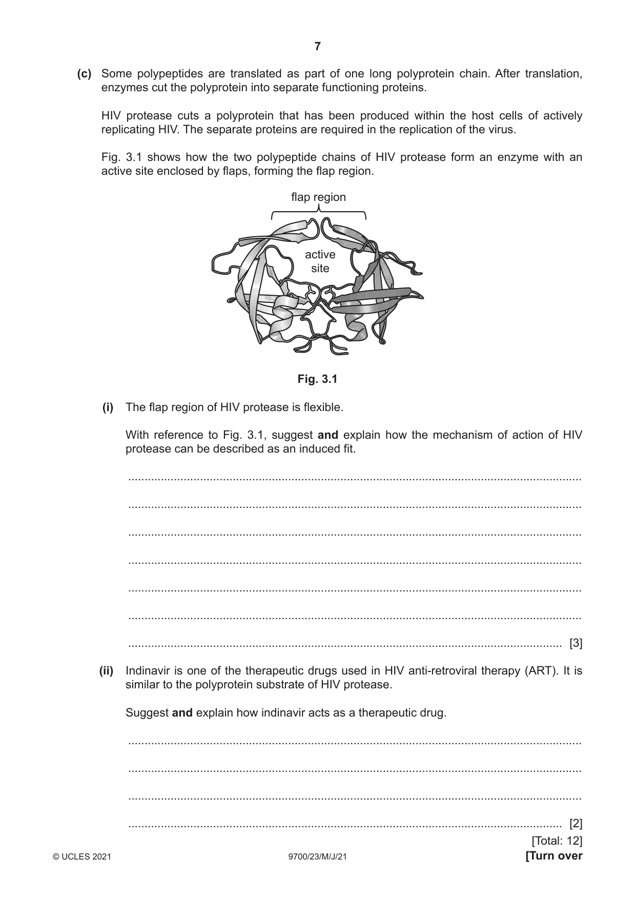
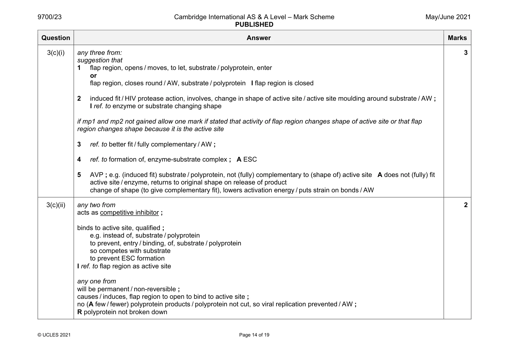
3 HIV protease is an enzyme composed of two identical polypeptide chains. Each polypeptide chain is 99 amino acids long. During translation, the amino acids are joined by peptide bonds to form the polypeptide chain. (a) Describe how a polypeptide of HIV protease is produced by the process of translation. ................................................................................................................................................... ................................................................................................................................................... ................................................................................................................................................... ................................................................................................................................................... ................................................................................................................................................... ................................................................................................................................................... ................................................................................................................................................... ................................................................................................................................................... ................................................................................................................................................... ............................................................................................................................................. [4] (b) The synthesised polypeptide has a primary protein structure and can form a tertiary protein structure after translation. There are more bond types in the tertiary structure. Compare the peptide bond formed during translation with the types of bond made during tertiary structure formation. ................................................................................................................................................... ................................................................................................................................................... ................................................................................................................................................... ................................................................................................................................................... ................................................................................................................................................... ................................................................................................................................................... ................................................................................................................................................... ............................................................................................................................................. [3] (c) Some polypeptides are translated as part of one long polyprotein chain. After translation, enzymes cut the polyprotein into separate functioning proteins. HIV protease cuts a polyprotein that has been produced within the host cells of actively replicating HIV. The separate proteins are required in the replication of the virus. Fig. 3.1 shows how the two polypeptide chains of HIV protease form an enzyme with an active site enclosed by flaps, forming the flap region. flap region active site Fig. 3.1 (i) The flap region of HIV protease is flexible. With reference to Fig. 3.1, suggest and explain how the mechanism of action of HIV protease can be described as an induced fit. ........................................................................................................................................... ........................................................................................................................................... ........................................................................................................................................... ........................................................................................................................................... ........................................................................................................................................... ........................................................................................................................................... ..................................................................................................................................... [3] (ii) Indinavir is one of the therapeutic drugs used in HIV anti-retroviral therapy (ART). It is similar to the polyprotein substrate of HIV protease. Suggest and explain how indinavir acts as a therapeutic drug. ........................................................................................................................................... ........................................................................................................................................... ........................................................................................................................................... ..................................................................................................................................... [2] [Total: 12]
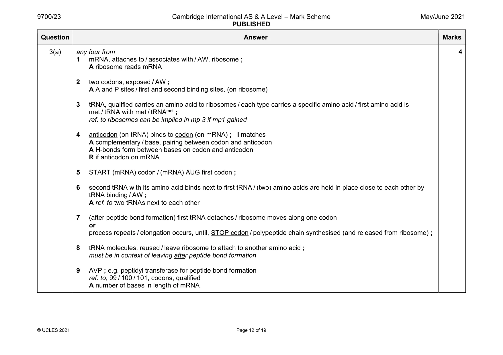
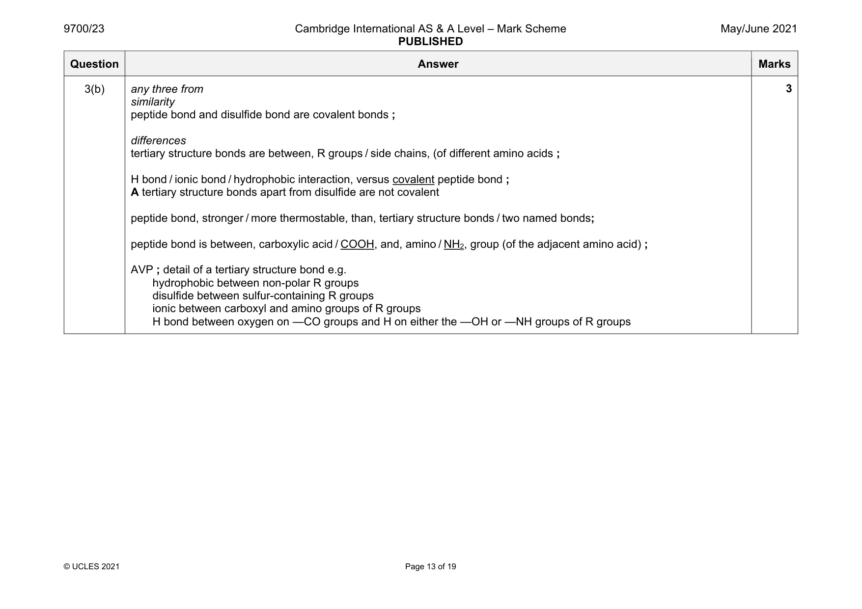
Mark scheme: 3(a) any four from 1 mRNA, attaches to / associates with / AW, ribosome ; A ribosome reads mRNA 2 two codons, exposed / AW ; A A and P sites / first and second binding sites, (on ribosome) 3 tRNA, qualified carries an amino acid to ribosomes / each type carries a specific amino acid / first amino acid is met / tRNA with met / tRNAmet ; ref. to ribosomes can be implied in mp 3 if mp1 gained 4 anticodon (on tRNA) binds to codon (on mRNA) ; I matches A complementary / base, pairing between codon and anticodon A H-bonds form between bases on codon and anticodon R if anticodon on mRNA 5 START (mRNA) codon / (mRNA) AUG first codon ; 6 second tRNA with its amino acid binds next to first tRNA / (two) amino acids are held in place close to each other by tRNA binding / AW ; A ref. to two tRNAs next to each other 7 (after peptide bond formation) first tRNA detaches / ribosome moves along one codon or process repeats / elongation occurs, until, STOP codon / polypeptide chain synthesised (and released from ribosome) ; 8 tRNA molecules, reused / leave ribosome to attach to another amino acid ; must be in context of leaving after peptide bond formation 9 AVP ; e.g. peptidyl transferase for peptide bond formation ref. to, 99 / 100 / 101, codons, qualified A number of bases in length of mRNA 4 Question Answer Marks 3(b) any three from similarity peptide bond and disulfide bond are covalent bonds ; differences tertiary structure bonds are between, R groups / side chains, (of different amino acids ; H bond / ionic bond / hydrophobic interaction, versus covalent peptide bond ; A tertiary structure bonds apart from disulfide are not covalent peptide bond, stronger / more thermostable, than, tertiary structure bonds / two named bonds; peptide bond is between, carboxylic acid / COOH, and, amino / NH2, group (of the adjacent amino acid) ; AVP ; detail of a tertiary structure bond e.g. hydrophobic between non-polar R groups disulfide between sulfur-containing R groups ionic between carboxyl and amino groups of R groups H bond between oxygen on —CO groups and H on either the —OH or —NH groups of R groups 3 Question Answer Marks 3(c)(i) any three from: suggestion that 1 flap region, opens / moves, to let, substrate / polyprotein, enter or flap region, closes round / AW, substrate / polyprotein I flap region is closed 2 induced fit / HIV protease action, involves, change in shape of active site / active site moulding around substrate / AW ; I ref. to enzyme or substrate changing shape if mp1 and mp2 not gained allow one mark if stated that activity of flap region changes shape of active site or that flap region changes shape because it is the active site 3 ref. to better fit / fully complementary / AW ; 4 ref. to formation of, enzyme-substrate complex ; A ESC 5 AVP ; e.g. (induced fit) substrate / polyprotein, not (fully) complementary to (shape of) active site A does not (fully) fit active site / enzyme, returns to original shape on release of product change of shape (to give complementary fit), lowers activation energy / puts strain on bonds / AW 3 3(c)(ii) any two from acts as competitive inhibitor ; binds to active site, qualified ; e.g. instead of, substrate / polyprotein to prevent, entry / binding, of, substrate / polyprotein so competes with substrate to prevent ESC formation I ref. to flap region as active site any one from will be permanent / non-reversible ; causes / induces, flap region to open to bind to active site ; no (A few / fewer) polyprotein products / polyprotein not cut, so viral replication prevented / AW ; R polyprotein not broken down 2
Q4 · Phloem sap is transported within phloem sieve tubes
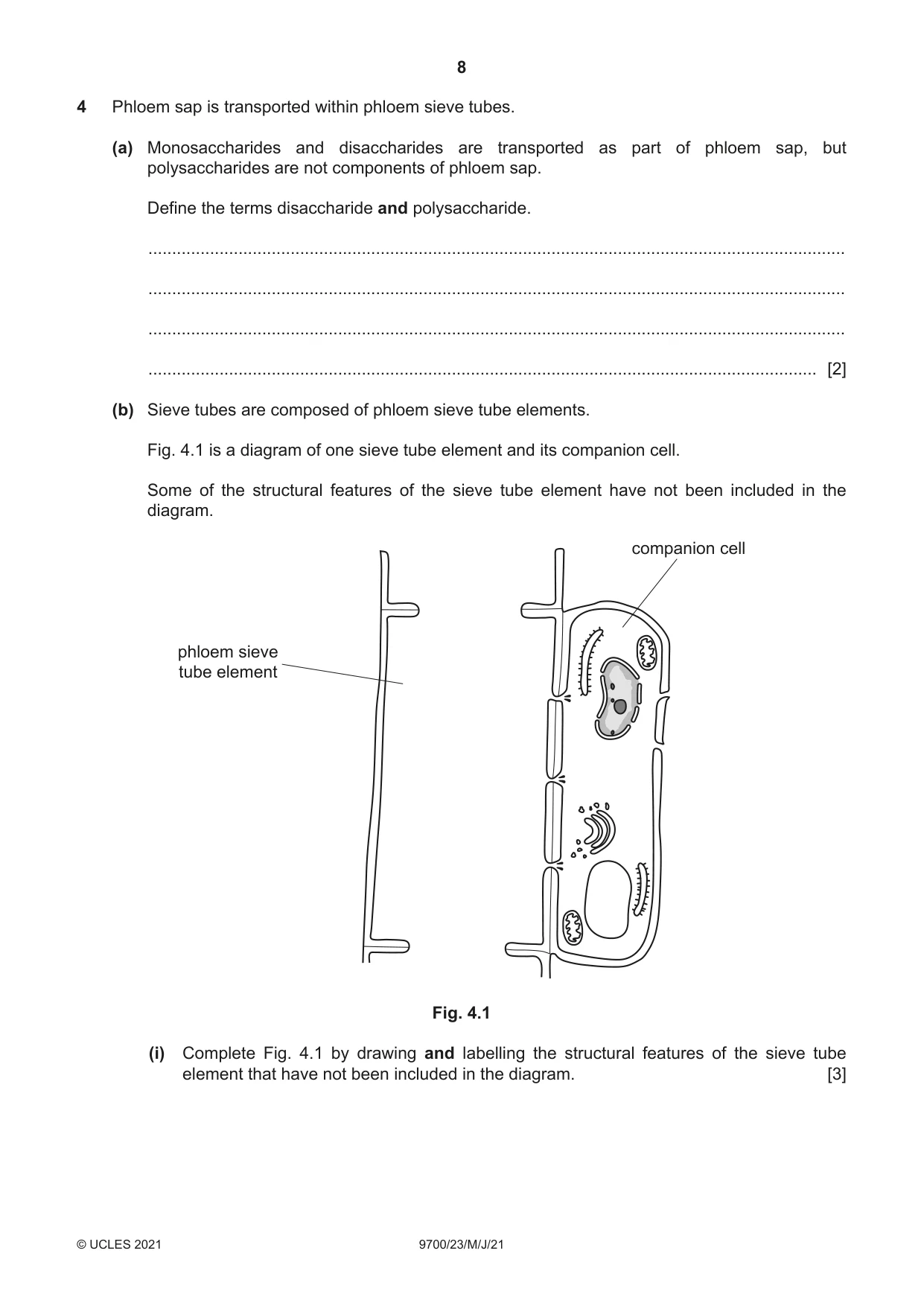
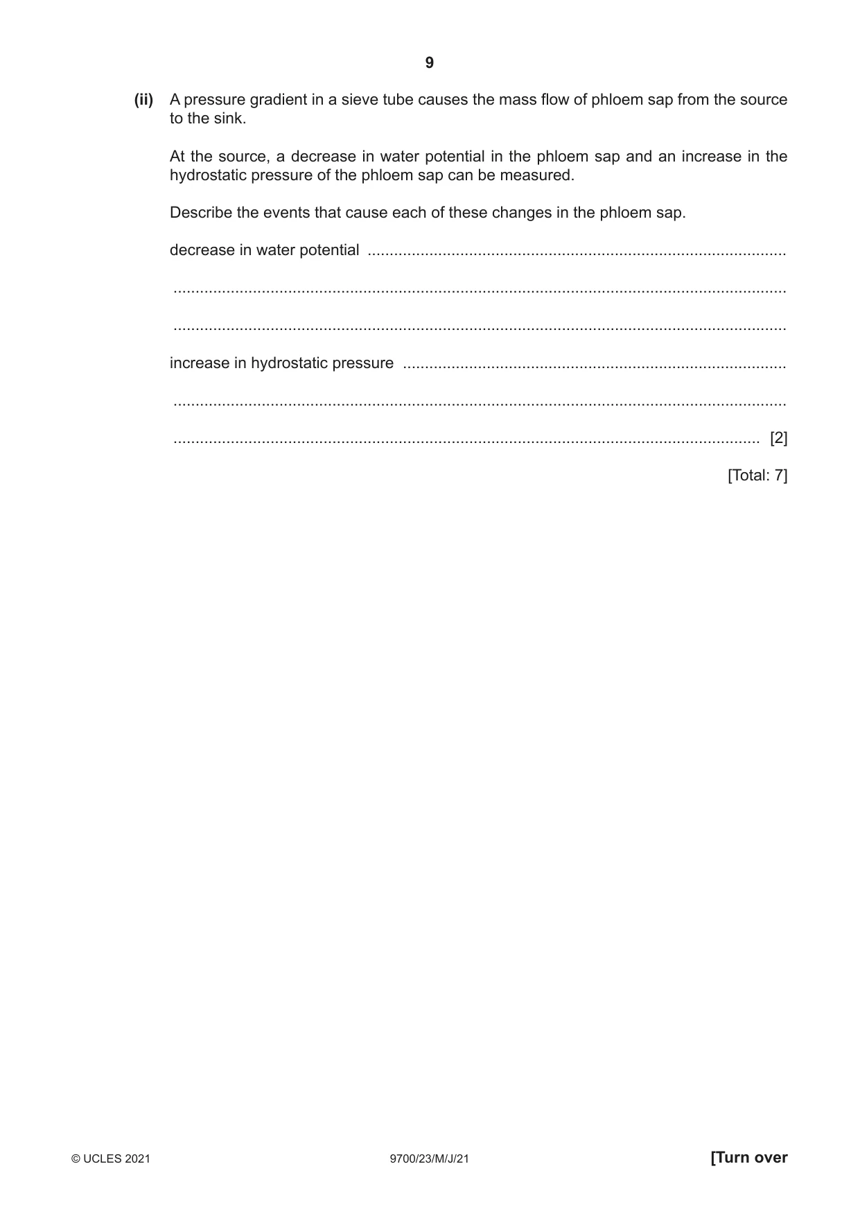
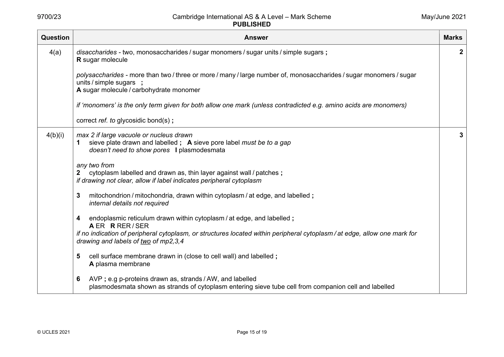
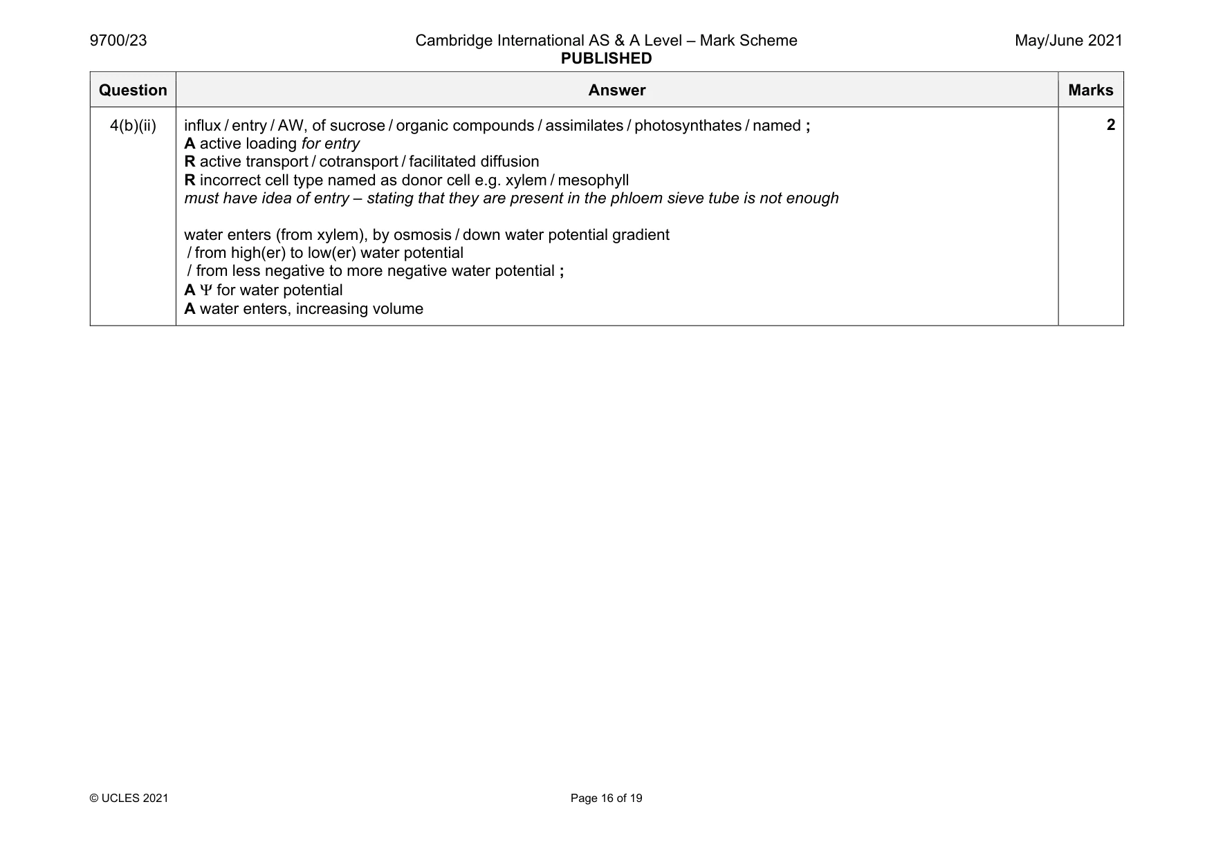
4 Phloem sap is transported within phloem sieve tubes. (a) Monosaccharides and disaccharides are transported as part of phloem sap, but polysaccharides are not components of phloem sap. Define the terms disaccharide and polysaccharide. ................................................................................................................................................... ................................................................................................................................................... ................................................................................................................................................... ............................................................................................................................................. [2] (b) Sieve tubes are composed of phloem sieve tube elements. Fig. 4.1 is a diagram of one sieve tube element and its companion cell. Some of the structural features of the sieve tube element have not been included in the diagram. companion cell phloem sieve tube element Fig. 4.1 (i) Complete Fig. 4.1 by drawing and labelling the structural features of the sieve tube element that have not been included in the diagram. [3] (ii) A pressure gradient in a sieve tube causes the mass flow of phloem sap from the source to the sink. At the source, a decrease in water potential in the phloem sap and an increase in the hydrostatic pressure of the phloem sap can be measured. Describe the events that cause each of these changes in the phloem sap. decrease in water potential ............................................................................................... ........................................................................................................................................... ........................................................................................................................................... increase in hydrostatic pressure ....................................................................................... ........................................................................................................................................... ..................................................................................................................................... [2] [Total: 7]
Mark scheme: 4(a) disaccharides - two, monosaccharides / sugar monomers / sugar units / simple sugars ; R sugar molecule polysaccharides - more than two / three or more / many / large number of, monosaccharides / sugar monomers / sugar units / simple sugars ; A sugar molecule / carbohydrate monomer if ‘monomers’ is the only term given for both allow one mark (unless contradicted e.g. amino acids are monomers) correct ref. to glycosidic bond(s) ; 4(b)(i) max 2 if large vacuole or nucleus drawn 1 sieve plate drawn and labelled ; A sieve pore label must be to a gap doesn’t need to show pores I plasmodesmata any two from 2 cytoplasm labelled and drawn as, thin layer against wall / patches ; if drawing not clear, allow if label indicates peripheral cytoplasm 3 mitochondrion / mitochondria, drawn within cytoplasm / at edge, and labelled ; internal details not required 4 endoplasmic reticulum drawn within cytoplasm / at edge, and labelled ; A ER R RER / SER if no indication of peripheral cytoplasm, or structures located within peripheral cytoplasm / at edge, allow one mark for drawing and labels of two of mp2,3,4 5 cell surface membrane drawn in (close to cell wall) and labelled ; A plasma membrane 6 AVP ; e.g p-proteins drawn as, strands / AW, and labelled plasmodesmata shown as strands of cytoplasm entering sieve tube cell from companion cell and labelled 3 Question Answer Marks 4(b)(ii) influx / entry / AW, of sucrose / organic compounds / assimilates / photosynthates / named ; A active loading for entry R active transport / cotransport / facilitated diffusion R incorrect cell type named as donor cell e.g. xylem / mesophyll must have idea of entry – stating that they are present in the phloem sieve tube is not enough water enters (from xylem), by osmosis / down water potential gradient / from high(er) to low(er) water potential / from less negative to more negative water potential ; A Ψ for water potential A water enters, increasing volume 2
Q5 · Four types of cell that can be seen in a prepared slide of blood taken from a mammal
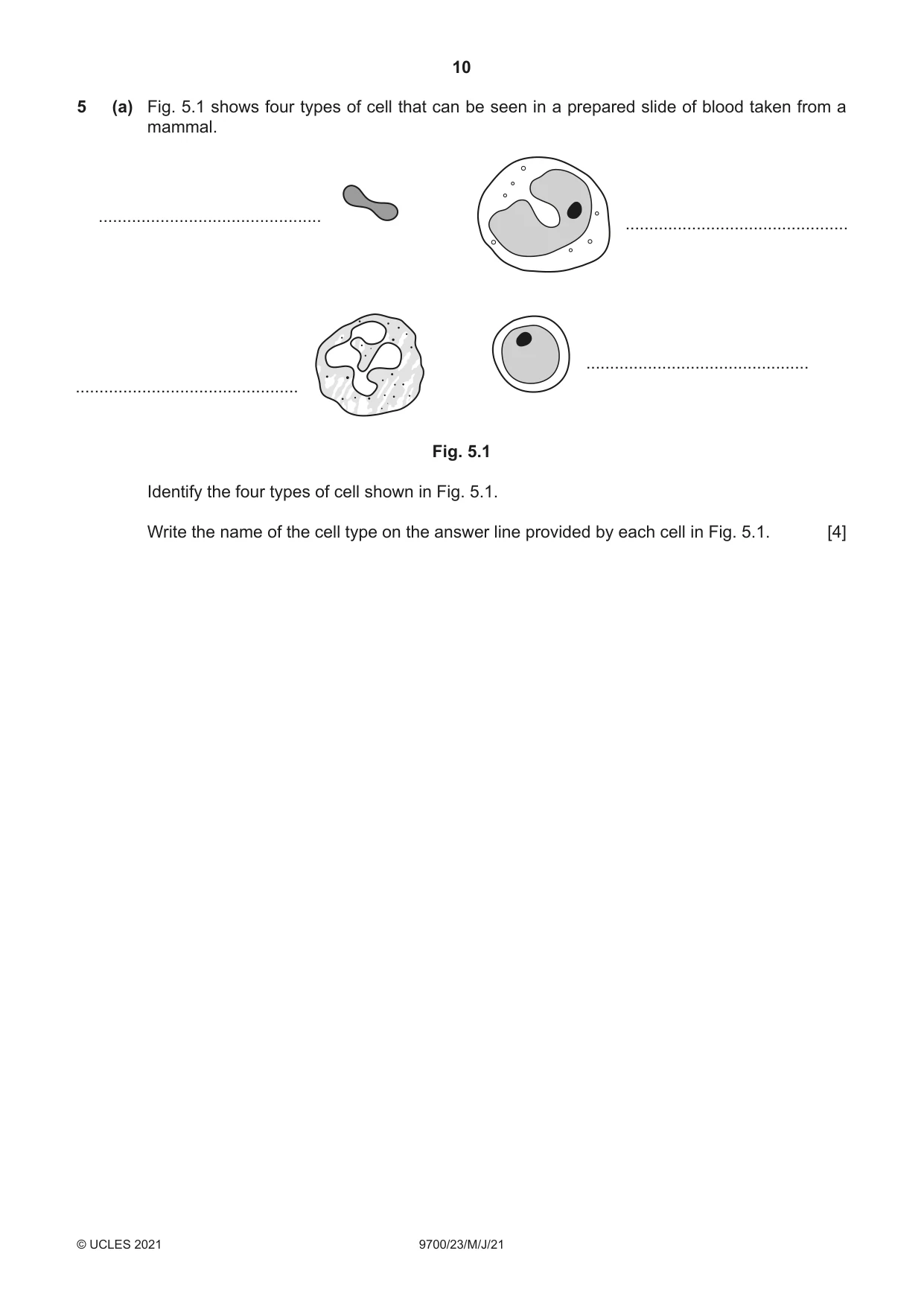
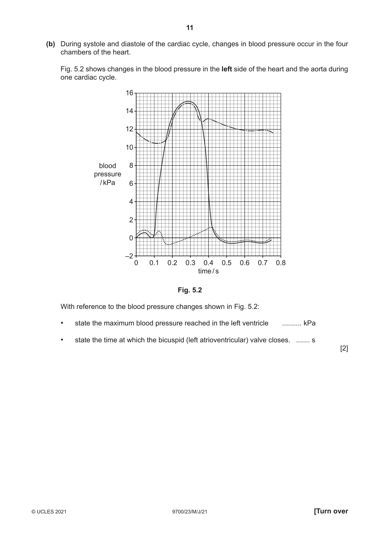
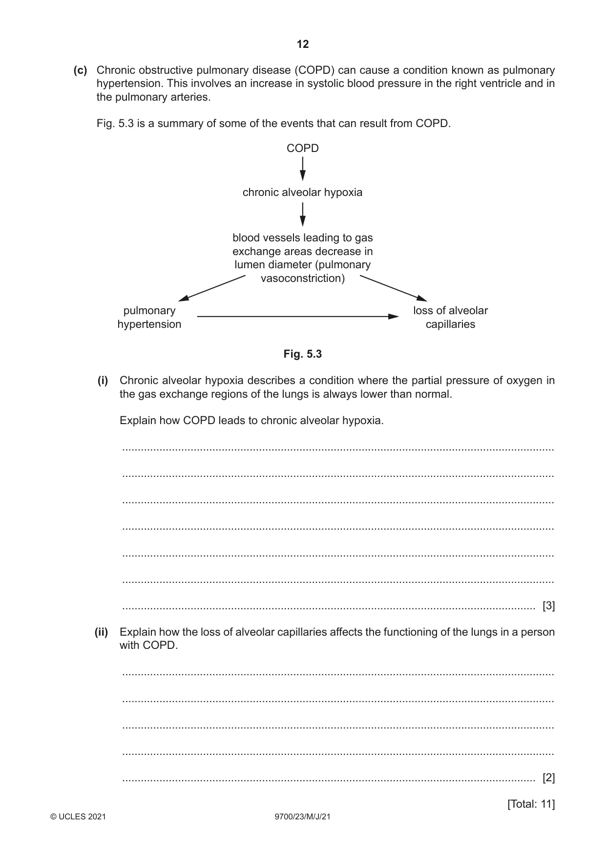
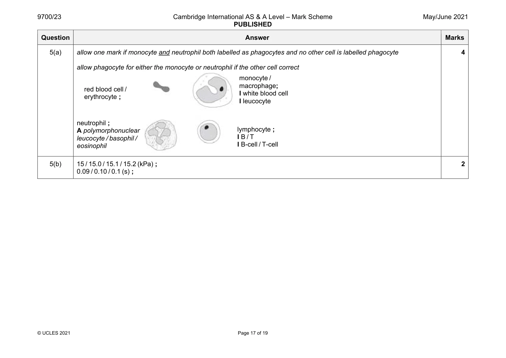
5 (a) Fig. 5.1 shows four types of cell that can be seen in a prepared slide of blood taken from a mammal. ............................................... ............................................... ............................................... ............................................... Fig. 5.1 Identify the four types of cell shown in Fig. 5.1. Write the name of the cell type on the answer line provided by each cell in Fig. 5.1. [4] (b) During systole and diastole of the cardiac cycle, changes in blood pressure occur in the four chambers of the heart. Fig. 5.2 shows changes in the blood pressure in the left side of the heart and the aorta during one cardiac cycle. 16 14 12 10 blood 8 pressure / kPa 6 4 2 0 –2 0 0.1 0.2 0.3 0.4 0.5 0.6 0.7 0.8 time / s Fig. 5.2 With reference to the blood pressure changes shown in Fig. 5.2: • state the maximum blood pressure reached in the left ventricle .......... kPa • state the time at which the bicuspid (left atrioventricular) valve closes. ....... s [2] (c) Chronic obstructive pulmonary disease (COPD) can cause a condition known as pulmonary hypertension. This involves an increase in systolic blood pressure in the right ventricle and in the pulmonary arteries. Fig. 5.3 is a summary of some of the events that can result from COPD. COPD chronic alveolar hypoxia blood vessels leading to gas exchange areas decrease in lumen diameter (pulmonary vasoconstriction) pulmonary loss of alveolar hypertension capillaries Fig. 5.3 (i) Chronic alveolar hypoxia describes a condition where the partial pressure of oxygen in the gas exchange regions of the lungs is always lower than normal. Explain how COPD leads to chronic alveolar hypoxia. ........................................................................................................................................... ........................................................................................................................................... ........................................................................................................................................... ........................................................................................................................................... ........................................................................................................................................... ........................................................................................................................................... ..................................................................................................................................... [3] (ii) Explain how the loss of alveolar capillaries affects the functioning of the lungs in a person with COPD. ........................................................................................................................................... ........................................................................................................................................... ........................................................................................................................................... ........................................................................................................................................... ..................................................................................................................................... [2] [Total: 11]
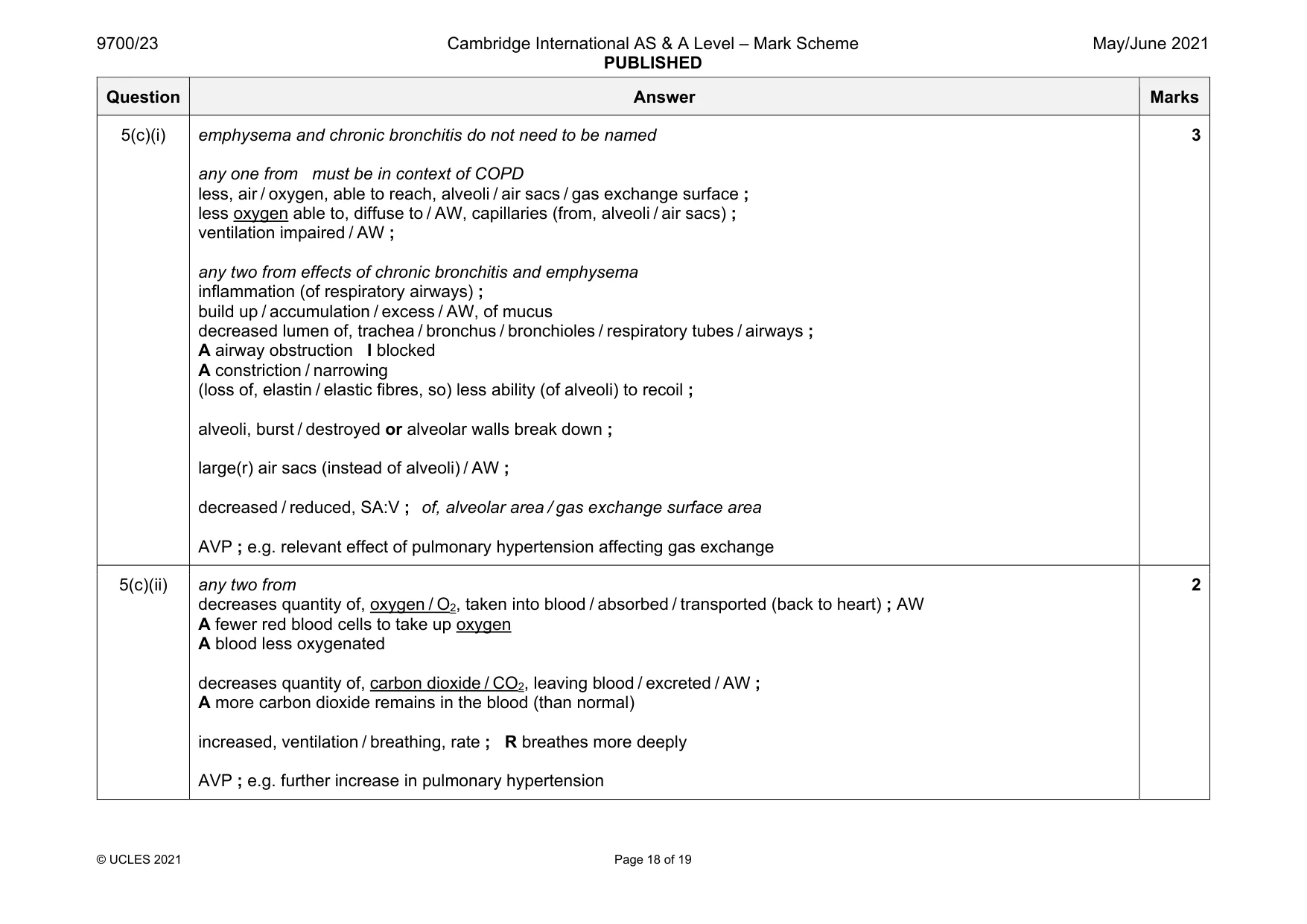
Mark scheme: 5(a) allow one mark if monocyte and neutrophil both labelled as phagocytes and no other cell is labelled phagocyte allow phagocyte for either the monocyte or neutrophil if the other cell correct 4 5(b) 15 / 15.0 / 15.1 / 15.2 (kPa) ; 0.09 / 0.10 / 0.1 (s) ; 2 red blood cell / erythrocyte ; monocyte / macrophage; I white blood cell I leucocyte lymphocyte ; I B / T I B-cell / T-cell neutrophil ; A polymorphonuclear leucocyte / basophil / eosinophil Question Answer Marks 5(c)(i) emphysema and chronic bronchitis do not need to be named any one from must be in context of COPD less, air / oxygen, able to reach, alveoli / air sacs / gas exchange surface ; less oxygen able to, diffuse to / AW, capillaries (from, alveoli / air sacs) ; ventilation impaired / AW ; any two from effects of chronic bronchitis and emphysema inflammation (of respiratory airways) ; build up / accumulation / excess / AW, of mucus decreased lumen of, trachea / bronchus / bronchioles / respiratory tubes / airways ; A airway obstruction I blocked A constriction / narrowing (loss of, elastin / elastic fibres, so) less ability (of alveoli) to recoil ; alveoli, burst / destroyed or alveolar walls break down ; large(r) air sacs (instead of alveoli) / AW ; decreased / reduced, SA:V ; of, alveolar area / gas exchange surface area AVP ; e.g. relevant effect of pulmonary hypertension affecting gas exchange 3 5(c)(ii) any two from decreases quantity of, oxygen / O2, taken into blood / absorbed / transported (back to heart) ; AW A fewer red blood cells to take up oxygen A blood less oxygenated decreases quantity of, carbon dioxide / CO2, leaving blood / excreted / AW ; A more carbon dioxide remains in the blood (than normal) increased, ventilation / breathing, rate ; R breathes more deeply AVP ; e.g. further increase in pulmonary hypertension 2
Q6 · The best time to obtain a clear image of chromosomes during a mitotic cell cycle is…
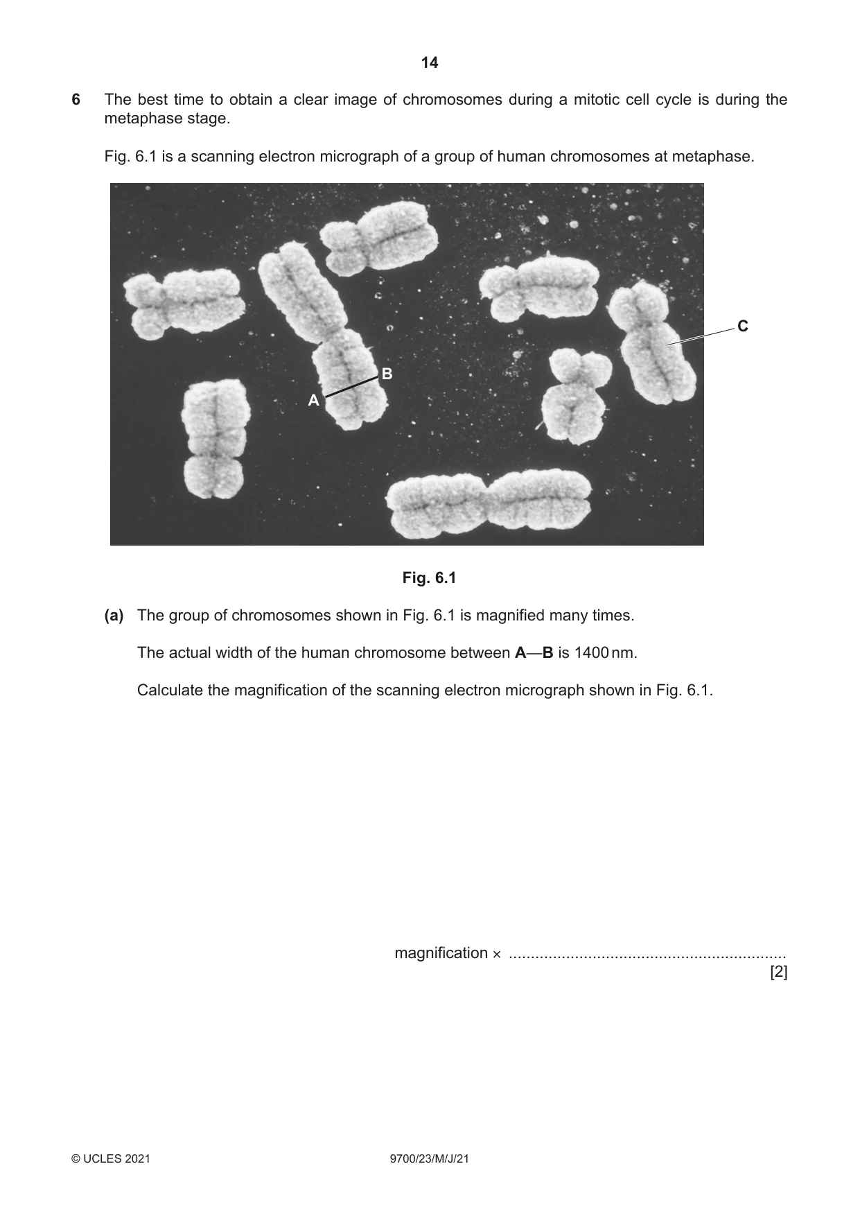
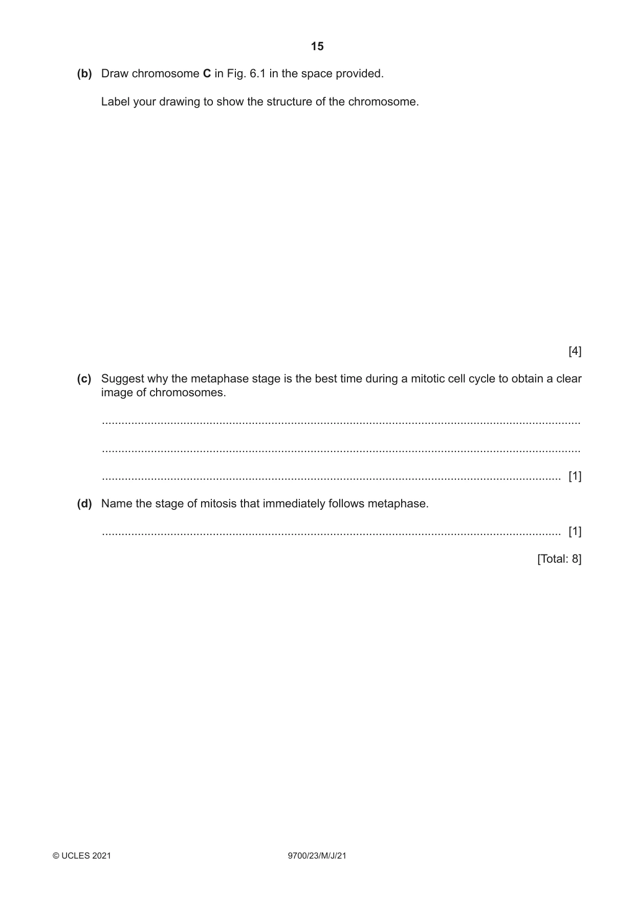
6 The best time to obtain a clear image of chromosomes during a mitotic cell cycle is during the metaphase stage. Fig. 6.1 is a scanning electron micrograph of a group of human chromosomes at metaphase. C B A Fig. 6.1 (a) The group of chromosomes shown in Fig. 6.1 is magnified many times. The actual width of the human chromosome between A—B is 1400 nm. Calculate the magnification of the scanning electron micrograph shown in Fig. 6.1. magnification × ............................................................... [2] (b) Draw chromosome C in Fig. 6.1 in the space provided. Label your drawing to show the structure of the chromosome. [4] (c) Suggest why the metaphase stage is the best time during a mitotic cell cycle to obtain a clear image of chromosomes. ................................................................................................................................................... ................................................................................................................................................... ............................................................................................................................................. [1] (d) Name the stage of mitosis that immediately follows metaphase. ............................................................................................................................................. [1] [Total: 8]
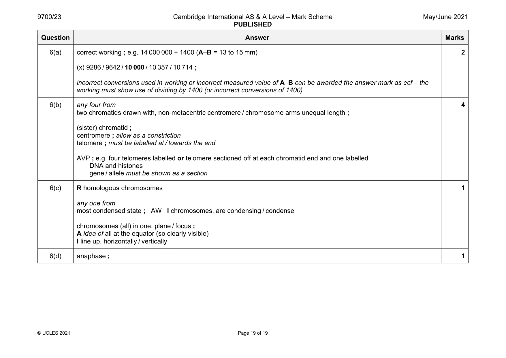
Mark scheme: 6(a) correct working ; e.g. 14 000 000 ÷ 1400 (A–B = 13 to 15 mm) (x) 9286 / 9642 / 10 000 / 10 357 / 10 714 ; incorrect conversions used in working or incorrect measured value of A–B can be awarded the answer mark as ecf – the working must show use of dividing by 1400 (or incorrect conversions of 1400) 2 6(b) any four from two chromatids drawn with, non-metacentric centromere / chromosome arms unequal length ; (sister) chromatid ; centromere ; allow as a constriction telomere ; must be labelled at / towards the end AVP ; e.g. four telomeres labelled or telomere sectioned off at each chromatid end and one labelled DNA and histones gene / allele must be shown as a section 4 6(c) R homologous chromosomes any one from most condensed state ; AW I chromosomes, are condensing / condense chromosomes (all) in one, plane / focus ; A idea of all at the equator (so clearly visible) I line up. horizontally / vertically 1 6(d) anaphase ; 1
What was in this paper
The subtopics covered by these 6 questions, and how many questions each got. Open one in a new tab to see every Cambridge question on it.
What you needed in this session
Cambridge’s own grade thresholds for 2021 May/June, Paper 2 · Variant 3. A higher threshold means an easier paper — the bar moves with how the cohort did.