Cambridge A Level Biology 9700 — 2021 May/June Paper 2 · Variant 1
9700/21/M/J/21 · 6 questions · 60 marks · ≈68 min
The question paper and its mark scheme, free to read here and free to download. This is Cambridge’s own paper, exactly as it was sat.
Question paper16 pages
Mark scheme16 pages
Answers below. Sit the paper first if you are practising.
Questions as text
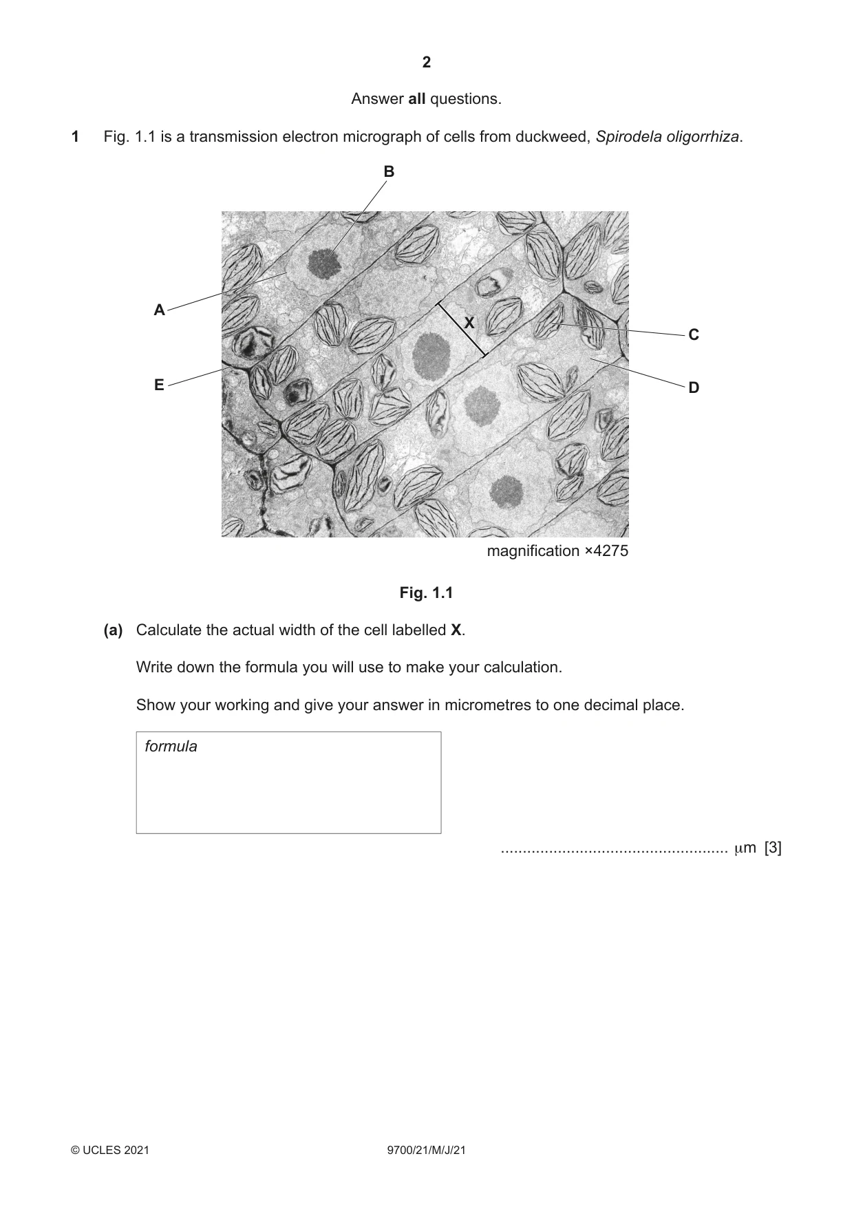
Q1 · A transmission electron micrograph of cells from duckweed, Spirodela oligorrhiza
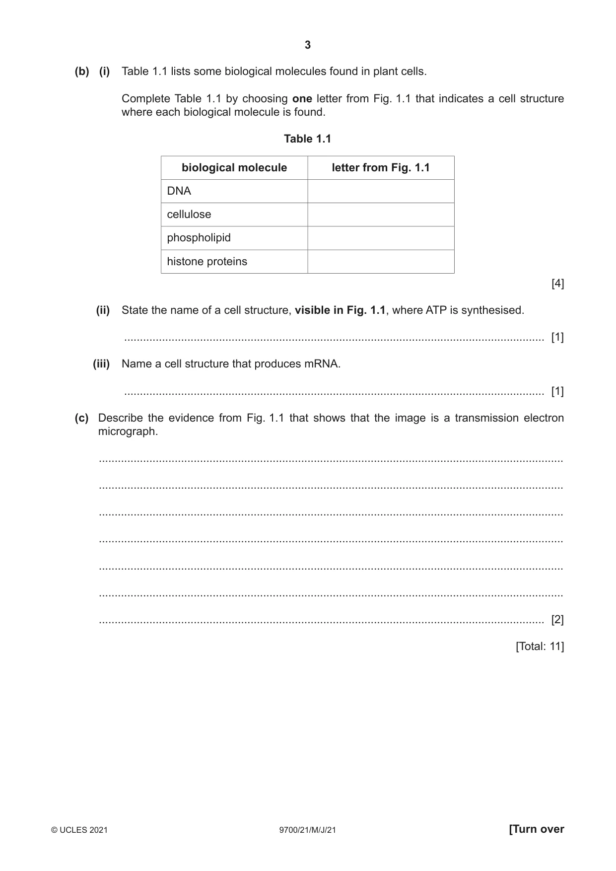
1 Fig. 1.1 is a transmission electron micrograph of cells from duckweed, Spirodela oligorrhiza. B A X C E D magnification ×4275 Fig. 1.1 (a) Calculate the actual width of the cell labelled X. Write down the formula you will use to make your calculation. Show your working and give your answer in micrometres to one decimal place. formula .................................................... µm [3] (b) (i) Table 1.1 lists some biological molecules found in plant cells. Complete Table 1.1 by choosing one letter from Fig. 1.1 that indicates a cell structure where each biological molecule is found. Table 1.1 biological molecule letter from Fig. 1.1 DNA cellulose phospholipid histone proteins [4] (ii) State the name of a cell structure, visible in Fig. 1.1, where ATP is synthesised. ..................................................................................................................................... [1] (iii) Name a cell structure that produces mRNA. ..................................................................................................................................... [1] (c) Describe the evidence from Fig. 1.1 that shows that the image is a transmission electron micrograph. ................................................................................................................................................... ................................................................................................................................................... ................................................................................................................................................... ................................................................................................................................................... ................................................................................................................................................... ................................................................................................................................................... ............................................................................................................................................. [2] [Total: 11]
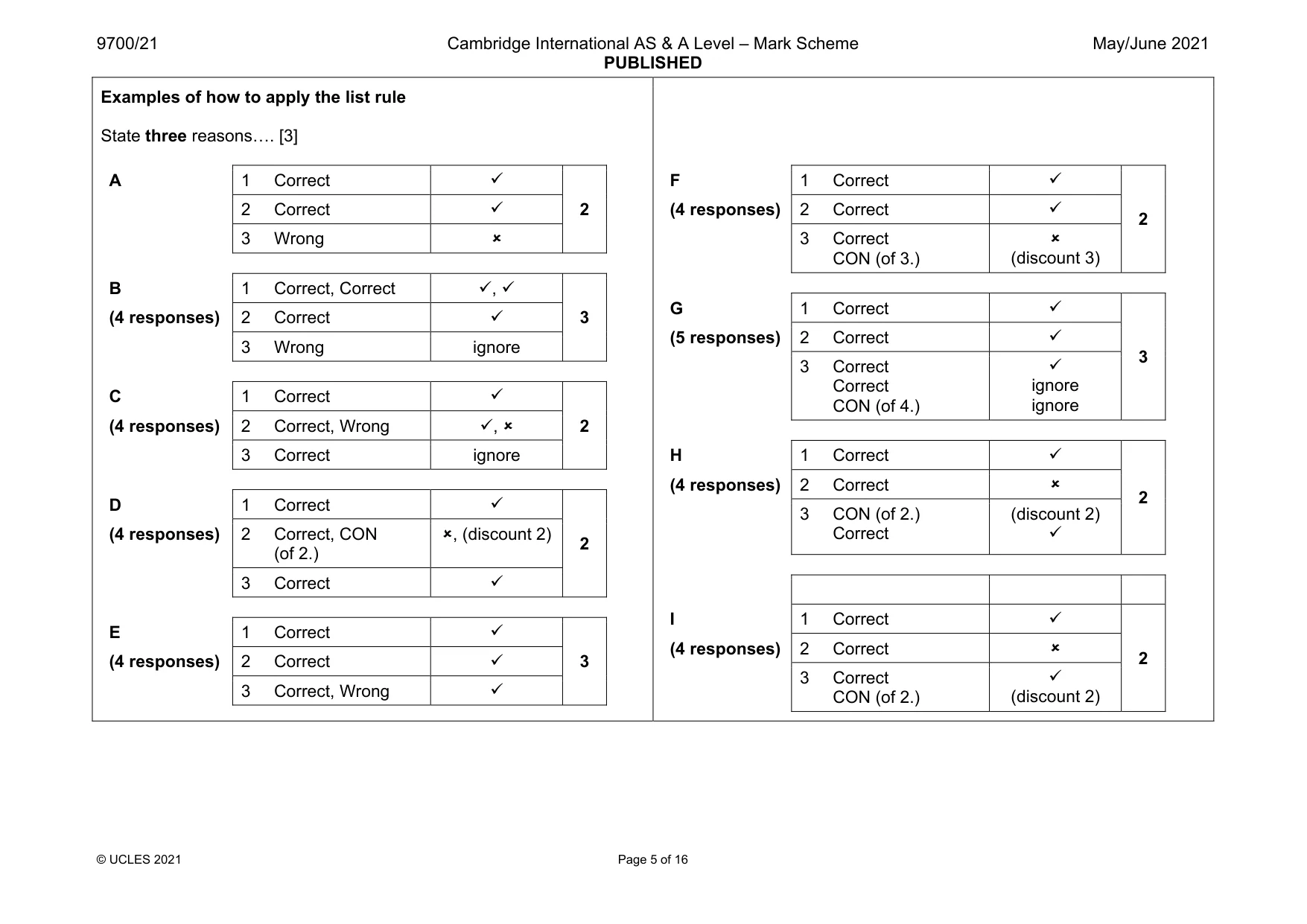
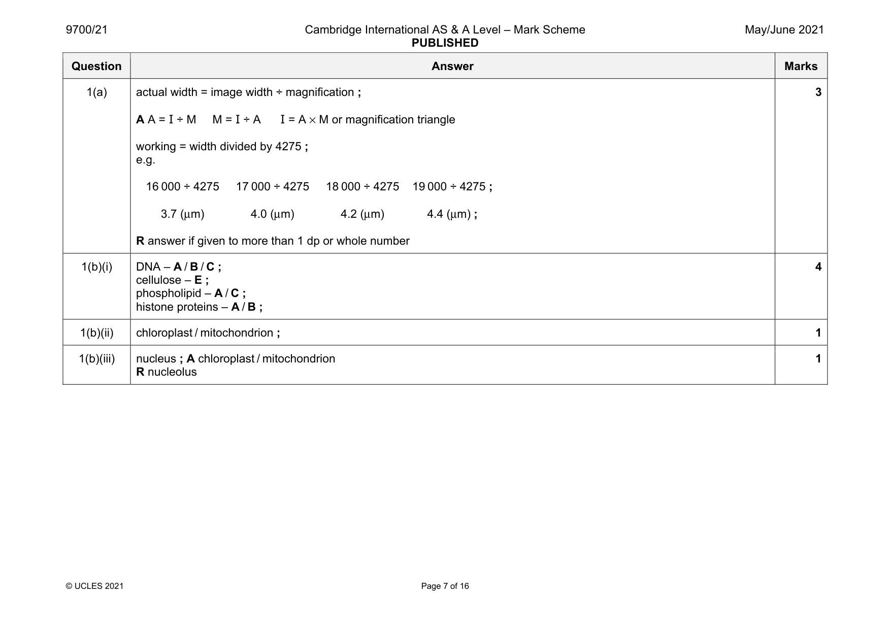
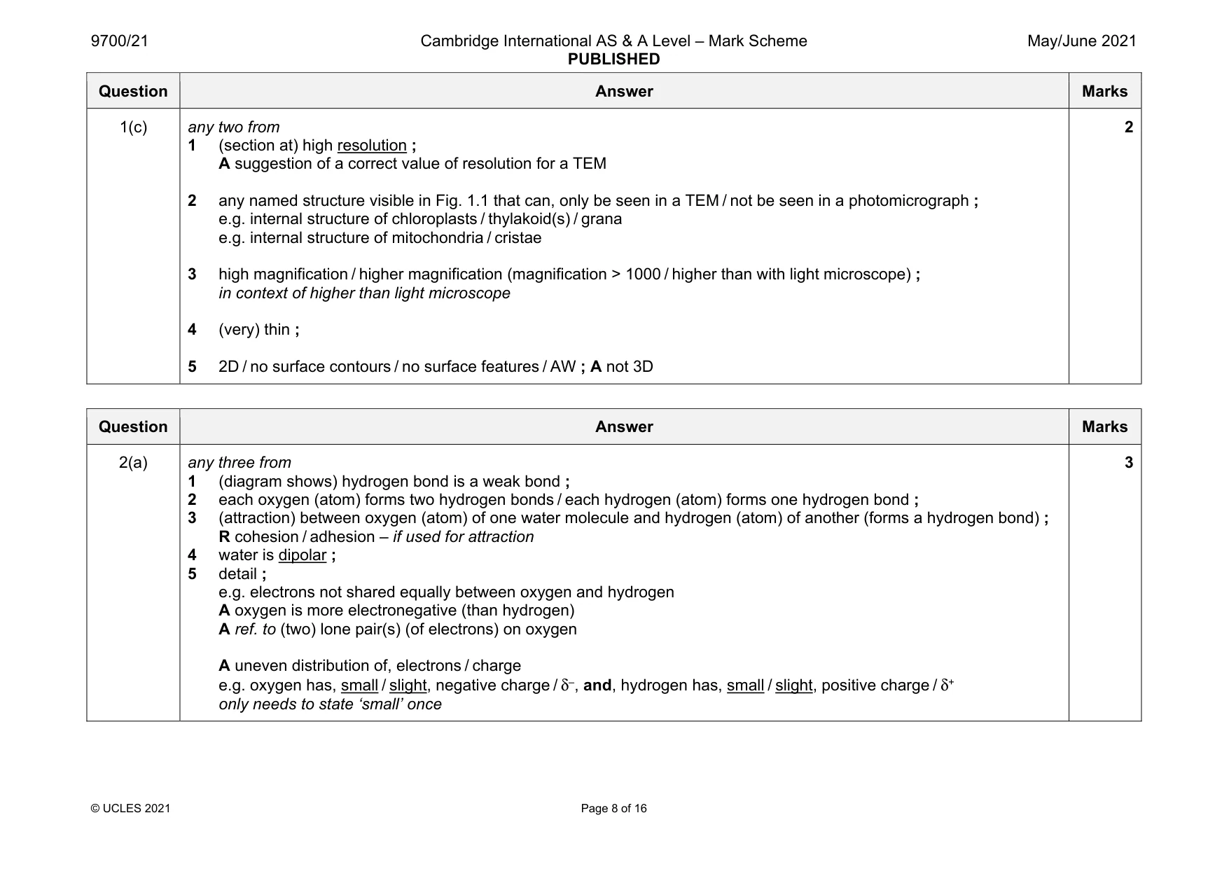
Mark scheme: 1(a) actual width = image width ÷ magnification ; A A = I ÷ M M = I ÷ A I = A × M or magnification triangle working = width divided by 4275 ; e.g. 16 000 ÷ 4275 17 000 ÷ 4275 18 000 ÷ 4275 19 000 ÷ 4275 ; 3.7 (μm) 4.0 (μm) 4.2 (μm) 4.4 (μm) ; R answer if given to more than 1 dp or whole number 3 1(b)(i) DNA – A / B / C ; cellulose – E ; phospholipid – A / C ; histone proteins – A / B ; 4 1(b)(ii) chloroplast / mitochondrion ; 1 1(b)(iii) nucleus ; A chloroplast / mitochondrion R nucleolus 1 Question Answer Marks 1(c) any two from 1 (section at) high resolution ; A suggestion of a correct value of resolution for a TEM 2 any named structure visible in Fig. 1.1 that can, only be seen in a TEM / not be seen in a photomicrograph ; e.g. internal structure of chloroplasts / thylakoid(s) / grana e.g. internal structure of mitochondria / cristae 3 high magnification / higher magnification (magnification > 1000 / higher than with light microscope) ; in context of higher than light microscope 4 (very) thin ; 5 2D / no surface contours / no surface features / AW ; A not 3D 2
More questions on Cells as the basic units of living organisms
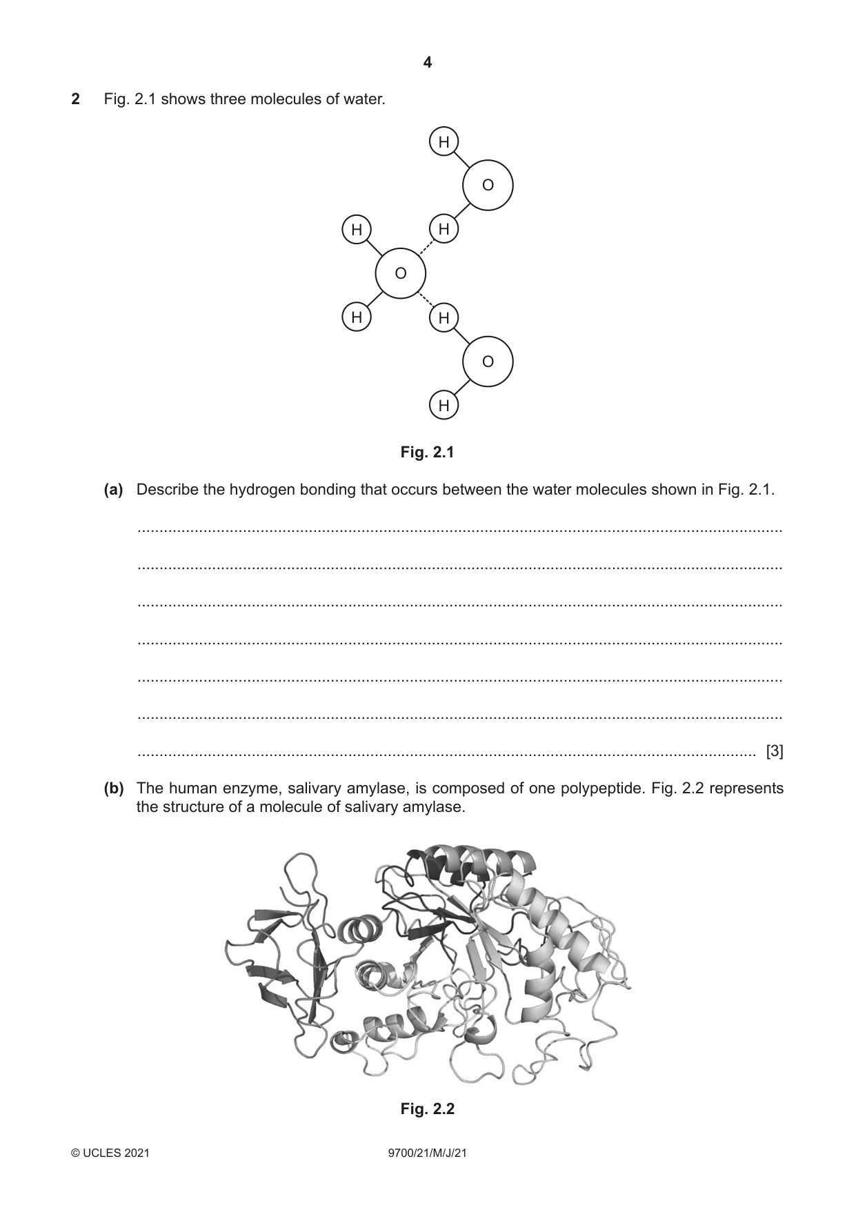
Q2 · Three molecules of water
2 Fig. 2.1 shows three molecules of water. H O H H O H H O H Fig. 2.1 (a) Describe the hydrogen bonding that occurs between the water molecules shown in Fig. 2.1. ................................................................................................................................................... ................................................................................................................................................... ................................................................................................................................................... ................................................................................................................................................... ................................................................................................................................................... ................................................................................................................................................... ............................................................................................................................................. [3] (b) The human enzyme, salivary amylase, is composed of one polypeptide. Fig. 2.2 represents the structure of a molecule of salivary amylase. Fig. 2.2 (i) Explain the role of hydrogen bonding in maintaining the secondary structure of proteins, such as salivary amylase. ........................................................................................................................................... ........................................................................................................................................... ..................................................................................................................................... [1] (ii) Explain the role of hydrogen bonding in maintaining the tertiary structure of proteins such as salivary amylase. ........................................................................................................................................... ........................................................................................................................................... ........................................................................................................................................... ........................................................................................................................................... ..................................................................................................................................... [2] (c) Outline the importance of water as a solvent in plants. ................................................................................................................................................... ................................................................................................................................................... ................................................................................................................................................... ................................................................................................................................................... ................................................................................................................................................... ................................................................................................................................................... ............................................................................................................................................. [3] [Total: 9]
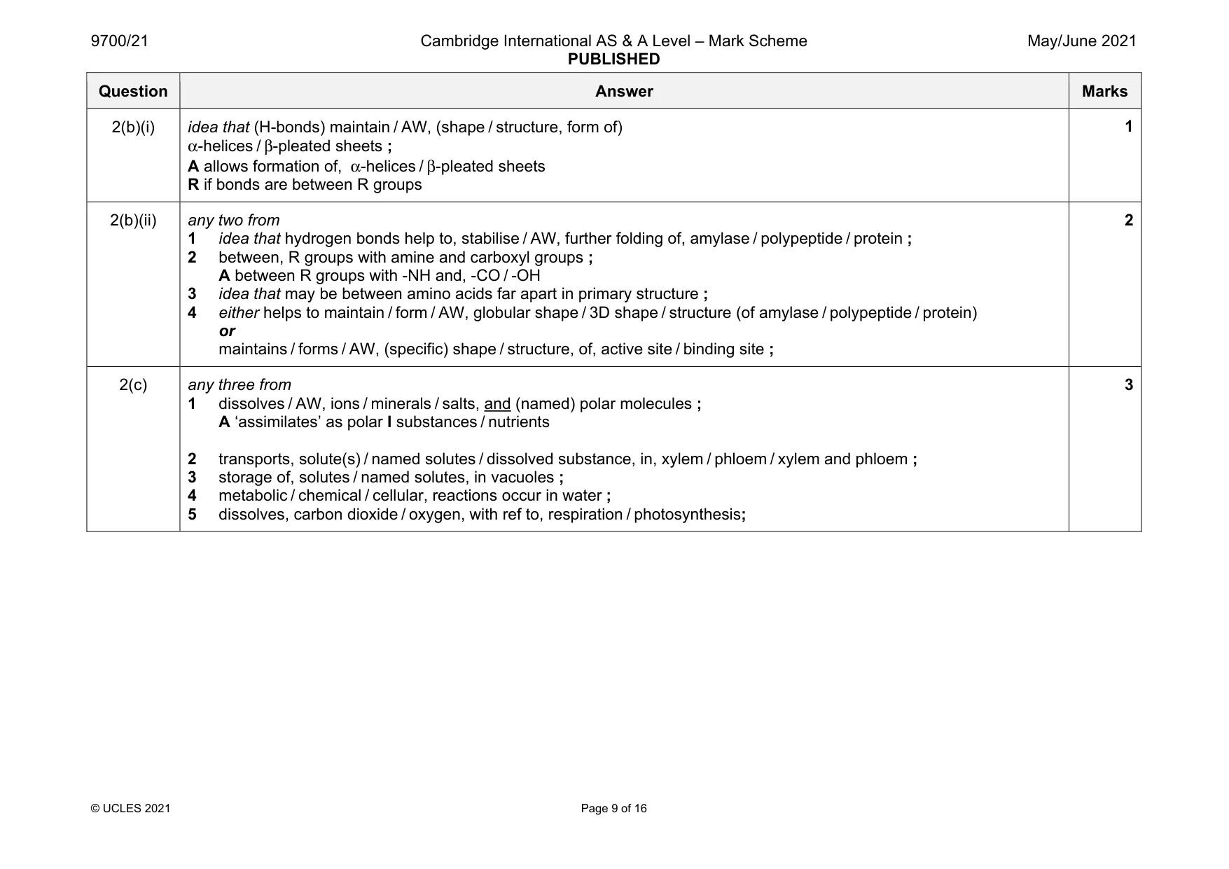
Mark scheme: 2(a) any three from 1 (diagram shows) hydrogen bond is a weak bond ; 2 each oxygen (atom) forms two hydrogen bonds / each hydrogen (atom) forms one hydrogen bond ; 3 (attraction) between oxygen (atom) of one water molecule and hydrogen (atom) of another (forms a hydrogen bond) ; R cohesion / adhesion – if used for attraction 4 water is dipolar ; 5 detail ; e.g. electrons not shared equally between oxygen and hydrogen A oxygen is more electronegative (than hydrogen) A ref. to (two) lone pair(s) (of electrons) on oxygen A uneven distribution of, electrons / charge e.g. oxygen has, small / slight, negative charge / δ–, and, hydrogen has, small / slight, positive charge / δ+ only needs to state ‘small’ once 3 Question Answer Marks 2(b)(i) idea that (H-bonds) maintain / AW, (shape / structure, form of) α-helices / β-pleated sheets ; A allows formation of, α-helices / β-pleated sheets R if bonds are between R groups 1 2(b)(ii) any two from 1 idea that hydrogen bonds help to, stabilise / AW, further folding of, amylase / polypeptide / protein ; 2 between, R groups with amine and carboxyl groups ; A between R groups with -NH and, -CO / -OH 3 idea that may be between amino acids far apart in primary structure ; 4 either helps to maintain / form / AW, globular shape / 3D shape / structure (of amylase / polypeptide / protein) or maintains / forms / AW, (specific) shape / structure, of, active site / binding site ; 2 2(c) any three from 1 dissolves / AW, ions / minerals / salts, and (named) polar molecules ; A ‘assimilates’ as polar I substances / nutrients 2 transports, solute(s) / named solutes / dissolved substance, in, xylem / phloem / xylem and phloem ; 3 storage of, solutes / named solutes, in vacuoles ; 4 metabolic / chemical / cellular, reactions occur in water ; 5 dissolves, carbon dioxide / oxygen, with ref to, respiration / photosynthesis; 3
Q3 · Visking tubing can be used to investigate the properties of cell membranes
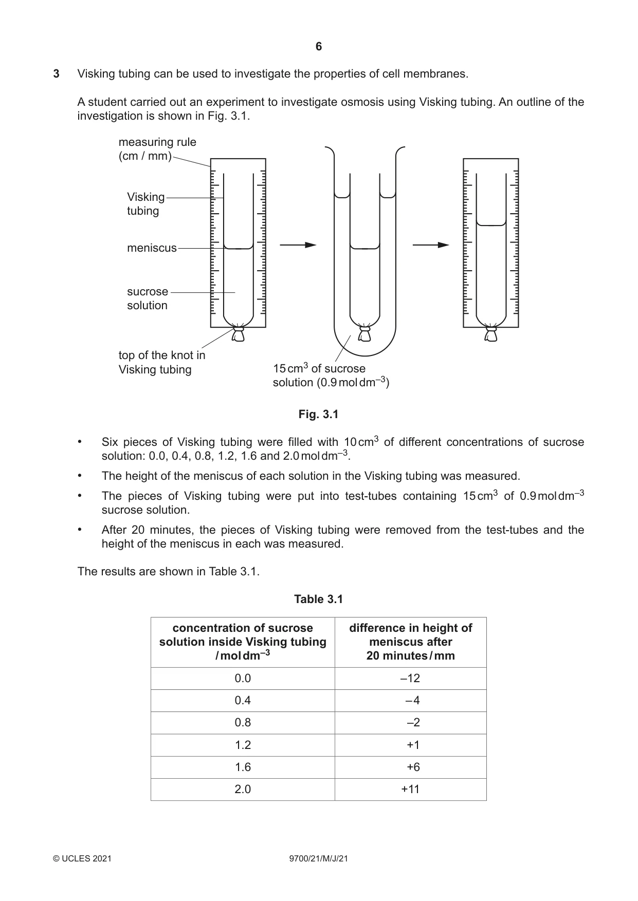
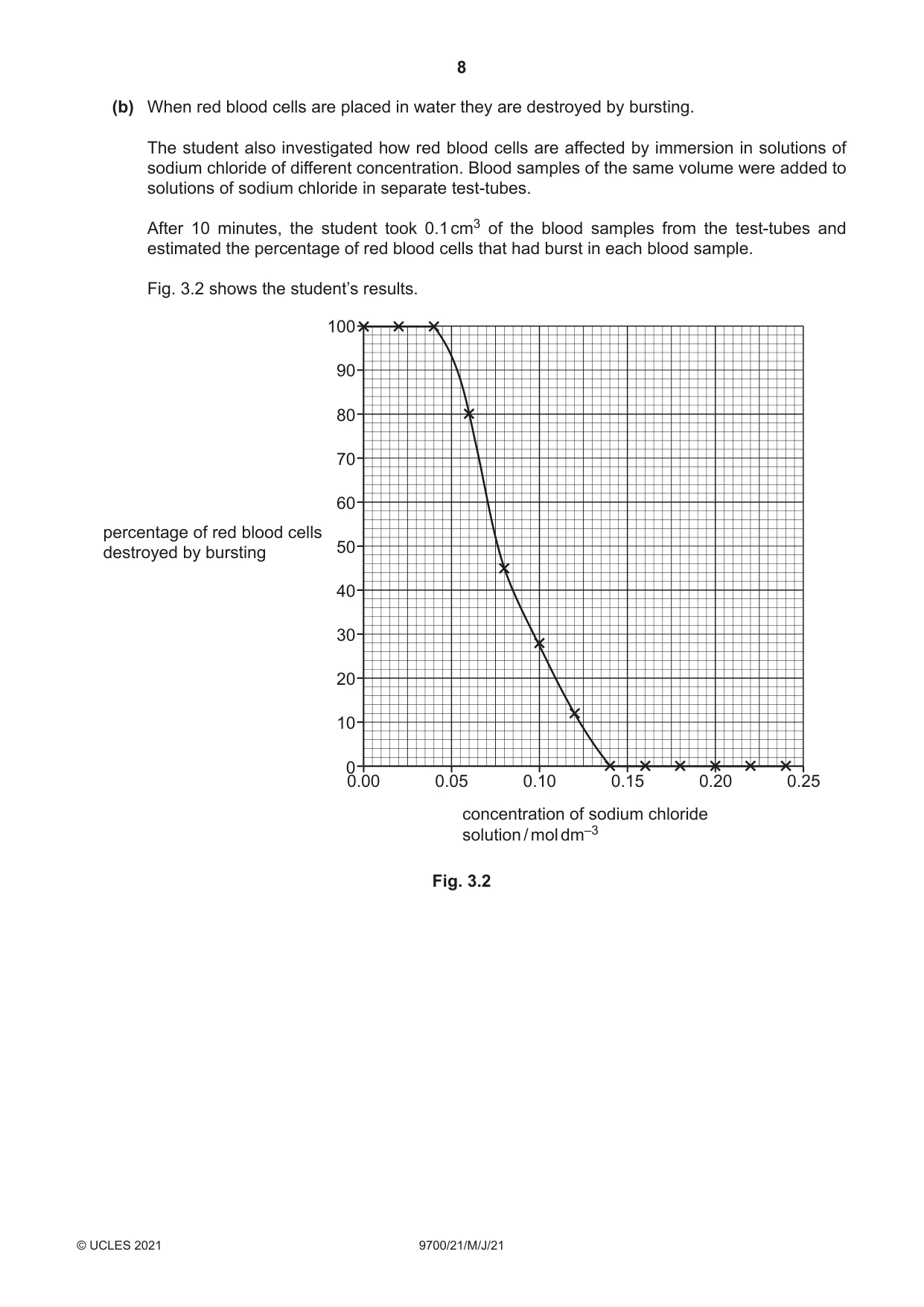
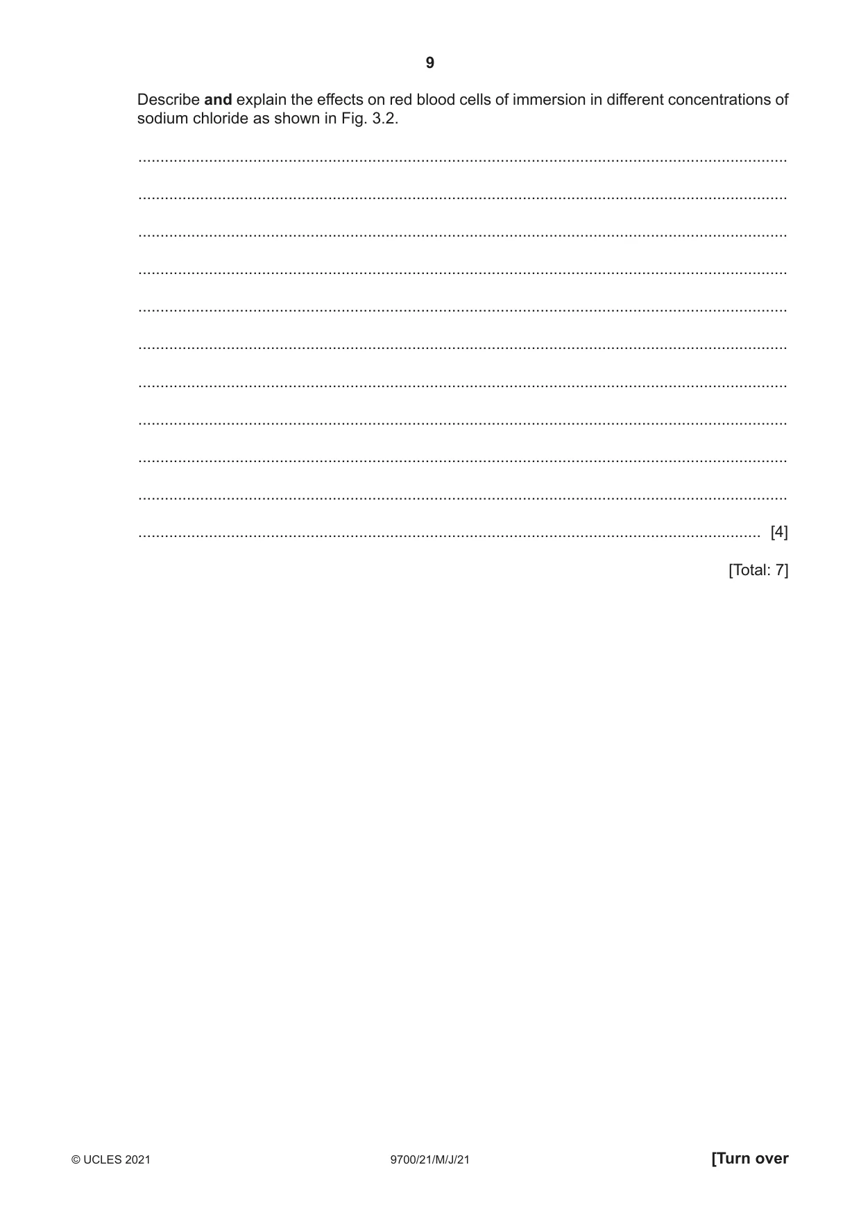
3 Visking tubing can be used to investigate the properties of cell membranes. A student carried out an experiment to investigate osmosis using Visking tubing. An outline of the investigation is shown in Fig. 3.1. measuring rule (cm / mm) Visking tubing meniscus sucrose solution top of the knot in Visking tubing 15 cm3 of sucrose solution (0.9 mol dm–3) Fig. 3.1 • Six pieces of Visking tubing were filled with 10 cm3 of different concentrations of sucrose solution: 0.0, 0.4, 0.8, 1.2, 1.6 and 2.0 mol dm–3. • The height of the meniscus of each solution in the Visking tubing was measured. • The pieces of Visking tubing were put into test-tubes containing 15 cm3 of 0.9 mol dm–3 sucrose solution. • After 20 minutes, the pieces of Visking tubing were removed from the test-tubes and the height of the meniscus in each was measured. The results are shown in Table 3.1. Table 3.1 concentration of sucrose difference in height of solution inside Visking tubing meniscus after / mol dm–3 20 minutes / mm 0.0 –12 0.4 – 4 0.8 –2 1.2 +1 1.6 +6 2.0 +11 (a) The Visking tubing used by the student was not permeable to sucrose. Explain the results shown in Table 3.1. ................................................................................................................................................... ................................................................................................................................................... ................................................................................................................................................... ................................................................................................................................................... ................................................................................................................................................... ................................................................................................................................................... ................................................................................................................................................... ............................................................................................................................................. [3] (b) When red blood cells are placed in water they are destroyed by bursting. The student also investigated how red blood cells are affected by immersion in solutions of sodium chloride of different concentration. Blood samples of the same volume were added to solutions of sodium chloride in separate test-tubes. After 10 minutes, the student took 0.1 cm3 of the blood samples from the test-tubes and estimated the percentage of red blood cells that had burst in each blood sample. Fig. 3.2 shows the student’s results. 100 90 80 70 60 percentage of red blood cells 50destroyed by bursting 40 30 20 10 00.00 0.05 0.10 0.15 0.20 0.25 concentration of sodium chloride solution / mol dm–3 Fig. 3.2 Describe and explain the effects on red blood cells of immersion in different concentrations of sodium chloride as shown in Fig. 3.2. ................................................................................................................................................... ................................................................................................................................................... ................................................................................................................................................... ................................................................................................................................................... ................................................................................................................................................... ................................................................................................................................................... ................................................................................................................................................... ................................................................................................................................................... ................................................................................................................................................... ................................................................................................................................................... ............................................................................................................................................. [4] [Total: 7]
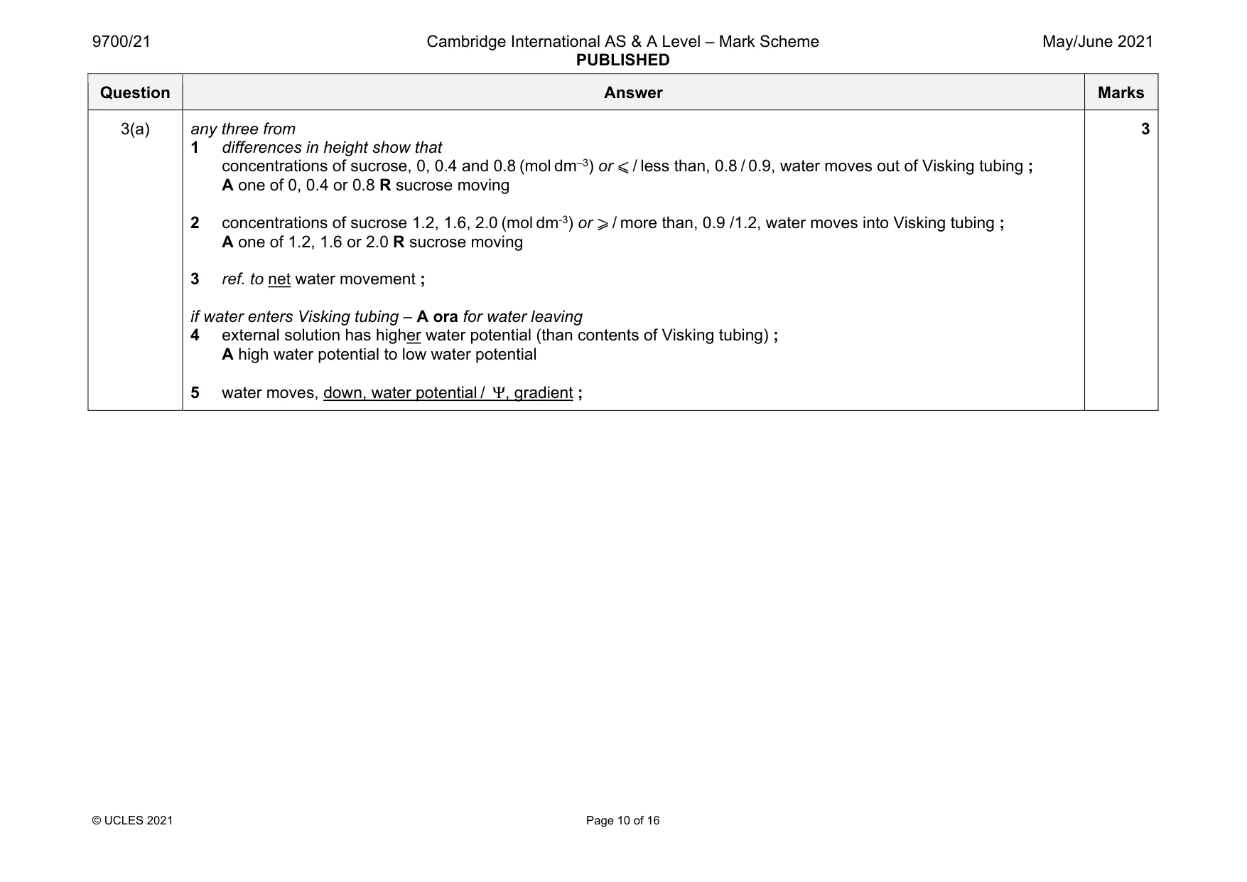
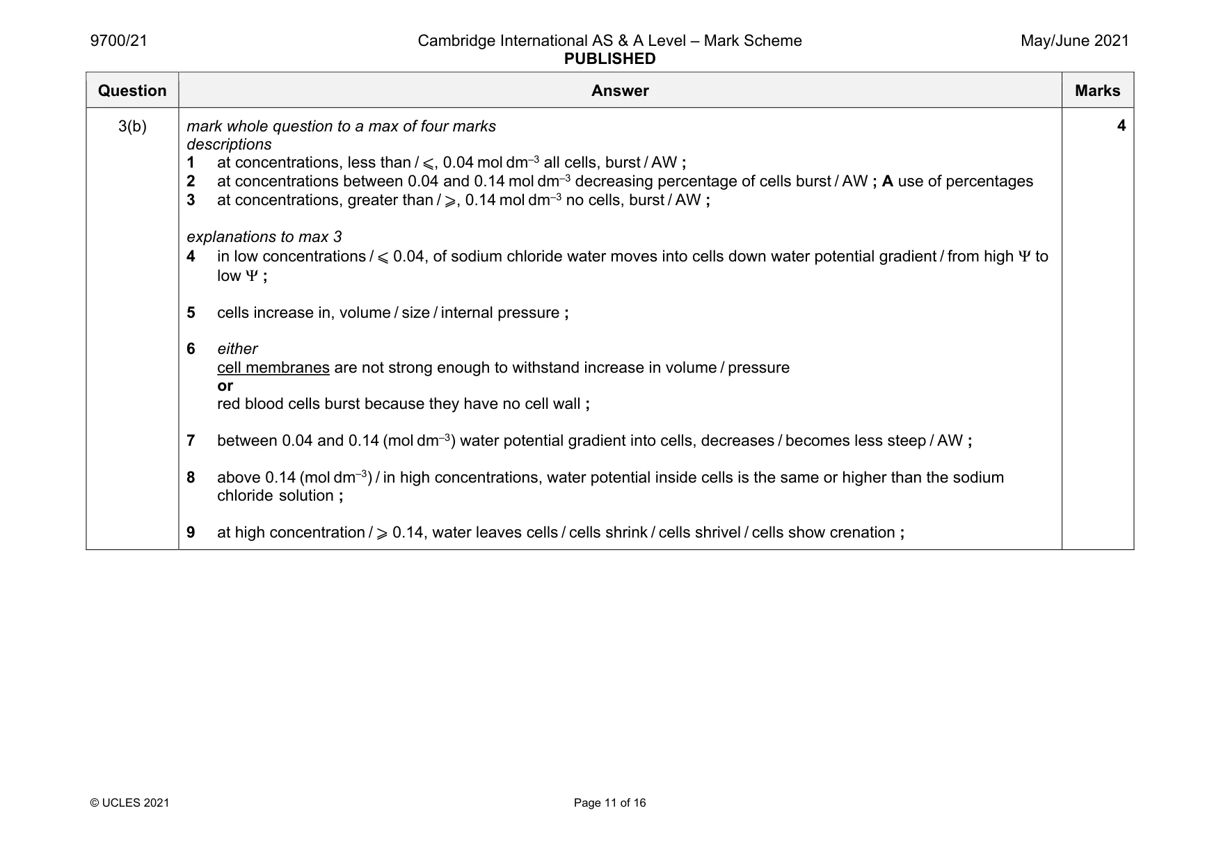
Mark scheme: 3(a) any three from 1 differences in height show that concentrations of sucrose, 0, 0.4 and 0.8 (mol dm–3) or ⩽ / less than, 0.8 / 0.9, water moves out of Visking tubing ; A one of 0, 0.4 or 0.8 R sucrose moving 2 concentrations of sucrose 1.2, 1.6, 2.0 (mol dm-3) or ⩾ / more than, 0.9 /1.2, water moves into Visking tubing ; A one of 1.2, 1.6 or 2.0 R sucrose moving 3 ref. to net water movement ; if water enters Visking tubing – A ora for water leaving 4 external solution has higher water potential (than contents of Visking tubing) ; A high water potential to low water potential 5 water moves, down, water potential / Ψ, gradient ; 3 Question Answer Marks 3(b) mark whole question to a max of four marks descriptions 1 at concentrations, less than / ⩽, 0.04 mol dm–3 all cells, burst / AW ; 2 at concentrations between 0.04 and 0.14 mol dm–3 decreasing percentage of cells burst / AW ; A use of percentages 3 at concentrations, greater than / ⩾, 0.14 mol dm–3 no cells, burst / AW ; explanations to max 3 4 in low concentrations / ⩽ 0.04, of sodium chloride water moves into cells down water potential gradient / from high Ψ to low Ψ ; 5 cells increase in, volume / size / internal pressure ; 6 either cell membranes are not strong enough to withstand increase in volume / pressure or red blood cells burst because they have no cell wall ; 7 between 0.04 and 0.14 (mol dm–3) water potential gradient into cells, decreases / becomes less steep / AW ; 8 above 0.14 (mol dm–3) / in high concentrations, water potential inside cells is the same or higher than the sodium chloride solution ; 9 at high concentration / ⩾ 0.14, water leaves cells / cells shrink / cells shrivel / cells show crenation ; 4
Q4 · The induced-fit hypothesis and the lock-and-key hypothesis are used to describe the mode…
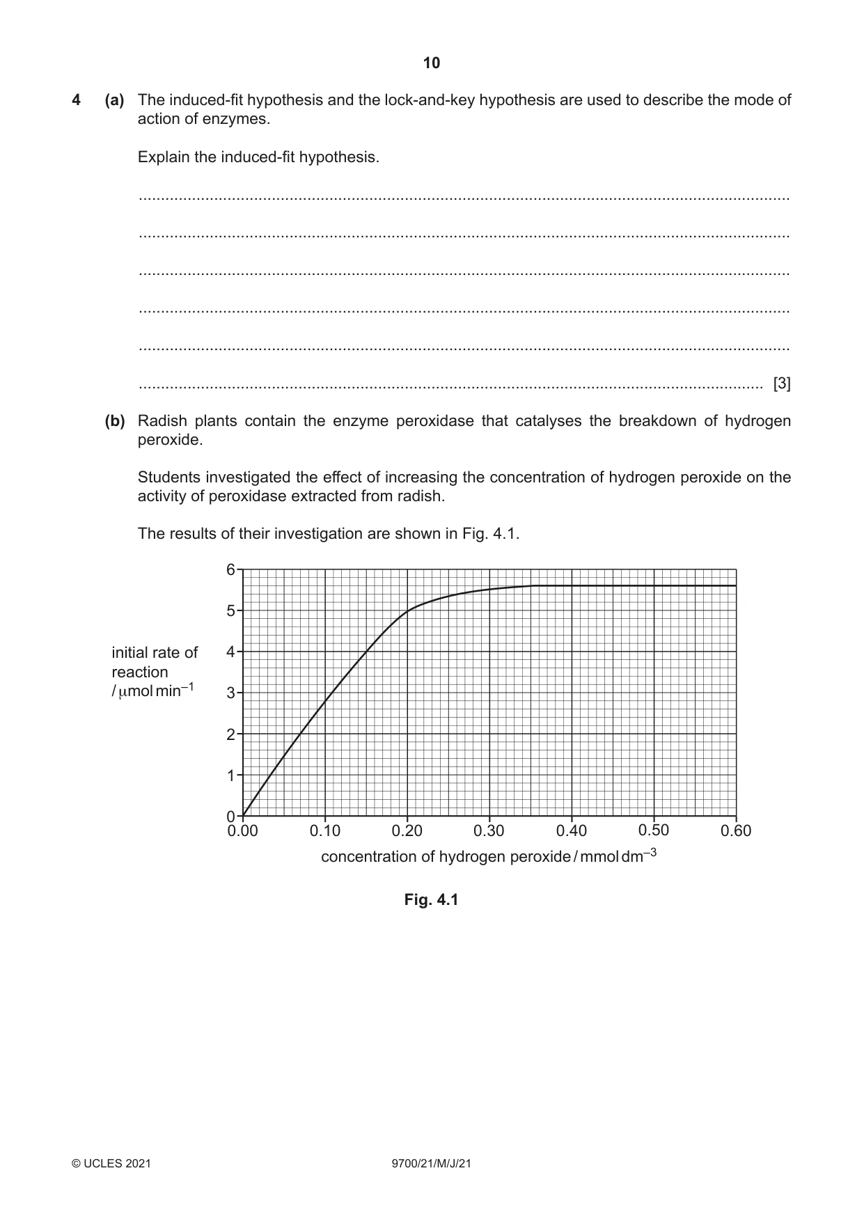
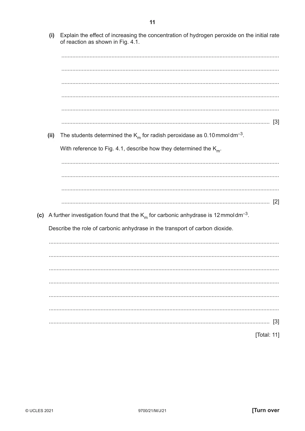
4 (a) The induced-fit hypothesis and the lock-and-key hypothesis are used to describe the mode of action of enzymes. Explain the induced-fit hypothesis. ................................................................................................................................................... ................................................................................................................................................... ................................................................................................................................................... ................................................................................................................................................... ................................................................................................................................................... ............................................................................................................................................. [3] (b) Radish plants contain the enzyme peroxidase that catalyses the breakdown of hydrogen peroxide. Students investigated the effect of increasing the concentration of hydrogen peroxide on the activity of peroxidase extracted from radish. The results of their investigation are shown in Fig. 4.1. 6 5 initial rate of 4 reaction / μmol min–1 3 2 1 0 0.00 0.10 0.20 0.30 0.40 0.50 0.60 concentration of hydrogen peroxide / mmol dm–3 Fig. 4.1 (i) Explain the effect of increasing the concentration of hydrogen peroxide on the initial rate of reaction as shown in Fig. 4.1. ........................................................................................................................................... ........................................................................................................................................... ........................................................................................................................................... ........................................................................................................................................... ........................................................................................................................................... ..................................................................................................................................... [3] (ii) The students determined the Km for radish peroxidase as 0.10 mmol dm–3. With reference to Fig. 4.1, describe how they determined the Km. ........................................................................................................................................... ........................................................................................................................................... ........................................................................................................................................... ..................................................................................................................................... [2] (c) A further investigation found that the Km for carbonic anhydrase is 12 mmol dm–3. Describe the role of carbonic anhydrase in the transport of carbon dioxide. ................................................................................................................................................... ................................................................................................................................................... ................................................................................................................................................... ................................................................................................................................................... ................................................................................................................................................... ................................................................................................................................................... ............................................................................................................................................. [3] [Total: 11]
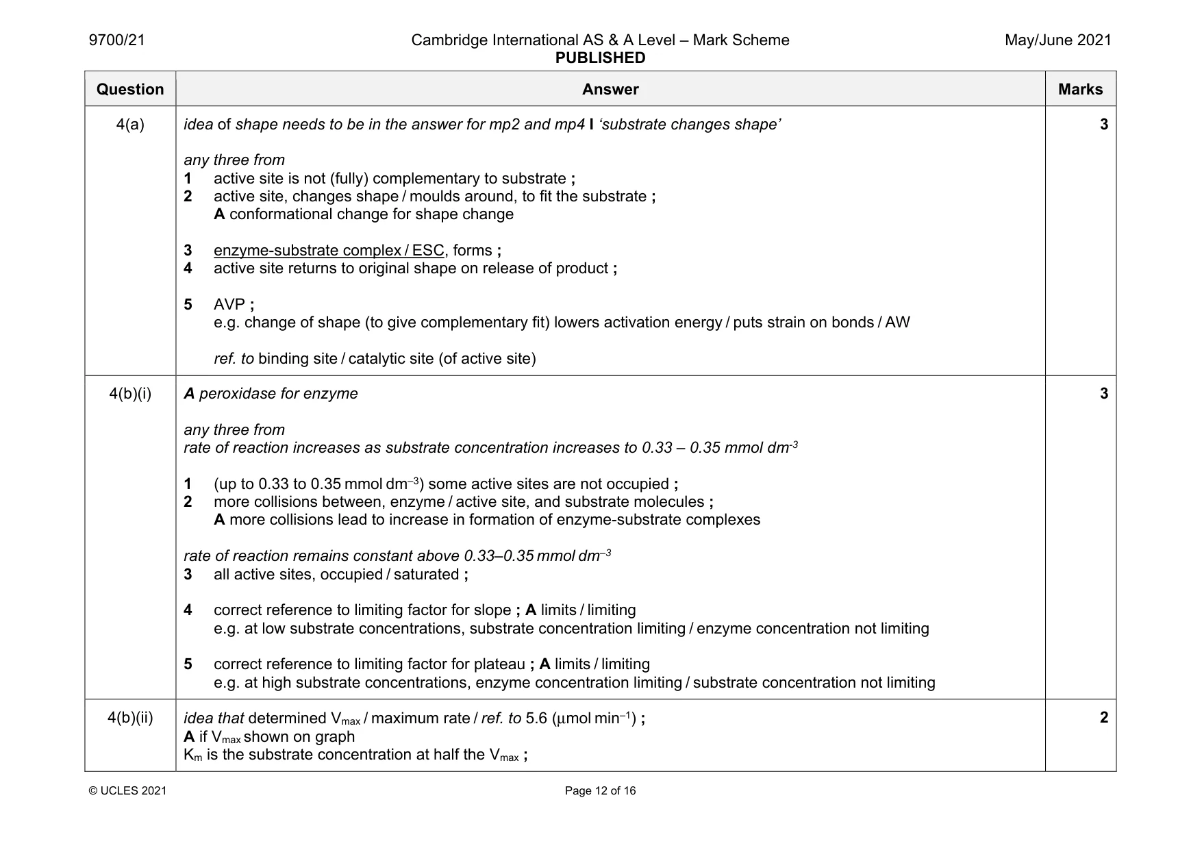
Mark scheme: 4(a) idea of shape needs to be in the answer for mp2 and mp4 I ‘substrate changes shape’ any three from 1 active site is not (fully) complementary to substrate ; 2 active site, changes shape / moulds around, to fit the substrate ; A conformational change for shape change 3 enzyme-substrate complex / ESC, forms ; 4 active site returns to original shape on release of product ; 5 AVP ; e.g. change of shape (to give complementary fit) lowers activation energy / puts strain on bonds / AW ref. to binding site / catalytic site (of active site) 3 4(b)(i) A peroxidase for enzyme any three from rate of reaction increases as substrate concentration increases to 0.33 – 0.35 mmol dm-3 1 (up to 0.33 to 0.35 mmol dm–3) some active sites are not occupied ; 2 more collisions between, enzyme / active site, and substrate molecules ; A more collisions lead to increase in formation of enzyme-substrate complexes rate of reaction remains constant above 0.33–0.35 mmol dm–3 3 all active sites, occupied / saturated ; 4 correct reference to limiting factor for slope ; A limits / limiting e.g. at low substrate concentrations, substrate concentration limiting / enzyme concentration not limiting 5 correct reference to limiting factor for plateau ; A limits / limiting e.g. at high substrate concentrations, enzyme concentration limiting / substrate concentration not limiting 3 4(b)(ii) idea that determined Vmax / maximum rate / ref. to 5.6 (μmol min–1) ; A if Vmax shown on graph Km is the substrate concentration at half the Vmax ; 2 Question Answer Marks 4(c) any three from in respiring tissue 1 (carbonic anhydrase) catalyses the reaction between water and carbon dioxide in, red (blood) cells / erythrocytes ; 2 to form carbonic acid, which dissociates to form HCO3– and H+ ; A from equation A use of word ion(s) 3 HCO3–, moves / passes / diffuses (through membrane proteins from red blood cells) into plasma ; 4 (activity of enzyme) maintains (steep) concentration gradient for diffusion of carbon dioxide into red blood cells ; 5 idea that so helps to remove large quantities of carbon dioxide ; in lungs 6 (carbonic anhydrase) converts HCO3- back into carbon dioxide so can be, excreted / removed / exhaled ; if described earlier A catalyses reverse reaction so carbon dioxide can be, excreted / AW 3
Q5 · Much success was made in reducing the number of cases of malaria between 2000 and 2015
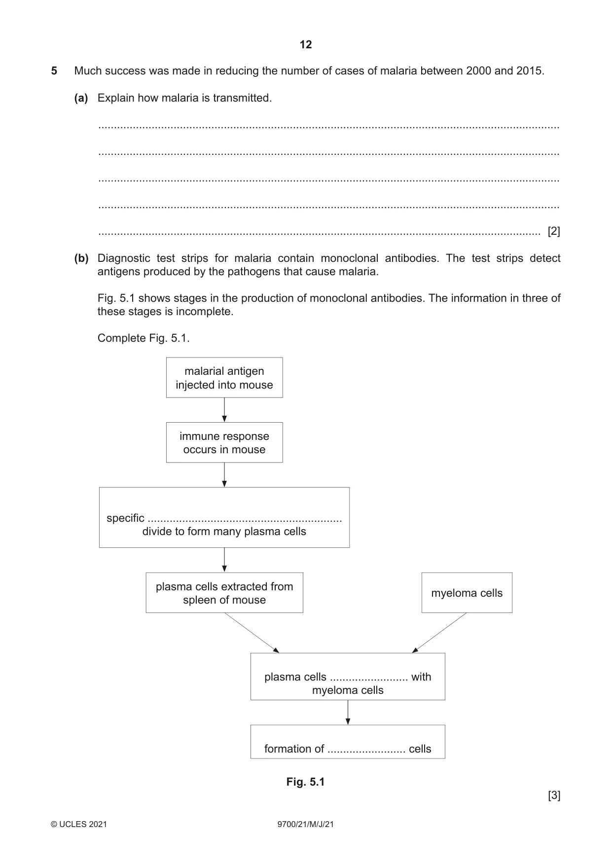
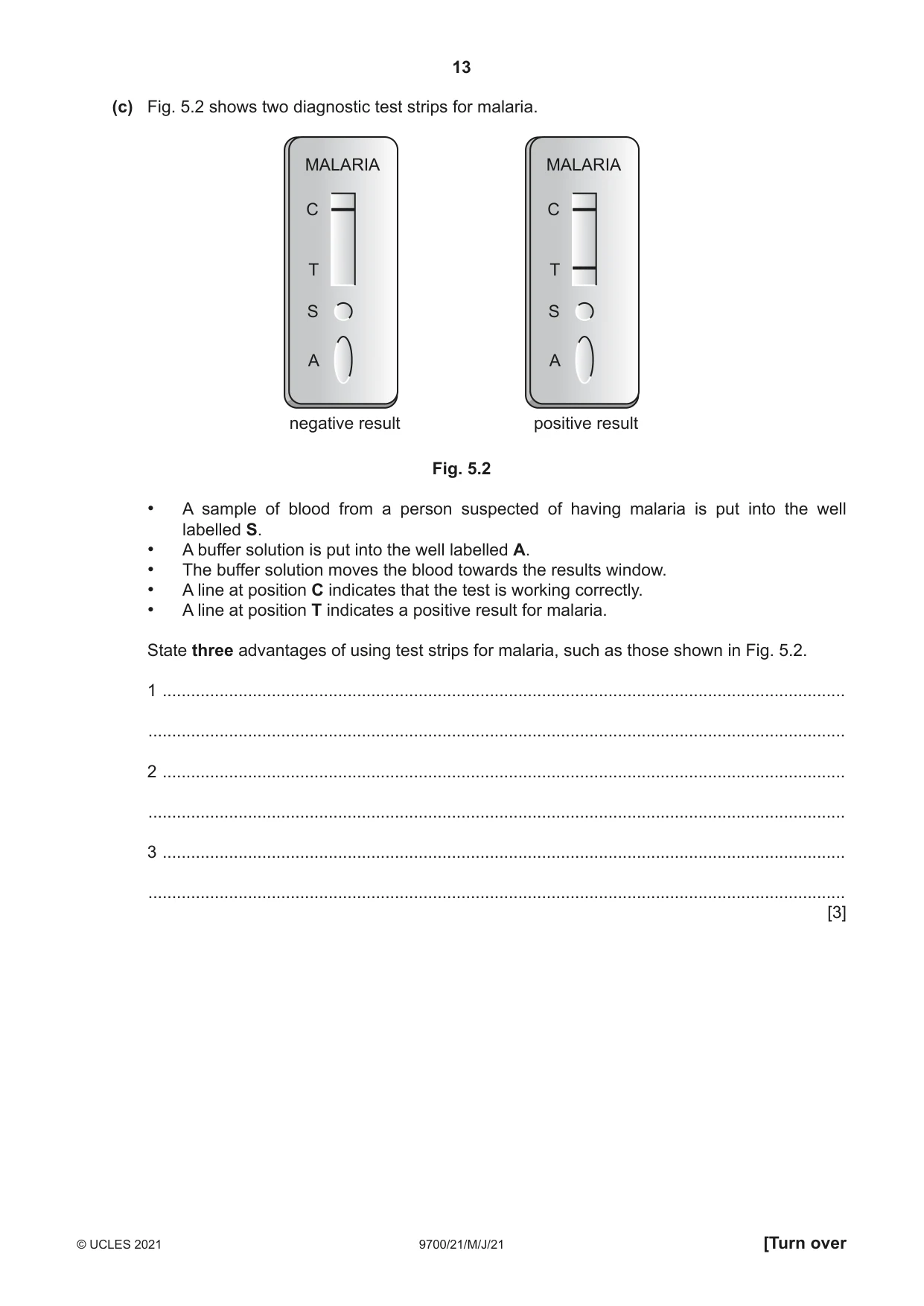
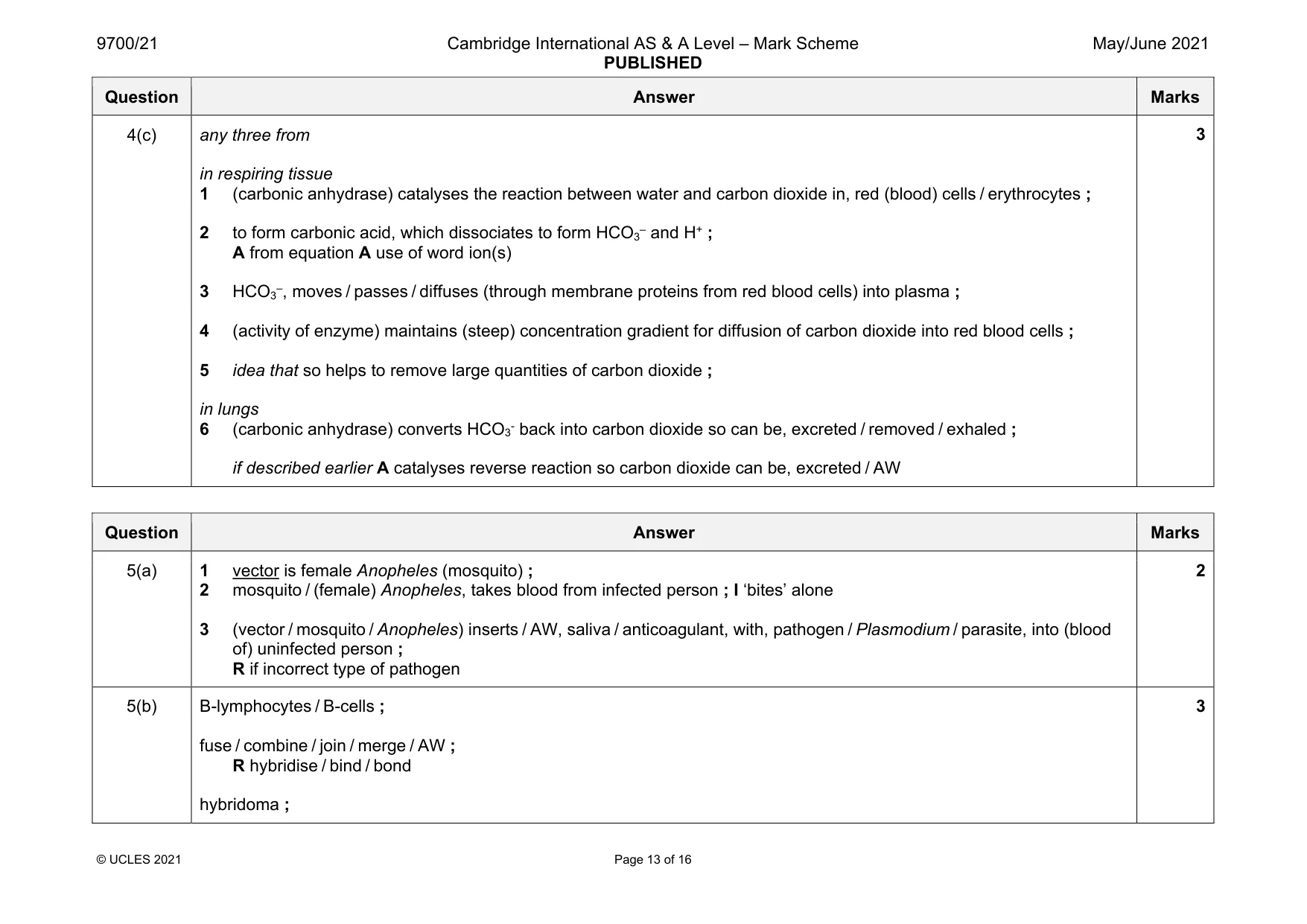
5 Much success was made in reducing the number of cases of malaria between 2000 and 2015. (a) Explain how malaria is transmitted. ................................................................................................................................................... ................................................................................................................................................... ................................................................................................................................................... ................................................................................................................................................... ............................................................................................................................................. [2] (b) Diagnostic test strips for malaria contain monoclonal antibodies. The test strips detect antigens produced by the pathogens that cause malaria. Fig. 5.1 shows stages in the production of monoclonal antibodies. The information in three of these stages is incomplete. Complete Fig. 5.1. malarial antigen injected into mouse immune response occurs in mouse specific .............................................................. divide to form many plasma cells plasma cells extracted from myeloma cells spleen of mouse plasma cells ......................... with myeloma cells formation of ......................... cells Fig. 5.1 [3] (c) Fig. 5.2 shows two diagnostic test strips for malaria. MALARIA MALARIA C C T T S S A A negative result positive result Fig. 5.2 • A sample of blood from a person suspected of having malaria is put into the well labelled S. • A buffer solution is put into the well labelled A. • The buffer solution moves the blood towards the results window. • A line at position C indicates that the test is working correctly. • A line at position T indicates a positive result for malaria. State three advantages of using test strips for malaria, such as those shown in Fig. 5.2. 1 ................................................................................................................................................ ................................................................................................................................................... 2 ................................................................................................................................................ ................................................................................................................................................... 3 ................................................................................................................................................ ................................................................................................................................................... [3] (d) The highest number of cases of malaria occur in sub-Saharan Africa and South-East Asia. Discuss the factors that determine the global distribution of malaria. ................................................................................................................................................... ................................................................................................................................................... ................................................................................................................................................... ................................................................................................................................................... ................................................................................................................................................... ................................................................................................................................................... ................................................................................................................................................... ................................................................................................................................................... ................................................................................................................................................... ............................................................................................................................................. [5] [Total: 13]
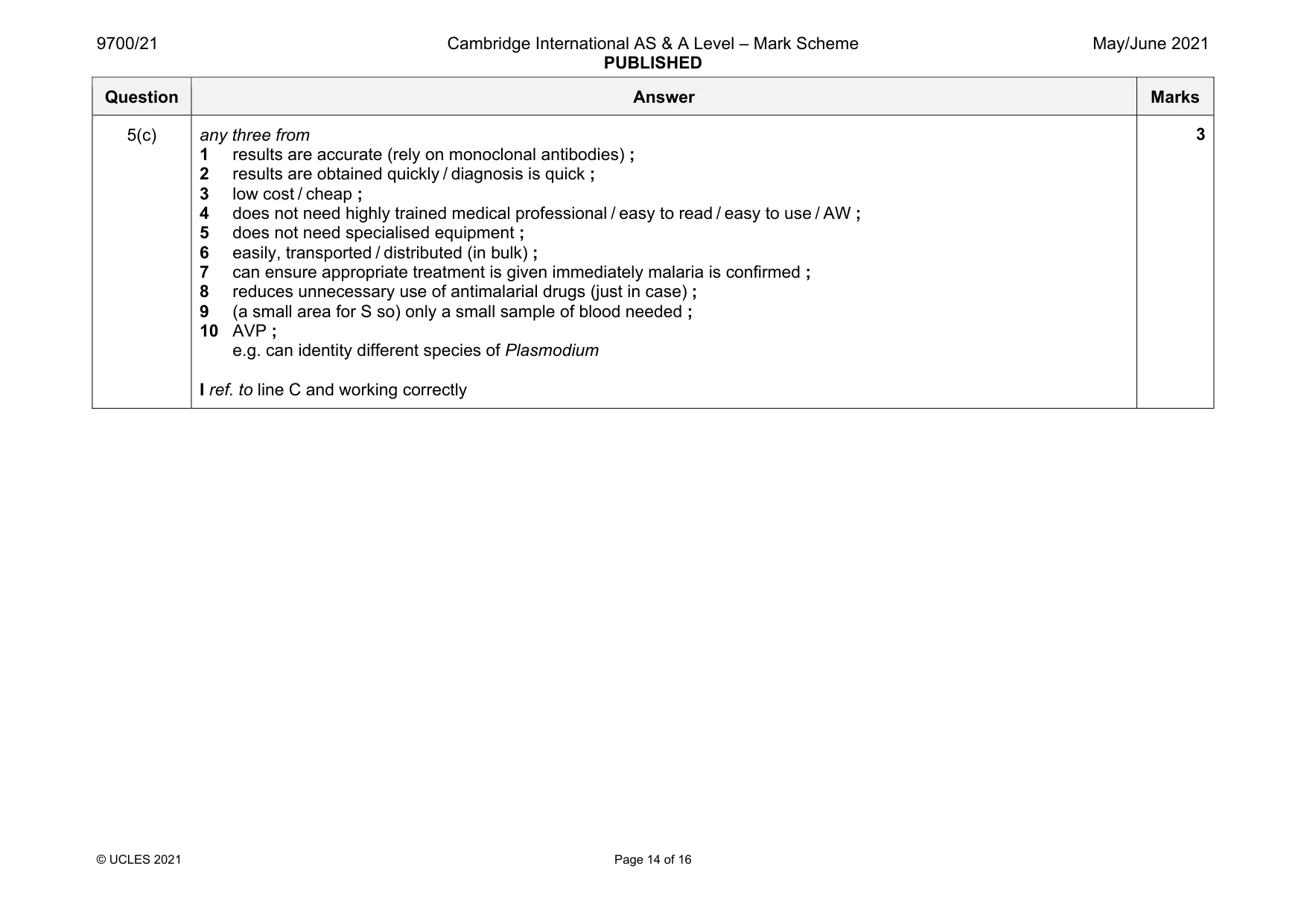
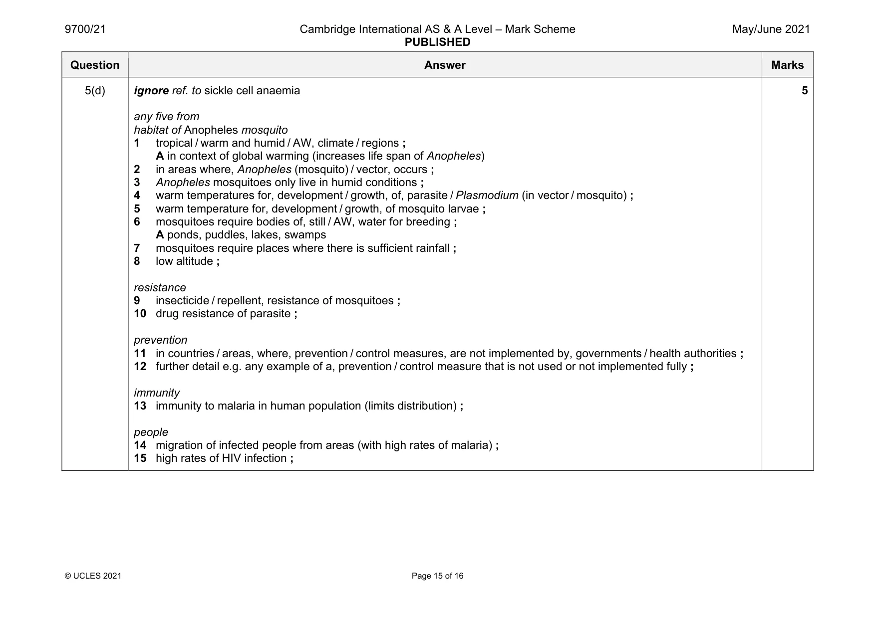
Mark scheme: 5(a) 1 vector is female Anopheles (mosquito) ; 2 mosquito / (female) Anopheles, takes blood from infected person ; I ‘bites’ alone 3 (vector / mosquito / Anopheles) inserts / AW, saliva / anticoagulant, with, pathogen / Plasmodium / parasite, into (blood of) uninfected person ; R if incorrect type of pathogen 5(b) B-lymphocytes / B-cells ; fuse / combine / join / merge / AW ; R hybridise / bind / bond hybridoma ; 3 Question Answer Marks 5(c) any three from 1 results are accurate (rely on monoclonal antibodies) ; 2 results are obtained quickly / diagnosis is quick ; 3 low cost / cheap ; 4 does not need highly trained medical professional / easy to read / easy to use / AW ; 5 does not need specialised equipment ; 6 easily, transported / distributed (in bulk) ; 7 can ensure appropriate treatment is given immediately malaria is confirmed ; 8 reduces unnecessary use of antimalarial drugs (just in case) ; 9 (a small area for S so) only a small sample of blood needed ; 10 AVP ; e.g. can identity different species of Plasmodium I ref. to line C and working correctly 3 Question Answer Marks 5(d) ignore ref. to sickle cell anaemia any five from habitat of Anopheles mosquito 1 tropical / warm and humid / AW, climate / regions ; A in context of global warming (increases life span of Anopheles) 2 in areas where, Anopheles (mosquito) / vector, occurs ; 3 Anopheles mosquitoes only live in humid conditions ; 4 warm temperatures for, development / growth, of, parasite / Plasmodium (in vector / mosquito) ; 5 warm temperature for, development / growth, of mosquito larvae ; 6 mosquitoes require bodies of, still / AW, water for breeding ; A ponds, puddles, lakes, swamps 7 mosquitoes require places where there is sufficient rainfall ; 8 low altitude ; resistance 9 insecticide / repellent, resistance of mosquitoes ; 10 drug resistance of parasite ; prevention 11 in countries / areas, where, prevention / control measures, are not implemented by, governments / health authorities ; 12 further detail e.g. any example of a, prevention / control measure that is not used or not implemented fully ; immunity 13 immunity to malaria in human population (limits distribution) ; people 14 migration of infected people from areas (with high rates of malaria) ; 15 high rates of HIV infection ; 5
Q6 · Lysosomes are cell structures that contain enzymes known as acid hydrolases
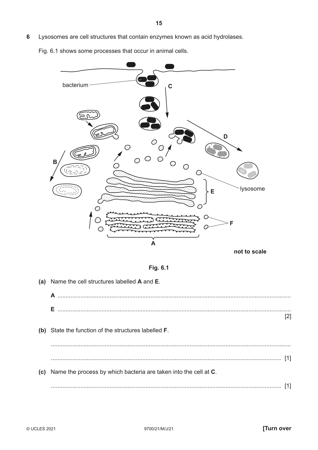
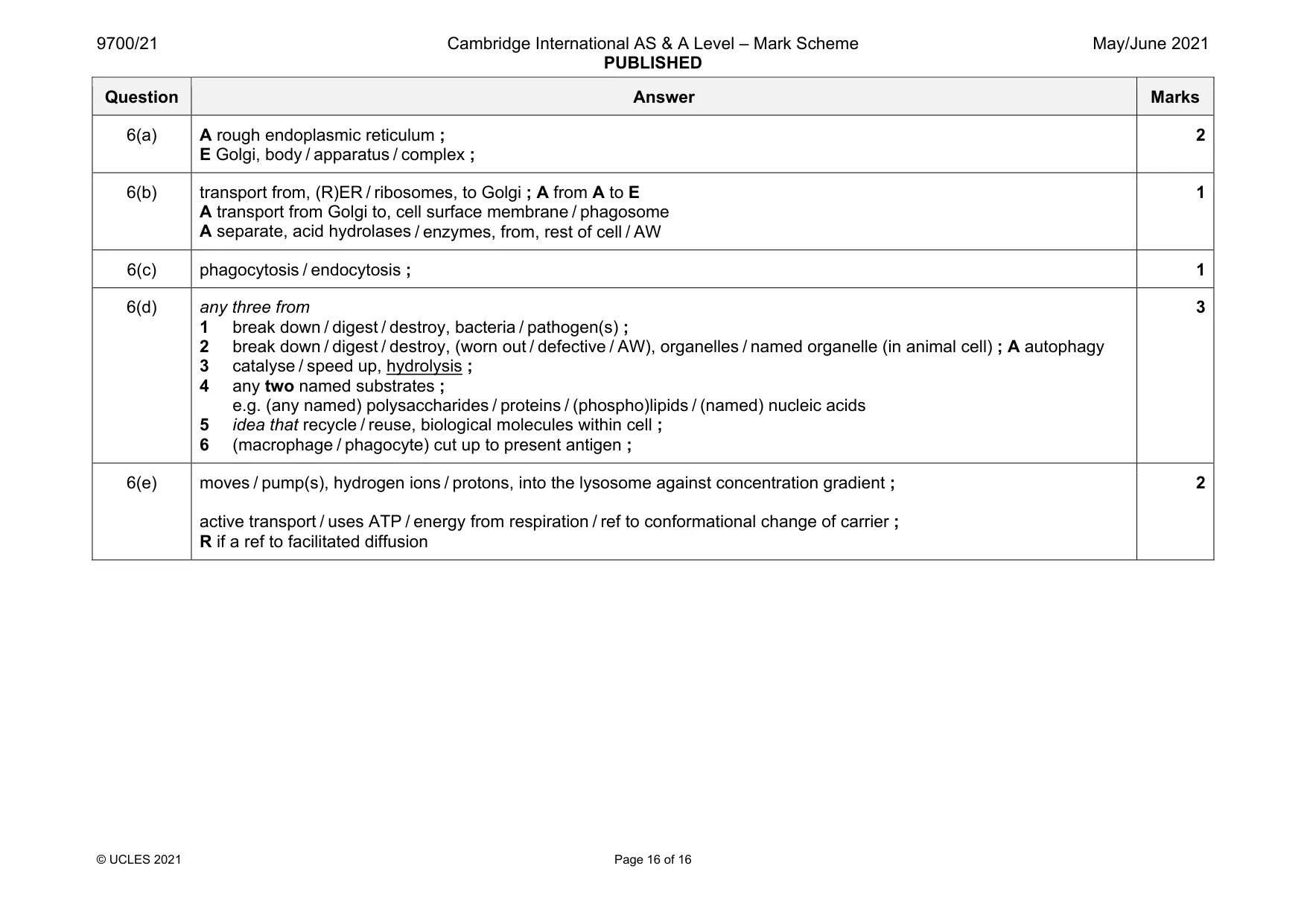
6 Lysosomes are cell structures that contain enzymes known as acid hydrolases. Fig. 6.1 shows some processes that occur in animal cells. bacterium C D B lysosomee E F A not to scale Fig. 6.1 (a) Name the cell structures labelled A and E. A ............................................................................................................................................... E ............................................................................................................................................... [2] (b) State the function of the structures labelled F. ................................................................................................................................................... ............................................................................................................................................. [1] (c) Name the process by which bacteria are taken into the cell at C. ............................................................................................................................................. [1] (d) With reference to the processes occurring at B and at D in Fig. 6.1, outline the role of acid hydrolases in lysosomes. ................................................................................................................................................... ................................................................................................................................................... ................................................................................................................................................... ................................................................................................................................................... ................................................................................................................................................... ................................................................................................................................................... ............................................................................................................................................. [3] (e) Carrier proteins in the membranes of lysosomes maintain a lower pH than the surrounding cytoplasm by moving hydrogen ions. Suggest how the carrier proteins maintain the lower pH within the lysosomes. ................................................................................................................................................... ................................................................................................................................................... ................................................................................................................................................... ................................................................................................................................................... ............................................................................................................................................. [2] [Total: 9]
Mark scheme: 6(a) A rough endoplasmic reticulum ; E Golgi, body / apparatus / complex ; 2 6(b) transport from, (R)ER / ribosomes, to Golgi ; A from A to E A transport from Golgi to, cell surface membrane / phagosome A separate, acid hydrolases / enzymes, from, rest of cell / AW 1 6(c) phagocytosis / endocytosis ; 1 6(d) any three from 1 break down / digest / destroy, bacteria / pathogen(s) ; 2 break down / digest / destroy, (worn out / defective / AW), organelles / named organelle (in animal cell) ; A autophagy 3 catalyse / speed up, hydrolysis ; 4 any two named substrates ; e.g. (any named) polysaccharides / proteins / (phospho)lipids / (named) nucleic acids 5 idea that recycle / reuse, biological molecules within cell ; 6 (macrophage / phagocyte) cut up to present antigen ; 3 6(e) moves / pump(s), hydrogen ions / protons, into the lysosome against concentration gradient ; active transport / uses ATP / energy from respiration / ref to conformational change of carrier ; R if a ref to facilitated diffusion 2
More questions on Cells as the basic units of living organisms
What was in this paper
The subtopics covered by these 6 questions, and how many questions each got. Open one in a new tab to see every Cambridge question on it.
What you needed in this session
Cambridge’s own grade thresholds for 2021 May/June, Paper 2 · Variant 1. A higher threshold means an easier paper — the bar moves with how the cohort did.