Cambridge A Level Biology 9700 — 2017 Oct/Nov Paper 2 · Variant 3
9700/23/O/N/17 · 6 questions · 60 marks · ≈68 min
The question paper and its mark scheme, free to read here and free to download. This is Cambridge’s own paper, exactly as it was sat.
Question paper16 pages
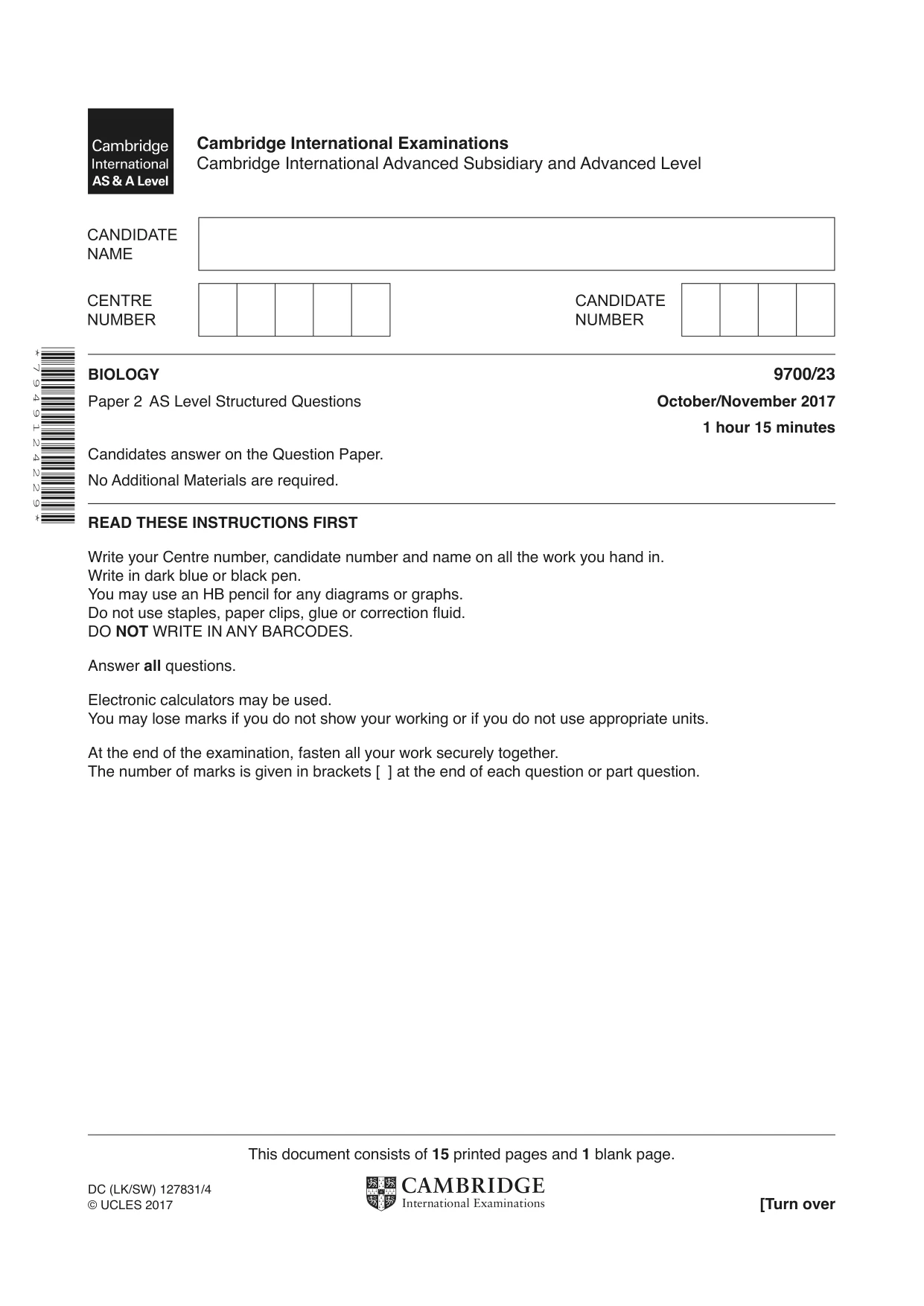

Mark scheme12 pages
Answers below. Sit the paper first if you are practising.
Questions as text
Q1 · A transmission electron micrograph of a part of an animal cell
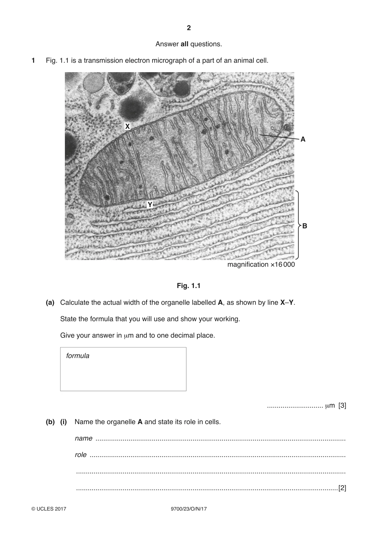
1 Fig. 1.1 is a transmission electron micrograph of a part of an animal cell. X A Y B magnification ×16 000 Fig. 1.1 (a) Calculate the actual width of the organelle labelled A, as shown by line X–Y. State the formula that you will use and show your working. Give your answer in µm and to one decimal place. formula ............................. µm [3] (b) (i) Name the organelle A and state its role in cells. name ................................................................................................................................. role .................................................................................................................................... ........................................................................................................................................... .......................................................................................................................................[2] (ii) Name the cell structure labelled B and state one reason for your answer. name ................................................................................................................................. reason ............................................................................................................................... ........................................................................................................................................... .......................................................................................................................................[2] [Total: 7]
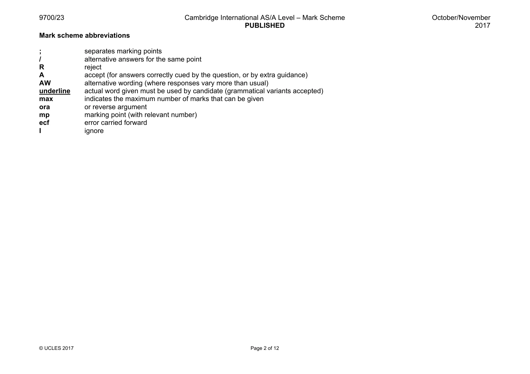
Mark scheme: 1(a) actual length = image length / magnification ; A A = I / M M = I / A I = A × M or magnification triangle 2.5 (µm) ;; for 40 mm X–Y length A 2.6 (for 40 / 41 mm) A 2.4 (for 38 / 39 mm) max 1 for incorrect or no answer but correct calculation e.g. 40 000 ÷ 16 000 correct answer but to more than one decimal place correct measurement and correct calculation but incorrect conversion 3 1(b)(i) mitochondrion ; A mitochondria max 1 for function produces / makes / synthesises / provides / AW, ATP or releases / provides / supplies, energy or aerobic respiration ; AVP ; e.g. part of the urea cycle β-oxidation of fat oxidative phosphorylation 2 1(b)(ii) rough endoplasmic reticulum ; A rough ER I RER ribosomes are attached ; accept mp2 if organelle identified as endoplasmic reticulum / RER 2
More questions on Cells as the basic units of living organisms
Q2 · Explain why the wall of the left ventricle in the heart is thicker than the wall of the…
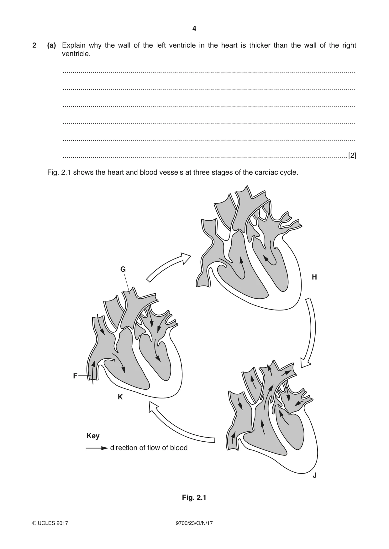
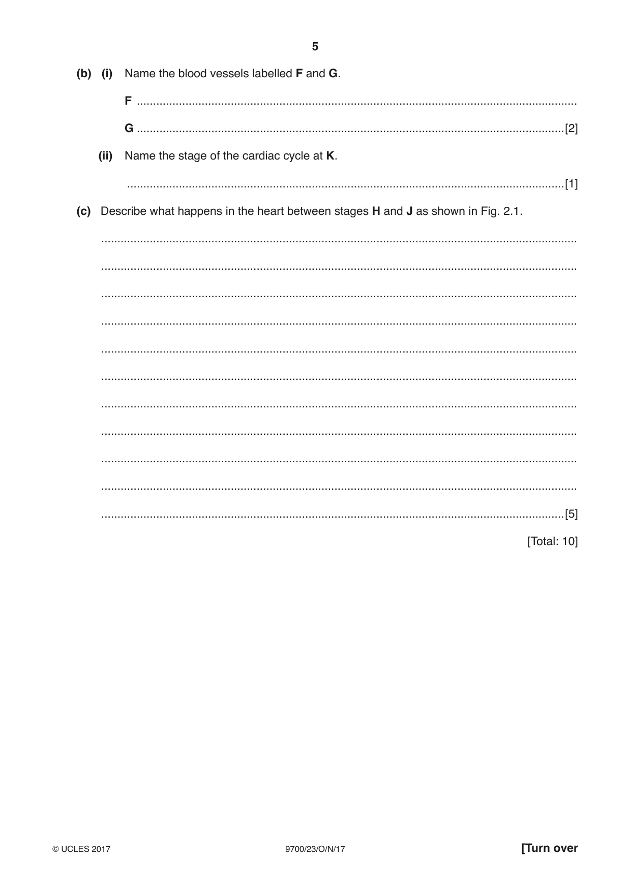
2 (a) Explain why the wall of the left ventricle in the heart is thicker than the wall of the right ventricle. ................................................................................................................................................... ................................................................................................................................................... ................................................................................................................................................... ................................................................................................................................................... ................................................................................................................................................... ...............................................................................................................................................[2] Fig. 2.1 shows the heart and blood vessels at three stages of the cardiac cycle. G H F K Key direction of flow of blood J Fig. 2.1 (b) (i) Name the blood vessels labelled F and G. F ........................................................................................................................................ G ....................................................................................................................................[2] (ii) Name the stage of the cardiac cycle at K. .......................................................................................................................................[1] (c) Describe what happens in the heart between stages H and J as shown in Fig. 2.1. ................................................................................................................................................... ................................................................................................................................................... ................................................................................................................................................... ................................................................................................................................................... ................................................................................................................................................... ................................................................................................................................................... ................................................................................................................................................... ................................................................................................................................................... ................................................................................................................................................... ................................................................................................................................................... ...............................................................................................................................................[5] [Total: 10]
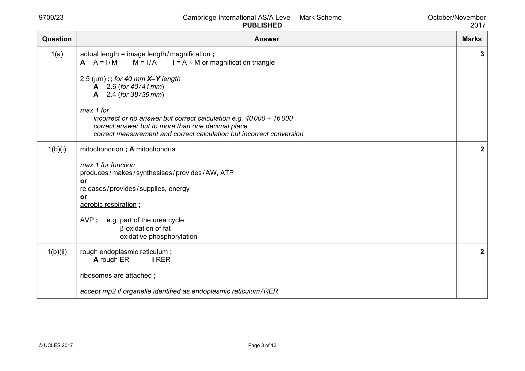
Mark scheme: 2(a) two from 1 to generate a high(er) blood pressure (during systole) ; A force I withstands / AW, high blood pressure 2 to overcome high(er) resistance (in systemic circuit than in pulmonary circuit) ; 3 to transport blood a greater distance / greater distance in systemic circuit ; 4 ref. to right ventricle generating low pressure to avoid damaging (capillaries in the) lungs / AW ; 2 2(b)(i) F (inferior / posterior) vena cava ; I superior G pulmonary artery ; 2 2(b)(ii) diastole ; I ventricular / atrial 1 2(c) five from 1 wave of excitation / wave of depolarisation / impulses, from, atrioventricular node / AVN ; 2 pass(es) down Purkyne fibres (to ventricles) ; A Bundle of His 3 (both) ventricles contract / ventricular systole ; 4 from the base (upwards) / AW ; 5 blood pressure in ventricles, increases / becomes higher ; 6 blood pressure in ventricles greater than in atria ; 7 atrioventricular / AV / tricuspid and bicuspid, valves close ; 8 blood pressure in ventricles greater than in, artery / aorta / pulmonary artery ; 9 semi-lunar / pulmonary and aortic, valves open ; A blood flows through 10 blood flows, into arteries / to lungs and rest of the body ; at end of description only 11 atria relaxing / atrial diastole / blood flows into atria ; 5
Q3 · The structure of an alveolus and surrounding structures in a mammalian lung
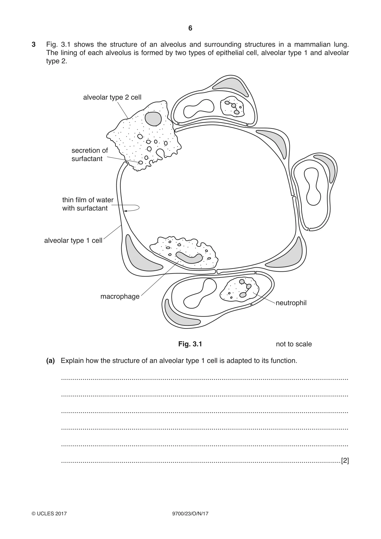
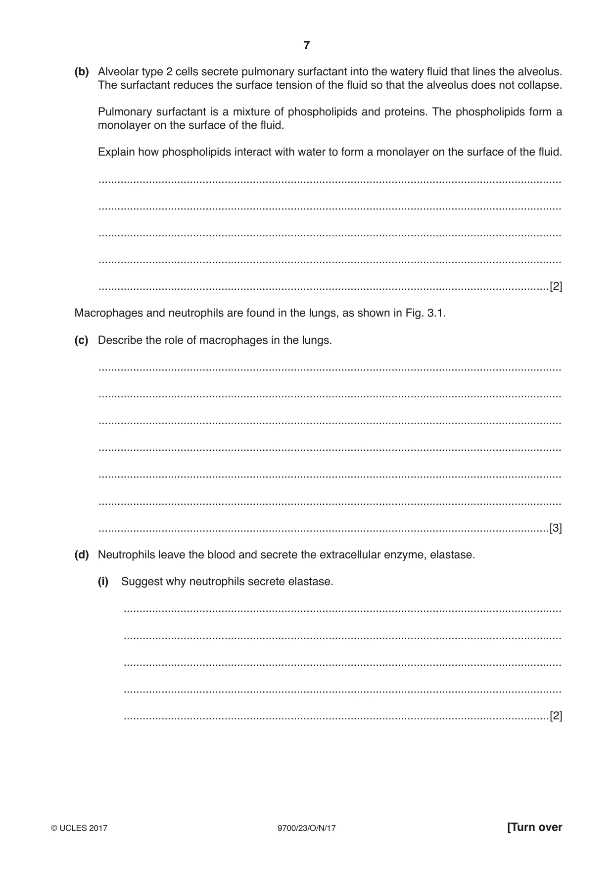
3 Fig. 3.1 shows the structure of an alveolus and surrounding structures in a mammalian lung. The lining of each alveolus is formed by two types of epithelial cell, alveolar type 1 and alveolar type 2. alveolar type 2 cell secretion of surfactant thin film of water with surfactant alveolar type 1 cell macrophage neutrophil Fig. 3.1 not to scale (a) Explain how the structure of an alveolar type 1 cell is adapted to its function. ................................................................................................................................................... ................................................................................................................................................... ................................................................................................................................................... ................................................................................................................................................... ................................................................................................................................................... ...............................................................................................................................................[2] (b) Alveolar type 2 cells secrete pulmonary surfactant into the watery fluid that lines the alveolus. The surfactant reduces the surface tension of the fluid so that the alveolus does not collapse. Pulmonary surfactant is a mixture of phospholipids and proteins. The phospholipids form a monolayer on the surface of the fluid. Explain how phospholipids interact with water to form a monolayer on the surface of the fluid. ................................................................................................................................................... ................................................................................................................................................... ................................................................................................................................................... ................................................................................................................................................... ...............................................................................................................................................[2] Macrophages and neutrophils are found in the lungs, as shown in Fig. 3.1. (c) Describe the role of macrophages in the lungs. ................................................................................................................................................... ................................................................................................................................................... ................................................................................................................................................... ................................................................................................................................................... ................................................................................................................................................... ................................................................................................................................................... ...............................................................................................................................................[3] (d) Neutrophils leave the blood and secrete the extracellular enzyme, elastase. (i) Suggest why neutrophils secrete elastase. ........................................................................................................................................... ........................................................................................................................................... ........................................................................................................................................... ........................................................................................................................................... .......................................................................................................................................[2] (ii) The protein alpha-1 antitrypsin is produced in cells in the liver and is transported to the lungs, where it inhibits the action of elastase. Some people produce a different form of this protein that remains within liver cells. These people are at an increased risk of developing emphysema, in which alveolar walls break down. Emphysema is one of the conditions associated with chronic obstructive pulmonary disease (COPD). Explain why these people are at increased risk of developing emphysema. ........................................................................................................................................... ........................................................................................................................................... ........................................................................................................................................... ........................................................................................................................................... ........................................................................................................................................... .......................................................................................................................................[3] [Total: 12]
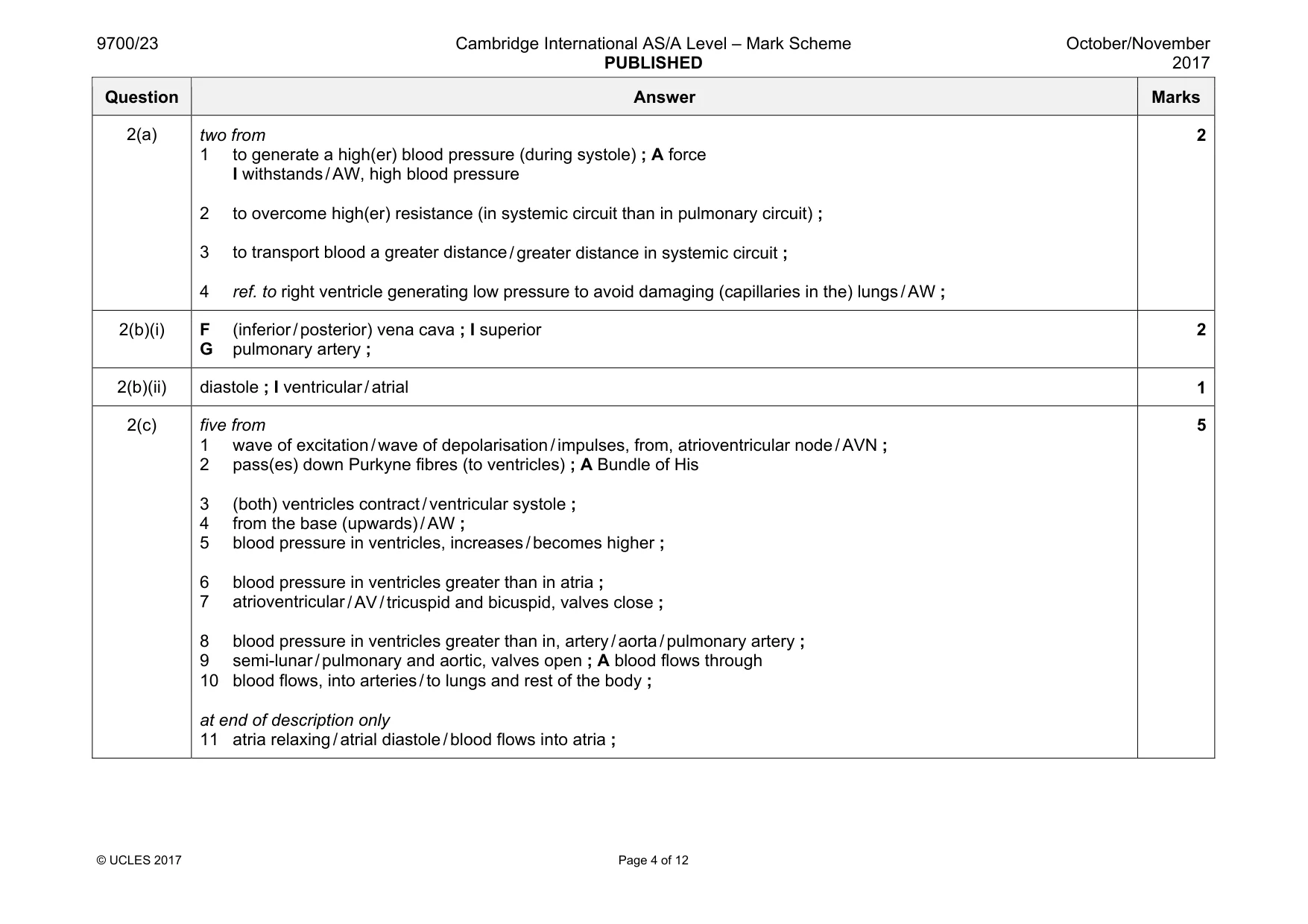
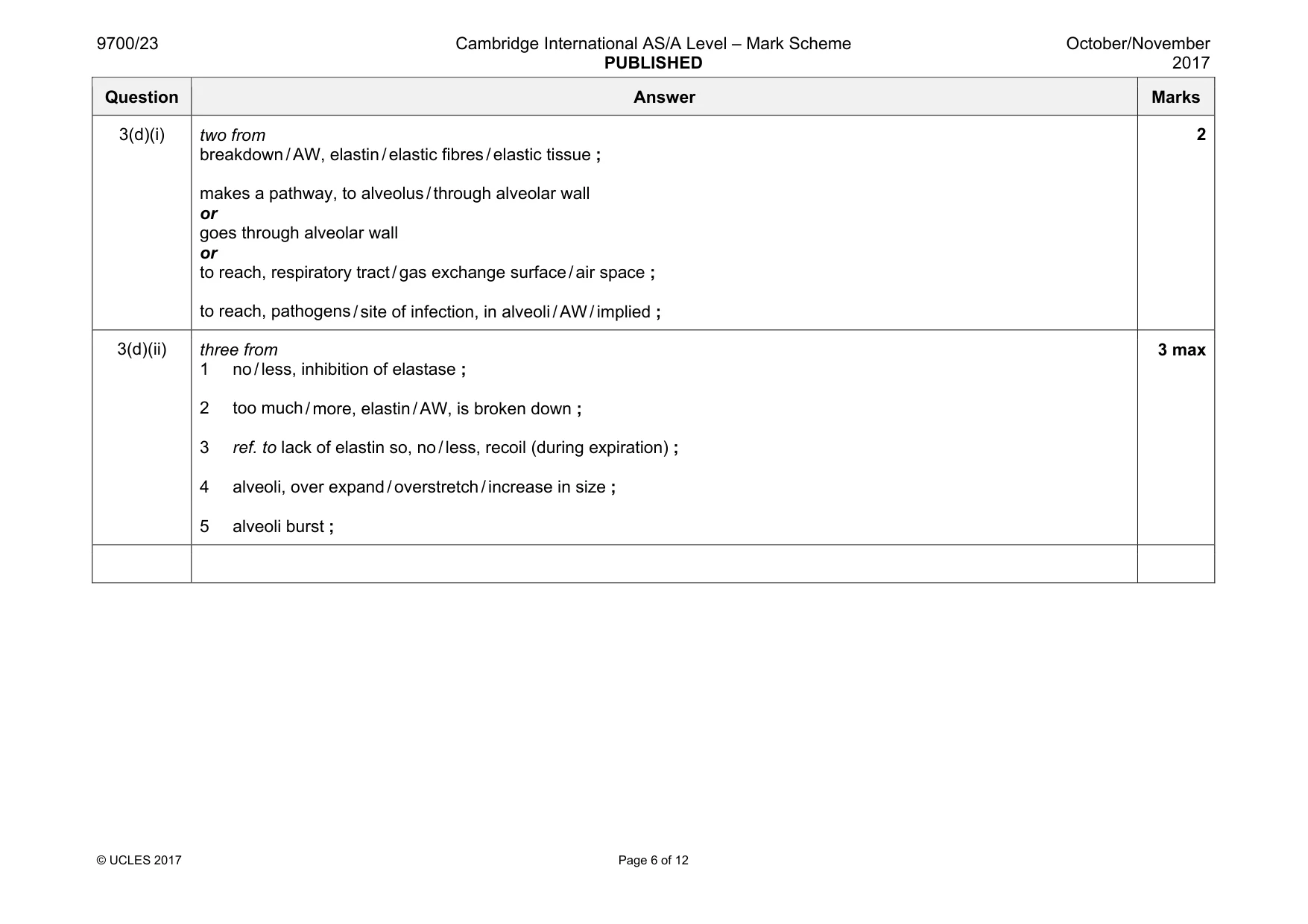
Mark scheme: 3(a) (cell) thin / squamous / flat / pavement ; R cell wall I one cell thick short distance for, diffusion of (named) gases / gas exchange ; A diffusion between (air in) alveolus and (blood in) capillary 2 3(b) two from phosphate / ‘heads’, are, polar / hydrophilic and, fatty acids / hydrocarbon chains / ‘tails’, are, non-polar / hydrophobic ; A hydrophilic / polar, heads and hydrophobic tails tails, face away from fluid or water / project into air ; A diagram heads form hydrogen bonds with water ; max 1 if answer is about a bilayer 2 3(c) three from 1 prevention of infections (of, gas exchange system / named part) ; A in context of a named disease (TB, pneumonia, influenza) 2 prevent (named) pathogen entering, rest of body / blood ; 3 idea that macrophages patrol / move around / AW, alveoli / lungs ; 4 (carry out) phagocytosis / endocytosis ; A are phagocytes 5 engulf / remove / destroy / kill / digest , pathogens ; 6 macrophages are, antigen presenting cells / APCs ; A description of antigen presentation A part of the immune system 7 AVP ; e.g. reduce excess surfactants I ref. to mucus 3 Question Answer Marks 3(d)(i) two from breakdown / AW, elastin / elastic fibres / elastic tissue ; makes a pathway, to alveolus / through alveolar wall or goes through alveolar wall or to reach, respiratory tract / gas exchange surface / air space ; to reach, pathogens / site of infection, in alveoli / AW / implied ; 2 3(d)(ii) three from 1 no / less, inhibition of elastase ; 2 too much / more, elastin / AW, is broken down ; 3 ref. to lack of elastin so, no / less, recoil (during expiration) ; 4 alveoli, over expand / overstretch / increase in size ; 5 alveoli burst ; 3 max
Q4 · Malaria is a disease transmitted by a vector
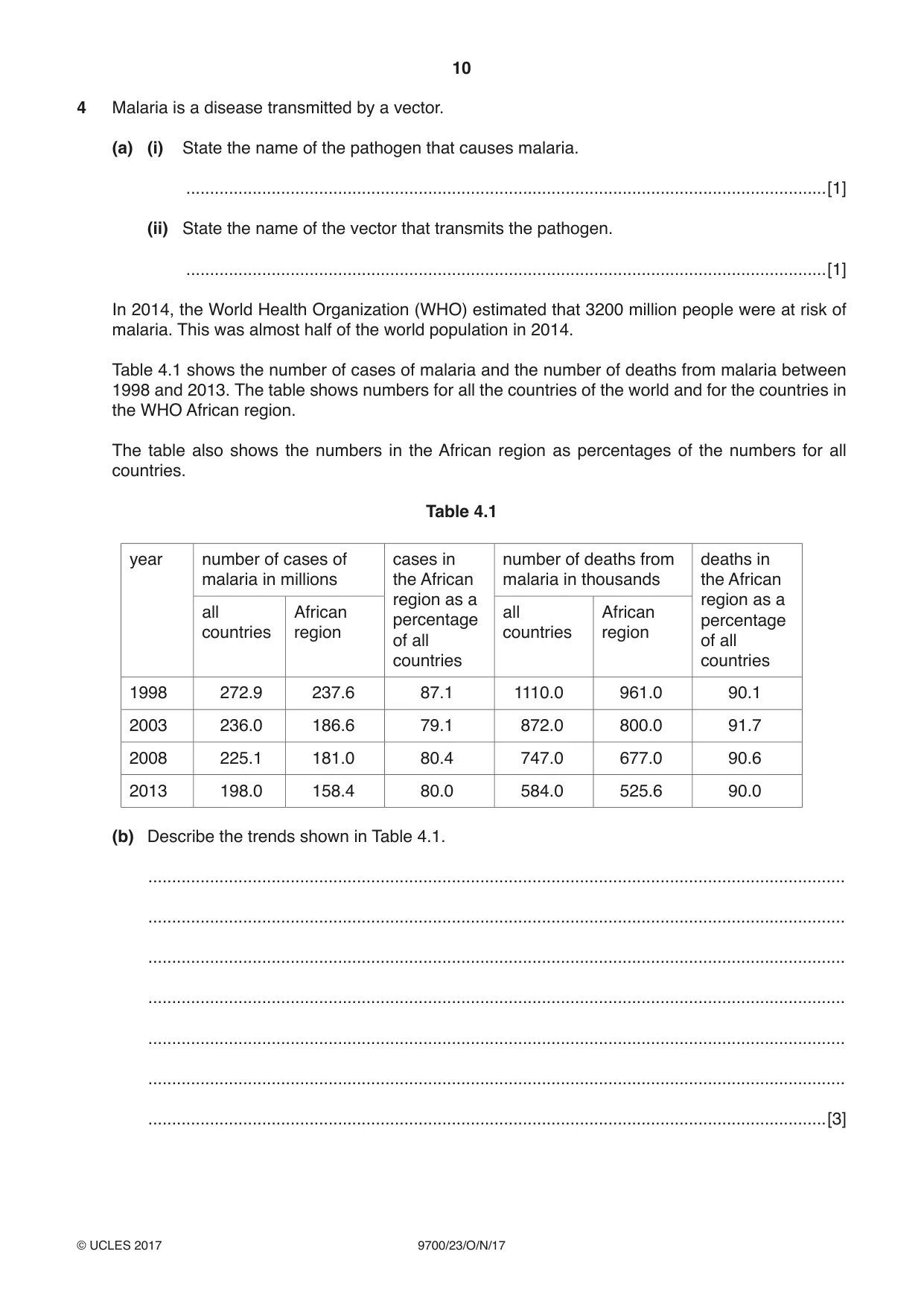
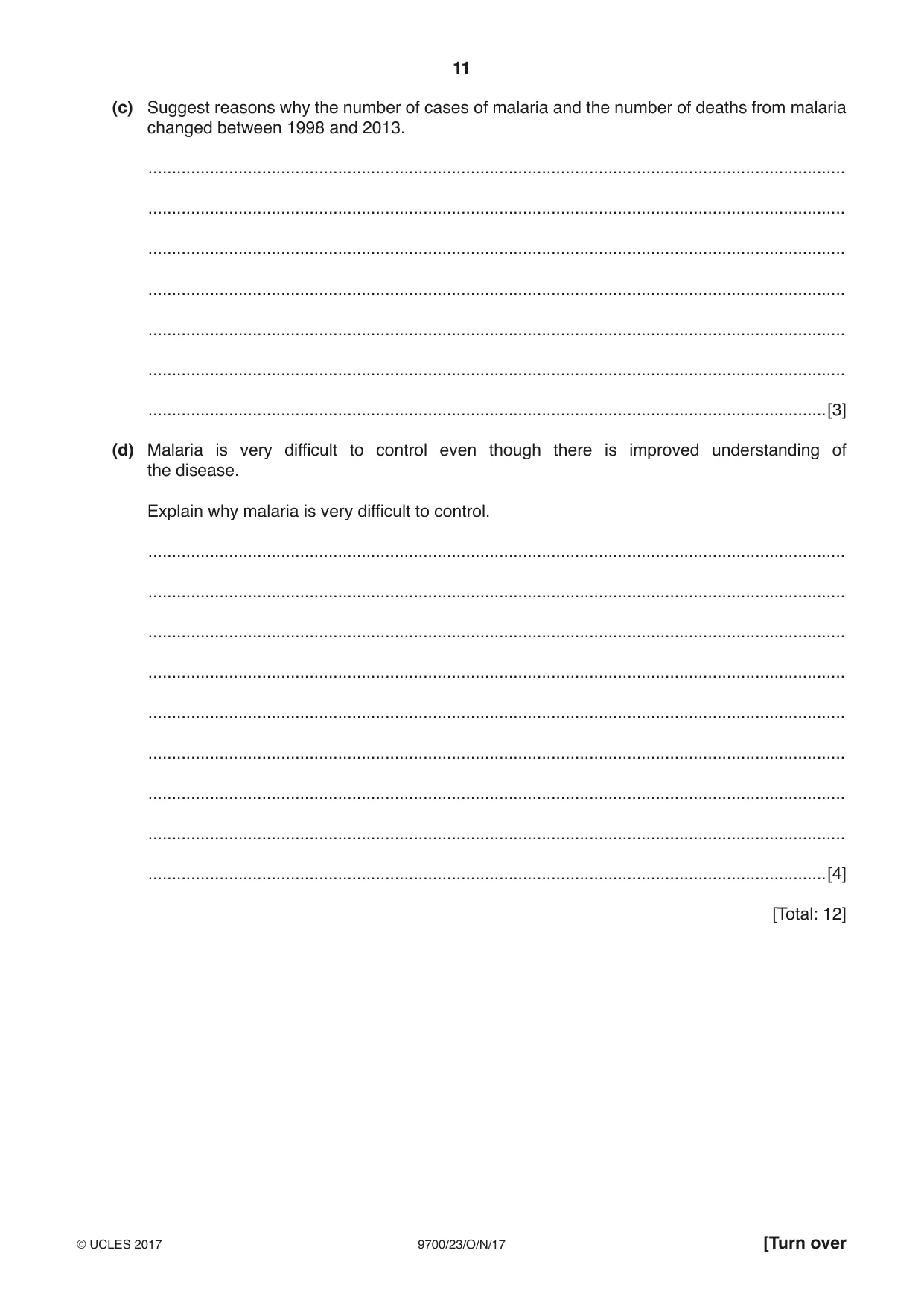
4 Malaria is a disease transmitted by a vector. (a) (i) State the name of the pathogen that causes malaria. .......................................................................................................................................[1] (ii) State the name of the vector that transmits the pathogen. .......................................................................................................................................[1] In 2014, the World Health Organization (WHO) estimated that 3200 million people were at risk of malaria. This was almost half of the world population in 2014. Table 4.1 shows the number of cases of malaria and the number of deaths from malaria between 1998 and 2013. The table shows numbers for all the countries of the world and for the countries in the WHO African region. The table also shows the numbers in the African region as percentages of the numbers for all countries. Table 4.1 year number of cases of cases in number of deaths from deaths in malaria in millions the African malaria in thousands the African region as a region as a all African all African percentage percentage countries region countries region of all of all countries countries 1998 272.9 237.6 87.1 1110.0 961.0 90.1 2003 236.0 186.6 79.1 872.0 800.0 91.7 2008 225.1 181.0 80.4 747.0 677.0 90.6 2013 198.0 158.4 80.0 584.0 525.6 90.0 (b) Describe the trends shown in Table 4.1. ................................................................................................................................................... ................................................................................................................................................... ................................................................................................................................................... ................................................................................................................................................... ................................................................................................................................................... ................................................................................................................................................... ...............................................................................................................................................[3] (c) Suggest reasons why the number of cases of malaria and the number of deaths from malaria changed between 1998 and 2013. ................................................................................................................................................... ................................................................................................................................................... ................................................................................................................................................... ................................................................................................................................................... ................................................................................................................................................... ................................................................................................................................................... ...............................................................................................................................................[3] (d) Malaria is very difficult to control even though there is improved understanding of the disease. Explain why malaria is very difficult to control. ................................................................................................................................................... ................................................................................................................................................... ................................................................................................................................................... ................................................................................................................................................... ................................................................................................................................................... ................................................................................................................................................... ................................................................................................................................................... ................................................................................................................................................... ...............................................................................................................................................[4] [Total: 12]
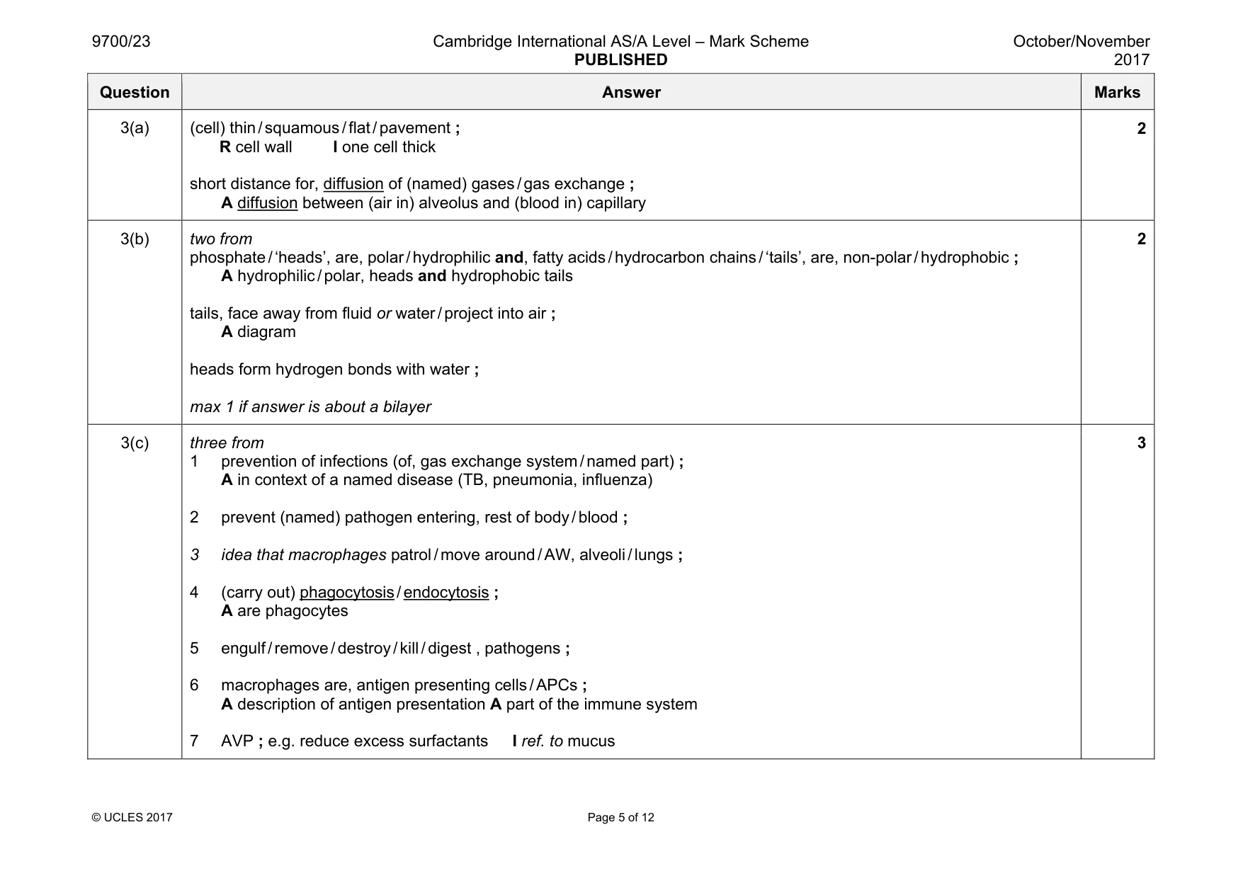
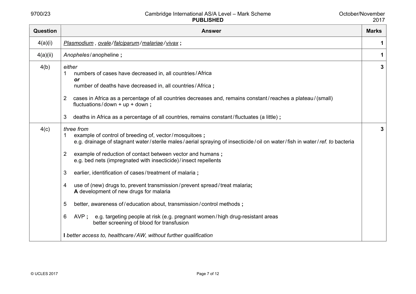
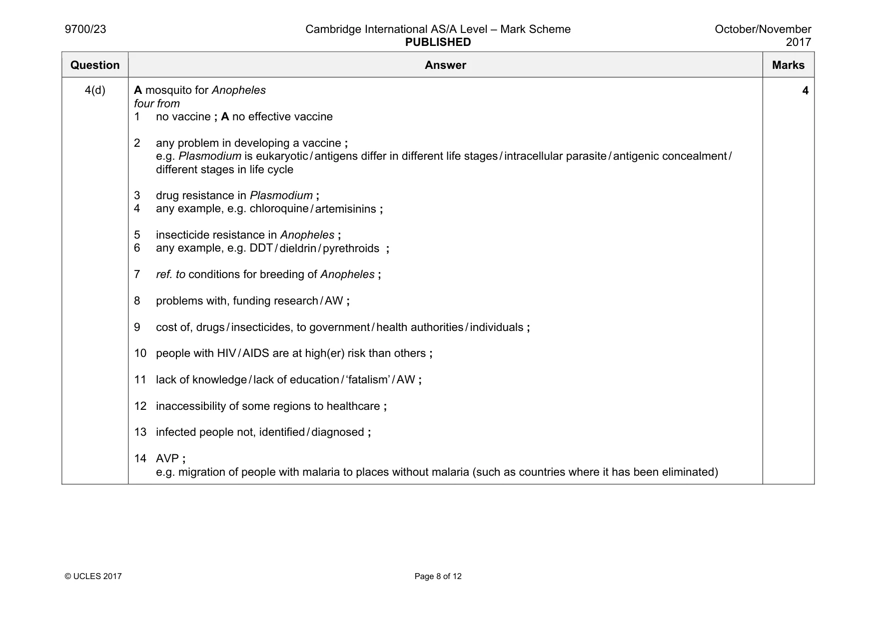
Mark scheme: 4(a)(i) Plasmodium , ovale / falciparum / malariae / vivax ; 1 4(a)(ii) Anopheles / anopheline ; 1 4(b) either 1 numbers of cases have decreased in, all countries / Africa or number of deaths have decreased in, all countries / Africa ; 2 cases in Africa as a percentage of all countries decreases and, remains constant / reaches a plateau / (small) fluctuations / down + up + down ; 3 deaths in Africa as a percentage of all countries, remains constant / fluctuates (a little) ; 3 4(c) three from 1 example of control of breeding of, vector / mosquitoes ; e.g. drainage of stagnant water / sterile males / aerial spraying of insecticide / oil on water / fish in water / ref. to bacteria 2 example of reduction of contact between vector and humans ; e.g. bed nets (impregnated with insecticide) / insect repellents 3 earlier, identification of cases / treatment of malaria ; 4 use of (new) drugs to, prevent transmission / prevent spread / treat malaria; A development of new drugs for malaria 5 better, awareness of / education about, transmission / control methods ; 6 AVP ; e.g. targeting people at risk (e.g. pregnant women / high drug-resistant areas better screening of blood for transfusion I better access to, healthcare / AW, without further qualification 3 Question Answer Marks 4(d) A mosquito for Anopheles four from 1 no vaccine ; A no effective vaccine 2 any problem in developing a vaccine ; e.g. Plasmodium is eukaryotic / antigens differ in different life stages / intracellular parasite / antigenic concealment / different stages in life cycle 3 drug resistance in Plasmodium ; 4 any example, e.g. chloroquine / artemisinins ; 5 insecticide resistance in Anopheles ; 6 any example, e.g. DDT / dieldrin / pyrethroids ; 7 ref. to conditions for breeding of Anopheles ; 8 problems with, funding research / AW ; 9 cost of, drugs / insecticides, to government / health authorities / individuals ; 10 people with HIV / AIDS are at high(er) risk than others ; 11 lack of knowledge / lack of education / ‘fatalism’ / AW ; 12 inaccessibility of some regions to healthcare ; 13 infected people not, identified / diagnosed ; 14 AVP ; e.g. migration of people with malaria to places without malaria (such as countries where it has been eliminated) 4
Q5 · The tree species Caryocar brasiliense grows in areas where there is very little rainfall…
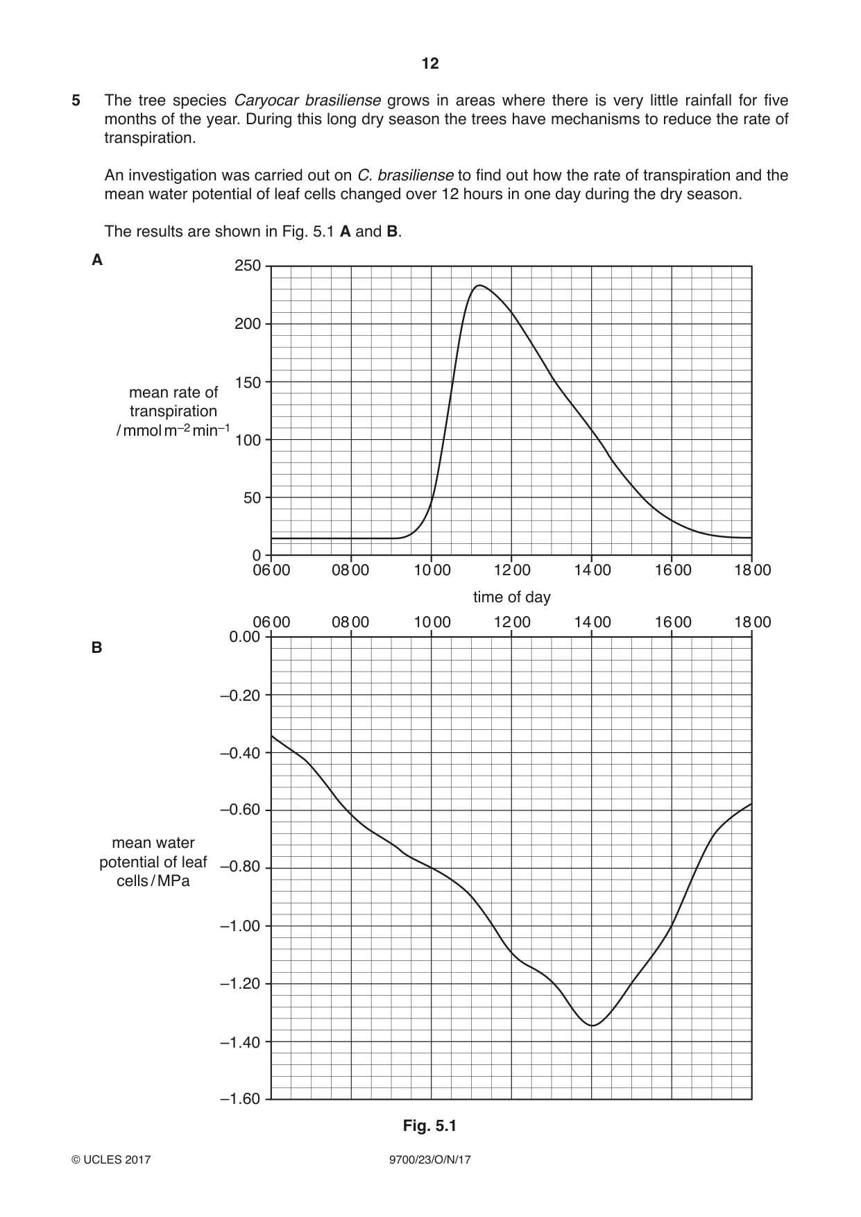
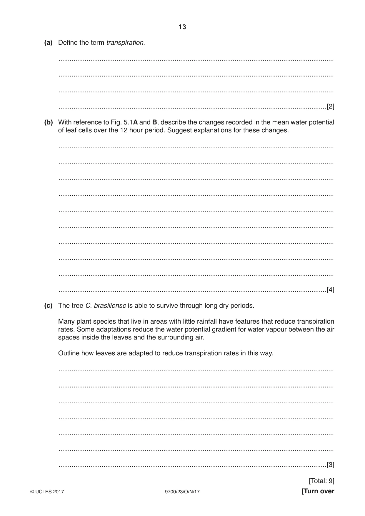
5 The tree species Caryocar brasiliense grows in areas where there is very little rainfall for five months of the year. During this long dry season the trees have mechanisms to reduce the rate of transpiration. An investigation was carried out on C. brasiliense to find out how the rate of transpiration and the mean water potential of leaf cells changed over 12 hours in one day during the dry season. The results are shown in Fig. 5.1 A and B. A 250 200 150 mean rate of transpiration / mmol m–2 min–1 100 50 0 06 00 08 00 10 00 12 00 14 00 16 00 18 00 time of day 06 00 08 00 10 00 12 00 14 00 16 00 18 00 0.00 B –0.20 –0.40 –0.60 mean water potential of leaf –0.80 cells / MPa –1.00 –1.20 –1.40 –1.60 Fig. 5.1 (a) Define the term transpiration. ................................................................................................................................................... ................................................................................................................................................... ................................................................................................................................................... ...............................................................................................................................................[2] (b) With reference to Fig. 5.1A and B, describe the changes recorded in the mean water potential of leaf cells over the 12 hour period. Suggest explanations for these changes. ................................................................................................................................................... ................................................................................................................................................... ................................................................................................................................................... ................................................................................................................................................... ................................................................................................................................................... ................................................................................................................................................... ................................................................................................................................................... ................................................................................................................................................... ................................................................................................................................................... ...............................................................................................................................................[4] (c) The tree C. brasiliense is able to survive through long dry periods. Many plant species that live in areas with little rainfall have features that reduce transpiration rates. Some adaptations reduce the water potential gradient for water vapour between the air spaces inside the leaves and the surrounding air. Outline how leaves are adapted to reduce transpiration rates in this way. ................................................................................................................................................... ................................................................................................................................................... ................................................................................................................................................... ................................................................................................................................................... ................................................................................................................................................... ................................................................................................................................................... ...............................................................................................................................................[3] [Total: 9]
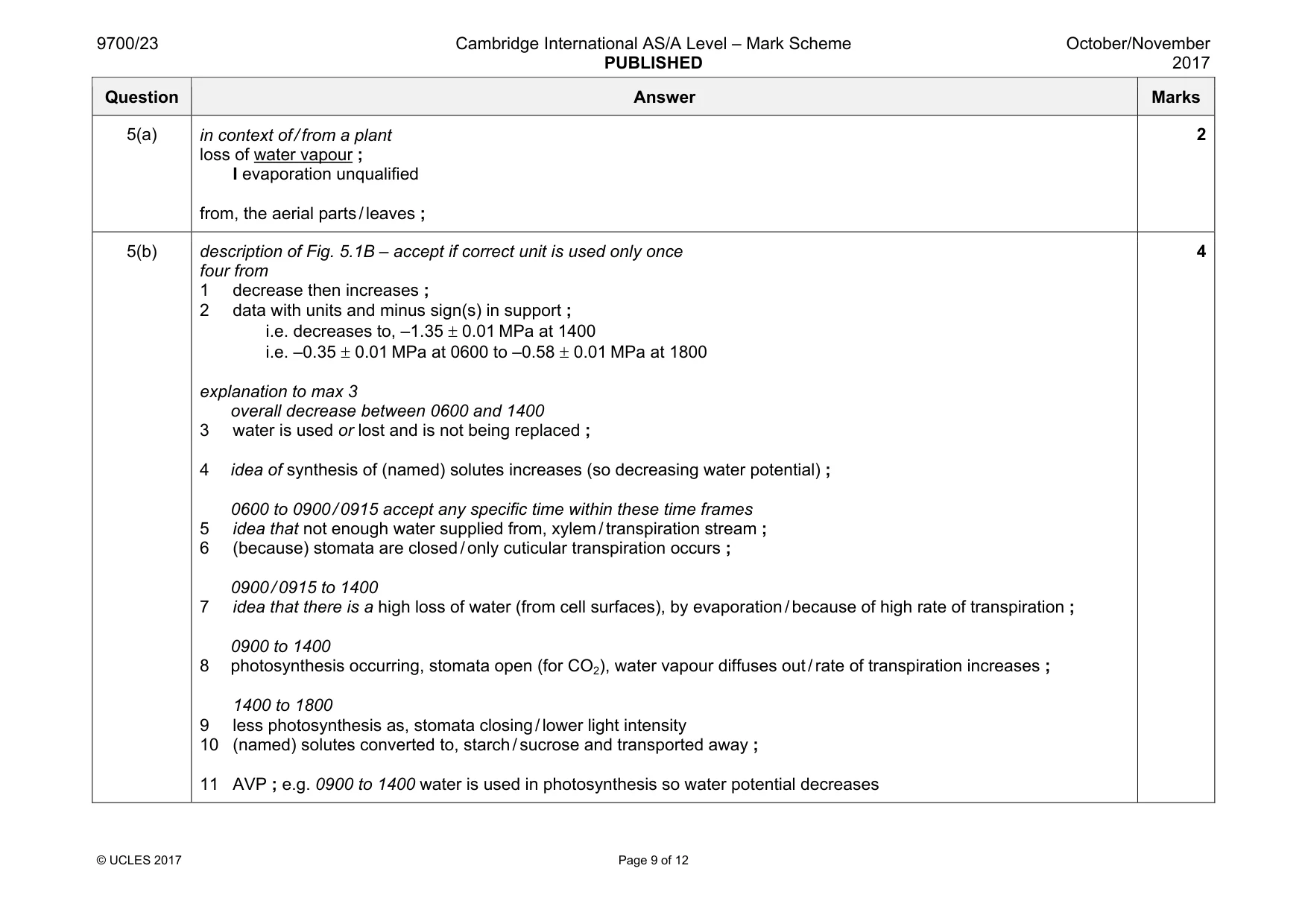
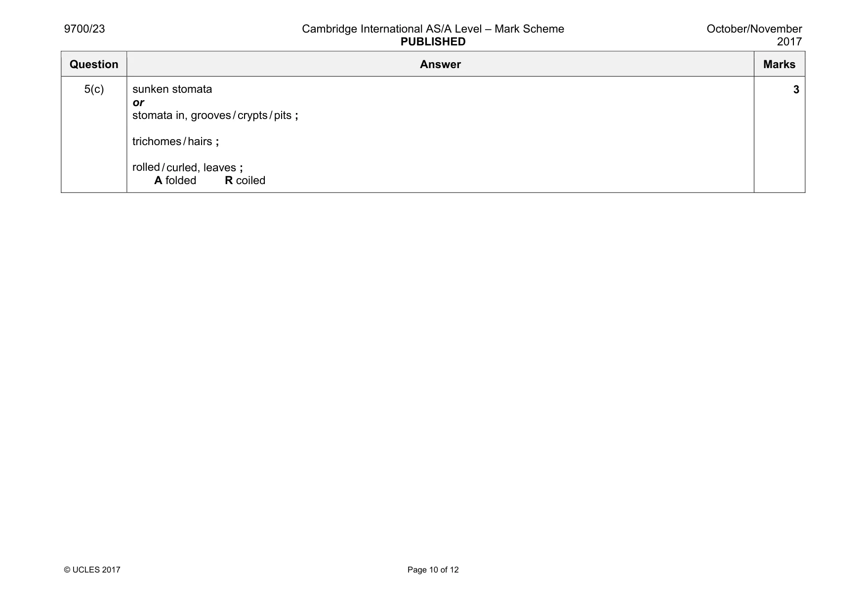
Mark scheme: 5(a) in context of / from a plant loss of water vapour ; I evaporation unqualified from, the aerial parts / leaves ; 2 5(b) description of Fig. 5.1B – accept if correct unit is used only once four from 1 decrease then increases ; 2 data with units and minus sign(s) in support ; i.e. decreases to, –1.35 ± 0.01 MPa at 1400 i.e. –0.35 ± 0.01 MPa at 0600 to –0.58 ± 0.01 MPa at 1800 explanation to max 3 overall decrease between 0600 and 1400 3 water is used or lost and is not being replaced ; 4 idea of synthesis of (named) solutes increases (so decreasing water potential) ; 0600 to 0900 / 0915 accept any specific time within these time frames 5 idea that not enough water supplied from, xylem / transpiration stream ; 6 (because) stomata are closed / only cuticular transpiration occurs ; 0900 / 0915 to 1400 7 idea that there is a high loss of water (from cell surfaces), by evaporation / because of high rate of transpiration ; 0900 to 1400 8 photosynthesis occurring, stomata open (for CO2), water vapour diffuses out / rate of transpiration increases ; 1400 to 1800 9 less photosynthesis as, stomata closing / lower light intensity 10 (named) solutes converted to, starch / sucrose and transported away ; 11 AVP ; e.g. 0900 to 1400 water is used in photosynthesis so water potential decreases 4 Question Answer Marks 5(c) sunken stomata or stomata in, grooves / crypts / pits ; trichomes / hairs ; rolled / curled, leaves ; A folded R coiled 3
Q6 · Extracellular growth factors are involved in the control of cell cycles in some mammalian…
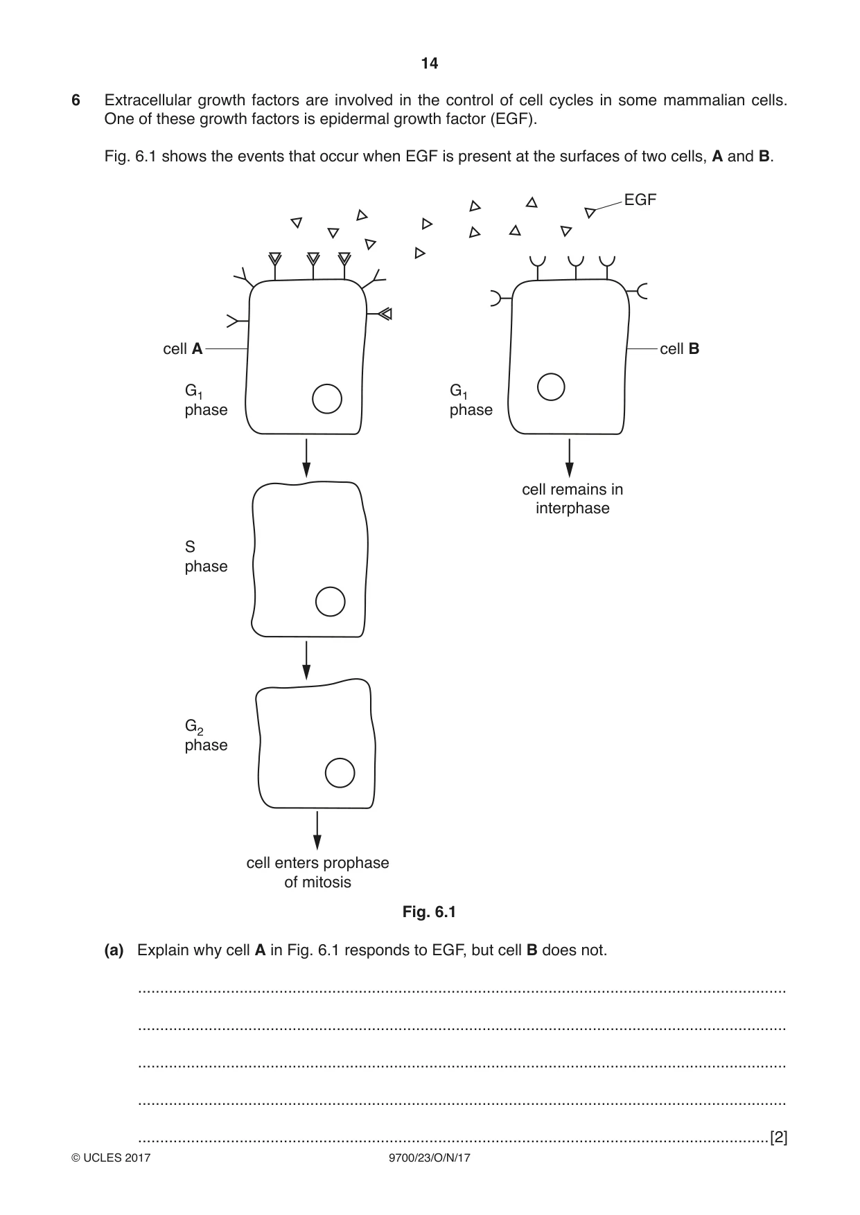
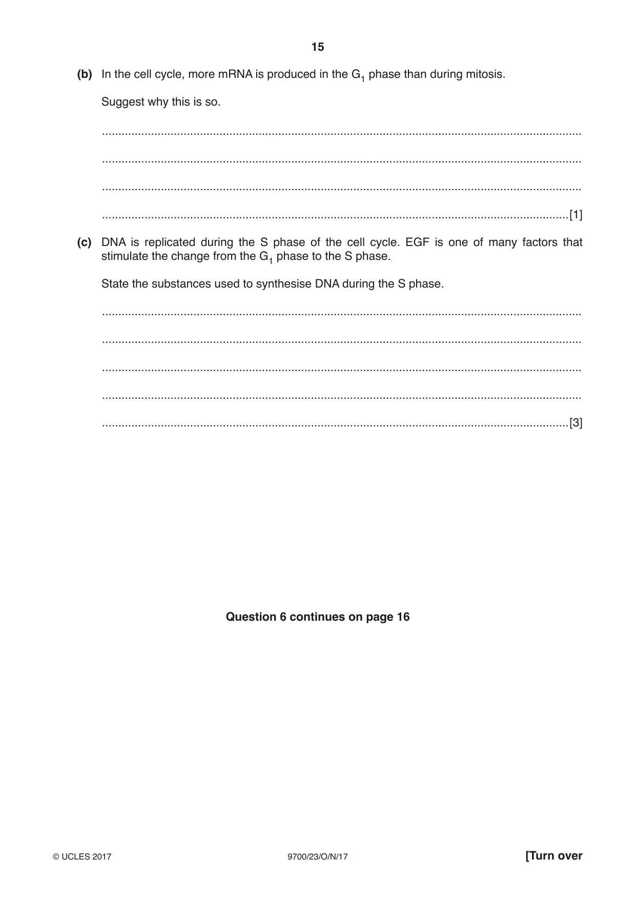
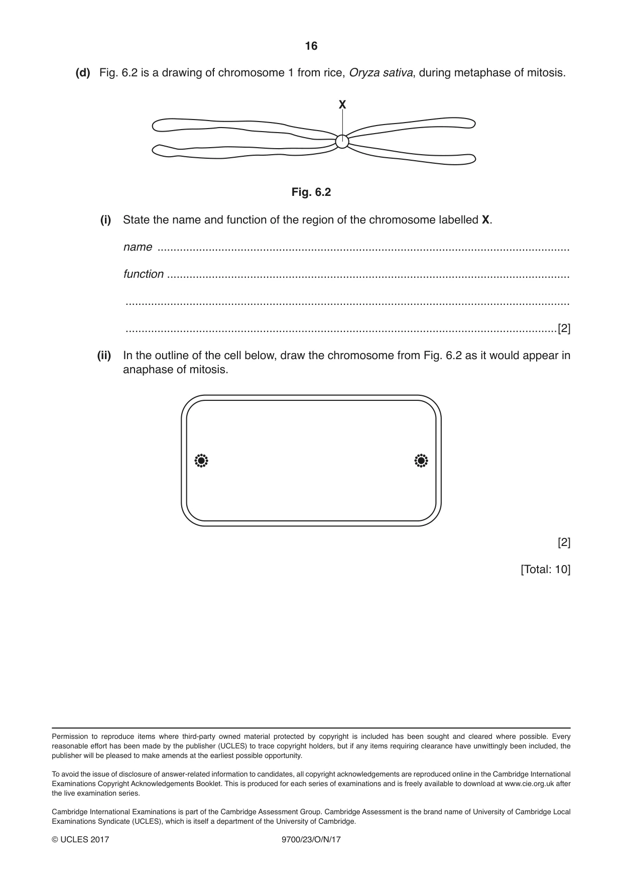
6 Extracellular growth factors are involved in the control of cell cycles in some mammalian cells. One of these growth factors is epidermal growth factor (EGF). Fig. 6.1 shows the events that occur when EGF is present at the surfaces of two cells, A and B. EGF cell A cell B G1 G1 phase phase cell remains in interphase S phase G2 phase cell enters prophase of mitosis Fig. 6.1 (a) Explain why cell A in Fig. 6.1 responds to EGF, but cell B does not. ................................................................................................................................................... ................................................................................................................................................... ................................................................................................................................................... ................................................................................................................................................... ...............................................................................................................................................[2] (b) In the cell cycle, more mRNA is produced in the G1 phase than during mitosis. Suggest why this is so. ................................................................................................................................................... ................................................................................................................................................... ................................................................................................................................................... ...............................................................................................................................................[1] (c) DNA is replicated during the S phase of the cell cycle. EGF is one of many factors that stimulate the change from the G1 phase to the S phase. State the substances used to synthesise DNA during the S phase. ................................................................................................................................................... ................................................................................................................................................... ................................................................................................................................................... ................................................................................................................................................... ...............................................................................................................................................[3] Question 6 continues on page 16 (d) Fig. 6.2 is a drawing of chromosome 1 from rice, Oryza sativa, during metaphase of mitosis. X Fig. 6.2 (i) State the name and function of the region of the chromosome labelled X. name ................................................................................................................................. function .............................................................................................................................. ........................................................................................................................................... .......................................................................................................................................[2] (ii) In the outline of the cell below, draw the chromosome from Fig. 6.2 as it would appear in anaphase of mitosis. [2] [Total: 10]
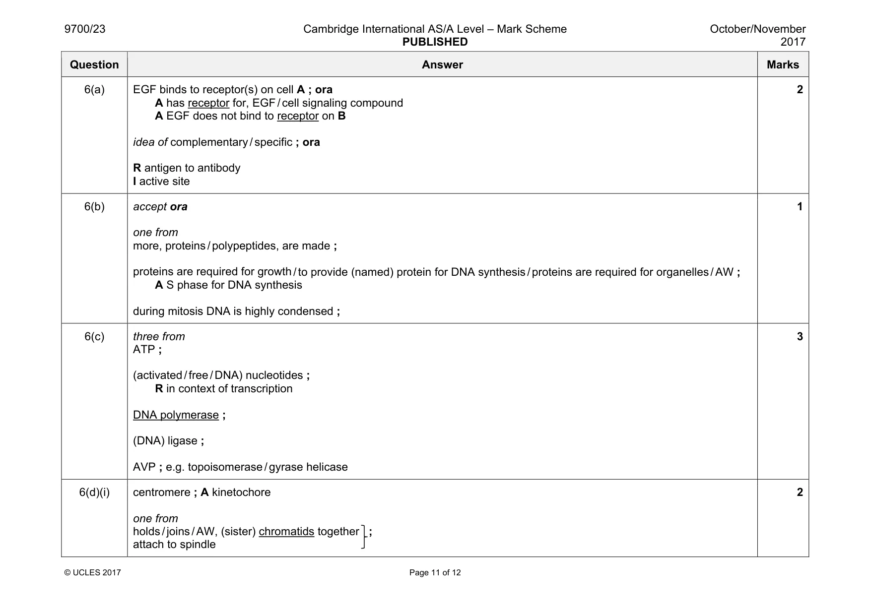
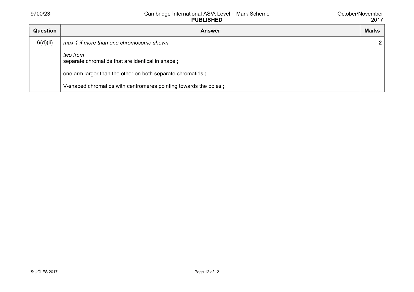
Mark scheme: 6(a) EGF binds to receptor(s) on cell A ; ora A has receptor for, EGF / cell signaling compound A EGF does not bind to receptor on B idea of complementary / specific ; ora R antigen to antibody I active site 2 6(b) accept ora one from more, proteins / polypeptides, are made ; proteins are required for growth / to provide (named) protein for DNA synthesis / proteins are required for organelles / AW ; A S phase for DNA synthesis during mitosis DNA is highly condensed ; 1 6(c) three from ATP ; (activated / free / DNA) nucleotides ; R in context of transcription DNA polymerase ; (DNA) ligase ; AVP ; e.g. topoisomerase / gyrase helicase 3 6(d)(i) centromere ; A kinetochore one from holds / joins / AW, (sister) chromatids together ; attach to spindle 2 Question Answer Marks 6(d)(ii) max 1 if more than one chromosome shown two from separate chromatids that are identical in shape ; one arm larger than the other on both separate chromatids ; V-shaped chromatids with centromeres pointing towards the poles ; 2
What was in this paper
The subtopics covered by these 6 questions, and how many questions each got. Open one in a new tab to see every Cambridge question on it.
What you needed in this session
Cambridge’s own grade thresholds for 2017 Oct/Nov, Paper 2 · Variant 3. A higher threshold means an easier paper — the bar moves with how the cohort did.