Cambridge A Level Biology 9700 — 2017 Oct/Nov Paper 2 · Variant 2
9700/22/O/N/17 · 6 questions · 60 marks · ≈68 min
The question paper and its mark scheme, free to read here and free to download. This is Cambridge’s own paper, exactly as it was sat.
Question paper16 pages
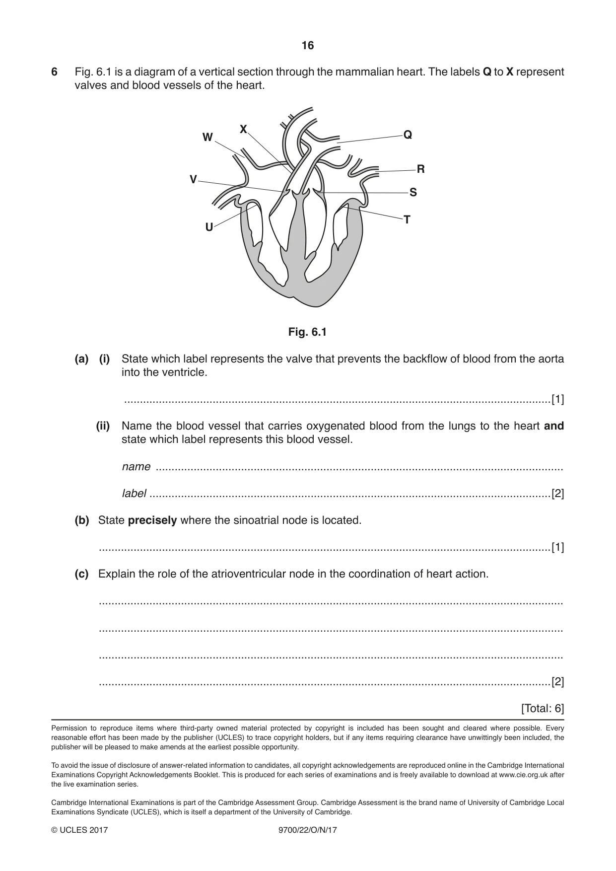
Mark scheme14 pages
Answers below. Sit the paper first if you are practising.
Questions as text
Q1 · The root apical meristem is a region of undifferentiated cells in the root tips of plants
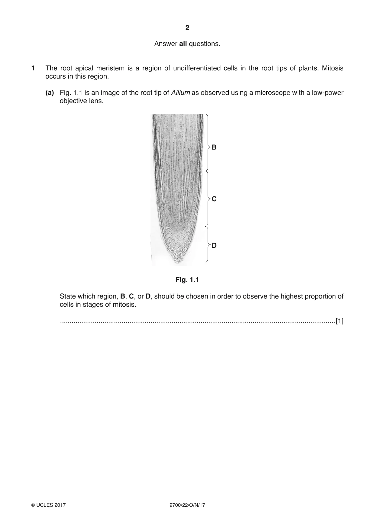
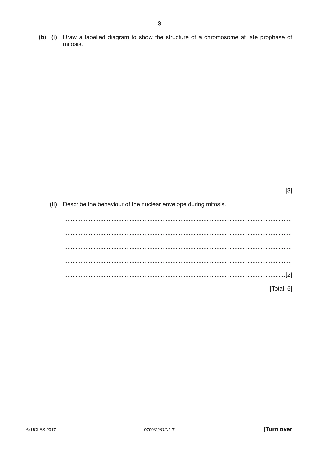
1 The root apical meristem is a region of undifferentiated cells in the root tips of plants. Mitosis occurs in this region. (a) Fig. 1.1 is an image of the root tip of Allium as observed using a microscope with a low-power objective lens. B C D Fig. 1.1 State which region, B, C, or D, should be chosen in order to observe the highest proportion of cells in stages of mitosis. ...............................................................................................................................................[1] (b) (i) Draw a labelled diagram to show the structure of a chromosome at late prophase of mitosis. [3] (ii) Describe the behaviour of the nuclear envelope during mitosis. ........................................................................................................................................... ........................................................................................................................................... ........................................................................................................................................... ........................................................................................................................................... .......................................................................................................................................[2] [Total: 6]
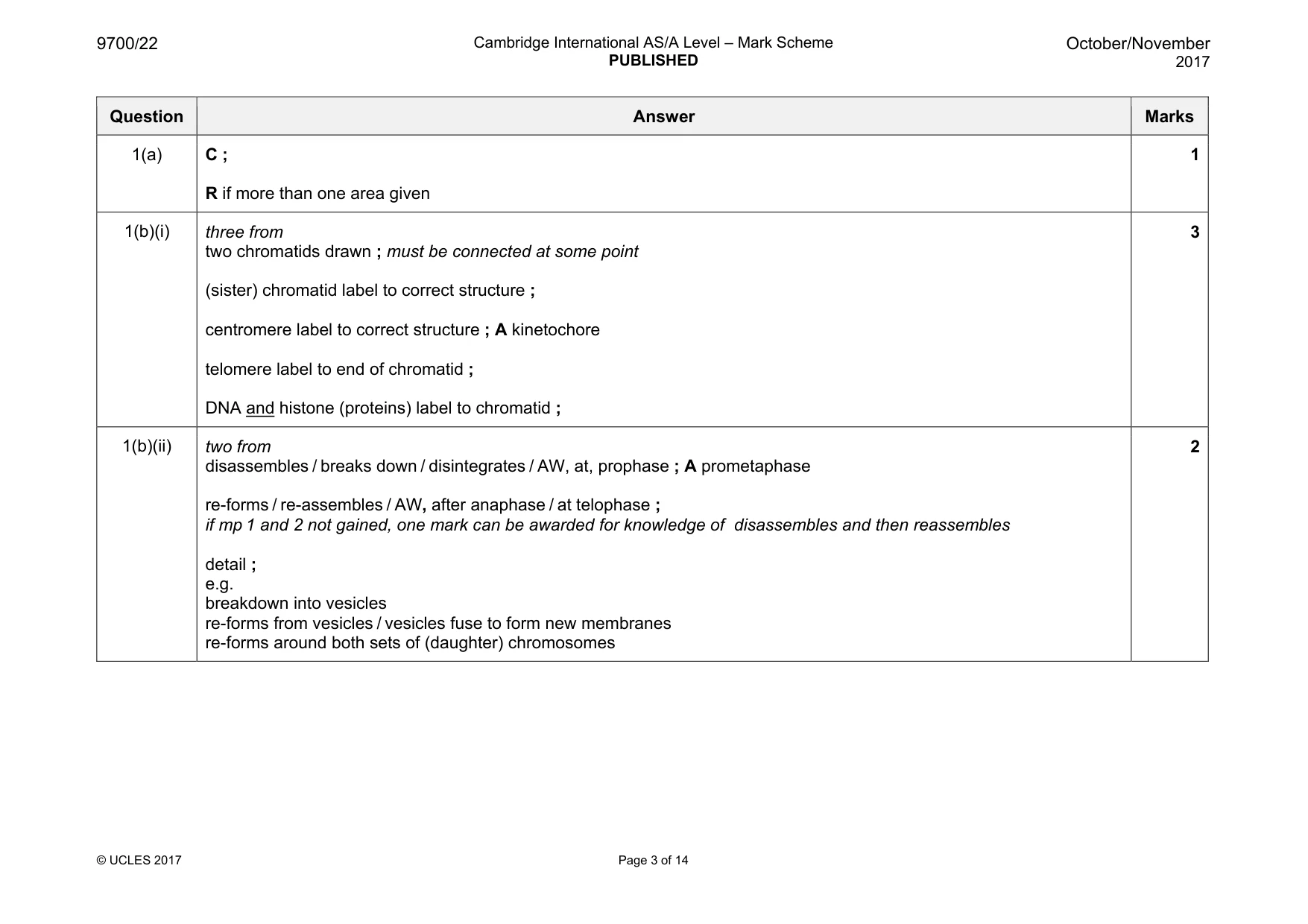
Mark scheme: 1(a) C ; R if more than one area given 1 1(b)(i) three from two chromatids drawn ; must be connected at some point (sister) chromatid label to correct structure ; centromere label to correct structure ; A kinetochore telomere label to end of chromatid ; DNA and histone (proteins) label to chromatid ; 3 1(b)(ii) two from disassembles / breaks down / disintegrates / AW, at, prophase ; A prometaphase re-forms / re-assembles / AW, after anaphase / at telophase ; if mp 1 and 2 not gained, one mark can be awarded for knowledge of disassembles and then reassembles detail ; e.g. breakdown into vesicles re-forms from vesicles / vesicles fuse to form new membranes re-forms around both sets of (daughter) chromosomes 2 Question 2
Q2 · Triglycerides and phospholipids are types of lipid
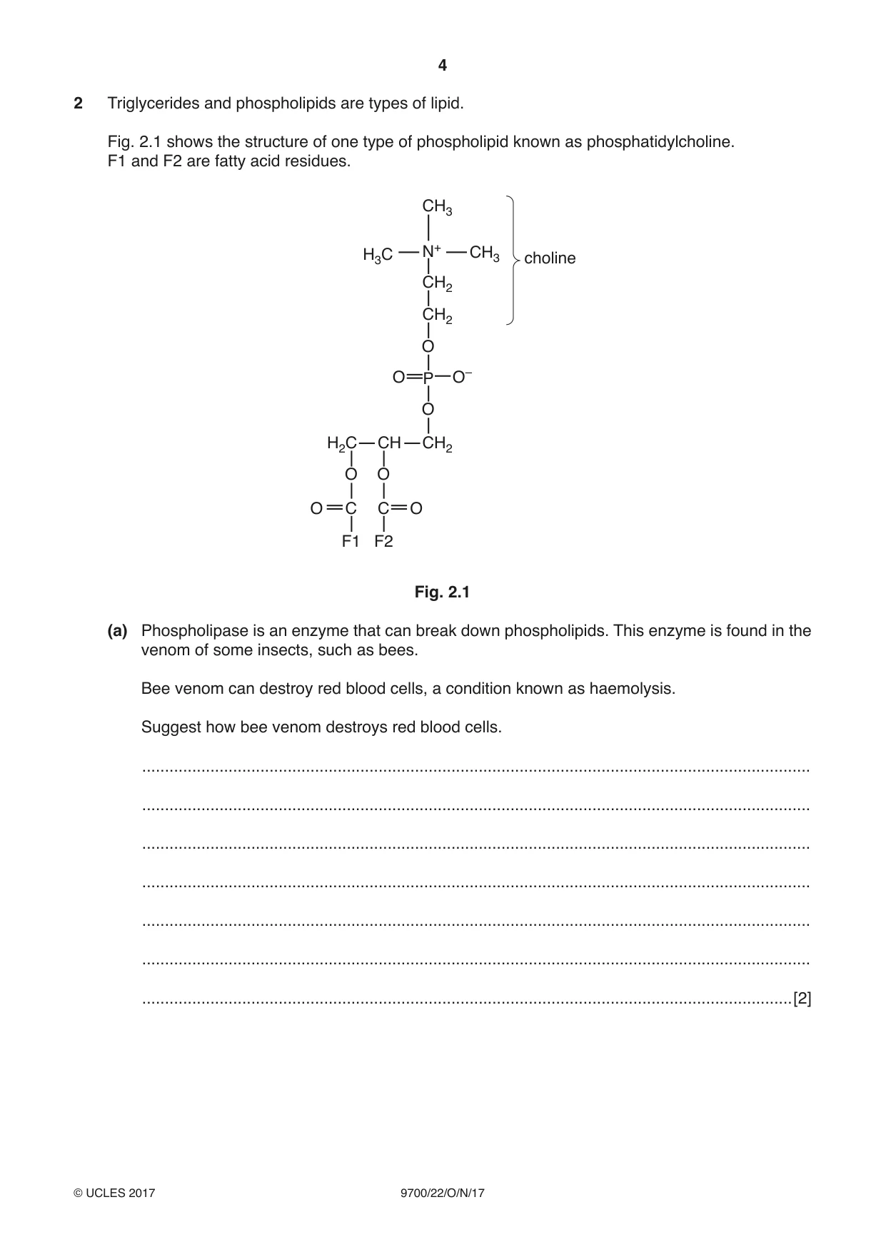
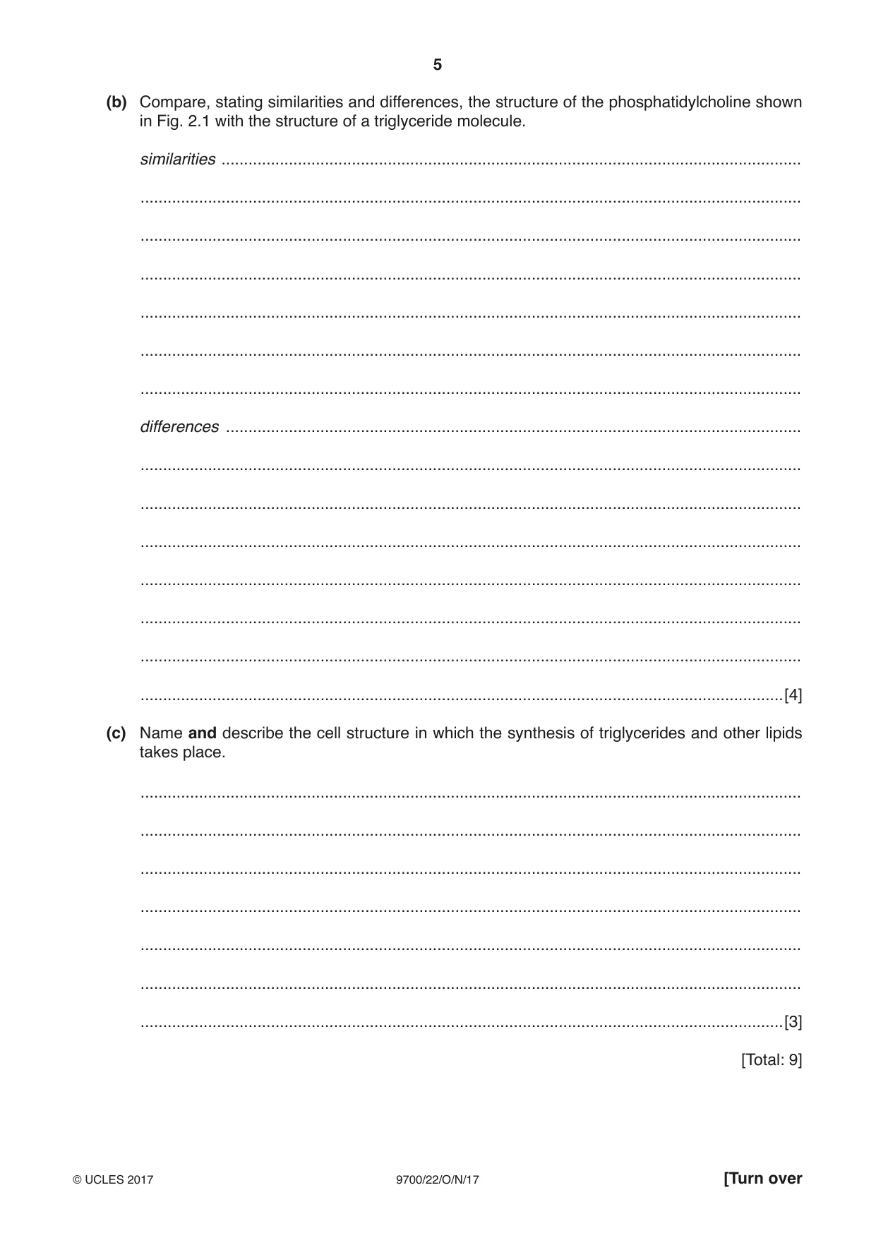
2 Triglycerides and phospholipids are types of lipid. Fig. 2.1 shows the structure of one type of phospholipid known as phosphatidylcholine. F1 and F2 are fatty acid residues. CH3 H3C N+ CH3 choline CH2 CH2 O O P O– O H2C CH CH2 O O O C C O F1 F2 Fig. 2.1 (a) Phospholipase is an enzyme that can break down phospholipids. This enzyme is found in the venom of some insects, such as bees. Bee venom can destroy red blood cells, a condition known as haemolysis. Suggest how bee venom destroys red blood cells. ................................................................................................................................................... ................................................................................................................................................... ................................................................................................................................................... ................................................................................................................................................... ................................................................................................................................................... ................................................................................................................................................... ...............................................................................................................................................[2] (b) Compare, stating similarities and differences, the structure of the phosphatidylcholine shown in Fig. 2.1 with the structure of a triglyceride molecule. similarities ................................................................................................................................. ................................................................................................................................................... ................................................................................................................................................... ................................................................................................................................................... ................................................................................................................................................... ................................................................................................................................................... ................................................................................................................................................... differences ................................................................................................................................ ................................................................................................................................................... ................................................................................................................................................... ................................................................................................................................................... ................................................................................................................................................... ................................................................................................................................................... ................................................................................................................................................... ...............................................................................................................................................[4] (c) Name and describe the cell structure in which the synthesis of triglycerides and other lipids takes place. ................................................................................................................................................... ................................................................................................................................................... ................................................................................................................................................... ................................................................................................................................................... ................................................................................................................................................... ................................................................................................................................................... ...............................................................................................................................................[3] [Total: 9]
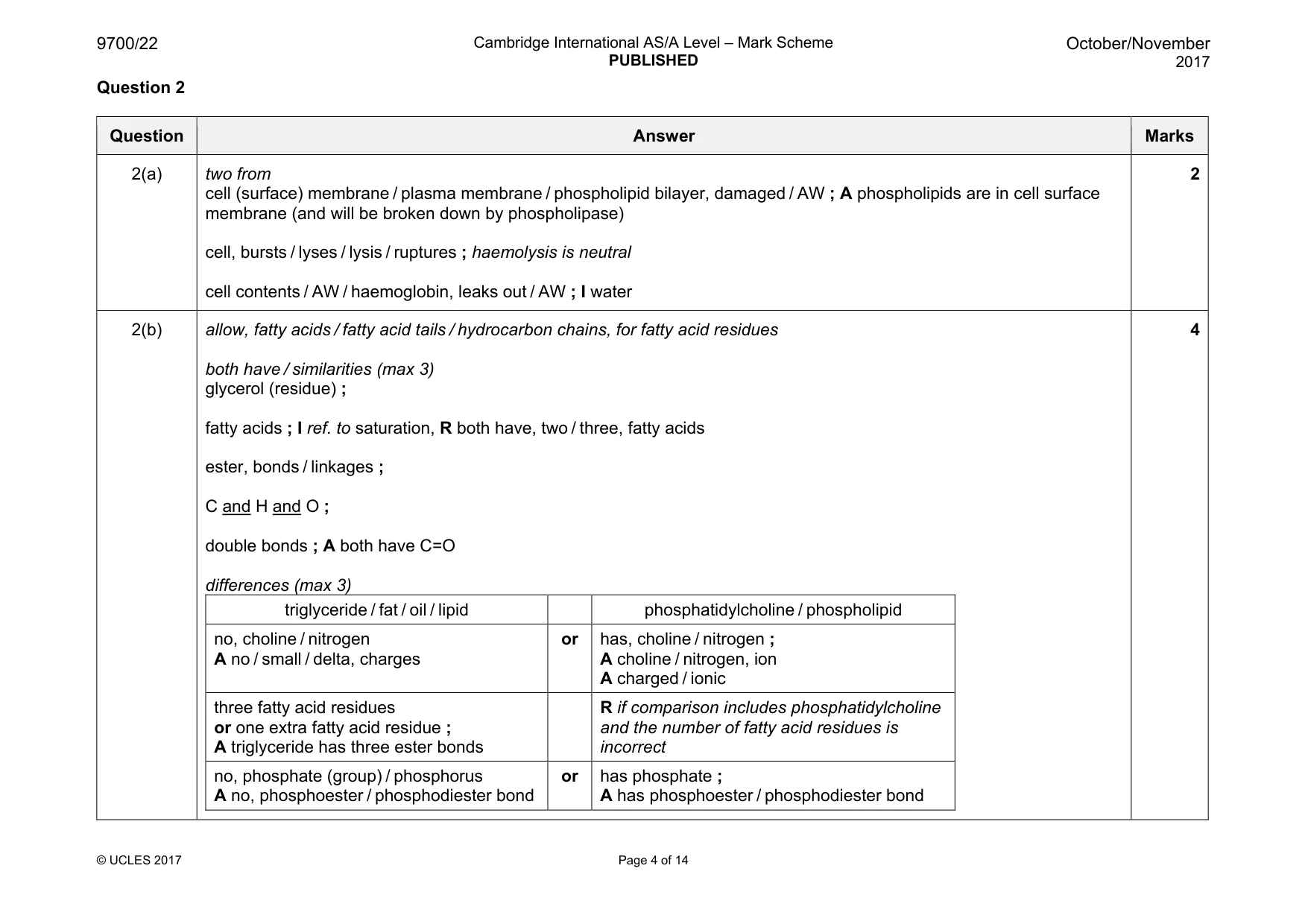
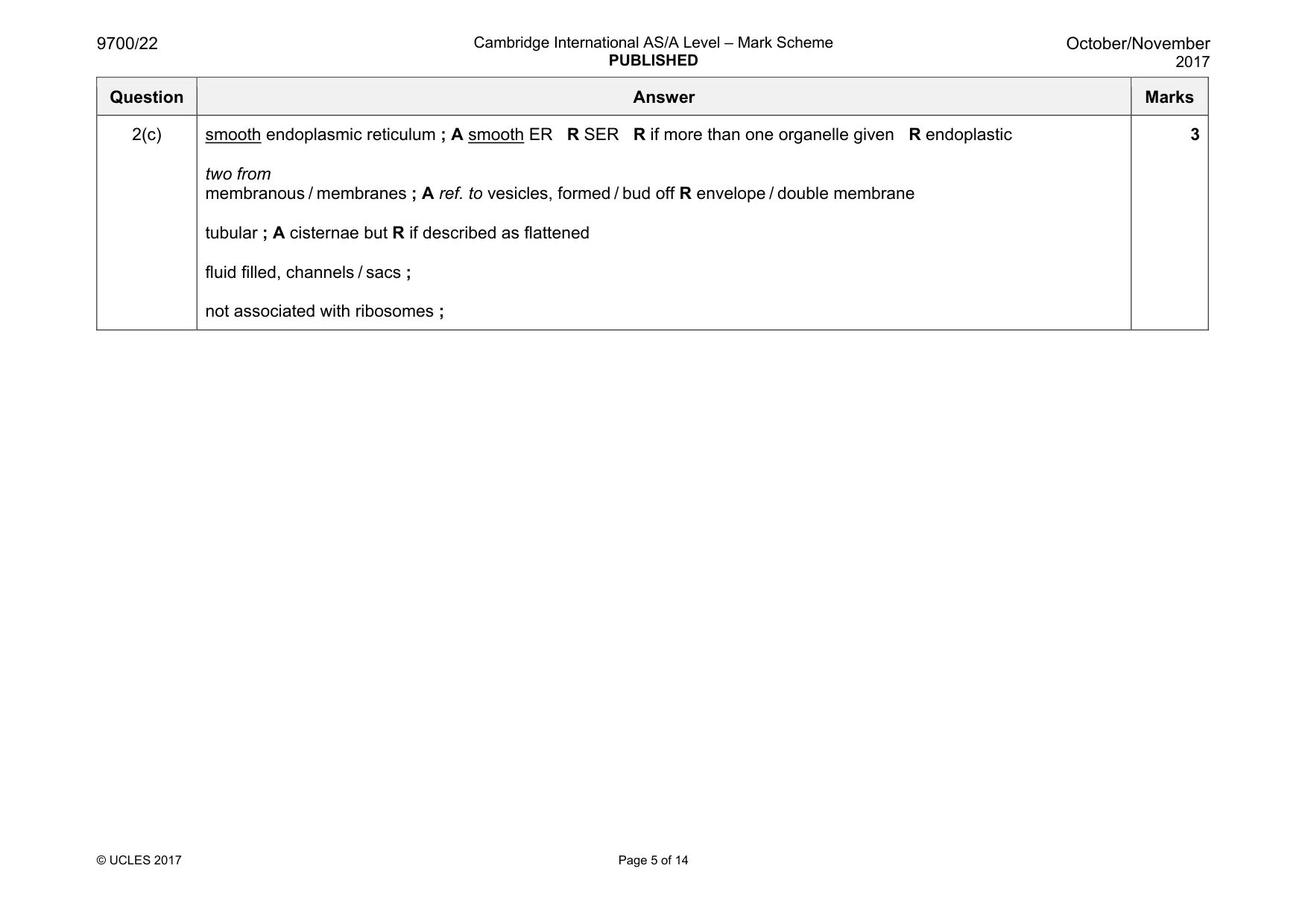
Mark scheme: 2(a) two from cell (surface) membrane / plasma membrane / phospholipid bilayer, damaged / AW ; A phospholipids are in cell surface membrane (and will be broken down by phospholipase) cell, bursts / lyses / lysis / ruptures ; haemolysis is neutral cell contents / AW / haemoglobin, leaks out / AW ; I water 2 2(b) allow, fatty acids / fatty acid tails / hydrocarbon chains, for fatty acid residues both have / similarities (max 3) glycerol (residue) ; fatty acids ; I ref. to saturation, R both have, two / three, fatty acids ester, bonds / linkages ; C and H and O ; double bonds ; A both have C=O differences (max 3) triglyceride / fat / oil / lipid phosphatidylcholine / phospholipid no, choline / nitrogen A no / small / delta, charges or has, choline / nitrogen ; A choline / nitrogen, ion A charged / ionic three fatty acid residues or one extra fatty acid residue ; A triglyceride has three ester bonds R if comparison includes phosphatidylcholine and the number of fatty acid residues is incorrect no, phosphate (group) / phosphorus A no, phosphoester / phosphodiester bond or has phosphate ; A has phosphoester / phosphodiester bond 4 Question Answer Marks 2(c) smooth endoplasmic reticulum ; A smooth ER R SER R if more than one organelle given R endoplastic two from membranous / membranes ; A ref. to vesicles, formed / bud off R envelope / double membrane tubular ; A cisternae but R if described as flattened fluid filled, channels / sacs ; not associated with ribosomes ; 3 Question 3
Q3 · Hydrolytic enzymes can function within the cell or can be secreted by the cell, where…
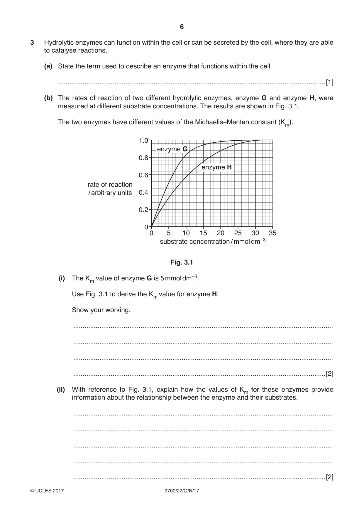
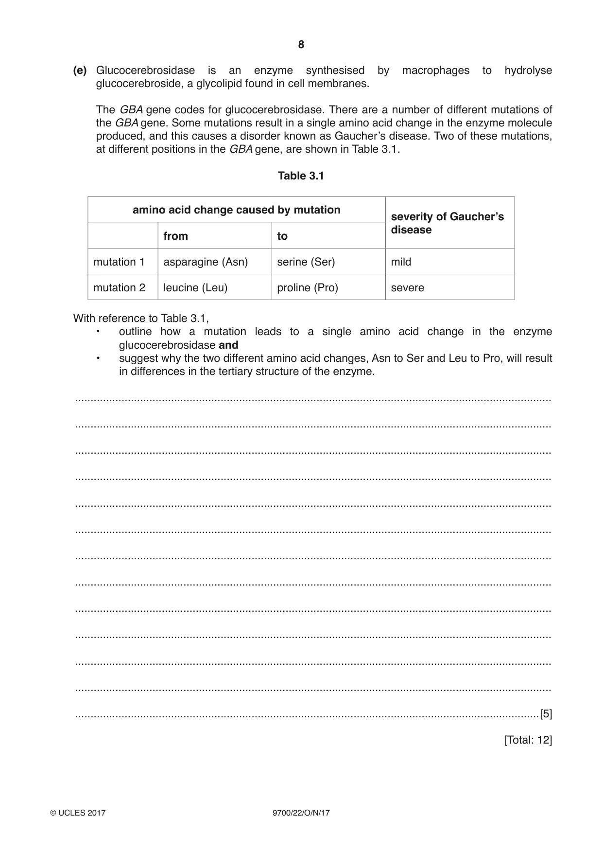
3 Hydrolytic enzymes can function within the cell or can be secreted by the cell, where they are able to catalyse reactions. (a) State the term used to describe an enzyme that functions within the cell. ...............................................................................................................................................[1] (b) The rates of reaction of two different hydrolytic enzymes, enzyme G and enzyme H, were measured at different substrate concentrations. The results are shown in Fig. 3.1. The two enzymes have different values of the Michaelis–Menten constant (Km). 1.0 enzyme G 0.8 enzyme H 0.6 rate of reaction / arbitrary units 0.4 0.2 0 0 5 10 15 20 25 30 35 substrate concentration / mmol dm–3 Fig. 3.1 (i) The Km value of enzyme G is 5 mmol dm–3. Use Fig. 3.1 to derive the Km value for enzyme H. Show your working. ........................................................................................................................................... ........................................................................................................................................... ........................................................................................................................................... .......................................................................................................................................[2] (ii) With reference to Fig. 3.1, explain how the values of Km for these enzymes provide information about the relationship between the enzyme and their substrates. ........................................................................................................................................... ........................................................................................................................................... ........................................................................................................................................... ........................................................................................................................................... .......................................................................................................................................[2] Cells can break down an old or damaged organelle, such as a mitochondrion, by enclosing the organelle in a membrane formed from the endoplasmic reticulum. This forms a vacuole. Vesicles containing hydrolytic enzymes fuse with the vacuole and the organelle is then digested. (c) Name the vesicles in the cell that contain hydrolytic enzymes. ...............................................................................................................................................[1] (d) The cell has internal chemical messengers that signal when an old or damaged mitochondrion should be broken down. Suggest one feature involving mitochondrial structure or function that could lead to the release of these internal signals. ................................................................................................................................................... ...............................................................................................................................................[1] Question 3 continues on page 8 (e) Glucocerebrosidase is an enzyme synthesised by macrophages to hydrolyse glucocerebroside, a glycolipid found in cell membranes. The GBA gene codes for glucocerebrosidase. There are a number of different mutations of the GBA gene. Some mutations result in a single amino acid change in the enzyme molecule produced, and this causes a disorder known as Gaucher’s disease. Two of these mutations, at different positions in the GBA gene, are shown in Table 3.1. Table 3.1 amino acid change caused by mutation severity of Gaucher’s disease from to mutation 1 asparagine (Asn) serine (Ser) mild mutation 2 leucine (Leu) proline (Pro) severe With reference to Table 3.1, • outline how a mutation leads to a single amino acid change in the enzyme glucocerebrosidase and • suggest why the two different amino acid changes, Asn to Ser and Leu to Pro, will result in differences in the tertiary structure of the enzyme. .......................................................................................................................................................... .......................................................................................................................................................... .......................................................................................................................................................... .......................................................................................................................................................... .......................................................................................................................................................... .......................................................................................................................................................... .......................................................................................................................................................... .......................................................................................................................................................... .......................................................................................................................................................... .......................................................................................................................................................... .......................................................................................................................................................... .......................................................................................................................................................... ......................................................................................................................................................[5] [Total: 12]
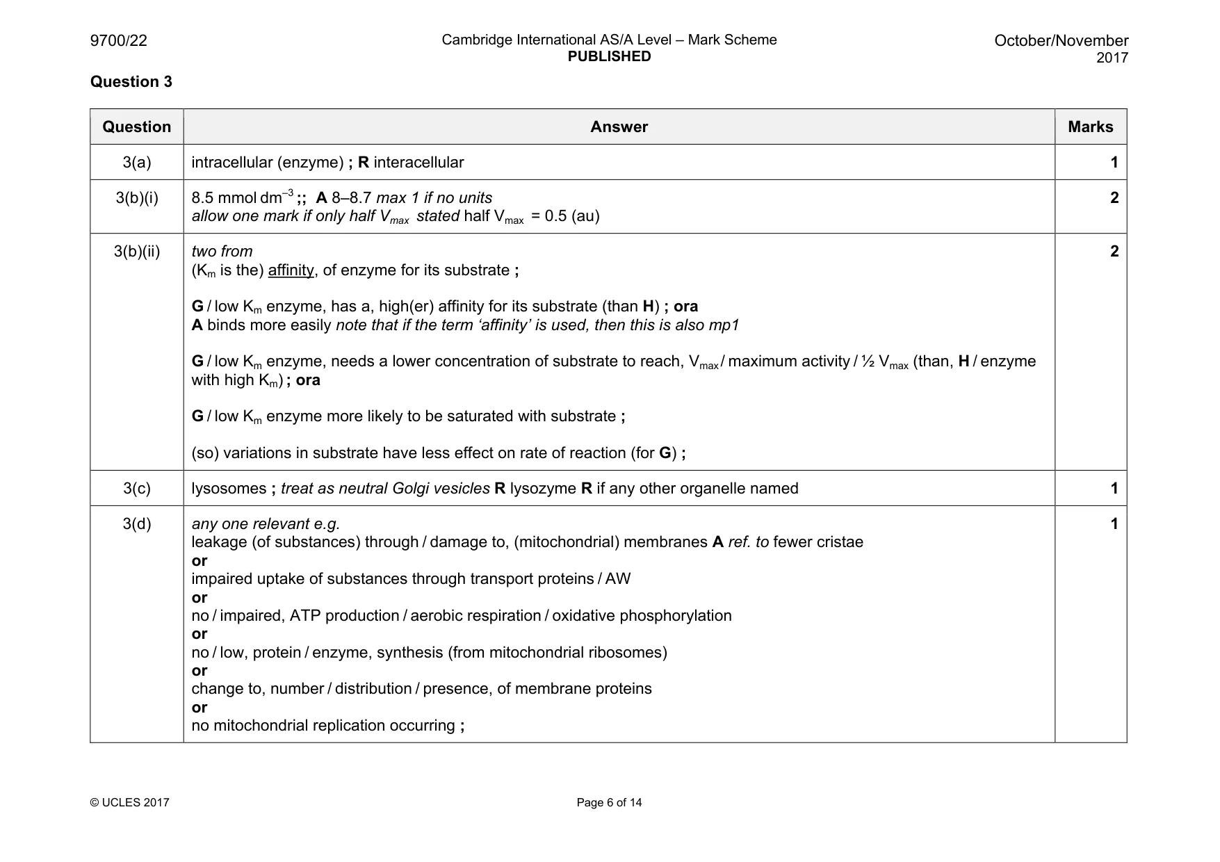
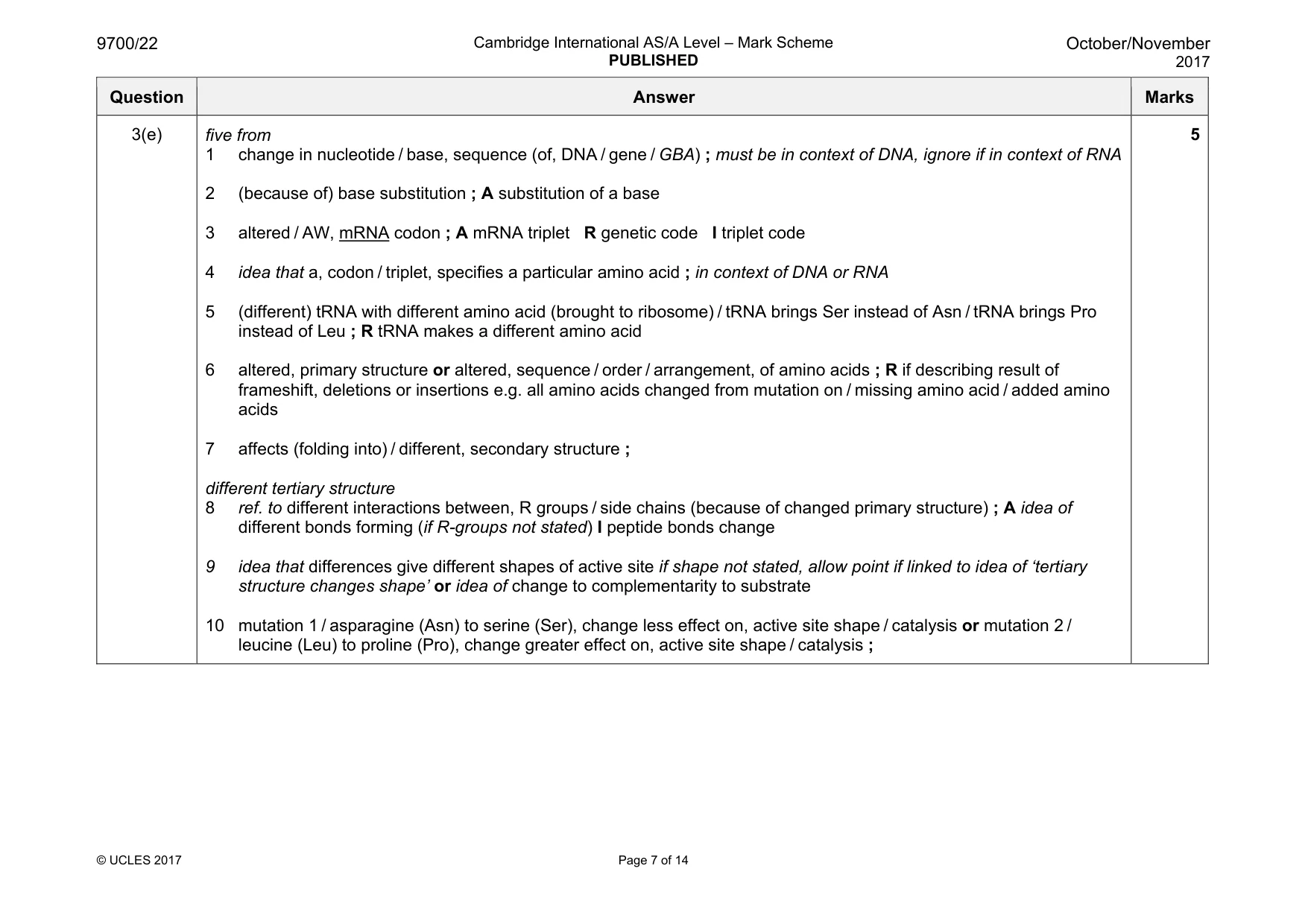
Mark scheme: 3(a) intracellular (enzyme) ; R interacellular 1 3(b)(i) 8.5 mmol dm–3 ;; A 8–8.7 max 1 if no units allow one mark if only half Vmax stated half Vmax = 0.5 (au) 2 3(b)(ii) two from (Km is the) affinity, of enzyme for its substrate ; G / low Km enzyme, has a, high(er) affinity for its substrate (than H) ; ora A binds more easily note that if the term ‘affinity’ is used, then this is also mp1 G / low Km enzyme, needs a lower concentration of substrate to reach, Vmax / maximum activity / ½ Vmax (than, H / enzyme with high Km) ; ora G / low Km enzyme more likely to be saturated with substrate ; (so) variations in substrate have less effect on rate of reaction (for G) ; 2 3(c) lysosomes ; treat as neutral Golgi vesicles R lysozyme R if any other organelle named 1 3(d) any one relevant e.g. leakage (of substances) through / damage to, (mitochondrial) membranes A ref. to fewer cristae or impaired uptake of substances through transport proteins / AW or no / impaired, ATP production / aerobic respiration / oxidative phosphorylation or no / low, protein / enzyme, synthesis (from mitochondrial ribosomes) or change to, number / distribution / presence, of membrane proteins or no mitochondrial replication occurring ; 1 Question Answer Marks 3(e) five from 1 change in nucleotide / base, sequence (of, DNA / gene / GBA) ; must be in context of DNA, ignore if in context of RNA 2 (because of) base substitution ; A substitution of a base 3 altered / AW, mRNA codon ; A mRNA triplet R genetic code I triplet code 4 idea that a, codon / triplet, specifies a particular amino acid ; in context of DNA or RNA 5 (different) tRNA with different amino acid (brought to ribosome) / tRNA brings Ser instead of Asn / tRNA brings Pro instead of Leu ; R tRNA makes a different amino acid 6 altered, primary structure or altered, sequence / order / arrangement, of amino acids ; R if describing result of frameshift, deletions or insertions e.g. all amino acids changed from mutation on / missing amino acid / added amino acids 7 affects (folding into) / different, secondary structure ; different tertiary structure 8 ref. to different interactions between, R groups / side chains (because of changed primary structure) ; A idea of different bonds forming (if R-groups not stated) I peptide bonds change 9 idea that differences give different shapes of active site if shape not stated, allow point if linked to idea of ‘tertiary structure changes shape’ or idea of change to complementarity to substrate 10 mutation 1 / asparagine (Asn) to serine (Ser), change less effect on, active site shape / catalysis or mutation 2 / leucine (Leu) to proline (Pro), change greater effect on, active site shape / catalysis ; 5 Question 4
Q4 · A transmission electron micrograph of the bacterium that causes cholera, Vibrio cholerae
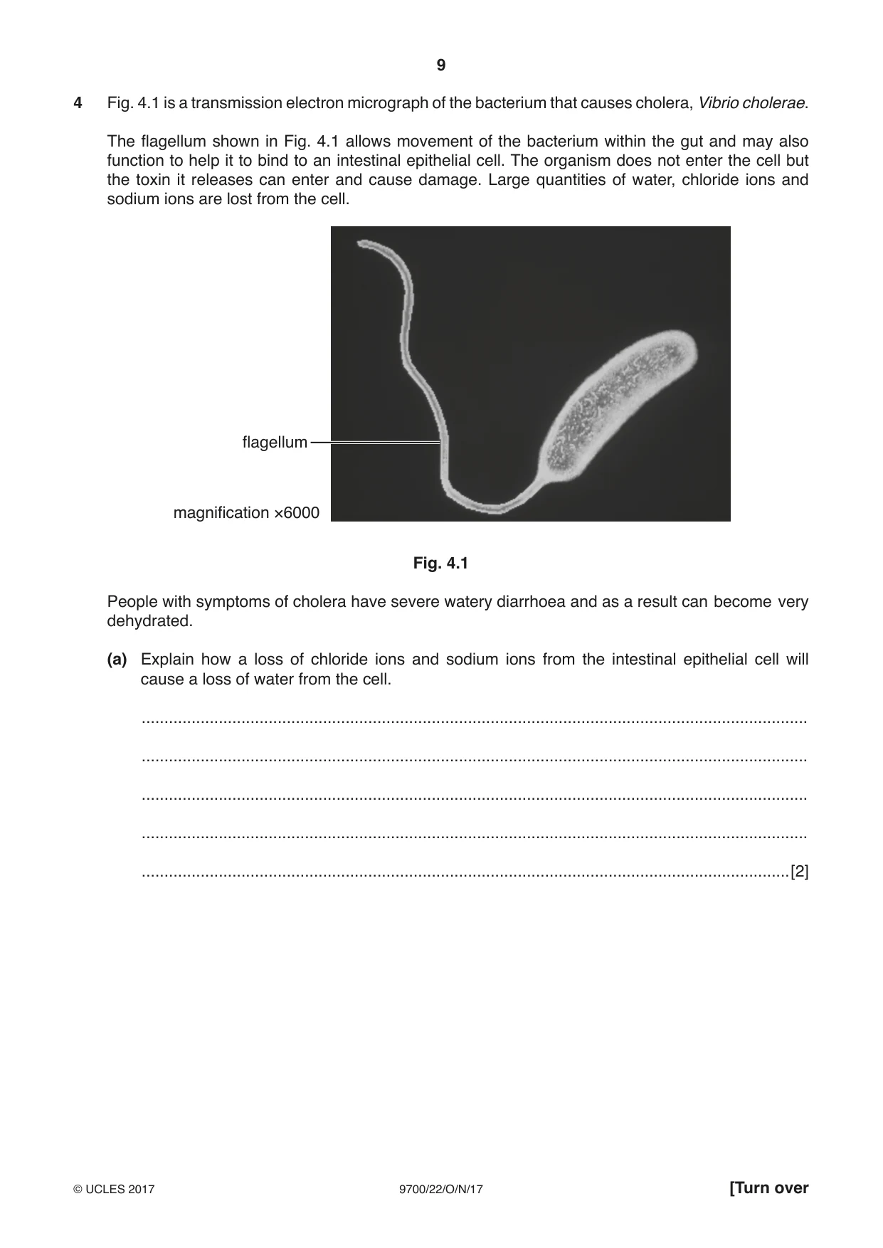
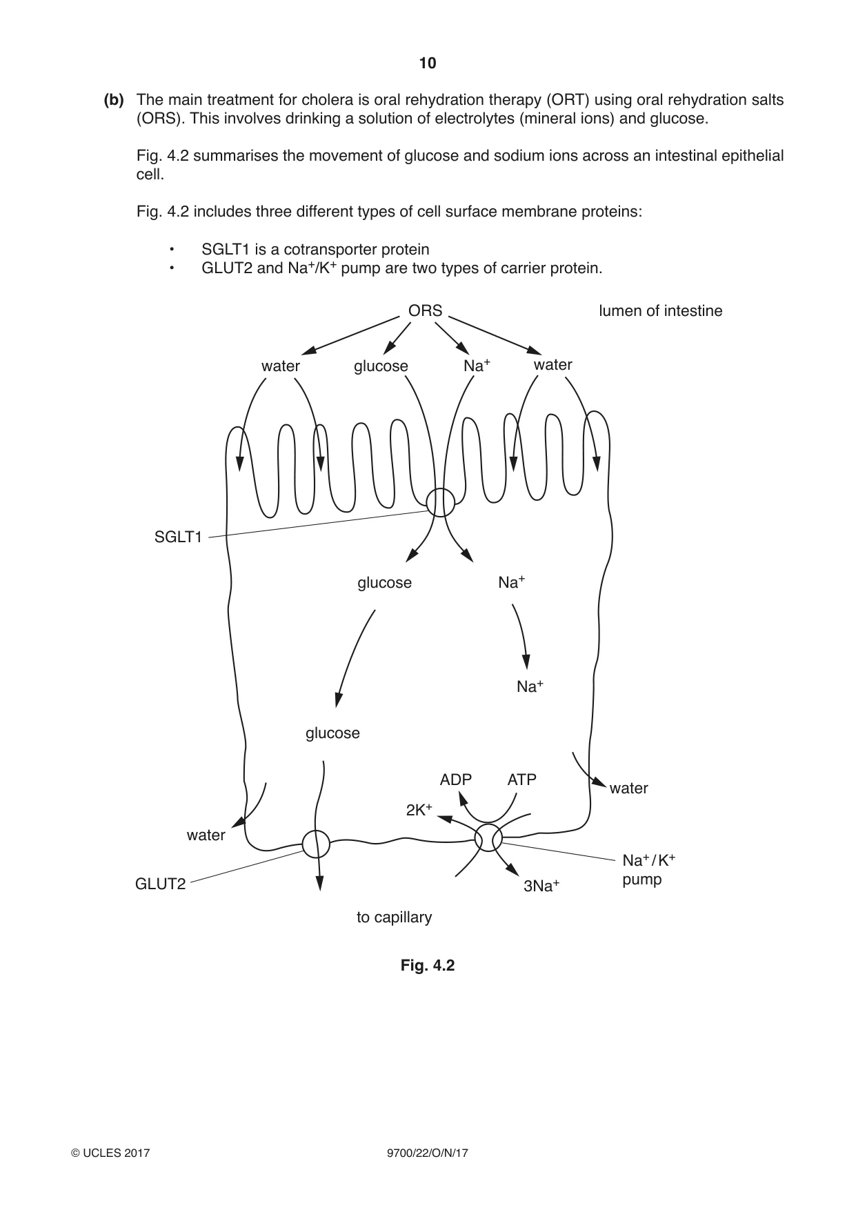
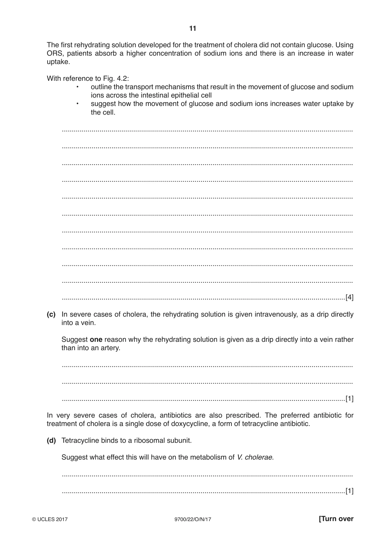
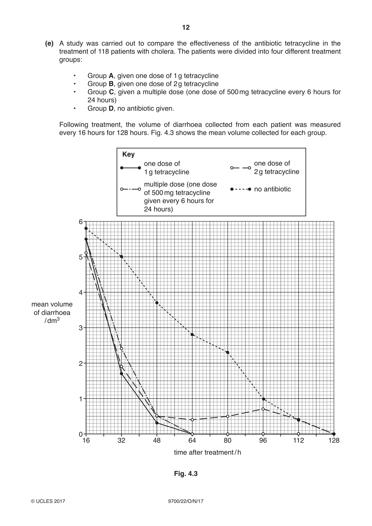
4 Fig. 4.1 is a transmission electron micrograph of the bacterium that causes cholera, Vibrio cholerae. The flagellum shown in Fig. 4.1 allows movement of the bacterium within the gut and may also function to help it to bind to an intestinal epithelial cell. The organism does not enter the cell but the toxin it releases can enter and cause damage. Large quantities of water, chloride ions and sodium ions are lost from the cell. flagellum magnification ×6000 Fig. 4.1 People with symptoms of cholera have severe watery diarrhoea and as a result can become very dehydrated. (a) Explain how a loss of chloride ions and sodium ions from the intestinal epithelial cell will cause a loss of water from the cell. ................................................................................................................................................... ................................................................................................................................................... ................................................................................................................................................... ................................................................................................................................................... ...............................................................................................................................................[2] (b) The main treatment for cholera is oral rehydration therapy (ORT) using oral rehydration salts (ORS). This involves drinking a solution of electrolytes (mineral ions) and glucose. Fig. 4.2 summarises the movement of glucose and sodium ions across an intestinal epithelial cell. Fig. 4.2 includes three different types of cell surface membrane proteins: • SGLT1 is a cotransporter protein • GLUT2 and Na+/K+ pump are two types of carrier protein. ORS lumen of intestine water glucose Na+ water SGLT1 glucose Na+ Na+ glucose ADP ATP water 2K+ water Na+ / K+ pump GLUT2 3Na+ to capillary Fig. 4.2 The first rehydrating solution developed for the treatment of cholera did not contain glucose. Using ORS, patients absorb a higher concentration of sodium ions and there is an increase in water uptake. With reference to Fig. 4.2: • outline the transport mechanisms that result in the movement of glucose and sodium ions across the intestinal epithelial cell • suggest how the movement of glucose and sodium ions increases water uptake by the cell. ................................................................................................................................................... ................................................................................................................................................... ................................................................................................................................................... ................................................................................................................................................... ................................................................................................................................................... ................................................................................................................................................... ................................................................................................................................................... ................................................................................................................................................... ................................................................................................................................................... ................................................................................................................................................... ...............................................................................................................................................[4] (c) In severe cases of cholera, the rehydrating solution is given intravenously, as a drip directly into a vein. Suggest one reason why the rehydrating solution is given as a drip directly into a vein rather than into an artery. ................................................................................................................................................... ................................................................................................................................................... ...............................................................................................................................................[1] In very severe cases of cholera, antibiotics are also prescribed. The preferred antibiotic for treatment of cholera is a single dose of doxycycline, a form of tetracycline antibiotic. (d) Tetracycline binds to a ribosomal subunit. Suggest what effect this will have on the metabolism of V. cholerae. ................................................................................................................................................... ...............................................................................................................................................[1] (e) A study was carried out to compare the effectiveness of the antibiotic tetracycline in the treatment of 118 patients with cholera. The patients were divided into four different treatment groups: • Group A, given one dose of 1 g tetracycline • Group B, given one dose of 2 g tetracycline • Group C, given a multiple dose (one dose of 500 mg tetracycline every 6 hours for 24 hours) • Group D, no antibiotic given. Following treatment, the volume of diarrhoea collected from each patient was measured every 16 hours for 128 hours. Fig. 4.3 shows the mean volume collected for each group. Key one dose of one dose of 1 g tetracycline 2 g tetracycline multiple dose (one dose no antibiotic of 500 mg tetracycline given every 6 hours for 24 hours) 6 5 4 mean volume of diarrhoea / dm3 3 2 1 0 16 32 48 64 80 96 112 128 time after treatment / h Fig. 4.3 (i) Describe the trends shown in Fig. 4.3. ........................................................................................................................................... ........................................................................................................................................... ........................................................................................................................................... ........................................................................................................................................... ........................................................................................................................................... ........................................................................................................................................... ........................................................................................................................................... ........................................................................................................................................... ........................................................................................................................................... .......................................................................................................................................[3] (ii) Explain whether the results support, or do not support, treating very severe cases of cholera with tetracycline. ........................................................................................................................................... ........................................................................................................................................... ........................................................................................................................................... ........................................................................................................................................... .......................................................................................................................................[2] (f) Some strains of V. cholerae are antibiotic resistant. Explain why this means that medical practitioners prefer to treat cholera with a single dose of antibiotic, rather than a multiple dose of the same antibiotic. ................................................................................................................................................... ................................................................................................................................................... ................................................................................................................................................... ................................................................................................................................................... ................................................................................................................................................... ...............................................................................................................................................[2] (g) Most people who have recovered from cholera rarely become ill again from the disease. In these people, antibodies have been identified that will bind either to the cholera toxin, or to the bacterial flagellum, or to the main bacterial cell. Explain why the antibodies are different, each one specific to its target. ................................................................................................................................................... ................................................................................................................................................... ................................................................................................................................................... ................................................................................................................................................... ................................................................................................................................................... ................................................................................................................................................... ...............................................................................................................................................[3] (h) Evidence suggests that newborn babies of mothers who have had cholera have immunity to the disease. State precisely the type of immunity these babies are likely to have. ...............................................................................................................................................[1] [Total: 19]
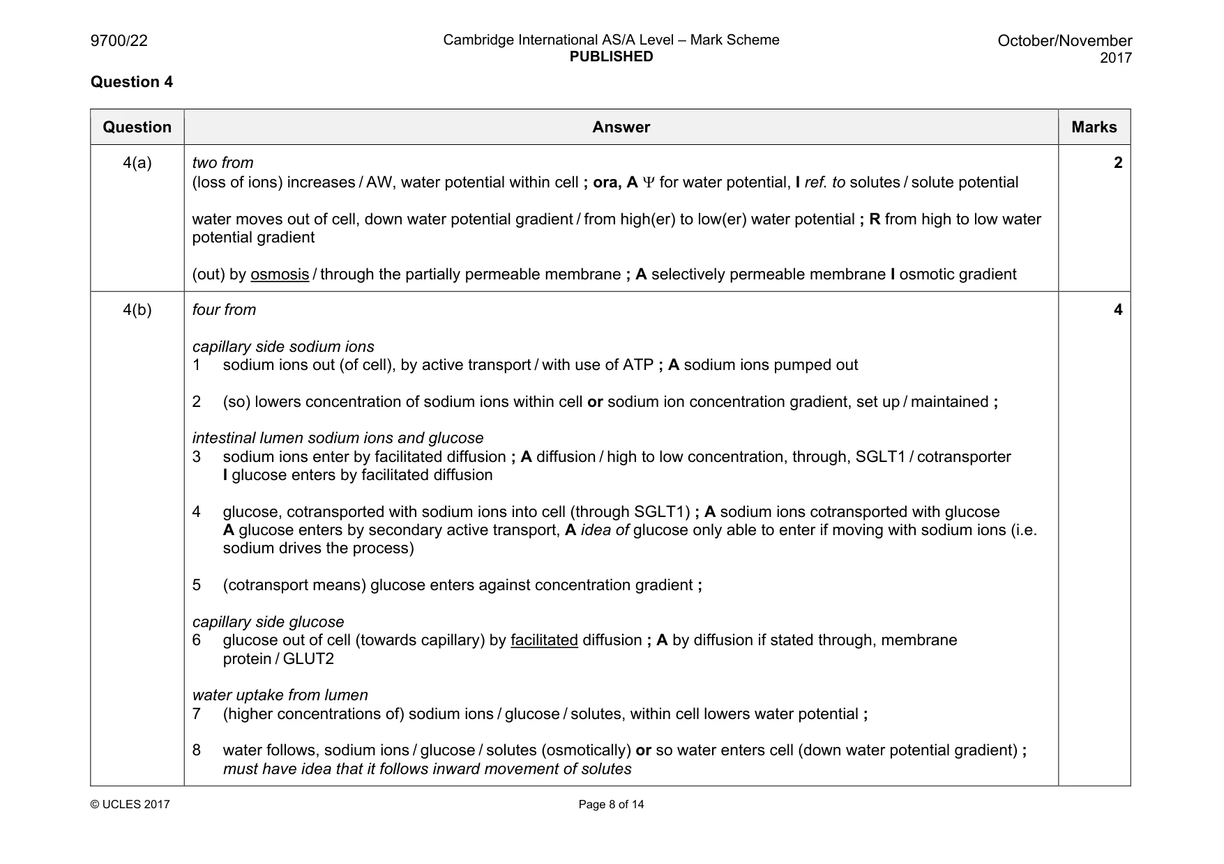
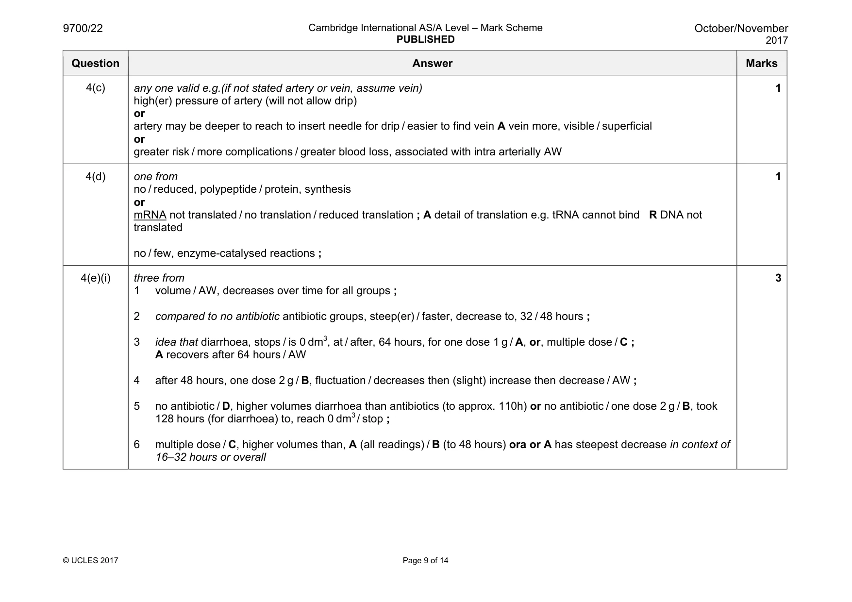
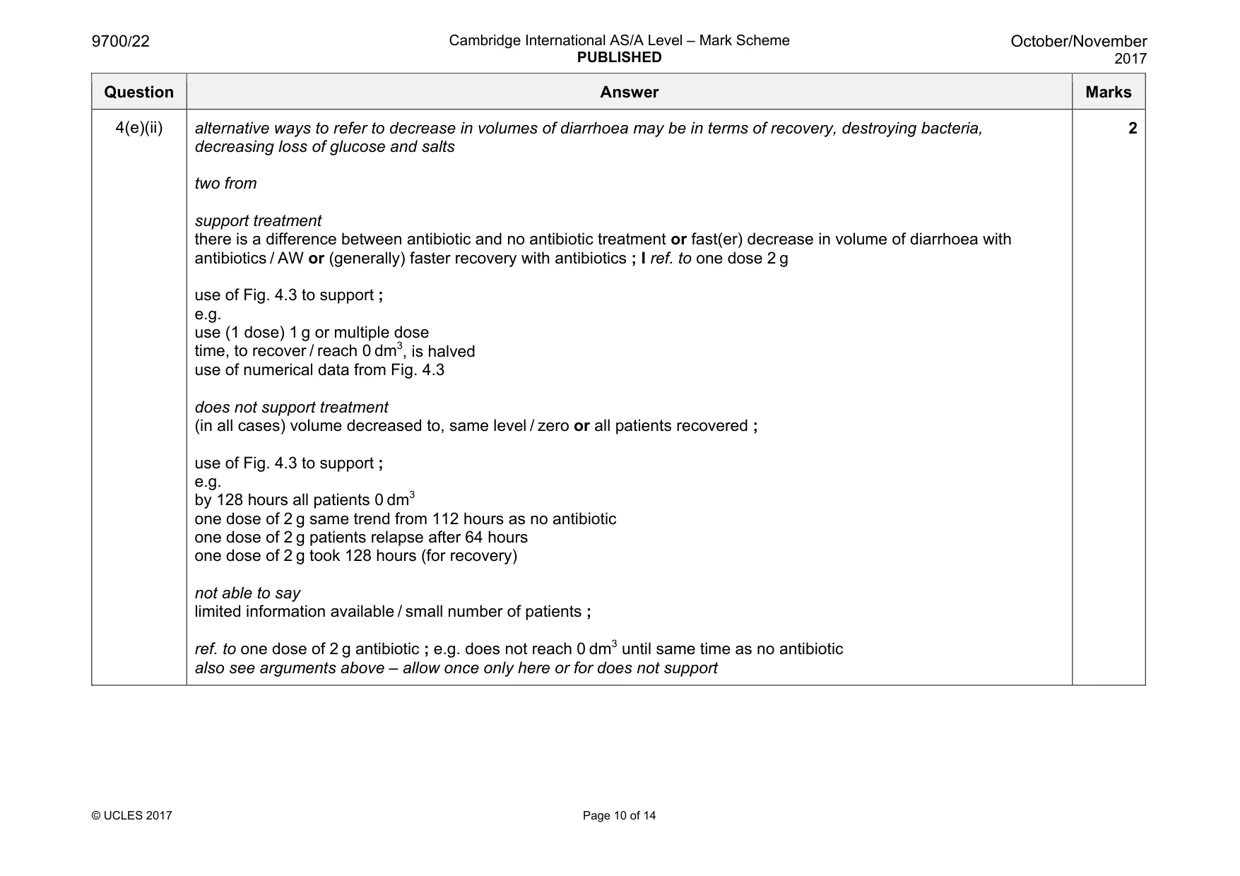
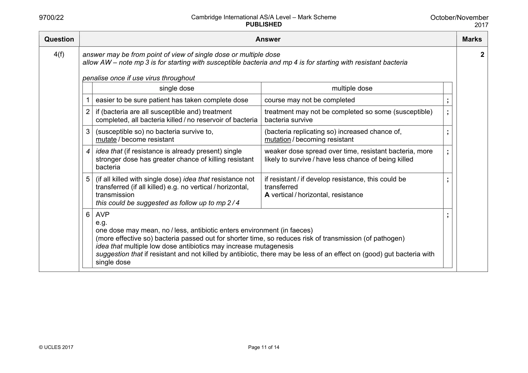
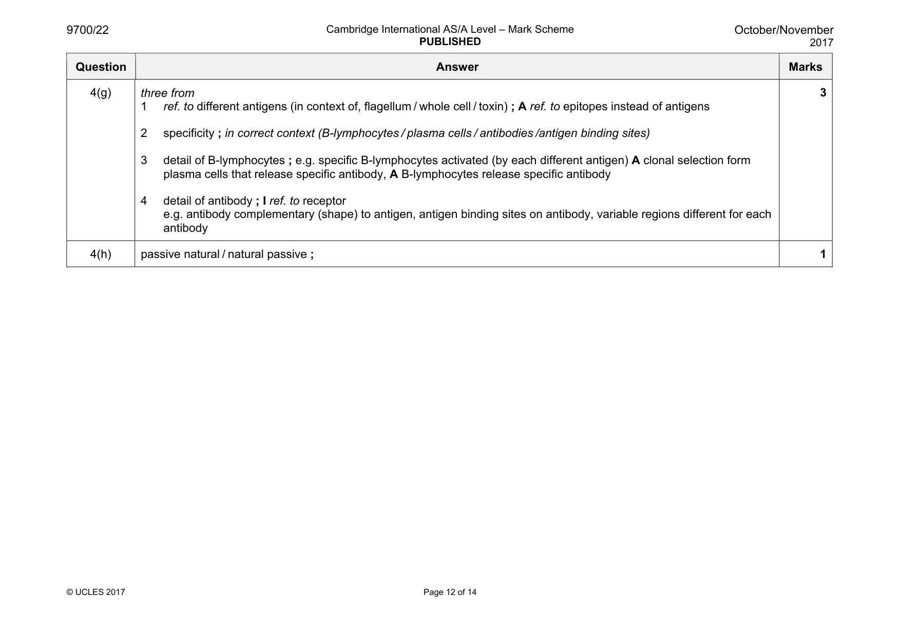
Mark scheme: 4(a) two from (loss of ions) increases / AW, water potential within cell ; ora, A Ψ for water potential, I ref. to solutes / solute potential water moves out of cell, down water potential gradient / from high(er) to low(er) water potential ; R from high to low water potential gradient (out) by osmosis / through the partially permeable membrane ; A selectively permeable membrane I osmotic gradient 2 4(b) four from capillary side sodium ions 1 sodium ions out (of cell), by active transport / with use of ATP ; A sodium ions pumped out 2 (so) lowers concentration of sodium ions within cell or sodium ion concentration gradient, set up / maintained ; intestinal lumen sodium ions and glucose 3 sodium ions enter by facilitated diffusion ; A diffusion / high to low concentration, through, SGLT1 / cotransporter I glucose enters by facilitated diffusion 4 glucose, cotransported with sodium ions into cell (through SGLT1) ; A sodium ions cotransported with glucose A glucose enters by secondary active transport, A idea of glucose only able to enter if moving with sodium ions (i.e. sodium drives the process) 5 (cotransport means) glucose enters against concentration gradient ; capillary side glucose 6 glucose out of cell (towards capillary) by facilitated diffusion ; A by diffusion if stated through, membrane protein / GLUT2 water uptake from lumen 7 (higher concentrations of) sodium ions / glucose / solutes, within cell lowers water potential ; 8 water follows, sodium ions / glucose / solutes (osmotically) or so water enters cell (down water potential gradient) ; must have idea that it follows inward movement of solutes 4 Question Answer Marks 4(c) any one valid e.g.(if not stated artery or vein, assume vein) high(er) pressure of artery (will not allow drip) or artery may be deeper to reach to insert needle for drip / easier to find vein A vein more, visible / superficial or greater risk / more complications / greater blood loss, associated with intra arterially AW 1 4(d) one from no / reduced, polypeptide / protein, synthesis or mRNA not translated / no translation / reduced translation ; A detail of translation e.g. tRNA cannot bind R DNA not translated no / few, enzyme-catalysed reactions ; 1 4(e)(i) three from 1 volume / AW, decreases over time for all groups ; 2 compared to no antibiotic antibiotic groups, steep(er) / faster, decrease to, 32 / 48 hours ; 3 idea that diarrhoea, stops / is 0 dm3, at / after, 64 hours, for one dose 1 g / A, or, multiple dose / C ; A recovers after 64 hours / AW 4 after 48 hours, one dose 2 g / B, fluctuation / decreases then (slight) increase then decrease / AW ; 5 no antibiotic / D, higher volumes diarrhoea than antibiotics (to approx. 110h) or no antibiotic / one dose 2 g / B, took 128 hours (for diarrhoea) to, reach 0 dm3 / stop ; 6 multiple dose / C, higher volumes than, A (all readings) / B (to 48 hours) ora or A has steepest decrease in context of 16–32 hours or overall 3 Question Answer Marks 4(e)(ii) alternative ways to refer to decrease in volumes of diarrhoea may be in terms of recovery, destroying bacteria, decreasing loss of glucose and salts two from support treatment there is a difference between antibiotic and no antibiotic treatment or fast(er) decrease in volume of diarrhoea with antibiotics / AW or (generally) faster recovery with antibiotics ; I ref. to one dose 2 g use of Fig. 4.3 to support ; e.g. use (1 dose) 1 g or multiple dose time, to recover / reach 0 dm3, is halved use of numerical data from Fig. 4.3 does not support treatment (in all cases) volume decreased to, same level / zero or all patients recovered ; use of Fig. 4.3 to support ; e.g. by 128 hours all patients 0 dm3 one dose of 2 g same trend from 112 hours as no antibiotic one dose of 2 g patients relapse after 64 hours one dose of 2 g took 128 hours (for recovery) not able to say limited information available / small number of patients ; ref. to one dose of 2 g antibiotic ; e.g. does not reach 0 dm3 until same time as no antibiotic also see arguments above – allow once only here or for does not support 2 Question Answer Marks 4(f) answer may be from point of view of single dose or multiple dose allow AW – note mp 3 is for starting with susceptible bacteria and mp 4 is for starting with resistant bacteria penalise once if use virus throughout single dose multiple dose 1 easier to be sure patient has taken complete dose course may not be completed ; 2 if (bacteria are all susceptible and) treatment completed, all bacteria killed / no reservoir of bacteria treatment may not be completed so some (susceptible) bacteria survive ; 3 (susceptible so) no bacteria survive to, mutate / become resistant (bacteria replicating so) increased chance of, mutation / becoming resistant ; 4 idea that (if resistance is already present) single stronger dose has greater chance of killing resistant bacteria weaker dose spread over time, resistant bacteria, more likely to survive / have less chance of being killed ; 5 (if all killed with single dose) idea that resistance not transferred (if all killed) e.g. no vertical / horizontal, transmission this could be suggested as follow up to mp 2 / 4 if resistant / if develop resistance, this could be transferred A vertical / horizontal, resistance ; 6 AVP e.g. one dose may mean, no / less, antibiotic enters environment (in faeces) (more effective so) bacteria passed out for shorter time, so reduces risk of transmission (of pathogen) idea that multiple low dose antibiotics may increase mutagenesis suggestion that if resistant and not killed by antibiotic, there may be less of an effect on (good) gut bacteria with single dose ; 2 Question Answer Marks 4(g) three from 1 ref. to different antigens (in context of, flagellum / whole cell / toxin) ; A ref. to epitopes instead of antigens 2 specificity ; in correct context (B-lymphocytes / plasma cells / antibodies /antigen binding sites) 3 detail of B-lymphocytes ; e.g. specific B-lymphocytes activated (by each different antigen) A clonal selection form plasma cells that release specific antibody, A B-lymphocytes release specific antibody 4 detail of antibody ; I ref. to receptor e.g. antibody complementary (shape) to antigen, antigen binding sites on antibody, variable regions different for each antibody 3 4(h) passive natural / natural passive ; 1 Question 5
Q5 · Descriptions A, B and C relate to the movement of water from the soil to the xylem in…
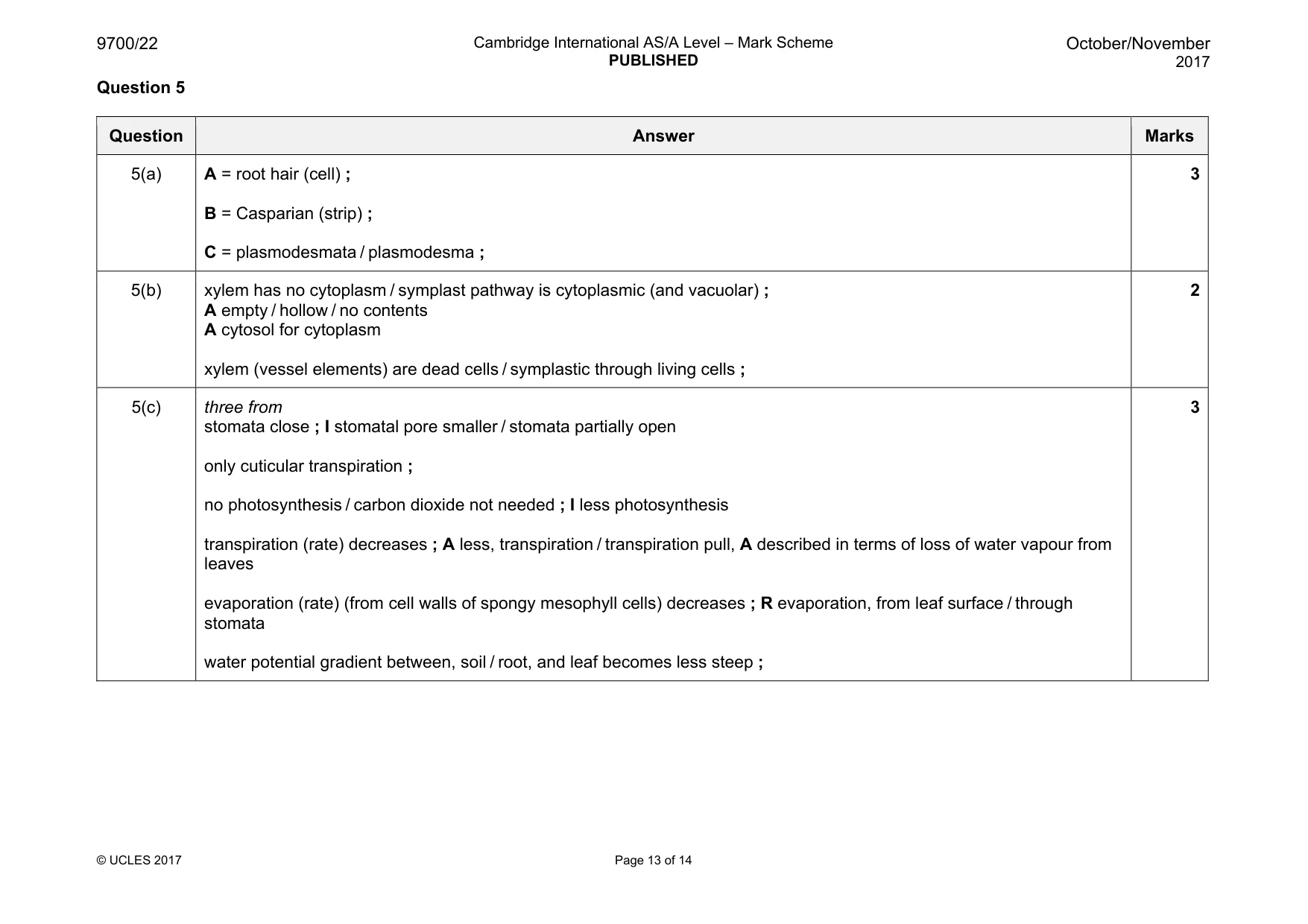
5 (a) Descriptions A, B and C relate to the movement of water from the soil to the xylem in roots. State the correct term to match each of the descriptions A, B and C. A The specialised root epidermal cell that provides a large surface area for the uptake of water from the soil. ........................................................................................................................................... B The band of suberin in the cell walls of the endodermis that prevents the movement of water by the apoplastic pathway. ........................................................................................................................................... C The cell structures that allow water to pass from one cell to the next as part of the symplastic pathway. ........................................................................................................................................... [3] (b) Explain, with reference to the structure of xylem vessel elements, why water does not take a symplastic pathway in the xylem to the leaves. ................................................................................................................................................... ................................................................................................................................................... ................................................................................................................................................... ................................................................................................................................................... ...............................................................................................................................................[2] (c) Explain why the rate of movement of water in the xylem may slow down at night. ................................................................................................................................................... ................................................................................................................................................... ................................................................................................................................................... ................................................................................................................................................... ................................................................................................................................................... ................................................................................................................................................... ...............................................................................................................................................[3] [Total: 8]
Mark scheme: 5(a) A = root hair (cell) ; B = Casparian (strip) ; C = plasmodesmata / plasmodesma ; 3 5(b) xylem has no cytoplasm / symplast pathway is cytoplasmic (and vacuolar) ; A empty / hollow / no contents A cytosol for cytoplasm xylem (vessel elements) are dead cells / symplastic through living cells ; 2 5(c) three from stomata close ; I stomatal pore smaller / stomata partially open only cuticular transpiration ; no photosynthesis / carbon dioxide not needed ; I less photosynthesis transpiration (rate) decreases ; A less, transpiration / transpiration pull, A described in terms of loss of water vapour from leaves evaporation (rate) (from cell walls of spongy mesophyll cells) decreases ; R evaporation, from leaf surface / through stomata water potential gradient between, soil / root, and leaf becomes less steep ; 3 Question 6
Q6 · A diagram of a vertical section through the mammalian heart
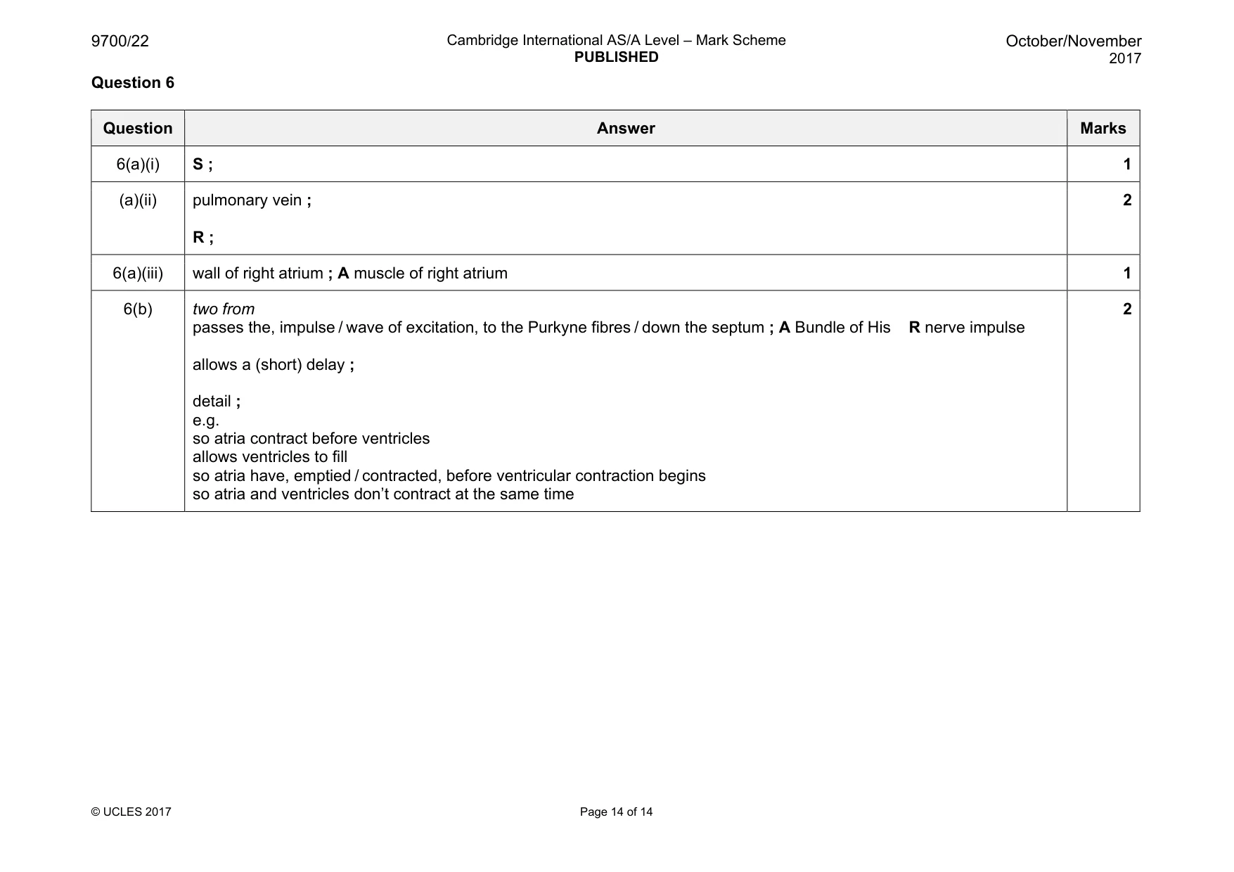
6 Fig. 6.1 is a diagram of a vertical section through the mammalian heart. The labels Q to X represent valves and blood vessels of the heart. X W Q R V S T U Fig. 6.1 (a) (i) State which label represents the valve that prevents the backflow of blood from the aorta into the ventricle. .......................................................................................................................................[1] (ii) Name the blood vessel that carries oxygenated blood from the lungs to the heart and state which label represents this blood vessel. name ................................................................................................................................. label ...............................................................................................................................[2] (b) State precisely where the sinoatrial node is located. ...............................................................................................................................................[1] (c) Explain the role of the atrioventricular node in the coordination of heart action. ................................................................................................................................................... ................................................................................................................................................... ................................................................................................................................................... ...............................................................................................................................................[2] [Total: 6]
Mark scheme: 6(a)(i) S ; 1 (a)(ii) pulmonary vein ; R ; 2 6(a)(iii) wall of right atrium ; A muscle of right atrium 1 6(b) two from passes the, impulse / wave of excitation, to the Purkyne fibres / down the septum ; A Bundle of His R nerve impulse allows a (short) delay ; detail ; e.g. so atria contract before ventricles allows ventricles to fill so atria have, emptied / contracted, before ventricular contraction begins so atria and ventricles don’t contract at the same time 2
What was in this paper
The subtopics covered by these 6 questions, and how many questions each got. Open one in a new tab to see every Cambridge question on it.
What you needed in this session
Cambridge’s own grade thresholds for 2017 Oct/Nov, Paper 2 · Variant 2. A higher threshold means an easier paper — the bar moves with how the cohort did.