Cambridge IGCSE Biology (9-1) 0970 — 2024 Oct/Nov Paper 4 · Variant 2
0970/42/O/N/24 · 6 questions · 80 marks · ≈90 min
The question paper and its mark scheme, free to read here and free to download. This is Cambridge’s own paper, exactly as it was sat.
Question paper16 pages

Mark scheme9 pages
Answers below. Sit the paper first if you are practising.
Questions as text
Q1 · Organisms from the genus Chlorella are protoctists
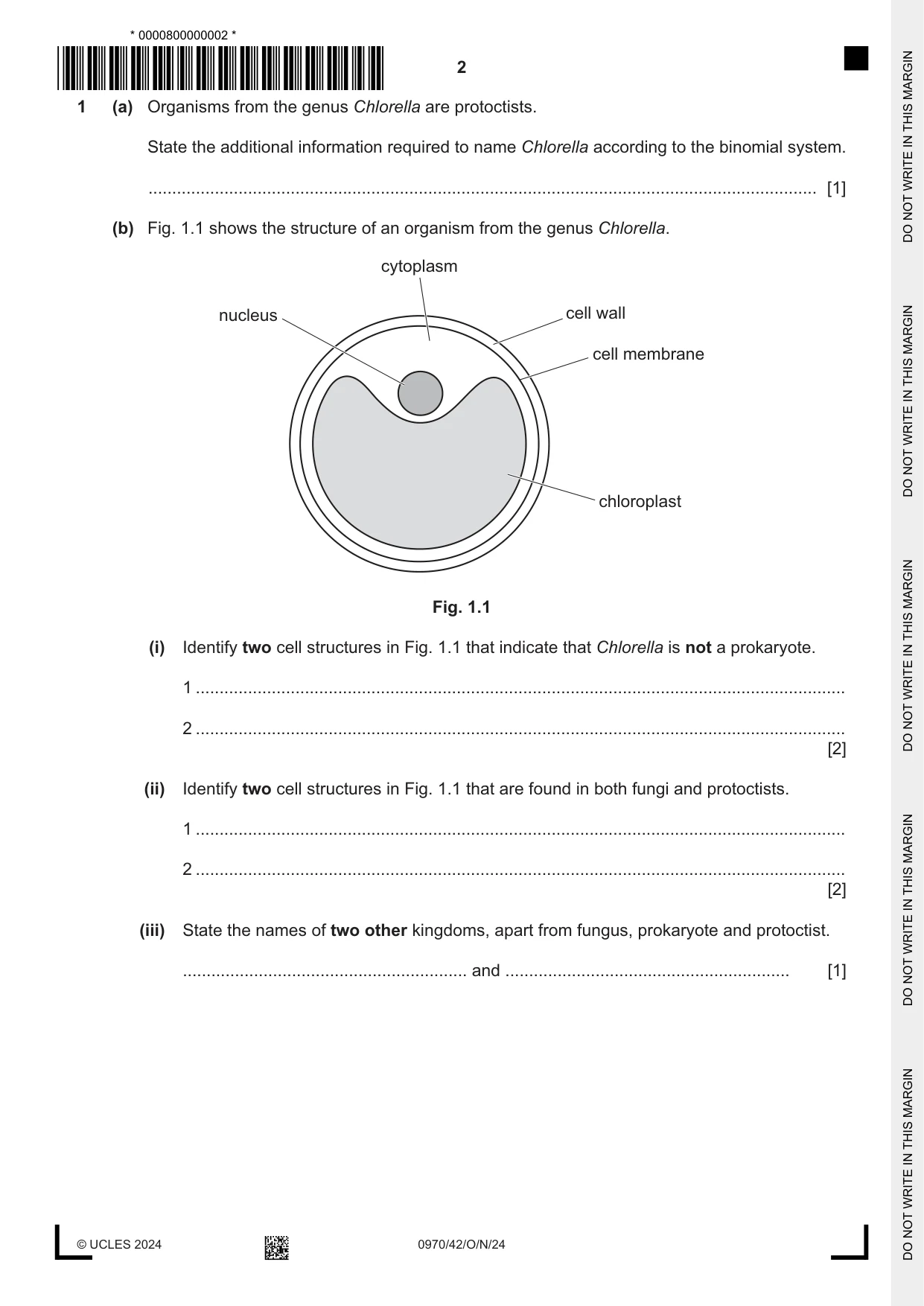
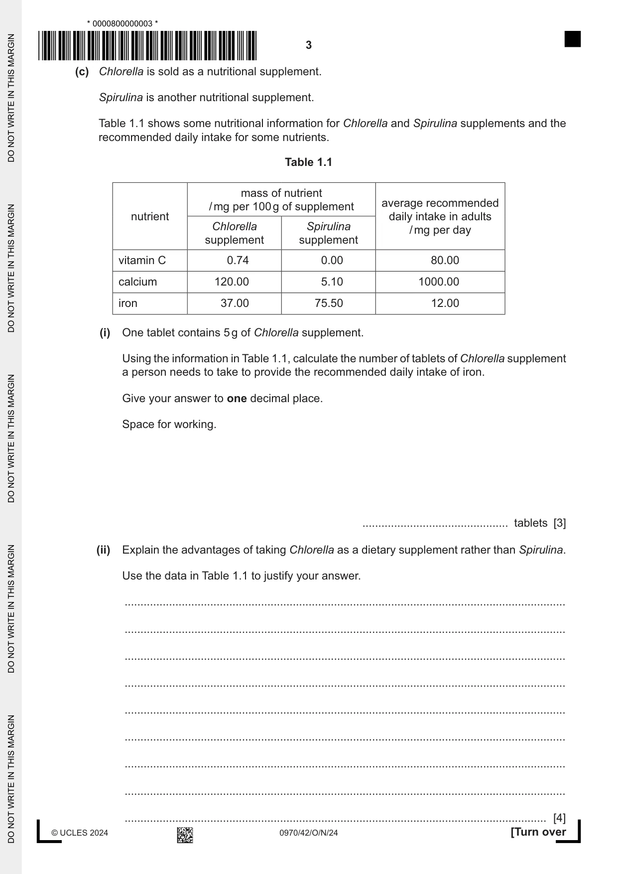
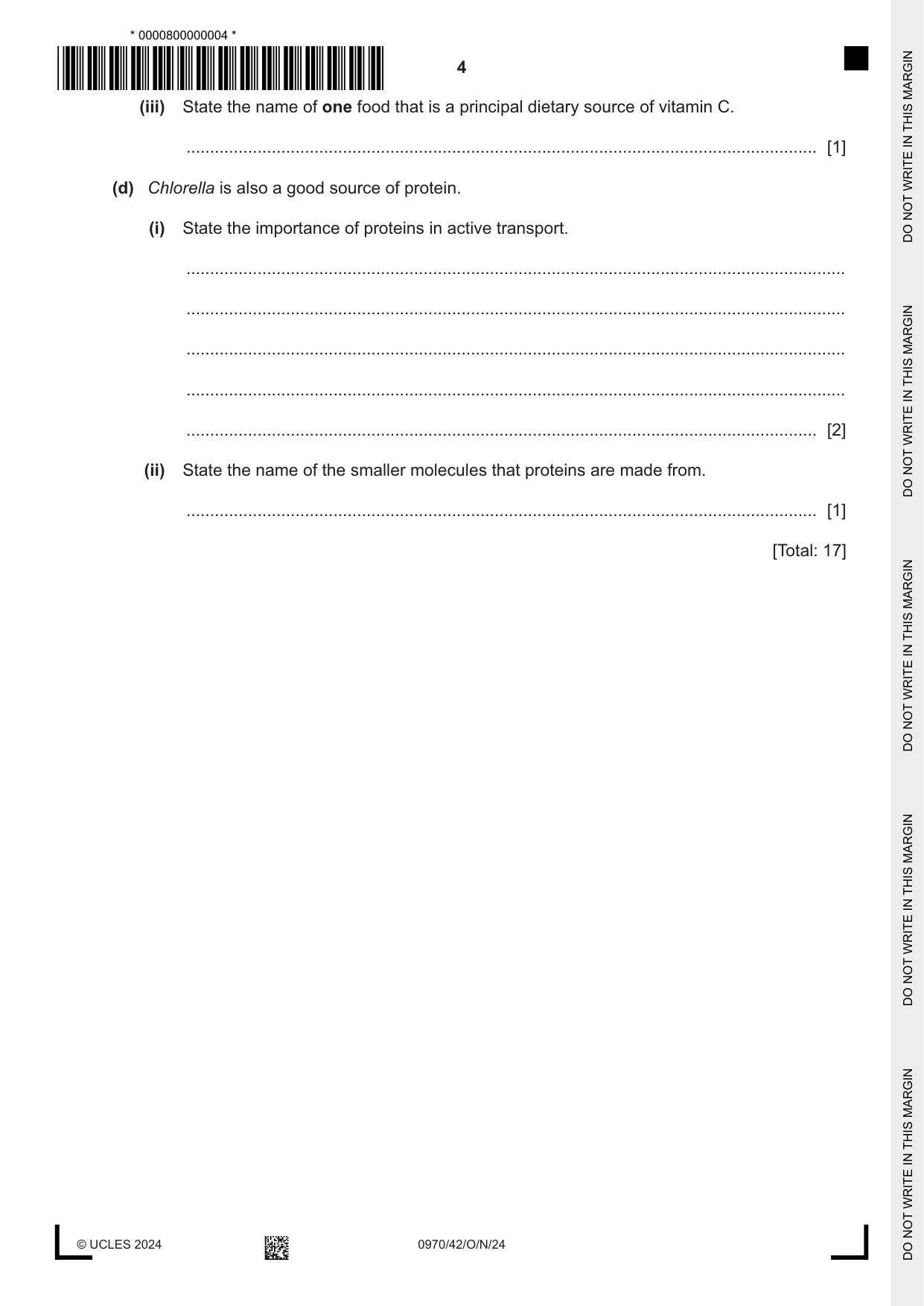
1 (a) Organisms from the genus Chlorella are protoctists. State the additional information required to name Chlorella according to the binomial system. ............................................................................................................................................. [1] (b) Fig. 1.1 shows the structure of an organism from the genus Chlorella. cytoplasm nucleus cell wall cell membrane chloroplast Fig. 1.1 (i) Identify two cell structures in Fig. 1.1 that indicate that Chlorella is not a prokaryote. 1 ......................................................................................................................................... 2 ......................................................................................................................................... [2] (ii) Identify two cell structures in Fig. 1.1 that are found in both fungi and protoctists. 1 ......................................................................................................................................... 2 ......................................................................................................................................... [2] (iii) State the names of two other kingdoms, apart from fungus, prokaryote and protoctist. ............................................................ and ............................................................ [1] (c) Chlorella is sold as a nutritional supplement. Spirulina is another nutritional supplement. Table 1.1 shows some nutritional information for Chlorella and Spirulina supplements and the recommended daily intake for some nutrients. Table 1.1 mass of nutrient average recommended / mg per 100 g of supplement nutrient daily intake in adults Chlorella Spirulina / mg per day supplement supplement vitamin C 0.74 0.00 80.00 calcium 120.00 5.10 1000.00 iron 37.00 75.50 12.00 (i) One tablet contains 5 g of Chlorella supplement. Using the information in Table 1.1, calculate the number of tablets of Chlorella supplement a person needs to take to provide the recommended daily intake of iron. Give your answer to one decimal place. Space for working. .............................................. tablets [3] (ii) Explain the advantages of taking Chlorella as a dietary supplement rather than Spirulina. Use the data in Table 1.1 to justify your answer. ........................................................................................................................................... ........................................................................................................................................... ........................................................................................................................................... ........................................................................................................................................... ........................................................................................................................................... ........................................................................................................................................... ........................................................................................................................................... ........................................................................................................................................... ..................................................................................................................................... [4] (iii) State the name of one food that is a principal dietary source of vitamin C. ..................................................................................................................................... [1] (d) Chlorella is also a good source of protein. (i) State the importance of proteins in active transport. ........................................................................................................................................... ........................................................................................................................................... ........................................................................................................................................... ........................................................................................................................................... ..................................................................................................................................... [2] (ii) State the name of the smaller molecules that proteins are made from. ..................................................................................................................................... [1] [Total: 17]
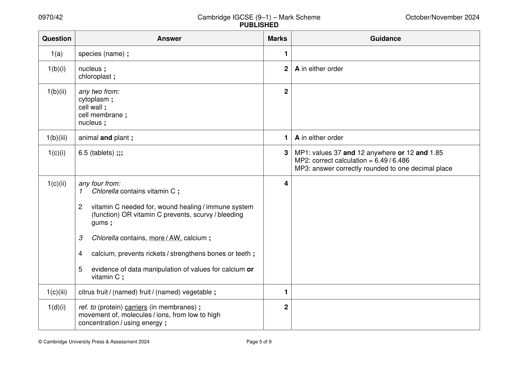
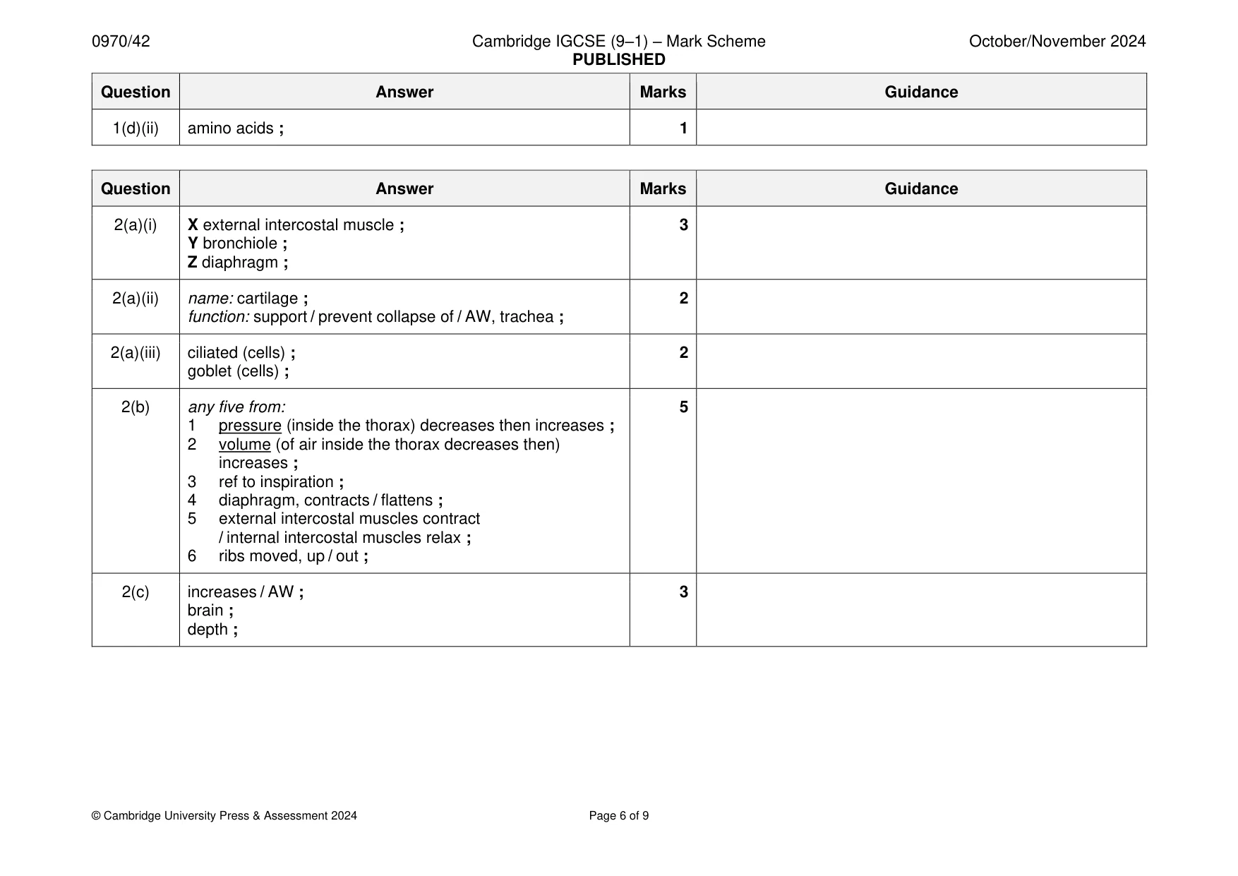
Mark scheme: Question Answer Marks Guidance 1(a) species (name) ; 1 1(b)(i) nucleus ; 2 A in either order chloroplast ; 1(b)(ii) any two from: 2 cytoplasm ; cell wall ; cell membrane ; nucleus ; 1(b)(iii) animal and plant ; 1 A in either order 1(c)(i) 6.5 (tablets) ;;; 3 MP1: values 37 and 12 anywhere or 12 and 1.85 MP2: correct calculation = 6.49 / 6.486 MP3: answer correctly rounded to one decimal place 1(c)(ii) any four from: 4 1 Chlorella contains vitamin C ; 2 vitamin C needed for, wound healing / immune system (function) OR vitamin C prevents, scurvy / bleeding gums ; 3 Chlorella contains, more / AW, calcium ; 4 calcium, prevents rickets / strengthens bones or teeth ; 5 evidence of data manipulation of values for calcium or vitamin C ; 1(c)(iii) citrus fruit / (named) fruit / (named) vegetable ; 1 1(d)(i) ref. to (protein) carriers (in membranes) ; 2 movement of, molecules / ions, from low to high concentration / using energy ; 1(d)(ii) amino acids ; 1
Q2 · A diagram of the gas exchange system in humans
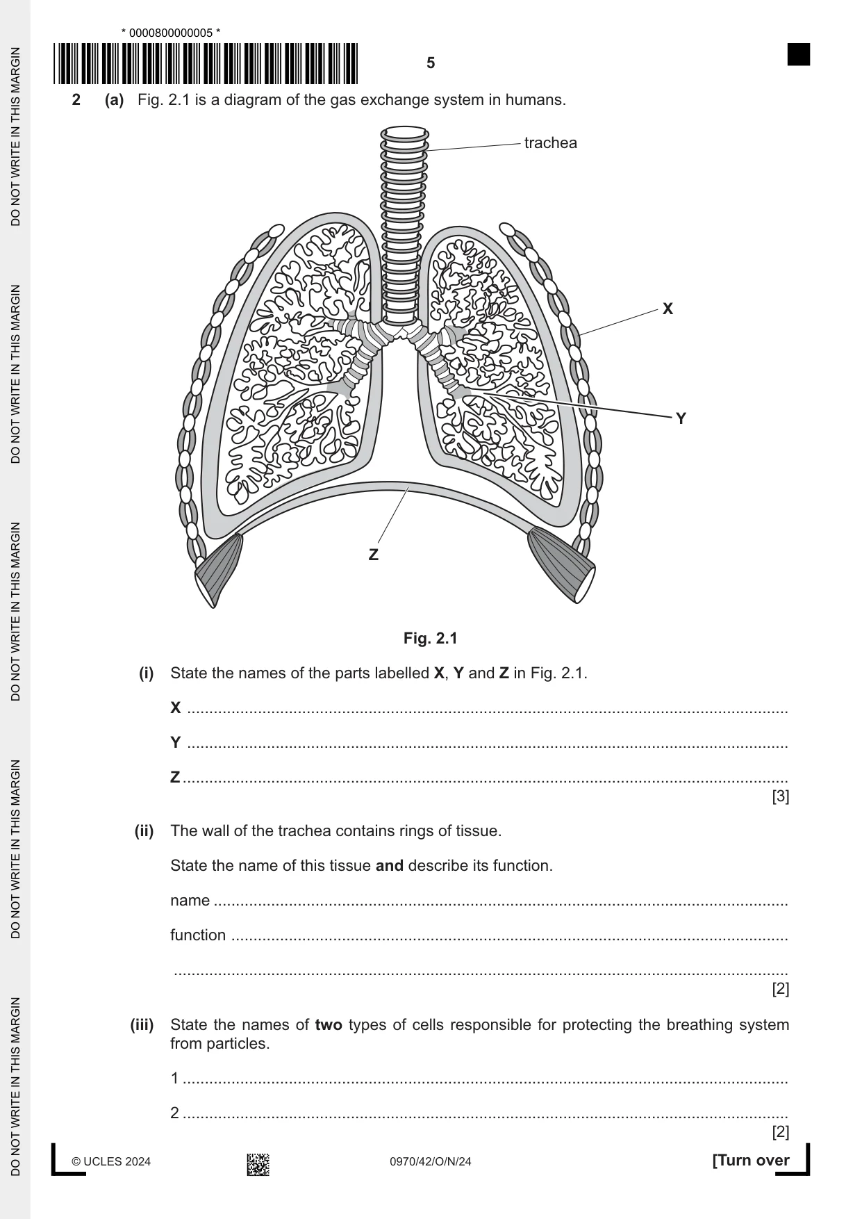
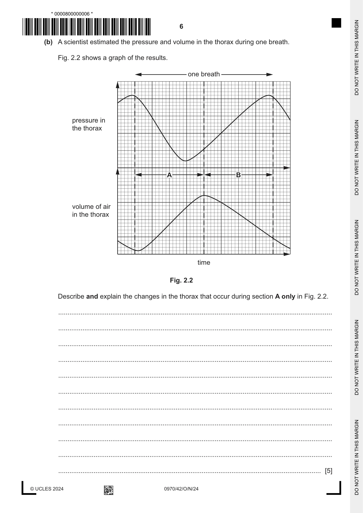
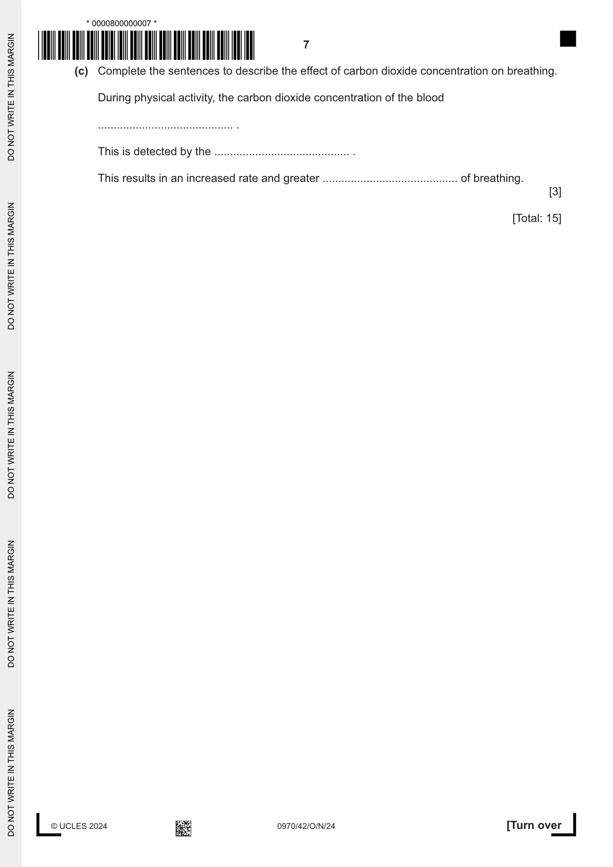
2 (a) Fig. 2.1 is a diagram of the gas exchange system in humans. trachea X Y Z Fig. 2.1 (i) State the names of the parts labelled X, Y and Z in Fig. 2.1. X ........................................................................................................................................ Y ........................................................................................................................................ Z ......................................................................................................................................... [3] (ii) The wall of the trachea contains rings of tissue. State the name of this tissue and describe its function. name .................................................................................................................................. function .............................................................................................................................. ........................................................................................................................................... [2] (iii) State the names of two types of cells responsible for protecting the breathing system from particles. 1 ......................................................................................................................................... 2 ......................................................................................................................................... [2] (b) A scientist estimated the pressure and volume in the thorax during one breath. Fig. 2.2 shows a graph of the results. one breath pressure in the thorax A B volume of air in the thorax time Fig. 2.2 Describe and explain the changes in the thorax that occur during section A only in Fig. 2.2. ................................................................................................................................................... ................................................................................................................................................... ................................................................................................................................................... ................................................................................................................................................... ................................................................................................................................................... ................................................................................................................................................... ................................................................................................................................................... ................................................................................................................................................... ................................................................................................................................................... ................................................................................................................................................... ............................................................................................................................................. [5] (c) Complete the sentences to describe the effect of carbon dioxide concentration on breathing. During physical activity, the carbon dioxide concentration of the blood ........................................... . This is detected by the ........................................... . This results in an increased rate and greater ........................................... of breathing. [3] [Total: 15]
Mark scheme: 2(a)(i) X external intercostal muscle ; 3 Y bronchiole ; Z diaphragm ; 2(a)(ii) name: cartilage ; 2 function: support / prevent collapse of / AW, trachea ; 2(a)(iii) ciliated (cells) ; 2 goblet (cells) ; 2(b) any five from: 5 1 pressure (inside the thorax) decreases then increases ; 2 volume (of air inside the thorax decreases then) increases ; 3 ref to inspiration ; 4 diaphragm, contracts / flattens ; 5 external intercostal muscles contract / internal intercostal muscles relax ; 6 ribs moved, up / out ; 2(c) increases / AW ; 3 brain ; depth ;
Q3 · A kidney nephron and its associated blood vessels
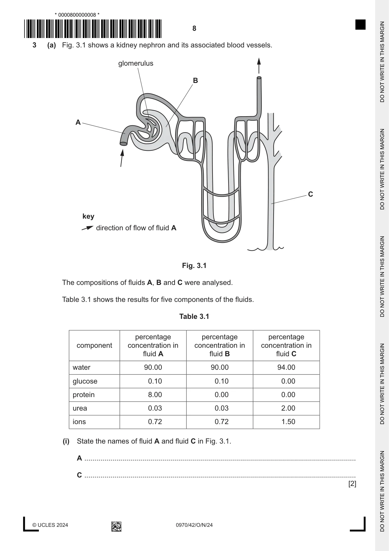
3 (a) Fig. 3.1 shows a kidney nephron and its associated blood vessels. glomerulus B A C key direction of flow of fluid A Fig. 3.1 The compositions of fluids A, B and C were analysed. Table 3.1 shows the results for five components of the fluids. Table 3.1 percentage percentage percentage component concentration in concentration in concentration in fluid A fluid B fluid C water 90.00 90.00 94.00 glucose 0.10 0.10 0.00 protein 8.00 0.00 0.00 urea 0.03 0.03 2.00 ions 0.72 0.72 1.50 (i) State the names of fluid A and fluid C in Fig. 3.1. A ........................................................................................................................................ C ........................................................................................................................................ [2] (ii) Using the information in Fig. 3.1 and Table 3.1, describe and explain the differences in the compositions of fluids A, B and C. ........................................................................................................................................... ........................................................................................................................................... ........................................................................................................................................... ........................................................................................................................................... ........................................................................................................................................... ........................................................................................................................................... ........................................................................................................................................... ........................................................................................................................................... ........................................................................................................................................... ........................................................................................................................................... ..................................................................................................................................... [5] (b) Outline how blood glucose concentration is controlled. ................................................................................................................................................... ................................................................................................................................................... ................................................................................................................................................... ................................................................................................................................................... ................................................................................................................................................... ................................................................................................................................................... ................................................................................................................................................... ................................................................................................................................................... ................................................................................................................................................... ................................................................................................................................................... ................................................................................................................................................... ................................................................................................................................................... ............................................................................................................................................. [6] (c) A gene mutation may be involved in the development of type 1 diabetes. (i) Describe what is meant by a gene mutation. ........................................................................................................................................... ........................................................................................................................................... ..................................................................................................................................... [2] (ii) Outline the treatment of type 1 diabetes. ........................................................................................................................................... ........................................................................................................................................... ........................................................................................................................................... ........................................................................................................................................... ........................................................................................................................................... ........................................................................................................................................... ..................................................................................................................................... [3] [Total: 18]
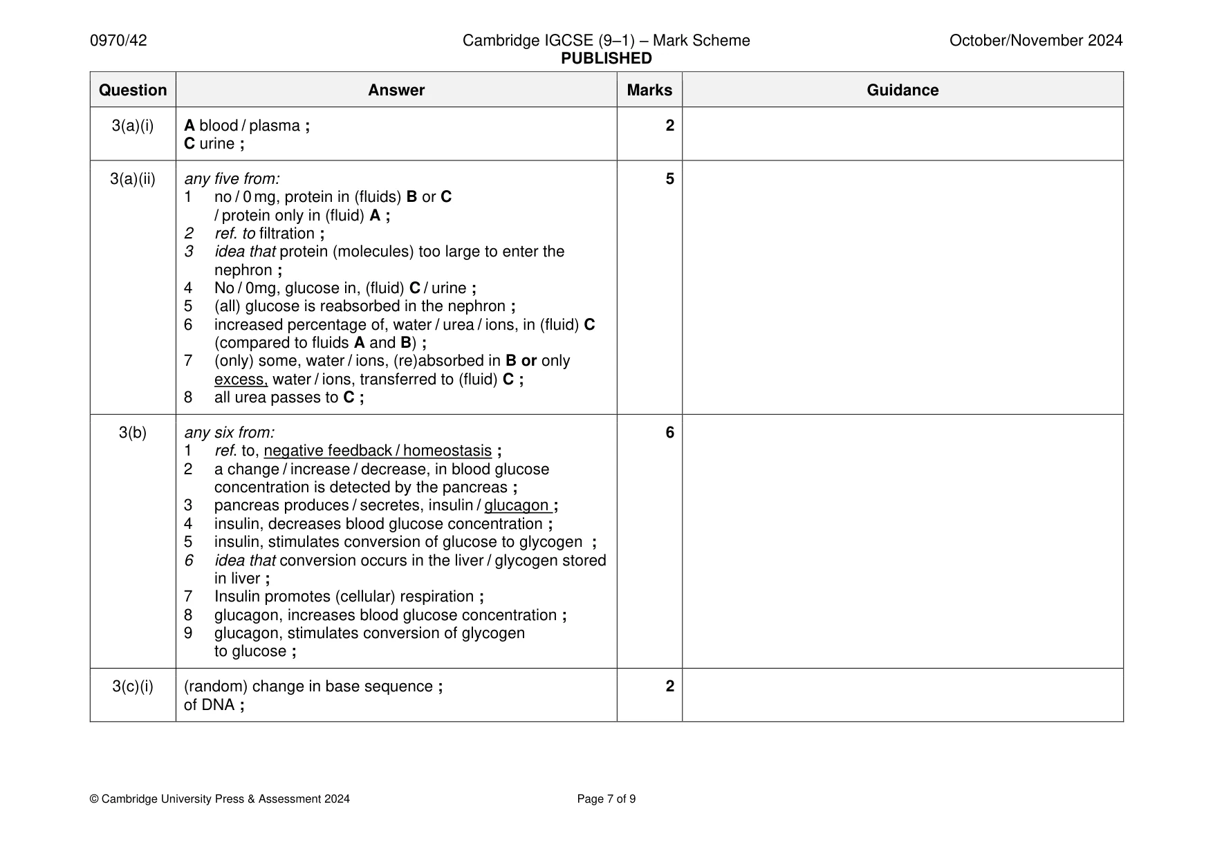
Mark scheme: 3(a)(i) A blood / plasma ; 2 C urine ; 3(a)(ii) any five from: 5 1 no / 0 mg, protein in (fluids) B or C / protein only in (fluid) A ; 2 ref. to filtration ; 3 idea that protein (molecules) too large to enter the nephron ; 4 No / 0mg, glucose in, (fluid) C / urine ; 5 (all) glucose is reabsorbed in the nephron ; 6 increased percentage of, water / urea / ions, in (fluid) C (compared to fluids A and B) ; 7 (only) some, water / ions, (re)absorbed in B or only excess, water / ions, transferred to (fluid) C ; 8 all urea passes to C ; 3(b) any six from: 6 1 ref. to, negative feedback / homeostasis ; 2 a change / increase / decrease, in blood glucose concentration is detected by the pancreas ; 3 pancreas produces / secretes, insulin / glucagon ; 4 insulin, decreases blood glucose concentration ; 5 insulin, stimulates conversion of glucose to glycogen ; 6 idea that conversion occurs in the liver / glycogen stored in liver ; 7 Insulin promotes (cellular) respiration ; 8 glucagon, increases blood glucose concentration ; 9 glucagon, stimulates conversion of glycogen to glucose ; 3(c)(i) (random) change in base sequence ; 2 of DNA ; 3(c)(ii) any three from: 3 insulin, pump / injections ; monitor glucose concentration of the blood or urine ; idea of control of carbohydrate intake ; exercise (after eating) ; AVP ;
Q4 · A diagram of two flowers from different plants of the same species
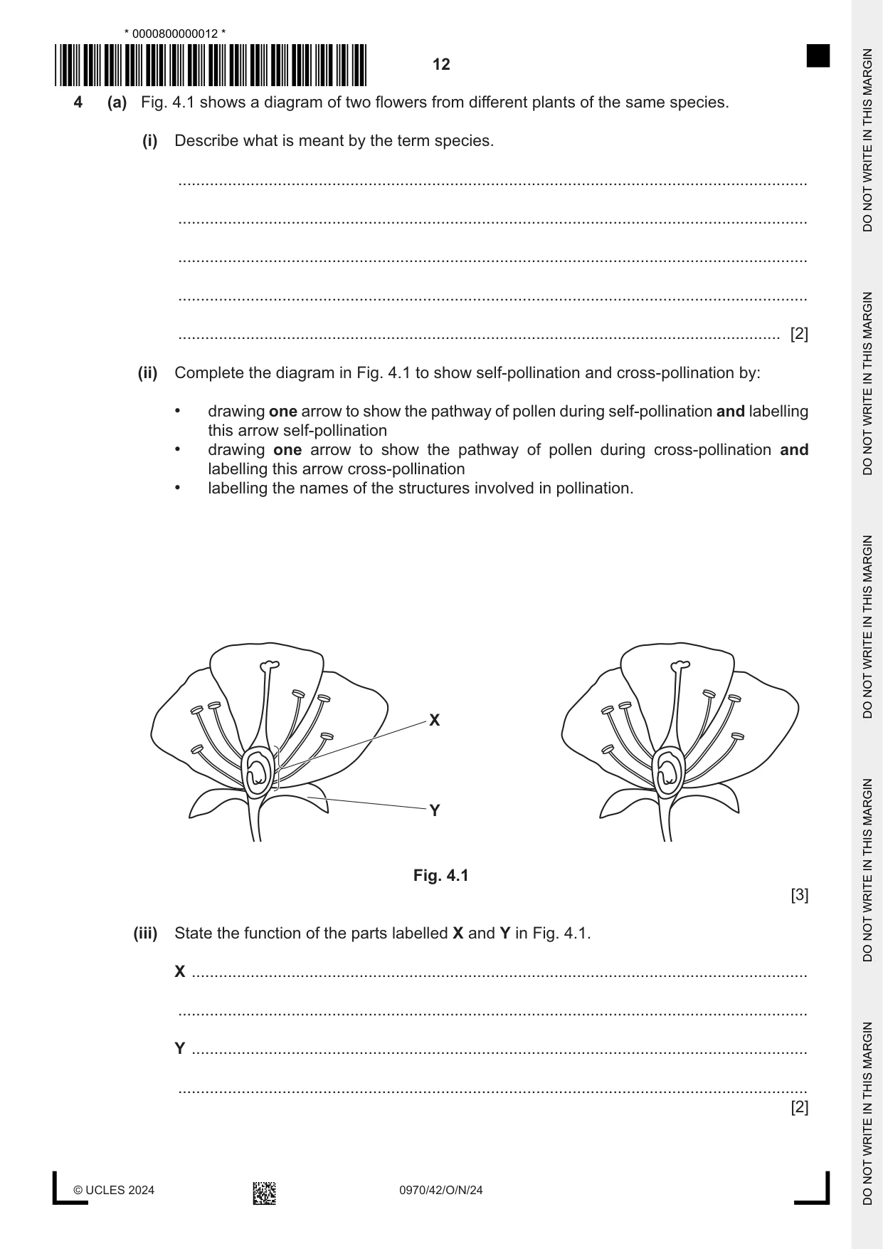
4 (a) Fig. 4.1 shows a diagram of two flowers from different plants of the same species. (i) Describe what is meant by the term species. ........................................................................................................................................... ........................................................................................................................................... ........................................................................................................................................... ........................................................................................................................................... ..................................................................................................................................... [2] (ii) Complete the diagram in Fig. 4.1 to show self‑pollination and cross‑pollination by: • drawing one arrow to show the pathway of pollen during self‑pollination and labelling this arrow self‑pollination • drawing one arrow to show the pathway of pollen during cross‑pollination and labelling this arrow cross‑pollination • labelling the names of the structures involved in pollination. X Y Fig. 4.1 [3] (iii) State the function of the parts labelled X and Y in Fig. 4.1. X ........................................................................................................................................ ........................................................................................................................................... Y ........................................................................................................................................ ........................................................................................................................................... [2] (b) Explain why self‑pollination that results in production of offspring is a form of sexual reproduction and not asexual reproduction. ................................................................................................................................................... ................................................................................................................................................... ................................................................................................................................................... ................................................................................................................................................... ................................................................................................................................................... ................................................................................................................................................... ............................................................................................................................................. [3] (c) State the type of environmental conditions that hydrophytes are adapted to live in. ............................................................................................................................................. [1] [Total: 11]
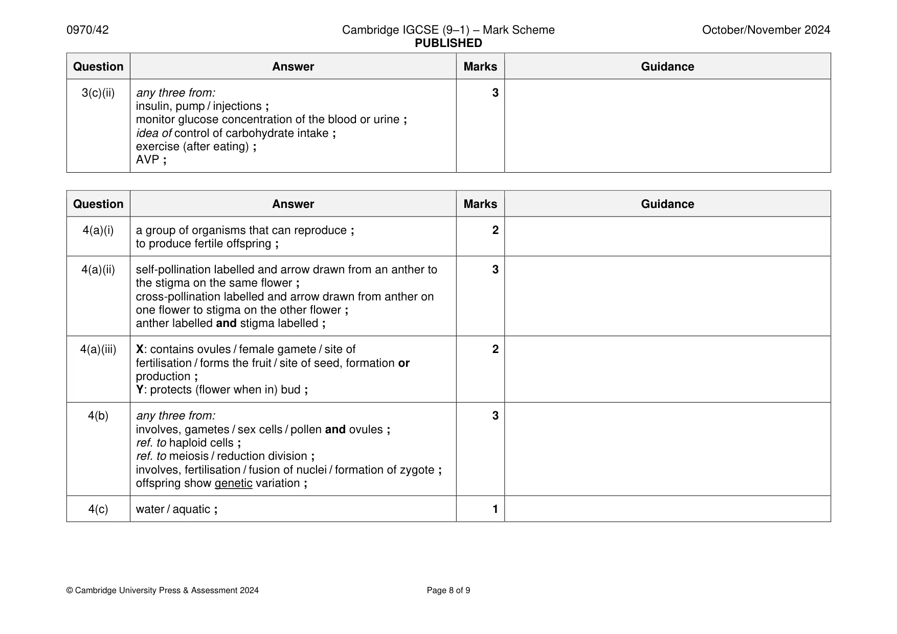
Mark scheme: 4(a)(i) a group of organisms that can reproduce ; 2 to produce fertile offspring ; 4(a)(ii) self-pollination labelled and arrow drawn from an anther to 3 the stigma on the same flower ; cross-pollination labelled and arrow drawn from anther on one flower to stigma on the other flower ; anther labelled and stigma labelled ; 4(a)(iii) X: contains ovules / female gamete / site of 2 fertilisation / forms the fruit / site of seed, formation or production ; Y: protects (flower when in) bud ; 4(b) any three from: 3 involves, gametes / sex cells / pollen and ovules ; ref. to haploid cells ; ref. to meiosis / reduction division ; involves, fertilisation / fusion of nuclei / formation of zygote ; offspring show genetic variation ; 4(c) water / aquatic ; 1
More questions on Concept and uses of classification systems
Q5 · A pedigree diagram showing the inheritance of blood group in one family
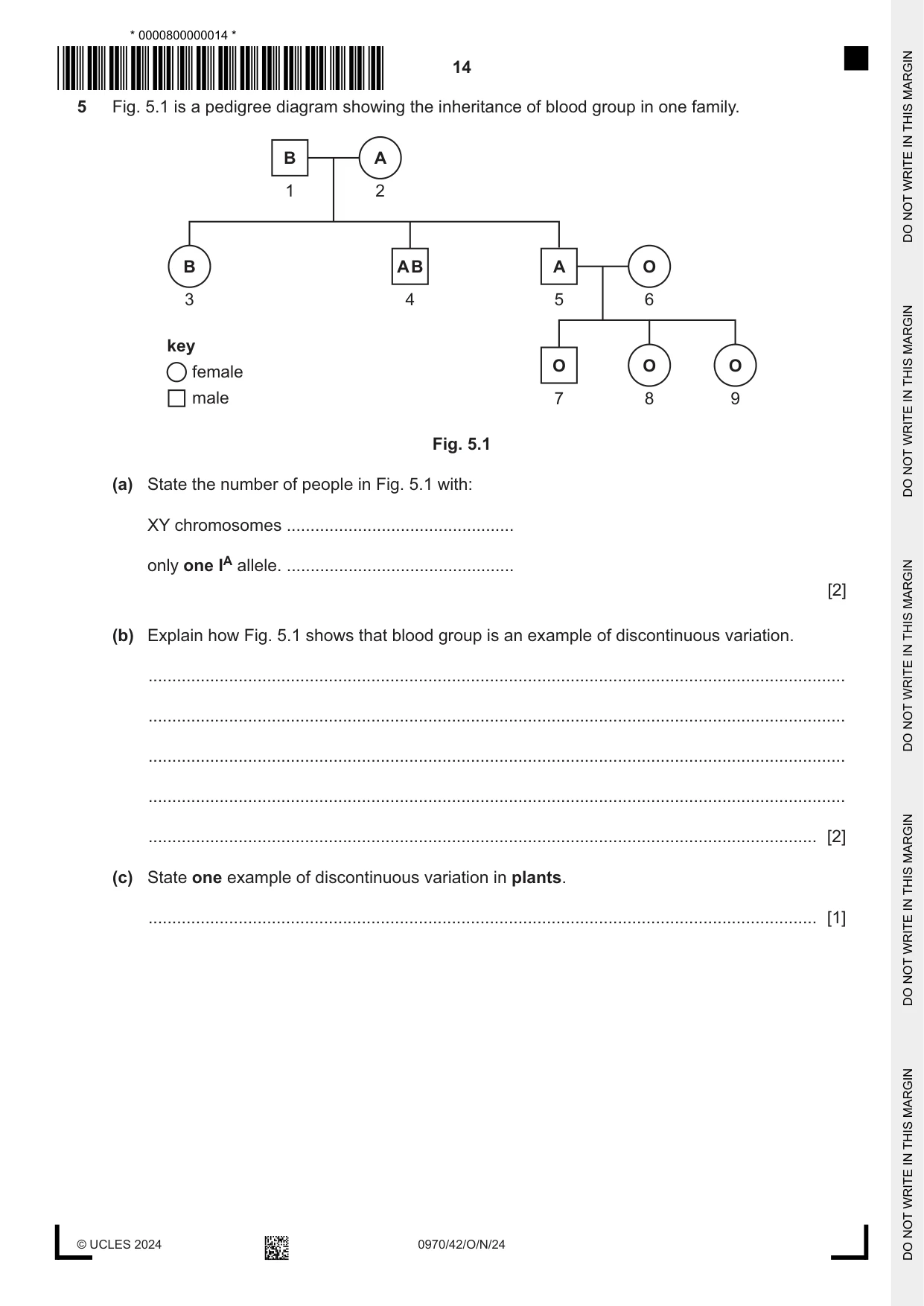
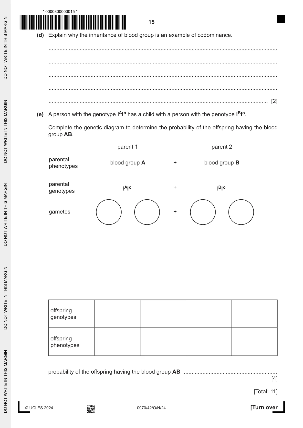
5 Fig. 5.1 is a pedigree diagram showing the inheritance of blood group in one family. B A 1 2 B AB A O 3 4 5 6 key O O O female male 7 8 9 Fig. 5.1 (a) State the number of people in Fig. 5.1 with: XY chromosomes ................................................ only one IA allele. ................................................ [2] (b) Explain how Fig. 5.1 shows that blood group is an example of discontinuous variation. ................................................................................................................................................... ................................................................................................................................................... ................................................................................................................................................... ................................................................................................................................................... ............................................................................................................................................. [2] (c) State one example of discontinuous variation in plants. ............................................................................................................................................. [1] (d) Explain why the inheritance of blood group is an example of codominance. ................................................................................................................................................... ................................................................................................................................................... ................................................................................................................................................... ................................................................................................................................................... ............................................................................................................................................. [2] (e) A person with the genotype IAIo has a child with a person with the genotype IBIo. Complete the genetic diagram to determine the probability of the offspring having the blood group AB. parent 1 parent 2 parental blood group A + blood group B phenotypes parental A o + B o genotypes gametes + offspring genotypes offspring phenotypes probability of the offspring having the blood group AB ............................................................. [4] [Total: 11]
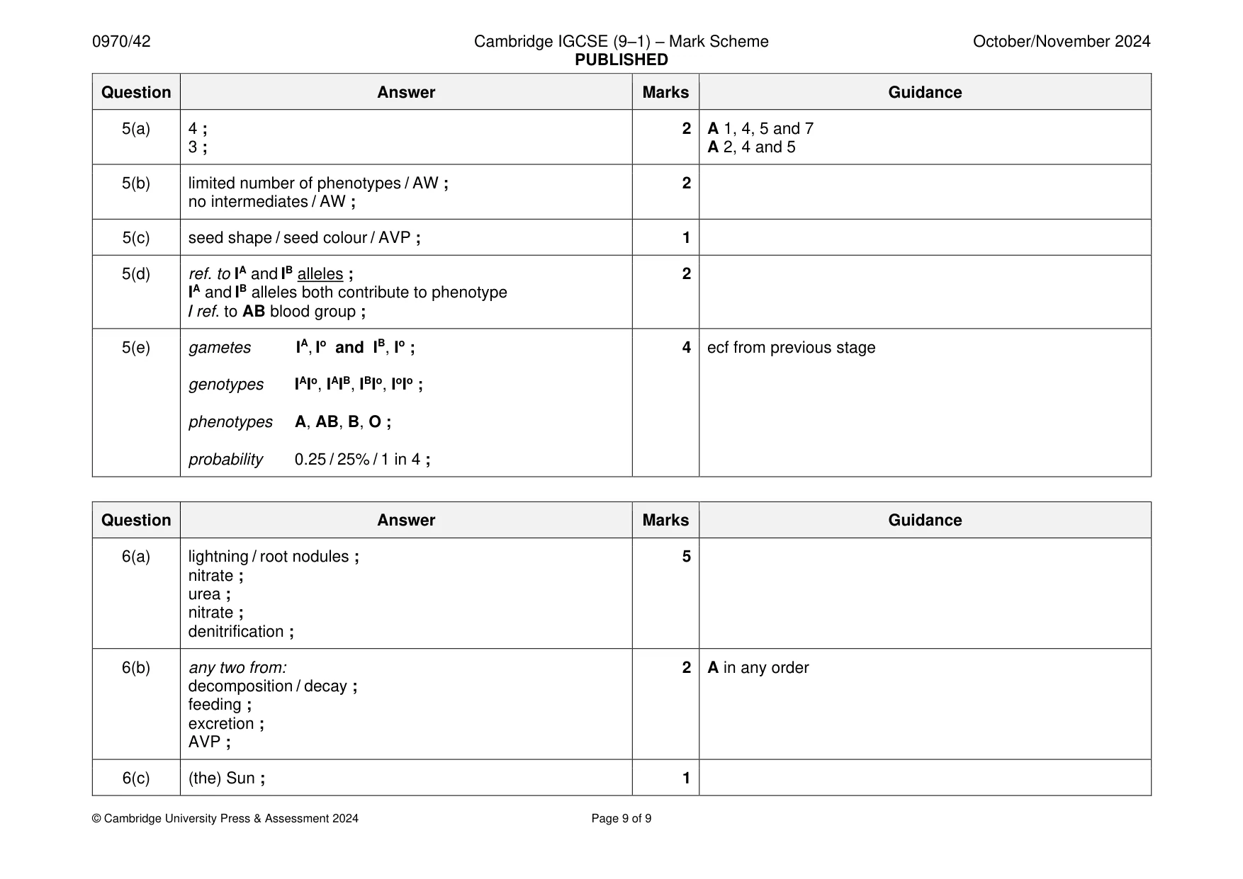
Mark scheme: 5(a) 4 ; 2 A 1, 4, 5 and 7 3 ; A 2, 4 and 5 5(b) limited number of phenotypes / AW ; 2 no intermediates / AW ; 5(c) seed shape / seed colour / AVP ; 1 5(d) ref. to IA and IB alleles ; 2 IA and IB alleles both contribute to phenotype / ref. to AB blood group ; 5(e) gametes IA, Io and IB, Io ; 4 ecf from previous stage genotypes IAIo, IAIB, IBIo, IoIo ; phenotypes A, AB, B, O ; probability 0.25 / 25% / 1 in 4 ;
Q6 · The flow chart in Fig
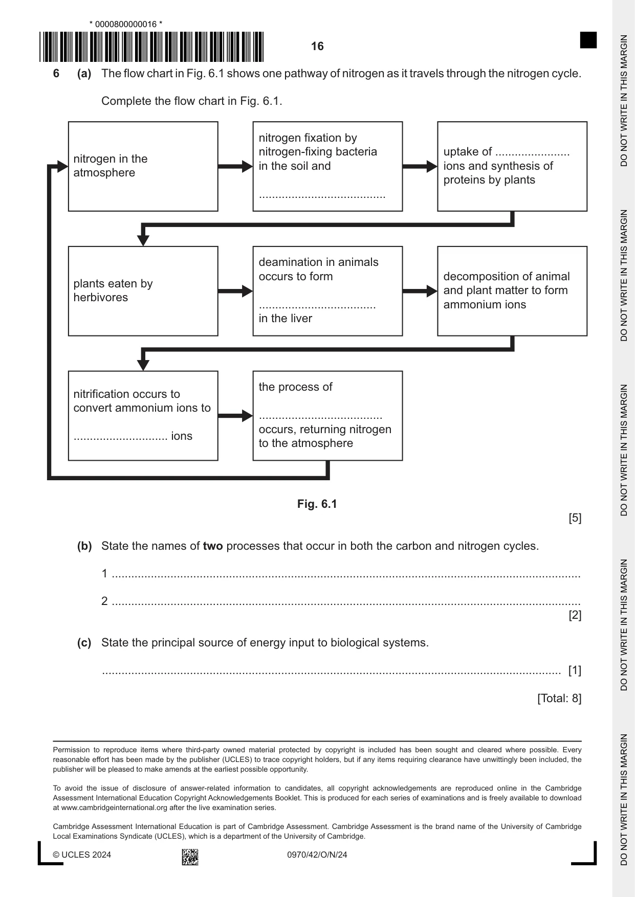
6 (a) The flow chart in Fig. 6.1 shows one pathway of nitrogen as it travels through the nitrogen cycle. Complete the flow chart in Fig. 6.1. nitrogen fixation by nitrogen-fixing bacteria uptake of ....................... nitrogen in the in the soil and ions and synthesis of atmosphere proteins by plants ....................................... deamination in animals occurs to form decomposition of animal plants eaten by and plant matter to form herbivores .................................... ammonium ions in the liver the process of nitrification occurs to convert ammonium ions to ...................................... occurs, returning nitrogen ............................. ions to the atmosphere Fig. 6.1 [5] (b) State the names of two processes that occur in both the carbon and nitrogen cycles. 1 ................................................................................................................................................ 2 ................................................................................................................................................ [2] (c) State the principal source of energy input to biological systems. ............................................................................................................................................. [1] [Total: 8]
Mark scheme: 6(a) lightning / root nodules ; 5 nitrate ; urea ; nitrate ; denitrification ; 6(b) any two from: 2 A in any order decomposition / decay ; feeding ; excretion ; AVP ; 6(c) (the) Sun ; 1
What was in this paper
The subtopics covered by these 6 questions, and how many questions each got. Open one in a new tab to see every Cambridge question on it.
What you needed in this session
Cambridge’s own grade thresholds for 2024 Oct/Nov, Paper 4 · Variant 2. A higher threshold means an easier paper — the bar moves with how the cohort did.