Cambridge A Level Biology 9700 — 2019 May/June Paper 2 · Variant 1
9700/21/M/J/19 · 6 questions · 60 marks · ≈68 min
The question paper and its mark scheme, free to read here and free to download. This is Cambridge’s own paper, exactly as it was sat.
Question paper16 pages


Mark scheme13 pages
Answers below. Sit the paper first if you are practising.
Questions as text
Q1 · Antibody molecules are proteins that show primary structure, secondary structure…
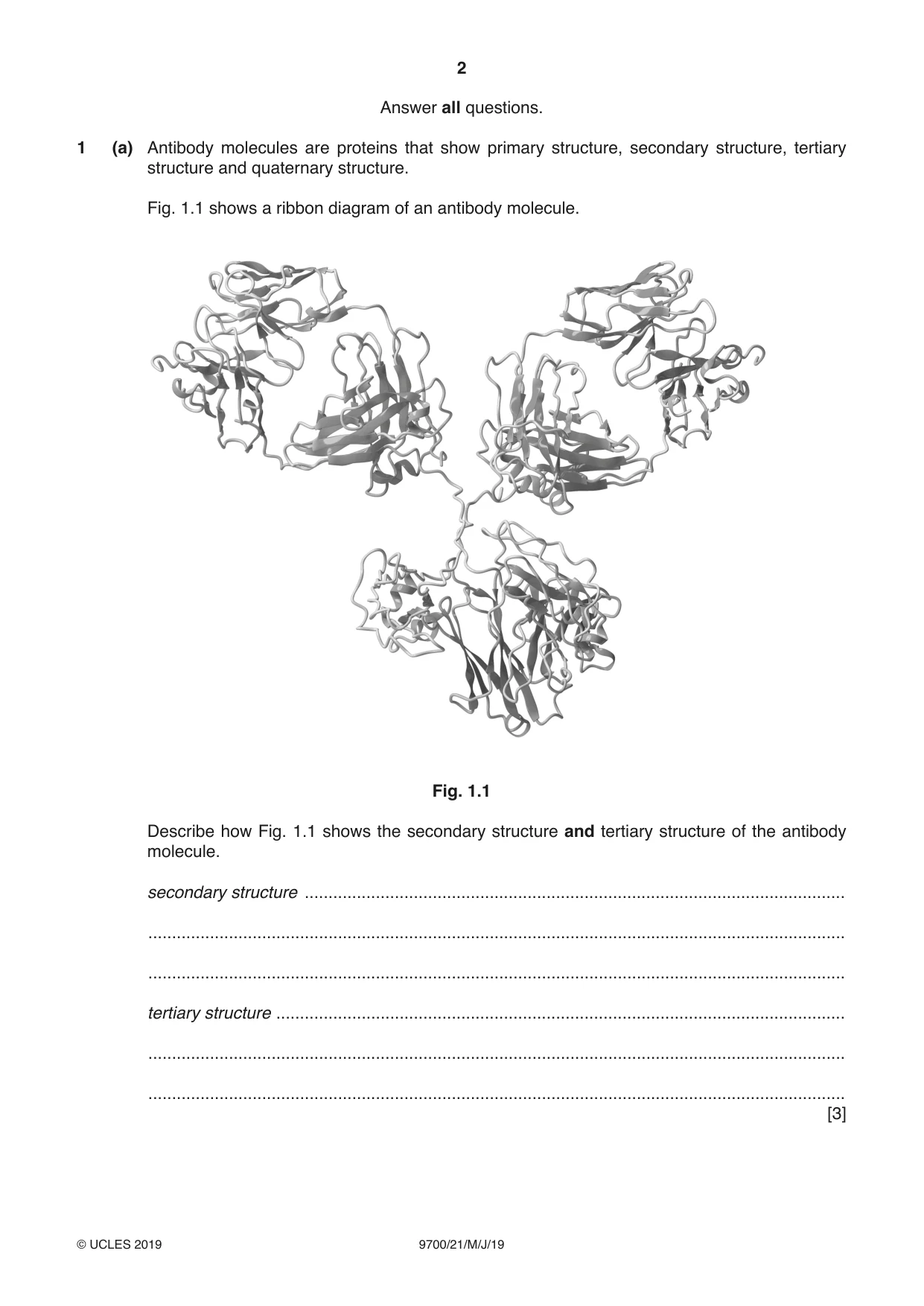
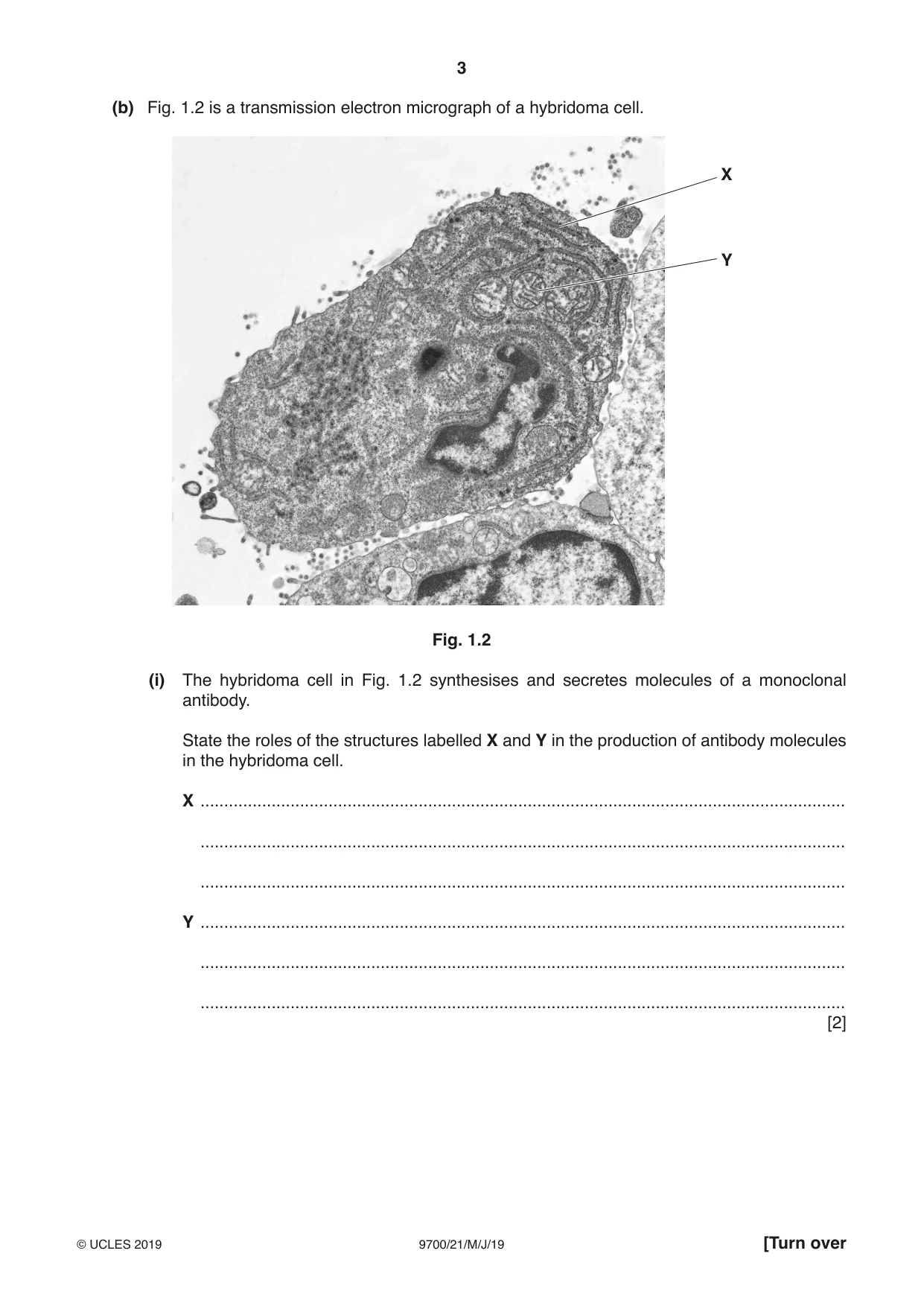
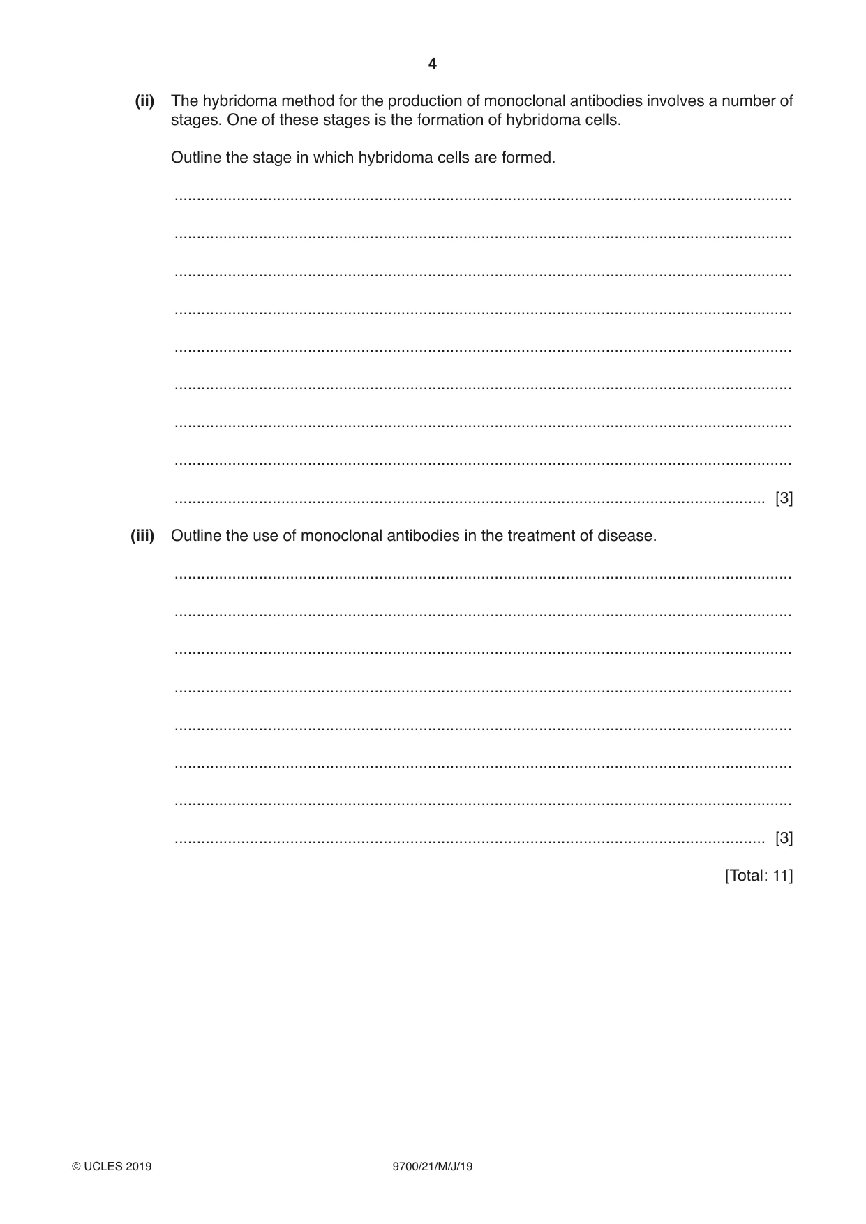
1 (a) Antibody molecules are proteins that show primary structure, secondary structure, tertiary structure and quaternary structure. Fig. 1.1 shows a ribbon diagram of an antibody molecule. Fig. 1.1 Describe how Fig. 1.1 shows the secondary structure and tertiary structure of the antibody molecule. secondary structure .................................................................................................................. ................................................................................................................................................... ................................................................................................................................................... tertiary structure ........................................................................................................................ ................................................................................................................................................... ................................................................................................................................................... [3] (b) Fig. 1.2 is a transmission electron micrograph of a hybridoma cell. X Y Fig. 1.2 (i) The hybridoma cell in Fig. 1.2 synthesises and secretes molecules of a monoclonal antibody. State the roles of the structures labelled X and Y in the production of antibody molecules in the hybridoma cell. X ........................................................................................................................................ ........................................................................................................................................ ........................................................................................................................................ Y ........................................................................................................................................ ........................................................................................................................................ ........................................................................................................................................ [2] (ii) The hybridoma method for the production of monoclonal antibodies involves a number of stages. One of these stages is the formation of hybridoma cells. Outline the stage in which hybridoma cells are formed. ........................................................................................................................................... ........................................................................................................................................... ........................................................................................................................................... ........................................................................................................................................... ........................................................................................................................................... ........................................................................................................................................... ........................................................................................................................................... ........................................................................................................................................... ..................................................................................................................................... [3] (iii) Outline the use of monoclonal antibodies in the treatment of disease. ........................................................................................................................................... ........................................................................................................................................... ........................................................................................................................................... ........................................................................................................................................... ........................................................................................................................................... ........................................................................................................................................... ........................................................................................................................................... ..................................................................................................................................... [3] [Total: 11]
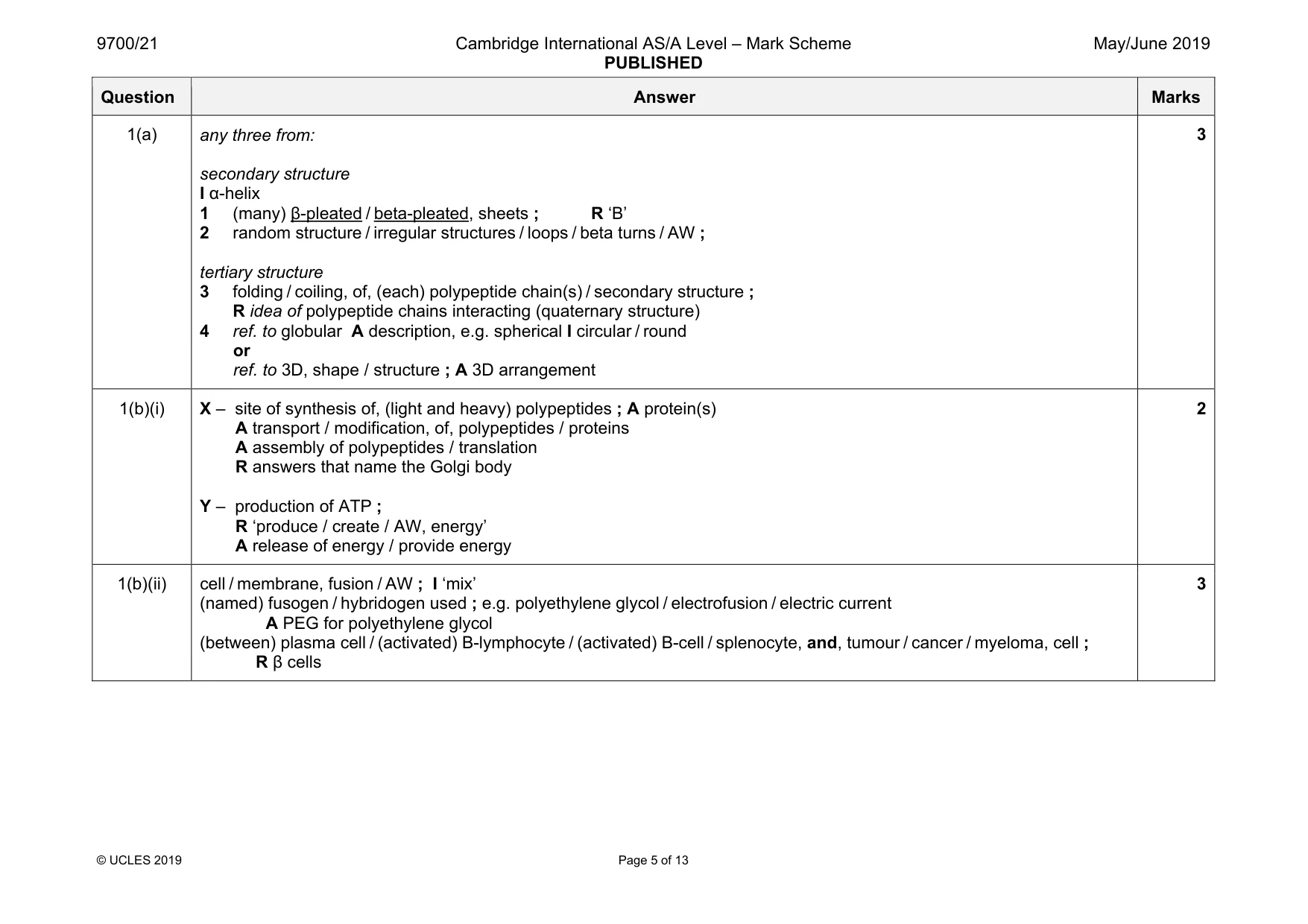
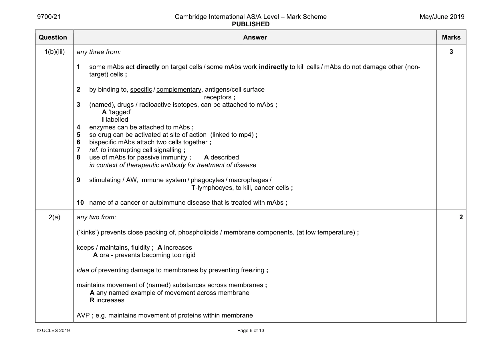
Mark scheme: 1(a) any three from: secondary structure I α-helix 1 (many) β-pleated / beta-pleated, sheets ; R ‘B’ 2 random structure / irregular structures / loops / beta turns / AW ; tertiary structure 3 folding / coiling, of, (each) polypeptide chain(s) / secondary structure ; R idea of polypeptide chains interacting (quaternary structure) 4 ref. to globular A description, e.g. spherical I circular / round or ref. to 3D, shape / structure ; A 3D arrangement 3 1(b)(i) X – site of synthesis of, (light and heavy) polypeptides ; A protein(s) A transport / modification, of, polypeptides / proteins A assembly of polypeptides / translation R answers that name the Golgi body Y – production of ATP ; R ‘produce / create / AW, energy’ A release of energy / provide energy 2 1(b)(ii) cell / membrane, fusion / AW ; I ‘mix’ (named) fusogen / hybridogen used ; e.g. polyethylene glycol / electrofusion / electric current A PEG for polyethylene glycol (between) plasma cell / (activated) B-lymphocyte / (activated) B-cell / splenocyte, and, tumour / cancer / myeloma, cell ; R β cells 3 Question Answer Marks 1(b)(iii) any three from: 1 some mAbs act directly on target cells / some mAbs work indirectly to kill cells / mAbs do not damage other (non- target) cells ; 2 by binding to, specific / complementary, antigens/cell surface receptors ; 3 (named), drugs / radioactive isotopes, can be attached to mAbs ; A ‘tagged’ I labelled 4 enzymes can be attached to mAbs ; 5 so drug can be activated at site of action (linked to mp4) ; 6 bispecific mAbs attach two cells together ; 7 ref. to interrupting cell signalling ; 8 use of mAbs for passive immunity ; A described in context of therapeutic antibody for treatment of disease 9 stimulating / AW, immune system / phagocytes / macrophages / T-lymphocyes, to kill, cancer cells ; 10 name of a cancer or autoimmune disease that is treated with mAbs ; 3
Q2 · Linoleic acid is an unsaturated fatty acid that is found in some triglycerides and some…
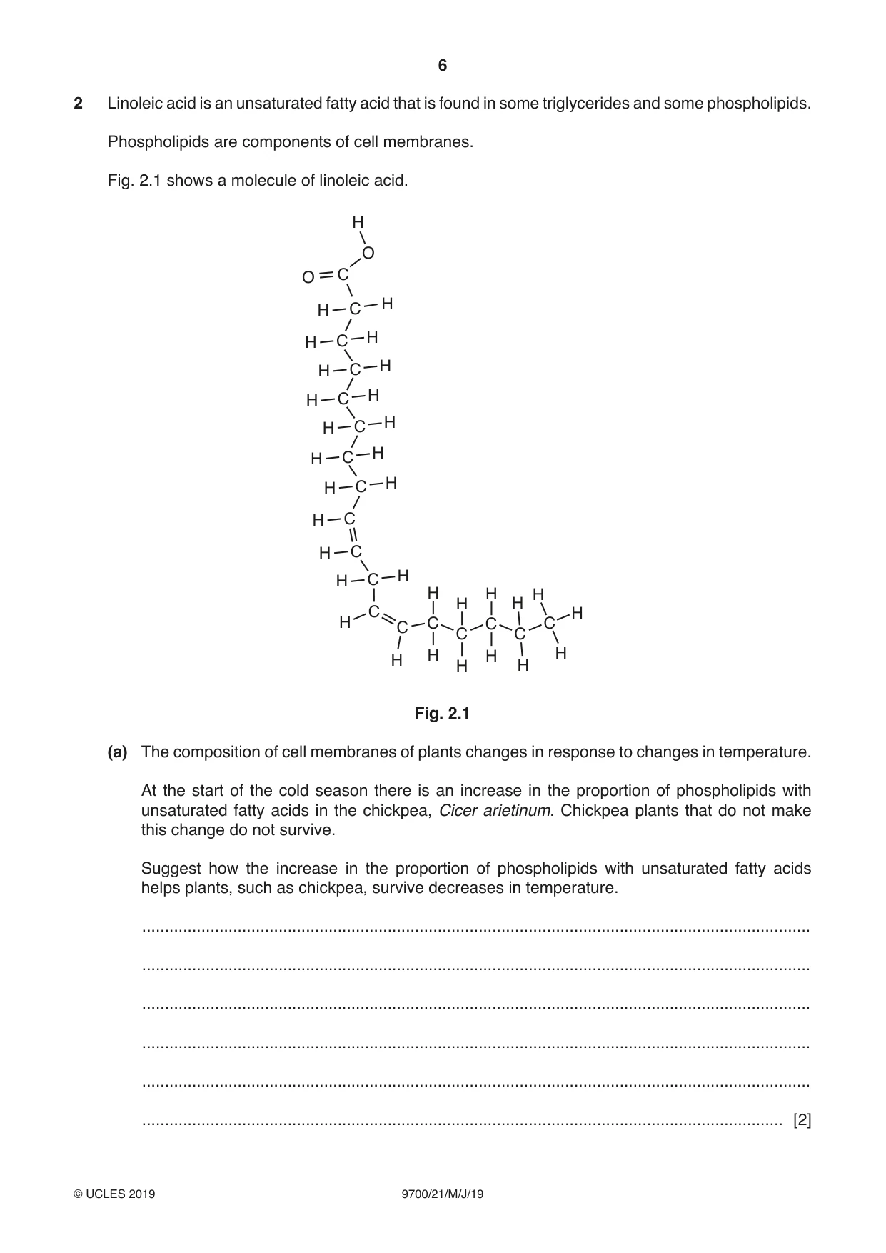
2 Linoleic acid is an unsaturated fatty acid that is found in some triglycerides and some phospholipids. Phospholipids are components of cell membranes. Fig. 2.1 shows a molecule of linoleic acid. H O O C H H C H C H H C H H C H H C H H C H H C H H C H C H C H H H H H H C H H C C C C C C H H H H H H Fig. 2.1 (a) The composition of cell membranes of plants changes in response to changes in temperature. At the start of the cold season there is an increase in the proportion of phospholipids with unsaturated fatty acids in the chickpea, Cicer arietinum. Chickpea plants that do not make this change do not survive. Suggest how the increase in the proportion of phospholipids with unsaturated fatty acids helps plants, such as chickpea, survive decreases in temperature. ................................................................................................................................................... ................................................................................................................................................... ................................................................................................................................................... ................................................................................................................................................... ................................................................................................................................................... ............................................................................................................................................. [2] (b) (i) State why triglycerides and phospholipids cannot be described as polymers. ........................................................................................................................................... ........................................................................................................................................... ..................................................................................................................................... [1] (ii) State two differences in the structure of triglycerides and phospholipids. 1 ........................................................................................................................................ ........................................................................................................................................ 2 ........................................................................................................................................ ........................................................................................................................................ [2] (c) Platelets metabolise linoleic acid to produce a molecule known as thromboxane. Thromboxane is released by platelets when blood loss occurs. Thromboxane acts on smooth muscle cells in the walls of arteries. This causes arteries to constrict, which reduces blood flow. Explain why the constriction of arteries following blood loss is an example of cell signalling. ................................................................................................................................................... ................................................................................................................................................... ................................................................................................................................................... ................................................................................................................................................... ................................................................................................................................................... ................................................................................................................................................... ................................................................................................................................................... ................................................................................................................................................... ................................................................................................................................................... ................................................................................................................................................... ............................................................................................................................................. [4] [Total: 9]
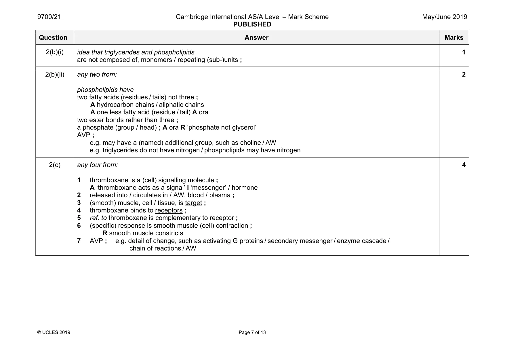
Mark scheme: 2(a) any two from: (‘kinks’) prevents close packing of, phospholipids / membrane components, (at low temperature) ; keeps / maintains, fluidity ; A increases A ora - prevents becoming too rigid idea of preventing damage to membranes by preventing freezing ; maintains movement of (named) substances across membranes ; A any named example of movement across membrane R increases AVP ; e.g. maintains movement of proteins within membrane 2 Question Answer Marks 2(b)(i) idea that triglycerides and phospholipids are not composed of, monomers / repeating (sub-)units ; 1 2(b)(ii) any two from: phospholipids have two fatty acids (residues / tails) not three ; A hydrocarbon chains / aliphatic chains A one less fatty acid (residue / tail) A ora two ester bonds rather than three ; a phosphate (group / head) ; A ora R ‘phosphate not glycerol’ AVP ; e.g. may have a (named) additional group, such as choline / AW e.g. triglycerides do not have nitrogen / phospholipids may have nitrogen 2 2(c) any four from: 1 thromboxane is a (cell) signalling molecule ; A ‘thromboxane acts as a signal’ I ‘messenger’ / hormone 2 released into / circulates in / AW, blood / plasma ; 3 (smooth) muscle, cell / tissue, is target ; 4 thromboxane binds to receptors ; 5 ref. to thromboxane is complementary to receptor ; 6 (specific) response is smooth muscle (cell) contraction ; R smooth muscle constricts 7 AVP ; e.g. detail of change, such as activating G proteins / secondary messenger / enzyme cascade / chain of reactions / AW 4
Q3 · Neutrase® is an enzyme that is used to hydrolyse proteins in solution
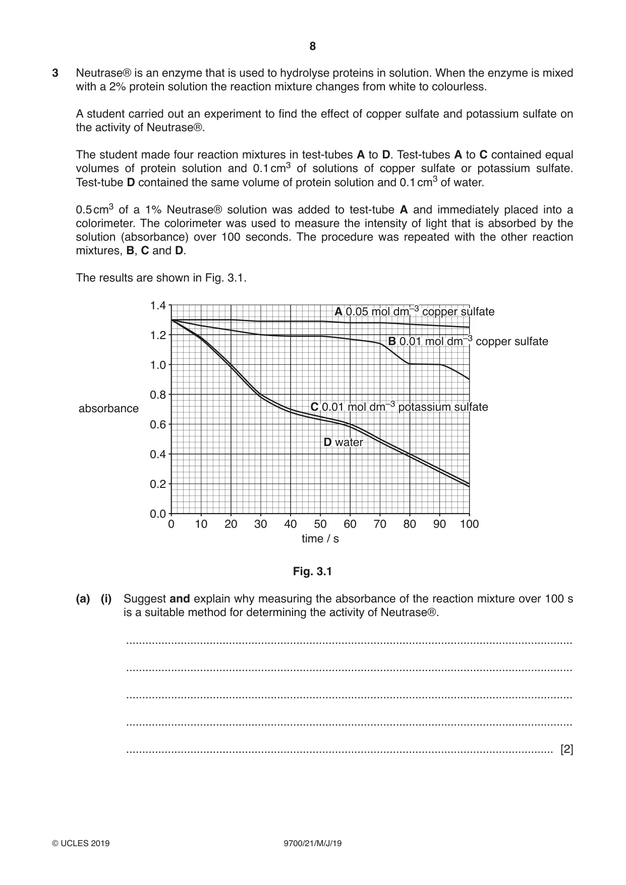
3 Neutrase® is an enzyme that is used to hydrolyse proteins in solution. When the enzyme is mixed with a 2% protein solution the reaction mixture changes from white to colourless. A student carried out an experiment to find the effect of copper sulfate and potassium sulfate on the activity of Neutrase®. The student made four reaction mixtures in test-tubes A to D. Test-tubes A to C contained equal volumes of protein solution and 0.1 cm3 of solutions of copper sulfate or potassium sulfate. Test-tube D contained the same volume of protein solution and 0.1 cm3 of water. 0.5 cm3 of a 1% Neutrase® solution was added to test-tube A and immediately placed into a colorimeter. The colorimeter was used to measure the intensity of light that is absorbed by the solution (absorbance) over 100 seconds. The procedure was repeated with the other reaction mixtures, B, C and D. The results are shown in Fig. 3.1. 1.4 A 0.05 mol dm–3 copper sulfate 1.2 B 0.01 mol dm–3 copper sulfate 1.0 0.8 absorbance C 0.01 mol dm–3 potassium sulfate 0.6 D water 0.4 0.2 0.0 0 10 20 30 40 50 60 70 80 90 100 time / s Fig. 3.1 (a) (i) Suggest and explain why measuring the absorbance of the reaction mixture over 100 s is a suitable method for determining the activity of Neutrase®. ........................................................................................................................................... ........................................................................................................................................... ........................................................................................................................................... ........................................................................................................................................... ..................................................................................................................................... [2] (ii) With reference to Fig. 3.1: • describe the effects of copper sulfate solution and potassium sulfate solution on the activity of Neutrase® • suggest explanations for the effects that you have described. ........................................................................................................................................... ........................................................................................................................................... ........................................................................................................................................... ........................................................................................................................................... ........................................................................................................................................... ........................................................................................................................................... ........................................................................................................................................... ........................................................................................................................................... ........................................................................................................................................... ........................................................................................................................................... ........................................................................................................................................... ........................................................................................................................................... ........................................................................................................................................... ........................................................................................................................................... ..................................................................................................................................... [5] (b) Neutrase® can be immobilised in alginate. Immobilised Neutrase® is used in the food industry to produce foods with high nutritional content. Explain the advantages of using immobilised enzymes, such as Neutrase®, compared with using the same enzymes free in solution. ................................................................................................................................................... ................................................................................................................................................... ................................................................................................................................................... ................................................................................................................................................... ............................................................................................................................................. [2] [Total: 9]
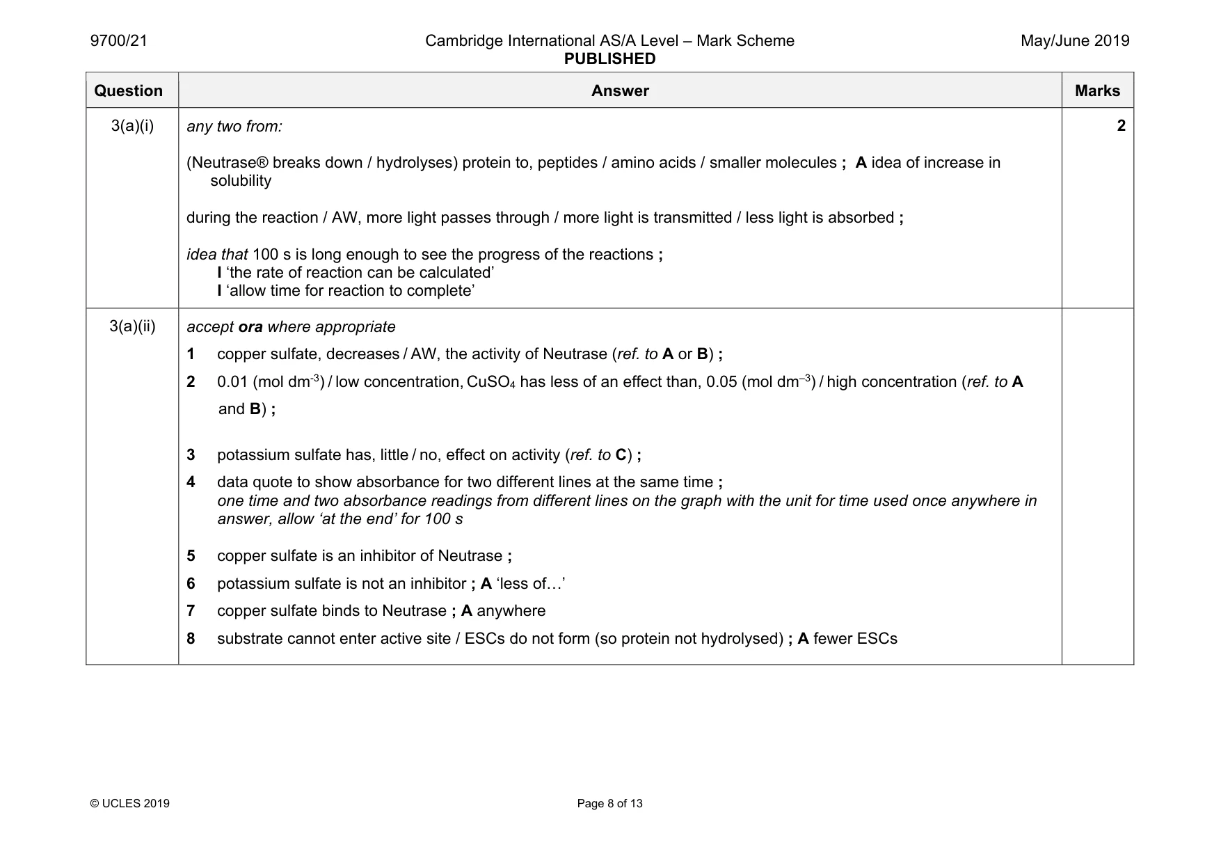
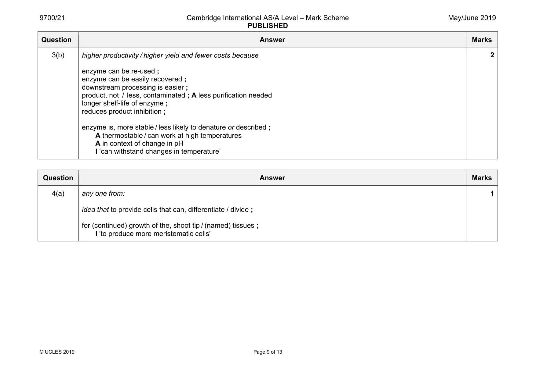
Mark scheme: 3(a)(i) any two from: (Neutrase® breaks down / hydrolyses) protein to, peptides / amino acids / smaller molecules ; A idea of increase in solubility during the reaction / AW, more light passes through / more light is transmitted / less light is absorbed ; idea that 100 s is long enough to see the progress of the reactions ; I ‘the rate of reaction can be calculated’ I ‘allow time for reaction to complete’ 2 3(a)(ii) accept ora where appropriate 1 copper sulfate, decreases / AW, the activity of Neutrase (ref. to A or B) ; 2 0.01 (mol dm-3) / low concentration, CuSO4 has less of an effect than, 0.05 (mol dm–3) / high concentration (ref. to A and B) ; 3 potassium sulfate has, little / no, effect on activity (ref. to C) ; 4 data quote to show absorbance for two different lines at the same time ; one time and two absorbance readings from different lines on the graph with the unit for time used once anywhere in answer, allow ‘at the end’ for 100 s 5 copper sulfate is an inhibitor of Neutrase ; 6 potassium sulfate is not an inhibitor ; A ‘less of«’ 7 copper sulfate binds to Neutrase ; A anywhere 8 substrate cannot enter active site / ESCs do not form (so protein not hydrolysed) ; A fewer ESCs Question Answer Marks 3(b) higher productivity / higher yield and fewer costs because enzyme can be re-used ; enzyme can be easily recovered ; downstream processing is easier ; product, not / less, contaminated ; A less purification needed longer shelf-life of enzyme ; reduces product inhibition ; enzyme is, more stable / less likely to denature or described ; A thermostable / can work at high temperatures A in context of change in pH I ‘can withstand changes in temperature’ 2
Q4 · Meristematic tissue is found in the growing regions of plants, such as shoot tips
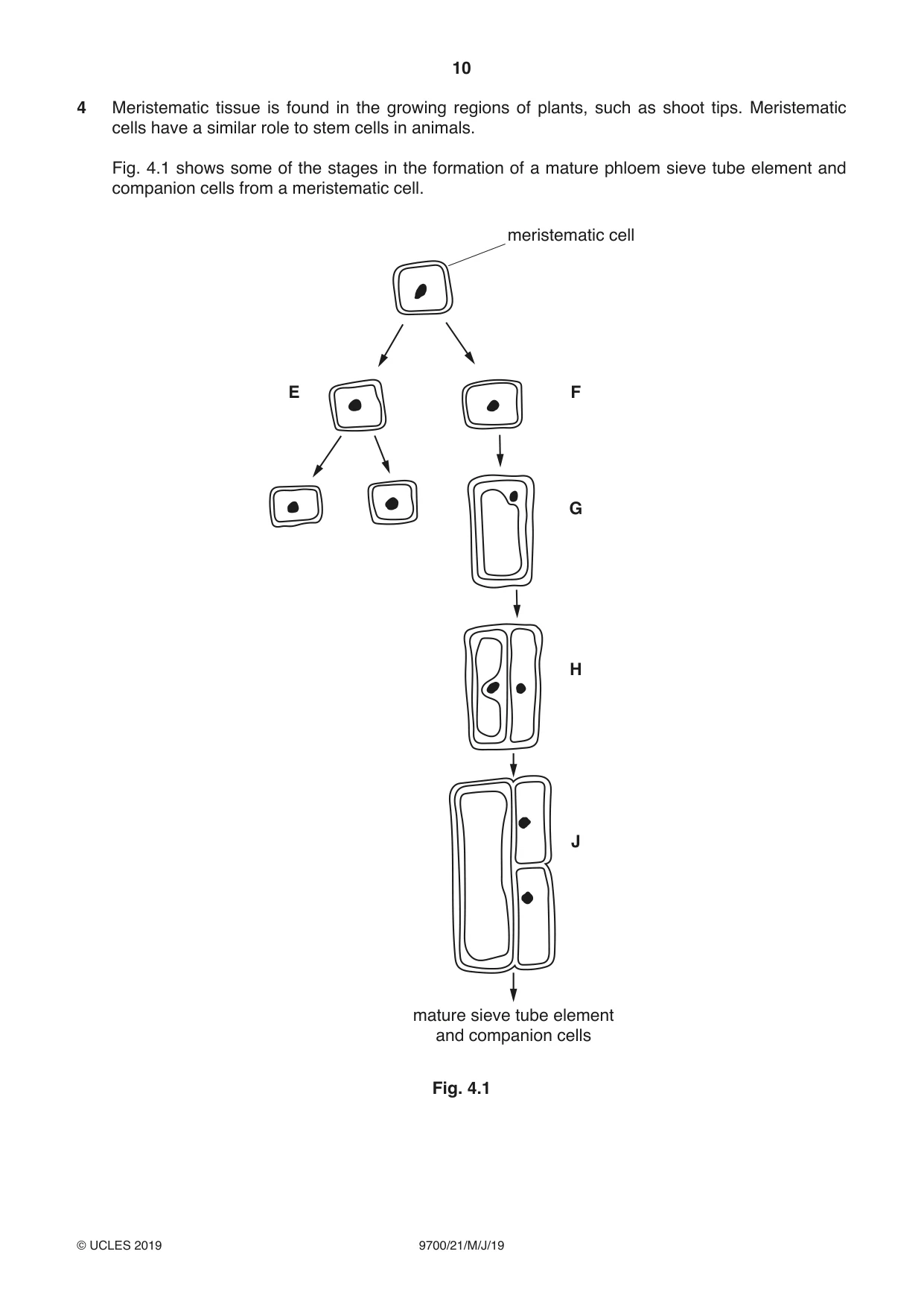
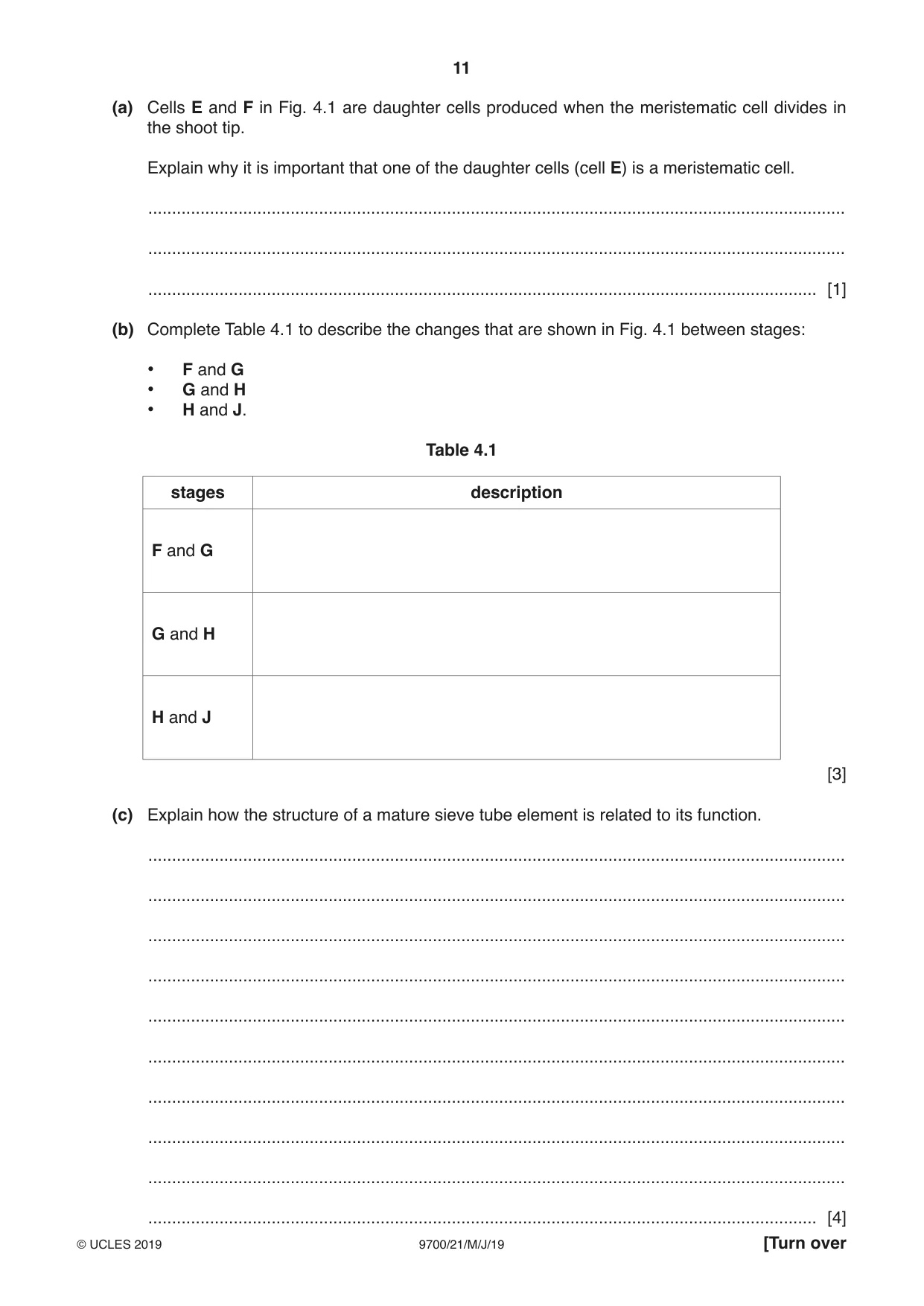
4 Meristematic tissue is found in the growing regions of plants, such as shoot tips. Meristematic cells have a similar role to stem cells in animals. Fig. 4.1 shows some of the stages in the formation of a mature phloem sieve tube element and companion cells from a meristematic cell. meristematic cell E F G H J mature sieve tube element and companion cells Fig. 4.1 (a) Cells E and F in Fig. 4.1 are daughter cells produced when the meristematic cell divides in the shoot tip. Explain why it is important that one of the daughter cells (cell E) is a meristematic cell. ................................................................................................................................................... ................................................................................................................................................... ............................................................................................................................................. [1] (b) Complete Table 4.1 to describe the changes that are shown in Fig. 4.1 between stages: • F and G • G and H • H and J. Table 4.1 stages description F and G G and H H and J [3] (c) Explain how the structure of a mature sieve tube element is related to its function. ................................................................................................................................................... ................................................................................................................................................... ................................................................................................................................................... ................................................................................................................................................... ................................................................................................................................................... ................................................................................................................................................... ................................................................................................................................................... ................................................................................................................................................... ................................................................................................................................................... ............................................................................................................................................. [4] (d) Describe the functions of companion cells in transport in the phloem. ................................................................................................................................................... ................................................................................................................................................... ................................................................................................................................................... ................................................................................................................................................... ............................................................................................................................................. [2] [Total: 10]
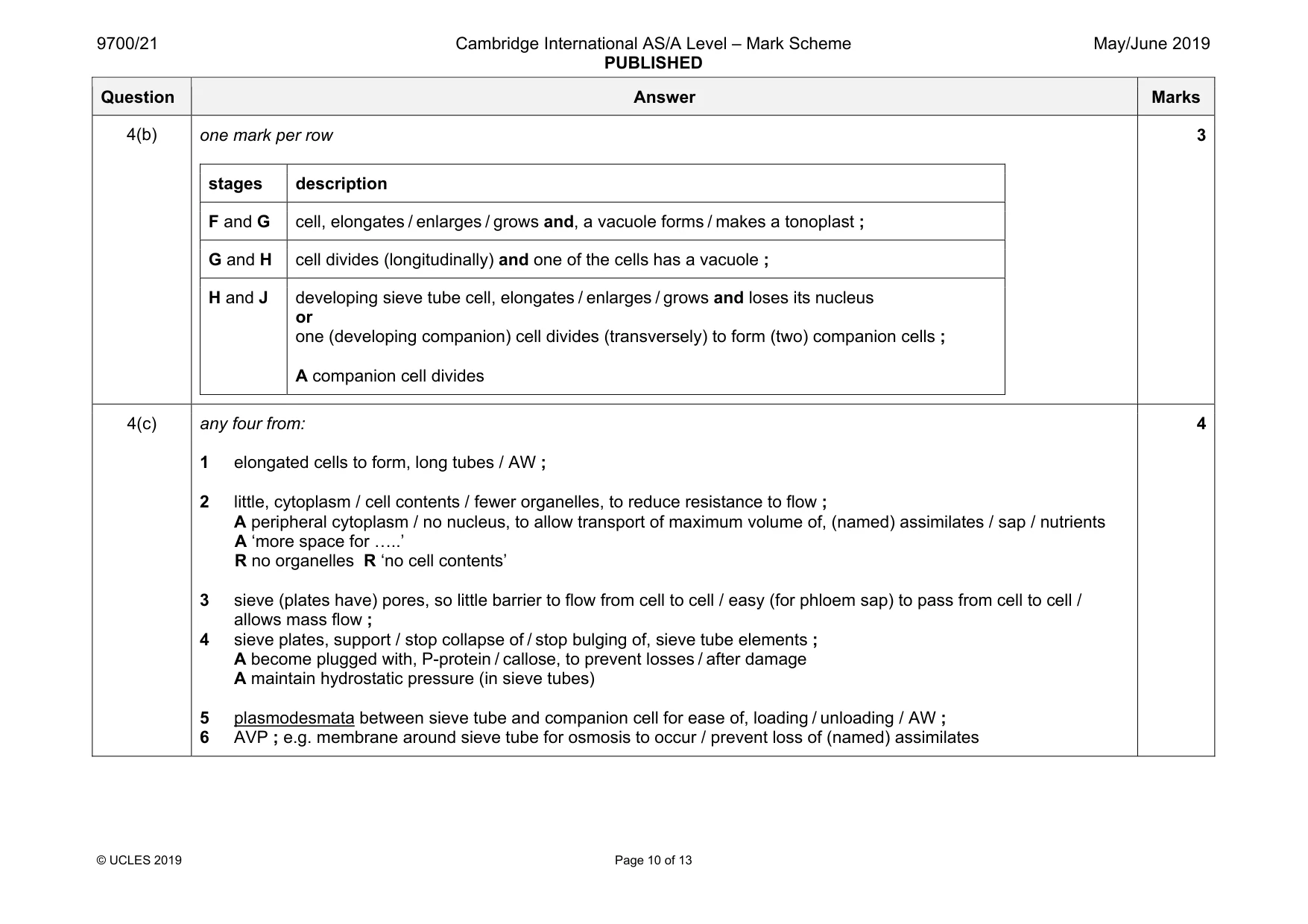
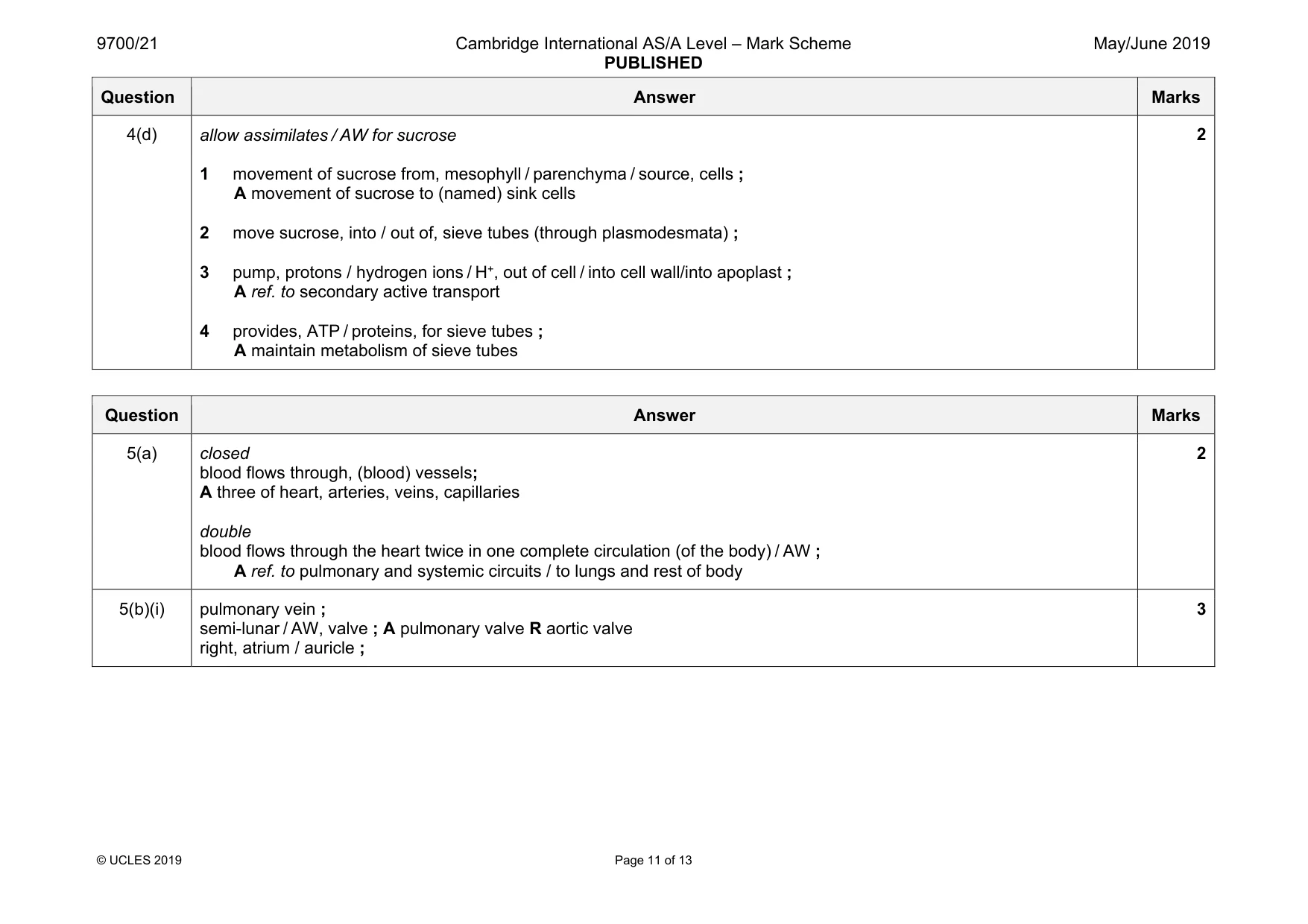
Mark scheme: 4(a) any one from: idea that to provide cells that can, differentiate / divide ; for (continued) growth of the, shoot tip / (named) tissues ; I 'to produce more meristematic cells' 1 Question Answer Marks 4(b) one mark per row stages description F and G cell, elongates / enlarges / grows and, a vacuole forms / makes a tonoplast ; G and H cell divides (longitudinally) and one of the cells has a vacuole ; H and J developing sieve tube cell, elongates / enlarges / grows and loses its nucleus or one (developing companion) cell divides (transversely) to form (two) companion cells ; A companion cell divides 3 4(c) any four from: 1 elongated cells to form, long tubes / AW ; 2 little, cytoplasm / cell contents / fewer organelles, to reduce resistance to flow ; A peripheral cytoplasm / no nucleus, to allow transport of maximum volume of, (named) assimilates / sap / nutrients A ‘more space for «..’ R no organelles R ‘no cell contents’ 3 sieve (plates have) pores, so little barrier to flow from cell to cell / easy (for phloem sap) to pass from cell to cell / allows mass flow ; 4 sieve plates, support / stop collapse of / stop bulging of, sieve tube elements ; A become plugged with, P-protein / callose, to prevent losses / after damage A maintain hydrostatic pressure (in sieve tubes) 5 plasmodesmata between sieve tube and companion cell for ease of, loading / unloading / AW ; 6 AVP ; e.g. membrane around sieve tube for osmosis to occur / prevent loss of (named) assimilates 4 Question Answer Marks 4(d) allow assimilates / AW for sucrose 1 movement of sucrose from, mesophyll / parenchyma / source, cells ; A movement of sucrose to (named) sink cells 2 move sucrose, into / out of, sieve tubes (through plasmodesmata) ; 3 pump, protons / hydrogen ions / H+, out of cell / into cell wall/into apoplast ; A ref. to secondary active transport 4 provides, ATP / proteins, for sieve tubes ; A maintain metabolism of sieve tubes 2
Q5 · The mammalian circulatory system is described as a closed double circulation
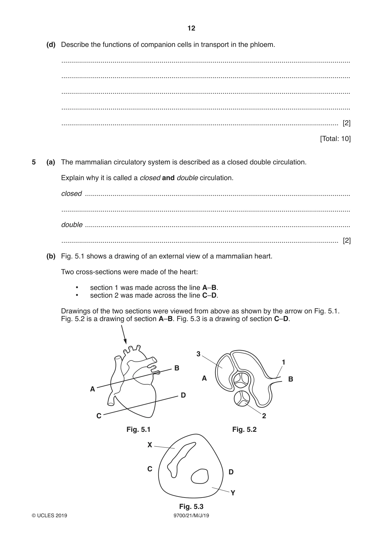
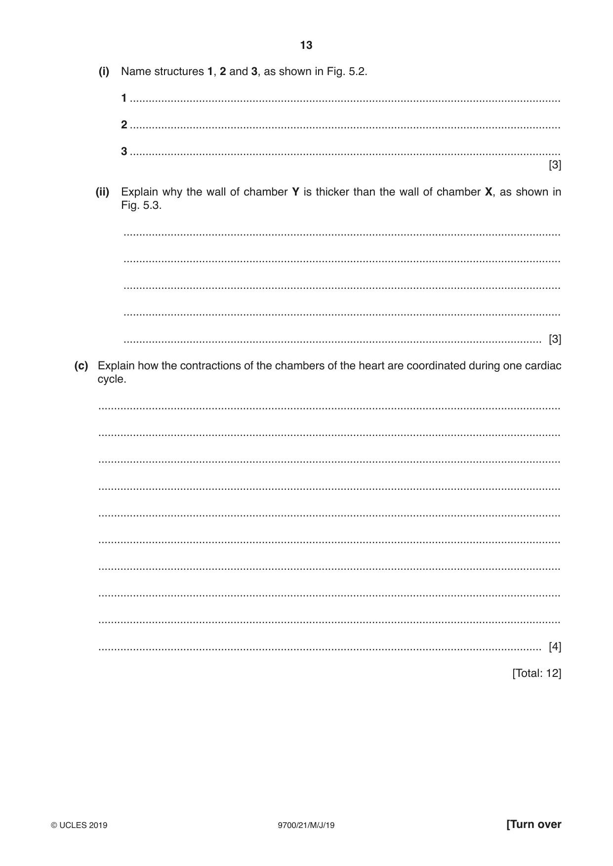
5 (a) The mammalian circulatory system is described as a closed double circulation. Explain why it is called a closed and double circulation. closed ....................................................................................................................................... ................................................................................................................................................... double ....................................................................................................................................... ............................................................................................................................................. [2] (b) Fig. 5.1 shows a drawing of an external view of a mammalian heart. Two cross-sections were made of the heart: • section 1 was made across the line A–B. • section 2 was made across the line C–D. Drawings of the two sections were viewed from above as shown by the arrow on Fig. 5.1. Fig. 5.2 is a drawing of section A–B. Fig. 5.3 is a drawing of section C–D. 3 1 B A B A D C 2 Fig. 5.1 Fig. 5.2 X C D Y Fig. 5.3 (i) Name structures 1, 2 and 3, as shown in Fig. 5.2. 1 ......................................................................................................................................... 2 ......................................................................................................................................... 3 ......................................................................................................................................... [3] (ii) Explain why the wall of chamber Y is thicker than the wall of chamber X, as shown in Fig. 5.3. ........................................................................................................................................... ........................................................................................................................................... ........................................................................................................................................... ........................................................................................................................................... ..................................................................................................................................... [3] (c) Explain how the contractions of the chambers of the heart are coordinated during one cardiac cycle. ................................................................................................................................................... ................................................................................................................................................... ................................................................................................................................................... ................................................................................................................................................... ................................................................................................................................................... ................................................................................................................................................... ................................................................................................................................................... ................................................................................................................................................... ................................................................................................................................................... ............................................................................................................................................. [4] [Total: 12]
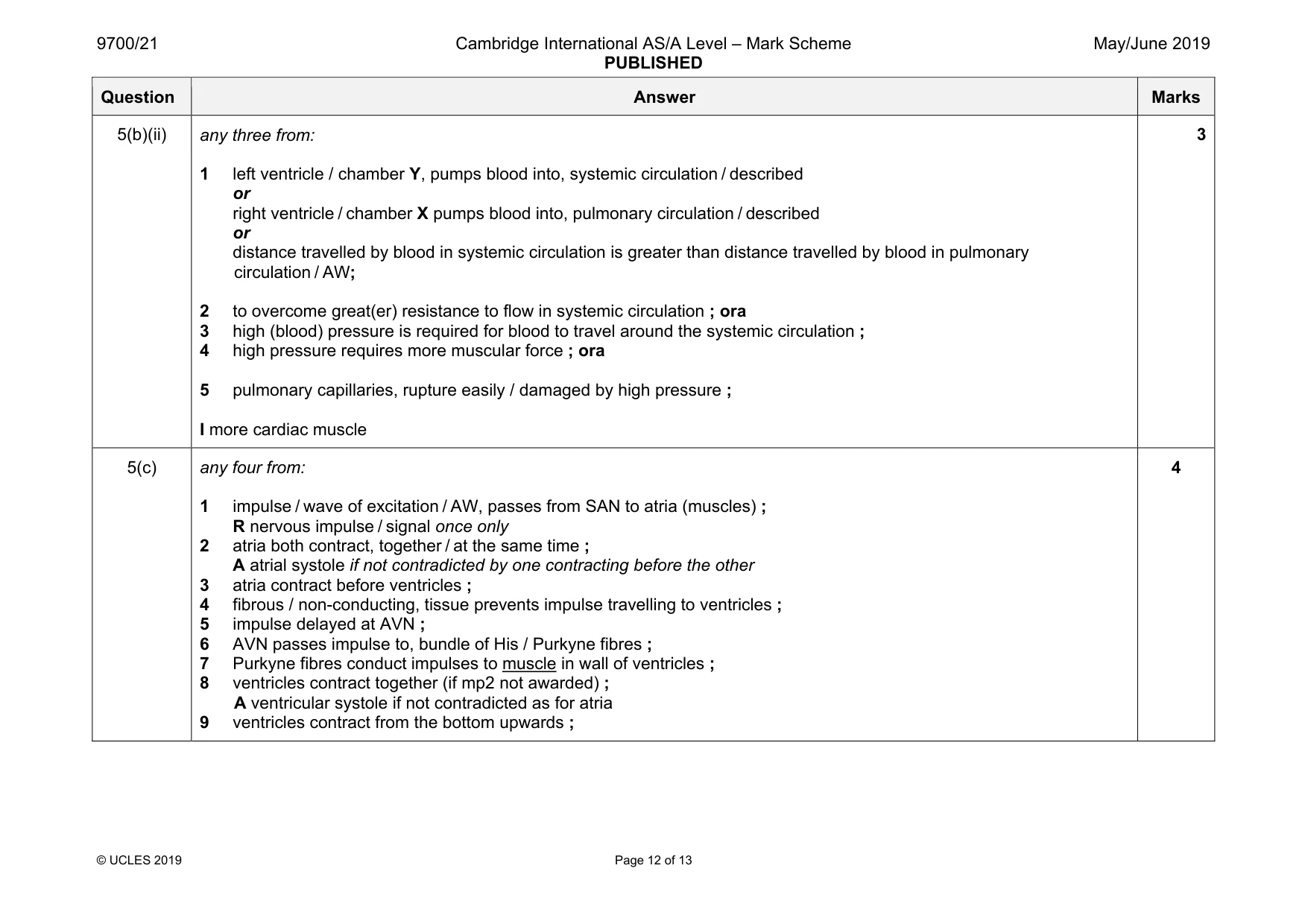
Mark scheme: 5(a) closed blood flows through, (blood) vessels; A three of heart, arteries, veins, capillaries double blood flows through the heart twice in one complete circulation (of the body) / AW ; A ref. to pulmonary and systemic circuits / to lungs and rest of body 2 5(b)(i) pulmonary vein ; semi-lunar / AW, valve ; A pulmonary valve R aortic valve right, atrium / auricle ; 3 Question Answer Marks 5(b)(ii) any three from: 1 left ventricle / chamber Y, pumps blood into, systemic circulation / described or right ventricle / chamber X pumps blood into, pulmonary circulation / described or distance travelled by blood in systemic circulation is greater than distance travelled by blood in pulmonary circulation / AW; 2 to overcome great(er) resistance to flow in systemic circulation ; ora 3 high (blood) pressure is required for blood to travel around the systemic circulation ; 4 high pressure requires more muscular force ; ora 5 pulmonary capillaries, rupture easily / damaged by high pressure ; I more cardiac muscle 3 5(c) any four from: 1 impulse / wave of excitation / AW, passes from SAN to atria (muscles) ; R nervous impulse / signal once only 2 atria both contract, together / at the same time ; A atrial systole if not contradicted by one contracting before the other 3 atria contract before ventricles ; 4 fibrous / non-conducting, tissue prevents impulse travelling to ventricles ; 5 impulse delayed at AVN ; 6 AVN passes impulse to, bundle of His / Purkyne fibres ; 7 Purkyne fibres conduct impulses to muscle in wall of ventricles ; 8 ventricles contract together (if mp2 not awarded) ; A ventricular systole if not contradicted as for atria 9 ventricles contract from the bottom upwards ; 4
Q6 · The DNA in the nucleus is known as nuclear DNA
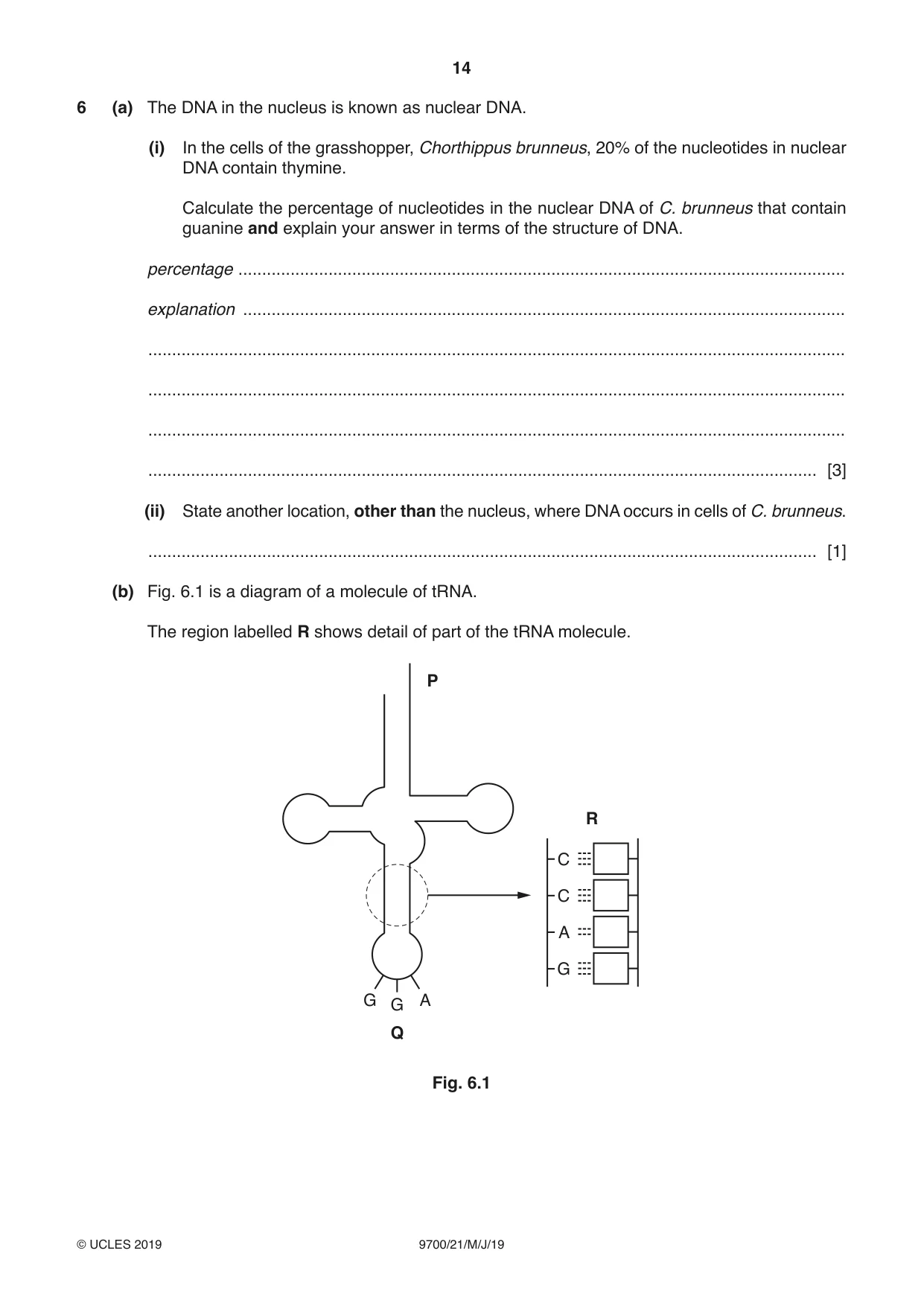
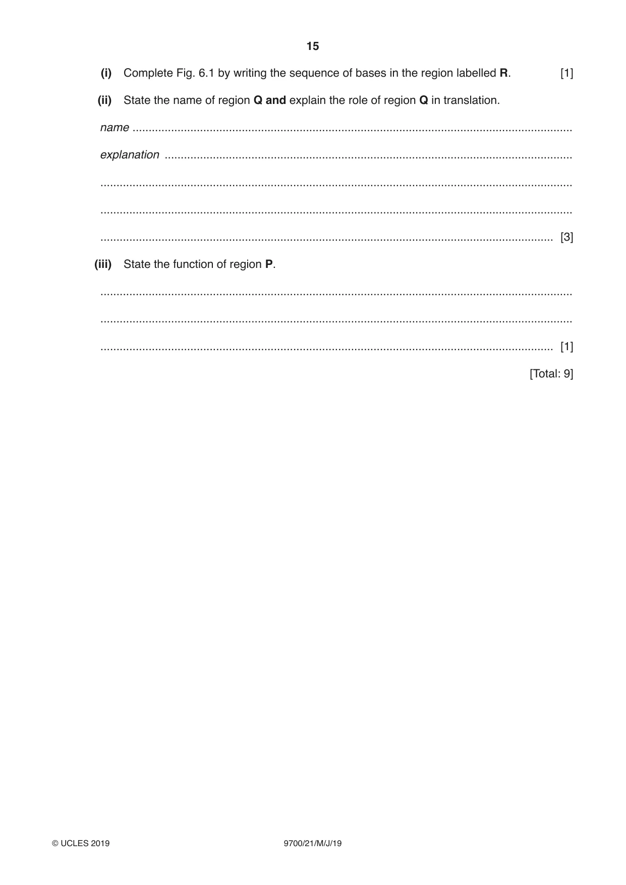
6 (a) The DNA in the nucleus is known as nuclear DNA. (i) In the cells of the grasshopper, Chorthippus brunneus, 20% of the nucleotides in nuclear DNA contain thymine. Calculate the percentage of nucleotides in the nuclear DNA of C. brunneus that contain guanine and explain your answer in terms of the structure of DNA. percentage ................................................................................................................................ explanation ............................................................................................................................... ................................................................................................................................................... ................................................................................................................................................... ................................................................................................................................................... ............................................................................................................................................. [3] (ii) State another location, other than the nucleus, where DNA occurs in cells of C. brunneus. ............................................................................................................................................. [1] (b) Fig. 6.1 is a diagram of a molecule of tRNA. The region labelled R shows detail of part of the tRNA molecule. P R C C A G G G A Q Fig. 6.1 (i) Complete Fig. 6.1 by writing the sequence of bases in the region labelled R. [1] (ii) State the name of region Q and explain the role of region Q in translation. name ......................................................................................................................................... explanation ............................................................................................................................... ................................................................................................................................................... ................................................................................................................................................... ............................................................................................................................................. [3] (iii) State the function of region P. ................................................................................................................................................... ................................................................................................................................................... ............................................................................................................................................. [1] [Total: 9]
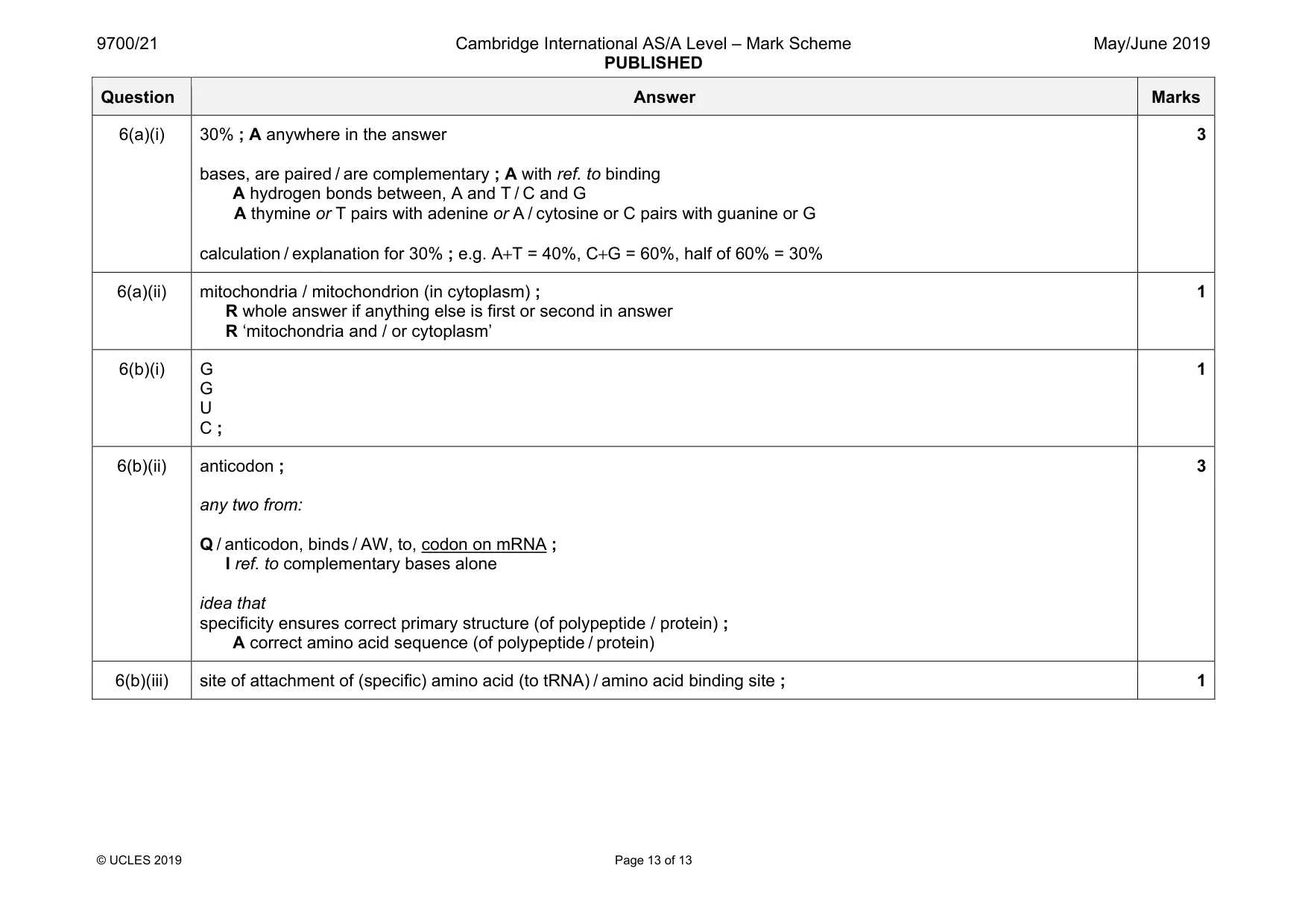
Mark scheme: 6(a)(i) 30% ; A anywhere in the answer bases, are paired / are complementary ; A with ref. to binding A hydrogen bonds between, A and T / C and G A thymine or T pairs with adenine or A / cytosine or C pairs with guanine or G calculation / explanation for 30% ; e.g. A+T = 40%, C+G = 60%, half of 60% = 30% 3 6(a)(ii) mitochondria / mitochondrion (in cytoplasm) ; R whole answer if anything else is first or second in answer R ‘mitochondria and / or cytoplasm’ 1 6(b)(i) G G U C ; 1 6(b)(ii) anticodon ; any two from: Q / anticodon, binds / AW, to, codon on mRNA ; I ref. to complementary bases alone idea that specificity ensures correct primary structure (of polypeptide / protein) ; A correct amino acid sequence (of polypeptide / protein) 3 6(b)(iii) site of attachment of (specific) amino acid (to tRNA) / amino acid binding site ; 1
What was in this paper
The subtopics covered by these 6 questions, and how many questions each got. Open one in a new tab to see every Cambridge question on it.
What you needed in this session
Cambridge’s own grade thresholds for 2019 May/June, Paper 2 · Variant 1. A higher threshold means an easier paper — the bar moves with how the cohort did.