Cambridge A Level Biology 9700 — 2019 Feb/March Paper 2 · Variant 2
9700/22/F/M/19 · 6 questions · 60 marks · ≈68 min
The question paper and its mark scheme, free to read here and free to download. This is Cambridge’s own paper, exactly as it was sat.
Question paper16 pages
Mark scheme12 pages
Answers below. Sit the paper first if you are practising.
Questions as text
Question 1
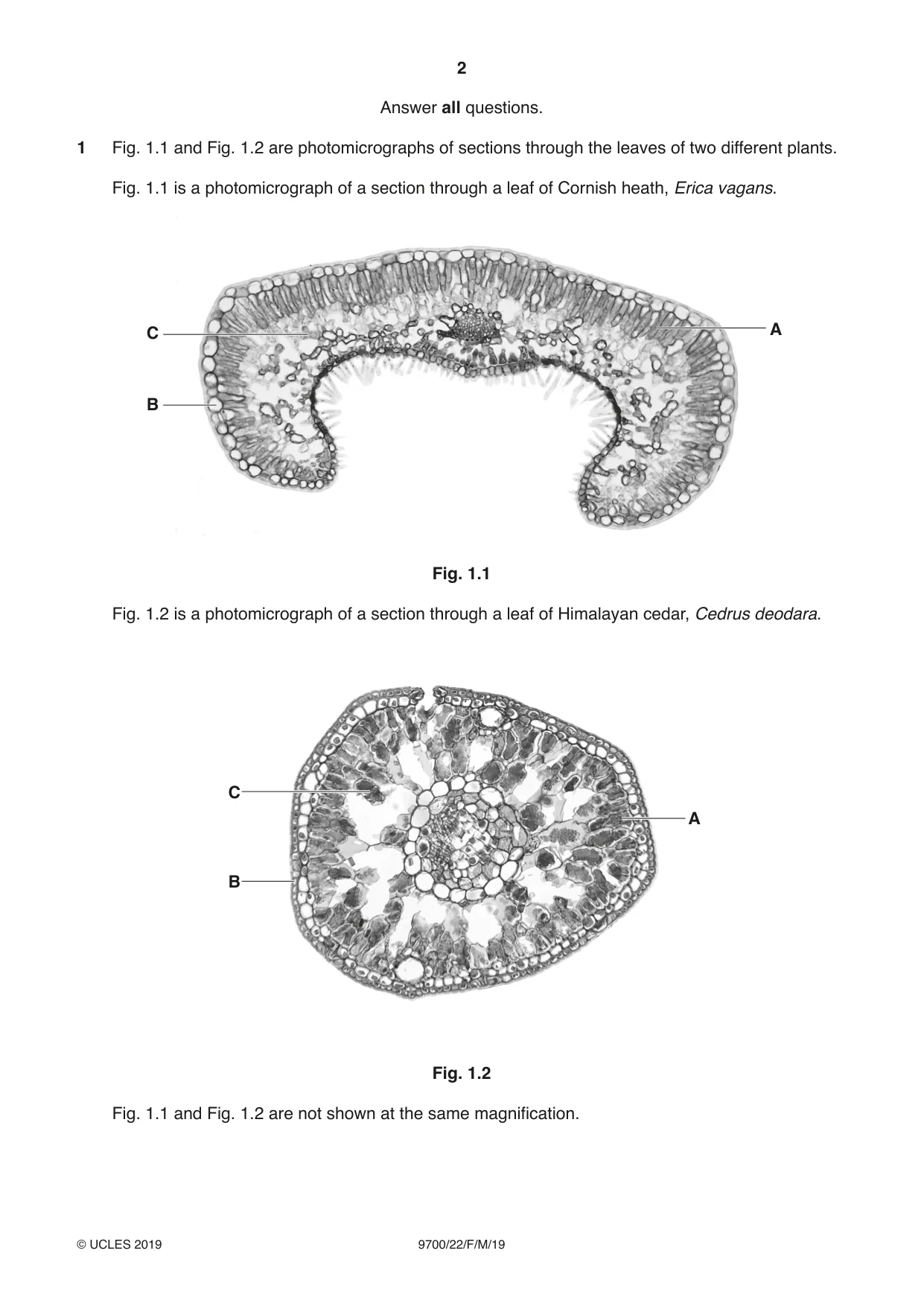
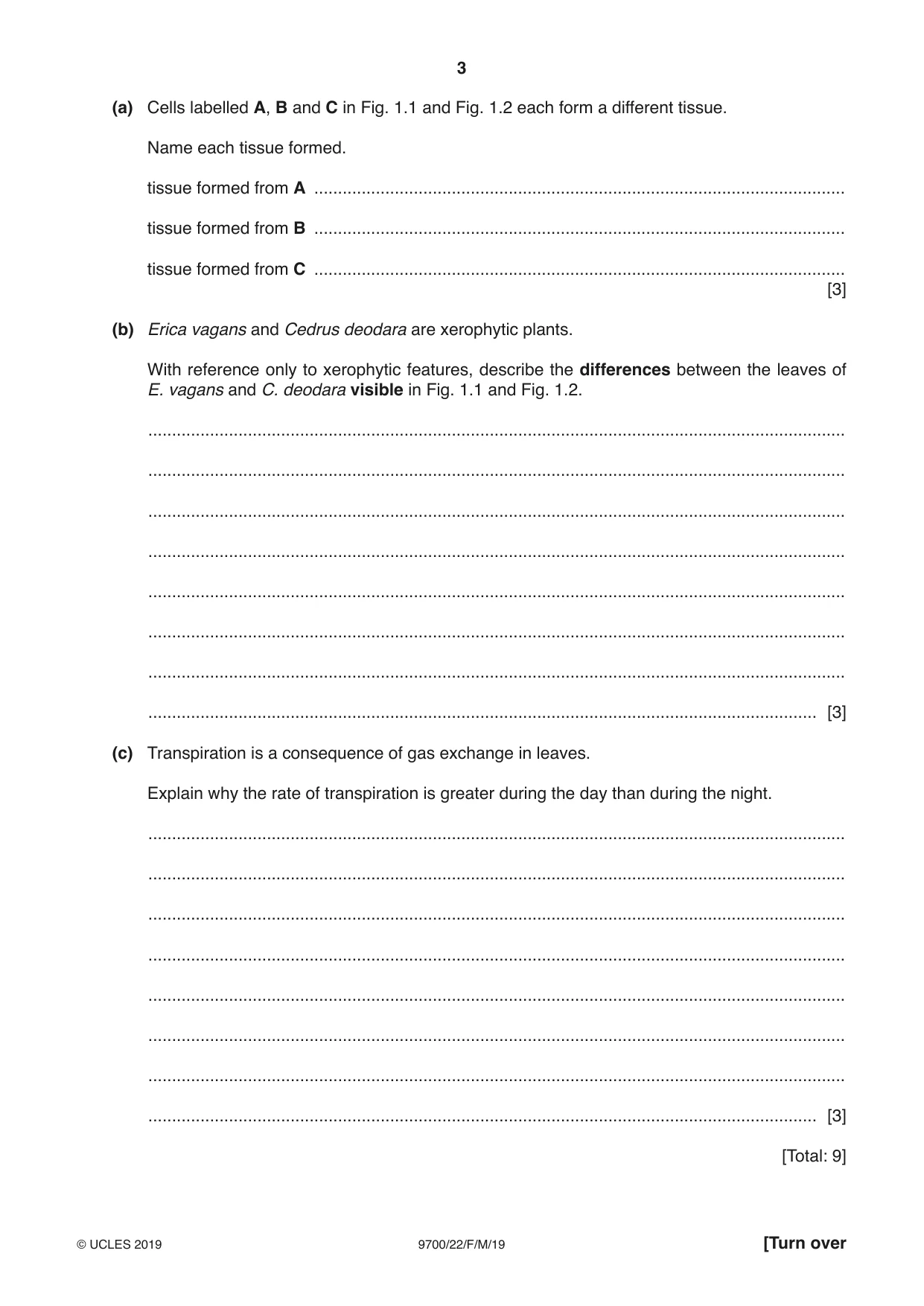
1 Fig. 1.1 and Fig. 1.2 are photomicrographs of sections through the leaves of two different plants. Fig. 1.1 is a photomicrograph of a section through a leaf of Cornish heath, Erica vagans. C A B Fig. 1.1 Fig. 1.2 is a photomicrograph of a section through a leaf of Himalayan cedar, Cedrus deodara. C A B Fig. 1.2 Fig. 1.1 and Fig. 1.2 are not shown at the same magnification. (a) Cells labelled A, B and C in Fig. 1.1 and Fig. 1.2 each form a different tissue. Name each tissue formed. tissue formed from A ................................................................................................................ tissue formed from B ................................................................................................................ tissue formed from C ................................................................................................................ [3] (b) Erica vagans and Cedrus deodara are xerophytic plants. With reference only to xerophytic features, describe the differences between the leaves of E. vagans and C. deodara visible in Fig. 1.1 and Fig. 1.2. ................................................................................................................................................... ................................................................................................................................................... ................................................................................................................................................... ................................................................................................................................................... ................................................................................................................................................... ................................................................................................................................................... ................................................................................................................................................... ............................................................................................................................................. [3] (c) Transpiration is a consequence of gas exchange in leaves. Explain why the rate of transpiration is greater during the day than during the night. ................................................................................................................................................... ................................................................................................................................................... ................................................................................................................................................... ................................................................................................................................................... ................................................................................................................................................... ................................................................................................................................................... ................................................................................................................................................... ............................................................................................................................................. [3] [Total: 9]
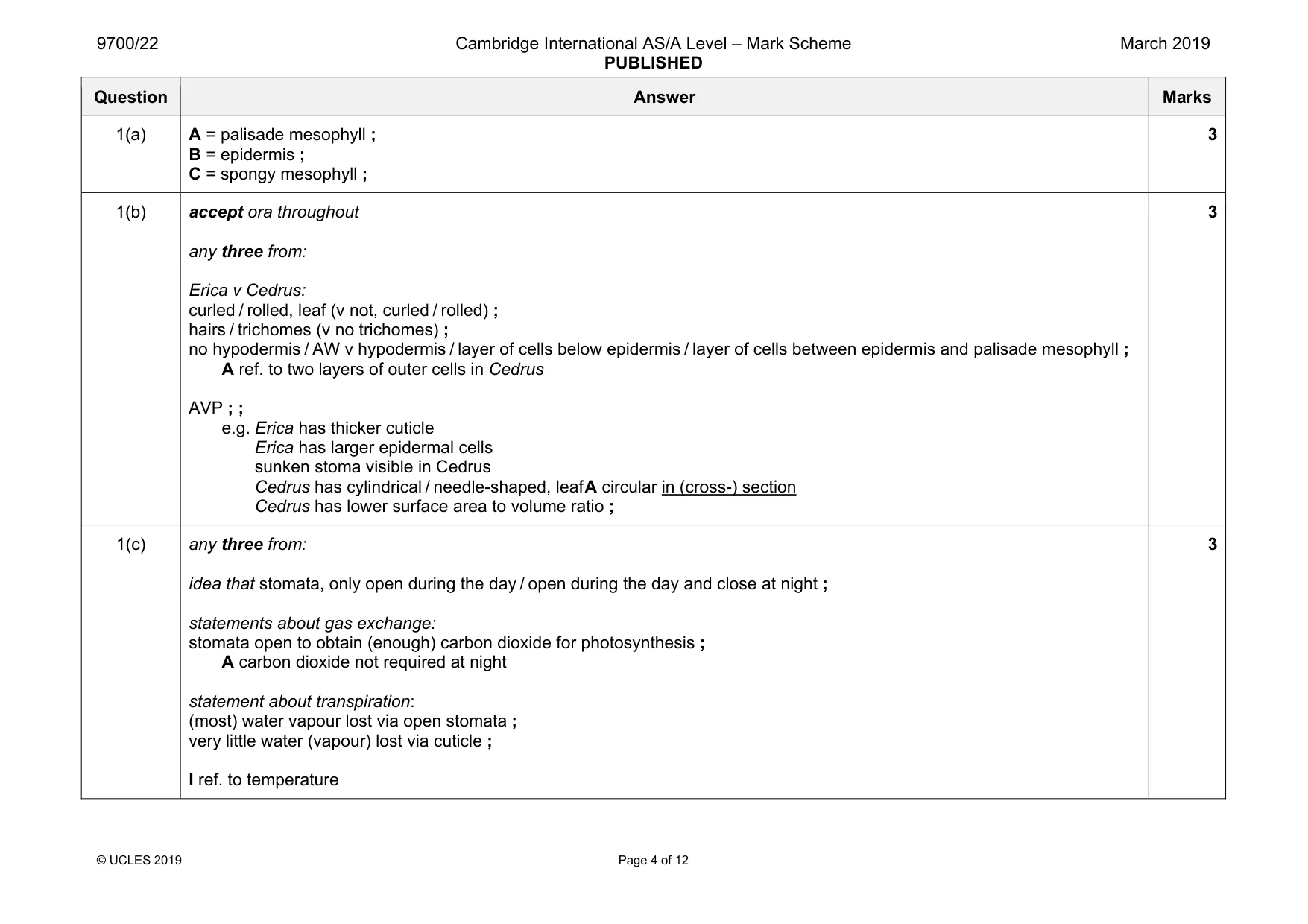
Mark scheme: 1(a) A = palisade mesophyll ; B = epidermis ; C = spongy mesophyll ; 3 1(b) accept ora throughout any three from: Erica v Cedrus: curled / rolled, leaf (v not, curled / rolled) ; hairs / trichomes (v no trichomes) ; no hypodermis / AW v hypodermis / layer of cells below epidermis / layer of cells between epidermis and palisade mesophyll ; A ref. to two layers of outer cells in Cedrus AVP ; ; e.g. Erica has thicker cuticle Erica has larger epidermal cells sunken stoma visible in Cedrus Cedrus has cylindrical / needle-shaped, leaf A circular in (cross-) section Cedrus has lower surface area to volume ratio ; 3 1(c) any three from: idea that stomata, only open during the day / open during the day and close at night ; statements about gas exchange: stomata open to obtain (enough) carbon dioxide for photosynthesis ; A carbon dioxide not required at night statement about transpiration: (most) water vapour lost via open stomata ; very little water (vapour) lost via cuticle ; I ref. to temperature 3
Q2 · In mammalian red blood cells, carbonic anhydrase has an important role in the transport…
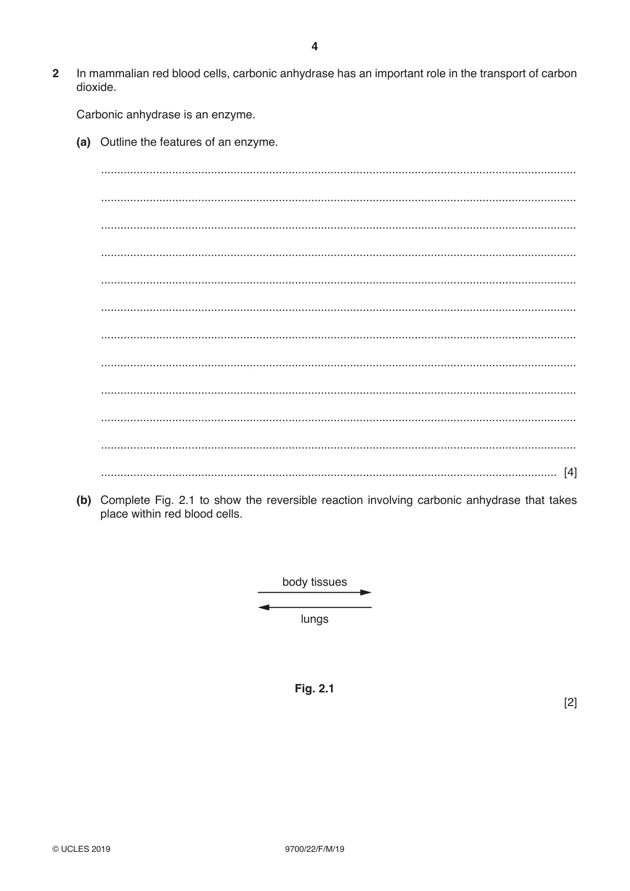
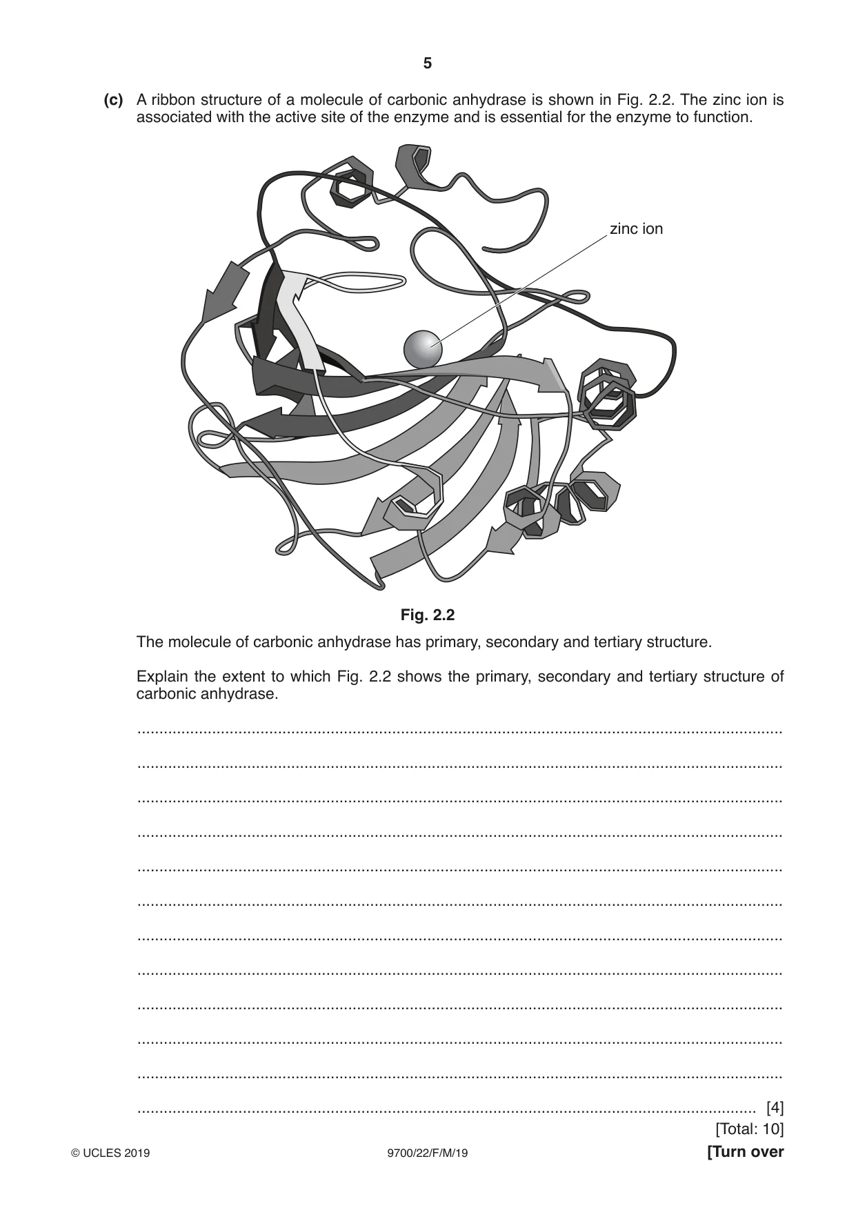
2 In mammalian red blood cells, carbonic anhydrase has an important role in the transport of carbon dioxide. Carbonic anhydrase is an enzyme. (a) Outline the features of an enzyme. ................................................................................................................................................... ................................................................................................................................................... ................................................................................................................................................... ................................................................................................................................................... ................................................................................................................................................... ................................................................................................................................................... ................................................................................................................................................... ................................................................................................................................................... ................................................................................................................................................... ................................................................................................................................................... ................................................................................................................................................... ............................................................................................................................................. [4] (b) Complete Fig. 2.1 to show the reversible reaction involving carbonic anhydrase that takes place within red blood cells. body tissues lungs Fig. 2.1 [2] (c) A ribbon structure of a molecule of carbonic anhydrase is shown in Fig. 2.2. The zinc ion is associated with the active site of the enzyme and is essential for the enzyme to function. zinc ion Fig. 2.2 The molecule of carbonic anhydrase has primary, secondary and tertiary structure. Explain the extent to which Fig. 2.2 shows the primary, secondary and tertiary structure of carbonic anhydrase. ................................................................................................................................................... ................................................................................................................................................... ................................................................................................................................................... ................................................................................................................................................... ................................................................................................................................................... ................................................................................................................................................... ................................................................................................................................................... ................................................................................................................................................... ................................................................................................................................................... ................................................................................................................................................... ................................................................................................................................................... ............................................................................................................................................. [4]
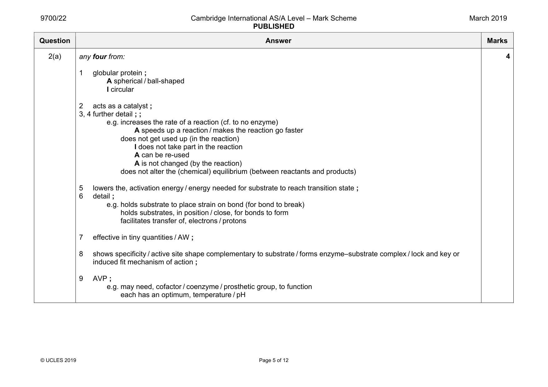
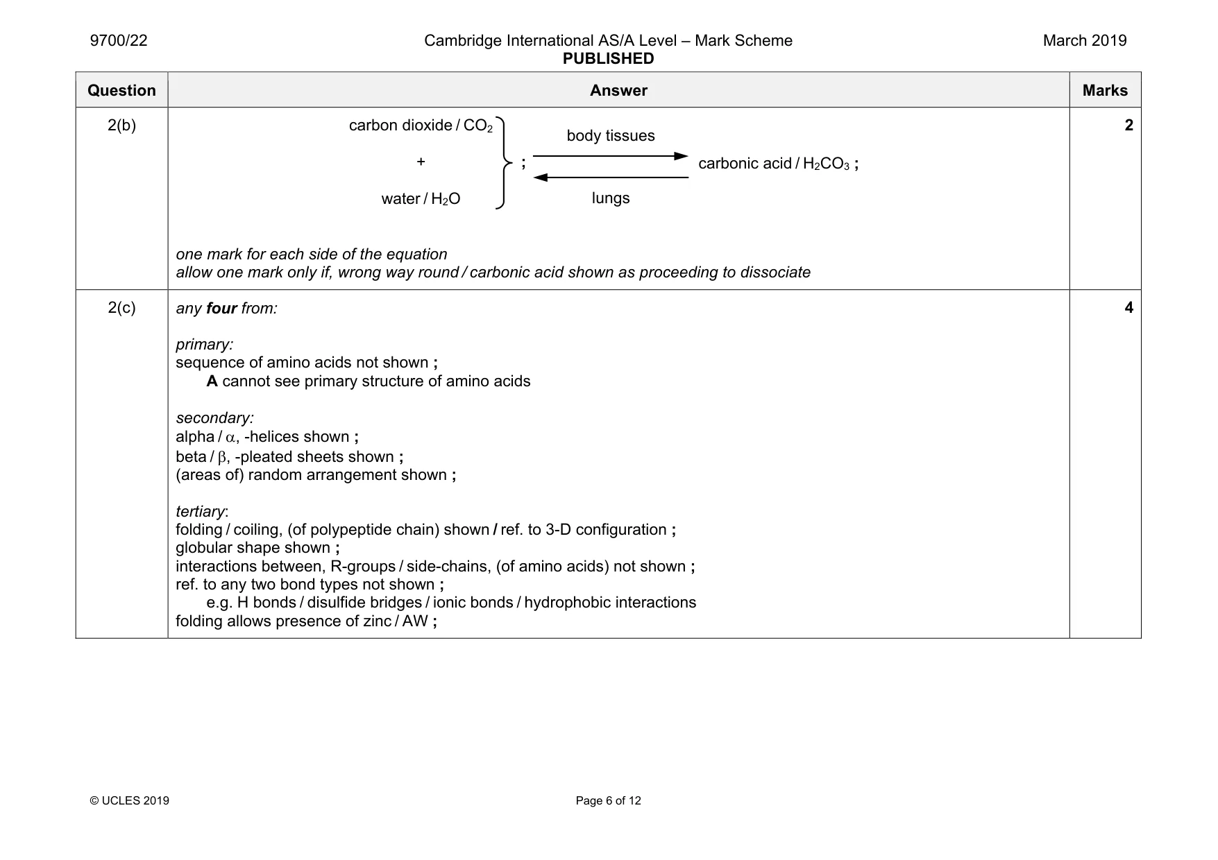
Mark scheme: 2(a) any four from: 1 globular protein ; A spherical / ball-shaped I circular 2 acts as a catalyst ; 3, 4 further detail ; ; e.g. increases the rate of a reaction (cf. to no enzyme) A speeds up a reaction / makes the reaction go faster does not get used up (in the reaction) I does not take part in the reaction A can be re-used A is not changed (by the reaction) does not alter the (chemical) equilibrium (between reactants and products) 5 lowers the, activation energy / energy needed for substrate to reach transition state ; 6 detail ; e.g. holds substrate to place strain on bond (for bond to break) holds substrates, in position / close, for bonds to form facilitates transfer of, electrons / protons 7 effective in tiny quantities / AW ; 8 shows specificity / active site shape complementary to substrate / forms enzyme–substrate complex / lock and key or induced fit mechanism of action ; 9 AVP ; e.g. may need, cofactor / coenzyme / prosthetic group, to function each has an optimum, temperature / pH 4 Question Answer Marks 2(b) one mark for each side of the equation allow one mark only if, wrong way round / carbonic acid shown as proceeding to dissociate 2 2(c) any four from: primary: sequence of amino acids not shown ; A cannot see primary structure of amino acids secondary: alpha / α, -helices shown ; beta / β, -pleated sheets shown ; (areas of) random arrangement shown ; tertiary: folding / coiling, (of polypeptide chain) shown / ref. to 3-D configuration ; globular shape shown ; interactions between, R-groups / side-chains, (of amino acids) not shown ; ref. to any two bond types not shown ; e.g. H bonds / disulfide bridges / ionic bonds / hydrophobic interactions folding allows presence of zinc / AW ; 4 body tissues lungs carbonic acid / H2CO3 ; carbon dioxide / CO2 + water / H2O ;
Q3 · In 2015, the World Health Organization (WHO) published the Global Technical Strategy for…
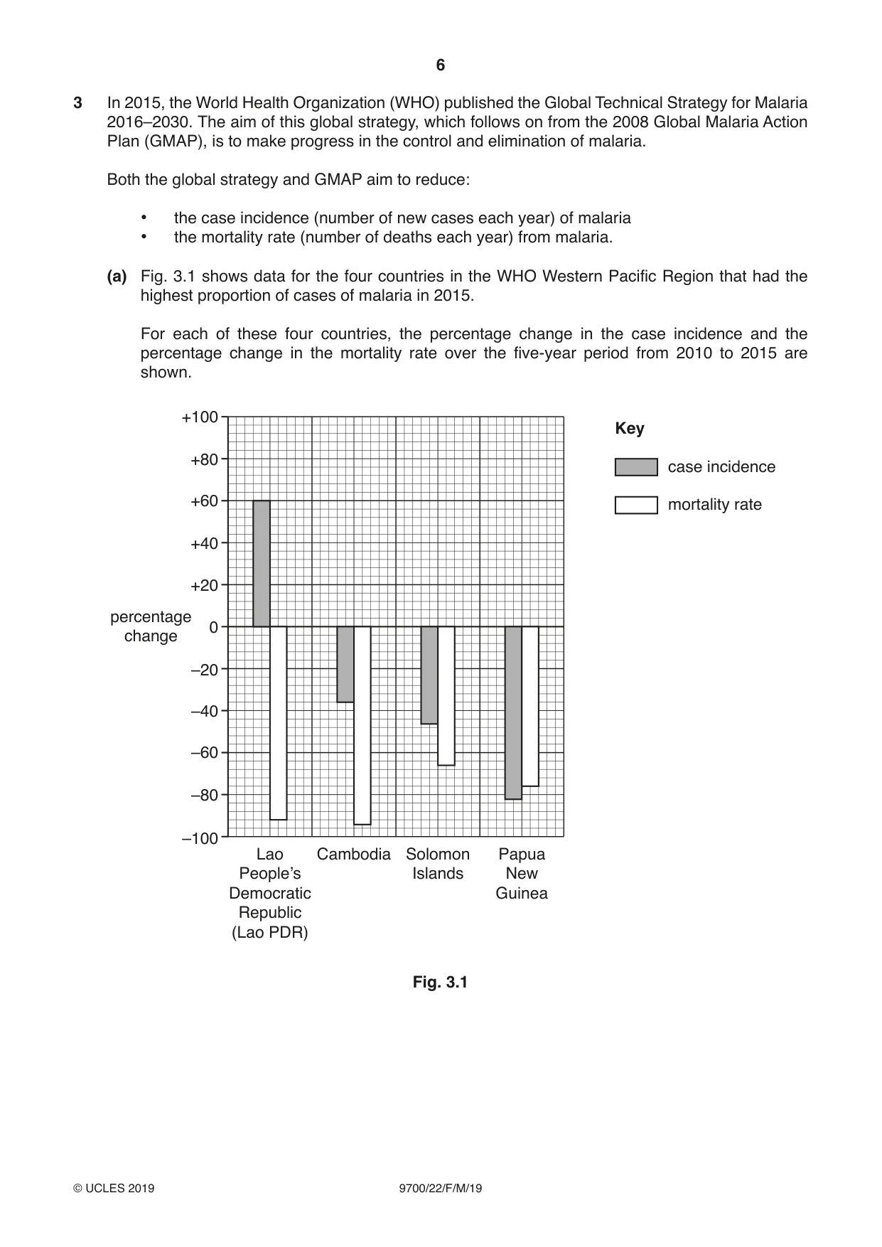
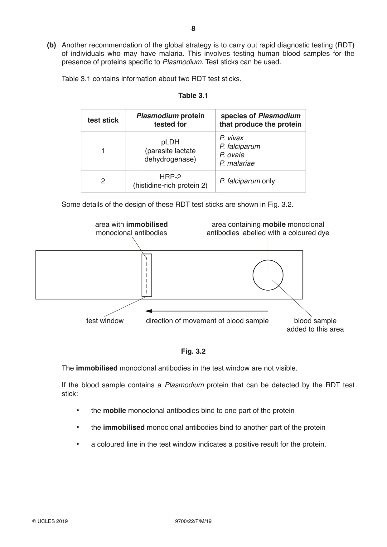
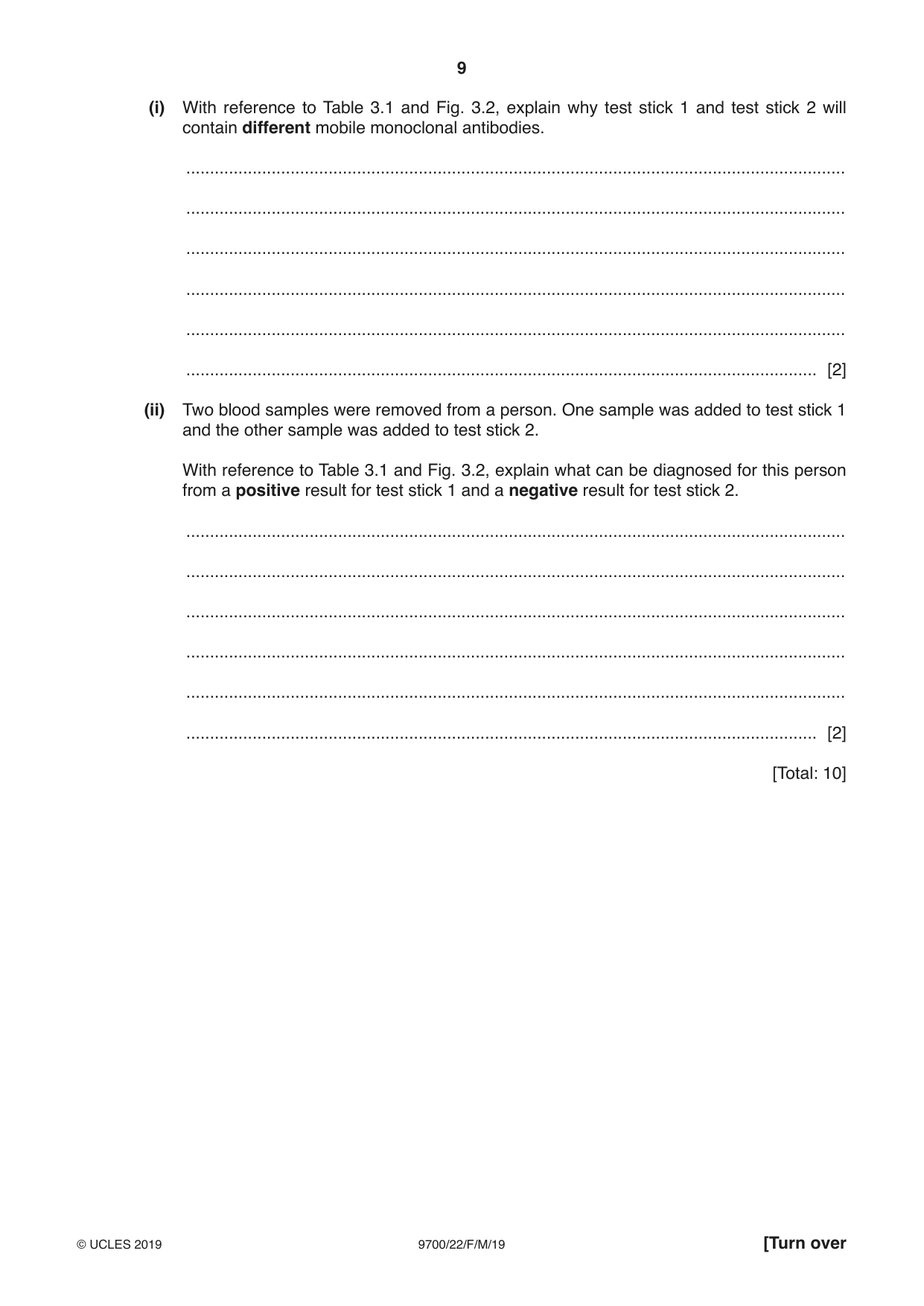
3 In 2015, the World Health Organization (WHO) published the Global Technical Strategy for Malaria 2016–2030. The aim of this global strategy, which follows on from the 2008 Global Malaria Action Plan (GMAP), is to make progress in the control and elimination of malaria. Both the global strategy and GMAP aim to reduce: • the case incidence (number of new cases each year) of malaria • the mortality rate (number of deaths each year) from malaria. (a) Fig. 3.1 shows data for the four countries in the WHO Western Pacific Region that had the highest proportion of cases of malaria in 2015. For each of these four countries, the percentage change in the case incidence and the percentage change in the mortality rate over the five-year period from 2010 to 2015 are shown. +100 Key +80 case incidence +60 mortality rate +40 +20 percentage 0 change –20 –40 –60 –80 –100 Lao Cambodia Solomon Papua People’s Islands New Democratic Guinea Republic (Lao PDR) Fig. 3.1 (i) With reference to Fig. 3.1, describe the progress made in the control of malaria in the four countries between 2010 and 2015. ........................................................................................................................................... ........................................................................................................................................... ........................................................................................................................................... ........................................................................................................................................... ........................................................................................................................................... ........................................................................................................................................... ........................................................................................................................................... ........................................................................................................................................... ........................................................................................................................................... ........................................................................................................................................... ........................................................................................................................................... ..................................................................................................................................... [4] (ii) All the countries shown in Fig. 3.1 supplied households at risk of malaria with insecticide-treated nets (ITNs). This is one of the recommendations in the GMAP and the global strategy. Describe and explain the role of ITNs. ........................................................................................................................................... ........................................................................................................................................... ........................................................................................................................................... ........................................................................................................................................... ........................................................................................................................................... ..................................................................................................................................... [2] (b) Another recommendation of the global strategy is to carry out rapid diagnostic testing (RDT) of individuals who may have malaria. This involves testing human blood samples for the presence of proteins specific to Plasmodium. Test sticks can be used. Table 3.1 contains information about two RDT test sticks. Table 3.1 Plasmodium protein species of Plasmodium test stick tested for that produce the protein P. vivax pLDH P. falciparum 1 (parasite lactate P. ovale dehydrogenase) P. malariae HRP-2 2 P. falciparum only (histidine-rich protein 2) Some details of the design of these RDT test sticks are shown in Fig. 3.2. area with immobilised area containing mobile monoclonal monoclonal antibodies antibodies labelled with a coloured dye test window direction of movement of blood sample blood sample added to this area Fig. 3.2 The immobilised monoclonal antibodies in the test window are not visible. If the blood sample contains a Plasmodium protein that can be detected by the RDT test stick: • the mobile monoclonal antibodies bind to one part of the protein • the immobilised monoclonal antibodies bind to another part of the protein • a coloured line in the test window indicates a positive result for the protein. (i) With reference to Table 3.1 and Fig. 3.2, explain why test stick 1 and test stick 2 will contain different mobile monoclonal antibodies. ........................................................................................................................................... ........................................................................................................................................... ........................................................................................................................................... ........................................................................................................................................... ........................................................................................................................................... ..................................................................................................................................... [2] (ii) Two blood samples were removed from a person. One sample was added to test stick 1 and the other sample was added to test stick 2. With reference to Table 3.1 and Fig. 3.2, explain what can be diagnosed for this person from a positive result for test stick 1 and a negative result for test stick 2. ........................................................................................................................................... ........................................................................................................................................... ........................................................................................................................................... ........................................................................................................................................... ........................................................................................................................................... ..................................................................................................................................... [2] [Total: 10]
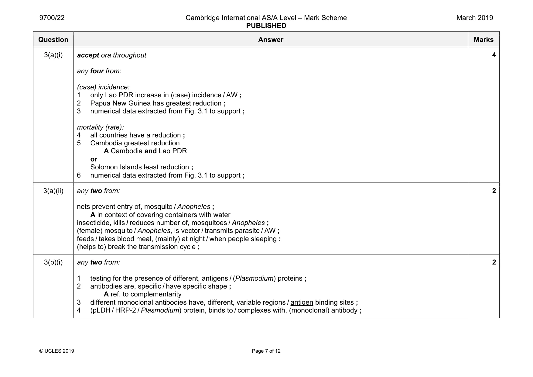
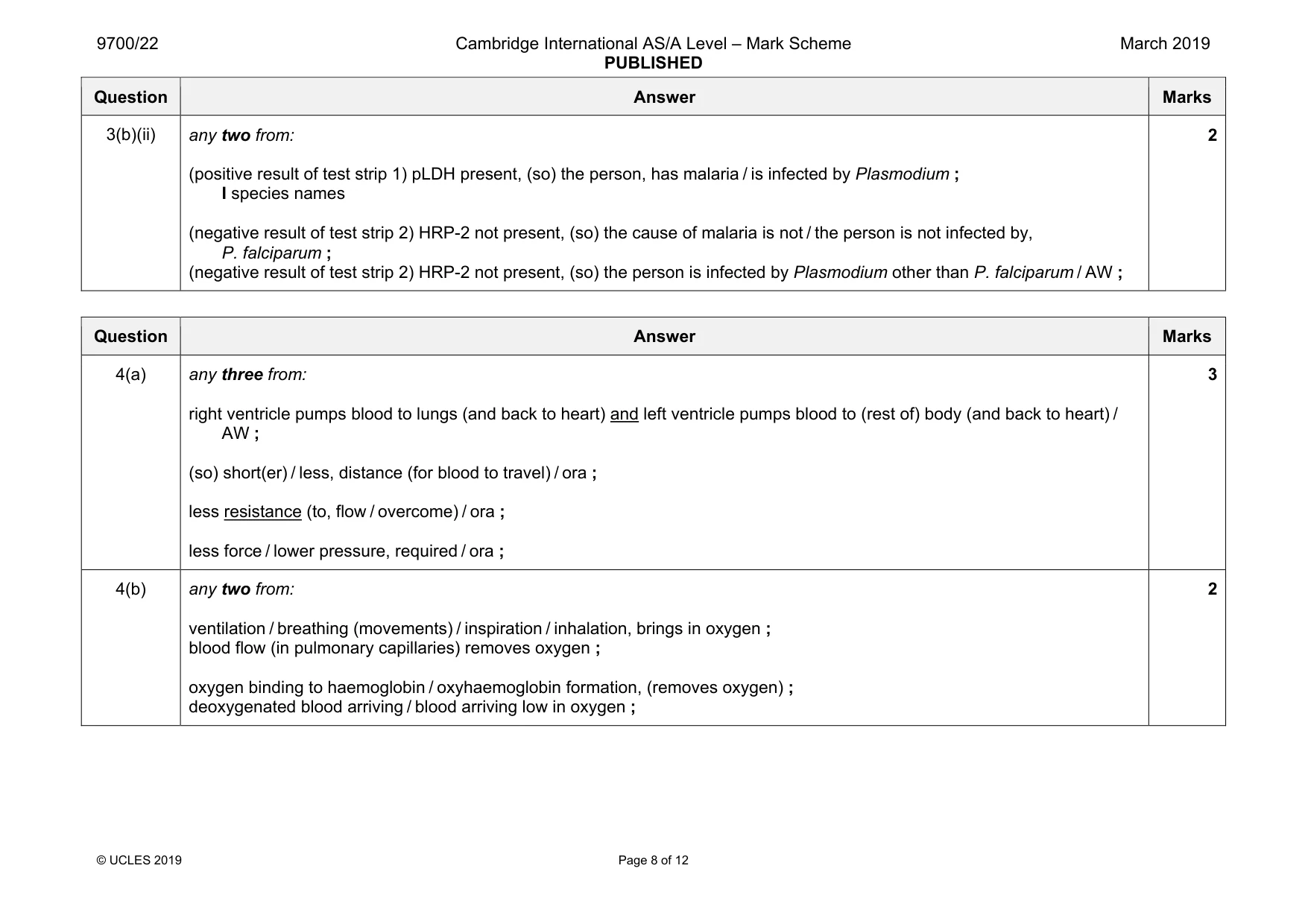
Mark scheme: 3(a)(i) accept ora throughout any four from: (case) incidence: 1 only Lao PDR increase in (case) incidence / AW ; 2 Papua New Guinea has greatest reduction ; 3 numerical data extracted from Fig. 3.1 to support ; mortality (rate): 4 all countries have a reduction ; 5 Cambodia greatest reduction A Cambodia and Lao PDR or Solomon Islands least reduction ; 6 numerical data extracted from Fig. 3.1 to support ; 4 3(a)(ii) any two from: nets prevent entry of, mosquito / Anopheles ; A in context of covering containers with water insecticide, kills / reduces number of, mosquitoes / Anopheles ; (female) mosquito / Anopheles, is vector / transmits parasite / AW ; feeds / takes blood meal, (mainly) at night / when people sleeping ; (helps to) break the transmission cycle ; 2 3(b)(i) any two from: 1 testing for the presence of different, antigens / (Plasmodium) proteins ; 2 antibodies are, specific / have specific shape ; A ref. to complementarity 3 different monoclonal antibodies have, different, variable regions / antigen binding sites ; 4 (pLDH / HRP-2 / Plasmodium) protein, binds to / complexes with, (monoclonal) antibody ; 2 Question Answer Marks 3(b)(ii) any two from: (positive result of test strip 1) pLDH present, (so) the person, has malaria / is infected by Plasmodium ; I species names (negative result of test strip 2) HRP-2 not present, (so) the cause of malaria is not / the person is not infected by, P. falciparum ; (negative result of test strip 2) HRP-2 not present, (so) the person is infected by Plasmodium other than P. falciparum / AW ; 2
Q4 · In a double circulatory system, blood passes through the heart twice in one complete…
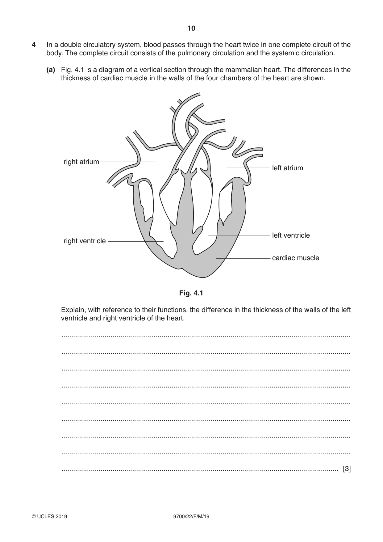
4 In a double circulatory system, blood passes through the heart twice in one complete circuit of the body. The complete circuit consists of the pulmonary circulation and the systemic circulation. (a) Fig. 4.1 is a diagram of a vertical section through the mammalian heart. The differences in the thickness of cardiac muscle in the walls of the four chambers of the heart are shown. right atrium left atrium left ventricle right ventricle cardiac muscle Fig. 4.1 Explain, with reference to their functions, the difference in the thickness of the walls of the left ventricle and right ventricle of the heart. ................................................................................................................................................... ................................................................................................................................................... ................................................................................................................................................... ................................................................................................................................................... ................................................................................................................................................... ................................................................................................................................................... ................................................................................................................................................... ................................................................................................................................................... ............................................................................................................................................. [3] (b) The alveolus is the gas exchange surface in mammals. For efficient oxygen uptake, a steep diffusion gradient is maintained between the alveolar air and the blood. Suggest how the steep diffusion gradient for oxygen is maintained at the gas exchange surface. ................................................................................................................................................... ................................................................................................................................................... ................................................................................................................................................... ................................................................................................................................................... ............................................................................................................................................. [2] (c) Compared to when they were non-smokers, the ability of people who smoke tobacco to deliver oxygen to their body tissues is reduced. Two causes of this reduction include: • a decrease in the volume of air per breath moving towards the alveoli • a decrease in the ability of red blood cells to carry oxygen. (i) Suggest one reason why smoking tobacco, even after only a short time, may cause a decrease in the volume of air per breath moving towards the alveoli. ........................................................................................................................................... ..................................................................................................................................... [1] (ii) Explain why smoking tobacco causes a decrease in the ability of red blood cells to carry oxygen. ........................................................................................................................................... ........................................................................................................................................... ........................................................................................................................................... ........................................................................................................................................... ..................................................................................................................................... [2] [Total: 8]
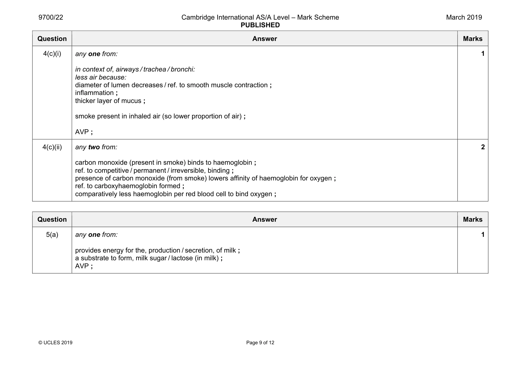
Mark scheme: 4(a) any three from: right ventricle pumps blood to lungs (and back to heart) and left ventricle pumps blood to (rest of) body (and back to heart) / AW ; (so) short(er) / less, distance (for blood to travel) / ora ; less resistance (to, flow / overcome) / ora ; less force / lower pressure, required / ora ; 3 4(b) any two from: ventilation / breathing (movements) / inspiration / inhalation, brings in oxygen ; blood flow (in pulmonary capillaries) removes oxygen ; oxygen binding to haemoglobin / oxyhaemoglobin formation, (removes oxygen) ; deoxygenated blood arriving / blood arriving low in oxygen ; 2 Question Answer Marks 4(c)(i) any one from: in context of, airways / trachea / bronchi: less air because: diameter of lumen decreases / ref. to smooth muscle contraction ; inflammation ; thicker layer of mucus ; smoke present in inhaled air (so lower proportion of air) ; AVP ; 1 4(c)(ii) any two from: carbon monoxide (present in smoke) binds to haemoglobin ; ref. to competitive / permanent / irreversible, binding ; presence of carbon monoxide (from smoke) lowers affinity of haemoglobin for oxygen ; ref. to carboxyhaemoglobin formed ; comparatively less haemoglobin per red blood cell to bind oxygen ; 2
Q5 · Human prolactin (hPRL) is a globular protein
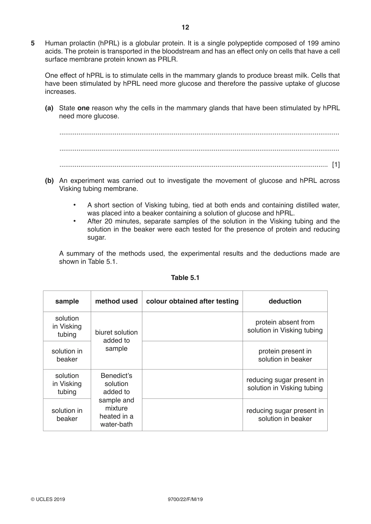
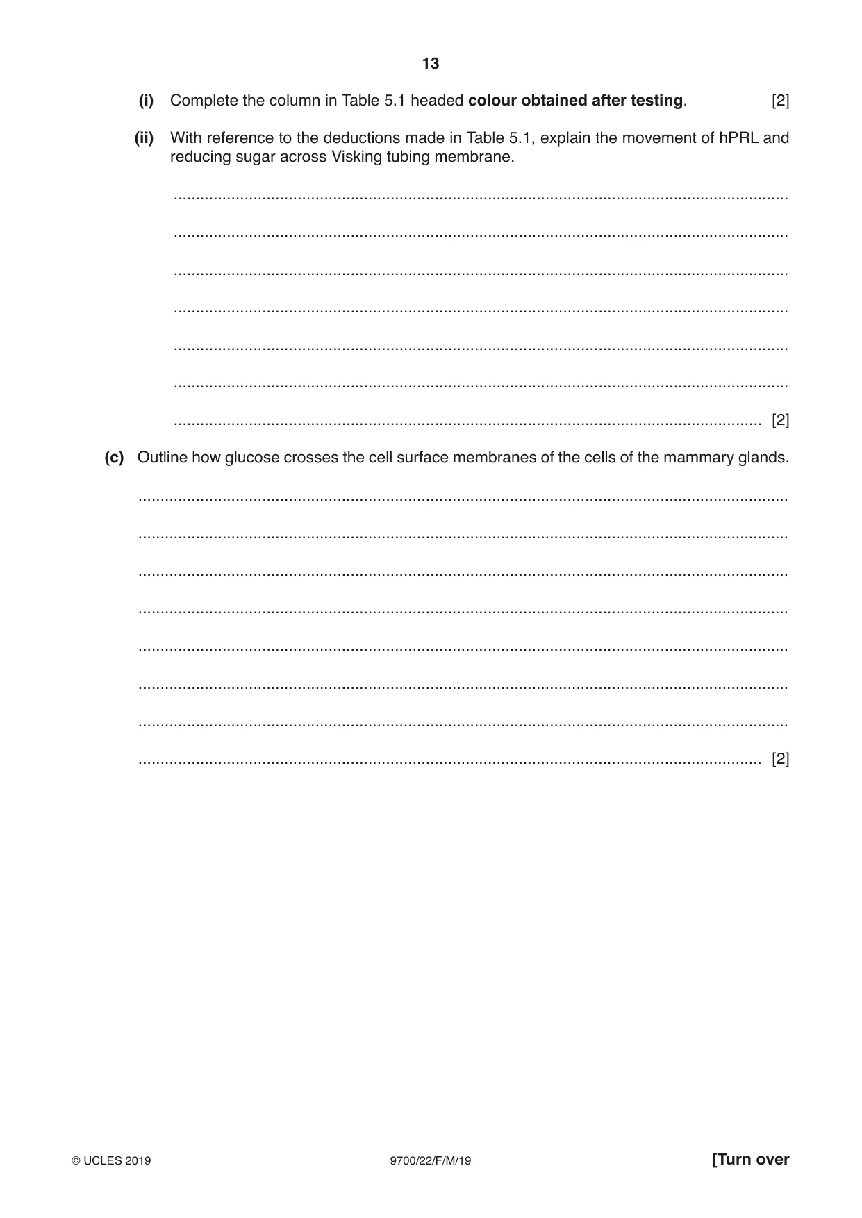
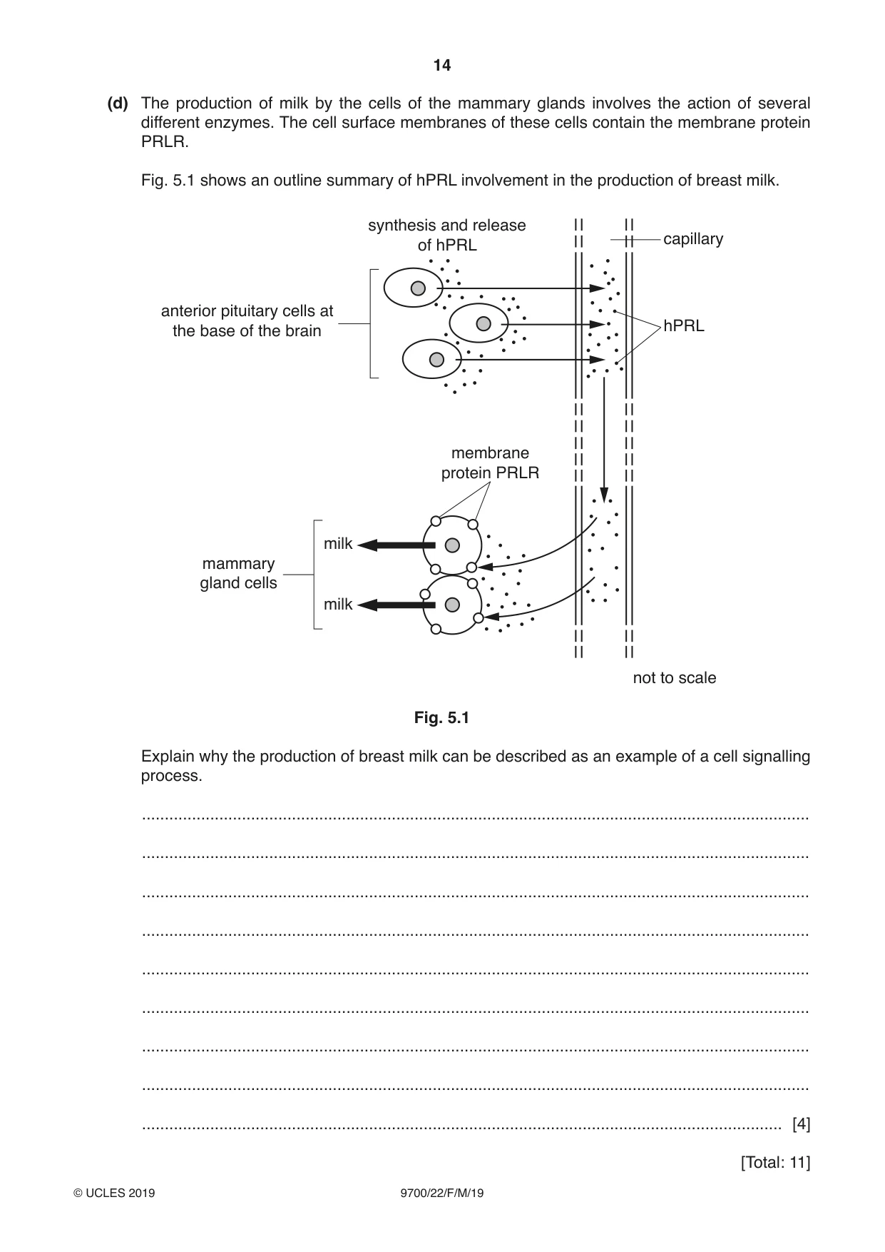
5 Human prolactin (hPRL) is a globular protein. It is a single polypeptide composed of 199 amino acids. The protein is transported in the bloodstream and has an effect only on cells that have a cell surface membrane protein known as PRLR. One effect of hPRL is to stimulate cells in the mammary glands to produce breast milk. Cells that have been stimulated by hPRL need more glucose and therefore the passive uptake of glucose increases. (a) State one reason why the cells in the mammary glands that have been stimulated by hPRL need more glucose. ................................................................................................................................................... ................................................................................................................................................... ............................................................................................................................................. [1] (b) An experiment was carried out to investigate the movement of glucose and hPRL across Visking tubing membrane. • A short section of Visking tubing, tied at both ends and containing distilled water, was placed into a beaker containing a solution of glucose and hPRL. • After 20 minutes, separate samples of the solution in the Visking tubing and the solution in the beaker were each tested for the presence of protein and reducing sugar. A summary of the methods used, the experimental results and the deductions made are shown in Table 5.1. Table 5.1 sample method used colour obtained after testing deduction solution protein absent from in Visking solution in Visking tubing biuret solution tubing added to sample solution in protein present in beaker solution in beaker solution Benedict’s reducing sugar present in in Visking solution solution in Visking tubing tubing added to sample and mixture solution in reducing sugar present in heated in a beaker solution in beaker water-bath (i) Complete the column in Table 5.1 headed colour obtained after testing. [2] (ii) With reference to the deductions made in Table 5.1, explain the movement of hPRL and reducing sugar across Visking tubing membrane. ........................................................................................................................................... ........................................................................................................................................... ........................................................................................................................................... ........................................................................................................................................... ........................................................................................................................................... ........................................................................................................................................... ..................................................................................................................................... [2] (c) Outline how glucose crosses the cell surface membranes of the cells of the mammary glands. ................................................................................................................................................... ................................................................................................................................................... ................................................................................................................................................... ................................................................................................................................................... ................................................................................................................................................... ................................................................................................................................................... ................................................................................................................................................... ............................................................................................................................................. [2] (d) The production of milk by the cells of the mammary glands involves the action of several different enzymes. The cell surface membranes of these cells contain the membrane protein PRLR. Fig. 5.1 shows an outline summary of hPRL involvement in the production of breast milk. synthesis and release capillary of hPRL anterior pituitary cells at hPRL the base of the brain membrane protein PRLR milk mammary gland cells milk not to scale Fig. 5.1 Explain why the production of breast milk can be described as an example of a cell signalling process. ................................................................................................................................................... ................................................................................................................................................... ................................................................................................................................................... ................................................................................................................................................... ................................................................................................................................................... ................................................................................................................................................... ................................................................................................................................................... ................................................................................................................................................... ............................................................................................................................................. [4] [Total: 11]
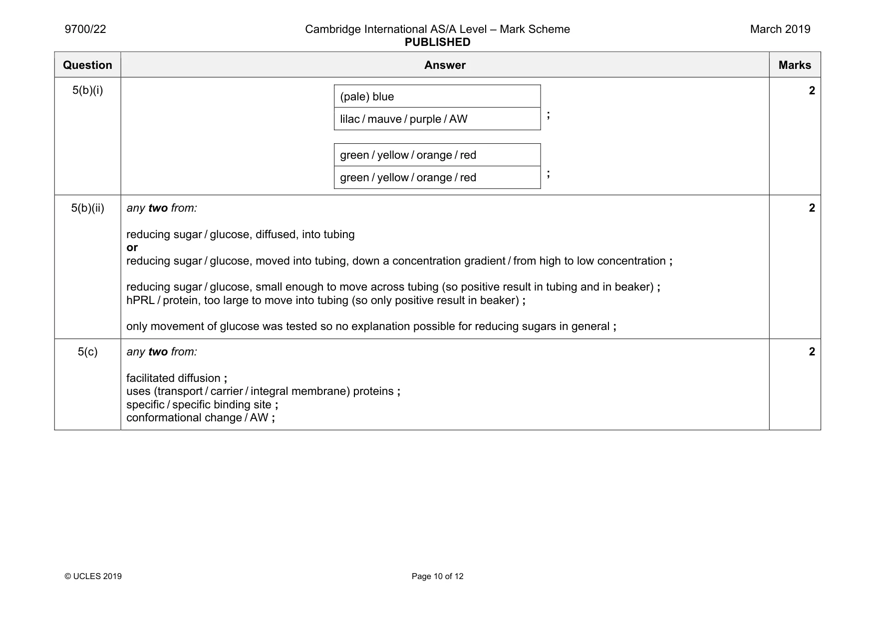
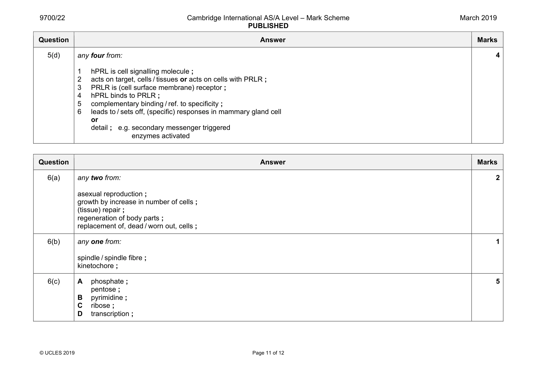
Mark scheme: 5(a) any one from: provides energy for the, production / secretion, of milk ; a substrate to form, milk sugar / lactose (in milk) ; AVP ; 1 Question Answer Marks 5(b)(i) (pale) blue ; lilac / mauve / purple / AW green / yellow / orange / red ; green / yellow / orange / red 2 5(b)(ii) any two from: reducing sugar / glucose, diffused, into tubing or reducing sugar / glucose, moved into tubing, down a concentration gradient / from high to low concentration ; reducing sugar / glucose, small enough to move across tubing (so positive result in tubing and in beaker) ; hPRL / protein, too large to move into tubing (so only positive result in beaker) ; only movement of glucose was tested so no explanation possible for reducing sugars in general ; 2 5(c) any two from: facilitated diffusion ; uses (transport / carrier / integral membrane) proteins ; specific / specific binding site ; conformational change / AW ; 2 Question Answer Marks 5(d) any four from: 1 hPRL is cell signalling molecule ; 2 acts on target, cells / tissues or acts on cells with PRLR ; 3 PRLR is (cell surface membrane) receptor ; 4 hPRL binds to PRLR ; 5 complementary binding / ref. to specificity ; 6 leads to / sets off, (specific) responses in mammary gland cell or detail ; e.g. secondary messenger triggered enzymes activated 4
Q6 · Plant and animal cells carry out mitosis to form two genetically identical cells from one…
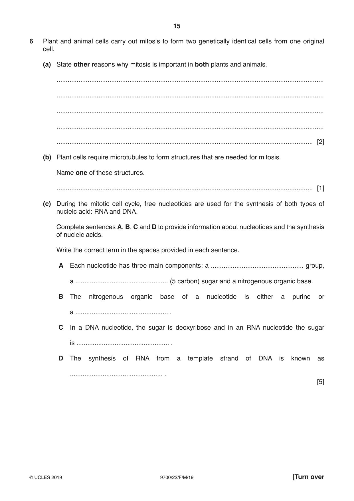
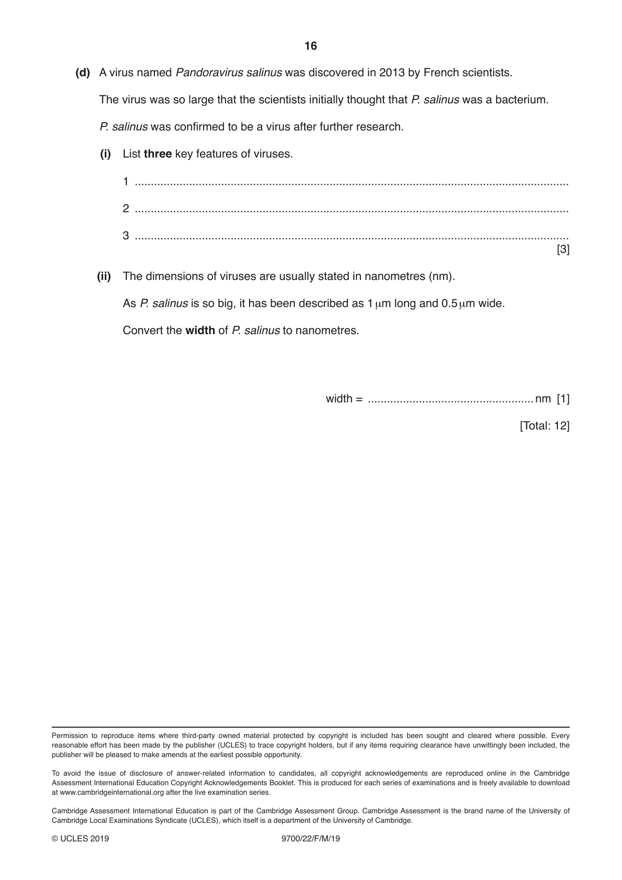
6 Plant and animal cells carry out mitosis to form two genetically identical cells from one original cell. (a) State other reasons why mitosis is important in both plants and animals. ................................................................................................................................................... ................................................................................................................................................... ................................................................................................................................................... ................................................................................................................................................... ............................................................................................................................................. [2] (b) Plant cells require microtubules to form structures that are needed for mitosis. Name one of these structures. ............................................................................................................................................. [1] (c) During the mitotic cell cycle, free nucleotides are used for the synthesis of both types of nucleic acid: RNA and DNA. Complete sentences A, B, C and D to provide information about nucleotides and the synthesis of nucleic acids. Write the correct term in the spaces provided in each sentence. A Each nucleotide has three main components: a ................................................... group, a ................................................... (5 carbon) sugar and a nitrogenous organic base. B The nitrogenous organic base of a nucleotide is either a purine or a ................................................... . C In a DNA nucleotide, the sugar is deoxyribose and in an RNA nucleotide the sugar is ................................................... . D The synthesis of RNA from a template strand of DNA is known as ................................................... . [5] (d) A virus named Pandoravirus salinus was discovered in 2013 by French scientists. The virus was so large that the scientists initially thought that P. salinus was a bacterium. P. salinus was confirmed to be a virus after further research. (i) List three key features of viruses. 1 ........................................................................................................................................ 2 ........................................................................................................................................ 3 ........................................................................................................................................ [3] (ii) The dimensions of viruses are usually stated in nanometres (nm). As P. salinus is so big, it has been described as 1 µm long and 0.5 µm wide. Convert the width of P. salinus to nanometres. width = ....................................................nm [1] [Total: 12]
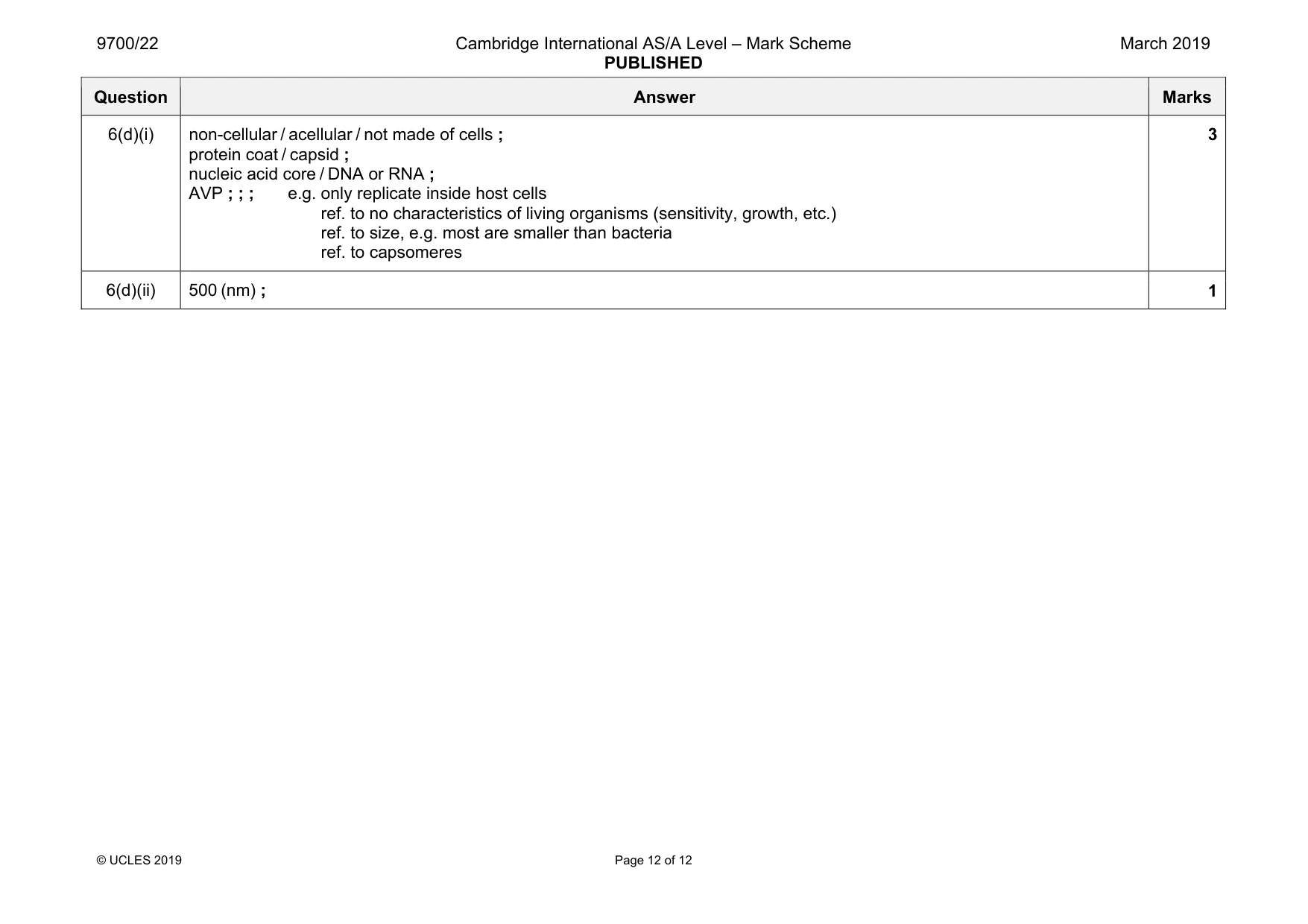
Mark scheme: 6(a) any two from: asexual reproduction ; growth by increase in number of cells ; (tissue) repair ; regeneration of body parts ; replacement of, dead / worn out, cells ; 2 6(b) any one from: spindle / spindle fibre ; kinetochore ; 1 6(c) A phosphate ; pentose ; B pyrimidine ; C ribose ; D transcription ; 5 Question Answer Marks 6(d)(i) non-cellular / acellular / not made of cells ; protein coat / capsid ; nucleic acid core / DNA or RNA ; AVP ; ; ; e.g. only replicate inside host cells ref. to no characteristics of living organisms (sensitivity, growth, etc.) ref. to size, e.g. most are smaller than bacteria ref. to capsomeres 3 6(d)(ii) 500 (nm) ; 1
More questions on Structure of nucleic acids and replication of DNA
What was in this paper
The subtopics covered by these 6 questions, and how many questions each got. Open one in a new tab to see every Cambridge question on it.
What you needed in this session
Cambridge’s own grade thresholds for 2019 Feb/March, Paper 2 · Variant 2. A higher threshold means an easier paper — the bar moves with how the cohort did.